contributions to anatomic pathology, over the years
TRANSCRIPT

Contributions to Anatomic Pathology, over the years

Anatomic Pathology, part 1
G.B. Morgagni
Rudolf Wirchow
Xavier Bichat

Anatomic Pathology, part 1
• Anatomic pathology materials: morphological samples taken for diagnostic purposes
• Anatomo-Pathologic studies are carried out on samples from:
1. Clinical autopsies 2. Surgical resections 3. Biopsies 4. Cytological preparations

Anatomic Pathology, part 1
Clinical autopsy: Aims at defining the causes of death 1. Perinatal autopsies (23-24 weeks or 500 gr >> 7
days of extrauterine life): malformations. 2. Pediatric autopsies (7 days of life >> 15 years):
lymphoma, leukemia, sudden death syndromes, infections.
3. Adult autopsies: cardiovascular disease, cancer and degenerative processes.

Anatomic Pathology, part 1

Anatomic Pathology, part 1
Cytopathologic studies Morphologic alterations of cells obtained by: - Spontaneous or induced exfoliation - Fine needle aspiration Allows the diagnosis of: - early dysplastic processes (screening) - neoplastic/pre-neoplastic lesions - inflammations/infections

Anatomic Pathology, part 1

Anatomic Pathology, part 1

Anatomic Pathology, part 1
Cytologic samples are processed quickly and easily: - smearing - centrifuging and smearing - staining (Papanicolaou) and observed under the light microscope

Anatomic Pathology, part 1

Anatomic Pathology, part 1
¨ Histopathological samples: -diagnostic biopsies -surgical samples ¨ Diagnostic biopsies allow histologic diagnoses -small -cylindrical (fine needle aspiration biopsy) -only part of a lesion or an organ Examples: renal and hepatic needle biopsies, samples from digestive endoscopy and bronchoscopy


Anatomic Pathology, part 1
¨ Surgical specimens are larger >> whole organs ¨ The lesion is removed completely
¨ The anatomo-pathologic study is necessary for: - diagnosis - extension of the lesion and excision margins - prognosis - therapeutic strategies


Anatomic Pathology, part 1
¨ Identification and dispatch material -request for histologic or cytologic exam adequately filled in -appropriate container, correctly identified -appropriate fixative (type and quantity) -timely dispatch to the anatomic pathology labs

Check-in
• Registration: ¤ Personal data (name, age, sex, occupation) ¤ Privacy statement
• Payment (direct/indirect): ¤ In-house (hospital) ¤ Day-hospital ¤ Outpatients (N.H.S. = S.S.N.) ¤ Private practice

Check-in
• Congruence • Tacking of anomalies • Pertinent clinical data • Fixation time


Anatomic Pathology, part 1
¨ Macroscopic examination of samples aims at: - Exactly describing the type of material:
size & alterations shape colour consistency margins (china ink) relations with adjacent structures
- Selecting appropriate area for microscopic examination

Sampling
Sampling
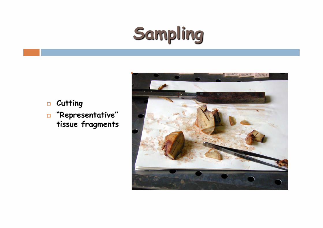
¨ Cutting ¨ “Representative”
tissue fragments

Sampling
¨ Fragments allotted into cassettes ¨ Proper identification by numbers/letters
Miometrio Leiomioma Cervice

Sampling


Anatomic Pathology, part 1
¨ Processing tissues for observation with light microscope -tissue fixation -dehydration/embedding (paraffin) -inclusion -cutting -deparaffinization or rehydration -staining -coveslipping -observation under light microscope

Anatomic Pathology, part 1
¨ Tissue fixation: interruption of degradation processes that start soon after cell death (autolysis and putrefaction)
-preserving architecture -preserving cell composition ¨ Autolysis: cellular autodigestion by enzymes (rupture
of lysosomal membranes) ¨ Putrefaction: bacterial superimposition on autolysis

Anatomic Pathology, part 1
¨ Fixatives may be: -chemical -physical (freezing) ¨ Chemical fixatives: make tissue proteins insoluble
and refractory to autolysis ¨ Simple fixatives ¨ Composed fixatives (fixative mixtures)
¨ Formalin: aqueous solution of 10% at pH 7

Anatomic Pathology, part 1
¨ Sampling ¨ Dehydration ¨ Inclusion: paraffin wax -tissue solidity -preservation of architectural relationships -thin (3-4µm) regular and homogeneous sections


Embedding
¨ Tissue samples are embedded in paraffin


Anatomic Pathology, part 1
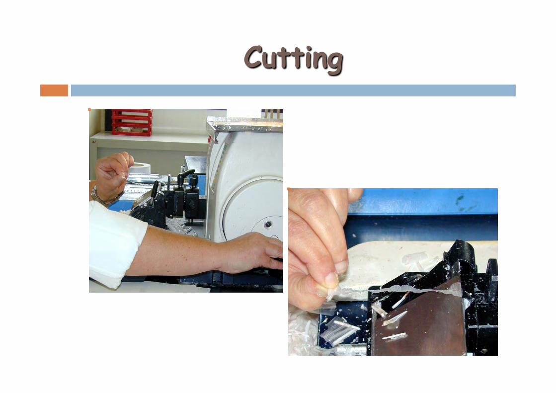
¨ Cut: Microtome (sections 3-4 microns) ¨ Rehydration ¨ Staining: H&E - Haematoxylin (nucleus) - Eosin (cytoplasm) - Giemsa: blood cells; MGG; silver impregnation ¨ Coverslipping: synthetic resin ¨ Observation under a light microscope ¨ Histological diagnosis


Cutting
¨ Thin sections (3-5 µm) adhered to holding glass slides
Microtome / seriotome
Cutting

Cutting

Staining

Staining
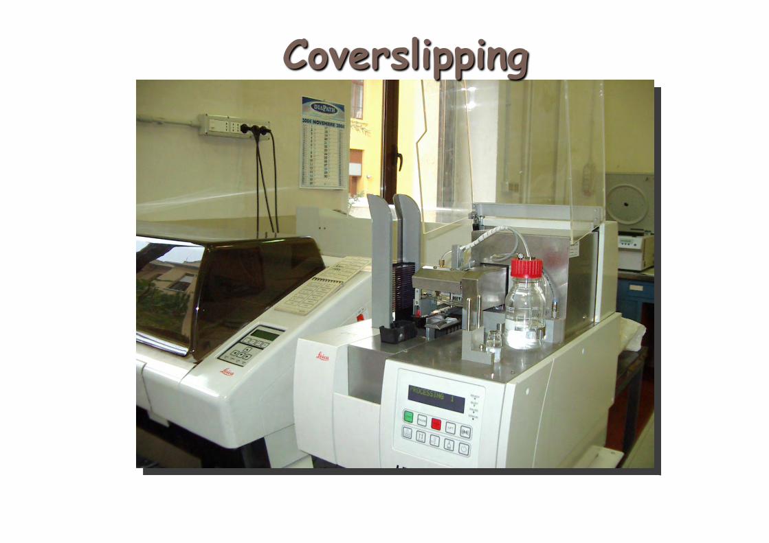
Coverslipping

Anatomic Pathology, part 1
¨ Anatomic-pathology diagnosis uses: -histological examination (morphological examination N/C)
-histochemical methods (chemicals linked to tissues): PAS, Sudan, Perls ....
-immunohistochemical methods (Ag/Ab reaction highlighted by a chromogen, studying tissue and cellular antigens) -electronic microscopy (observation of subcellular structures on TEM, external morphology and molecular composition on SEM) semi-thin or yultra-thin sections
-cytometry and flow cytometry (based on image analyzers)
-molecular biology (genetic study of diseases)

Examination and reporting
• Collection of slides • Microscopic
examination • Additional stains
¤ Histochemical ¤ immunohistochemical ¤ Molecular hybridization
• Report

Stains

Immunohistochemistry

Fluorescence

Electronic miscroscopy

Molecular biology

Anatomic Pathology, part 1
¨ Intraoperative examination (frozen sections): histological exam required by the surgeon during a operation, which could modify the surgical approach:
-neoplastic lymph nodes -margins of surgical resection -sample suitability (adequate cellularity) -confirmation of diagnostic suspicion

Anatomic Pathology, part 1
¨ Surgical samples will be: -frozen (cryopreserved) -sectioned by a cryostat -stained with H&E and observed under a light microscope (OM) Diagnosis is achieved in 70-80% of cases, may be incomplete (no grade, no stage) or only partially reflect what found on permanent sections (better morphological preservation, additional sampling, etc.)