controlling g-quadruplex formation via lipid …ohfwurqlf 6xssohphqwdu\ 0dwhuldo (6, iru...
TRANSCRIPT

Controlling G-quadruplex formation via lipid modification of oligonucleotide sequences
Brune Vialet,† # Arnaud Gissot,†# Romain Delzor,† and Philippe Barthélémy†* † Inserm, U1212, CNRS 5320, Bordeaux, F-33076 France, Université de Bordeaux, F-33076, Bordeaux, France. # These two authors contributed equally to the work.
Supplementaryinformation
Electronic Supplementary Material (ESI) for ChemComm.This journal is © The Royal Society of Chemistry 2017

2
Listofacronyms:
G4:G-quadruplex
LON:LipidOligoNucleotide
TheOligonucleotidesusedinthisstudyarenamedafterthepresence(orabsence)oftheG4-proneDNAsequence:
x-OLIGONUCLEOTIDEy
x:referstothenatureofthelipideitheramonooctadecylchain(1-)ordialkylatedketal(k-)
y:G4:G4-pronesequenceorsc:scramblesequence
ex: 1-LONG4:n-C18H37-5'-TTAGTTGGGGTTCAGTTGG-3'(G4-pronesequenceinbold)
SeeFig.2andS1forthedetailedlistofoligonucleotidesused.
AnalyticalTechniques:
PAGE:PolyAcrylamideGelElectrophoresis
CD:CircularDichroism
DLS:DynamicLightScattering

3
Experimentalsection:Automated DNA synthesis and purification of LONs. LONs were synthesized using the phosphoramidite methodology on an automated Expedite 8909 DNA synthesizer at the µmol scale on 1000 Å primer support (loading: 30-100 µmol/g, Link technologies, Synbase Control Pore Glass). Prior to use, the phosphoramidites 1 and 2 were dried over P2O5 overnight and then dissolved in dry CH2Cl2/CH3CN 1/1 to a 0.1 M concentration (lipidic phosphoramidites do not dissolve in pure acetonitrile). N-benzylthiotetrazole was used for activation of the phosphoramidite prior to coupling. The phosphoramidite 1 and 2 were manually coupled at the 5’ end of sequence on the solid support by passing (via syringes) the activator and the phosphoramidite solution (0.5 mL) back and forth several times for 10 min. Cleavage from the solid support and base deprotection was achieved using 1 mL of a saturated aqueous NH4OH solution for 4 h at 55°C. The supernatant was collected and the CPG beads were washed 3 times with 200 µL of milliQ water. The solutions were pooled and evaporated (speed vac). The crude LONs were dissolved in 0.5 mL of water and purified (except k-LONG4) on a semi-preparative C4-reverse phase HPLC column (Macherey Nagel, Nucleosil, 5µm, 250mm), flow rate: 5 mL/min, using buffer A (0.1M triethylammonium acetate pH 7.1 / CH3CN (95/5, V/V)) and buffer B 0.1M triethylammonium acetate pH 7.1 / CH3CN (20/80, V/V), (see Figure S2 for details). The LONs eluted after 6,5 min. The LON containing fractions were pooled and evaporated to dryness and dissolved in autoclaved milliQ water. The (L)ONs were then dialyzed against a 10mM LiCl solution (not to favor G4 formation) followed by water. Yields were acceptable following this protocol (15-30% yield after purification of the crude material). k-LONs were purified by preparative PAGE using conventional protocols with 20*20*0.2 cm 20% polyacrylamide gels at a limiting power of 15W. Importantly, we found that the quantity of k-LONG4 that could be loaded on the gel did not exceed ca. 100 nmoles of crude material in ca. 300µL of loading sample. Higher amounts of crude LON led to substantial trailing of the LON band probably because of the formation of the micelles in that case even in the presence of heat + 7M urea in the running buffer. Using smaller quantities, the k-LONG4 band is well defined (UV-shadow) and cut out directly with a clean scalpel. The gel slab was chopped into fine particles to elute the LON. We have been unsuccessful at eluting k-LONG4 from the gel using different eluting buffers with additional heating (up to 90°C) and/or sonication and/or freeze and thaw protocols. Very small quantities of k-LONG4 were systematically obtained for each of these tests. We therefore developed an original electroelution protocol for the purification of these LONs (see Figure S3). In short, the electrical wire that was originally connected to the negative pole of the generator (black wire, Figure S3) was modified manually by wrapping a platinum wire around the naked copper wire. The latter was immersed in the eluting buffer contained in a plastic pipette that was chopped off at both edges to 1) facilitate pouring of the eluting buffer on top and 2) increase the cross section at the bottom to minimize resistance to the current flow. The bottom of the syringe was blocked by polymerizing 0.5mL of a 8% polyacrylamide solution (the bottom of the pipette being temporarily blocked by wrapping a parafilm foil around). After polymerization, the gel was prerun to remove any unpolymerized materials prior to loading the crushed acrylamide gel slab containing the LON (the TBE eluting buffer from the pipette was withdrawn beforehand for practical reasons). A dialysis tubing (with a cutoff of 2 kDa) was adapted and wrapped with a parafilm foil around the bottom of the pipette to recover the sample after electroelution. This allowed the dialysis tubing to be immersed in a large quantity of eluting buffer at the bottom in the electrophoresis tank. This step is crucial as a high power is required for elution (using a 50 mL spin tube instead in the absence of dialysis tubing led to a rapid increase in temperature of the solution followed by a short-circuit). The

4
elution was carried out with an electrical power of 7-10 W to allow enough heat dissipation in the gel to favor denaturation of the LONs. The LONs were finally dialyzed in a similar manner as described above for the other (L)ONs. Mass spectra were recorded on a MALDI-Tof-ToF mass spectrometer (Ultraflex, Bruker Daltonics, Bremen, Germany). Best results were obtained in the linear mode with positive-ion detection. Mass spectra were acquired with an ion source voltage 1 of 25kV, an ion source voltage 2 of 23.5kV, a lens voltage of 6kV, by accumulating the ion signals from 1000 laser shots at constant laser fluence with a 100Hz laser. External mass calibration was achieved using a mixture of oligonucleotides dT12-dT18 (Sigma). 1:1 mixture of samples of LONs (~20-50µM) and matrix was spotted on a MALDI target and air-dried before analysis. The performance of the following matrices was evaluated: 2,5-dihydroxybenzoic acid (DHB), 2,4,6-trihydroxy-acetophenone (THAP), 3-hydroxypicolinic acid (3-HPA) and 2,6-dihydroxyacetophenone (DHA). THAP at a concentration of 20 mg/mL in a 4:1 mixture of ethanol and 100mM aqueous ammonium citrate was shown to yield optimal mass spectral results (see Figure S1 for mass values).
PAGE / agarose electrophoresis. Electrophoresis experiments were performed according to standard procedures with 1% agarose gels. PAGE were carried out with 17% polyacrylamide gels (acrylamide-bis acrylamide 19:1, 40% w/v) and run with a 100V limiting tension for native PAGE experiments.
Dynamic light scattering (DLS). Particle size was determined using a Zetasizer 3000 HAS MALVERN. Experiments were realized with samples containing different concentration of LONs dissolved in deionized water or phosphate buffer. Measurements were performed at 25°C.
Viscosizing or Taylor dispersion analyses were recorded on a Viscosizer TD (Malvern Instruments Ltd., Malvern, UK equipped with a 254nm UV filter close to the λmax of oligonucleotides. Prior to analysis, the non-coated capillary has been prepared by injecting 1M NaOH during 30min at 3000mbar followed by 10min rinse with water at 3000mbar. The cellulose coated capillary has been prepared by injecting water during 30 min at 3000mbar. Internal Material (non-coated): fused silica, internal diameter: 75µm, outer diameter: 360µm, L1: 45cm, L2:85cm, Total Length: 130 cm. The samples were injected (pressure: 50mbar) analyzed at 25°C (Mobilization pressure: 140mbar). Taylorgramms were recorded and analyzed with the viscosizer TD software 2.01 with a one component fit. Washing: 1min of water between each sample, pressure: 3000mbar (Results shown in Figure S7). The coated cellulose capillary was in general necessary for the analysis of LONs with the noticeable exception of k-LONG4. Unless the LON forms stable micellar assemblies as in the case of k-LONG4, unspecific adsorption was observed onto the uncoated capillary with the other LONs as evidenced by a trailing in the absorption curve of the chromatogram.
TEM analyses were performed at the Bordeaux Imaging Center (BIC) of the University of Bordeaux using an Hitachi H7650 at a voltage of 80kV. For sample preparation a drop of 100 nM solution of 1-LONG4, 2X selex salts was placed on a carbon film 200 Mesh copper grid and left to dry for 10 min. A drop of 1% uranyl acetate solution as a negative stain for 1 min was then added to the copper grid and left to dry.
NMR analysis were performed at the Institut Européen de Chimie Biologie (IECB) using a 700MHz NMR Bruker spectrometer. Samples concentrations were 100µM. Experiments were performed at 25°.
5
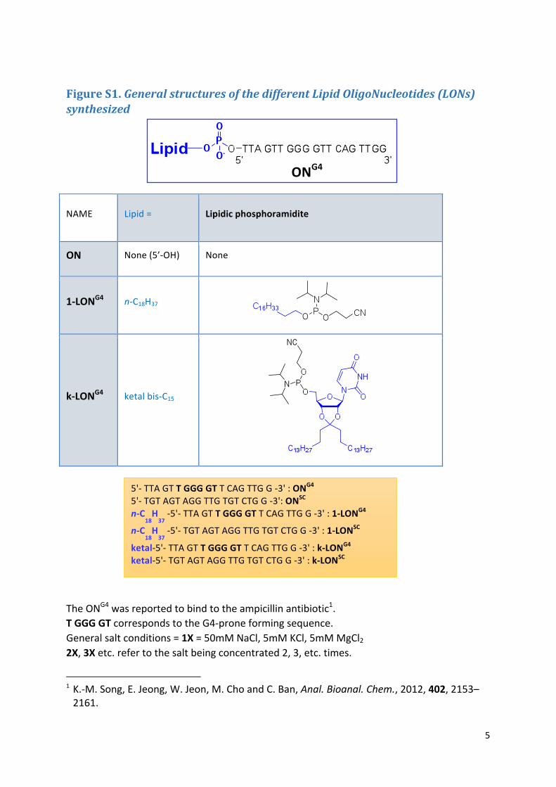
FigureS1.GeneralstructuresofthedifferentLipidOligoNucleotides(LONs)synthesized
NAME Lipid= Lipidicphosphoramidite
ON None(5’-OH) None
1-LONG4 n-C18H37
k-LONG4 ketalbis-C15
TheONG4wasreportedtobindtotheampicillinantibiotic1.TGGGGTcorrespondstotheG4-proneformingsequence.Generalsaltconditions=1X=50mMNaCl,5mMKCl,5mMMgCl22X,3Xetc.refertothesaltbeingconcentrated2,3,etc.times.
1K.-M.Song,E.Jeong,W.Jeon,M.ChoandC.Ban,Anal.Bioanal.Chem.,2012,402,2153–2161.
ONG4
5'-TTAGTTGGGGTTCAGTTGG-3':ONG4 5'-TGTAGTAGGTTGTGTCTGG-3':ONSC n-C
18H37-5'-TTAGTTGGGGTTCAGTTGG-3':1-LONG4
n-C18H37-5'-TGTAGTAGGTTGTGTCTGG-3':1-LONSC
ketal-5'-TTAGTTGGGGTTCAGTTGG-3':k-LONG4 ketal-5'-TGTAGTAGGTTGTGTCTGG-3':k-LONSC
6
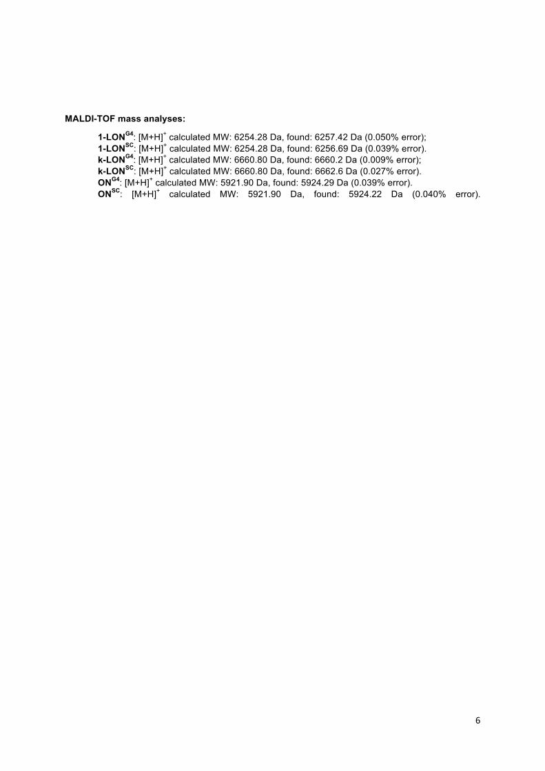
MALDI-TOF mass analyses:
1-LONG4: [M+H]+ calculated MW: 6254.28 Da, found: 6257.42 Da (0.050% error); 1-LONSC: [M+H]+ calculated MW: 6254.28 Da, found: 6256.69 Da (0.039% error). k-LONG4: [M+H]+ calculated MW: 6660.80 Da, found: 6660.2 Da (0.009% error); k-LONSC: [M+H]+ calculated MW: 6660.80 Da, found: 6662.6 Da (0.027% error). ONG4: [M+H]+ calculated MW: 5921.90 Da, found: 5924.29 Da (0.039% error). ONSC: [M+H]+ calculated MW: 5921.90 Da, found: 5924.22 Da (0.040% error).

7
FigureS2.CrudeoligoPAGEprofilesofk-LONSCandk-LONG4andtheircorrespondingRP-HPLCpurificationchromatograms.
Top: Similar denaturing PAGE profiles for the crude k-LONG4 and k-LONSC afterautomated chemical synthesis (before purification): both LONs are expected to bepresentinroughlythesameproportionsinthecrudeinHPLC.Bottom:HPLCpurificationof the corresponding crudes (the Y axis corresponds to the absorbance recorded at260nminnormalizedarbitraryunits).
RP-HPLC conditions: C4 semi preparative column (Macherey Nagel, Nucleosil, 5µm, 250mm), flowrate5mL/minmobilephaseeluentA:0.1MtriethylammoniumacetatepH7.1/CH3CN(95/5,V/V),eluentB:0.1MtriethylammoniumacetatepH7.1/CH3CN(20/80,V/V).
Methoddiagram.Startat15%B,increaseto90%B,thendecreaseto15%B.Totalmethodtime17min.
0
20
40
60
80
100
0 2 4 6 8 10 12 14 16 18
%eluen
tB
Time(min)
8
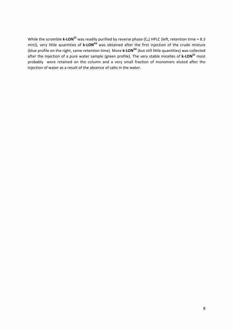
Whilethescramblek-LONSCwasreadilypurifiedbyreversephase(C4)HPLC(left,retentiontime=8.3min)), very little quantities of k-LONG4 was obtained after the first injection of the crudemixture(blueprofileontheright,sameretentiontime).Morek-LONG4(butstilllittlequantities)wascollectedafterthe injectionofapurewatersample(greenprofile).Theverystablemicellesofk-LONSCmostprobably were retained on the column and a very small fraction of monomers eluted after theinjectionofwaterasaresultoftheabsenceofsaltsinthewater.

9
FigureS3.Experimentalsetupfortheelectro-elutionofk-LONG4frompreparativepolyacrylamidegel.
togenerator(+)
togenerator(-)
Crushed polyacrylamide gelcontainingthepurifiedLONontop(0.5mLof8%polyacrylamidewaspolymerized at the bottom downof the pipette to prevent leakageofthecrushedgel)
Platinum wire dippedinto 1X TBE runningbuffer complementedwith7Murea.
DialysistubingimmersedintoTBEbuffer
TBEelectrophoresisbuffer

10
-10
-5
0
5
10
15
20
25
220 245 270 295 320
ELLIPT
ICITY(m
deg)
nm
FigureS4.EvidenceforaparalleltetramolecularG-quadruplexwith1-LONG4.A B
Conditions: [LON]=5µM(diluted fromoriginal30µMsolutions),1X salts,phosphatebufferpH6.920mM.
C
1:nosalts2:1X:50mMNaCl,5mMKCl,5mMMgCl23:55mMLiCl,5mMMgCl24: control intramolecular 77-mer G4
-10
-5
0
5
10
15
20
25
220 245 270 295 320
nm
1-LONG4
1-LONSCONG4 ONSC
K+ Na
+ Li
+ Mg
2+
1 2 3 4
77merintra- molecular
G4 4*19=76mer intermolecular
G4

11
D
E

12
A:Whilethescramblesequence1-LONSCdoesnotshowanynoticeablefolding,1-LONG4exhibittheCDprofileobservedforparallelG-quadruplexes(RM:notemperaturegradientcycleshavebeenusedhere). B: in line with PAGE results, the unmodified ONG4 do not give any CD G4-signature.C: Native PAGE of 1-LONG4 in different salts conditions (left, 100mM salts) and with a 77-merintramolecularG-quadruplexestructure(lane4)asacontrol(right).ThebandfortheintramolecularG4(77-mer)migratedsimilarlyastheretardedtpG4bandfrom1-LONG4 (4*19-mer).Thetrailingofthe retardedband in thegel isdue to theabsenceof EDTA in thegel. In fact, the trailing ismorevisiblewhenMg2+ispresentinthedepositandresultsfromthepartialdisruptionof1-LONG4micellaraggregatesascanbeseenbytheabsenceoftrailingintheabsenceofaddedsalts(lane1).D:Salt-dependencyforthestabilizationof1-LONG4micellaraggregates.Whilenomicellesarevisibleintheabsenceofsalt (0X lanes),micellesareonlyvisible in thepresenceofMg2+ (1X lane). Interestingly,only1-LONG4tpG4arevisibleinthepresenceofonlyK+.Micellesareneverthelesspresentinsolutionunder these conditions (DLS data not shown), they are not stable enough to survive theelectrophoresisconditions.

13
FigureS5.Partitioningorlackofpartitioningfork-LONSCandk-LONG4monomersrespectivelyintotritonorSDSmicelles.
Withorw/o17mMTriton(>cmc) sameconditionswith23mMSDS(>cmc)
Conditions:[LON]=16µM,1Xsalts,TBbuffer0.5X,U=50Vlimitingfortheelectrophoresis.
TheLONsweredenaturedat95°Cinthepresenceofthesalts.Thedetergentwasaddedthereafterwhile the sample was still warm and the mixture was left renaturing at 20°C for 8h beforeelectrophoresis.
RM:thetrailingofthebandsresultsfromtheabsenceofEDTAintherunningbuffer.
k-
LONSC
k-
LONSC
+SDS
k-
LONG4
k-
LONSC
k-
LONG4
+triton
k-
LONSC
+triton

14
FigureS6.G4signaturefork-LONG4.A) CircularDichroism:
Conditions: [LON]=5µM(diluted fromoriginal30µMsolutions),1X salts,phosphatebufferpH6.920mM.
In line with PAGE results under low salts conditions, the G4 structure was not detected for theunmodified ONG4. Both k-LONG4 and 1-LONG4 CD signatures are in agreement with a parallel G4folding.Interestingly,k-LONSCshowedatendencytoformG4structurestoo.ThemicellisationoftheLONleadstoahighlocalconcentrationofG-richDNAsequencesthatmaybeinpartresponsiblefortheformationoftheseG-quadruplexes.

15
B) Agarosegelelectrophoresis:
Conditions:[k-LONG4]=30µMindifferentsaltconditions,in20mMphosphatebufferpH6.9.Agarosegel1%,TBbuffer0.5X,100Vfortheelectrophoresis.(0Xcorrespondstotheabsenceofaddedsalts,theconcentrationsareexpressedinmM).
1Xsalts:50mMNaCl,5mMKCl,5mMMgCl2
ComparedtoNa+,K+isamorepotentstabilizerofthemicellaraggregatesfromk-LONG4asjudgedbythesharperandmoreretardedmicellarbandsobtainedat50mMsaltconcentrations.
Gene Ruler 1kb
0X 1X 10 50 100 10 50 100 10 50 100
salts
Na+
K+
Mg2
+

16
FigureS7.Agarosegel(1%)andDLSexperimentswithk-LONSC.
Conditions:[LON]=20µM,1Xsalts(left)and2X(right),TBbuffer0.5X.
Trailingofthek-LONSCbandisclearlyvisibleinthepresenceof1Xsalts(left).Whenthesaltconcentrationisdoubled(right),themonomerbandappears.Thetrailingofthebandswithk-LONSC most likely results from unspecific interactions between the scrambleoligonucleotidesandtheagarosegelmatrix(seebelow).
k-
LONG4
k-
LONG4
k-
LONsc
k-
LONsc
Gene Ruler 1kb
17
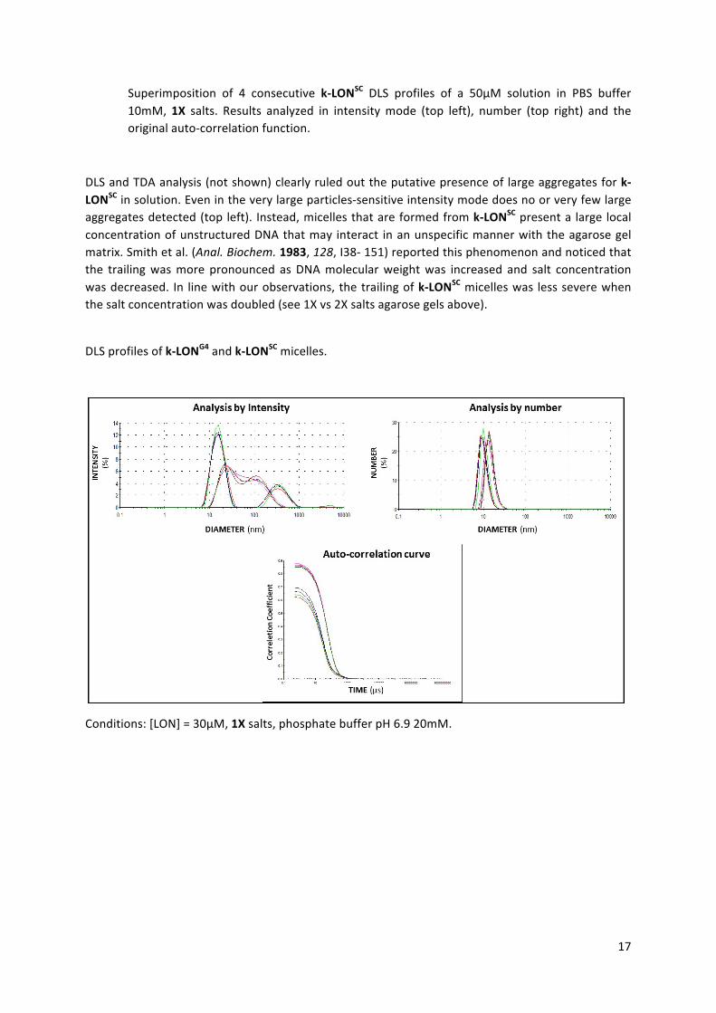
Superimposition of 4 consecutive k-LONSC DLS profiles of a 50µM solution in PBS buffer10mM, 1X salts. Results analyzed in intensitymode (top left), number (top right) and theoriginalauto-correlationfunction.
DLSandTDAanalysis(notshown)clearlyruledouttheputativepresenceof largeaggregatesfork-LONSCinsolution.Evenintheverylargeparticles-sensitiveintensitymodedoesnoorveryfewlargeaggregatesdetected(top left). Instead,micellesthatareformedfromk-LONSCpresenta large localconcentrationofunstructuredDNAthatmay interact inanunspecificmannerwiththeagarosegelmatrix.Smithetal.(Anal.Biochem.1983,128,I38-151)reportedthisphenomenonandnoticedthatthe trailingwasmore pronounced asDNAmolecularweightwas increased and salt concentrationwasdecreased. In linewithourobservations, thetrailingofk-LONSCmicelleswas lessseverewhenthesaltconcentrationwasdoubled(see1Xvs2Xsaltsagarosegelsabove).
DLSprofilesofk-LONG4andk-LONSCmicelles.
Conditions:[LON]=30µM,1Xsalts,phosphatebufferpH6.920mM.

18
FigureS8.TDAofk-LONG4andk-LONSCmicellarsizes.
k-LONG4:λ=254nm;hydrodynamicradius6,216nm;diffusioncoefficient39,5µm2/s.
k-LONsc:λ=254nm;hydrodynamicradius5,787nm;diffusioncoefficient42,4µm2/s.
TDAof30µMsolutionsofk-LONG4(top)andk-LONSC(bottom)in3XsaltsinPBSbuffer10mMpH6.9.Thebluecurvecorrespondstotheabsorptionsignalinthefirstanalysiswindow,thegreenoneinthesecond window. The red and violet lines correspond to the fitting curve from which thehydrodynamicradiusisextracted.
19
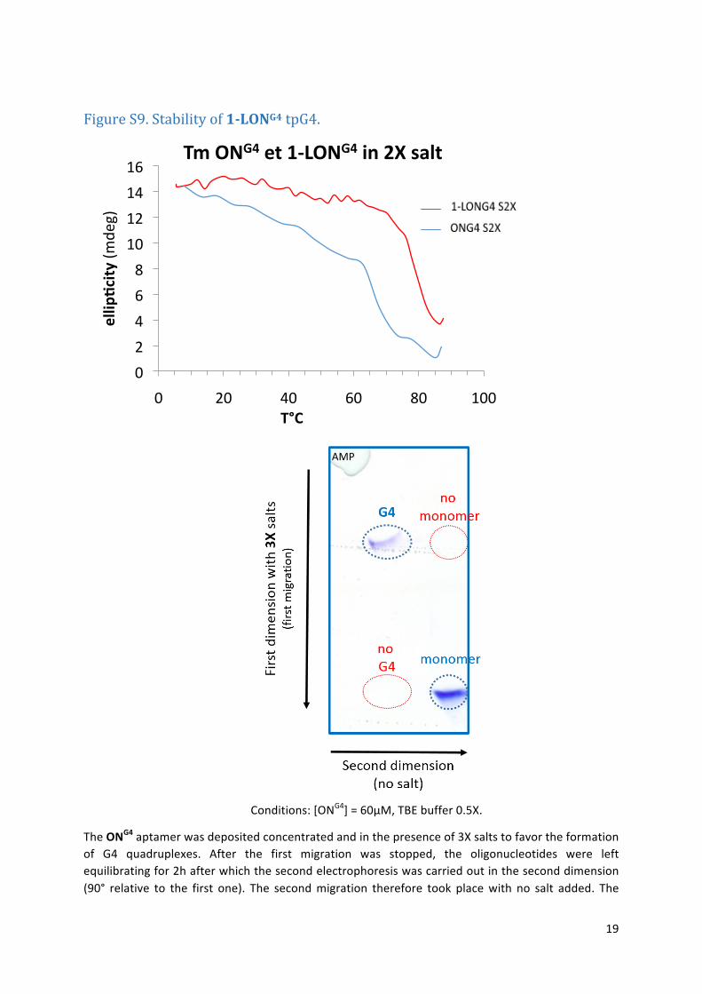
FigureS9.Stabilityof1-LONG4tpG4.
Conditions:[ONG4]=60µM,TBEbuffer0.5X.
TheONG4aptamerwasdepositedconcentratedandinthepresenceof3Xsaltstofavortheformationof G4 quadruplexes. After the first migration was stopped, the oligonucleotides were leftequilibratingfor2hafterwhichthesecondelectrophoresiswascarriedoutintheseconddimension(90° relative to the first one). The secondmigration therefore took placewith no salt added. The
0
2
4
6
8
10
12
14
16
0 20 40 60 80 100
ellip
_city(m
deg)
T°C
TmONG4et1-LONG4in2Xsalt
20
absence of added salts that promote the formation of the G4 structures and the dilution of thesample in the gelmay explain the absence of G4 in the secondmigration of themonomer band(bottomleft,circledinred).Thisexperimentclearlydemonstratesthatonceformed,thetpG4isinertas nomonomer band is observed in the seconddimension of theG4 band (top right in red). It isworthemphasizingthattheG4remainedstablefor2hdespitetheabsenceofstabilizingsaltsinthegelduringthecourseofthesecondmigration.
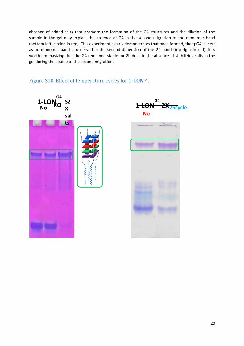
FigureS10.Effectoftemperaturecyclesfor1-LONG4.
1-LONG4
25cycles KCl0.1M
S2Xsalts
Nosalts No
cycle
25cycles
1-LONG42X
salts

21
FigureS11.Effectoftemperaturecyclesfor1-LONG4.
CDspectraof1-LONG4without(red)andwithtemperaturecycles(25cycles,blue)ofa5µMsolutioninthepresenceof2Xsaltsand20mMphosphatebuffer.
-10
-5
0
5
10
15
20
210 230 250 270 290 310 330
Elipicity(m
deg)
λ(nm)
nocycle25cycles

22
FigureS12.Effectofcationsonthethermalstabilityof1-LONG4andONG4tpG4s.
1-LONG4
CDMeltingcurvesof1-LONG4inthepresenceofdifferentcations(5µMLONsolutionsinphosphatebuffer20mM).
Nomeltingisobservedinthepresenceof100mMmagnesium(green)orpotassium(darkgrey):Tm>95°C.Incontrast,theTmisverylow<20°Cinthesameconditionswithsodium.Wehadtoincreasethesodiumconcentrationto250mMtoobservethetransition(lightbluecurve).SodiumisclearlyacompetitorfortpG4bindingastheTmisdecreasedinthepresenceofamixtureofthe3salts(redandbluecurves,sameconditionsthelatterwithadditionaltemperaturecyclesapplied).
0
5
10
15
20
25
30
25 35 45 55 65 75 85 95 105
CD(m
deg)
T(°C)
Tm1-LONG4
S2X
S2Xnocycle
100mMKCl
100mMMgCl2
100mMNaCl
250mMNaCl

23
ONG4
SameexperimentswithONG4.Thesamebehaviorisobserved.
0
10
20
30
40
50
60
70
20 30 40 50 60 70 80 90 100
CD(m
deg)
T(°C)
250mMLiCl
250MmMgCl2
250MmKCl
250NaCl

24
FigureS13:Effectsofcationsatintraandextracellularconcentrationsonthethermalstabilityof1-LONG4andk-LONG4
CD spectra of 1-LONG4 at intracellular (blue, 140Mm KCl, 12mM NaCl, and 1mM MgCl2 ) andextracellular (red, 5mMKCl and145mMNaCl) salt concentrationsat37°C.Underextracellular saltconditions1-LONG4isnotfoldedjustasthecontrolONG4(green,seemaintextfordetails).
-15
-10
-5
0
5
10
15
20
25
220 230 240 250 260 270 280 290 300 310
CD(m
deg)
λ(nm)
CD1-LONG4andONG4
0
5
10
15
20
25
30
0 20 40 60
CD(m
deg)
T(°C)
Tm1-LONG4

25
CDmeltingcurveof1-LONG4extractedat265nmatintracellularcationconcentrations(blue)andextracellularconcentrations(red).
CDspectraofk-LONG4at37°Cofintracellular(blue)andextracellular(red)concentrations.
CDmeltingcurveextractedat265nmof1-LONG4atintracellular(blue)andextracellular(red)concentrations:theperfecttetramolecularparallelG4meltsonlyatextracellularsaltconcentrations.ThesteadydecreaseobservedatlowtemperaturescorrespondtothemeltingofmismatchparallelG4s.
-15
-10
-5
0
5
10
15
20
25
220 240 260 280 300 320
CD(m
deg)
λ(nm)
CDk-LONG4
0
5
10
15
20
25
30
0 20 40 60 80 100
CD(m
deg)
T(°C)
Tmk-LONG4

26