converse: chapter 54 craniofacial microsomia john … · converse: chapter 54 craniofacial...
TRANSCRIPT
1
Converse: Chapter 54
Craniofacial Microsomia
John Marquis Converse, Joseph G. McCarthy, Donald Wood-Smith, Peter J. Coccaro
Among the congenital otocephalic syndromes, the term "first and second branchial archsyndrome" designates, in the United States, a characteristic congenital malformation whichis usually unilateral but occasionally bilateral. In the German literature, the deformity hasbeen termed "dysostosis otomandibularis". Caronni (1971) has coined the term "auriculo-branchiogenic dysplasia". Stark and Saunders (1962) referred to a similar clinical associationof physical findings as the first branchial arch syndrome or the oral-mandibular-auricularsyndrome.
The term "first and second branchial arch syndrome" is not entirely satisfactory indifferentiating specific syndromes, for there are also other malformations derived frommaldevelopment of these branchial arches. Gorlin and Pindborg (1964) reviewed the variousnames by which the condition has been described and advocated the term "hemifacialmicrosomia", which implies that the deformity is exclusively unilateral and spares thecranium. Pruzansky (1971) used the term "otocraniocephalic syndromes" to describeaberrations in the development of the first and second branchial arches.
At the Center for Craniofacial Anomalies of the Institute of Reconstructive PlasticSurgery of the New York University Medical Center, the term "hemicraniofacial microsomia"is preferred for the unilateral form, and the designation "bilateral craniofacial microsomia" isreserved for the bilateral type.
Differential Diagnosis
The deformity of craniofacial microsomia, whether unilateral or bilateral, ischaracterized by varying degrees of a regional hypoplasia affecting the temporomandibularand pterygo-mandibular complexes (skeletal and neuromuscular structures). The bilateral formmust be distinguished from mandibulofacial dysostosis (Treacher Collins syndrome) (seechapter 55), which shows a well-defined genetic pattern (Franceschetti and Klein, 1949) withthe deformity being transmitted in an irregular dominant fashion; the gene is unstable and hasa weak degree of penetrance. The genetic component in hemicraniofacial microsomia is lessapparent, and the syndrome may be considered to be genetically heterogeneous - ie, as havingmore than one cause responsible for the defect. Tessier (1976) has pointed out thatcraniofacial microsomia has features in common with mandibulofacial dysostosis (TreacherCollins syndrome), such as the temporozygomatic defect and orbital deformities. Specificcharacteristics of hemicraniofacial microsomia are the malformations of the ramus and facialparalysis due to the involvement of the temporal bone in the malformation. Tessier (1976) hasdescribed three types of clefts that are present in mandibulofacial dysostosis (see Chapter 46).
Cervical spine abnormalities have been noted in many of the affected patients. TheGoldenhar syndrome (oculo-auriculo-vertebral dysplasia (1952)) shows similar characteristicswith, in addition, one or more epibulbar dermoids; it may be considered as a variant of
2
craniofacial microsomia. According to Tessier (1976), a frontozygomatic cleft (No 8) is acharacteristic of Goldenhar syndrome (see Chapter 46).
A jaw malformation similar to that seen in bilateral craniofacial microsomia may beobserved in patients who have suffered postnatal trauma or infection which has affected thecondylar cartilage and, as a result, mandibular growth. It has been suggested, bothexperimentally and clinically, by several authors that impaired growth of the condyle mayresult in an underdevelopment not only of the mandible but also of the craniofacial osseouscomplex on the affected side (Sarnat and Engel, 1951; Brodie, 1964; Sarnat and Laskin,1964). It is, however, usually relatively easy to distinguish postnatal deformities from thoseresulting from prenatal (genetic or intrauterine environmental) factors.
In postnatal traumatic deformity, the deformity is restricted to the jaws and the auricleis spared; the soft tissues are not deficient, and there is no temporal bone deformity. Theearlier the traumatic insult occurs, the more severe the deformity, and there is often associatedtemporomandibular joint ankylosis.
In contrast, in craniofacial microsomia the deformity is more widespread, withinvolvement of the temporal bone, middle ear, mastoid process, external ear, and base of theskull, and often soft tissue hypoplasia on the affected side.
Exceptions to these differentiating features exist. A jaw malformation characteristicof hemicraniofacial microsomia may be present from birth with minimal auricular or temporalbone development. The authors have observed two such cases of hemicraniofacial microsomiain which audiograms showed a normal level of hearing in both ears and no deformity of thetemporal bone could be detected by roentgenography. These patients represent variations ofhemicraniofacial microsomia rather than a separate syndrome (mandibular dysostosis, Nagerand de Reynier, 1948).
Patients with severe orbital clefts (see Chapter 46) and hypoplasia of the maxilla alsohave an occlusal slant and a short mandibular ramus but the condylar deformity, which is thehallmark of craniofacial microsomia, is lacking. In craniofacial microsomia, the mandibularanomaly is a developmental deformity that is secondary to a hypoplasia of the temporal bone;the hypoplastic maxilla is secondary to the mandibular hypoplasia.
History
Grabb (1965) cited the teratological tablets written in approximately 2000 BC, by theChaldeans of Mesopotamia as the earliest recordings of malformations of the first and secondbranchial arches in man. Bartholinus (1654) described a child with absence of the externalauditory canal, and Lachmund (1688) reported a female with microtia and agenesis of theexternal auditory canal. Thompson (1845) drew attention to the fact that the clinicalexpression of the syndrome was in structures derived from the first and second branchialarches and the intervening cleft.
In recent times the publications of Kazanjian (1939), Altman (1951), Meurmann(1957), François (1961), Stark and Saunders (1962), Gorlin and Pindborg (1964), Grabb(1965), Pruzansky (1969), Longacre, DeStefano, and Holmstrand (1963), Obwegeser (1970,
3
1974), and Converse and associates (1973a, b, 1974) have focused attention on the clinicalfindings and the reconstruction of the individual deformities.
Clinical Spectrum of the Syndrome
Incidence. In a study of birth records at several hospitals in the United States, thebirth incidence was determined to be 1 in 5642 births (Grabb, 1965). Poswillo (1973) citedan incidence of 1 in 4000. Following the administration of thalidomide to pregnant womenin Germany between the years 1959 and 1962, 2000 less severe cases of first and secondbranchial arch malformations were reported (Kleinsasser and Schlothane, 1964).
Sex. In the series of 102 patients reported by Grabb (1965), 63 were males and 39were females.
Unilateral vs. Bilateral. In a series of 74 patients, Meurmann (1957) reported eightpatients with bilateral involvement; Dupertuis and Musgrave (1959) noted a unilateral tobilateral incidence of 6 to 1; Grabb (1965) cited bilateral involvement in 12 of 102 patients;Converse, Wood-Smith, McCarthy, Coccaro, and Becker (1974) studied 15 patients withbilateral craniofacial microsomia out of a total series of 280 patients. The pathologic findingsand treatment of bilateral craniofacial microsomia are discussed in a subsequent section of thechapter.
Variations of the Syndrome. The deformity in craniofacial microsomia varies inextent and degree. As in many other craniofacial anomalies, it is often difficult to classify theindividual deformity.
In the severe form, all of the structures derived from the first and second branchialarches are hypoplastic, while in other types, either the auricular or jaw dysplasias may bepredominant.
It is in the latter cases that the associated deformities are less evident. There may bemany shades of expression, according to the degree of involvement of the structures derivedfrom the first and second arches and the involvement of the adjacent skeletal structures:
1. The ear deformity may be maximal, while the jaw deformity is not apparent onclinical examination. Roentgenographic studies, including tomography, demonstrate, however,that all cases of external auditory canal and auricular hypoplasia with middle ear deformityhave mandibular changes on the affected side.
2. The ear deformity may be less severe, and the jaw deformity may not be evident;roentgenographic analysis may show a disparity of the skeletal structures. In these cases ofminor jaw deformities, careful clinical examination will often show a slight deviation of themandible to the affected side. The midsagittal plane extends through the interspace betweenthe upper central incisors (midincisor point), but the lower midincisor point may be off centerin relation to it.
3. The characteristic jaw deformity of hemicraniofacial microsomia may be presentwithout gross auricular or temporal bone maldevelopment. These cases are difficult to
4
differentiate from postnatal deformities caused by injury, but the diagnosis becomes evidentif the deformity was present at birth. This type of deformity may also be complicated byassociated malformations such microphthalmos. The auricle may be normal in shape,protruding, or low-set, with a normal hearing mechanism.
4. "Formes frustes" or microforms are more frequent than is generally acknowledged.They must be searched for in cases of slight facial asymmetry and in auricular malformationswithout manifest jaw deformity. The patients shown is an example of a "forme fruste". Thedeformity is characterized by a soft tissue deficiency in the right parotid-masseteric area (theparotid gland was hypoplastic), a protruding right auricle (with normal hearing) and a slightdegree of macrostomia involving the right oral commissure. The dental occlusal relationshipswere adequate. Correction was achieved by insertion of a dermis-fat graft that restoredadequate facial contour, a protruding ear operation according to the technique of Converse andassociates (1955, 1963), and closure of the macrostomia.
Grabb (1965) proposed a classification of six groups according to the specificanatomical abnormalities but emphasized the overlapping among the individual groups.
Embryology
The ear can be taken as a frame of reference in the otocraniocephalic syndromes(Pruzansky, 1971) because of its developmental relationship with the jaw (Table 54-1). Abrief review of the phylogeny and ontogeny of the auricle and hearing apparatus is helpfulin understanding the mechanism of the malformation in the craniofacial microsomiasyndrome.
The two principal divisions of the organ of hearing come from different embryonicanlagen. The sensory end organ in the inner ear is derived from the ectodermal otocyst, whilethe sound-conducting apparatus in the external and middle ear comes from the gill structures.
The membranous labyrinth has its beginning in the 3.5 week old human embryo (Arey,1946) as a thickening of the ectoderm on the side of the head - the otic placode (see Chapter35). This area is enfolded to become the otic pit, then pinches off to become the otocyst. Bymeans of a series of folds, the otocyst differentiates in the 3 month old fetus into theendolymphatic duct and sac, the semicircular endolymphatic ducts, the utricle, the saccule,and the cochlear duct, which contains the organ of Corti. By the fifth month of fetal life, thesensory end organ of the ear attains adult form and size, as the cartilaginous otic capsuleossifies.
It is speculated that our piscine ancestors swam in seas not yet as salty as today'soceans and that man's endolymph, entrapped by the enfolding otocyst, closely resembles inchemical composition the dilute salt water of that primeval sea. Our ancient aquatic forebearsdid not require any special mechanism to transmit sound to the inner ear. As in today's fish,sound was readily transmitted from the sea through the skin to the fluid of the inner ear.
However, when these enterprising ancestors struggled out of the seas onto dry land,a new problem appeared. A mechanical device was needed to convert air vibrations of largeamplitude and small force into fluid vibrations of small amplitude and large force. The gill
5
structures, no longer needed for breathing, became converted into such a mechanism. The firstbranchial groove became the external auditory meatus and canal, while the first pharyngealpouch became the eustachian tube and middle ear. Instead of the branchial groove andpharyngeal pouch connecting to become a gill cleft, a thin intervening layer of tissueremained to form the tympanic membrane.
The mandible, incus, and malleus developed from the cartilage of the first branchialarch (Meckel's cartilage), while the stapes (with the exception of the footplate, whichoriginates from the otic capsule), styloid process, and hyoid bone developed from the cartilageof the second arch (Reichert's cartilage) (See Table 54-1). The large area of the tympanicmembrane, connected by the lever system of the ossicular chain to the small area of the ovalwindow, provided the ear with an effective mechanism to overcome the sound barrier betweenair and water.
Table 54-1. Structures Derived From the First and Second Branchial Arches andthe Otic Capsule
First branchial archMaxillary process Maxilla
Palatine boneZygoma
Mandibular process Trigeminal nerveAnterior part of auricleMandibleHead of malleusBody of incusTympanic boneSphenomandibular ligament
First branchial groove External auditory meatusTympanic membrane
First pharyngeal pouch Eustachian tubeMiddle ear cavity
Second branchial arch Facial nervePosterior part of auricleManubrium of malleus, long process of incus,stapedial superstructure, and tympanic surfaceStapedial artery, styloid process, stylohyoidligamentLesser cornu of hyoid
Otic capsule Vestibular surface of stapes, internal acousticmeatusInner ear.
The gill structures destined to form the sound-conducting apparatus and the jaws firstappear in the human embryo at 4 weeks, at about the same time as the otic pit.
By the third fetal month, the pinna has been formed from the first and secondbranchial arches on either side of the first branchial groove; the latter is the primary shallow,
6
funnel-shaped external auditory meatus. From the inner end of the primary meatus, a solidcord of ectodermal cells extends further inward, with a bulblike enlargement adjacent to themiddle ear. It is not until the seventh fetal month that this cord canalizes, beginning mediallyto form the tympanic membrane and then extending laterally to join with the primary meatusto form the completed external auditory meatus. The external and middle ear, althoughcapable of transmitting sound to the inner ear, are not yet of adult form and size.
In the seventh fetal month, pneumatization of the temporal bone begins, withexcavations lined by mucous membrane extending out from the middle ear cavity while thejellylike mesodermal tissue in the middle ear cavity begins to resolve. At birth the eustachiantube inflates; the fetal mesodermal tissue in the middle ear and antrum continues to resorbuntil the epithelium lies close to the periosteum, and pneumatization of the temporal boneproceeds.
The external auditory meatus, entirely cartilaginous at birth (except for the narrowincomplete ring of the tympanic bone), deepens by growth of the tympanic bone to form theadult osseous meatus. Except for some pneumatization of the petrous apex that may continueinto adult life, the external and middle ear finally reach adult form and size in late childhood(in contrast to the inner ear, which becomes adult in fetal life). It is generally accepted thatthe first branchial arch furnishes the anterior part of the auricle (see Table 54-1); the secondarch provides the structures of the remaining external ear (Wood-Jones and Wen-I-Chuan,1934). The maxilla, palatine bone, and zygoma develop from the maxillary process of the firstbranchial arch, the mandible from the mandibular process.
Meckel's cartilage, the primary jaw of lower vertebrates, represents the temporaryskeleton of the first pharyngeal arch; the two symmetrical cartilaginous bars in early fetal lifedescribe a parabolic arch that serves as a model and guide in the early morphogenesis of themandible.
Three main regions of Meckel's cartilage should be considered: (1) the distal portion,which becomes incorporated into the anterior part of the body of the mandible; (2) a middleportion, which gives rise to the sphenomandibular ligament and contributes to the mylohyoidgroove of the mandible; (3) the proximal or intratympanic portion, which differentiates intothe malleus, the incus, and the anterior malleolar ligament.
Embryology of the face is discussed in more detail in Chapter 53.
Etiopathogenesis
Unlike mandibulofacial dysostosis, the genetic component in craniofacial microsomiais ill-defined, and there is no evidence of genetic transmission except in a few patients. Grabb(1965) reported that only 4 of 102 patients studied had one sibling or one parent withmaldevelopment of the first and second branchial arch syndrome. Hanhart (1949) describeda form of inheritable auricular hypoplasia, and Rogers (1968) listed several family studies byother authors which suggested a possible hereditary basis for auricular deformities. Summitt(1969) reported a pedigree of the Goldenhar syndrome which was compatible with autosomaldominant transmission.
7
A thorough search for minor deformities (such as preauricular tags or a receding chin)in other members of the family may provide a clue that the affected genes are not absent inthe patient. As Fisher (1961) has stated, "A high index of suspicion based primarily on acareful family and maternal history, and understanding of the anomaly's causative factors andkeenness of observation are important in identifying congenital abnormalities - many of whichcan be detected only after one has searched for them."
Consequently current etiopathogenic theories favor an intrauterine factor (or factors)which affects the embryo and has the following three characteristics:
1. The factor varies in its intensity and penetrance.
2. The factor strikes at varying periods in the course of prenatal development - fromthe first to the seventh month.
3. The damage is produced in varying loci in the fetus or embryo, along any point ofthe developing first and second branchial arches; the damage may be localized (segmental)or widespread.
The etiopathogenesis has not been adequately explained to date. The theory ofmesodermal deficiency of Hoffstetter and Veau has been involved by Stark and Saunders(1962).
Others have suggested that vascular defects of the stapedial artery may account formaldevelopment of the first and second branchial arches (McKenzie and Craig, 1955). Thestapedial artery, a temporary vascular supply for the primordia of the first and secondbranchial arches, appears as a collateral of the hyoid artery and anastomoses with thepharyngeal artery; it is ultimately replaced by the finite external carotid system. Willie-Jorgensen (1962) felt that abnormalities of the latter artery were responsible for anomalies ofthe first and second branchial arches. A decreased heat pattern over the region of the externalmaxillary artery has also been demonstrated and is interpreted as evidence of a vasculardeficiency (Ide, Miller, and Wollschlaeger, 1970).
Poswillo (1973) has produced phenocopies of craniofacial microsomia following theadministration of triazene to the mouse and thalidomide to the monkey. Embryonic hematomaformation with spreading hemorrhage prior to the formation of the stapedial artery wasdemonstrated. The extent and size of the hematoma correlated with the size of the anomalousdefect. In the mouse experimental model, all of the variations found in the human syndromecould be reproduced. The spectrum of defects was broad: a small aural hematoma producinga residual deformity of only the external ear and auditory ossicles; and larger hemorrhagiclesions affecting the condyle, mandibular ramus, and zygoma.
As mentioned earlier, Kleinsasser and Schlothane (1964) reported a large number ofnewborns with first and second branchial anomalies following the widespread use ofthalidomide during pregnancy.
8
Pathology
The deformity in hemicraniofacial microsomia usually has the three major features ofauricular, mandibular, and maxillary hypoplasia. The hypoplasia, however, also involvesadjacent anatomical structures: the zygoma, the pterygoid processes of the sphenoid bone, thetemporal bone (the middle ear and less frequently the internal ear; the mastoid process issmall, and on the roentgenogram is seen to be acellular), the facial nerve, the facialmusculature of expression, the muscles of mastication, the parotid, the cutaneous andsubcutaneous tissues, and the tongue, soft palate, pharynx, and floor of the nose.
While the jaw and ear deformities are the most conspicuous in the majority of patients,the development of the first and second branchial arches and the structures derived therefromare intimately interlinked with those of the chondrocranium and membranous bones of theskull; associated deformities of the temporal bone and other cranial bones are inevitable. Inextreme forms of the dysplasia, extensive hemicraniofacial involvement is evident. AsPruzansky (1971) has stated, maldevelopment in one area may trigger a "domino effect", withinvolvement of the entire craniofacial bone community.
In addition to the skeletal deficiency, varying amounts of soft tissue hypoplasia,microphthalmos, anophthalmos, cranial nerve palsy, cleft lip and palate, and lateral facialclefts are frequent accompanying deformities.
The Jaw Deformity
The most conspicuous deformity of hemifacial microsomia is the hypoplasia of themandible on the affected side. The ramus is short or virtually absent, and the body of themandible curves upward to join the short ramus. The chin is deviated to the affected side; thebody of the mandible on the unaffected side does not follow its usual curvature but assumesa flattened contour with a straightened gonial angle.
Ramus and condyle malformations vary from minimal hypoplasia of the condyle toits complete absence in association with hypoplasia or agenesis of the ramus. In all patients,condylar anomalies can be demonstrated, and this finding may represent the hallmark of thesyndrome. As a consequence, the spatial relationships of the distorted, deformed, and/ordeficient anatomical parts, as well as the neuromuscular components identified with theseparts, become of paramount importance in the diagnosis and the planning of treatment.
Attempts at classification of the mandibular anomaly have been made (Pruzansky,1971):
Grade I: Hypoplasia is minimal or slight.
Grade II: The condyle and ramus are small; the head of the condyle is flattened; theglenoid fossa is absent; the condyle is hinged on a flat, often convex, infratemporal surface;the coronoid process may be absent.
Grade III: The ramus is reduced to a thin lamina of bone, or it is completely absent.
9
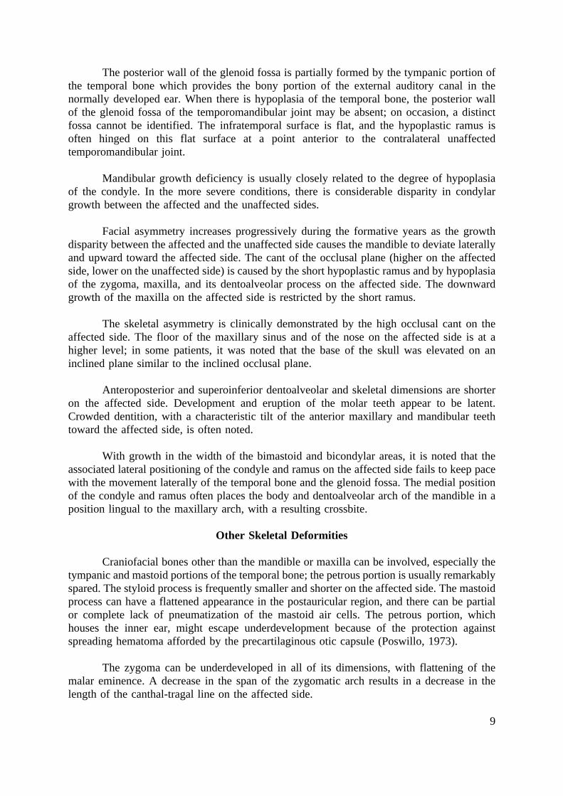
The posterior wall of the glenoid fossa is partially formed by the tympanic portion ofthe temporal bone which provides the bony portion of the external auditory canal in thenormally developed ear. When there is hypoplasia of the temporal bone, the posterior wallof the glenoid fossa of the temporomandibular joint may be absent; on occasion, a distinctfossa cannot be identified. The infratemporal surface is flat, and the hypoplastic ramus isoften hinged on this flat surface at a point anterior to the contralateral unaffectedtemporomandibular joint.
Mandibular growth deficiency is usually closely related to the degree of hypoplasiaof the condyle. In the more severe conditions, there is considerable disparity in condylargrowth between the affected and the unaffected sides.
Facial asymmetry increases progressively during the formative years as the growthdisparity between the affected and the unaffected side causes the mandible to deviate laterallyand upward toward the affected side. The cant of the occlusal plane (higher on the affectedside, lower on the unaffected side) is caused by the short hypoplastic ramus and by hypoplasiaof the zygoma, maxilla, and its dentoalveolar process on the affected side. The downwardgrowth of the maxilla on the affected side is restricted by the short ramus.
The skeletal asymmetry is clinically demonstrated by the high occlusal cant on theaffected side. The floor of the maxillary sinus and of the nose on the affected side is at ahigher level; in some patients, it was noted that the base of the skull was elevated on aninclined plane similar to the inclined occlusal plane.
Anteroposterior and superoinferior dentoalveolar and skeletal dimensions are shorteron the affected side. Development and eruption of the molar teeth appear to be latent.Crowded dentition, with a characteristic tilt of the anterior maxillary and mandibular teethtoward the affected side, is often noted.
With growth in the width of the bimastoid and bicondylar areas, it is noted that theassociated lateral positioning of the condyle and ramus on the affected side fails to keep pacewith the movement laterally of the temporal bone and the glenoid fossa. The medial positionof the condyle and ramus often places the body and dentoalveolar arch of the mandible in aposition lingual to the maxillary arch, with a resulting crossbite.
Other Skeletal Deformities
Craniofacial bones other than the mandible or maxilla can be involved, especially thetympanic and mastoid portions of the temporal bone; the petrous portion is usually remarkablyspared. The styloid process is frequently smaller and shorter on the affected side. The mastoidprocess can have a flattened appearance in the postauricular region, and there can be partialor complete lack of pneumatization of the mastoid air cells. The petrous portion, whichhouses the inner ear, might escape underdevelopment because of the protection againstspreading hematoma afforded by the precartilaginous otic capsule (Poswillo, 1973).
The zygoma can be underdeveloped in all of its dimensions, with flattening of themalar eminence. A decrease in the span of the zygomatic arch results in a decrease in thelength of the canthal-tragal line on the affected side.
10
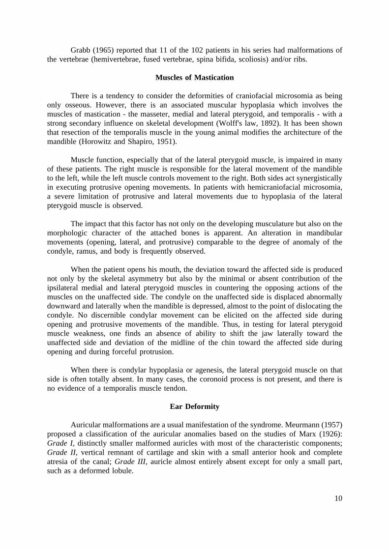
Grabb (1965) reported that 11 of the 102 patients in his series had malformations ofthe vertebrae (hemivertebrae, fused vertebrae, spina bifida, scoliosis) and/or ribs.
Muscles of Mastication
There is a tendency to consider the deformities of craniofacial microsomia as beingonly osseous. However, there is an associated muscular hypoplasia which involves themuscles of mastication - the masseter, medial and lateral pterygoid, and temporalis - with astrong secondary influence on skeletal development (Wolff's law, 1892). It has been shownthat resection of the temporalis muscle in the young animal modifies the architecture of themandible (Horowitz and Shapiro, 1951).
Muscle function, especially that of the lateral pterygoid muscle, is impaired in manyof these patients. The right muscle is responsible for the lateral movement of the mandibleto the left, while the left muscle controls movement to the right. Both sides act synergisticallyin executing protrusive opening movements. In patients with hemicraniofacial microsomia,a severe limitation of protrusive and lateral movements due to hypoplasia of the lateralpterygoid muscle is observed.
The impact that this factor has not only on the developing musculature but also on themorphologic character of the attached bones is apparent. An alteration in mandibularmovements (opening, lateral, and protrusive) comparable to the degree of anomaly of thecondyle, ramus, and body is frequently observed.
When the patient opens his mouth, the deviation toward the affected side is producednot only by the skeletal asymmetry but also by the minimal or absent contribution of theipsilateral medial and lateral pterygoid muscles in countering the opposing actions of themuscles on the unaffected side. The condyle on the unaffected side is displaced abnormallydownward and laterally when the mandible is depressed, almost to the point of dislocating thecondyle. No discernible condylar movement can be elicited on the affected side duringopening and protrusive movements of the mandible. Thus, in testing for lateral pterygoidmuscle weakness, one finds an absence of ability to shift the jaw laterally toward theunaffected side and deviation of the midline of the chin toward the affected side duringopening and during forceful protrusion.
When there is condylar hypoplasia or agenesis, the lateral pterygoid muscle on thatside is often totally absent. In many cases, the coronoid process is not present, and there isno evidence of a temporalis muscle tendon.
Ear Deformity
Auricular malformations are a usual manifestation of the syndrome. Meurmann (1957)proposed a classification of the auricular anomalies based on the studies of Marx (1926):Grade I, distinctly smaller malformed auricles with most of the characteristic components;Grade II, vertical remnant of cartilage and skin with a small anterior hook and completeatresia of the canal; Grade III, auricle almost entirely absent except for only a small part,such as a deformed lobule.
11
In a comprehensive study, Pruzansky (1971), using air and bone conductionaudiometry and temporal bone tomography, evaluated 57 patients with craniofacialmicrosomia. It was observed that the degree of auricular deformity as classified above doesnot correlate exactly with hearing function. The type of hearing loss, although usuallyassumed to be conductive in origin, can be determined only by audiometry. Tomography, notauricular morphology, is the only indicator of middle ear structure. In addition, the unaffectedear may also harbor abnormalities in structure and function and should be evaluated. Therewas, however, a direct relationship between the degree of severity of auricular malformationand ipsilateral mandibular deformity. Reconstruction of the auricle and middle ear in patientswith craniofacial microsomia is discussed in Chapter 35.
Deformities of the Nervous System
Nervous system abnormalities in patients with craniofacial microsomia have receivedlittle attention in the medical literature (Gorlin, Jue, Jacobsen and Goldschmidt, 1963).
Patients in the series at the Institute of Reconstructive Plastic Surgery have beeninvestigated by complete neurologic examination, skull radiography, computerized axialtomography, and electromyography (Aleksic and coworkers, 1975a, b, c, d).
Cerebral Anomalies. A wide variety of cerebral anomalies exists in craniofacialmicrosomia and may include ipsilateral cerebral hypoplasia (Aleksic and associates, 1975a),hypoplasia of the corpus callosum (Timm, 1960), hydrocephalus of the communicating(Timm, 1960; Aleksic and associates, 1975c) and obstructive (Herrmann and Optiz, 1969)types, intracranial lipoma (Gaupp and Jantz, 1942; Aleksic and associates, 1975c), andunilateral hypoplasia of the brain stem and cerebellum (Mathies, 1966; Aleksic and associates,1975c). Other associated abnormalities include mental retardation (Gorlin and coworkers,1963), epilepsy, and electroencephalographic findings suggestive of epilepsy (Franceschetti,Klein and Brocher, 1959; Christiaens, Walbaum, Farriaux and Fontain, 1966; Aleksic andassociates, 1975c).
Cranial Nerve Anomalies. Cranial nerve abnormalities are frequent inhemicraniofacial microsomia and can range from arhinencephaly of the bilateral (Virchow,1864; Timm, 1960) and unilateral (Aleksic and associates, 1975a) types to unilateral agenesisand hypoplasia of the optic nerve with secondary changes in the lateral geniculate body andvisual cortex (Aleksic and associates, 1975d), congenital ophthalmoplegia and Duane'sretraction syndrome (Bowen, Collum and Rees, 1971), hypoplasia of the trochlear andabducens nuclei and nerves (Aleksic and associates, 1976), congenital trigeminal anesthesia(Sugar, 1967), and aplasia of the trigeminal nerve and motor and sensory nucleus (Aleksicand associates, 1975b). The most common cranial nerve anomaly is facial paralysis secondaryto agenesis of the facial muscles, aberrant pathway of the facial nerve in the temporal bone(Bellucci, 1972; Sekhar and associates, 1975), or hypoplasia of the intracranial portion of thefacial nerve and facial nucleus in the brain stem (Aleksic and associates, 1975c). Congenitalhearing loss may be due to a malformed inner ear (Aleksic and Sekhar, 1976), hypoplasia ofthe cochlear nerve and brain stem auditory nuclei (Aleksic and associates, 1975c), orhypoplasia and impaired function of the ninth through the twelfth cranial nerves (Mathies,1966; Berkman and Feingold, 1968). In conclusion, any cranial nerve can be clinicallyinvolved in patients with craniofacial microsomia, and it is likely that hypoplasia or agenesis
12
of a portion of the entire cranial nerve trunk and corresponding brain stem nuclei representsthe pathoanatomic substrate of the clinical dysfunction.
Electromyographic abnormalities have been briefly described in the literature (Grabb,1965). Several of the authors' patients showed diminished or low-normal motor conductionvelocity of the facial nerves, usually unilateral, while electromyographic abnormalities rangedfrom absence of the muscle to long polyphasic potential with incomplete interference patternon active innervation (Aleksic and coworkers, 1975c). No cases of fibrillation potentials haveyet been documented. The interpretation of the electrodiagnostic data has been somewhathampered by the fact that many patients have undergone earlier surgical procedures in theregion of the mandible and auricle.
Soft Tissue Deformities
In addition to the anomalies of the muscles of mastication and nervous system, thereis often a generalized soft tissue hypoplasia which involves the skin, the subcutaneous tissue,and the facial musculature of expression. The musculature of the soft palate and of the tongueis occasionally less developed on the affected side. The not infrequent hypoplasia or aplasiaof the parotid gland, previously noted by Entin (1958), places the branches of the facial nervein a superficial and vulnerable position. The latter finding has obvious surgical implications.The shortness of soft tissues on the affected side is made evident by the shorter distancebetween the mastoid process and the angle of the mouth or the lateral canthus of the eye. Theskin and subcutaneous tissue also show varying degrees of atrophy, particularly in the parotid-masseteric and auriculomastoid areas.
In the series reported by Grabb (1965), 10 per cent of the patients had malformationsof the eye and eyelids and/or palate.
Transverse facial clefting ranging from macrostomia to a full thickness defect of thecheek can be present (Converse and coworkers, 1974). The clefts probably result from failureof fusion of the maxillary and mandibular processes. In embryologic development, the lateralcommissure of the oral fissure is initially situated at the point of bifurcation of the maxillaryand mandibular processes. With fusion of the latter and development of the muscles ofmastication, the original broad mouth is reduced in size. In addition, the parotid glands,originally located near the embryonic oral commissure grow laterally toward the developingear, but the parotid duct papillae remain in their more medial position (Converse andcoworkers, 1974).
Treatment
Corrective Jaw Surgery
Correction of the jaw deformity in craniofacial microsomia has been attempted eitherby expansion for contour restoration or by osteotomies to change the jaw asymmetry andcrossbite.
Contour Restoration Operation. Early expansion of local contour to maintain thegrowth of the soft tissues on the affected side has been achieved by serial autogenous split
13
rib grafts (Longacre, De Stefano and Holmstrand, 1961; Longacre, 1968), by allografts ofcartilage or bone (Stark and Saunders, 1962), and by preserved cartilage allografts andinorganic implants (Brown, Fryer and Ohlwiler, 1960).
Other techniques for improvement of contour in the adult include correcting thedeficient mandible either with a transplant taken from the lower portion of the unaffected halfof the mandible (Gorski and Tarczynska, 1969) or by bone graft onlays (Converse andShapiro, 1954; Longacre, De Stefano and Holmstrand, 1961). The only long-term report ofresults obtained by these contour-restoring procedures is that of Longacre (1968), whoreported maintenance of contour in craniofacial dysostoses following serial autogenous onlaybone grafts.
Such surgery, however, fails to improve dental occlusion or function and also fails toalign the facial skeleton in a position conducive to subsequent growth.
Osteotomies of the Jaws. Correction of the jaw asymmetry and crossbite byosteotomies and bone grafts has been done in children and adults, and the timing of thesurgery has been the subject of controversy.
Poswillo (1974) cautioned against reconstruction of the facial bones before age 12. Hefelt that the trauma associated with surgery could destroy the functional matrix (Moss, 1968)of part of the facial skeleton and profoundly interfere with subsequent facial growth.
Obwegeser (1974) also advocated postponement of definitive jaw surgery until growthof the facial skeleton has ceased, as he felt osteotomies performed during childhood interferedwith growth of the facial skeleton.
Converse, Horowitz, Coccaro and Wood-Smith (1973b), however, recommendedsurgery during childhood with the following objectives: (1) improving the symmetry of themandible by bilateral vertical osteotomies of the ramus during the period of mixed dentition;(2) providing for downgrowth of the maxilla by lengthening the shortened mandibular ramus;(3) restoring the adequate dental occlusion; and (4) expanding the facial skeleton at an earlyage to fill out the soft tissues on the affected side of the face.
Osteotomies in the Adult. Correction of the jaw asymmetry and of the crossbite bymandibular osteotomy and bone grafts has been performed in the adult (Limberg, 1928;Converse and Shapiro, 1952; Dingman and Grabb, 1963, 1964).
Obwegeser (1970) has obtained satisfactory results in adults by a combined Le FortI osteotomy of the maxilla and a bilateral sagittal section of the mandibular ramus. Thisprocedure cannot be employed in the patient during the period of mixed dentition, as themaxillary osteotomy would injure or destroy the unerupted teeth.
Obwegeser (1974) has advocated that in severe cases the temporomandibular jointshould be initially reconstructed with a costochondral rib graft. Any associated hypoplasia ofthe temporal bone, zygomatic arch, and lateral orbit rim should also be corrected. At asubsequent operation, the lower half of the facial skeleton can be rotated inferiorly and
14
anteriorly by a combination of a Le Fort I osteotomy, a bilateral sagittal split osteotomy ofthe mandible, and a double-step horizontal advancement osteotomy.
Osteotomies in Childhood. A metatarsal head transplant, as a substitute for themissing growth center of the mandibular condyle, has been advocated (Glahn and Winther,1967). Others who used this procedure earlier found little or no growth of the grafts and noparticular improvement in joint function (Dingman and Grabb, 1964; Freeman, 1965).
Converse and Rushton (1941) operated on a 12 year old patient with the jaw deformityof craniofacial microsomia. A horizontal osteotomy of the ramus was performed above theinferior alveolar foramen, and iliac bone graft was inserted between the fragments after theplacement of a bite-block to open the occlusion on the affected side.
A similar procedure was performed by Osborne (1964) in patients with craniofacialmicrosomia and in those with asymmetrical growth resulting from injury. Osborne advocatedsurgery before the age of 6 years in order to give the maxilla an opportunity to develop afterthe release of the upward pressure exerted on the affected side by the hypoplastic mandible.Longitudinal studies of two of Osborne's cases of congenital origin were done by Knowles(1966), who was able to document maxillary growth.
Delaire (1970) recommended even earlier intervention between the ages of 4 and 6years. He elongated the shorter ramus with an inverted L osteotomy and inserted costal bonegrafts into the gap formed at the horizontal branch of the L.
Converse and his associates (1973b) advocated a two-stage procedure during childhoodto correct the mandibular asymmetry, to allow expansion of the constricted maxilla, and toprevent further contraction of the overlying soft tissue.
The first stage, usually done when the child is 8 or 9 years old and in the mixeddentition period when mandibular growth is less active, corrects the vertical and horizontalhypoplasia of the malformed half of the mandible. A bilateral vertical osteotomy through theramus is performed, permitting a forward, lateral, and downward movement of the malformedmandibular body toward the unaffected side. The rotation and displacement result in a bonygap in the deficient ramus on the affected side, reflecting the amount of downward and medialdisplacement of the mandibular body. Interposition iliac bone grafts are placed in the defectand maintained in position by wedging them between the fragments and by securing themwith interosseous stainless steel wires. The iliac bone grafts are removed according to thetechnique of Crockford and Converse (1972).
The second stage of the corrective treatment is undertaken during adolescence andcompletes the treatment. Although the first stage achieves satisfactory occlusal relationshipsbetween the maxillary and mandibular dentoalveolar arches, the mandible usually remainsasymmetrical because of the disparity in shape of the two halves of the bone. Thedisproportionate growth between the affected and the unaffected halves of the mandible alsocauses varying degrees of asymmetry and malocclusion. It is during the second stage of thetreatment that final symmetry, definitive occlusion, and mandibular contour are achieved.
15
Planning the Treatment. Prior to surgery, casts of the dentition are made, and anocclusal bite-block is fabricated which will be used to maintain the sectioned mandibularsegment in the most desirable mediolateral and superoinferior position. The plaster cast("model") of the mandibular teeth is placed in the position the mandible will occupy after thecorrective surgical procedure. The occlusal plane is to be lowered on the affected side, andafter the bilateral vertical osteotomy of the ramus, an interocclusal space (open bite) will beestablished on the affected side. The surgically created void permits growth of the mandibularand maxillary dentoalveolar process and facilitates the eruption of the permanent dentition.Moreover, it minimizes or eliminates the preexisting severe cant of the occlusal plane(resulting from condylar growth disparity.
The bite-block is fixed into position during the surgical procedure, and intermaxillarywire fixation is employed. In addition, the lengthening procedure provides spaces to realignthe teeth by orthodontic means, thus eliminating the crowded condition of the dentitioncharacteristic of hemicraniofacial microsomia.
The purpose of the vertical section through the unaffected ramus is to facilitate therotation of the mandible without unduly disturbing the temporomandibular joint.
It is not unusual that on the affected side the ramus is slender and obliquely directeddownward and forward; the coronoid process is usually absent. On occasion, the posterioraspect of the body rests on the infratemporal surface because the ramus so short. The latterfinding is unusual, and in nearly all of the patients, the size of the affected ramus is sufficientfor a vertical osteotomy.
If the tooth bud of the third molar sits in the projected path of the osteotomy, it shouldbe extracted.
Operative Technique. Both mandibular rami are approached through submandibularincisions placed in one of the natural skin folds of the neck. The dissection is extendedsubcutaneously upward until the mandibular angle and the posterior portion of the mandibularbody are exposed. Care is exercised to avoid injury to the marginal mandibular branch of thefacial nerve.
On the unaffected side of the mandible, the insertions of the masseter muscle areraised subperiosteally from the lateral surface of the ramus, and the posterior border of theramus is freed of its muscular and ligamentous insertions. The neck of the condyle, thesigmoid notch, and the coronoid process can be easily identified on the unaffected side. Onthe medial aspect of the ramus, the medial pterygoid muscle is raised from its area ofinsertion on the posteroinferior portion of the bone. A vertical osteotomy is performed, withthe line of the osteotomy extending from the posterior portion of the sigmoid notch downwardto a point anterior to the mandibular angle.
The purpose of the osteotomy through the unaffected ramus is to prevent a rotationof the condyle within the glenoid fossa when the mandible is shifted laterally - an essentialrequirement for correction of the deformity. The osteotomy through the unaffected side alsopermits bone grafting of the defect resulting from the osteotomy or overlapping the fragmentsas required.
16
A similar vertical osteotomy is done through the ramus on the defective side. Themandible is then rotated toward the unaffected side and the teeth are maintained inintermaxillary fixation according to the preoperative plan. After intermaxillary fixation hasbeen established according to the correction occlusion, a bite-block is inserted to correctedthe canted occlusal plane. The gap between the fragments of the ramus on the deficient sideis filled with bone grafts.
While split rib grafts have been used in the past, iliac bone is preferred and is removedfrom the area below the anterosuperior spine without disturbing the cartilaginous crest (seeChapter 13). Additional bone grafts for contour are usually added over the lateral portion ofthe defective ramus and along the lower border of the body and angle of the mandible.
Bone grafts are occasionally required on the contralateral side if advancement of themandibular body results in a gap between the fragments of the unaffected ramus. In othercases it may be necessary to overlap the fragments of the unaffected ramus when considerablelateral rotation of the mandible is indicated.
The cutaneous wounds are closed in a two-layer fashion. Suction drains are notroutinely employed.
Interval Between the First and Second Stages. After a period of 10 to 12 weeks, thebite-block is removed and a second occlusal guide plane is placed over the biting surfaces ofthe molar teeth on the affected side to serve two important functions: (1) to maintain thenewly acquired mandibular position and to counter any muscular pull which tends to deviatethe mandible toward the unaffected side, particularly during protrusion and elevation, thusenhancing muscular activity on the deficient side; (2) to establish a necessary interocclusalstop on the unaffected side, ie, to maintain the interocclusal space which was established onthe affected side by the osteotomy.
During this interim period of growth and development, orthodontic bands and arch-wire appliances are placed on the molar and incisor teeth of the maxilla and mandible. Theyserve to correct incisor relationships, to maintain adequate arch length, and to permitmanagement of the bicuspids as they emerge. Thus they facilitate the movement of the teethto more effective levels of occlusion during the transition from deciduous to permanent teeth.
The orthodontic therapy between the first and second surgical stages has threeobjectives - guiding, functional, and corrective: guiding, to improve dental relationships andtake advantage of maxillary and mandibular growth; functional, to enhance growth potentialon the affected side and contribute toward maintaining the mandible in the midline;corrective, to alter intradental and interdental relationships as needed and to establish aninterocclusal and muscular balance anteroposteriorly and superoinferiorly between the maxillaand mandible on both sides.
Second Stage. The second surgical stage is completed during adolescence and requiresone or more operative procedures. Although the osteotomy of the first stage may haveachieved a degree of symmetry, one cannot expect symmetrical growth of both halves of themandible. The disparity of growth between the bone-grafted half and the unaffected half willrequire additional surgery to achieve adequate contour.
17
1. Onlay bone grafts may be placed over the defective side of the mandible or maxillaat any time during the period of growth, as demonstrated by Longacre (1968). Onlay bonegrafts may also be needed to restore contour of the flattened "unaffected" half of themandibular body.
2. A horizontal advancement osteotomy of the anteroinferior portion of the body ofthe mandible is usually indicated. In addition to anterior advancement, the segment of bonemust also be shifted toward the unaffected side to place the mental symphysis in the midline.Despite late appositional growth over the symphysis of the mandible in males, an effort toexpand the soft tissues in this area should be considered, and an earlier operation may beindicated. The horizontal advancement osteotomy may need to be combined with placementof iliac bone as onlay grafts over the deficient half of the mandible. Onlay bone grafts torestore contour may also be placed over the temporal bone and zygoma, as emphasized byObwegeser (1974).
3. Because of the disparity in growth between the grafted half of the mandible and theunaffected half, elongation by a second bilateral osteotomy of the rami with bone graftingmay be required.
A Le Fort I osteotomy, not feasible at the first stage because of the presence of toothbuds of the secondary dentition in the maxilla, becomes a practical surgical procedure duringadolescence. The downward tilt of the maxilla on the defective side leaves a defect in thebody of the maxilla which must be filled with bone grafts.
Restoration of Soft Tissue Contour. Soft tissue hypoplasia is a characteristic featureof the syndrome. The soft tissue hypoplasia is usually not as severe or diffuse as in hemifacialatrophy (Romberg's disease). It is usually most conspicuous in the parotid-masseteric andauriculo-mastoid areas. Hypoplasia of the facial musculature of expression or congenital facialparalysis caused by agenesis of the facial nerve results in a generalized deficit soft tissuecontour. Improvement in the soft tissue deficiency has been obtained by the insertion of atube flap in the preauricular area (Gillies and Millard, 1957), a de-epithelized flap (Brown andMcDowell, 1958), or a dermis-fat graft introduced subcutaneously (Davis, 1968). Additionalimprovement in contour can be obtained by limited use of silicone fluid (see Chapter 15).
De-epithelized microvascular free flaps of dermis and fat have been employed byFujino, Rytinzaburo and Sugimoto (1975) for correction of severe hemifacial atrophy usingdeltopectoral tissue, and by Wells and Edgerton (1977) using groin tissue based on theinferior epigastric vessels. The external maxillary vessels were anastomosed to the vessels ofthe transplanted tissue by microsurgical techniques.
Restoration of hearing and reconstruction of the microtic ear are discussed in Chapter35.
Longitudinal Studies. Many of the patients who have undergone reconstruction bythe above technique have been followed for up to 15 years at regular intervals.
18
Early Versus Late Reconstruction
Decisions for early surgery must be based upon the character of the anatomicaldeformity (the spatial relationship of the condylar process of the mandible to the temporalbone and the pterygoid process of the sphenoid bone) and the function activity of the musclesattached to these bones. Severe alterations of condyle-fossa relationships have a direct impactupon the length and direction of the lateral pterygoid muscle. The anatomical and functionalactivity of the lateral pterygoid muscle contributes significantly to condylar development and,in turn, to the growth of the mandible; with adequate muscular function, the mandible is welldeveloped and reflects the musculature balance to produce facial symmetry. The severedeviations in facial asymmetry of the hemicraniofacial microsomia patient appear to bedirectly related to the abnormal bony structures of the temporomandibular complex and to theimbalance of the functioning muscles attached to these bones.
Another important consideration for early surgery is the problem of soft tissueresistance to mandibular advancement. Deficient cutaneous tissues resisting the surgicalprojection of the mandible constitute an indication for early surgery.
Thus, four areas of analysis must be considered in any decision concerning earlysurgery:
1. the status of the bony structures that make up the pterygoid-mandibular complex;
2. the spatial relationships of these parts to the temporal and sphenoid bones;
3. the anatomical and functional characteristics of the muscles attached to thesestructures; and
4. the status of the soft tissues on the affected side compared with unaffected side.
It would follow that given adequate anatomical and functional skeletal structureswithin the pterygo-mandibular complex, acceptable mandibular growth might be achievedthrough oro-orthopedic treatment procedures, thus precluding early surgery (see Chapter 30).However, it would also follow that in the absence of adequate anatomical and functionalskeletal pterygomandibular components, little or no mandibular growth can be expectedthrough oro-orthopedic therapy and, thus, early surgery would be indicated.
It should be noted that in patients selected for early mandibular surgery, growth wasobserved and documented in longitudinal studies. Further studies have indicated that althoughnotable improvement in facial asymmetry was demonstrated following osteotomies of themandible, the desirable result was not stable. This finding emphasizes the fact that althoughthe skeletal structures were physically repositioned, the procedures did not satisfy theadjustments needed in the functioning muscles and ligaments associated with the skeletalunits. Following surgery, there still remained the muscular and ligamentous abnormalities thatcontinued to produce the imbalance of function and development that is so closely associatedwith the varying degrees of facial asymmetry.
19
Longitudinal studies suggest that the amount of growth on the defective side isinversely proportional to the extent of the hypoplasia of the ramus.
The rationale for early surgery in severe cases where minimal growth may be expectedis to obtain the beneficial effects of the altered relationships of the structures contributing todentoalveolar growth within the maxilla and mandible on the affected side. The surgicallengthening of the ramus and the lowering of the body of the mandible on the affected sideproduce the necessary interocclusal space needed for the growth and development of thedentoalveolar processes. The surgery combined with the bite-block treatment procedure alsoassists in maintaining the mandible in the midline.
In patients in whom the disparity of growth between the affected and unaffected halvesof the mandible becomes evident, a second operation during adolescence, similar to the first,will reestablish skeletal symmetry.
Bilateral Craniofacial Microsomia
There is little mention in the literature of the bilateral form of craniofacial microsomia.The relative incidence of the hemifacial and bilateral types was discussed earlier in thechapter.
Variations of the Syndrome. Converse, Wood-Smith, McCarthy, Coccaro, and Becker(1974), in a review of 15 patients with bilateral craniofacial microsomia, proposed aclassification and noted that the expression of the syndrome was as variable as in theunilateral type.
Some patients show only bilateral microtia without clinically obvious jaw deformities.In this group, however, tomographic studies demonstrated mild condylar blunting or surfaceirregularities.
The majority of patients display bilateral microtia, mandibular micrognathia, and aclass II dental occlusal relationship. While the ear deformities may not be symmetrical, bothhalves of the mandible appear equally hypoplastic, and there is an anterior open bitedeformity.
In addition to bilateral auricular anomalies and severe mandibular micrognathia,macrostomia or transverse facial clefts are seen in other patients. The clefts range from amacrostomia to full thickness buccal clefts extending from the oral commissures to theauricular tragus. These patients represent the most complete expression of the bilateralsyndrome.
Surgical Reconstruction of the Mandible. In the reconstruction of the micrognathicmandible in bilateral craniofacial microsomia, there are two goals: (1) restoration ofmandibular size, form, and position; and (2) correction of the soft tissue deficiency in themental region.
Restoration of mandibular size and form usually involves increasing the anteroposteriordimensions of both mandibular rami. This has been accomplished either by a sagittal split
20
osteotomy or by a vertical osteotomy of the rami with interposition bone grafts, as describedby Converse and associates (1973b).
Vertical osteotomy of the ramus with interposition of bone grafts is particularly suitedto increase the anteroposterior dimension of the ramus and also to add bone grafts to the thincondylar process and underdeveloped mandibular angle. The mandibular body is advanced tothe predetermined position, and one can provide for any slight asymmetry requiring moreadvancement on one side than the other. Intermaxillary fixation is established, the condylarfragments are recessed, and bone grafts are interposed between the fragments and added asonlay grafts.
When the rami are of sufficient size, the sagittal split technique can be employed. Thewide surface of contact between the split osseous fragments precludes the need for bonegrafting.
In some cases of mandibular micrognathia in bilateral facial microsomia, one alsoneeds to lengthen the body of the mandible by a step osteotomy of the body performedanterior to the mental foramen (Converse and Shapiro, 1952). With this technique, themandibular incisor teeth are brought into occlusion with their maxillary counterparts. Unlikethe sagittal split or vertical osteotomy techniques, there is a disruption in the continuity of thedentoalveolar arch.
Horizontal advancement osteotomy of the anteroinferior portion of the mandible mayalso be required to advance the mental symphysis. Horizontal advancement osteotomy aloneis sufficient to correct the mandibular micrognathia if the dental occlusal relationships areacceptable and additional improvement can be obtained by orthodontic therapy. Refinementsof the horizontal osteotomy include the double-tiered osteotomy (Neuner, 1965) or theaddition of iliac bone grafts to the osteotomy site. The technique of the various osteotomiesis discussed in Chapter 30.
A frequent complication in both hemicraniofacial and bilateral craniofacial microsomiais the soft tissue deficiency that restricts the surgical advancement of the mandible and isresponsible for the long-term resorption of the advanced bone. The hypoplasia is apparent notonly in the cutaneous tissues over the mandibular symphysis but also in the mucosal liningof the floor of the mouth.
Attempts have been made to counteract the soft tissue resistance by widely detachingthe suprahyoid musculature through the "degloving" procedure (see Chapter 30), whichexposes the entire anterior portion of the mandible in a subperiosteal plane. Postoperativeforward traction of the symphysis can also be exerted by the cranial "halo" device ofGeorgiade and Nash (19660 and the "stick-and-carrot" outrigger appliance.
The patient shown was born with transverse facial clefts, low-set microtic auricles,deafness, and mandibular micrognathia. The facial clefts had been repaired in infancy. Theproblem of reconstruction required a number of operations: (1) A surgical-orthodonticpremolar set-back osteotomy was performed to correct the position of the anterior maxillarydentoalveolar segment. (2) The tenuous right ramus was reconstructed by a vertical osteotomycombined with bone grafting; the left ramus underwent a sagittal split osteotomy. These

21
operations increased the anteroposterior dimensions of each ramus. (3) An intraoral skin graftinlay remedied the soft tissue deficiency and distended the cutaneous tissue over the mentalarea. (4) A bilateral step osteotomy and iliac bone grafting were used to elongate the bodyof the mandible. The patient has an orthodontic appliance for final alignment of the teeth.
An unusual focus of bone and ectopic dentition in the region of the pterygoidprocesses was resected when the patient was 7 years old.