cooperation of the btb-zinc finger protein, abrupt, with ... · rhogef2 or src64b, in the...
TRANSCRIPT

RESEARCH ARTICLE
Cooperation of the BTB-Zinc finger protein, Abrupt, withcytoskeletal regulators in Drosophila epithelial tumorigenesisNezaket Turkel1,*,§, Marta Portela1,#,§, Carole Poon1,‡, Jason Li2, Anthony M. Brumby1 andHelena E. Richardson1,3,4,#,¶
ABSTRACTThe deregulation of cell polarity or cytoskeletal regulators is acommon occurrence in human epithelial cancers. Moreover, there isaccumulating evidence in human epithelial cancer that BTB-ZFgenes, such as Bcl6 and ZBTB7A, are oncogenic. From ourprevious studies in the vinegar fly, Drosophila melanogaster, wehave identified a cooperative interaction between a mutation inthe apico-basal cell polarity regulator Scribble (Scrib) andoverexpression of the BTB-ZF protein Abrupt (Ab). Herein, weshow that co-expression of ab with actin cytoskeletal regulators,RhoGEF2 or Src64B, in the developing eye-antennal epithelialtissue results in the formation of overgrown amorphous tumours,whereas ab andDRac1 co-expression leads to non-cell autonomousovergrowth. Together with ab, these genes affect the expression ofdifferentiation genes, resulting in tumours locked in a progenitor cellfate. Finally, we show that the expression of two mammalian genesrelated to ab,Bcl6 andZBTB7A, which are oncogenes inmammalianepithelial cancers, significantly correlate with the upregulation ofcytoskeletal genes or downregulation of apico-basal cell polarityneoplastic tumour suppressor genes in colorectal, lung and otherhuman epithelial cancers. Altogether, this analysis has revealed thatupregulation of cytoskeletal regulators cooperate with Abrupt inDrosophila epithelial tumorigenesis, and that high expression ofhuman BTB-ZF genes, Bcl6 and ZBTB7A, shows significantcorrelations with cytoskeletal and cell polarity gene expression inspecific epithelial tumour types. This highlights the need for furtherinvestigation of the cooperation between these genes in mammaliansystems.
KEY WORDS: Drosophila, Eye-antennal disc, Apico-basal cellpolarity, Actin cytoskeletal regulators, BTB-ZF, Abrupt, RhoGEF2,Rac1, Src, Scribble
INTRODUCTIONCancer is a cooperative process involving many mutations thatlead to the deregulation of the normal controls that regulate cellproliferation, survival, differentiation and migration, amongstother processes (Hanahan and Weinberg, 2011). Understandingthe molecular events that occur during cooperativetumorigenesis is critical in order to develop therapeutics tocombat cancer. The model organism, Drosophila melanogaster(vinegar fly), has proven to be an excellent model for thediscovery of new tumorigenic genes and the dissection of theirroles in tumour progression, and has proven relevance to humancancer (Brumby and Richardson, 2005; Cheng et al., 2013;Gonzalez, 2013; Rudrapatna et al., 2012; Stefanatos and Vidal,2011).
Recently, the disruption of apical-basal cell polarity, which affectscell adhesion and signalling pathways and leads to an epithelial tomesenchymal transition (EMT), has been realized as a newhallmarkofcancer (Elsum et al., 2012; Hanahan and Weinberg, 2011; Humbertet al., 2008). Central to cell polarity regulation are the Scribblemodule(Scribble (Scrib),Dlg andLgl), theCrumbsmodule (Crumbs, Pals andPatj) and the Par module (Par6, Par3 and aPKC), which undergopositive or negative interactions in the establishment andmaintenanceof the apical and basolateral domains of an epithelial cell. Wholeorganism or tissue-specific depletion of genes in the Scribble modulelead to a loss of cell polarity and aberrant signalling, leading to theformation of neoplastic tumour inDrosophila epithelial tissues (Elsumet al., 2012; Humbert et al., 2008). However, when scrib, dlg or lgl aremutated in patches of cells within the developing eye-antennal tissue,despite deregulation of signalling pathways and cell proliferation,tissue overgrowth does not ensue due to cell differentiation and JunN-terminal Kinase (JNK)-mediated apoptosis (Brumby andRichardson, 2003; Doggett et al., 2011; Grzeschik et al., 2007, 2010;Igaki et al., 2006;Uhlirova andBohmann, 2006; Uhlirova et al., 2005).In investigating cooperation between polarity loss and oncogenicpathways in epithelial tumorigenesis, we discovered that activation ofthe small GTPase, Ras (Ras85DV12, referred to as RasACT herein) oractivated Notch (NotchICD, referred to as NotchACT herein) cooperatedwith scrib loss-of-function to formmassive invasive tumours (Brumbyand Richardson, 2003). Subsequent analysis showed that cooperationdepended upon activation of the JNKanddownregulation of theHipponegative tissue growth control pathways, thereby promoting tumourgrowth, inhibitingdifferentiation andpromoting an invasive phenotype(Doggett et al., 2011; Igaki et al., 2006; Leong et al., 2009; Uhlirovaand Bohmann, 2006; Uhlirova et al., 2005). This mechanism isconserved in mammalian cells and mouse models, where depletion orknockout of scrib leads to hyperplasia, and additional expression of theRas oncogene (Ha-RasV12) cooperates with scrib loss-of-functionto promote tumorigenesis (Dow et al., 2008; Elsum et al., 2013;Godde et al., 2014; Pearson et al., 2011). Moreover, similar to thatobserved inDrosophila, theexpressionof JNKis able to cooperatewithReceived 26 May 2015; Accepted 5 June 2015
1Cell Cycle and Development Laboratory, Peter MacCallum Cancer Centre,Melbourne, Victoria 3002, Australia. 2Bioinformatics Core Facility, Peter MacCallumCancer Centre, Melbourne, Victoria 3002, Australia. 3Sir Peter MacCallumDepartment of Oncology, Department of Anatomy and Neuroscience, Departmentof Biochemistry and Molecular Biology, University of Melbourne, Melbourne,Victoria 3010, Australia. 4School of Molecular Sciences, La Trobe University,Melbourne, Victoria 3086, Australia.*Presentaddress:DepartmentofGeneticsandBioengineering, Facultyof Engineeringand Architecture, Yeditepe University, 26 Agustos Campus, Kayisdagi Cad.,Kayisdagi, Istanbul 34755, Turkey. ‡Present address: Cell growth and ProliferationLaboratory, Peter MacCallum Cancer Centre, Melbourne, Victoria 3002, Australia.#Present address: Department of Biochemistry and Genetics, La Trobe Instituteof Molecular Sciences, La Trobe University, Melbourne, Victoria 3086, Australia§These authors contributed equally to this work
¶Author for correspondence ([email protected])
This is an Open Access article distributed under the terms of the Creative Commons AttributionLicense (http://creativecommons.org/licenses/by/3.0), which permits unrestricted use,distribution and reproduction in any medium provided that the original work is properly attributed.
1024
© 2015. Published by The Company of Biologists Ltd | Biology Open (2015) 4, 1024-1039 doi:10.1242/bio.012815
BiologyOpen
by guest on July 28, 2020http://bio.biologists.org/Downloaded from

Ha-RasV12 to promote invasive growth in 3D matrigel cultures(Brumby et al., 2011).To further investigate cooperative tumorigenesis, we carried out a
screen for genes that when over-expressed in eye-antennal discclones act similarly to RasACT or NotchACT in cooperation with scribloss-of-function (Turkel et al., 2013). In this screen, we identifiedabrupt (ab), which in cooperation with scrib loss-of-functionpromotes the retention of a progenitor-like cell state by blockingexpression of differentiation genes, as well as promoting tumourgrowth and invasion. Abrupt encodes a Broad-Complex, Tramtrack,Bric-a-brac domain (BTB)-zinc-finger (ZF) transcription factor withroles in neuromuscular junction and dendrite morphogenesis,ovarian border cell migration and imaginal disc epithelialdevelopment (Grieder et al., 2007; Hattori et al., 2013; Hu et al.,1995; Jang et al., 2009). BTB-ZF transcription factors are a largefamily of proteins, with 47 human members, many of which havebeen shown to be associated with cancer (Costoya, 2007; Kelly andDaniel, 2006). The most well known of the BTB-ZF mammalianfamily members are Bcl6 and ZBTB7 (LRF/Pokemon), whichfunction as proto-oncogenes in lymphomas, leukaemias and solidcancers (Hatzi and Melnick, 2014; Maeda et al., 2005). In solidcancers, Bcl6 is upregulated in breast, colorectal and squamous headand neck epithelial cancers, and contributes to their growth andprogression (Sena et al., 2014; Walker et al., 2014; Worsham et al.,2012; Wu et al., 2014). ZBTB7A is upregulated in colorectal,bladder, breast, prostate, non-small cell lung cancer and liver cancersand reducing its expression blocks tumour development (Aggarwalet al., 2010, 2011; Guo et al., 2014; Jeon et al., 2008; Liu et al., 2012;Qu et al., 2010; Zhang et al., 2013; Zhao et al., 2013, 2008).In a Drosophila genetic screen for Ras-cooperating genes (using
ey>RasACT, where expression of RasACT is driven via the eyelesspromoter throughout the developing eye), we identified the actincytoskeletal regulatory genes, RhoGEF2 andDRac1 (Brumby et al.,2011). These genes enhanced the ey>RasACT hyperplastic adult eyephenotype and also resulted in morphological and differentiationdefects (Brumby et al., 2011). Furthermore, RhoGEF2 and DRac1showed neoplastic growth in cooperation with RasACT in a clonalcontext in the eye-antennal disc (Brumby et al., 2011). DRac1(Drosophila Rac1) is a member of Rho/Rac/Cdc42 small-GTPasesuperfamily, key regulators of the actin cytoskeleton (Jaffe and Hall,2005; Szczepanowska, 2009), and is involved in morphological cellshape changes during Drosophila development (Harden et al.,1995; Settleman, 1999; Van Aelst and D’Souza-Schorey, 1997).Indeed, constitutive activation of Rac1 during tube morphogenesisof the Drosophila salivary gland causes changes in epithelial cellmorphology, resembling an epithelial to mesenchymal transition(EMT) by mislocalization or loss of expression of the apical polarityregulators, Crumbs and aPKC, and the adherens junction proteinsE-cadherin and β-catenin (Pirraglia et al., 2006; Pirraglia andMyat, 2010). It is therefore likely that these downstream effects ofRac1 also contribute to its cooperative effects with RasACT intumorigenesis in the eye-antennal disc (Brumby et al., 2011).RhoGEF2 is a guanine nucleotide exchange factor (GEF)
(Schmidt and Hall, 2002) that acts via activating the smallGTPase, Rho1, in morphological cell shape changes duringDrosophila development (Barrett et al., 1997; Häcker andPerrimon, 1998; Mulinari et al., 2008; Nikolaidou and Barrett,2004; Padash Barmchi et al., 2005; Rogers et al., 2004). Consistentwith RhoGEF2 functioning via Rho1, we also found that anactivated allele of Rho1 (Rho1V14) was also a RasACT cooperatingoncogene in epithelial tumorigenesis (Brumby et al., 2011).RhoGEF2 cooperates with RasACT in tumorigenesis by activating
the Rho1-Rok-MyoII-JNK pathway (Khoo et al., 2013).Interestingly, MyoII activity (pMRLC) is increased in scrib−
RasACT eye-antennal disc clones and contributes to scrib RasACT
tumorigenesis (Kulshammer and Uhlirova, 2013), as does JNKactivation (Igaki et al., 2006; Leong et al., 2009; Uhlirova andBohmann, 2006).
Furthermore, in this genetic screen, we identified anothercytoskeletal regulator, Src42A, a Drosophila homolog of the Srctyrosine kinase (Thomas and Brugge, 1997), but were unable toconfirm its cooperative interaction with RasACTwith an independenttransgene (Brumby et al., 2011). However, we found thatoverexpression of the second Drosophila Src family member,Src64B, using a transgenic line (Dodson et al., 1998), showed strongcooperation with RasACTwhen expressed globally in the developingeye or in eye-antennal disc MARCM clones (C.P., A.B., H.R.,unpublished data). Src64B also functions in regulation of the actincytoskeleton and cell shape changes during development inDrosophila (Dodson et al., 1998; Guarnieri et al., 1998; Kelsoet al., 2002; O’Reilly et al., 2006; Roulier et al., 1998; Strong andThomas, 2011; Takahashi et al., 1996). Depending on the context,upregulation of Src64B or Src42A activity (via overexpression of theSrc genes or Csk downregulation) can lead to either increasedproliferation, or apoptosis and invasion (Pedraza et al., 2004; Readet al., 2004; Vidal et al., 2006, 2007). Recent studies have alsoshown that overexpression of Src42A or Src64B inDrosophila adultintestinal progenitor cells results in progenitor cell over-proliferation(Cordero et al., 2014; Kohlmaier et al., 2014). Furthermore, in thedeveloping wing epithelium blocking apoptosis in tissuesexpressing Src64B results in overgrowth (Fernández et al., 2014),and in the eye-antennal epithelium Src64B or Src42A upregulation(or downregulation of the Src negative regulator, Csk) cooperateswith RasACT to result in neoplastic tumour formation (Enomoto andIgaki, 2013; Vidal et al., 2010, 2007).
Since RhoGEF2, DRac1 or Src are cooperating oncogenes withRasACT, and ab overexpression phenocopies RasACT or NotchACT incooperative tumorigenesis with scrib loss-of-function (Turkel et al.,2013), we sought to determinewhether ab could also cooperate withRhoGEF2, DRac1 or Src64B in tumorigenesis. Herein, we describethe effect of co-expression of ab with RhoGEF2, DRac1 or Src64Bin the developing eye-antennal epithelium. We show that co-expression of ab with RhoGEF2 or Src64B results in neoplastictumour formation, whereas ab and DRac1 co-expression leads tonon-cell autonomous overgrowth. We show that together with abthese genes affect the expression of differentiation genes. Finally,we investigate whether the expression of two mammalian genesrelated to ab, Bcl-6 and ZBTB7A, which are oncogenic inmammalian cancer, are correlated with the upregulation ofcytoskeletal genes or downregulation of apico-basal cell polarityneoplastic tumour suppressor genes in human epithelial cancers.
RESULTSCooperation of abrupt with RhoGEF2To determine if ab cooperates with Ras-cooperative oncogene,RhoGEF2, to drive tumorigenesis, we generated clones expressingab and RhoGEF2 using the MARCM system (Lee and Luo, 1999),and compared tumour development to scrib− ab-expressing clonesin the Drosophila developing eye-antennal epithelium (Fig. 1). Ourprevious studies have shown that the overexpression of ab inotherwise wild-type eye disc clones promoted antennal discovergrowth, but did not block photoreceptor differentiation.Mutation of scrib alone in clones results in cell morphologychanges and disorganisation, but does not dramatically affect
1025
RESEARCH ARTICLE Biology Open (2015) 4, 1024-1039 doi:10.1242/bio.012815
BiologyOpen
by guest on July 28, 2020http://bio.biologists.org/Downloaded from

differentiation as revealed by Elav staining or lead to tissueovergrowth and larvae enter pupariation normally at day 5/6 afteregg deposition (AED) (Brumby and Richardson, 2003; Turkel et al.,2013). However, scrib− ab-expressing clones (marked by GFP)overgrow at the expense of the surrounding normal tissue (GFPnegative) over an extended larval period and form massive tumours(Fig. 1A,B), which fuse with the surrounding tissue and invadeinto the brain (Turkel et al., 2013), similar to that observed forscrib− RasACT tumours (Brumby and Richardson, 2003). scrib−
ab-expressing clones showed cell morphology defects, as revealed
by F-actin staining (Fig. 1A2,B2), and an inhibition ofphotoreceptor cell differentiation in the eye epithelium, asrevealed by Elav staining (arrowheads, Fig. 1A1-A4).
RhoGEF2 expression in mosaic disc produced small clones withincreased F-actin levels, and cell morphology and differentiationdefects (arrowheads, Fig. 1C1,C3,C4) (Brumby et al., 2011; Khooet al., 2013). At day 5/6 AED ab RhoGEF2 co-expressing cloneswere smaller than the surrounding wild-type clones (Fig. 1D1,D4)and accumulated F-actin (Fig. 1D2). ab RhoGEF2mosaic discs alsoshowed non-cell autonomous effects, as the surrounding wild-type
Fig. 1. RhoGEF2 cooperates with ab to form large tumours. Confocal planar images of mosaic larval eye-antennal discs stained for F-actin (with Phalloidin,grey or red in merge) and Elav (grey or blue in merge); mutant clones are GFP+ and wild-type tissue is GFP− (grey or green in merge). Eye-antennal discs areorientated with posterior to the left in this and all other figures. (A) ab scrib1 mosaic eye-antennal disc at day 5 AED. (B) ab scrib1 mosaic eye-antennal discat day 8 AED. (C) RhoGEF2 mosaic eye-antennal disc at day 5 AED. (D) ab RhoGEF2 mosaic eye-antennal disc at day 5 AED. (E) ab RhoGEF2 mosaiceye-antennal disc at day 8 AED. Arrowheads point to patches of mutant tissue showing differentiation defects. Genotypes: (A,B) ey-FLP, UAS-GFP;; UAS-ab55,FRT82B, scrib1/tubGAL4; FRT82B, tubGAL80. (C) ey-FLP, UAS-GFP; UAS-RhoGEF2; FRT82B/tubGAL4; FRT82B, tubGAL80. (D,E) ey-FLP, UAS-GFP;UAS-RhoGEF2; UAS-ab55, FRT82B/tubGAL4; FRT82B, tubGAL80. Scale bars=50 μM.
1026
RESEARCH ARTICLE Biology Open (2015) 4, 1024-1039 doi:10.1242/bio.012815
BiologyOpen
by guest on July 28, 2020http://bio.biologists.org/Downloaded from

tissue exhibited folding and distortion around the clonal tissue atday 5 (Fig. 1D2). At day 8/9 AED, ab RhoGEF2 eye disc cloneswere overgrown relative to wild-type tissue (Fig. 1E), althoughfolded wild-type tissue was present around clonal tissue. However,antennal disc clones did not overgrow and remained a similar size asday 5 clones. Differentiation as marked by Elav was reduced in eyedisc clones throughout larval development (arrowheads, Fig. 1D1-D4,E1-E4). The effect of ab RhoGEF2 cooperation led to a failurein pupation and the formation of giant larvae (not shown), similar toRhoGEF2 RasACT cooperation (Khoo et al., 2013). However, incomparison to scrib− ab tumours, which exhibit fusion of the twoeye-antennal discs that is associated with an invasive phenotype(Turkel et al., 2013), ab RhoGEF2 did not show strong invasiveproperties, since the two eye-antennal discs did not fuse together(data not shown). Indeed, the cooperative tumorigenic effect of abRhoGEF2 was most similar to RasACT RhoGEF2 cooperation, withthe exception of the effect on the antennal disc (Brumby et al., 2011;Khoo et al., 2013). Taken together, these data show that RhoGEF2is capable of cooperating with ab to produce overgrown,undifferentiated and amorphous tumours.
Cooperation of abrupt with Src64BSince Src64B can cooperate with RasACT (see introduction),we wished to determine if ab also cooperates with Src64B.When expressed alone, Src64B resulted in large clones in theantennal and the anterior portion of the eye disc, which showedhigh levels of F-actin accumulation (Fig. 2A2). Clones in theposterior differentiated region of the eye disc proper were verysmall and did not noticeably affect differentiation, although
larger clones were observed in the overlying peripodial layerleading to the displacement of the underlying differentiated tissue(Fig. 2A1,A3,A4). Src64B-expressing mosaic larvae pupatednormally, but were delayed in development and eclosed 1–2 daysafter their control counterparts (not shown). Co-expression ofSrc64B and ab resulted in large clones in the antenna and the eyediscs, including the posterior region of the eye disc at day 5/6 AED(Fig. 2B1), however these were not significantly overgrown relativeto thewild-type tissue. However at day 8/9 AED, Src64B ab eye discclones were clearly overgrown relative to wild-type tissue. Src64Bab co-expressing clones had rounded-edges with high levels of F-actin at day 5/6 and day 8 AED (Fig. 2B2,C2). Differentiation, asrevealed by Elav staining, was abolished in clones in the posteriorregion of the eye disc (arrowheads, Fig. 2B1-B4,C1-C4). Theoverall size of Day 8/9 Src64B ab mosaic eye-antennal discs wereovergrown relative to wild-type mosaic eye-antennal discs, howeverthere was slightly more wild-type tissue remaining at day 8/9 AEDcompared to scrib− abmosaic discs (compare Fig. 2Cwith Fig. 1B).Src64B ab cooperation led to the formation of giant larvae and afailure of pupation (not shown), however they did not result in thefusion of the two eye-antennal discs (not shown), as occurs withscrib− ab tumours. Altogether, these data indicates that abcooperates with Src64B to promote overgrown, undifferentiatedand amorphous tumours.
Cooperation of abrupt with DRac1Since DRac1 and RasACT cooperate to form invasive tumours inthe eye-antennal epithelium (Brumby et al., 2011), we soughtto investigate if ab and DRac1 also cooperate in tumorigenesis.
Fig. 2. Src64B cooperates with ab to form large tumours.Confocal planar images of mosaic larval eye-antennal discs stained for F-actin (with Phalloidin, greyor red in merge) and Elav (grey or blue in merge); mutant clones are GFP+ and wild-type tissue is GFP− (grey or green in merge). (A)Src64Bmosaic eye-antennaldisc at day 5 AED. (B) ab Src64B mosaic eye-antennal disc at day 5 AED. (C) ab Src64B mosaic eye-antennal disc at day 8 AED. Arrowheads point to patchesof mutant tissue showing differentiation defects. Genotypes: (A) ey-FLP, UAS-GFP; UAS-Src64B; FRT82B/tubGAL4; FRT82B, tubGAL80. (B,C) ey-FLP,UAS-GFP; UAS-Src64B; UAS-ab55, FRT82B/tubGAL4; FRT82B, tubGAL80. Scale bars=50 μM.
1027
RESEARCH ARTICLE Biology Open (2015) 4, 1024-1039 doi:10.1242/bio.012815
BiologyOpen
by guest on July 28, 2020http://bio.biologists.org/Downloaded from

In mosaic eye-antennal discs at day 5 AED, DRac1 over-expressionproduced small clones with cell morphology defects (althoughF-actin levels were only slightly increased, Fig. 3A2) and disruptedElav expression (yellow arrowheads, Fig. 3A1-A4). Over-expression of ab with DRac1 resulted in large clones mostly inthe anterior region of the eye disc (Fig. 3B), although overall therewas less mutant clonal tissue in the eye-antennal disc compared withthe wild-type mosaic eye-antennal disc. At later times (day 8/9AED), DRac1 ab co-expression resulted in strong non-cellautonomous effects, as indicated by the highly folded wild-typetissue surrounding the clonal tissue and greater representation ofGFP− tissue (Fig. 3C). DRac1 ab co-expression resulted in roundedclones with elevated F-actin levels at day 5 AED (white arrowheads,Fig. 3B1,B2,B4) although at day 8/9 AED F-actin appearedelevated throughout the tissue (Fig. 3C2). In the posterior region ofthe eye disc, DRac1 ab expressing clones showed reduced Elavexpression (yellow arrowheads, Fig. 3B1-B4,C1-C4). DRac1 ablarvae were delayed in development and pupated 1–2 days after thewild-type controls (data not shown). Most died at the pupal stage,
however the occasional adult emerged (∼1/50 of expected numbers)with overgrown distorted eyes (Fig. 3D1) compared with thewild-type controls (Fig. 3D2). In summary, although ab cooperatedwith DRac1, this overgrowth was non-cell autonomous and thecooperation was not sufficient to form neoplastic tumours asobserved with DRac1 RasACT (Brumby et al., 2011).
Comparison of cooperative interactions relative to scrib− abtumoursThe comparative overgrowth at day 5/6 AED and day 8/9 AED forexpression of the actin cytoskeletal genes with ab relative to scrib−
ab is summarized in Fig. 4. To determine the relative overgrowth ofthe mutant tissue to wild-type tissue we quantified the volume ofGFP+ tissue to total eye-antennal disc volume for all genotypes atday 5/6 and day 8/9 (Fig. 4A,B). At day 5/6 AED the GFP+ tumourvolume relative to the total disc volume for RhoGEF2 ab, Src64B aband DRac1 ab was similar to the FRT control, but scrib− ab clonaltissue was slightly reduced relative to wild-type tissue (Fig. 4A).However, at day 8/9 AED, scrib− ab GFP+ tumours represented the
Fig. 3. Co-expression of DRac1with ab results in non-cell autonomous overgrowth.Confocal planar images of mosaic larval eye-antennal discs stained forF-actin (with Phalloidin, grey or red in merge) and Elav (grey or blue in merge); mutant clones are GFP+ and wild-type tissue is GFP− (grey or green in merge).(A)DRac1mosaic eye-antennal disc at day 5 AED. (B) ab DRac1mosaic eye-antennal disc at day 5 AED. (C) ab DRac1mosaic eye-antennal disc at day 8 AED.(D1) ab DRac1 escaper adult fly heads, side and dorsal views. (D2) wild-type (control) adult fly heads, side and dorsal views. Yellow arrowheads point topatches of mutant tissue showing differentiation defects. Note that in panel C differentiation was observed in the wild-type tissue in the posterior region of the eyedisc, but the highly folded nature of the wild-type tissue makes this difficult to image in a single Z section. White arrowheads point to an example of elevatedF-actin. Genotypes: (A) ey-FLP, UAS-GFP; UAS-DRac1; FRT82B/tubGAL4; FRT82B, tubGAL80. (B,C,D1) ey-FLP, UAS-GFP; UAS-DRac1; UAS-ab55,FRT82B/tubGAL4; FRT82B, tubGAL80. (D2) ey-GAL4. Scale bars=50 μM.
1028
RESEARCH ARTICLE Biology Open (2015) 4, 1024-1039 doi:10.1242/bio.012815
BiologyOpen
by guest on July 28, 2020http://bio.biologists.org/Downloaded from

majority of the overgrown discs (Fig. 4B). Src64B abGFP+ tumourswere also more greatly represented relative to the wild-type tissue,however although the whole tissue was overgrown RhoGEF2 abGFP+ tumours did not overgrow relative to the wild-type tissue(Fig. 4B). By contrast, DRac1 ab clones were underrepresented inthe overgrown discs, suggesting that non-cell autonomousovergrowth had occurred (Fig. 4B). All cooperative interactionsaffected differentiation of photoreceptor development as judged byELAV staining (Fig. 4C). Relative to scrib− ab cooperativetumorigenesis, co-expression of the cytoskeletal genes with abresulted in less potent cooperative overgrowth at day 8/9 AED(Fig. 4A-C), which was correlated with non-cell autonomous tissuegrowth effects. Except for DRac1 ab, all cytoskeletal genes showedsimilar properties in cooperation with ab as with RasV12 (Fig. 4C).
Cooperation of abrupt with RhoGEF2, Src64B or DRac1affects tissue growth via effects on cell proliferation and celldeathIn order to assess how ab was cooperating with RhoGEF2, Src64Bor DRac1 relative to scrib− to affect tissue growth, we performedEdU labelling to reveal proliferating cells and TUNEL labelling todetect dying cells in mosaic eye-antennal discs from all genotypesat day 5/6 and day 8/9 (Figs 5 and 6). The EdU labellingexperiment revealed that relative to the FRT control where cellproliferation ceases in the posterior region of the eye disc(Fig. 5A), scrib− ab, RhoGEF2 ab, and Src64B ab GFP+ clonesshowed increased numbers of EdU+ cells in the posterior region aswell as throughout the eye-antennal discs at day 5 and day 8 AED(Fig. 5B,C,F-J), however DRac1 ab GFP+ clones showed areduction in EdU incorporation relative to the surrounding wild-typetissue (Fig. 5D,E,J). The analysis of cell death, revealed that therewere more dying cells in the wild-type tissue (GFP−) in scrib− ab,
RhoGEF2 ab, and Src64B ab mosaic discs at day 5 and day 8 AED(Fig. 6B,C,F-J) versus the FRT control that showed only low levelsof TUNEL+ cells (Fig. 6A). Conversely, DRac1 ab GFP+ clonesshowed more dying cells relative to the wild-type tissue at day 8AED (Fig. 6E,J), although similarly low numbers of TUNEL+ cellswere present in the mutant tissue (GFP+) versus wild-type tissue(GFP−) at day 5 AED (Fig. 6D,J). Altogether, these results showthat increased cell proliferation of the mutant tissue and increasedcell death of the wild-type tissue occurs in scrib− ab, RhoGEF2 ab,and Src64B ab mosaic discs, whilst the opposite occurs in DRac1ab mosaic discs. The EdU and TUNEL patterns are generallyconsistent with the tissue overgrowth data at day 8 (Fig. 4B), withthe exception of RhoGEF2 ab, where the tumour did not overgrowrelative to the wild-type tissue. Since EdU measures S phase cells,it is possible there might be delays in G2/M phase in the mutanttissue in this genotype to account for this effect. At day 5, thetumour volume was similar to wild-type for all samples, except forscrib− ab where mutant tissue was less represented (Fig. 4A),therefore the EdU and TUNEL data at day 5 does not reflecttumour volume at this stage, but predicts what occurs later intumour development (i.e. day 8).
Cooperation of abrupt with RhoGEF2, Src64B or DRac1affects expression of critical eye and antennaldifferentiation genesWe have previously shown, by ChIP sequencing of Ab targets andexpression array analysis, that Ab regulates the expression of eye-antennal cell fate genes and that this effect is enhanced or alteredin ab scrib− tumours (Turkel et al., 2013). Since co-expression ofab with RhoGEF2, Src64B or DRac1 also affects expression ofthe eye differentiation factor, Elav, we sought to determinewhether other cell fate genes in eye and antennal development
Fig. 4. Quantification of clonal overgrowth andsummary of tumorigenic properties of ab withscrib−, RhoGEF2, Src64B or DRac1.(A) Quantification of Day 5/6 larval eye-antennaldisc GFP+ clonal volume relative to total discvolume normalized to the Day 5 FRP control.(B) Quantification of Day 8/9 larval eye-antennaldisc GFP+ clonal volume relative to total discvolume, normalized to the Day 5 FRT control. Thevolume of GFP+ clonal tissue relative to total discvolume was measured from confocal sectionscovering the whole eye-antennal disc of at least 3samples per genotype. The data is presented as apercentage of GFP+ tissue versus total tissuevolume. The data was compared using unpairedt-test (two-tailed with 99% confidence level);error bars represent standard error of the mean(s.e.m.). *P<0.05, **P<0.007, and ****P<0.0001.(C) Summary of the tumorigenic phenotypes incomparison with RasV12-driven tumours.
1029
RESEARCH ARTICLE Biology Open (2015) 4, 1024-1039 doi:10.1242/bio.012815
BiologyOpen
by guest on July 28, 2020http://bio.biologists.org/Downloaded from

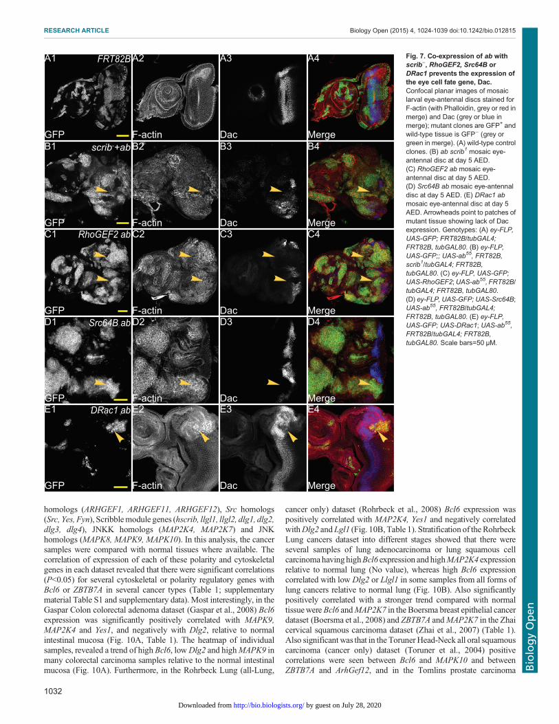
were also affected in these tumours. In eye development,Dachshund (Dac) is one of the earliest transcriptional regulatorsthat drives cell fate determination in the developing eye (Chenet al., 1997; Shen and Mardon, 1997), and expression of Dac isblocked in ab scrib− tumours (Turkel et al., 2013). We thereforewished to determine if this was also the case in ab cytoskeletalgene cooperative tumours.In wild-type eye-antennal discs, Dac is expressed in a broad band
in the middle of the eye disc and also in a crescent in the antennaldisc (Fig. 7A). scrib1 ab clones do not express Dac in the eye disc(arrowheads, Fig. 7B1-B4) or in the antennal disc. Dac expression isonly slightly reduced in scrib1 clones and unaffected in aboverexpressing clones in the eye disc (Turkel et al., 2013). InRhoGEF2 ab clones in the eye disc, Dac expression was blocked(arrowheads, Fig. 7C1-C4). Similarly, Dac expression was blockedin Src64B ab clones (arrowheads, Fig. 7D1-D4) and in DRac1 abclones (arrowheads, Fig. 5E1-E4). Dac expression was also blocked
in the antennal disc in ab RhoGEF2, ab Src64B or ab DRac1 co-expressing clones (Fig. 7C-E; data not shown). Thus, similarly to abscrib− tumours, ab cytoskeletal gene tumours appear to be blockedin differentiation prior to Dac expression.
In antennal disc differentiation, initial expression domains of thetranscription factors Homothorax (Hth), Cut (Ct) and Distal-less(Dll) during 2nd instar larval development establish the earlyproximo-distal axis of the antenna (Dominguez and Casares, 2005).Wehave previously shown that scrib− ab clones retain the expressionof Dll within the growing tumour, but downstream regulated genes,such asDac, are not retained (Turkel et al., 2013).We therefore testedif Dll was still expressed in ab cytoskeletal gene tumours.
In wild-type antennal discs, Dll is expressed in more distallydestined cells in the antennae (Fig. 8A), and scrib− ab clones retainthis expression (arrowheads, Fig. 8B1-B4). Co-expression ofRhoGEF2 with ab did not block Dll expression, and instead anenlarged Dll-expression domain was observed (arrowheads,
Fig. 5. Comparison of cell proliferation levels in ab with scrib1, DRac1, RhoGEF2 or Src64B. Confocal planar images of mosaic larval eye-antennal discslabelled with EdU for S-phases (grey or red in merge) and DAPI (blue in merge); mutant clones are GFP+ and wild-type tissue is GFP− (green in merge).Arrowheads point to patches of tissue showing alterations in cell proliferation. (A) wild-type control clones. (B) ab scrib1 mosaic eye-antennal disc at day 5.(C) ab scrib1 mosaic eye-antennal disc at day 8. (D) DRac1 ab mosaic eye-antennal disc at day 5. (E) DRac1 ab mosaic eye-antennal disc at day 8.(F) RhoGEF2 ab mosaic eye-antennal disc at day 5. (G) RhoGEF2 ab mosaic eye-antennal disc at day 8. (H) Src64B ab mosaic eye-antennal disc at day 5.(I) Src64B ab mosaic eye-antennal disc at day 8. (J) Quantification showing the percentage of EdU positive tissue in wild-type versus mutant clones of thelisted genotypes. Error bars represent s.e.m. Genotypes: (A) ey-FLP, UAS-GFP; FRT82B/tubGAL4; FRT82B, tubGAL80. (B-C) ey-FLP, UAS-GFP;; UAS-ab55,FRT82B, scrib1/tubGAL4; FRT82B, tubGAL80. (D-E) ey-FLP, UAS-GFP; UAS-DRac1; UAS-ab55, FRT82B/tubGAL4; FRT82B, tubGAL80. (F-G) ey-FLP,UAS-GFP; UAS-RhoGEF2; UAS-ab55, FRT82B/tubGAL4; FRT82B, tubGAL80. (H-I) ey-FLP, UAS-GFP; UAS-Src64B; UAS-ab55, FRT82B/tubGAL4;FRT82B, tubGAL80. Scale bars=50 μM.
1030
RESEARCH ARTICLE Biology Open (2015) 4, 1024-1039 doi:10.1242/bio.012815
BiologyOpen
by guest on July 28, 2020http://bio.biologists.org/Downloaded from

Fig. 8C1-C4), probably due to a partial duplication of the antennae,which is sometimes observed in ab-expressing clones (Turkel et al.,2013). Surprisingly, Src64B ab clones showed reduced expressionof Dll (arrowheads, Fig. 8D1-D4) and distortion of the antennalstructures due to cell morphology changes (Fig. 8D2). In DRac1 abclones, normal expression of Dll was also observed (arrowheads,Fig. 8E1-E4). Altogether, these results show that RhoGEF2 ab andDRac1 ab are similar to scrib− ab in cell fate status, however Src64Bab tumours are blocked at an earlier progenitor cell state than scrib−
ab tumours (summarized in Fig. 9).
Correlation in expression of oncogenic BTB-Zinc fingergenes, Bcl6 and ZBTB7A, with apico-basal cell polarity andcytoskeletal genes in human epithelial cancerSince we have shown here that ab cooperates with the cytoskeletalregulators, RhoGEF2 and Src64B, to result in cooperativetumorigenesis, we wished to determine whether the expression of
human homologs of these genes showed cooperation with BTB-Znfinger genes in human cancers. Since our previous studies had alsoshown that the cell polarity tumour suppressor, scrib, showedcooperative tumorigenesis with ab (Turkel et al., 2013), we alsosought to determinewhether human homologs of the Scribblemodulewere downregulated in human tumours, showing high expression ofBTB-Zn finger genes. Furthermore, since we have shown that theJNK signalling pathway was important in the invasive properties ofthese tumours and sufficient to cooperate with RasACT inDrosophilaand mammalian invasive tumour growth (Brumby et al., 2011), wewished to examine the correlation of expression of the human JNKKand JNK homologs with BTB-Zn finger genes in human cancer. Ofthe human BTB-Zn finger genes, there is greatest evidence for Bcl6and ZBTB7A as oncogenes in human epithelial cancer (seeIntroduction), so we focused our analysis on these genes. UsingOncomine, we analysed collections of human epithelial cancers forexpression correlation with Bcl6 or ZBTB7A and human RhoGEF2
Fig. 6. Comparison of cell death levels in ab with scrib1, DRac1, RhoGEF2 or Src64B. Confocal planar images of mosaic larval eye-antennal discslabelled with TUNEL as an apoptotic marker (grey or red in merge) and DAPI (blue in merge); mutant clones are GFP+ and wild-type tissue is GFP− (green inmerge). Arrowheads point to patches of tissue showing alterations in cell death. (A) wild-type control clones. (B) ab scrib1 mosaic eye-antennal disc at day5. (C) ab scrib1 mosaic eye-antennal disc at day 8. (D) DRac1 ab mosaic eye-antennal disc at day 5. (E) DRac1 ab mosaic eye-antennal disc at day 8.(F) RhoGEF2 ab mosaic eye-antennal disc at day 5. (G) RhoGEF2 ab mosaic eye-antennal disc at day 8. (H) Src64B ab mosaic eye-antennal disc at day 5.(I) Src64B ab mosaic eye-antennal disc at day 8. (J) Quantification showing the number of TUNEL positive cells in wild-type versus mutant clones of the listedgenotypes. Error bars represent s.e.m. Genotypes: (A) ey-FLP, UAS-GFP; FRT82B/tubGAL4; FRT82B, tubGAL80. (B-C) ey-FLP, UAS-GFP;; UAS-ab55,FRT82B, scrib1/tubGAL4; FRT82B, tubGAL80. (D-E) ey-FLP, UAS-GFP; UAS-DRac1; UAS-ab55, FRT82B/tubGAL4; FRT82B, tubGAL80. (F-G) ey-FLP,UAS-GFP; UAS-RhoGEF2; UAS-ab55, FRT82B/tubGAL4; FRT82B, tubGAL80. (H-I) ey-FLP, UAS-GFP; UAS-Src64B; UAS-ab55, FRT82B/tubGAL4;FRT82B, tubGAL80. Scale bars=50 μM.
1031
RESEARCH ARTICLE Biology Open (2015) 4, 1024-1039 doi:10.1242/bio.012815
BiologyOpen
by guest on July 28, 2020http://bio.biologists.org/Downloaded from
homologs (ARHGEF1, ARHGEF11, ARHGEF12), Src homologs(Src, Yes, Fyn), Scribblemodule genes (hscrib, llgl1, llgl2, dlg1, dlg2,dlg3, dlg4), JNKK homologs (MAP2K4, MAP2K7) and JNKhomologs (MAPK8, MAPK9, MAPK10). In this analysis, the cancersamples were compared with normal tissues where available. Thecorrelation of expression of each of these polarity and cytoskeletalgenes in each dataset revealed that there were significant correlations(P<0.05) for several cytoskeletal or polarity regulatory genes withBcl6 or ZBTB7A in several cancer types (Table 1; supplementarymaterial Table S1 and supplementary data). Most interestingly, in theGaspar Colon colorectal adenoma dataset (Gaspar et al., 2008) Bcl6expression was significantly positively correlated with MAPK9,MAP2K4 and Yes1, and negatively with Dlg2, relative to normalintestinal mucosa (Fig. 10A, Table 1). The heatmap of individualsamples, revealed a trend of high Bcl6, lowDlg2 and highMAPK9 inmany colorectal carcinoma samples relative to the normal intestinalmucosa (Fig. 10A). Furthermore, in the Rohrbeck Lung (all-Lung,
cancer only) dataset (Rohrbeck et al., 2008) Bcl6 expression waspositively correlated with MAP2K4, Yes1 and negatively correlatedwithDlg2 andLgl1 (Fig. 10B, Table 1). Stratification of the RohrbeckLung cancers dataset into different stages showed that there wereseveral samples of lung adenocarcinoma or lung squamous cellcarcinomahavinghighBcl6 expression and highMAP2K4 expressionrelative to normal lung (No value), whereas high Bcl6 expressioncorrelated with low Dlg2 or Llgl1 in some samples from all forms oflung cancers relative to normal lung (Fig. 10B). Also significantlypositively correlated with a stronger trend compared with normaltissuewereBcl6 andMAP2K7 in the Boersma breast epithelial cancerdataset (Boersma et al., 2008) and ZBTB7A andMAP2K7 in the Zhaicervical squamous carcinoma dataset (Zhai et al., 2007) (Table 1).Also significant was that in the Toruner Head-Neck all oral squamouscarcinoma (cancer only) dataset (Toruner et al., 2004) positivecorrelations were seen between Bcl6 and MAPK10 and betweenZBTB7A and ArhGef12, and in the Tomlins prostate carcinoma
Fig. 7. Co-expression of ab withscrib−, RhoGEF2, Src64B orDRac1 prevents the expression ofthe eye cell fate gene, Dac.Confocal planar images of mosaiclarval eye-antennal discs stained forF-actin (with Phalloidin, grey or red inmerge) and Dac (grey or blue inmerge); mutant clones are GFP+ andwild-type tissue is GFP− (grey orgreen in merge). (A) wild-type controlclones. (B) ab scrib1 mosaic eye-antennal disc at day 5 AED.(C) RhoGEF2 ab mosaic eye-antennal disc at day 5 AED.(D) Src64B ab mosaic eye-antennaldisc at day 5 AED. (E) DRac1 abmosaic eye-antennal disc at day 5AED. Arrowheads point to patches ofmutant tissue showing lack of Dacexpression. Genotypes: (A) ey-FLP,UAS-GFP; FRT82B/tubGAL4;FRT82B, tubGAL80. (B) ey-FLP,UAS-GFP;; UAS-ab55, FRT82B,scrib1/tubGAL4; FRT82B,tubGAL80. (C) ey-FLP, UAS-GFP;UAS-RhoGEF2;UAS-ab55, FRT82B/tubGAL4; FRT82B, tubGAL80.(D) ey-FLP, UAS-GFP; UAS-Src64B;UAS-ab55, FRT82B/tubGAL4;FRT82B, tubGAL80. (E) ey-FLP,UAS-GFP; UAS-DRac1; UAS-ab55,FRT82B/tubGAL4; FRT82B,tubGAL80. Scale bars=50 μM.
1032
RESEARCH ARTICLE Biology Open (2015) 4, 1024-1039 doi:10.1242/bio.012815
BiologyOpen
by guest on July 28, 2020http://bio.biologists.org/Downloaded from

dataset (Tomlins et al., 2007) Bcl6 expression was positivelycorrelated with ArhGef11 and MAPK8 (Table 1). Significantpositive correlations were also observed in the Collisson Pancreaticadenocarcinoma (cancer only) dataset (Collisson et al., 2011)between ZBTB7A and Src (Table 1). Furthermore, in the Grutzmannpancreatic ductal adenocarcinoma dataset (Grützmann et al., 2004),although of borderline significance, a positive correlation wasobserved between ZBTB7A and MAP2K7 that showed a strongertrend compared with normal tissue (Table 1). Thus, taken together,these data show that in certain epithelial cancers the upregulation ofBcl6 or ZBTB7A expression is significantly correlated with reducedexpression of Dlg2 or Llgl1 cell polarity genes or high expression ofArhGef11, ArhGef12, MAP2K4, MAP2K7, MAPK8, MAPK9,MAPK10, Src or Yes1 cytoskeletal genes. Based on our functionaldata in Drosophila and mammalian cells (this study; Brumby et al.,2011; Khoo et al., 2013; Turkel et al., 2013; C.P., A.B., H.R.,unpublished data), we would expect the concordant expression of
Bcl6 or ZBTB7A with these genes should result in tumour growth,morphology changes, differentiation blockage and invasiveproperties.
DISCUSSIONIn this study, we have shown that over-expression of the Ab BTB-ZF protein cooperates with upregulation of RhoGEF2 or Src64B intumorigenesis, whereas Ab and DRac1 do not cooperate.Furthermore, we show that expression of Ab with each of thesecytoskeletal regulators results in disruption to differentiation, in thatthe photoreceptor cell marker, Elav, and the early cell fate gene,Dac, are not expressed, although the antennal cell fate gene, Dll, isretained in all except ab Src64B co-expressing clones. Finally, wehave found significant correlations in human epithelial cancerdatasets between the high expression of BTB-ZF oncogenes, Bcl6and ZBTB7A, and low expression of Dlg2 or Llgl1 cell polaritygenes or high expression of ArhGef11, ArhGef12, MAP2K4,
Fig. 8. Co-expression of ab withSrc64B reduces expression of theantennal cell fate gene, Dll, butexpression is retained in ab scrib−,abRhoGEF2 and abDRac1 clones.Confocal planar images of mosaiclarval antennal discs stained forF-actin (with Phalloidin, grey or redin merge) and Dll (grey or blue inmerge); mutant clones are GFP+ andwild-type tissue is GFP− (grey orgreen in merge). (A) wild-type controlclones in the antennal disc at day 5AED. (B) ab scrib1 mosaic antennaldisc at day 5 AED. (C) RhoGEF2 abmosaic antennal disc at day 5 AED.(D) Src64B ab mosaic antennal discat day 5 AED. (E) DRac1 ab mosaicantennal disc at day 5 AED.Arrowheads point to Dll expression.Genotypes: (A) ey-FLP, UAS-GFP;FRT82B/tubGAL4; FRT82B,tubGAL80. (B) eyFLP, UAS-GFP;;UAS-ab55, FRT82B, scrib1/tubGAL4;FRT82B, tubGAL80. (C) eyFLP,UAS-GFP; UAS-RhoGEF2; UAS-ab55, FRT82B/tubGAL4; FRT82B,tubGAL80. (D) eyFLP, UAS-GFP;UAS-Src64B; UAS-ab55, FRT82B/tubGAL4; FRT82B, tubGAL80.(E) eyFLP, UAS-GFP; UAS-DRac1;UAS-ab55, FRT82B/tubGAL4;FRT82B, tubGAL80. Scalebars=50 μM.
1033
RESEARCH ARTICLE Biology Open (2015) 4, 1024-1039 doi:10.1242/bio.012815
BiologyOpen
by guest on July 28, 2020http://bio.biologists.org/Downloaded from

MAP2K7, MAPK8, MAPK9, MAPK10, Src or Yes1 cytoskeletalgenes. This data suggests that cooperation between these genes mayoccur in some human epithelial cancers.
Comparison of tumorigenic propertiesRhoGEF2 ab or Src64B ab tumours showed overgrowth during anextended larval period resulting in giant larvae and loss of
differentiation (Fig. 4C). However, unlike scrib− ab tumours therewas also non-cell autonomous proliferation and the tumours did notappear to be as invasive as scrib− ab tumours, although a moredetailed analysis of this is required. By contrast, co-expression ofDRac1 and ab did not result in cooperative tumorigenesis, but rathernon-cell autonomous proliferation. Relative to the cooperation ofthese cytoskeletal genes with RasV12 (Brumby et al., 2011; Khoo
Fig. 9. Summary of the effects of ab scrib−, ab Src64B,ab RhoGEF2 and ab DRac1 on Dac and Dll expression.A schematic of an eye-antennal disc showing the normalexpression of Dac and Dll, and the effect of ab scrib−, ab Src64B,ab RhoGEF2 and ab DRac1 on their expression patterns.
Table 1 . Significant correlations in gene expression
Gene expression datasets BTB-Zn gene Polarity or cytoskeletal gene P value Pearson R Notes
Positive CorrelationsTomlins Prostate all Bcl6 ARHGEF11 5,60×10⁻³ 0.45 ATomlins Prostate Carcinoma Bcl6 ARHGEF11 1,40×10⁻² 0.57 *, #Toruner Head-Neck all oral squamous carcinoma (cancer only) ZBTB7A ARHGEF12 1,70×10⁻² 0.59 *Toruner Head-Neck all oral squamous carcinoma (cancer only) Bcl6 MAPK10 1,10×10⁻² 0.62 *Tomlins Prostate all Bcl6 MAPK8 2,00×10⁻² 0.46 ATomlins Prostate Carcinoma Bcl6 MAPK8 2,20×10⁻² 0.57 *Gasper colon all Bcl6 MAPK9 3,20×10⁻¹³ 0.71 AGaspar colorectal adenoma Bcl6 MAPK9 4,40×10⁻⁹ 0.69 *, #Gaspar colon normal Bcl6 MAPK9 3,10×10⁻⁶ 0.82 NGaspar colorectal adenoma Bcl6 MAP2K4 6,80×10⁻³ 0.36 *, #Rohrbeck all-Lung (cancer only) Bcl6 MAP2K4 2,50×10⁻³ 0.45 *Boersma Breast epithelial cancer Bcl6 MAP2K7 2,30×10⁻³ 0.43 *, #Grutzmann Pancreatic ductal adenocarcinoma ZBTB7A MAP2K7 5,10×10⁻² 0.6 *, #, §Grutzmann Pancreas all (no met) ZBTB7A MAP2K7 1,20×10⁻² 0.52 AZhai Cervix Cervical squamous ZBTB7A MAP2K7 1,30×10⁻² 0.46 *, #Zhai Cervix all ZBTB7A SCRIB 3,40×10⁻² 0.34 ACollisson Pancreas all-adenocarcinoma ZBTB7A SRC 3,10×10⁻² 0.42 *Gasper colon all Bcl6 YES1 1,60×10⁻⁵ 0.47 AGaspar colorectal adenoma Bcl6 YES1 1,40×10⁻⁴ 0.49 *, #Gaspar colon normal Bcl6 YES1 3,40×10⁻² 0.45 NRohrbeck all-Lung (cancer only) Bcl6 YES1 9,60×10⁻³ 0.4 *
Negative correlationsGasper colon all Bcl6 DLG2 2,20×10⁻¹⁵ −0.75 AGaspar colorectal adenoma Bcl6 DLG2 7,40×10⁻¹³ −0.79 *, #Gaspar colon normal Bcl6 DLG2 8,40×10⁻⁴ −0.66 NRohrbeck all-Lung (cancer only) Bcl6 DLG2 2,00×10⁻² −0.36 *Rohrbeck all-Lung (cancer only) Bcl6 LLGL1 3,80×10⁻³ −0.44 *
A, all (normal and cancer); N, normal; * Bcl6/ZBTB7 high and correlated as expected with the test gene; # trend is stronger in cancer comparedwith normal tissue;§ borderline significance with positive correlation.
1034
RESEARCH ARTICLE Biology Open (2015) 4, 1024-1039 doi:10.1242/bio.012815
BiologyOpen
by guest on July 28, 2020http://bio.biologists.org/Downloaded from

et al., 2013; C.P., A.B., H.R., unpublished data), RhoGEF2 orSrc64B cooperation with ab showed similar properties (Fig. 4C). Bycontrast, DRac1 RasV12 tumours showed strong cell-autonomousovergrowth and invasive properties, whereas DRac1 ab expressingcells did not overgrow relative to wild-type tissue, but instead thesurrounding wild-type tissue was induced to overgrow (Fig. 4C).The phenomenon of non-cell autonomous overgrowth observed in
DRac1 ab mosaic eye-antennal discs (and to some extent in abRhoGEF2 and ab Src64B mosaic discs) is similar to the effect that“undead” cells (cells where apoptosis is initiated by activation ofinitiator caspases, but effector caspase activation is blocked – and thuscell death – by expression of the inhibitor, p35) have upon theirsurrounding wild-type neighbours (Martin et al., 2009; Morata et al.,2011; Perez-Garijo et al., 2009; Ryoo and Bergmann, 2012). Thisoccurs by the release of Wingless (Wg) and Decapentaplegic (Dpp)and perhaps other morphogens from the undead cells, which promotecompensatory proliferation in the surrounding wild-type cells. Thesimilarity of these phenotypes suggests that DRac1 ab expressingcells might be in an “undead” state, and release Dpp andWg, therebyinducing proliferative overgrowth of the surrounding wild-type cells.Alternatively, these cells might be deficient in mitochondrialfunction, which together with expression of a cell-survival factor,such as RasV12, results in non-cell autonomous overgrowth withoutevidence of caspase activation (Ohsawa et al., 2012). In this scenario,the mitochondrial dysfunction results in increased reactive oxygenspecies (ROS) that activate JNK signalling, which subsequentlyinactivates Hippo pathway signalling, leading to increased expressionof the target genes Wingless and Unpaired (Upd) that activate Wgsignalling and Jak/Stat signalling, respectively, in the neighbouringwild-type cells.However, sincewe observedTUNEL-positive cells inDRac1 ab, RhoGEF2 ab and Src64B ab expressing clones, it is more
likely that the first of thesemechanisms is responsible for the non-cellautonomous overgrowth, however this requires further investigation.Interestingly, in undead cells JNK activation is required for Dpp andWg production and non-cell autonomous overgrowth (Morata et al.,2011; Perez-Garijo et al., 2009). Furthermore, strong activation ofJNK signalling together with RasV12 results in non-cell autonomousovergrowth (Uhlirova et al., 2005), although at presumably lowerlevels of JNK activation, cell autonomous overgrowth occurs(Brumby et al., 2011; Igaki et al., 2006; Uhlirova and Bohmann,2006). Therefore it is possible that the different effects on non-cellautonomous versus autonomous cell overgrowth inDRac1 ab versusRhoGEF2aborSrc64Bab-expressing cellsmight dependon the levelof JNK activation. Nonetheless, at early stages, ab-driven RhoGEF2,Src64B or DRac1 tumours were similar in inducing non-cellautonomous effects, but at later times the RhoGEF2 ab and Src64Bab-expressing cells showed more predominant autonomous cellovergrowth, whilst theDRac1 ab expressing cells did not, suggestingthat there are likely to be molecular differences between DRac1 andRhoGEF2 or Src64B in their cooperative interactions with ab thatimpact on cell proliferation or survival of the tumour cells.
Our profiling ofAb targets and deregulated genes revealed that dac,dan, eya and ct eye-antennal differentiation genes were repressed,along with changes in expression of cell growth/proliferation andsurvival genes thatwould be expected to promote tumorigenic growthin cooperationwith scrib loss-of-function (Turkel et al., 2013). scrib−
ab tumours showed downregulation of Dac, but the antennal cell fateexpression domain of Dll was not affected (Turkel et al., 2013).Similarly, ab expression with either of the cytoskeletal genes resultedin repression of Dac, however Src64B ab tumours additionallyrepressed Dll, in contrast to DRac1 ab, RhoGEF2 ab and scrib− abtumours where Dll was unaffected. This data suggests that Src64B
Fig. 10. Heatmaps of expression of Bcl6 relative to polarity or cytoskeletal regulatory genes in the Gaspar colon and Rohrbeck Lung datasets.(A) Gaspar Colon data set. (B) Rohrbeck Lung data set. Samples are stratified into normal tissue (intestinal mucosa for Gaspar Colon or no value for RohrbeckLung) and cancer grades for Rohrbeck Lung. Relative expression levels of the indicated gene probesets are indicated. The Gaspar Colon dataset has 3 probes toBcl6 and Yes1, and 2 probes toDlg2 andMAPK9. Red is high expression and blue is low expression. The outlined samples indicate those whereBcl6 is high andDlg2 or Llgl1 are low or MAP2K4, MAPK9 or Yes1 are high.
1035
RESEARCH ARTICLE Biology Open (2015) 4, 1024-1039 doi:10.1242/bio.012815
BiologyOpen
by guest on July 28, 2020http://bio.biologists.org/Downloaded from

expression exerts an additional effect on ab-expressing cells to inhibitDll gene expression and differentiation. Src upregulation activates theJNK and Stat signalling pathways, affects adherens junction functionand repressesHippo signalling (Enomoto and Igaki, 2013; Kohlmaieret al., 2014; Ma et al., 2013; Read et al., 2004; Sotillos et al., 2013;Vidal et al., 2006). Furthermore, recent studies have shown thatoverexpression of Src64B in the Drosophila intestinal stem cells canalter differentiation and result in amplification of progenitor cell pools(Cordero et al., 2014; Kohlmaier et al., 2014). scribmutant cells alsoupregulate JNK, downregulate the E-cadherin/β-catenin adhesioncomplex and repress Hippo signalling (Brumby and Richardson,2003; Doggett et al., 2011; Igaki et al., 2006; Leong et al., 2009;Uhlirova and Bohmann, 2006). Furthermore, the Jak/Stat ligand,Upd3, is also upregulated in the scrib− cells, where it drives tumourovergrowth, and is also required to activate Jak/Stat signalling in thewild-type neighbouring cells in cell competition (Bunker et al., 2015;Chen et al., 2012; Schroeder et al., 2013). RhoGEF2 and DRac1 alsoupregulate JNK signalling (Brumby et al., 2011; Khoo et al., 2013),and might also repress Hippo signalling to promote tissue growth,since regulators of actin cytoskeletal tension, such as activated Rokand Myosin II regulatory light chain, induce Yki target geneexpression (Fernandez et al., 2011; Halder et al., 2012; Rauskolbet al., 2014; Sansores-Garcia et al., 2011). However, inDrosophila itis unknown if RhoGEF2 or DRac1 affect Jak/Stat signalling. Sincescrib loss-of-function and Src activation deregulate similar pathways,the precise mechanism by which Src64B cooperates with ab to blockexpression of Dll in the developing eye-antennal disc remains to bedetermined.
Cooperation of BTB-ZF transcription factors withderegulated cytoskeletal or polarity genes in human cancerOur finding that therewas a significant correlation between increasedexpression of human BTB-ZF oncogenic genes, Bcl6 or ZBTB7A,and downregulation of the cell polarity genes, Dlg2 and Llgl1, orhomologs of JNKK (MAPK2K4, MAPK2K7), JNK (MAPK8,MAPK9, MAPK10), RhoGEF2 (ArhGEF11, ArhGEF12) or Src(Yes1, Src) cytoskeletal genes in various epithelial cancers, suggeststhat the concordant expression of these genes might be contributingto human epithelial cancer initiation and progression. Whilst thisstudy only focused on two of the 47 BTB-ZF genes in the humangenome, it raises the question of whether other BTB-ZF genes mightalso show correlations with the expression of cytoskeletal or cellpolarity genes in human epithelial cancers. However, tissue andcancer-grade specific effects might be observed, as a recentlypublished study revealed that ZBTB7Awas commonly deleted in latestage oesophageal, bladder, colorectal, lung, ovarian and uterinecancers (Liu et al., 2014). Moreover, they found that low ZBTB7Aexpression correlates with poor prognosis in colon cancer patients,suggesting that ZBTB7A plays a tumour suppressor function in thesecancers. Interestingly, this study also found that in colon cancerxenografts, ZBTB7A represses the expression of genes in theglycolytic pathway, a metabolic pathway that is required foraggressive tumour growth, and that inhibition of this pathwayreduces tumour growth. Pertinent to this finding, we found thatblocking glycolytic pathways in Drosophila polarity-impairedtumours, impedes tumour growth without substantially affectingnormal tissues (Willoughby et al., 2013), suggesting thatdownregulation of the Scribble polarity module might upregulateglycolytic metabolic pathways and be dependent on them for tumourgrowth and survival. It is therefore possible that the cooperationbetween ab and scrib− or cytoskeletal genes inDrosophilamay alsoreflect a need for upregulation of the glycolytic pathway. In human
epithelial cancers, the correlations observed between elevatedZBTB7A expression and reduced expression of the Scribblepolarity module gene (or high expression of cytoskeletal genes)might also indicate a requirement for glycolytic pathway activationfor tumorigenesis. Further studies are clearly required to examine thecooperative effects of Bcl6 or ZBTB7Awith deregulated cytoskeletalor cell polarity genes in human epithelial cell lines andmousemodelsin order to discern whether our findings in Drosophila are indeedconserved in mammalian systems.
Identifying cooperative interactions in cancer is likely to providenovel therapeutic approaches in combating the tumour. Indeed,recently a small molecule inhibitor targeting Bcl6 has beendeveloped, and combining this with a Stat3 inhibitor resulted inenhanced cell killing in triple negative breast cancer cell lines(Walker et al., 2014). Since in Drosophila and human cells, Srcupregulates Stat activity (Cordero et al., 2014; Frame, 2004;Kohlmaier et al., 2014; Read et al., 2004; Sotillos et al., 2013),tumours showing high Bcl6 and Src or Yes1 expression would bepredicted to be sensitive to this combined therapeutic regime.Interestingly, a predominance of the significant correlations thatwere observed in the human epithelial cancer datasets with eitherBcl6 or ZBTB7A involved upregulation of JNKK and JNK familygenes. Since JNK signalling is central to many cooperativeinteractions examined by us and others (Brumby et al., 2011;Brumby and Richardson, 2003; Enomoto and Igaki, 2013; Igakiet al., 2006; Leong et al., 2009; Turkel et al., 2013; Uhlirova andBohmann, 2006), inhibiting the JNK pathway in addition to Bcl6 inBcl6-driven cancers might also be a promising therapeutic approachto combat these cancers. In summary, our functional studies inDrosophila and bioinformatics analysis of human cancers hasshown that cooperative tumorigenic interactions occur betweenBTB-ZF genes and cell polarity or cytoskeletal genes, and warrantsfurther investigation to determine whether restoring normalexpression of these genes or downstream pathways in humancancer cells can reduce tumorigenesis.
MATERIALS AND METHODSDrosophila stocksThe following Drosophila stocks were used: ey-FLP1, UAS-mCD8-GFP;;Tub-GAL4, FRT82B, Tub-GAL80 (Lee and Treisman, 2001);UAS-ab55 (III)(Cook et al., 2004); UAS-RhoGEF2 (II) (Padash Barmchi et al., 2005);UAS-Src64B (II) (R. Cagan, Mount Sinai School of Medicine, New York,USA); UAS-DRac1 (II) (Luo et al., 1994); scrib1 (Bilder and Perrimon,2000) and ey-GAL4 (Bloomington Stock Centre). FRT82B recombinantstocks were generated for all transgenic lines for mosaic analysis.
Mosaic analysisClonal analysis utilised MARCM (mosaic analysis with repressible cellmarker) (Lee and Luo, 1999) with FRT82B and ey-FLP1 to induce clonesand mCD8-GFP expression to mark mutant tissue. All fly crosses werecarried out at 25°C and grown on standard fly media.
ImmunostainingThird-instar larval eye-antennal discs were dissected in phosphate-bufferedsaline (PBS), fixed in 4% paraformaldehyde for 30 min, and washed in PBS+0.1% Triton X-100 (PBT). Samples were blocked in 2% NGS in PBTwith1.5% saponin for 1 h in room temperature and then incubated in primaryantibodies over night at 4°C in 2% NGS in PBT. Samples were then washedtwo times in PBT for 30 min before addition of the secondary antibody. EdUand TUNEL labelling were performed as previously described (Turkel et al.,2013).
Antibodies used were: mouse anti-Elav (DSHB, 1/20), mouse anti-Dll(Duncan et al., 1998, 1/500) and mouse anti-Dac (DSHB, 1/10). Secondaryantibodies were: anti-mouse Alexa 568 or 633 (Invitrogen) at 1/400 dilution.
1036
RESEARCH ARTICLE Biology Open (2015) 4, 1024-1039 doi:10.1242/bio.012815
BiologyOpen
by guest on July 28, 2020http://bio.biologists.org/Downloaded from

F-actin was detected with phalloidin–tetramethylrhodamine isothioblueate(TRITC; Sigma, 0.3 µM, 1/1000) and DNA was detected using DAPIstaining. Samples were mounted in 80% (v/v) glycerol/PBS.
ImagingImages of fixed and mounted samples onto the glass slides were capturedusing BioRad, Olympus Fluoview FV100 and Leica TCS SP5 confocallaser microscopes. Single optical sections were selected in FluoViewsoftware before being processed in Adobe Photoshop CS6 and assembledinto figures in Adobe Illustrator CS6.
Adult flies were frozen at −20°C before imaging in order to facilitatepositioning them under the microscope. Images were captured on LumeneraInfinity 1 camera attached to Olympus SZX7 dissection microscope andprocessed using Adobe Photoshop CS3.
Quantification of clone volumeVolumetric clone analysis was performed using Volocity 3D ImageAnalysis Software (Perkin-Elmer). To determine the ratio of clonal tissuevolume to total volume of the eye-antennal disc for each genotype and timepoint, GFP+ clonal tissue relative to total disc area (as marked by Phallodinto visualize the cells) was measured from confocal Z sections encompassingthe entire eye-antennal disc. The data for each genotypewas compared usingGraphPad Prism 6 using unpaired t-tests. Error bars represent s.e.m. and thesignificance was set at P<0.05.
Quantification of EdU and TUNEL stainingFor TUNEL and EdU labelling, 6 to 10 discs for each genotype wereanalysed. TUNEL was quantified using Photoshop 5.1 Extended. EDU wasquantified using a program designed by David Tapiador, available at https://github.com/nogates/counting-semaphore.
Analysis of published datasetsUsing Oncomine (Research Premium Edition), we identified 18 publishedgene expression data sets that contain epithelial cancer samples. Datawas filtered down to the genes of interest and was downloaded forfurther analysis. Eleven of the 18 data sets that have at least 30 samples andcontain at least three quarters of our query genes were analysed forcorrelation of expression levels between BCL6/ZBTB7A and each of thegenes in our gene panel. These data sets were: Boersma Breast (Boersmaet al., 2008), Collisson Pancreas all-adenocarcinoma (Collisson et al., 2011),Gaspar Colon (Gaspar et al., 2008), Grützmann Pancreas (Grützmann et al.,2004), Ma Breast 2 (Ma et al., 2004), Ma Breast 4 (Ma et al., 2009),Rohrbeck Lung (Rohrbeck et al., 2008), Skrzypczak Colorectal 2(Skrzypczak et al., 2010), Tomlins Prostate (Tomlins et al., 2007),Toruner Head-Neck all-oral squamous carcinoma (Toruner et al., 2004)and Zhai Cervix (Zhai et al., 2007). Where data is available, samples arestratified into normal (no cancer) and cancer for separate analysis to identifycancer-specific gene expression correlations. All analyses were done usingthe R software package.
AcknowledgementsWe thank David Tapiador for developing the program for quantification of EdUlabelled tissues. We also acknowledge Bloomington, Vienna RNAi and NationalInstitute of Genetics (Japan) Stock Centers for the provision of fly strains andOzDrosfor quarantine and handling of flies imported into Australia and Flybase for its wealthof information.
Competing interestsThe authors declare no competing or financial interests.
Author contributionsN.T., M.P., H.E.R. and A.M.B. conceived and designed the experiments. N.T., M.P.and C.P. performed the experiments. N.T., M.P., C.P., J.L., A.M.B. and H.E.R.analysed the data. N.T., M.P. and H.E.R. wrote the paper.
FundingH.E.R. is supported by a Senior Research Fellowship from the National Healthand Medical Research Council (NHMRC) Australia, M.P. from funding from theCancer Council Victoria Australia, and C.P. was supported by an Australian
Postgraduate Award. This work was supported by grants from the NHMRC, #400211to H.E.R. and A.M.B., and #509051 to A.M.B., and Peter Mac Internal funds toH.E.R.
Supplementary materialSupplementary material available online athttp://bio.biologists.org/lookup/suppl/doi:10.1242/bio.012815/-/DC1
ReferencesAggarwal, A., Hunter, W. J., III, Aggarwal, H., Silva, E. D., Davey, M. S., Murphy,
R. F. and Agrawal, D. K. (2010). Expression of leukemia/lymphoma-related factor(LRF/POKEMON) in human breast carcinoma and other cancers. Exp. Mol.Pathol. 89, 140-148.
Aggarwal, H., Aggarwal, A., Hunter, W. J., III, Yohannes, P., Khan, A. U. andAgrawal, D. K. (2011). Expression of leukemia/lymphoma related factor (LRF/Pokemon) in human benign prostate hyperplasia and prostate cancer. Exp. Mol.Pathol. 90, 226-230.
Barrett, K., Leptin, M. and Settleman, J. (1997). The Rho GTPase and a putativeRhoGEF mediate a signaling pathway for the cell shape changes in Drosophilagastrulation. Cell 91, 905-915.
Bilder, D. and Perrimon, N. (2000). Localization of apical epithelial determinants bythe basolateral PDZ protein Scribble. Nature 403, 676-680.
Boersma, B. J., Reimers, M., Yi, M., Ludwig, J. A., Luke, B. T., Stephens, R. M.,Yfantis, H. G., Lee, D. H., Weinstein, J. N. and Ambs, S. (2008). A stromal genesignature associated with inflammatory breast cancer. Int. J. Cancer 122,1324-1332.
Brumby, A. M. and Richardson, H. E. (2003). scribble mutants cooperate withoncogenic Ras or Notch to cause neoplastic overgrowth in Drosophila. EMBO J.22, 5769-5779.
Brumby, A. M. and Richardson, H. E. (2005). Using Drosophila melanogaster tomap human cancer pathways. Nat. Rev. Cancer 5, 626-639.
Brumby, A. M., Goulding, K. R., Schlosser, T., Loi, S., Galea, R., Khoo, P.,Bolden, J. E., Aigaki, T., Humbert, P. O. and Richardson, H. E. (2011).Identification of novel Ras-cooperating Oncogenes in Drosophila melanogaster: aRhoGEF/Rho-family/JNK Pathway is a Central Driver of Tumorigenesis.Genetics188, 105-125.
Bunker, B. D., Nellimoottil, T. T., Boileau, R. M., Classen, A. K. and Bilder, D.(2015). The transcriptional response to tumorigenic polarity loss in Drosophila.Elife 4, e03189.
Chen, R., Amoui, M., Zhang, Z. and Mardon, G. (1997). Dachshund and eyesabsent proteins form a complex and function synergistically to induce ectopic eyedevelopment in Drosophila. Cell 91, 893-903.
Chen, C.-L., Schroeder, M. C., Kango-Singh, M., Tao, C. and Halder, G. (2012).Tumor suppression by cell competition through regulation of the Hippo pathway.Proc. Natl. Acad. Sci. USA 109, 484-489.
Cheng, L.Y., Parsons, L. M. and Richardson, H. E. (2013). Modelling cancer inDrosophila: The next generation. In Encyclopedia Life Sciences (eLS).Chichester: John Wiley & Sons Ltd.
Collisson,E.A., Sadanandam,A.,Olson, P., Gibb,W. J., Truitt, M.,Gu,S.,Cooc, J.,Weinkle, J., Kim, G. E., Jakkula, L. et al. (2011). Subtypes of pancreatic ductaladenocarcinoma and their differing responses to therapy. Nat. Med. 17, 500-503.
Cook, O., Biehs, B. and Bier, E. (2004). brinker and optomotor-blind actcoordinately to initiate development of the L5 wing vein primordium inDrosophila. Development 131, 2113-2124.
Cordero, J. B., Ridgway, R. A., Valeri, N., Nixon, C., Frame, M. C., Muller, W. J.,Vidal, M. and Sansom, O. J. (2014). c-Src drives intestinal regeneration andtransformation. EMBO J. 33, 1474-1491.
Costoya, J. A. (2007). Functional analysis of the role of POK transcriptionalrepressors. Brief Funct. Genomic Proteomic 6, 8-18.
Dodson, G. S., Guarnieri, D. J. and Simon, M. A. (1998). Src64 is required forovarian ring canal morphogenesis during Drosophila oogenesis. Development125, 2883-2892.
Doggett, K., Grusche, F. A., Richardson, H. E. and Brumby, A. M. (2011). Loss ofthe Drosophila cell polarity regulator Scribbled promotes epithelial tissueovergrowth and cooperation with oncogenic Ras-Raf through impaired Hippopathway signaling. BMC Dev. Biol. 11, 57.
Dominguez, M. and Casares, F. (2005). Organ specification-growth controlconnection: new in-sights from the Drosophila eye-antennal disc. Dev. Dyn.232, 673-684.
Dow, L. E., Elsum, I. A., King, C. L., Kinross, K. M., Richardson, H. E. andHumbert, P. O. (2008). Loss of humanScribble cooperates with H-Ras to promotecell invasion through deregulation of MAPK signalling. Oncogene 27, 5988-6001.
Duncan, D. M., Burgess, E. A. and Duncan, I. (1998). Control of distal antennalidentity and tarsal development in Drosophila by spineless-aristapedia, a homologof the mammalian dioxin receptor. Genes Dev. 12, 1290-1303.
Elsum, I., Yates, L., Humbert, P. O. and Richardson, H. E. (2012). The Scribble-Dlg-Lgl polarity module in development and cancer: from flies to man. EssaysBiochem. 53, 141-168.
1037
RESEARCH ARTICLE Biology Open (2015) 4, 1024-1039 doi:10.1242/bio.012815
BiologyOpen
by guest on July 28, 2020http://bio.biologists.org/Downloaded from

Elsum, I. A., Yates, L. L., Pearson, H. B., Phesse, T. J., Long, F., O’Donoghue,R., Ernst, M., Cullinane, C. and Humbert, P. O. (2013). Scrib heterozygositypredisposes to lung cancer and cooperates with KRas hyperactivation toaccelerate lung cancer progression in vivo. Oncogene 33, 5523-5533.
Enomoto, M. and Igaki, T. (2013). Src controls tumorigenesis via JNK-dependentregulation of the Hippo pathway in Drosophila. EMBO Rep. 14, 65-72.
Fernandez, B. G., Gaspar, P., Bras-Pereira, C., Jezowska, B., Rebelo, S. R. andJanody, F. (2011). Actin-Capping Protein and the Hippo pathway regulate F-actinand tissue growth in Drosophila. Development 138, 2337-2346.
Fernandez, B. G., Jezowska, B. and Janody, F. (2014). Drosophila actin-CappingProtein limits JNK activation by the Src proto-oncogene. Oncogene 33,2027-2039.
Frame, M. C. (2004). Newest findings on the oldest oncogene; how activated srcdoes it. J. Cell Sci. 117, 989-998.
Gaspar, C., Cardoso, J., Franken, P., Molenaar, L., Morreau, H., Moslein, G.,Sampson, J., Boer, J. M., de Menezes, R. X. and Fodde, R. (2008). Cross-species comparison of human and mouse intestinal polyps reveals conservedmechanisms in adenomatous polyposis coli (APC)-driven tumorigenesis.Am. J. Pathol. 172, 1363-1380.
Godde, N. J., Sheridan, J. M., Smith, L. K., Pearson, H. B., Britt, K. L., Galea,R. C., Yates, L. L., Visvader, J. E. and Humbert, P. O. (2014). Scribblemodulates the MAPK/Fra1 pathway to disrupt luminal and ductal integrity andsuppress tumour formation in the mammary gland. PLoS Genet. 10, e1004323.
Gonzalez, C. (2013). Drosophila melanogaster: a model and a tool to investigatemalignancy and identify new therapeutics. Nat. Rev. Cancer 13, 172-183.
Grieder, N. C., Charlafti, I., Kloter, U., Jackle, H., Schafer, U. and Gehring, W. J.(2007). Misexpression screen in Drosophila melanogaster aiming to reveal novelfactors involved in formation of body parts. Genetics 175, 1707-1718.
Grutzmann, R., Pilarsky, C., Ammerpohl, O., Luttges, J., Bohme, A., Sipos, B.,Foerder, M., Alldinger, I., Jahnke, B., Schackert, H. K. et al. (2004). Geneexpression profiling of microdissected pancreatic ductal carcinomas using high-density DNA microarrays. Neoplasia 6, 611-622.
Grzeschik, N. A., Amin, N., Secombe, J., Brumby, A. M. and Richardson, H. E.(2007). Abnormalities in cell proliferation and apico-basal cell polarity areseparable in Drosophila lgl mutant clones in the developing eye. Dev. Biol. 311,106-123.
Grzeschik, N. A., Parsons, L. M., Allott, M. L., Harvey, K. F. and Richardson,H. E. (2010). Lgl, aPKC, and Crumbs regulate the Salvador/Warts/Hippo pathwaythrough two distinct mechanisms. Curr. Biol. 20, 573-581.
Guarnieri, D. J., Dodson, G. S. and Simon, M. A. (1998). SRC64 regulates thelocalization of a Tec-family kinase required for Drosophila ring canal growth. Mol.Cell 1, 831-840.
Guo, C., Zhu, K., Sun, W., Yang, B., Gu, W., Luo, J., Peng, B. and Zheng, J.(2014). The effect of Pokemon on bladder cancer epithelial-mesenchymaltransition. Biochem. Biophys. Res. Commun. 443, 1226-1231.
Hacker, U. and Perrimon, N. (1998). DRhoGEF2 encodes a member of the Dblfamily of oncogenes and controls cell shape changes during gastrulation inDrosophila. Genes Dev. 12, 274-284.
Halder, G., Dupont, S. and Piccolo, S. (2012). Transduction of mechanical andcytoskeletal cues by YAP and TAZ. Nat. Rev. Mol. Cell Biol. 13, 591-600.
Hanahan, D. andWeinberg, R. A. (2011). Hallmarks of cancer: the next generation.Cell 144, 646-674.
Harden, N., Loh, H. Y., Chia,W. and Lim, L. (1995). A dominant inhibitory version ofthe small GTP-binding protein Rac disrupts cytoskeletal structures and inhibitsdevelopmental cell shape changes in Drosophila. Development 121, 903-914.
Hattori, Y., Usui, T., Satoh, D., Moriyama, S., Shimono, K., Itoh, T., Shirahige, K.and Uemura, T. (2013). Sensory-neuron subtype-specific transcriptionalprograms controlling dendrite morphogenesis: genome-wide analysis of Abruptand Knot/Collier. Dev. Cell 27, 530-544.
Hatzi, K. and Melnick, A. (2014). Breaking bad in the germinal center: howderegulation of BCL6 contributes to lymphomagenesis. Trends Mol. Med. 20,343-352.
Hu, S., Fambrough, D., Atashi, J. R., Goodman, C. S. and Crews, S. T. (1995).The Drosophila abrupt gene encodes a BTB-zinc finger regulatory protein thatcontrols the specificity of neuromuscular connections. Genes Dev. 9, 2936-2948.
Humbert, P. O., Grzeschik, N. A., Brumby, A. M., Galea, R., Elsum, I. andRichardson, H. E. (2008). Control of tumourigenesis by the Scribble/Dlg/Lglpolarity module. Oncogene 27, 6888-6907.
Igaki, T., Pagliarini, R. A. andXu, T. (2006). Loss of cell polarity drives tumor growthand invasion through JNK activation in Drosophila. Curr. Biol. 16, 1139-1146.
Jaffe, A. B. and Hall, A. (2005). Rho GTPases: biochemistry and biology. Annu.Rev. Cell Dev. Biol. 21, 247-269.
Jang, A. C.-C., Chang, Y.-C., Bai, J. and Montell, D. (2009). Border-cell migrationrequires integration of spatial and temporal signals by the BTB protein Abrupt.Nat.Cell Biol. 11, 569-579.
Jeon, B.-N., Yoo, J.-Y., Choi, W.-I., Lee, C.-E., Yoon, H.-G. and Hur, M.-W. (2008).Proto-oncogene FBI-1 (Pokemon/ZBTB7A) represses transcription of the tumorsuppressor Rb gene via binding competition with Sp1 and recruitment of co-repressors. J. Biol. Chem. 283, 33199-33210.
Kelly, K. F. and Daniel, J. M. (2006). POZ for effect–POZ-ZF transcription factors incancer and development. Trends Cell Biol. 16, 578-587.
Kelso, R. J., Hudson, A. M. and Cooley, L. (2002). Drosophila Kelch regulatesactin organization via Src64-dependent tyrosine phosphorylation. J. Cell Biol. 156,703-713.
Khoo, P., Allan, K., Willoughby, L., Brumby, A. M. and Richardson, H. E. (2013).In Drosophila, RhoGEF2 cooperates with activated Ras in tumorigenesis througha pathway involving Rho1-Rok-Myosin-II and JNK signalling.Dis. Model. Mech. 6,661-678.
Kohlmaier, A., Fassnacht, C., Jin, Y., Reuter, H., Begum, J., Dutta, D. andEdgar, B. A. (2014). Src kinase function controls progenitor cell pools duringregeneration and tumor onset in the Drosophila intestine. Oncogene 34,2371-2384.
Kulshammer, E. and Uhlirova, M. (2013). The actin cross-linker Filamin/Cheeriomediates tumor malignancy downstream of JNK signaling. J. Cell Sci. 126,927-938.
Lee, T. and Luo, L. (1999). Mosaic analysis with a repressible cell marker for studiesof gene function in neuronal morphogenesis. Neuron 22, 451-461.
Lee, J. D. and Treisman, J. E. (2001). The role of Wingless signaling in establishingthe anteroposterior and dorsoventral axes of the eye disc. Development 128,1519-1529.
Leong, G. R., Goulding, K. R., Amin, N., Richardson, H. E. and Brumby, A. M.(2009). Scribble mutants promote aPKC and JNK-dependent epithelial neoplasiaindependently of Crumbs. BMC Biol. 7, 62.
Liu, K., Liu, F., Zhang, N., Liu, S. and Jiang, Y. (2012). Pokemon silencing leads toBim-mediated anoikis of human hepatoma cell QGY7703. Int. J. Mol. Sci. 13,5818-5831.
Liu, X.-S., Haines, J. E., Mehanna, E. K., Genet, M. D., Ben-Sahra, I., Asara, J. M.,Manning, B. D. and Yuan, Z.-M. (2014). ZBTB7A acts as a tumor suppressorthrough the transcriptional repression of glycolysis. Genes Dev. 28, 1917-1928.
Luo, L., Liao, Y. J., Jan, L. Y. and Jan, Y. N. (1994). Distinct morphogeneticfunctions of similar small GTPases: Drosophila Drac1 is involved in axonaloutgrowth and myoblast fusion. Genes Dev. 8, 1787-1802.
Ma, X.-J., Wang, Z., Ryan, P. D., Isakoff, S. J., Barmettler, A., Fuller, A., Muir, B.,Mohapatra, G., Salunga, R., Tuggle, J. T. et al. (2004). A two-gene expressionratio predicts clinical outcome in breast cancer patients treated with tamoxifen.Cancer Cell 5, 607-616.
Ma, X.-J., Dahiya, S., Richardson, E., Erlander, M. and Sgroi, D. C. (2009). Geneexpression profiling of the tumor microenvironment during breast cancerprogression. Breast Cancer Res. 11, R7.
Ma, X., Shao, Y., Zheng, H., Li, M., Li, W. and Xue, L. (2013). Src42A modulatestumor invasion and cell death via Ben/dUev1a-mediated JNK activation inDrosophila. Cell Death Dis. 4, e864.
Maeda, T., Hobbs, R. M. and Pandolfi, P. P. (2005). The transcription factorPokemon: a new key player in cancer pathogenesis. Cancer Res. 65, 8575-8578.
Martin, F. A., Perez-Garijo, A. and Morata, G. (2009). Apoptosis in Drosophila:compensatory proliferation and undead cells. Int. J. Dev. Biol. 53, 1341-1347.
Morata, G., Shlevkov, E. and Perez-Garijo, A. (2011). Mitogenic signaling fromapoptotic cells in Drosophila. Dev. Growth Differ. 53, 168-176.
Mulinari, S., Barmchi, M. P. and Hacker, U. (2008). DRhoGEF2 and diaphanousregulate contractile force during segmental groove morphogenesis in theDrosophila embryo. Mol. Biol. Cell 19, 1883-1892.
Nikolaidou, K. K. and Barrett, K. (2004). A Rho GTPase signaling pathway is usedreiteratively in epithelial folding and potentially selects the outcome of Rhoactivation. Curr. Biol. 14, 1822-1826.
Ohsawa, S., Sato, Y., Enomoto, M., Nakamura, M., Betsumiya, A. and Igaki, T.(2012). Mitochondrial defect drives non-autonomous tumour progression throughHippo signalling in Drosophila. Nature 490, 547-551.
O’Reilly, A. M., Ballew, A. C., Miyazawa, B., Stocker, H., Hafen, E. and Simon,M. A. (2006). Csk differentially regulates Src64 during distinct morphologicalevents in Drosophila germ cells. Development 133, 2627-2638.
Padash Barmchi, M., Rogers, S. and Hacker, U. (2005). DRhoGEF2 regulatesactin organization and contractility in the Drosophila blastoderm embryo. J. CellBiol. 168, 575-585.
Pearson, H. B., Perez-Mancera, P. A., Dow, L. E., Ryan, A., Tennstedt, P.,Bogani, D., Elsum, I., Greenfield, A., Tuveson, D. A., Simon, R. et al. (2011).SCRIB expression is deregulated in human prostate cancer, and its deficiency inmice promotes prostate neoplasia. J. Clin. Invest. 121, 4257-4267.
Pedraza, L. G., Stewart, R. A., Li, D.-M. and Xu, T. (2004). Drosophila Src-familykinases function with Csk to regulate cell proliferation and apoptosis. Oncogene23, 4754-4762.
Perez-Garijo, A., Shlevkov, E. and Morata, G. (2009). The role of Dpp and Wg incompensatory proliferation and in the formation of hyperplastic overgrowthscaused by apoptotic cells in the Drosophila wing disc. Development 136,1169-1177.
Pirraglia, C. and Myat, M. M. (2010). Genetic regulation of salivary glanddevelopment in Drosophila melanogaster. Front. Oral Biol. 14, 32-47.
Pirraglia, C., Jattani, R. and Myat, M. M. (2006). Rac function in epithelial tubemorphogenesis. Dev. Biol. 290, 435-446.
1038
RESEARCH ARTICLE Biology Open (2015) 4, 1024-1039 doi:10.1242/bio.012815
BiologyOpen
by guest on July 28, 2020http://bio.biologists.org/Downloaded from

Qu, H., Qu, D., Chen, F., Zhang, Z., Liu, B. and Liu, H. (2010). ZBTB7overexpression contributes to malignancy in breast cancer. Cancer Invest. 28,672-678.
Rauskolb, C., Sun, S., Sun, G., Pan, Y. and Irvine, K. D. (2014). Cytoskeletaltension inhibits Hippo signaling through an Ajuba-Warts complex. Cell 158,143-156.
Read, R. D., Bach, E. A. and Cagan, R. L. (2004). Drosophila C-terminal Src kinasenegatively regulates organ growth and cell proliferation through inhibition of theSrc, Jun N-terminal kinase, and STAT pathways. Mol. Cell. Biol. 24, 6676-6689.
Rogers, S. L., Wiedemann, U., Hacker, U., Turck, C. and Vale, R. D. (2004).Drosophila RhoGEF2 associates with microtubule plus ends in an EB1-dependent manner. Curr. Biol. 14, 1827-1833.
Rohrbeck, A., Neukirchen, J., Rosskopf, M., Pardillos, G. G., Geddert, H.,Schwalen, A., Gabbert, H. E., von Haeseler, A., Pitschke, G., Schott, M. et al.(2008). Gene expression profiling for molecular distinction and characterization oflaser captured primary lung cancers. J. Transl. Med. 6, 69.
Roulier, E. M., Panzer, S. and Beckendorf, S. K. (1998). The Tec29 tyrosinekinase is required during Drosophila embryogenesis and interacts with Src64 inring canal development. Mol. Cell 1, 819-829.
Rudrapatna, V. A., Cagan, R. L. and Das, T. K. (2012). Drosophila cancer models.Dev. Dyn. 241, 107-118.
Ryoo, H. D. and Bergmann, A. (2012). The role of apoptosis-induced proliferationfor regeneration and cancer. Cold Spring Harb. Perspect. Biol. 4, a008797.
Sansores-Garcia, L., Bossuyt, W., Wada, K.-I., Yonemura, S., Tao, C., Sasaki, H.and Halder, G. (2011). Modulating F-actin organization induces organ growth byaffecting the Hippo pathway. EMBO J. 30, 2325-2335.
Schmidt, A. and Hall, A. (2002). Guanine nucleotide exchange factors for RhoGTPases: turning on the switch. Genes Dev. 16, 1587-1609.
Schroeder, M. C., Chen, C.-L., Gajewski, K. and Halder, G. (2013). A non-cell-autonomous tumor suppressor role for Stat in eliminating oncogenic scribble cells.Oncogene 32, 4471-4479.
Sena, P., Mariani, F., Benincasa, M., De Leon, M. P., Di Gregorio, C., Mancini, S.,Cavani, F., Smargiassi, A., Palumbo, C. and Roncucci, L. (2014).Morphological and quantitative analysis of BCL6 expression in humancolorectal carcinogenesis. Oncol. Rep. 31, 103-110.
Settleman, J. (1999). Rho GTPases in development. Prog. Mol. Subcell. Biol. 22,201-229.
Shen, W. and Mardon, G. (1997). Ectopic eye development in Drosophila inducedby directed dachshund expression. Development 124, 45-52.
Skrzypczak, M., Goryca, K., Rubel, T., Paziewska, A., Mikula, M., Jarosz, D.,Pachlewski, J., Oledzki, J. and Ostrowski, J. (2010). Modeling oncogenicsignaling in colon tumors by multidirectional analyses of microarray data directedfor maximization of analytical reliability. PLoS ONE 5, e13091.
Sotillos, S., Krahn, M., Espinosa-Vazquez, J. M. and Hombria, J. C.-G. (2013).Src kinases mediate the interaction of the apical determinant Bazooka/PAR3 withSTAT92E and increase signalling efficiency in Drosophila ectodermal cells.Development 140, 1507-1516.
Stefanatos, R. K. A. and Vidal, M. (2011). Tumor invasion and metastasis inDrosophila: a bold past, a bright future. J. Genet. Genomics 38, 431-438.
Strong, T. C. and Thomas, J. H. (2011). Maternal and zygotic requirements forsrc64 during Drosophila cellularization. Genesis 49, 912-918.
Szczepanowska, J. (2009). Involvement of Rac/Cdc42/PAK pathway incytoskeletal rearrangements. Acta Biochim. Pol. 56, 225-234.
Takahashi, F., Endo, S., Kojima, T. and Saigo, K. (1996). Regulation of cell-cellcontacts in developing Drosophila eyes by Dsrc41, a new, close relative ofvertebrate c-src. Genes Dev. 10, 1645-1656.
Thomas, S. M. and Brugge, J. S. (1997). Cellular functions regulated by Src familykinases. Annu. Rev. Cell Dev. Biol. 13, 513-609.
Tomlins, S. A., Mehra, R., Rhodes, D. R., Cao, X., Wang, L., Dhanasekaran,S.M., Kalyana-Sundaram, S.,Wei, J. T., Rubin, M. A., Pienta, K. J. et al. (2007).Integrative molecular concept modeling of prostate cancer progression. Nat.Genet. 39, 41-51.
Toruner, G. A., Ulger, C., Alkan, M., Galante, A. T., Rinaggio, J., Wilk, R., Tian,B., Soteropoulos, P., Hameed, M. R., Schwalb, M. N. et al. (2004). Associationbetween gene expression profile and tumor invasion in oral squamous cellcarcinoma. Cancer Genet. Cytogenet. 154, 27-35.
Turkel, N., Sahota, V. K., Bolden, J. E., Goulding, K. R., Doggett, K., Willoughby,L. F., Blanco, E., Martin-Blanco, E., Corominas, M., Ellul, J. et al. (2013). TheBTB-zinc finger transcription factor abrupt acts as an epithelial oncogene inDrosophila melanogaster through maintaining a progenitor-like cell state. PLoSGenet. 9, e1003627.
Uhlirova, M. and Bohmann, D. (2006). JNK- and Fos-regulated Mmp1 expressioncooperates with Ras to induce invasive tumors in Drosophila. EMBO J. 25,5294-5304.
Uhlirova, M., Jasper, H. and Bohmann, D. (2005). Non-cell-autonomous inductionof tissue overgrowth by JNK/Ras cooperation in a Drosophila tumor model. Proc.Natl. Acad. Sci. USA 102, 13123-13128.
Van Aelst, L. and D’Souza-Schorey, C. (1997). Rho GTPases and signalingnetworks. Genes Dev. 11, 2295-2322.
Vidal, M., Larson, D. E. and Cagan, R. L. (2006). Csk-deficient boundary cells areeliminated from normal Drosophila epithelia by exclusion, migration, andapoptosis. Dev. Cell 10, 33-44.
Vidal, M., Warner, S., Read, R. and Cagan, R. L. (2007). Differing Src signalinglevels have distinct outcomes in Drosophila. Cancer Res. 67, 10278-10285.
Vidal, M., Salavaggione, L., Ylagan, L., Wilkins, M., Watson, M., Weilbaecher, K.and Cagan, R. (2010). A role for the epithelial microenvironment at tumorboundaries: evidence from Drosophila and human squamous cell carcinomas.Am. J. Pathol. 176, 3007-3014.
Walker, S. R., Liu, S., Xiang, M., Nicolais, M., Hatzi, K., Giannopoulou, E.,Elemento, O., Cerchietti, L., Melnick, A. and Frank, D. A. (2014). Thetranscriptional modulator BCL6 as a molecular target for breast cancer therapy.Oncogene 34, 1073-1082.
Willoughby, L. F., Schlosser, T., Manning, S. A., Parisot, J. P., Street, I. P.,Richardson, H. E., Humbert, P. O. and Brumby, A. M. (2013). An in vivo large-scale chemical screening platform using Drosophila for anti-cancer drugdiscovery. Dis. Model. Mech. 6, 521-529.
Worsham, M. J., Lu, M., Chen, K. M., Stephen, J. K., Havard, S. and Schweitzer,V. P. (2012). Malignant and nonmalignant gene signatures in squamous head andneck cancer. J. Oncol. 2012, 752860.
Wu, Q., Liu, X., Yan, H., He, Y.-H., Ye, S., Cheng, X.-W., Zhu, G.-L., Wu, W.-Y.,Wang, X.-N., Kong, X.-J. et al. (2014). B-cell lymphoma 6 protein stimulatesoncogenicity of human breast cancer cells. BMC Cancer 14, 418.
Zhai, Y., Kuick, R., Nan, B., Ota, I., Weiss, S. J., Trimble, C. L., Fearon, E. R. andCho, K. R. (2007). Gene expression analysis of preinvasive and invasive cervicalsquamous cell carcinomas identifies HOXC10 as a key mediator of invasion.Cancer Res. 67, 10163-10172.
Zhang, Y.-Q., Xiao, C.-X., Lin, B.-Y., Shi, Y., Liu, Y.-P., Liu, J.-J., Guleng, B. andRen, J.-L. (2013). Silencing of Pokemon enhances caspase-dependentapoptosis via fas- and mitochondria-mediated pathways in hepatocellularcarcinoma cells. PLoS ONE 8, e68981.
Zhao, Z.-H., Wang, S.-F., Yu, L., Wang, J., Chang, H., Yan, W.-L., Zhang, J. andFu, K. (2008). Overexpression of Pokemon in non-small cell lung cancer andforeshowing tumor biological behavior as well as clinical results. Lung Cancer 62,113-119.
Zhao, G.-T., Yang, L.-J., Li, X.-X., Cui, H.-L. and Guo, R. (2013). Expression of theproto-oncogene Pokemon in colorectal cancer–inhibitory effects of an siRNA.Asian Pac. J. Cancer Prev. 14, 4999-5005.
1039
RESEARCH ARTICLE Biology Open (2015) 4, 1024-1039 doi:10.1242/bio.012815
BiologyOpen
by guest on July 28, 2020http://bio.biologists.org/Downloaded from