coordination and control : the nervous and endocrine systems
TRANSCRIPT

Notes
MODULE - 2Forms and Functions of
Plants and animals
Coordination and Control : The Nervous and Endocrine Systems
BIOLOGY 366
Every organism performs movements and a number of other tasks for its survival.Besides, several other actions are continuously occurring inside the body that needto be properly timed and coordinated. All this is the outcome of two organ systems– the nervous and the endocrine (hormonal) systems.
OBJECTIVES
After completing this lesson, you will be able to :
describe the functions of the nervous system and list its subdivisions;list, draw and label the major parts of the human brain and spinal cord andexplain their functions;describe the nervous system of cockroachexplain the structure of a neuron, a nerve and describe the conduction ofimpulse through a nerve fibre and across the synapse;define reflex action and draw the components of the reflex arc;list various sensory receptors in human body and describe the structure andfunctioning of the sense organs–eye, ear, nose, tongue and skin;distinguish between exocrine and endocrine glands;list various endocrine glands and locate their position in human body;identify properties of hormones and mention their nature and manner offunctioning;differentiate between hormones and pheromones;name the various hormones secreted by pituitary, thyroid, parathyroid, thymus,adrenals, pancreas and reproductive organs in humans and mention theirfunctions;relate the hormonal imbalance with hormone related disorders in humans;state the effects of over functioning (hyperactivity) and hypoactivity(underfunctioning) of pituitary and thyroid;explain the feedback mechanism of hormonal control.
17
COORDINATION AND CONTROL : THENERVOUS AND ENDOCRINE SYSTEMS

Notes
MODULE - 2Forms and Functions of
Plants and animals
Coordination and Control : The Nervous and Endocrine Systems
367BIOLOGY
17.1 FUNCTIONS OF THE NERVOUS SYSTEMThe major functions of the nervous system in humans are as follows:
(i) It keeps us informed about the outside world through the sense organs.
(ii) It enables us to remember, think and to reason out.
(iii) It controls all voluntary muscular activities like running, speaking etc.
(iv) It regulates several involuntary activities such as breathing, beating of the heart,movement of food through the food canal, etc.
Thus, the nervous system makes our body parts work together in proper coordination,as one single integrated unit.
Some basic termsBefore you learn about the various aspects of the nervous system, get familiar withthe following related terms.
Stimulus : an agent or a sudden change of the external or the internal environmentthat results in a change in the activities of the organism.
Impulse : a wave of electrical disturbance that travels accross the nerve cell andits fibre.
Response : a change in the activity of the organism caused due to stimulus.
Receptors : The nerve cells which on receiving the stimulus, set up wave of impulsestowards the central nervous system (brain and spinal cord).
Effectors : muscles or glands, which on receiving the impulse from the brain orspinal cord contract or secrete substances.
Nerve : A bundle of axons (nerve fibres) of separate neurons connecting the centralnervous system with other parts of the body.
Sensory (afferent) nerve or the cell : bringing the impulse from the receptor(sensory organ) to the main nervous system.
Motor (efferent) nerve or the cell : Carrying the impulse from the main nervoussystem towards a muscle or a gland.
17.1.1 Nervous System in AnimalsVarious activities of an animal’s body are controlled and coordinated through twosystems viz. the nervous system and the endocrine system. We will discuss the nervoussystem of cockroach here. A detailed account of the nervous system in humans is givenin your text book lesson 16: module 2: Book l. Recall that the nervous system basicallyconsists of two parts:
(i) Central nervous system
(ii) Peripheral nervous system
The nervous system of cockroach also follows the same basic plan and consists of:
(i) Central nervous system
(ii) Peripheral nervous system
(iii) Sympathetic or visceral nervous system

Notes
MODULE - 2Forms and Functions of
Plants and animals
Coordination and Control : The Nervous and Endocrine Systems
BIOLOGY 368
Central Nervous SystemIt consists of brain or supra-oesophageal ganglion that lies above the oesophagusin the head. A sub-oesophageal ganglion lies below the oesophagus and is formed.The brain gives off a pair of short and stout circumoesophageal connectives thatmeet the sub-oesophageal ganglion. A double ventral nerve cord extends from thesub-oesophageal ganglion. It bears three thoracicand six abdominal ganglia (See figure below).
Peripheral Nervous SystemIt consists of nerves which are given off from theganglia so as to innervate all the parts of the body(See the figure).
Sympathetic Nervous SystemIt consists of frontal ganglion and a visceralganglion. Various nerves are given are given offfrom the visceral ganglion.
(a) Central Nervous System (CNS), consisting of brain and spinal cord. It is thesite of information processing (receiving information and responding to it).
(b) Peripheral Nervous System ( PNS), consisting of all the nerves entering andleaving the brain and the spinal cord.
Further division of these two components is shown in Fig. 17.1.
Fig 17.1 The basic components of nervous system
Brain
Segmentalganglia
Ventralnerve cord
Nervous System of Cockroach

Notes
MODULE - 2Forms and Functions of
Plants and animals
Coordination and Control : The Nervous and Endocrine Systems
369BIOLOGY
17.4 NERVOUS SYSTEM OF HUMANS
The central nervous system of humans includes a highly developed brains and spiralcard (Fig. 17.1). Peripheral Nervous system is made of nervous as shown in Fig.17.1.
17.4.1 The BrainThe brain is a very delicate organ lodged inside the cranium of the skull (Fig.17.2a)It is protected by three coverings, the meninges (meninx: membrane): an outertough duramater (dura: tough; mater: mother), a thin delicate web-like middlearachnoid (arachne: spider), and the innermost highly vascular piamater (pia:tender) richly supplied with blood vessels. The space between the membranes is filledwith a fluid called cerebrospinal fluid. There are cavities inside the brain, whichare also filled with the same fluid.
The brain consists of three main regions:
(i) forebrain consisting of cerebrum and diencephalon,
(ii) midbrain a small tubular part between the fore and the hindbrain,
(iii) hindbrain consists of cerebellum, pons, and medulla oblongata.
The individual parts of the brain are described below:
(a) Cerebrum. This is the largest part of the brain, divided into two (the right andthe left) parts called cerebral hemispheres. Their outer surface is highlyconvoluted with ridges and grooves. Each hemisphere is hollow internally andthe walls have two (an inner and an outer) regions. The outer region (cerebralcortex) contains cell bodies of the nerve cells and being grayish in colour it iscalled gray matter. The inner region is composed of whitish axon fibres and iscalled the white matter. Corpus callosum is a sheet of cris-cross nerve fibresconnecting the two cerebral hemispheres (Fig. 17.2b). Left side of the cerebrumcontrols the right side of the body and vice-versa.
Fig. 17.2 (a) Brain lodged inside cranium
Skull
Vertebrae cut through
Spinal Cord
Cerebellum
Membranes
Cerebrum
Notes
MODULE - 2Forms and Functions of
Plants and animals
Coordination and Control : The Nervous and Endocrine Systems
BIOLOGY 370
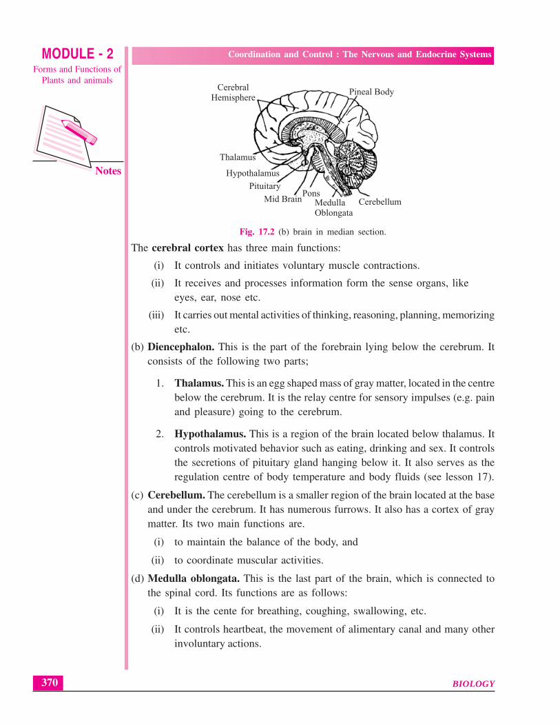
Fig. 17.2 (b) brain in median section.
The cerebral cortex has three main functions:
(i) It controls and initiates voluntary muscle contractions.
(ii) It receives and processes information form the sense organs, likeeyes, ear, nose etc.
(iii) It carries out mental activities of thinking, reasoning, planning, memorizingetc.
(b) Diencephalon. This is the part of the forebrain lying below the cerebrum. Itconsists of the following two parts;
1. Thalamus. This is an egg shaped mass of gray matter, located in the centrebelow the cerebrum. It is the relay centre for sensory impulses (e.g. painand pleasure) going to the cerebrum.
2. Hypothalamus. This is a region of the brain located below thalamus. Itcontrols motivated behavior such as eating, drinking and sex. It controlsthe secretions of pituitary gland hanging below it. It also serves as theregulation centre of body temperature and body fluids (see lesson 17).
(c) Cerebellum. The cerebellum is a smaller region of the brain located at the baseand under the cerebrum. It has numerous furrows. It also has a cortex of graymatter. Its two main functions are.
(i) to maintain the balance of the body, and
(ii) to coordinate muscular activities.
(d) Medulla oblongata. This is the last part of the brain, which is connected tothe spinal cord. Its functions are as follows:
(i) It is the cente for breathing, coughing, swallowing, etc.
(ii) It controls heartbeat, the movement of alimentary canal and many otherinvoluntary actions.
MedullaOblongata
PonsCerebellumMid Brain
Pituitary
Hypothalamus
Thalamus
CerebralHemisphere
Pineal Body
Notes
MODULE - 2Forms and Functions of
Plants and animals
Coordination and Control : The Nervous and Endocrine Systems
371BIOLOGY
In all, 12 pairs of nerves (cranial nerves) come out of the brain, some of theseare sensory, some motor and some are of mixed type.
17.4.2 The Spinal cord
The spinal cord extends form the medulla of the brain downward almost the wholelength of the backbone. It is also wrapped in the same three meninges as the brainand the space between them contains the same cerebrospinal fluid. The arrangementof the white and gray mater is reversed in it i.e. white matter is outside and thegray matter inside.
Fig. 17.6 shows the general structure of the spinal cord as seen in its cross section. Italso shows the manner in which the spinal nerves originate from it.
Functions of spinal cord.
(i) Carry out reflexes below the neck,
(ii) Conducts sensory impulses from the skin and muscles to the brain,
(iii) Conducts motor responses from the brain to the trunk and limbs.
INTEXT QUESTIONS 17.1
1. With the help of a flow chart write down the basic components of the nervoussystem in the space given below.
2. Name the ganglia which
(a) forms the brain
(b) lies below the oesophagus and is joined to brain.
3. Which part of nervous system of cockroach can be compared to our spinal cordthough our spinal cord is dorsal and this part of nervous system of cockroachis ventral?
Notes
MODULE - 2Forms and Functions of
Plants and animals
Coordination and Control : The Nervous and Endocrine Systems
BIOLOGY 372
4. Name the main parts of the brain.
............................................................................................................................
5. Mention the one functions each of :
(i) Cerebrum...................................................................................................
(ii) Cerebellum ................................................................................................
(iii) Medulla oblongata ....................................................................................
(iv) Hypothalamus............................................................................................
6. What are the
(i) gray matter, and........................................................................................
(ii) white matter made of? .............................................................................
7. Name the fluid in the cavities of the brain.
............................................................................................................................
17.4 PERIPHERAL NERVOUS SYSTEMThe peripheral nervous system consists of all nerves arising from the brain and thespinal cord. Overall, it consists of two kinds of pathways: the afferent (receiving)sensory pathways and efferent (carrying aways) motor pathways.
A. The afferent (receiving/sensory) pathways are included in two kinds of nerves.
Purely sensory nerves, for example the cranial nerves received from theeyes, ears, nose, etc.
Mixed cranial nerves like the fifth (facial nerve ) which contains sensoryfibres bringing sensations from the face but it also contains motor fibreswhich carry impulses away to the jaw muscles.
B. The efferent (sending) pathway may be subdivided into somatic and autonomicnervous systems.
(i) The somatic nervous system controls the voluntary muscles. It includesmost cranial nerves as well as the motor nerve fibres of the spinal nerves.Both these convey message from the CNS to the voluntary muscles.
(ii) Autonomic nervous system (ANS). This innervates the involuntarymuscles and the glands. It consists of a pair of chains of ganglia and nerveson either sides of the backbone (Fig. 17.3) This system is essentially amotor system, which regulates the involuntary actions of the internalorgans. It consists of two parts: (a) Sympathetic nervous system and (b)parasympathetic nervous system. (Fig. 17.3).

Notes
MODULE - 2Forms and Functions of
Plants and animals
Coordination and Control : The Nervous and Endocrine Systems
373BIOLOGY
Fig. 17.3 Autonomic nervous system - sympathetic and parasympathetic
Sympathetic nervous system prepares the body for facing emergencysituations and the parasympathetic nervous system reestablishes the normalconditions once the emergency is over.
The opposite effects of the two subdivisions of the autonomic nervous system onthe different organs are listed below in the table 17.1.
Table 17.1 Effects of autonomic nervous system
Organ Effect of Sympa- Effect of Parasympa- thetic Activity thetic activity
1. Eye pupil Dilated Constricted
2. Heart beat Speeded up Slowed down
3. Blood vessels
a. on skin Constricted Dilated
b. on muscles Dilated No effect
4. Bronchioles Dilated Constricted
5. Urinary bladder Muscles relaxed Muscles contract(feeling of urination)
Sphincter contracted Sphincter relaxed
6. Sweat secretion Increased No effect
Iris
Lacrimal gland
Salivary gland
Heart
Lungs
Stomach duodenumpancreas
Adrenal gland
Colon
Urinary Bladder
Gonads andsex organs
SympatheticParasympathetic

Notes
MODULE - 2Forms and Functions of
Plants and animals
Coordination and Control : The Nervous and Endocrine Systems
BIOLOGY 374
7. Blood sugar Increased No effect
8. Salivary secretion Stops Increased
9. Tear glands Activated Slowed down
10. Erector muscles of Stimulated (hair Relaxed (hair flattened)skin hair raised)
11. Adrenal glands Increased secretion of No effectAdrenalin
12. Intestine Peristalsis decreased Peristalsis increased
13. Stomach glands Decreased secretion Increased secretion
The autonomic nervous system is strongly influenced by emotions such asgrief, anger, fear, sexual stimulation, etc.
INTEXT QUESTIONS 17.2
1. What are the two subdivisions of the autonomic nervous system?
............................................................................................................................
2. Name the specific subdivisions of the autonomic nervous system concerned withthe following:
(i) Slowing down heart beat .........................................................................
(ii) Increasing salivary secretion.....................................................................
(iii) Dilatation of the pupil ..............................................................................
(iv) Increasing intestinal peristalsis .................................................................
(v) Muscle contraction of the urinary bladder giving the feeling the need forurination. ...................................................................................................
3. Why is the peripheral nervous system called so?
............................................................................................................................
4. State the alternative terms for sensory and motor nerves.
............................................................................................................................
17.5 NEURON – THE STRUCTURAL AND FUNCTIONAL UNIT OFNERVOUS SYSTEM (FIG. 17.4)
You have already studied about the nerve cell. This is to refresh your memory forrelating the structure of the neuron with the conduction of nerve impulse.
The cell body contains nucleus and cell organelles in the cytoplasm.

Notes
MODULE - 2Forms and Functions of
Plants and animals
Coordination and Control : The Nervous and Endocrine Systems
375BIOLOGY
Dendrites (short branching processes) extend out from the cell body. They bringsignals (impulses) from the receptor or from the axon endings of another neuron.There may be as many as 200 dendrites in a single neuron allowing as manyconnections with the axon endings of other neurons.
A long nerve fibre or axon carries the impulse from the cell body towards itsterminal branches which may either pass on the impulse to another neuron, orinto a muscle or gland to bring about the required action. Synapse is the pointof communication between one nerve cell and another or between nerve celland a muscle.
A sheath of fatty material (myelin) often covers the axon, and such nerve fibresare called medullated or myelinated fibres.
Fig. 17.4 The nerve cell
17.6 CONDUCTION OF NERVE IMPULSE ALONG THE NEURON ANDOVER THE SYNAPSE
The conduction of nerve impulse through the nerve fibre is electrical in nature andthe one through the synapse is chemical in nature.
Dendron
Dendrites
Perikaryon
Nucleus
Nissl granulesAxon
Nucleus
Medullary sheath
Node of Ranvier
Axon endings

Notes
MODULE - 2Forms and Functions of
Plants and animals
Coordination and Control : The Nervous and Endocrine Systems
BIOLOGY 376
A. Along the neuron–Electrical SignallingThe transmission (moving from one end to another) of the nerve impulse throughthe nerve fibre is electrochemical. It is not simply a flow of electrons through anelectric wire but it travels as a wave of depolarization (Fig. 17.5). Read thefollowing to understand depolasis atom.
In normal resting condition the outside of the nerve fibre carries positive (+) charge.In this condition nerve fibre is said to be polarized. The polarization is due to thepresence of more Na+ ions outside the cell membrane. Such state is maintained dueto the sodium ions being continuously pumped out by means of the sodiumpotassium pump and operated by active transport using ATP for energy.
Sodium potassium pump is a carrier protein on the plasma membranewhich transports sodium and potassium ions across the membrane. Normallyions move from the region of their high concentration to the region of theirlow concentration.
The changes when a stimulus arrives at the nerve fibre are as follows:The axon membrane at that spot becomes more permeable to Na+ ions, whichmove inward and bring about depolarization or localised change of charge frompositive to negative (see diagram) on that spot.This point of depolarization itself becomes the stimulus for the adjoining areaof the membrane, which in turn becomes depolarized.Meanwhile the previous area becomes repolarized due to active movement ofthe sodium ions to the outside of the membrane by means of what is called‘sodium pump’.And now the fibre is ready for the next wave of depolarization.
Thus a nerve impulse is a self- propagating wave of depolarization and repolarization
Fig. 17.5 Conduction of nerve impulse.
Direction of nerve impulse
ExcitedRegion
Restingregion
Recoveryregion
Excitedregion
Restingregion
Notes
MODULE - 2Forms and Functions of
Plants and animals
Coordination and Control : The Nervous and Endocrine Systems
377BIOLOGY
B. Over the Synapse – Chemical SignallingThe impulse travelling through a nerve fibre may reach either its destination. (muscleor gland) for action or the dendrites of another neuron for further transmission. Themeeting place is called synapse. The transmission over a synapse is a chemicalprocess. As the impulse reaches the terminal end of the axon, the following eventsoccur :
– a chemical acetylcholine is released by the end of the axon.
– acetylcholine stimulates the next neuron to start the new impulse.
– acteylcholine is soon broken down there to make the synapse ready for the nexttransmission.
In case the axon endings are branched and in contact with the dendrites of otherneurons the impulse will travel through all of them.
‘All or none’ principle. If the stimulus is strong enough (with a minimum threshold)to produce the impulse, the impulse will set up and travel at its own speed. Thresholdis the minimum strength of a stimulus that can initiate an impulse. Increasing theintensity of the stimulus cannot raise the speed of transmission.
17.7 REFLEX ACTIONReflex action is an automatic, quick and involuntary action in the body brought aboutby a stimulus. For example,
You instantaneously withdraw your hand on accidentally touching a hot plateor a sharp thorn.
Watering (salivation ) of the mouth takes place on seeing or just smelling afamiliar tasty food.
Two types of reflexes – simple and conditionedThe two examples of reflex action given above are basically different. The first oneis inborn or natural, which did not require previous learning. Such reflexes are calledsimple reflexes.
The other example is the outcome of repeated experience. Here the brain actuallyremembers the taste of food and works in an unconscious manner- such reflexesare called conditioned reflexes.
Some other examples of reflexes are as follows:
(A) Simple ReflexQuick closing of eyelids on noticing an object suddenly approaching the eye.
Coughing when the food swallowed enters the windpipe instead of the foodpipe.
Narrowing of the eye pupil in strong light.
If the foot of sleeping person is tickled, it is jerked away.
(B) Conditioned ReflexesApplying brakes in your vehicle (car or bicycle) on noticing someone suddenlycoming in front of it.

Notes
MODULE - 2Forms and Functions of
Plants and animals
Coordination and Control : The Nervous and Endocrine Systems
BIOLOGY 378
Tying shoe laces while talking to someone, not knowing whether you are firstputting the left lace over the right or the vice versa.
A dog runs away if it notices you kneeling down as if you are picking up astone for striking.
Standing up on seeing the teacher entering the classroom.
Mechanism of Reflex ActionSome reflexes are brought about through the brain (cerebral reflexes) such as theclosing of the eyelids due to approaching objects while other are brought aboutthrough the spinal cord (spinal reflexes). The pathway in a simple spinal reflex actionis represented in the diagram below (Fig.17.6).
Fig. 17.6 Nerve pathways in a simple reflex action
In this, there are five necessary parts:
The stimulus (prick, heat etc.) → receptor in the sensory organ → the afferent(sensory) nerve fibre running through the dorsal root of the spinal nerve bringingthe impulse into the spinal cord → a (motor) neuron sending out the commandthrough its efferent fibre in the ventral root of the spinal nerve → a muscle or thegland.
Mostly there occur an intermediate neuron between the axon ending of the afferentfibre and the motor neuron inside the spinal cord.
INTEXT QUESTIONS 17.3
1. Given below are a few examples of reflexes. Write against each, the categoryof reflex, whether simple or conditioned.
(i) Knee jerk...................................................................................................
Gray matter Ventral root
Spinal nerve
Effector (Muscle)
Receptor (skin)
Sensory neuron
Dorsal ganglionDorsal root
Spinal cordWhile matter

Notes
MODULE - 2Forms and Functions of
Plants and animals
Coordination and Control : The Nervous and Endocrine Systems
379BIOLOGY
(ii) Salivation on seeing a favorite dish.........................................................
(iii) Tying of shoe laces while talking ............................................................
(iv) Closing of eyelids if a strong beam of light is flashed across ...............
(vi) Mistaking a coiled rope as snake if you happen to step on it in darkness..................................................................................................................
17.8 SENSORY RECEPTORS (THE SENSE ORGANS)Sense organs are the organs through which we sense or detect changes in the externalenvironment. Each sense organ has special sensory cells, which receive the stimuliand transmit the impulses produced through the concerned nerve to the brain orthe spinal cord. The brain sorts out the impulses, interprets them and transmitsmessage for the required response. In human there are typically five sense receptors,eyes for seeing, ears for hearing, nose for smelling, tongue for taste and skin forsensing touch, pain, heat, etc.
17.8.1 The Eye (the sense of vision)The eye is nearly spherical in shape, bulging a little in front, and is able to rotatefreely in the bony socket. It is a hollow ball containing several structures inside(Fig.17.7).
The wall of the eyeball is made up of three layers: the sclera, choroid and retina.
Sclera is the outermost tough white layer. In front it is continued as thetransparent cornea.
Choroid is the middle layer. It is composed of connective tissue having a densenetwork of blood vessels. Its inner surface is dark brown or black. This preventsreflection, which would otherwise interfere with the clarity of the image.
Fig. 17.7 Vertical section of the human eye
Eye muscle
Sclera
Choroid
Retina
Yellow spot
Blind spot
Optic nerve
Vitreous Humour
Ciliary Muscle
Lens
Cornea
ConjunctivaPupil
Aqueous HumourIris
Suspensory ligament
Eye lid
Notes
MODULE - 2Forms and Functions of
Plants and animals
Coordination and Control : The Nervous and Endocrine Systems
BIOLOGY 380
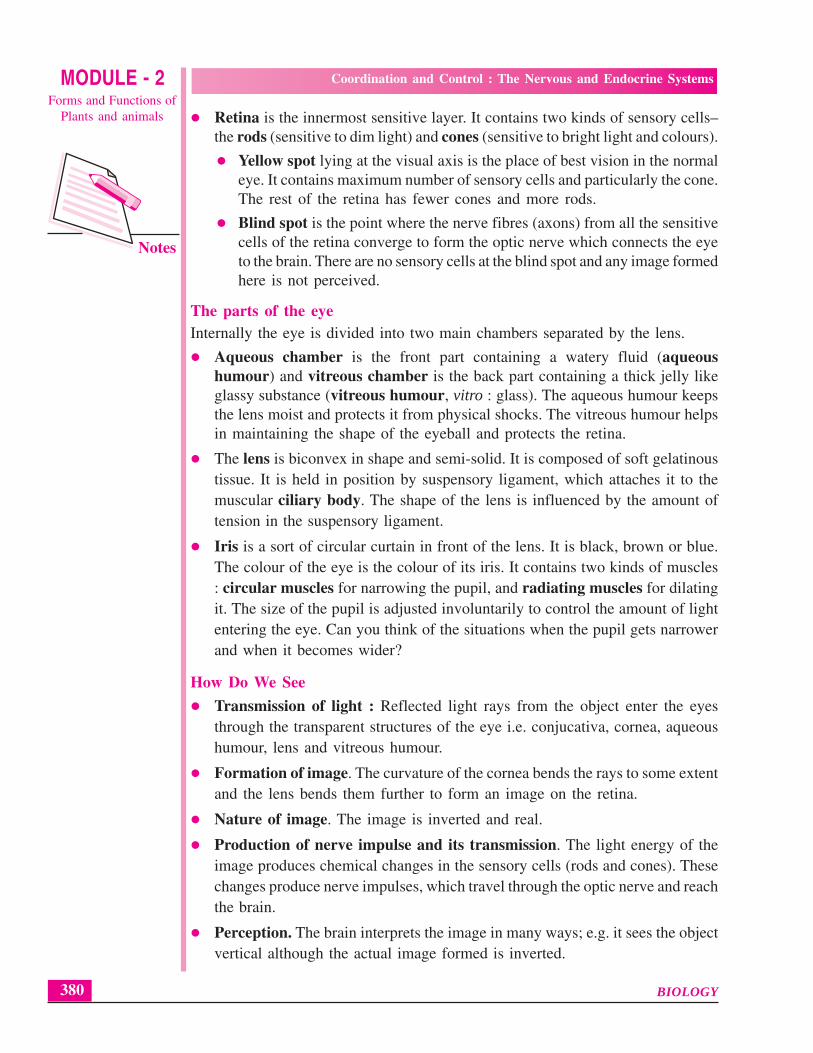
Retina is the innermost sensitive layer. It contains two kinds of sensory cells–the rods (sensitive to dim light) and cones (sensitive to bright light and colours).
Yellow spot lying at the visual axis is the place of best vision in the normaleye. It contains maximum number of sensory cells and particularly the cone.The rest of the retina has fewer cones and more rods.
Blind spot is the point where the nerve fibres (axons) from all the sensitivecells of the retina converge to form the optic nerve which connects the eyeto the brain. There are no sensory cells at the blind spot and any image formedhere is not perceived.
The parts of the eyeInternally the eye is divided into two main chambers separated by the lens.
Aqueous chamber is the front part containing a watery fluid (aqueoushumour) and vitreous chamber is the back part containing a thick jelly likeglassy substance (vitreous humour, vitro : glass). The aqueous humour keepsthe lens moist and protects it from physical shocks. The vitreous humour helpsin maintaining the shape of the eyeball and protects the retina.
The lens is biconvex in shape and semi-solid. It is composed of soft gelatinoustissue. It is held in position by suspensory ligament, which attaches it to themuscular ciliary body. The shape of the lens is influenced by the amount oftension in the suspensory ligament.
Iris is a sort of circular curtain in front of the lens. It is black, brown or blue.The colour of the eye is the colour of its iris. It contains two kinds of muscles: circular muscles for narrowing the pupil, and radiating muscles for dilatingit. The size of the pupil is adjusted involuntarily to control the amount of lightentering the eye. Can you think of the situations when the pupil gets narrowerand when it becomes wider?
How Do We See
Transmission of light : Reflected light rays from the object enter the eyesthrough the transparent structures of the eye i.e. conjucativa, cornea, aqueoushumour, lens and vitreous humour.
Formation of image. The curvature of the cornea bends the rays to some extentand the lens bends them further to form an image on the retina.
Nature of image. The image is inverted and real.
Production of nerve impulse and its transmission. The light energy of theimage produces chemical changes in the sensory cells (rods and cones). Thesechanges produce nerve impulses, which travel through the optic nerve and reachthe brain.
Perception. The brain interprets the image in many ways; e.g. it sees the objectvertical although the actual image formed is inverted.

Notes
MODULE - 2Forms and Functions of
Plants and animals
Coordination and Control : The Nervous and Endocrine Systems
381BIOLOGY
Accomodation (focusing). Focusing the image on retina is calledaccommodation. Changing the curvature of the elastic lens brings aboutaccommodation.
– For distant vision : The lens is more flattened or thinner; this is the normalcondition of the lens, which is kept stretched by the suspensory ligaments.
– For near vision : The ciliary muscles which are circular, contract and tendto reduce the circumference of the eyeball there. This releases the tensionon the suspensory ligament and the lens becomes thicker (more rounded)on account of its own elasticity.
A normal eye is constantly accommodating while walking, playingor just looking around.
Binocular vision. In all primates including humans, both eyes are placedforward. Each eye views at a slightly different angle. The images from the twoeyes are perceived overlapped inside the brain giving the impression of depth(3-dimensional/stereoscopic vision).
Three Common defects of the eye
1. Near sightedness (Myopia). Nearby objects are clearly seen but not the distantones by those suffering from myopia because the image of the object is formedin front of the retina. This can be corrected by using concave lens (worn in frames(spectacles) or as contact lenses).
2. Long sightedness (Hypermetropia). Distant objects are clearly seen but notthe nearby because the image of the object is formed behind the retina. Thiscan be corrected by convex lens (worn in frames as spectacles or as contactlenses).
3. Cataract (opacity of the lens). The lens usually loses its transparency and turnsopaque with age. Such a lens can be surgically removed and replaced by an intra-ocular lens.
INTEXT QUESTIONS 17.4
1. State the function of the following parts of the eye:
(i) Iris .............................................................................................................
(ii) Ciliary muscles ..........................................................................................
(iii) Pupil ..........................................................................................................
(iv) Vitreous humour .......................................................................................
(v) Retina ........................................................................................................

Notes
MODULE - 2Forms and Functions of
Plants and animals
Coordination and Control : The Nervous and Endocrine Systems
BIOLOGY 382
2. Name the following:
(i) Area of sharp vision in the eye ...............................................................
(ii) The kind of lens used for correcting near-sightedness ...........................
(iii) The condition in which the lens of the eye turns opaque .....................
(iv) The capacity of eye to focus objects at different distances ...................
17.8.2 The Ear-Sense of Hearing and Balance
The ear serves two sensory functions: hearing and maintaining balance of the body.The ear has three main parts – external ear, middle ear, and internal ear (Fig. 17.8)
Fig. 17.8 The human ear.
The external ear consists of the following :– an outwardly projecting ear to be called pinna supported by cartilage. It directs
the sound waves inwards.– The auditory canal through which the sound waves travel up to the ear drum
(tympanic membrane)The middle ear consists of the following:
An air-filled tympanic cavityThe tympanum or ear drumThree tiny bones-malleus (hammer) connected to the ear drum, incus (anvil)in between and stapes (stirrup) forming a contact with the oval window of theinternal ear.Eustachian tube connects the tympanic cavity with pharynx. It equalizes thepressure on both sides of the eardrum or tympanum :
The internal ear contains two main parts:(a) Cochlea – It is a long coiled structure which looks like the coils of the shell
of a snail. It has two and a half turns. The inner winding cavity of the cochleais divided into three parallel tubes of canals separated by membranes. The canalsare filled with a fluid called endolymph. The middle canal possesses sensory cells(organ of corti) for hearing.
Pinna
Incus
Malleus
Semicircular canals
Stapes at oval window
Auditiory Nerve
Cochlea
Base of skull
Round window
Eustachian tubeEar drum
Bone
Externalauditiorymeatus

Notes
MODULE - 2Forms and Functions of
Plants and animals
Coordination and Control : The Nervous and Endocrine Systems
383BIOLOGY
(b) Vestibule – is concerned with physical balance of the body. It consists of threesemicircular canals arranged at right angles to each other and a part joiningthe cochlea and differentiated into a utriculus and a sacculus. One end of eachsemicircular canal is widened to form an ampulla, which contains sensory cells,and the nerve fibres from them continue into auditory nerve.
Mechanism of hearing– The sound waves enter the auditory canal and cause the eardrum to vibrate
– The vibrations of the eardrum are transferred to malleus, to incus, and then tostapes. Stapes transfers the vibrations through oval window into the cochlea.
– These vibrations move the fluid in the cochlea. The organ of corti catches themovement of the fluid and transfers it to the auditory nerve that carries theimpulses to the brain
Perception of body balanceStatic balance due to gravity – Any bending or change in the body posture causesthe fluid inside the semicircular canals to move. The semi circular canals are arrangedin different planes. The sensory hairs in the ampulla of the canal pick up thesemovements and the impulses are transmitted through the auditory nerve.
Balance during motion – Utriculus and sacculus perceive dynamic equilibrium(while the body is in motion). Fine particles of calcium carbonate present in theendolymph press on the sensory hairs whenever the body is in some motion. Theimpulses are carried through the auditory nerve.
17.8.3 Tongue and Nose (Sense of taste and smell)The tongue perceives the taste and the nose perceives the smell. The perceptiondepend upon the nature of chemical substance coming in contact with the sensorycells. For taste there is a direct contact of the substance with the sensory cells locatedin the taste buds on the tongue. For smell, the molecules of the chemical are carriedinward by the air inhaled and they stimulate the sensory epithelium of the nose.
17.8.4 Skin (Touch and some other miscellaneous senses)There are a variety of nerve endings in the skin. Some of these are concerned withtouch (gentle pressure), some with deep pressure and others with cold, heat andpain.
The sense of hunger is due to receptors in the stomach wall. The sense of thirstis due to stimulation of nerves in the pharynx. And the sense of fatigue is locatedin the muscles.
INTEXT QUESTIONS 17.5
1. Which part of the ear is involved when:
(i) a gymnast performs various balancing feats. ..........................................
(ii) you hear a song. .......................................................................................

Notes
MODULE - 2Forms and Functions of
Plants and animals
Coordination and Control : The Nervous and Endocrine Systems
BIOLOGY 384
2. Name the following :
(i) The part into which the sound waves are directed by the ear pinna.
..................................................................................................................
(ii) The kind of balance with which the semi-circular canals are concerned.
..................................................................................................................
(iii) Any two sensations felt through free nerve endings in the skin.
..................................................................................................................
17.9 COORDINATION THROUGH HORMONES—THE ENDOCRINESYSTEM
Hormones are secretions from specific cells or glands in the body called endoerineglands Harmones are carried by blood to target organs. Their effect is producedin one or more specific parts only. Most hormones are secreted by special glandscalled the endocrine glands. These are also called ductless glands because theirsecretions are poured directly into the blood and not through ducts. Certainhormones are produced by other glands or body parts also, for example, the stomachand the duodenum.
17.9.1 Nature and Function of HormonesHormones are secreted from their source directly into the blood.
Blood carries the hormone to the target cells which respond to it.
Hormones regulate the physiological processes.
They are produced in very small quantities and are biologically very active.For example, adrenaline is active even at a concentration of 1 in 300 millionparts.
Their excess and deficiency, both, cause serious disorders.
Chemically, the hormones may be water-soluble proteins (peptides),glycoproteins and amines or lipid-soluble steroids.
The extra hormones are not stored in the body and are excreted out.
17.9.2 Hormone Secretors — the Endocrine GlandsIn humans there are more than a dozen tissues and organs that produce hormones.Most of these are shown in Fig. 17.8. These can be listed under two categories
(a) Exclusively endocrine : the pituitary, the thyroid, the parathyroid, thymusand the adrenals.
(b) Partially endocrine : The pancreas, gastric and duodenal epithelium, thegonads (testis in males and ovary in females) and placenta in females.

Notes
MODULE - 2Forms and Functions of
Plants and animals
Coordination and Control : The Nervous and Endocrine Systems
385BIOLOGY
Fig. 17.8 Location of principal endocrine glands in the human body
1. Pituitary — the master glandThe pituitary gland (also called hypophysis) (Fig. 17.9) is a small projection (aboutthe size of a pea) which hangs from the base of the mid-brain. It is connected tothe hypothalamus of the brain by the pituitary stalk. The hypothalamus, althougha part of the brain, also secretes some hormones.
Fig. 17.9 Pitutary gland
ThyroidParathyroids
Thymus
Duodenum (Not shown)
Adernal
Pituitary (Hypophysis)
Pineal body
Islets of langerhans (in pancreas)
Ovary
Placenta (not shown)
Testis
Hypothalamus
Anterior pituitary
Pars Intermedia
Posterior Pituitary
Hypophyseal stalk

Notes
MODULE - 2Forms and Functions of
Plants and animals
Coordination and Control : The Nervous and Endocrine Systems
BIOLOGY 386
The pituitary controls most other endocrine glands. It has two distinct parts: theanterior pituitary and the posterior pituitary. Various hormones produced fromthese two parts and their actions are listed below in Table 17.2.
Table 17.2 Pituitary hormones, their action and abnormalities due to itsoversecretion or undersecretion
Source Hormones Action and abnormalitiesproduced
Anterior lobe Growth hormone Promotes growth of whole body,of pituitary (GH), also known particularly of the skeleton.
as somatotropic Undersecretion in childhood lead tohormone (STH) Dwarfism; oversecretion in childhood
causes gigantism and in adult, acromegaly.
Tropic hormones 1. Thyroid stimulating hormone (TSH)(stimulate other stimulates thyroid.endocrine glands) 2. Adrenocorticotropic hormone (ACTH)
stimulates adrenal cortex.Gonadotropic hormones 3. Follicle stimulating hormone (FSH)
stimulates egg formation in females andsperm formation in males.
4. Luteinizing hormone (LH) stimulatesovulation and the formation of corpusluteum which produces the femalehormone progesterone andLH stimulates testis to produce the malehormone testosterone.
5. Prolactin stimulates milk production.
Posterior lobe Antidiuretic hormone Increases absorption of water from theof pituitary (ADH) or vasopressin kidney tubules (osmoregulation).
Its deficiency causes diabetes insipidus.
Oxytocin Stimulates contractions of the uterus duringchildbirth.
2. ThyroidThyroid is a bilobed structure situated in the front region of the neck(Fig. 17.10). It secretes two hormones—thyroxine and calcitonin.Thyroxine regulates basal metabolism i.e. the rate of cellular oxidation resultingin heat production. Controls growth and development, ossification of the bones,body temperature, mental development, etc.
Undersecretion of thyroxine (hypothyroidism) produces three conditions
Simple goitre. Enlargement of thyroid visible as a swelling in the neck. It iscaused due to iodine deficiency in food as iodine is needed for production ofthyroid hormones.
Cretinism. Poor body growth (dwarfism) and mental retardation
Myxoedema. Swelling of the face and hands. General sluggishness.

Notes
MODULE - 2Forms and Functions of
Plants and animals
Coordination and Control : The Nervous and Endocrine Systems
387BIOLOGY
Oversecretion of thyroxine (hyperthyroidism) produces exophthalmic goitre. Thiscondition causes marked increase in the metabolic rate, rapid heart beat, shortnessof breath and the eyes protrude out together with goitre in the neck.
Fig. 17.10 The thyroid gland
Calcitonin. It regulates the calcium and phosphate levels in the blood. If the calciumlevel in blood is high more calcitonin is secreted and the calcium ions are movedfrom the blood to the bones making them harder. The reverse happens when thecalcium level in the blood is low making the bones soft.
3. Parathyroids
These are two small pairs of glands wholly or partially embedded in the thyroidgland. Their secretion parathormone raises blood calcium level by stimulatingrelease of calcium from bones.
4. Thymus
It is located at the base of neck. It produces some hormones involved in maturationof T lymphocytes. It begins to atrophy after puberty.
5. Adrenals
The adrenals (ad: adjacent, renal; kidney) are a pair of glands situated like caps oneabove each kidney. Each adrenal consists of two parts: a central medulla and aperipheral cortex.
The adrenal medulla secretes adrenaline which,
increases heart beat accompanied by an increase in the blood pressure.
increases blood supply to the muscles while decreasing blood supply to thevisceral organs.
releases more glucose into the blood from the liver.
The adrenal cortex secretes two categories of hormones: glucocorticoids andmineralocorticoids.
Oesophagus
Parathyroidgland
Thyroid gland
Trachea (windpipe)
Larynx

Notes
MODULE - 2Forms and Functions of
Plants and animals
Coordination and Control : The Nervous and Endocrine Systems
BIOLOGY 388
(a) Glucocorticoids e.g. cortisoneIn response to stress it raises blood glucose through action of the liver includingdeamination of amino acids. During starvation and prolonged fasting therequired glucose is partly provided through this hormone.
It adapts the body to stresses such as extreme heat or cold, burns, infections,etc.
Some of the cortical hormones behave like sex hormones.
– Overgrowth of adrenal cortex in young children causes prematuresexual maturity.
– Overgrowth of adrenal cortex in mature females results in thedevelopment of male characters such as beard and deep voice.
– Overgrowth of adrenal cortex in mature males results in the developmentof some feminine characters such as enlargement of breasts.
(b) Mineralocorticoids e.g. aldosterone
This hormone is concerned with water retention. It increases reabsorption of sodiumand chloride ions in kidneys. Read the role of aldosteronl in increasing blood volumeand blood pressure in increasing blood volume and blood pressure in lesson 14(14.3.6)
6. PancreasPancreas is an endocrine as well as an exocrine gland. It has special groups of cellscalled Islets of Langerhans, which consists of three kinds of cells – alpha cellsproducing the harmone glucagon, beta cells producing harmone insulin and gammacells producing harmone somatostatin.
(i) Glucagon. It stimulates breakdown of glycogen to glucose in the liver, leadingto rise in the blood sugar level.
(ii) Insulin. It performs two principal tasks;
Promotes glucose utilization by the body cells.
Stimulates deposition of extra glucose in the blood as glycogen in the liver.
Gluccagon and insulin have oppsite functions.
Non-secretion or under secretion of insulin causes diabetes mellitus(hyperglycemia, meaning ‘more than normal sugar in blood’.
A diabetic person,
has higher glucose in blood;
excretes a great deal of urine loaded with sugar;
feels thirsty because of loss of water through too much urination;
loses weight and becomes weak. In some cases, the patient even loses theeyesight.
Oversecretion of insulin causes hypoglycemia or low blood sugar. The brain mayenter a state of coma if the level of sugar in blood becomes too low.
(iii) Somatostatin also called Growth Hormone-Inhibiting Hormone (GHIH)inhibits secretion of insulin as well as glucagon.

Notes
MODULE - 2Forms and Functions of
Plants and animals
Coordination and Control : The Nervous and Endocrine Systems
389BIOLOGY
7. Gonads (testis and ovary)
Testes in males possess two kinds of cells : the sperm-producing germinal cells andthe hormone-producing interstitial cells. The hormones produced are called androgensand the commonest one among them is testosterone.
The testosterone stimulates the development of the male characters during whichthe body at puberty starts developing facial hair, and their voice cracks and deepens.
Ovaries in females produce two kinds of hormones—estrogen and progesterone.Estrogen is secreted from the follicles of the ovary and stimulates the developmentof breasts and fat deposition on the hip in a mature woman. Estrogen prepares thewall of the uterus for receiving the fertilized egg.
Progesterone is secreted by the corpus luteum (follicle left after the release ofovum). It brings about the final changes in the uterus for the retention and growthof the foetus during pregnancy.
8. Placenta
Placenta of a pregnant woman produces certain hormones. One such hormone ishuman chorionic gonadotropin (HCG), which maintains the activity of corpusluteum in secreting progesterone continuously, when a women becomes pregnant.
9. Hormones from stomach and intestine
(i) Gastrin is the hormone secreted by the mucus membrane of the pyloricend of the stomach. It stimulates the gastric glands to secrete gastric juice.
(ii) Secretin is the hormone secreted by the inner lining of the duodenum.It stimulates the production of pancreatic juice while the hormonecholecystokinin stimulates release of bile from gall bladder.
17.10 THE FEEDBACK MECHANISM (CONTROL OF HORMONALSECRETION)
The amount of hormone released by an endocrine gland is determined by the body’sneed for the particular hormone at any given time. The product of the target tissueexerts an effect on the respective endocrine gland. This effect may be positive(‘secrete more’) or negative (‘secrete no more’ or ‘slow down’). This can beexplained by taking the example of thyroid gland.
Feed back mechanism of thyroid activity (Fig. 17.11). Hypothalamus releases ahormone TSH-RH (TSH- Releasing Hormone) which instructs the anterior pituitaryto release TSH (thyroid stimulating hormone). The TSH stimulates thyroid to releasethyroxine. If the level of thyroxine in blood increases, the pituitary stops the releaseof TSH. When the level of thyroxine falls in the blood, the thyroid gets stimulated

Notes
MODULE - 2Forms and Functions of
Plants and animals
Coordination and Control : The Nervous and Endocrine Systems
BIOLOGY 390
to secrete more of it. In feedback mechanism the starting point of an activity receivesback the information whether to continue or increase, or to slow down or even stop.
Fig. 17.11 Feed back mechanism in hormone action
(solid line = stimulation; broken line = suppression/inhibition)
17.11 COMPARISON OF HORMONAL AND NERVOUS COORDINATION
The table 17.2 below lists a few major differences between these two different kindsof control and regulating mechanisms.
Table 17.2 difference between hormonal and nervous control
Property Hormonal control Nervous control
1. Nature of signal
2. Speed of signal
3. Effect in the body
All hormones arechemical signal
Slow
General effect. Thehormones can influencecells in many differentparts of the body.
Nerve impulses areelectrical signals. Chemicalsignalling takes place atsynapses
Rapid. Between 0.7 metresper second and 120 metresper second
Localized effect – affectsonly the particular muscleor the gland

Notes
MODULE - 2Forms and Functions of
Plants and animals
Coordination and Control : The Nervous and Endocrine Systems
391BIOLOGY
4. Effect on growth
5. Capacity formodification
6. Duration ofeffect
17.12 PHEROMONES—THE CHEMICAL MESSENGERS AT SOCIALLEVEL
Pheromones are the secretions given out by an individual into the environment,which bring about a specific response in other members of the same species. Someof the examples of the pheromones are as follows:
Common ants march on the floor or walls in a trail on an invisible path laiddown by a secretion from their bodies. It helps them to reach the destinationone after another, as well as to return correctly to their own nest.
When disturbed honey bees give out an alarm pheromone from their stingat the back and mandibles in the mouth. This alerts the inmates of the hive toface the attack.
Females of a particular moth gives out a scent which can attract a male fromas much distance as 3-4 kilometers.
Introduction of a male mouse into a group of female mice shortens oestrus cycle(cycle of development of eggs in the ovary and ovulation).
Introduction of a strange male mouse of a different strain disturbs to the extentthat the newly pregnant females abort their foetuses. The source ofpheromone of the strange male mouse is in its urine.
INTEXT QUESTIONS 17.6
1. Name the following
(i) The organ in the neck on the trachea close to which thyroid is located
..................................................................................................................
(ii) The condition caused due to oversecretion of thyroxin
..................................................................................................................
(iii) The hormone concerned with facing dangers
..................................................................................................................
Can affect growth
Cannot be modified bylearning from previousexperience
Short term or long lasting.
Cannot affect growth
Can be modified by learningfrom previous experiences
Short – lived

Notes
MODULE - 2Forms and Functions of
Plants and animals
Coordination and Control : The Nervous and Endocrine Systems
BIOLOGY 392
(iv) The condition of passing much glucose in the urine
..................................................................................................................
(v) The source gland of ADH
..................................................................................................................
2. What are pheromones?
............................................................................................................................
WHAT YOU HAVE LEARNT
The coordination of body activities inside the body of an organism is broughtabout by two systems- the nervous and the endocrine systems.
The nervous system is composed of the central nervous system (brain and spinalcord) and the peripheral nervous system (cranial and spinal nerves and theautonomic nervous system).
The autonomic nervous system consists of a pair of chain of ganglia by the sideof spinal cord. It is largely concerned with the normal functioning of the visceralorgans.
The nervous system of cockroach is made of brain or cerepral gangha, suboesophaegeal ganglion, thoracic ganglia gangha and six abdominal ganglia fromwhich nerves come out.
Cerebrum is the largest part of the brain and is the seat of intelligence.
Cerebellum is the centre of balance.
Medulla oblongata controls breathing and heart beat.
Spinal cord is the centre for simple reflexes.
The sensitive layer of the eye is the retina which is composed of rods (sensitiveto dim light) and cones (sensitive to bright light and for colour vision).
The internal ear performs two tasks perception of sound by the cochlea and thatof disturbance in body balance by the semicircular canals, utriculus and sacculus.
The nose perceives chemical stimuli by the chemicals carried by the air and thetongue by direct contact with them.
Skin possesses receptors for touch, pain, heat cold etc.
Chemical coordination is brought about by hormones produced by the ductlessglands, that are carried by the blood and which act on the target cells or organsaway from their source.
There is a close link between the nervous and the endocrine systems, shownby the way in which the pituitary gland interacts with the hypothalamus of thebrain.

Notes
MODULE - 2Forms and Functions of
Plants and animals
Coordination and Control : The Nervous and Endocrine Systems
393BIOLOGY
Our endocrine glands include the pituitary, thyroid, parathyroid, thymus adrenals,pancreas, gonads and placenta.
The pituitary controls and regulates the activities of almost all other endocrineglands.
The undersecretion as well as the oversecretion of the hormones, both produceill effects.
Hormone levels are generally controlled by feed back mechanism.
Pheromones are secretions released outside in the enviroment, which produceresponse in other individuals of the same species.
TERMINAL QUESTIONS
1. Name the two divisions of the nervous system?
2. What is gray matter?
3. Name the chemical involved in the transmission of nerve impulse across asynapse.
4. Give two examples of sensory nerves.
5. Name the respective areas of the retina concerned with best vision and no vision.
6. What is the role of the eustachian tube in the ear?
7. Name the hormone and its source glands, whose deficiency leads to diabetesinsipidus.
8. What are pheromones?
9. Name and explain the event that happens immediately when a nerve fibre getsstimulated?
10. Are the endocrine glands and the ductless glands one and the same thing? Giveone example.
11. Describe any one example of condition reflex in the humans.
12. List the functions of medulla oblongata.
13. Differentiate between sympathetic and parasympathetic nervous systems.
14. What are the two principal tasks of insulin?
15. Explain the following terms: (i) synapse (ii) stimulus and (iii) impulse
16. Draw a diagram to show the arrangement of the bones inside the middle ear.
17. Write short notes on the following :
(i) myopia
(ii) taste buds
(iii) accommodation of the eye

Notes
MODULE - 2Forms and Functions of
Plants and animals
Coordination and Control : The Nervous and Endocrine Systems
BIOLOGY 394
18. How do sympathetic and parasympathetic nervous systems act differently on(i) pupil of the eye, and (ii) urinary bladder?
19. Draw a labelled diagram of the cross section of the spinal cord and the nervouspathway of a simple reflex concerned with it.
20. Explain the role of ciliary muscles in our eyes
21. Taking the example of thyroxine secretion, explain what is meant by feedbackmechanism?
ANSWERS TO INTEXT QUESTIONS
17.1 1. FIg. 16.1, page 337
2. (a) supraoesophageal ganglion (b) sub oesophageal ganglion
3. Ventral nerve cord
4. Cerebrum, cerebellum, medulla oblongata, thalamus and hypothalamus
5. (i) Cerebrum–intelligence/thinking/reasoning/memory;
(ii) Cerebellum– balance/muscular coordination
(iii) Medulla oblongata–involuntary actions
(iv) Hypothalamus–homeostasis
6. Gray matter–composed of neuron cell bodies
White matter–composed of axon fibres
7. Cerebrospinal fluid
17.2 1. Sympathetic nervous system and parasympathetic nervous system
2. (i) parasympathetic nervous system
(ii) parasympathetic nervous system
(iii) sympathetic nervous system
(iv) parasympathetic nervous system
(v) parasympathetic nervous system
3. because it connects the periphery (surface) of the body
4. sensory = afferent, motor = efferent

Notes
MODULE - 2Forms and Functions of
Plants and animals
Coordination and Control : The Nervous and Endocrine Systems
395BIOLOGY
17.3 1. (i) simple (ii) conditioned (iii) conditioned
(iv) simple (v) conditioned
17.4 1. (i) contracts and dilates pupil
(ii) helps in near vision/contracts to make lens thicker
(iii) controls amount of light entering the eye
(iv) maintains shape of the eye ball and protects retina
(v) produces nerve impulses into the optic nerve
2. (i) yellow spot (ii) concave lens
(iii) cataract (iv) accommodation
17.5 1. (i) vestibule (ii) cochlea
2. (i) auditory meateus
(ii) static balance
(iii) touch/pressure/warmth/cold/ ….
17.6 1. (i) larynx, (ii) cretinism, (iii) adrenaline (iv) diabetes mellitus,
(v) posterior pituitary
2. Pheromone is a secretion from one individual that is given out into theenvironment and which elicits a response in other members of the samespecies.