coordination and processing of dna ends during double - genetics
TRANSCRIPT

1
Coordination and processing of DNA ends during double-strand break repair: The role of the bacteriophage T4 Mre11/Rad50 (MR)
complex Joshua R. Almond*1, Bradley A. Stohr*1,2, Anil K. Panigrahi*3, Dustin
W. Albrecht§, Scott W. Nelson§ and Kenneth N. Kreuzer*
*Department of Biochemistry, Duke University Medical Center, Durham, NC
§ Department of Biochemistry, Biophysics and Molecular Biology, Iowa State University, Ames, IA
1 These two authors contributed equally as co-first-authors 2 Current address: Department of Pathology, University of California, San Francisco, CA 3 Current address: Department of Anesthesiology, Stanford University, Palo Alto, CA
Genetics: Early Online, published on August 26, 2013 as 10.1534/genetics.113.154872
Copyright 2013.

2
Running title: Bacteriophage T4 MR complex in DSB repair Keywords: DSB repair, Mre11-Rad50 complex, homologous recombination, recombination-dependent replication, end coordination Corresponding Author: Kenneth N Kreuzer Address: Box 3711, Duke University Medical Center, Durham, NC 27712 Email address: [email protected] Phone: 919 684 6466

3
ABSTRACT
The in vivo functions of the bacteriophage T4 Mre11/Rad50 complex (gp46/47) in
double-strand end processing, double-strand break repair, and recombination-dependent
replication were investigated. The complex is essential for T4 growth, but we wished to
investigate the in vivo function during productive infections. We therefore generated a
suppressed triple amber mutant in the Rad50 subunit to substantially reduce the level of complex
and thereby reduce phage growth. Growth-limiting amounts of the complex caused a concordant
decrease in phage genomic recombination-dependent replication. However, the efficiencies of
double-strand break repair and of plasmid-based recombination-dependent replication remained
relatively normal. Genetic analyses of linked markers indicated that double-strand ends were less
protected from nuclease erosion in the depleted infection, and also that end coordination during
repair was compromised. We discuss models for why phage genomic recombination-dependent
replication is more dependent on Mre11/Rad50 levels when compared to plasmid recombination-
dependent replication. We also tested the importance of the conserved histidine residue in
nuclease Motif I of the T4 Mre11 protein. Substitution with multiple different amino acids
(including serine) failed to support phage growth, completely blocked plasmid recombination-
dependent replication, and led to the stabilization of double-strand ends. We also constructed and
expressed an Mre11 mutant protein with the conserved histidine changed to serine. The mutant
protein was found to be completely defective for nuclease activities, but retained the ability to
bind the Rad50 subunit and double-stranded DNA. These results indicate that the nuclease
activity of Mre11 is critical for phage growth and recombination-dependent replication during T4
infections.

4
INTRODUCTION
The Mre11/Rad50 (MR) protein complex plays a central role in the response to double-
strand breaks (DSBs) in eukaryotic cells (Stracker and Petrini 2011). The MR complex is quickly
recruited to the site of double-strand breaks (DSBs), where its diverse functions include
checkpoint activation mediated by the ataxia-telangiectasia mutated (ATM) kinase (Maser et al.
1997; Nelms et al. 1998; Lee and Paull 2005). Mutations in the human Mre11 gene cause the
ataxia-telangiectasia-like disorder (ATLD), which is characterized by immunodeficiency,
predisposition to certain cancers, and cellular hypersensitivity to ionizing radiation (Stewart et al.
1999).
The MR protein complex has been highly conserved throughout evolution, with
homologs in Saccharomyces cerevisiae, Pyrococcus furiosus, Escherichia coli (SbcCD), and
bacteriophage T4 (gp46/47) (Sharples and Leach 1995; Connelly and Leach 2002). The basic
structure of the protein complex was elucidated in humans, S. cerevisiae, and E. coli (Connelly et
al. 1998; Anderson et al. 2001; de Jager et al. 2001; Hopfner et al. 2002). The core MR complex
consists of an Mre11 dimer, with each Mre11 subunit bound to a single Rad50. Each Rad50
subunit adopts a long coiled coil structure and can dimerize with a second Rad50 subunit through
their so-called zinc-hooks, a conserved CXXC motif at the apex of the coiled-coil domain that
forms a zinc tetrathiolate center upon dimerization. This Rad50 dimer serves as a flexible tether
connecting Mre11 proteins on each end (Hopfner et al. 2002). The eukaryotic Mre11 protein has
several conserved phosphoesterase motifs, and in addition binds to double-stranded, single-
stranded, and forked DNA structures (de Jager et al. 2001; Connelly and Leach 2002; Trenz et
al. 2006). The role of the Mre11 phosphoesterase activity in nucleolytic processing of the
double-strand ends (DSEs) during repair was initially confusing, since the protein complex has
an exonuclease activity with the wrong polarity for generating a 3’ resected end (Trujillo et al.
1998; Trujillo and Sung, 2001; Lewis et al. 2004; Llorente and Symington 2004; Krogh et al.
2005). However, recent research has clarified a key role of the MR complex nuclease activity.
MR (with partner proteins) removes a small segment of the 5’ terminal strand at a DSB and
thereby licenses other exonucleases for efficient resection to generate the long 3’ single-stranded
ends (Hopkins and Paull 2008; Mimitou and Symington 2008; Zhu et al. 2008).

5
The unique structure of the MR complex suggests that it could tether the ends of a DSB
to facilitate proper repair (Cromie and Leach 2001; Connelly and Leach 2002; Williams et al.
2008). Indeed, the human MR complex can link two linear DNA strands in vitro, with the
globular Mre11 proteins binding the DNA ends and the Rad50 zinc-hooks interacting to tether
the two DNA ends together (de Jager et al. 2001). A similar role in vivo is supported by the work
of Lobachev et al. (2004). While these results strongly argue that the MR complex plays a
tethering role in vivo, the effect of tethering on DSB repair fidelity is not clear. Mutations in the
MR complex can lead to genomic instability, but this instability could result from disruption of
other MR complex functions such as checkpoint activation and telomere maintenance
(Theunissen et al. 2003; Smith et al. 2005; Stracker and Petrini 2011).
Bacteriophage T4 provides a simple model system for DSB repair via homologous
recombination, which has been studied using genetic, biochemical and structural approaches.
The phage encodes the UvsX strand exchange protein (Rad51 homolog), recombination mediator
protein UvsY (Rad52 paralog), a branch specific DNA helicase UvsW (most similar to
eukaryotic Rad54), and the archetype single-strand binding protein gp32, all of which are
involved in the strand exchange reaction (Yonesaki and Minagawa 1985; Formosa and Alberts
1986; Hinton and Nossal 1986; Kodadek et al. 1989; Yonesaki and Minagawa 1989; Morrical
and Alberts 1990; Carles-Kinch et al. 1997; Gajewski et al. 2011). The mechanism of strand
exchange between a single-stranded circle and homologous linear duplex has been studied in
great detail in vitro, and crystallography has revealed 3-dimensional structures of all or parts of
the UvsX, UvsW and gp32 proteins (Shamoo et al. 1995; Sickmier et al. 2004; Gajewski et al.
2011). Advantages of the T4 system include the limited number of involved proteins and the
absence of complex post-translational modifications and other regulatory events in response to
DNA damage, allowing a simpler view of the core recombination reaction.
Phage T4 also has a well-conserved homolog of the MR complex, which is necessary for
DNA end processing in vivo but dispensable for strand exchange in vitro (in reactions that are
initiated with a single-stranded substrate). T4 MR complex consists of gp47 (the Mre11
homolog) and gp46 (the Rad50 homolog), which will hereafter be referred to as Mre11 and
Rad50. The structure of the T4 MR complex has not been determined. However, the T4 homolog
shows conservation of MR functional features, including the CXXC motif, Walker A and B
motifs, signature motif, H- and D-loops, and heptad repeat region (presumed extended coiled

6
coil) of the Rad50, along with the phosphoesterase motifs of Mre11 (Sharples and Leach 1995;
Connelly and Leach 2002). Prior work in T4 has demonstrated that the MR complex is required
for recombination-dependent replication (RDR) and DSB repair in vivo, and is absolutely
required for phage growth (reviewed in Kreuzer and Morrical 1994; Kreuzer and Drake 1994).
Interestingly, knockouts of the T4 MR complex are essentially lethal, while knockouts of other
recombination proteins (UvsX, UvsY, UvsW) reduce the burst but are not lethal (Wiberg 1966;
Cunningham and Berger 1977). This could reflect UvsXYW-independent recombination
reactions (perhaps single-strand annealing) or could reflect some essential role for the MR
complex outside of the RDR reaction (see Discussion). Consistent with results mentioned above
with the eukaryotic protein, genetic studies support a role for the T4 MR complex in end
coordination during DSB repair (Shcherbakov et al. 2006b) (also see below).
Until very recently, the T4 MR complex was recalcitrant to purification and biochemical
characterization. Limited success at purifying the complex was reported by Bleuit et al. (2001),
but only small amounts of protein were obtained and the purification procedure was not reliable.
However, Herdendorf et al. (2011) recently developed a robust procedure for purifying
milligram amounts of the complex. The purified complex has activities very similar to the
eukaryotic MR complex, including DNA-stimulated ATPase, 3’ to 5’ DNA (Mn++-dependent)
exonuclease, and single stranded endonuclease activities (Herdendorf et al. 2011). This advance
allowed characterization of the kinetics of ATP hydrolysis, modulation by partner proteins UvsY
and gp32, and multiple biochemical analyses of substitution mutants in various functional motifs
(Herdendorf et al. 2011; Herdendorf and Nelson, 2011; De la Rosa and Nelson, 2011; Albrecht
et al. 2012). Perhaps most interesting is the finding that UvsY and gp32 activate a Mg++-
dependent endonuclease activity that was postulated to be involved in end resection (Herdendorf
et al. 2011). One of the key questions that remain in the T4 system is whether the MR complex
nuclease itself catalyzes extensive resection (in spite of the incorrect directionality of the
exonuclease in vitro) or whether MR licenses another exonuclease(s), as in the eukaryotic
system.
In this study, we have used the bacteriophage T4 model system to address in vivo roles of
the MR complex. We find that reducing the amount of MR complex to growth-limiting (but not
lethal) levels results in a profound defect in RDR of the phage chromosome but only mild defects
in DSB repair or in plasmid models of RDR. We also provide genetic evidence that these

7
reduced levels of MR complex result in more DNA end erosion and a strong deficiency in end
coordination during DSB repair. Finally, we generated infections of phage with multiple
different substitutions in the conserved histidine residue in phosphoesterase motif I of Mre11.
Every tested substitution prevented phage growth and completely blocked RDR as measured by
a DSB-dependent plasmid model system. We conclude that the Mre11 nuclease activity is
critical for T4 growth and for RDR.
MATERIALS AND METHODS
Materials. Restriction enzymes and T4 DNA ligase were purchased from New England Biolabs
(Beverly, MA), 4-20% Mini-PROTEAN TGX gels and Precision Plus Dual Color Standards
from BIO-RAD (Hercules, CA), O’GeneRuler DNA Ladder Mix from Thermo Fisher Scientific
(Waltham, MA), Goat anti-Rabbit IR Dye 800CW from LI-COR Biosciences (Lincoln, NE), E.
coli Proteins-Agarose affinity gel (for removing E. coli antibodies) from Alpha Diagnostic
International (San Antonio, TX), Nytran Nylon Transfer Membrane from GE Healthcare
(Waukesha, WI), Random Primed DNA Labeling Kit from Roche Diagnostics (Indianapolis,
IN), and [α-32P] from PerkinElmer (Boston, MA). Luria Broth (LB) was formulated as follows:
Bacto-Tryptone (10 g/L), Yeast Extract (5 g/L), and NaCl (10 g/L).
Strains. Escherichia coli strains KL16-99 (CGSC #4206) (Hfr - e14- recA1 spoT1 thiE1
deoB13) and CSH108 (CGSC #8081) [F’128 Δ(gpt-lac)5 - ara(FG) gyrA-0(NalR) argE(Am)
rpoB0(rifR) thiE1 ] were obtained from the Yale University E. coli Genetic Resource Center
(New Haven, CT). The following E. coli strains were described previously: AB1 (non-
suppressing) (Kreuzer et al. 1988); MCS1 (supD) (Kreuzer et al. 1988), MV20 + (non-
suppressing, lysogen) (Stohr and Kreuzer 2002), MCS1+ (supD, lysogen) (Kreuzer et al.
1988), NapIV +/pSTS54 (non-suppressing, lysogen, RIIB expression plasmid) (Stohr and
Kreuzer 2002), CR63 (supD) (Edgar et al. 1964). The INTERCHANGE amber suppressor
strains were purchased from Promega (Madison, WI). Suppressor-containing plasmids from the
plasmid INTERCHANGE strains, along with plasmid pTD101, were moved into strain CSH108
8
for analysis of T4 RDR (Figure 6). The INTERCHANGE strains with chromosomal-borne
suppressors are derivatives of CSH108.
Bacteriophage T4tdSG2, which contains a deletion of the I-TevI open reading frame
(ORF), was generously provided by Marlene Belfort (State University of New York, Albany,
NY) (Bell-Pedersen et al. 1990). T4 strain BAS3 is a derivative of T4tdSG2 with an I-TevI ORF
deletion and an I-TevI recognition site interrupting the beginning of the rIIB gene (Stohr and
Kreuzer 2002). The I-TevI recognition site insert is 64-bp long, inserted between 168,894 and
168,895 bp of the T4 genome (T4 genome file NC_000866.4). T4 strain BAS4 is a double rII
mutant, carrying the UV294 allele in rIIA (deletion of an extra T in run of T’s at position 469-
473 bp of the T4 genome) and the UV232 allele in rIIB (insertion of extra T in run of T’s at
position 168,468-168,469 bp). T4 strain FC11 harbors a frameshift mutation in rIIB resulting
from deletion of one T in a run of T’s at position 168,875-168,879 (Shinedling et al. 1987). T4
strains used for plasmid DNA replication experiments are derivatives of strain K10, which
carries the following mutations: amB262 (gene 38), amS29 (gene 51), nd28 (denA), and rIIPT8
(denB-rIIΔ) (Selick et al. 1988).
The T4 derivatives with amber mutations at serine codons in gene 46, including the triple
amber mutant (46am3), were constructed by performing marker rescue from pBR322-derived
plasmids containing an internal fragment of gene 46, during an infection by T4tdSG2. The
following serine residues were mutated in the indicated phage strains: Ser-303 (46am1 and 46am3
mutants); Ser-293 and Ser-296 (46am2 and 46am3 mutants). The T4 K10 derivative with an amber
mutation at the His-10 residue of gene 47 was constructed using the T4 insertion/substitution
system (Selick et al. 1988). Additional T4 strains were constructed by genetic crosses.
Plasmids. Plasmids pBS4, pBS7, and pAC500 were described previously (Stohr and Kreuzer
2001; Stohr and Kreuzer 2002). Plasmid pBS4-0 is a derivative of pBS4 with the T4 replication
origin removed (but it retains the I-TevI recognition site; see diagram in Figure 4A). Plasmid
pTD101 contains the I-TevI recognition sequence, which suffers a DSB during bacteriophage T4
infection, flanked by direct repeats. It was derived from pTD001 (Tomso and Kreuzer 2000) as
follows. Plasmids pACYC184 and pTD001 were both digested with AvaI and HindIII. The
pACYC184 2848-bp fragment was ligated to the pTD001 2228-bp fragment to give pTD101.
9
Phage co-infections for growth and phage replication measurements. Phage co-infections were
performed as described (Stohr and Kreuzer 2002). In brief, bacteria were grown to an OD560 of
0.5 and co-infected with the indicated phage strains at the indicated multiplicity of infection
(MOI). After a 4-min adsorption at 37° without shaking, infections were continued with vigorous
shaking. For determination of plaque-forming units (PFU) (Figure 1A), infected cells were lysed
at indicated time points with chloroform at room temperature for 30 min, and cell debris was
removed by centrifugation (8000 x g for 10 min). Lysate dilutions were then plated on E. coli
CR63 to measure plaque formation. For determination of phage DNA replication during co-
infection (Figure 1D), DNA was isolated from cell aliquots taken at the indicated time points as
described previously (Stohr and Kreuzer 2001). Aliquots of isolated DNA were digested with
PacI, a restriction enzyme that cuts modified T4 DNA, and then subjected to Southern blotting
using a probe consisting of the HindIII fragment with the rIIA-rIIB junction, labeled using the
random primed method. Blots were visualized using a Phosphorimager and quantitated using
ImageQuant software (Molecular Dynamics, Sunnyvale, CA).
Co-conversion and end coordination analysis. Co-infections were initiated as described above,
and co-conversion and end coordination experiments were performed as described previously
(Stohr and Kreuzer 2002). In brief, infected cells were lysed with chloroform after a 45-min
infection, and cellular debris was removed by centrifugation. For co-conversion experiments,
total phage titers and rII+ recombinant phage titers were determined by plating lysate dilutions on
MCS1 (λ-) and MV20 λ+, respectively. For end coordination experiments, phage titers were
determined by plating on MV20 λ+ (supports growth of rII+ recombinants), MCS1 λ+ (supports
growth of rIIA- single mutants and rII+ recombinants), and NapIV λ+/pSTS54 (supports growth
of rIIB- single mutants and rII+ recombinants). Due to the low efficiency of plating on the NapIV
λ+/pSTS54 cell line, phage were preadsorbed to CR63 for 4 min before plating, as described
previously (Stohr and Kreuzer 2002). All co-conversion and end coordination analyses were
performed as previously described (Stohr and Kreuzer 2002).
Analysis of plasmid DSB formation and processing in T4-infected cells. The indicated bacterial
strains were pre-grown in LB containing appropriate antibiotic(s) with shaking at 37° to an
OD560 of 0.5, and the indicated phage strain was then added at an MOI of 3. The infected

10
cultures were incubated at 37° for 4 min without shaking to allow phage adsorption and then 40
min with vigorous shaking. Aliquots were taken at 20 and 40 min, and total cellular DNA was
purified using SDS/proteinase K treatment, phenol extraction, and dialysis as described
previously (Stohr and Kreuzer 2001). The purified DNA was digested with the indicated
restriction enzymes and subjected to agarose gel electrophoresis and Southern blotting. The
probe for experiments with pTD101 consisted of a 1,184-bp fragment of pACYC184 that had
been doubly digested with XmnI and DrdI. The probe for visualization of pBS4-0, pBS7 and
pAC500 was generated using the random primed labeling kit with a mix of plasmids pBS7 (also
hybridizes with pBS4-0) and pAC1000 (closely related to pAC500) as template.
Analysis of Mre11 expression levels after infection with 47am (His-10) mutant. The growth and
infection steps described just above for analysis of plasmid DSB formation and processing were
used to produce cells infected with the indicated T4 phage for 20 min. The infected cells were
collected by centrifugation in a microfuge and the pellets were resuspended by vortexing in 500
μl of a wash buffer (100 mM NaCl, 50 mM Tris-HCl pH 8, 1 mM EDTA). The cells were
recollected by centrifugation and the supernatant was removed. The pellets were resuspended by
vortexing in 25 μl of H20 and then 25 μl of 2X SDS Loading Buffer (2.7 M glycerol, 0.1 M Tris
pH 6.8, 2% SDS, 0.29 M 2-mercaptoethanol, bromophenol blue at 10 mg/L) was added. Samples
were boiled for 5 min and debris was removed by centrifugation in a microfuge for 10 min. Total
protein concentrations across samples were roughly equalized by subjecting each sample to
polyacrylamide gel electrophoresis in triplicate, followed by staining with Coomassie Blue and
quantitation using an Odyssey Infrared Imager (LI-COR Biosciences) and Odyssey Infrared
Imaging System Application Software (LI-COR Biosciences, Version 3.0). The average
intensities (using multiple bands) were compared between samples and then the volumes
adjusted for equal loading of total protein on the final gel (Western blot below).
The samples were analyzed by Western blotting using a gp46/47 rabbit primary antibody
that had been twice purified by passage over a total E. coli protein affinity gel. Samples were run
on 4-20% Mini-PROTEAN TGX gels and then transferred to a nitrocellulose membrane using an
iBlot (Invitrogen). The membrane was blocked with 5% non-fat milk buffer for 1 hour at room
temperature. Next, 0.1% Tween 20 and a 400-fold dilution of the primary antibody were added
into the blocking buffer and incubated overnight at 4° with shaking. The following day, the
11
membrane was rinsed once with TBS-T (0.14M NaCl, 0.02 M Tris HCl (pH 7.6), 0.1% Tween
20) followed by three 10-min washes with TBS-T at room temperature. The membrane was next
incubated with goat anti-rabbit IR Dye 800CW secondary antibody (1:20,000) in 5% non-fat
milk buffer for 1 h at room temperature with shaking. The membrane was rinsed with TBS-T
once followed by three 10-min washes with TBS-T at room temperature. Western blots were
scanned using an Odyssey Infrared Imager (LI-COR Biosciences) and analyzed with the
Odyssey Infrared Imaging System Application Software (LI-COR Biosciences, Version 3.0).
In vitro characterization of the H10S Mre11 mutant protein. The H10S mutation was generated
in the Mre11 expression vector pTYB1-gp47 (Herdendorf et al. 2011) using the Quickchange™
mutagenesis protocol (Stratagene). The sequence of the forward mutagenic primer was as
follows: 5’- gaaaattttaaatttaggtgattggagtttaggcgttaaagctgatgatg-3’, where the mutant codon is
shown in bold. The second mutagenic primer was the reverse complement of the forward. The
expression, purification, and biochemical assays were performed essentially as described
(Herdendorf et al. 2011).
RESULTS
Limiting the amount of MR complex by suppressing multiple amber mutations
The T4 MR complex is essential for viability (for review, see Mosig 1994). To
investigate the function of the complex during a viable infection, we wished to engineer a phage
mutant with reduced and growth-limiting levels of MR complex. Such a mutant could potentially
be used to demonstrate genetic defects due to limiting amounts of protein.
Many genetic studies of T4 utilize a serine suppressor (supD)-containing E. coli strain,
CR63, to suppress amber mutations. This serine suppressor has been shown to function with
efficiencies ranging from about 0.05 to just over 0.5 with various amber codons in different
sequence contexts (Miller and Albertini 1982; Bossi 1983). To reduce the levels of the MR
complex without changing the amino acid sequence, we changed serine codons in gene 46,
which encodes Rad50, into amber codons, and suppressed them with the serine suppressor. The
serine codons were all roughly in the middle of the gene-coding region (residues 293, 296 and

12
303, near the CXXC motif at residues 288-291). This region was chosen because a previously
analyzed amber mutation in this region is lethal when not suppressed, demonstrating that the
amber fragment does not substitute for the essential function(s) of the Rad50 subunit (data not
shown).
We first generated a single and a double amber mutant. The double amber mutant should
generate much less active Rad50 than the single (presumably the product of the efficiency of
suppression at the two sites). The single amber mutant looked identical to a wild-type control
when grown in the suppressing strain CR63, whereas the double amber mutant had a slightly
reduced plaque size. However, in liquid infections, neither mutant showed a significant reduction
in average burst size (data not shown). We therefore constructed a triple amber mutant to further
reduce the amount of Rad50. In this case, plaque size was strongly reduced, and the average
burst size went down to about a fifth of that of the wild type (Figure 1A; also see below).
Infections by this triple-amber mutant, called 46am3, provide an opportunity to analyze the
genetic effects of limiting MR complex in a T4 infection.
End processing and phage DNA replication during infections with limiting MR complex
Based on the phenotypes of gene 46 or 47 knockout mutants, the T4 MR complex is
required for the processing of DSEs and also for the bulk of T4 DNA replication, which occurs
by an RDR mechanism (for review, see Mosig 1994). We began by comparing DNA replication
and end processing in an MR knockout infection versus a limiting MR infection (i.e., the 46am3
mutant in a non-suppressing versus serine-suppressing host). As will be explained in more detail
below these were actually co-infections with two different phage strains. In each case, cells were
infected with a multiplicity of six phage that carry the normal I-TevI-encoding gene but no
inserted I-TevI site, along with a multiplicity of one phage that has a deletion of the I-TevI gene
but has an I-TevI site inserted in the rIIB gene. Both co-infecting phage are either wild-type or
triple-amber for the 46 gene. In this way, the bulk replication results shown first can be
compared directly with the genetic analyses of DSB repair below.
We isolated intracellular DNA at various times after infection, cleaved with a restriction
enzyme that cuts modified T4 DNA (PacI), hybridized a Southern blot with an rII probe, and
quantitated the total amount of phage genomic DNA (rII fragment) by Phosphorimager (image
of blot is in Figure 1D and quantitation in Figures 1B and 1C).

13
Beginning with the non-suppressing host, the 46am3 mutant had the expected DNA arrest
phenotype, with some DNA replication (presumably origin-dependent) early but very little
additional replication as the infection progressed (Figure 1B). In addition, DNA broken at the I-
TevI site was clearly present beginning at the 14-min time point, and the broken ends were
stabilized throughout the remainder of the infection (Figure 1D; I-TevI-cleaved DNA was
undetectable in the wild-type infection). These results are exactly as expected from previous
studies of gene 46/47 knockout mutations (Albright and Geiduschek 1983; Kreuzer et al. 1995;
Tomso and Kreuzer 2000).
Next, we consider the triple-amber mutant infection of the suppressing host, in which the
MR complex is limiting for growth. In this case, the amount of phage DNA replication was again
dramatically reduced compared to the 46+ control, and the phage burst was substantially reduced
compared to the wild-type (DNA amounts in Figure 1C; phage production in Figure 1A). In
addition, I-TevI-cleaved ends could be detected in DNA from the triple-amber mutant infection
but not in DNA from the 46+ infection, beginning with the 14-min time point (Figure 1D). The
intensity of the cleaved bands was, however, much weaker than in the non-suppressing host,
consistent with many of the DSB’s undergoing a repair reaction when the T4 MR complex is
present but limiting (also see below). We conclude that limiting amounts of MR complex causes
a modest reduction in the processing efficiency of DSB’s, as well as greatly reduced levels of
phage DNA replication.
Recombination of linked markers during DSB repair with limiting MR complex
We next analyzed the effects of this reduction in MR complex levels on homologous
recombination, using a genetic system for analyzing homologous recombination stimulated by
defined DSBs within the rIIB gene (Stohr and Kreuzer 2002). This system monitors the
contribution of nearby heteroalleles during the DSB repair reaction, and can also be used to
measure the coordination of DNA ends during repair.
We performed a series of genetic crosses, each involving two rII - alleles (Figure 2A).
One phage genome (BAS3 in Figure 2A) carried an insertion of a defined DSB site near the 5’
end of the rIIB gene. The insertion renders BAS3 phage rIIB- due to a frameshift and the inserted
site is cleaved efficiently by I-TevI endonuclease when it is available during T4 infections. The
second phage genome (lower DNA) carried a nearby rII heteroallele: FC11, UV232 or UV294,

14
located 15, 425, or 460 bp (respectively) from the site of the I-TevI site insertion. In each cross,
both phage strains have either a wild-type gene 46 or the 46am3 allele. In addition, the BAS3
phage with the I-TevI site insertion carries a deletion of the I-TevI reading frame, and this protein
is provided in trans by the phage that carries the FC11, UV232 or UV294 allele (see Figure 2A).
The phage with a functional I-TevI gene and the rII heteroallele is used at a multiplicity of
infection of six, while the BAS3 phage is used at a multiplicity of one. This ensures that nearly
every BAS3 chromosome with an I-TevI insertion site is cleaved successfully, and that most
DSB repair events use a chromosome without the I-TevI insertion site as a repair template (see
Stohr and Kreuzer, 2002). In our previous study, we showed that the frequency of recombination
events that generate rII+ phage (using a heteroallele close to UV294) was reduced by about 4.5
fold when the infecting phage strains were both deleted for the I-TevI coding sequence,
demonstrating a strong dependence on DSB induction (Stohr and Kreuzer 2002).
When the DSB site in the BAS3 phage chromosome is repaired using the lower phage
DNA as template, the site essentially undergoes a gene conversion event in which the insertion is
converted to rIIB+ sequence (see Stohr and Kreuzer 2002, for a discussion of our usage of the
terms gene conversion and co-conversion). At some frequency, the flanking rII marker is co-
converted with the DSB site, resulting in an rII- progeny molecule. An rII+ progeny molecule is
generated only when the flanking marker is NOT co-converted, as shown with the green line in
Figure 2A.
A variety of interrelated genetic parameters were calculated from the results of these
crosses (Table 1). First, we found that the frequency of rII+ progeny phage was only modestly
reduced in the MR-depleted infections with the heteroalleles UV294 and UV232, and was more
substantially reduced with the very close FC11 marker (the UV294 difference was not quite
statistically significant). As will be described below, the more substantial reduction with FC11
may reflect the details of end processing locally at the break. Considering that a large majority of
recombination events are dependent on DSB production (see above) and that MR depletion only
reduced the frequency of rII+ progeny by about 30% with the other two markers, we infer that
DSB repair efficiency is not much affected by MR depletion.
Another measure of the frequency of DSB repair is provided by the survival of the DNA
molecule that sustains the DSB. For this purpose, the distant I-TevI gene deletion serves as an
unlinked marker of the BAS3 chromosome. The calculated input fraction of the BAS3

15
chromosomes is 1/7 or 0.143 based on the multiplicities of infection. We measured the
proportion of I-TevI ORF deletion progeny phage from each infection by plaque hybridization to
follow the possible loss of BAS3 chromosomes. As shown in Table 1, the output percentages
were quite similar to the predicted input fraction for both the MR wild-type and MR-depleted
infections. We measured about 15% (on average) fewer I-TevI ORF deletion progeny phage from
the MR-depleted infections, which rose to statistical significance (p = 0.0014) only when we
combined the results from all nine infections (triplicates of the three heteroallele experiments).
This small reduction is again consistent with a slightly lower frequency of DSB repair in the
MR-depleted infection (although we cannot rule out the possibility that the small reduction
results from some subtle technical problem). In any case, the most striking conclusion is that
DSB repair again appears to be occurring at a near normal frequency in the MR-depleted
infections.
As explained in more detail elsewhere, the co-conversion frequency of the linked
heteroallele is calculated as 1 minus the ratio (rII+ output percentage / I-TevI ORF deletion
output percentage) (see Stohr and Kreuzer 2002). In our previous study of 46+ infections, we
found that the co-conversion frequency is very high near the break but drops off as a function of
distance from the DSB (Stohr and Kreuzer 2002). We argued that exonuclease action starting at
the break usually destroys the wild-type sequence on the cut chromosome when the flanking
marker is very close to the break, but that the exonuclease action decreases stochastically with
distance from the break (Stohr and Kreuzer 2002; also see Shcherbakov et al. 2006a).
For flanking markers that were 400-500 bp from the DSB site, such as UV232 and
UV294, the co-conversion frequency in MR-proficient infections had dropped to about 0.5
(Stohr and Kreuzer 2002). If the MR complex is the major exonuclease involved in end
processing, one might expect a substantial decrease in the co-conversion frequencies of markers
like these in the MR-limiting infection due to reduced exonuclease action. However, we found
that the frequencies of co-conversion for UV232 and UV294 were, if anything, slightly increased
by the (suppressed) 46am3 mutation (Table 1).
When we consider the same experiment with the FC11 marker, which is much closer to
the DSB site (15 bp away), the results were more dramatic. Here, we found that the co-
conversion frequency increased from 0.88 for the MR-proficient infection to 0.94 for the MR-
limiting infection, a result that is extremely significant (p = 0.0007). Therefore, the reduced

16
amount of Rad50 had a more significant effect on the behavior of a marker very close to a DSB
site; the wild-type sequence in the cut chromosome (across from the FC11 allele) was apparently
lost about twice as frequently in the MR-limiting infection than in the wild-type infection. This
result is consistent with models in which the normal level of MR complex actually protects DNA
very near the end from erosion (see Discussion).
Analysis of end coordination under conditions of limiting MR complex
The coordination of the two broken DNA ends during repair can be analyzed in a three-
marker cross, with the DSB site between two flanking markers and the chromosome with the
flanking markers being present in substantial excess (Figure 2B; note that in the subset of cells
infected with BAS3 phage, a large majority will have a single infecting BAS3 phage
chromosome with an average of 9 infecting BAS4 chromosomes). The flanking markers in this
experimental derivation are ones that give a frequency of co-conversion of about 0.5 (UV232
and UV294; see above). The basic rationale is that rII+ recombinants are generated at a much
higher frequency if the two broken ends are coordinated during the repair event, whereas
uncoordinated repair (ends apart) would generate primarily rII single mutant products from the
DSB repair event (see Stohr and Kreuzer 2002). Thus, the ratio of rII single mutants to rII+
phage recombinants reflects end coordination; a calculated ratio of about 16 reflects completely
uncoordinated repair, while a ratio in the range of one to four reflects well-coordinated repair
(the exact value depending on the repair model and assumptions within that model; see Stohr and
Kreuzer 2002). We previously measured rII single mutant / rII+ ratios of about 4.5 in MR-
proficient infections, indicating that DSB repair in T4 generally does involve substantial end
coordination (Stohr and Kreuzer 2002; also see Shcherbakov et al. 2006b).
We performed this same kind of experiment, directly comparing 46+ and 46am3 infections
(in the suppressing host). For technical reasons, we changed the rIIA marker from the one used in
our previous analysis (HB80) to one that was at a similar location (UV294; these two are only 20
bp apart from each other). The ratio of rIIB single mutants to rII+ recombinants was measured at
5.2 ± 0.43 for the 46+ infection (Table 2), close to the value measured by Stohr and Kreuzer
(2002). However, this ratio was increased very significantly to 8.2 ± 0.59 in the 46am3 infection
(Table 2; p = 0.002 for the difference, using two-tailed unpaired t-test). Therefore, depletion of
the T4 MR complex leads to an apparent reduction in the coordination of DNA ends during
17
repair of a DSB. A similar conclusion was reached using a distinct approach by Shcherbakov et
al. (2006b).
Also note that the frequency of rII+ progeny in this three-factor cross was reduced about
two-fold in the MR-limiting infection (Table 2). The increased frequency of co-conversion of the
two flanking markers in the MR-limiting infections should account for about half of this
decrease, again consistent with the conclusion that DSB repair is only very modestly decreased
in the MR-limiting infections.
Effects of MR depletion on plasmid-based model systems for DSB-induced RDR
Plasmid model systems provide useful windows for studying the details of phage T4 DSB
processing/repair and the RDR that is induced during the DSB repair reactions (Kreuzer et al.
1995; George and Kreuzer 1996; George et al. 2001; Stohr and Kreuzer 2002; Tomso and
Kreuzer 2000). These systems utilize phage derivatives carrying denA and denB mutations to
prevent phage-induced plasmid DNA breakdown. Plasmid pTD101 carries a 787-bp duplication
with an I-TevI site located in between the direct repeats. This plasmid is very similar to the
previously used plasmid pTD001, except that pTD101 is on a pACYC184 backbone while
pTD001 is on a pBR322 backbone (see Tomso and Kreuzer, 2000 for derivation of pTD001).
After infection by T4, induction of I-TevI leads to a DSB, and subsequent repair occurs
predominantly by an RDR mechanism that generates rolling circle products with only one copy
of the repeat per plasmid segment (Figure 3A) (Tomso and Kreuzer 2000; George et al. 2001).
As an independent means of analyzing the effect of MR depletion on T4 RDR reactions,
we moved the 46am3 mutation into a denA denB background and analyzed the phage-induced
repair and replication of I-TevI-cleaved pTD101. In a non-suppressing host strain, infection with
the 46+ control phage generated large amounts of replicated plasmid containing the expected
deletion of one copy of the repeat (“replicated” band in Figure 3B). Restriction enzyme AseI cuts
T4-modified DNA and cleaves the plasmid once, allowing resolution of the unreplicated plasmid
(full-length) and the replicated, deleted plasmid (replicated; Figure 3B; A digests). Addition of
enzyme HaeIII, which cuts unreplicated plasmid many times but is blocked by the T4 DNA
modifications, demonstrates that only the replicated, deleted plasmid band carries the T4 DNA
modifications diagnostic of T4-induced replication (Figure 3B; A/H digests). I-TevI-cleaved
DNA was processed efficiently in the 46+ infections and was thereby not detectable. When the

18
46am3 mutant infected the non-suppressing host, I-TevI-cleaved DNA was greatly stabilized and
T4-induced plasmid replication was completely blocked (Figure 3B, am3, note absence of DNA
in the A/H digests). As expected, the 46+ infections of the supD-containing host looked identical
to those in the non-suppressing host, with abundant RDR plasmid product generation (Figure
3B). Finally, turning to the MR-depleted infections (supD/am3), we found that production of the
plasmid RDR products was delayed but eventually reached levels that were nearly as high as in
the 46+ infection (Figure 3B). Only a very small amount of I-TevI-cleaved DNA was detected,
indicating that end processing occurred in an efficient manner.
Phage genomic RDR appears to be much more severely inhibited by MR depletion than
is RDR of the pTD101 plasmid model (compare results of Figure 1C and Figure 3B). We tested
one possible explanation, namely that MR depletion preferentially inhibits intermolecular RDR
(as in phage genomic) over intramolecular RDR (as in pTD101). For this purpose, we analyzed
RDR in a two-plasmid system that allows both inter- and intra-molecular RDR events from the
same DSB (Stohr and Kreuzer 2002). I-TevI-induced cleavage of plasmid pBS7 creates a broken
plasmid that can undergo intramolecular RDR to generate a deleted product or intermolecular
RDR when the broken end invades a partially homologous pAC500 plasmid (see diagram in
Figure 4B). As a control, we also analyzed infections of cells containing pBS4-0 (no duplication)
and pAC500, where only intermolecular RDR can occur. As expected, infection by the 46+
phage produced large amounts of replicated pAC500 (intermolecular) in both sets of infections,
and large amounts of replicated intramolecular product (pBS7 deletion) in only the
pBS7/pAC500 infection (Figure 4C). In the MR-depleted infections (am3), a delayed but fairly
robust production of plasmid RDR products was again observed, with no obvious discrepancy
between intermolecular and intramolecular (Figure 4C). We conclude that MR depletion does
not preferentially reduce either mode of RDR, and that some other explanation is needed for the
larger effect on phage genomic RDR (see Discussion).
Substitutions in the nuclease motif of the Mre11 subunit (gp47)
Studies in other systems have demonstrated the importance of the Mre11 phosphoesterase
motifs (DXH), including the highly conserved motif I (DXH) very near the N terminus (see
Introduction). We are interested in the possible importance of the T4 Mre11 (gp47) nuclease
activity, since the T4 MR complex is so important in DSE processing. To begin to test the

19
importance of functional motifs and potentially uncover viable separation-of-function mutants,
we mutated the highly conserved histidine (His-10) within motif I. Mutations in this motif of the
S. cerevisiae Mre11 protein resulted in informative partial-function mutants (Krogh et al. 2005).
Since any mutation of this highly conserved residue might turn out to be lethal in the T4 system,
we introduced an amber mutation in place of the His codon, and isolated the desired strain using
a host containing a histidine-tRNA suppressor. The presence of the desired amber mutation
(47amHis10) in the resulting phage was confirmed by amplifying the region containing the
mutation using the PCR and verifying the presence of an expected BfaI restriction enzyme site.
We then took advantage of a collection of strains that each carry a different suppressor
tRNA to introduce different amino acid substitutions at this residue (Kleina et al. 1990;
Normanly et al. 1990), with the histidine-tRNA suppressor strain serving as positive control. The
amber mutant phage failed to grow on multiple suppressor strains, implying that substitutions of
lysine, arginine, proline, leucine, tyrosine, serine, glutamic acid and glycine all result in lethality.
Both the parental K10 strain and the 47amHis10 strain contain unrelated amber mutations in genes
38 and 51 (involved in tail and head assembly, respectively). Since the parental K10 strain grows
in each of these suppressing hosts, the suppressor tRNA’s must be functional (several other
suppressing strains did not allow growth of either parental K10 or the 47amHis10 mutant,
presumably due to lack of suppression of the 38 and/or 51 mutation). We conclude that the
conserved histidine within phosphoesterase Motif I of the T4 Mre11 is essential for T4 growth.
To test the importance of the histidine residue in motif I in T4 DSB processing and RDR,
we used the plasmid RDR assay shown in Figure 3 above. Control infections with gene 47+
phage were conducted to test whether any of the suppressors interfere with T4 RDR for some
trivial reason (i.e., generating a dominant-negative replication protein due to read-through).
Seven of the tested suppressors are plasmid borne, and results with these strains are
shown in Figure 5. The 47+ infection generated robust levels of plasmid pTD101 RDR product in
the His, Cys, Pro, Glu, Arg and Phe suppressor strains (with some delay in the Glu suppressor
strain; Figure 5). However, infections of the Gly suppressor-containing strain consistently
generated reduced levels of plasmid RDR product. The 47amHis10 phage, in contrast, generated
plasmid RDR product only in the strain with the cognate His suppressor (Figure 5). Furthermore,
the I-TevI-cleaved plasmid bands were stabilized in each of the suppressor-containing strains
(except His suppressor) after infection by the 47amHis10 phage. We conclude that DSE processing

20
is inhibited and RDR is completely blocked by these six substitutions at the His10 codon of the
Mre11.
Five additional suppressors are located in the E. coli chromosome in a distinct genetic
background. Since the efficiency of suppression can vary between genetic backgrounds, we
introduced the His suppressor (on its pBR322-based plasmid) into the suppressor-free version of
this genetic background as a positive control. The 47+ phage generated ample amounts of
replicated pTD101 deletion product in either the suppressor-free or His suppressor-containing
host (Figure 6). Without any suppressor, the 47amHis10 phage generated no plasmid RDR product
but robust amounts of stabilized I-TevI-cleaved plasmid DNA, while the presence of the His
suppressor effectively suppressed these defects (Figure 6). We conclude that this is a suitable
genetic background to assess the effect of substitutions at the His-10 residue of the Mre11.
Turning to results with the five additional suppressors, the 47+ phage produced plasmid
RDR product with little or no stabilization of the broken ends (Figure 7). Somewhat reduced
levels of RDR product was seen in the Gln and Leu suppressor strains. In contrast, the 47amHis10
phage produced no plasmid RDR product but did show robust amounts of stabilized I-TevI-
cleaved plasmid DNA in each of the strains (Figure 7).
We conclude that a wide range of substitutions at the His-10 codon of Mre11 abolishes
the in vivo end processing and RDR activities of the T4 MR complex. These strong defects in
end processing and RDR could be due to very poor suppression and/or production of unstable
protein, which would trivialize the overall significance of these results. We therefore analyzed
the level of soluble Mre11 in parallel infections by performing Western blots. Roughly equal
amounts of total protein from 20-minute infections of suppressor strains were compared.
Considering the chromosomal suppressors, the tyrosine, serine, glutamine and leucine
suppressors all resulted in at least as much Mre11 as the positive control histidine suppressor,
while the lysine suppressor resulted in very little protein (Figure 8). Considering the plasmid-
based suppressors, the proline, glutamic acid and glycine suppressors resulted in more Mre11
than the histidine suppressor, while the arginine, phenylalanine and cysteine suppressors resulted
in less (data not shown). Since the histidine suppressor appeared to be fully functional in vivo at
its relative level of expression, we conclude that at least seven of the amino acid substitutions
had sufficient levels of MR complex to sustain biological function but yet did not do so (while
four others had lower levels of Mre11 which might possibly have contributed to their lack of
21
function). Therefore, at least seven substitutions of the His-10 codon block biological function
even when expressed at levels sufficient for function with the wild-type Mre11, verifying the
importance of the nuclease motif in Mre11 function.
The effect of the H10S mutation on the nuclease activity of Mre11
To confirm that mutation of the conserved motif I (DXH) of T4 Mre11 abolishes its
nuclease activity, we generated the H10S point mutant in our pTYB1-gp47 expression plasmid,
expressed the protein in E. coli BL21 (DE3) cells and purified it to homogeneity. The mutant
protein exhibited similar levels of expression and purified in an identical manner as the wild-type
protein (data not shown). As predicted, the H10S mutant was completely defective in nuclease
activity using two established nuclease assays. The first assay tests the ability of the MR
complex to perform multiple nucleotide excisions on a uniformly 32P-labeled 1680-bp linear
dsDNA and is dependent on hydrolysis of ATP by Rad50 (Herdendorf et al. 2011). As shown in
Figure 9A, the wild-type MR complex removes approximately ~45% of the nucleotides during a
rapid phase followed by a second, much slower phase. As described in Herdendorf et al. (2011),
the data can be fit to a single-exponential plus linear equation and control experiments have
assigned these phases as dsDNA exo and ssDNA exo/endo nuclease activities, respectively. The
MR-H10S complex has no measurable activity during the time course of the reaction, consistent
with the expected loss of nuclease activity due to the removal of this important metal ligating
residue (Moreau et al. 1999; Hopfner et al. 2001). The second nuclease assay is performed under
steady-state conditions and relies on the removal of the fluorescent nucleotide analog, 2-
aminopurine from the 3’ end of a 50 bp dsDNA substrate (Albrecht et al. 2012). In this assay the
Mre11 is activated by the presence of the Rad50 but ATP hydrolysis is not required. As seen in
Figure 9B, the MR-H10S complex is also completely defective in ATP-independent nuclease
activity.
To determine if H10S-Mre11 binds to both Rad50 and dsDNA, we performed ATP
hydrolysis assays (Herdendorf et al. 2011). This assay is performed in the absence of MnCl2 so
that the nuclease activity of the wild-type Mre11 is absent. As shown in Figure 9C, the Rad50
alone has relatively little ATP hydrolysis activity and the addition of either the Mre11 subunit or
dsDNA alone has little effect. However, addition of either the wild-type or H10S-Mre11 together
with DNA causes an approximately 20-fold activation in ATP hydrolysis activity of the Rad50.

22
This indicates that the H10S-Mre11 retains its ability to bind to the Rad50 and that the H10S-MR
complex binds to dsDNA in a normal fashion. Together these results indicate that the H10S-
Mre11 subunit is specifically defective in its nuclease activity but fully functional in complex
assembly and DNA binding.
DISCUSSION
The Mre11/Rad50 complex is a key factor in DSB repair and damage signaling that is
conserved widely in evolution. We have investigated the detailed roles of the T4 MR complex by
generating phage that produce growth-limiting amounts of the MR complex and also phage with
substitutions in a conserved nuclease/phosphoesterase motif in Mre11.
Reducing the amount of T4 MR complex by suppression of three amber codons in the
Rad50 gene with the cognate amino acid (i.e. wild-type protein sequence) resulted in a
substantial reduction in phage burst and dramatic reduction in phage RDR (Figure 1). The
amount of phage DNA replication was not much higher than achieved with the complete
knockout of MR protein upon infection of a suppressor-free host. We were able to investigate
some of the details of MR function by use of this growth-limiting (but not lethal) situation.
A surprising aspect of the results is that the MR-depleted infection was nearly as
defective in phage genomic RDR as the MR knockout infection, and yet the MR-depleted
infection produced a modest burst while MR knockouts are essentially lethal. In a general sense,
these results indicate that T4 requires some minimal level of MR complex to complete a growth-
essential function. One model is that a very modest amount of MR-dependent RDR is necessary
to generate concatameric DNA suitable for DNA packaging, and the MR-depleted infection can
achieve this level of RDR. Another model involves the ends of the infecting T4 chromosome,
which are bound by the end-protection protein gp2 (Silverstein and Goldberg 1976a; Silverstein
and Goldberg 1976b; Oliver and Goldberg 1977; Lipinska et al. 1989). Eukaryotic MR complex
is needed to liberate DNA from SPO11-DNA covalent complexes by means of an endonuclease
reaction near the bound protein (for review, see Paull 2010). By analogy, perhaps the T4 MR
complex is needed to generate free DNA ends in the infecting phage DNA by cleavage near the

23
bound gp2, with this reaction being debilitated in the MR-depleted infection but totally defective
in the MR-knockout infection.
Another interesting, and perhaps related, issue is that the MR-depleted infections
achieved relatively high levels of RDR when tested with the plasmid pTD101 system, in spite of
the very low level of phage genomic RDR (Figure 3). One simple model was that MR depletion
preferentially inhibits intermolecular over intramolecular RDR. We obtained evidence against
this model by testing a two-plasmid system where both modes of RDR can occur (Figure 4)
What then accounts for the different responses of plasmid versus phage genomic RDR? Perhaps
the above model for a role of MR complex in processing the natural, protein-bound, genomic
ends could explain the difference. The need for MR complex might be much higher with a
protein-bound DSE than with the free DSEs generated after I-TevI cleavage in the plasmid model
systems. This would also be consistent with the relatively normal DSB repair when the phage
genomic rIIB gene is cleaved with I-TevI (Tables 1 and 2).
Despite the fact that MR-depleted infections showed reduced burst size and a strong
reduction in phage genomic RDR, the efficiency and nature of DSB end processing and DSB
repair seemed relatively normal. We detected very little accumulation of I-TevI-broken DNA
with either a phage genomic or plasmid break (Figures 1D and 3), which indicates that the
broken ends were processed quickly and efficiently. Such broken ends are greatly stabilized in
MR-knockout infections, indicating that the reduced amount of MR was important for the end
processing. Also, the frequency of DSB repair as judged by genetic tests was only slightly
reduced by the MR deficiency.
Genetic analysis using closely linked markers did reveal some interesting alterations in
end processing of an inserted I-TevI site in the MR-depleted infections (Table 1). We found a
modestly reduced level of co-conversion of a linked marker approximately 425 bp from the
insertion but a dramatic reduction in co-conversion for a marker only 15 bp from the insertion
(Table 1). Some discussion of the molecular events underlying this genetic result is appropriate.
Note first that the insertion consists of 64 bp of sequence with an I-TevI cleavage site towards
one end (Stohr and Kreuzer 2002). Therefore, the two 3’ ends of the broken DNA contain 53 and
13 extra bases that must be processed for correct DSB repair (I-TevI cleavage is staggered by 2
bases, with 3’ end overhangs; see Bell-Pedersen et al. 1991). Thus, the marker that is 15 bp from
the insertion is actually 68 bp from the 3’ end of the broken DNA molecule. Regarding the

24
resection events on the broken DNA, Shcherbakov et al. (2006a) showed that the T4 exonuclease
DexA contributes to erosion of DNA only very near a break and the T4 DNA polymerase
proofreading exonuclease contributes to erosion of DNA both near and farther from a break
(both tested in an MR-proficient infection). The simplest model to explain our results is that the
reduced amount of MR complex in the suppressed triple amber mutant is allowing increased
access to DexA exonuclease (given the strong effect we see with the nearby marker FC11) and
perhaps also to the T4 DNA polymerase exonuclease.
Using the MR-depleted infection, we have also asked whether the T4 MR complex helps
to coordinate the repair of the two broken DNA ends. Results from a chromosomal DSB repair
assay indicate that depletion of the T4 MR complex favors repair products in which the two ends
of the break utilize different repair templates (Table 2). Using a distinct but related approach,
Shcherbakov et al. (2006b) reached the same conclusion. They analyzed DSB repair in the very
low numbers of progeny phage produced from MR-deficient infections, and also in the higher
numbers of progeny phage produced from MR-deficient phage that carry the das suppressor
allele (which is believed to activate exonuclease activity of T4 RNase H; see personal
communication in Shcherbakov et al. 2006b). These two studies together provide strong
evidence that the MR complex prevents dissociation of the ends of a DSB, ultimately promoting
a coordinated repair process. Perhaps the MR complex directly links the two ends together, as
supported by in vitro activity of the MR complex (de Jager et al. 2001). Alternatively, the MR
complex might link the broken ends to the repair template, which would also keep the ends
approximated (Hopfner et al. 2002).
Cromie and Leach (2001) proposed that by linking the two ends of a DSB, the MR
complex could limit the initiation of break-induced replication in favor of coordinated repair
with only limited DNA synthesis at the break site. However, the repair products generated in our
plasmid assays are fully replicated (also see George and Kreuzer 1996). Thus, our results imply
that the MR complex can prevent the dissociation of the DNA ends even when the ultimate
repair mechanism involves break-induced replication.
The need for coordinated repair of DSBs in eukaryotes is clear, as dissociation of the
broken ends would lead to genomic instability. The role for coordination in T4 is less clear given
the T4 life cycle. The extensive RDR during T4 infection leads to extensive recombination of the
numerous genome copies, such that the entire replicating pool may comprise one large

25
interconnected DNA mass (Matthews, 1994). In this context, the value of tethering of broken
DNA ends in a T4 infection is uncertain.
We have also approached the biological role of the Mre11 nuclease active site. We found
that at least eight different substitutions at the conserved His-10 residue abolish T4 growth, end
processing and RDR, demonstrating that this active site is critically important. Furthermore, we
showed that a His10 to Ser substitution abolishes the in vitro nuclease activity of the MR
complex, while having no effect on DNA binding by the MR complex or stimulation of the
Rad50 ATPase activity (Figure 9). These in vitro results can be directly compared with the
complete defects in end processing and RDR in vivo when the 47amHis10 mutant infected a serine-
suppressing host (Figure 7). The extreme phenotypes of the His10 substitutions in the Mre11
stand in contrast with the quite mild effects of substantial reductions in MR complex in the
suppressed triple amber (Rad50) infections.
As described above (see Introduction), the eukaryotic and P. furiosis MR complexes have
3’ to 5’ exonuclease activity as well as endonuclease activity, and the endonuclease activity on
the 5’ strand is important for activating 5’ to 3’ exonucleases for strand resection. The activities
of the T4 MR complex are very similar, although it is not yet clear whether it too activates other
exonucleases for 5’ to 3’ resection (see Herdendorf et al. 2011). As described above, UvsY and
gp32 activate a Mg++-dependent endonuclease activity in the T4 MR protein that may be
involved in end resection (Herdendorf et al. 2011). Our finding of a strict dependence of DSB
end processing and repair on the Mre11 nuclease active site demonstrates the key nature of the
nuclease activity of the T4 complex, but does not resolve the question of which enzyme catalyzes
the extensive 5’ end resection in the DSB repair reaction.
While the role of the Mre11 nuclease motif in yeast and mammalian systems is complex
and species-specific (Stracker and Petrini 2011), recent evidence in yeast and mammalian
systems suggests that the nuclease motif of Mre11 is critical for many of its DNA repair
functions. Mutations in the nuclease motif of the S. pombe Mre11 homolog caused
hypersensitivity to DNA damaging agents comparable to that of Mre11-deleted mutants
(Williams et al. 2008). Similarly, mice either lacking Mre11 entirely or harboring a nuclease-
deficient mutant Mre11 showed early embryonic lethality, and their cells were equally
hypersensitive to ionizing radiation (Buis et al. 2008). Intriguingly, while the murine cells with
nuclease-deficient Mre11 were defective in the activation of the ATR kinase, activation of the

26
ATM kinase after DNA damage was not significantly impacted (Buis et al. 2008). Thus, the
nuclease motif of Mre11 is critical for some but not all of its functions.
In summary, we have shown here that very low levels of wild-type MR complex results
in relatively normal processing of induced DSBs in the phage T4 chromosome and relatively
normal levels of plasmid RDR, but also leads to major deficiencies in both phage chromosomal
RDR and end coordination. Further studies are needed to elucidate why these processes respond
very differently to depleted MR levels, and to test specific models involving processing the gp2
protein bound at genomic ends and/or a key role for end coordination in phage chromosomal
RDR. We also found that the nuclease motif in Mre11 is critically important for the plasmid
model of RDR and for phage growth, with at least 7 substitutions providing no complementing
activity. One of these mutations was shown directly to abolish nuclease activity in vitro while
preserving the ability of the mutant protein to interact with Rad50 and DNA. Future studies can
now focus on the precise role of the nuclease motif, in either activating nucleolytic processing by
MR itself or by licensing some other nuclease for this key step in DSB repair.
ACKNOWLEDGMENTS
This work was supported by NIH Grant 5RO1 GM066934 to KK (and Stephen W White) and
NSF Grant MCB: 1121693 to SWN. JRA was supported in part by Grant T32CA009111 from
the National Cancer Institute. The content of this article is solely the responsibility of the authors
and does not necessarily represent the official views of the National Cancer Institute, the
National Institute of General Medical Sciences, the National Institutes of Health, or the National
Science Foundation. We thank Melissa Kline for her technical contributions to this work.

27
REFERENCES
Albrecht, D. W., T. J. Herdendorf and S. W. Nelson, 2012 Disruption of the bacteriophage T4
Mre11 dimer interface reveals a two-state mechanism for exonuclease activity. J Biol
Chem 287: 31371-31381.
Albright, L. M., and E. P. Geiduschek, 1983 Site-specific cleavage of bacteriophage T4 DNA
associated with the absence of gene 46 product function. J Virol 47: 77-88.
Anderson, D. E., K. M. Trujillo, P. Sung and H. P. Erickson, 2001 Structure of the Rad50 x
Mre11 DNA repair complex from Saccharomyces cerevisiae by electron microscopy. J
Biol Chem 276: 37027-37033.
Bell-Pedersen, D., S. Quirk, J. Clyman and M. Belfort, 1990 Intron mobility in phage T4 is
dependent upon a distinctive class of endonucleases and independent of DNA sequences
encoding the intron core: mechanistic and evolutionary implications. Nucleic Acids Res
18: 3763-3770.
Bell-Pedersen, D., S. M. Quirk, M. Bryk and M. Belfort, 1991 I-TevI, the endonuclease encoded
by the mobile td intron, recognizes binding and cleavage domains on its DNA target.
Proc Natl Acad Sci U S A 88: 7719-7723.
Bossi, L., 1983 Context effects: Translation of UAG codon by suppressor tRNA is affected by
the sequence following UAG in the message. J Mol Biol 164: 73-87.
Bleuit, J. S., H. Xu, Y. Ma, T. Wang, J. Liu et al., 2001 Mediator proteins orchestrate enzyme-
ssDNA assembly during T4 recombination-dependent DNA replication and repair. Proc
Natl Acad Sci U S A 98: 8298-8305.
Buis, J., Y. Wu, Y. Deng, J. Leddon, G. Westfield et al., 2008 Mre11 nuclease activity has
essential roles in DNA repair and genomic stability distinct from ATM activation. Cell
135: 85-96.
Carles-Kinch, K., J. W. George and K. N. Kreuzer, 1997 Bacteriophage T4 UvsW protein is a
helicase involved in recombination, repair and the regulation of DNA replication origins.
EMBO J 16: 4142-4151.
Connelly, J. C., L. A. Kirkham and D. R. Leach, 1998 The SbcCD nuclease of Escherichia coli is
a structural maintenance of chromosomes (SMC) family protein that cleaves hairpin
DNA. Proc Natl Acad Sci U S A 95: 7969-7974.

28
Connelly, J. C., and D. R. Leach, 2002 Tethering on the brink: the evolutionarily conserved
Mre11-Rad50 complex. Trends Biochem Sci 27: 410-418.
Cromie, G. A., and D. R. Leach, 2001 Recombinational repair of chromosomal DNA double-
strand breaks generated by a restriction endonuclease. Mol Microbiol 41: 873-883.
Cunningham, R. P., and H. Berger, 1977 Mutations affecting genetic recombination in
bacteriophage T4D. I. Pathway analysis. Virology 80: 67-82.
de JAGER, M., M. L. Dronkert, M. Modesti, C. E. Beerens, R. Kanaar et al., 2001a DNA-binding
and strand-annealing activities of human Mre11: implications for its roles in DNA
double-strand break repair pathways. Nucleic Acids Res 29: 1317-1325.
de JAGER, M., J. Van Noort, D. C. Van Gent, C. Dekker, R. Kanaar et al., 2001b Human
Rad50/Mre11 is a flexible complex that can tether DNA ends. Mol Cell 8: 1129-1135.
DE la Rosa, M. B., and S. W. Nelson, 2011 An interaction between the Walker A and D-loop
motifs is critical to ATP hydrolysis and cooperativity in bacteriophage T4 Rad50. J Biol
Chem 286: 26258-26266.
Edgar, R. S., G. H. Denhardt and R. H. Epstein, 1964 A Comparative genetic study of
conditional lethal mutations of bacteriophage T4D. Genetics 49: 635-648.
Formosa, T., and B. M. Alberts, 1986 Purification and characterization of the T4 bacteriophage
UvsX protein. J Biol Chem 261: 6107-6118.
Gajewski, S., M. R. Webb, V. Galkin, E. H. Egelman, K. N. Kreuzer et al., 2011 Crystal
structure of the phage T4 recombinase UvsX and its functional interaction with the T4
SF2 helicase UvsW. J Mol Biol 405: 65-76.
George, J. W., and K. N. Kreuzer, 1996 Repair of double-strand breaks in bacteriophage T4 by a
mechanism that involves extensive DNA replication. Genetics 143: 1507-1520.
George, J. W., B. A. Stohr, D. J. Tomso and K. N. Kreuzer, 2001 The tight linkage between
DNA replication and double-strand break repair in bacteriophage T4. Proc Natl Acad Sci
U S A 98: 8290-8297.
Herdendorf, T. J., D. W. Albrecht, S. J. Benkovic and S. W. Nelson, 2011 Biochemical
characterization of bacteriophage T4 Mre11-Rad50 complex. J Biol Chem 286: 2382-
2392.
Herdendorf, T. J., and S. W. Nelson, 2011 Functional evaluation of bacteriophage T4 Rad50
signature motif residues. Biochemistry 50: 6030-6040.

29
Hinton, D. M., and N. G. Nossal, 1986 Cloning of the bacteriophage T4 UvsX gene and
purification and characterization of the T4 UvsX recombination protein. J Biol Chem
261: 5663-5673.
Hopfner, K. P., L. Craig, G. Moncalian, R. A. Zinkel, T. Usui et al., 2002 The Rad50 zinc-hook
is a structure joining Mre11 complexes in DNA recombination and repair. Nature 418:
562-566.
Hopfner, K. P., A. Karcher, L. Craig, T. T. Woo, J. P. Carney et al., 2001 Structural biochemistry
and interaction architecture of the DNA double-strand break repair Mre11 nuclease and
Rad50-ATPase. Cell 105: 473-485.
Hopkins, B. B., and T. T. Paull, 2008 The P. furiosus Mre11/Rad50 complex promotes 5' strand
resection at a DNA double-strand break. Cell 135: 250-260.
Kleina, L. G., J. M. Masson, J. Normanly, J. Abelson and J. H. Miller, 1990 Construction of
Escherichia coli amber suppressor trna genes. II. Synthesis of additional tRNA genes and
improvement of suppressor efficiency. J Mol Biol 213: 705-717.
Kodadek, T., D. C. Gan and K. Stemke-Hale, 1989 The phage T4 UvsY recombination protein
stabilizes presynaptic filaments. J Biol Chem 264: 16451-16457.
Kreuzer, K. N., AND J.W. Drake, 1994a Repair of lethal DNA damage, pp. 89-97 in Molecular
Biology of Bacteriophage T4, edited by J.D. Karam. American Society for Microbiology,
Washington, D.C.
Kreuzer, K. N., AND S.W. Morrical, 1994b Initiation of DNA replication, pp. 28-42 in Molecular
Biology of Bacteriophage T4, edited by J.D. Karam. American Society for Microbiology,
Washington, D.C.
Kreuzer, K. N., H. W. Engman and W. Y. Yap, 1988 Tertiary initiation of replication in
bacteriophage T4. Deletion of the overlapping uvsY promoter/replication origin from the
phage genome. J Biol Chem 263: 11348-11357.
Kreuzer, K. N., M. Saunders, L. J. Weislo and H. W. Kreuzer, 1995 Recombination-dependent
DNA replication stimulated by double-strand breaks in bacteriophage T4. J Bacteriol
177: 6844-6853.
Krogh, B. O., B. Llorente, A. Lam and L. S. Symington, 2005 Mutations in Mre11
phosphoesterase motif I that impair Saccharomyces cerevisiae Mre11-Rad50-Xrs2
complex stability in addition to nuclease activity. Genetics 171: 1561-1570.

30
Lee, J. H., and T. T. Paull, 2005 ATM activation by DNA double-strand breaks through the
Mre11-Rad50-Nbs1 complex. Science 308: 551-554.
Lewis, L. K., F. Storici, S. Van Komen, S. Calero, P. Sung et al., 2004 Role of the nuclease
activity of Saccharomyces cerevisiae Mre11 in repair of DNA double-strand breaks in
mitotic cells. Genetics 166: 1701-1713.
Lipinska, B., A. S. Rao, B. M. Bolten, R. Balakrishnan and E. B. Goldberg, 1989 Cloning and
identification of bacteriophage T4 gene 2 product gp2 and action of gp2 on infecting
DNA in vivo. J Bacteriol 171: 488-497.
Llorente, B., and L. S. Symington, 2004 The Mre11 nuclease is not required for 5' to 3' resection
at multiple HO-induced double-strand breaks. Mol Cell Biol 24: 9682-9694.
Lobachev, K., E. Vitriol, J. Stemple, M. A. Resnick and K. Bloom, 2004 Chromosome
fragmentation after induction of a double-strand break is an active process prevented by
the RMX repair complex. Curr Biol 14: 2107-2112.
Maser, R. S., K. J. Monsen, B. E. Nelms and J. H. Petrini, 1997 hMre11 and hRad50 nuclear foci
are induced during the normal cellular response to DNA double-strand breaks. Mol Cell
Biol 17: 6087-6096.
Mathews, C. K., 1994 An overview of the T4 developmental program, pp. 1-8 in Molecular
Biology of Bacteriophage T4, edited by J.D. Karam. American Society for Microbiology,
Washington, D.C.
Miller, J.H., and A. M. Albertini, 1983 Effects of surrounding sequence on the suppression of
nonsense codons. J Mol Biol 164: 59-71.
Mimitou, E. P., and L. S. Symington, 2008 Sae2, Exo1 and Sgs1 collaborate in DNA double-
strand break processing. Nature 455: 770-774.
Moreau, S., J. R. Ferguson and L. S. Symington, 1999 The nuclease activity of Mre11 is required
for meiosis but not for mating type switching, end joining, or telomere maintenance. Mol
Cell Biol 19: 556-566.
Morrical, S. W., and B. M. Alberts, 1990 The UvsY protein of bacteriophage T4 modulates
recombination-dependent DNA synthesis in vitro. J Biol Chem 265: 15096-15103.
Mosig, G., 1994 Homologous recombination, pp. 54-82 in Molecular Biology of Bacteriophage
T4, edited by J.D. Karam. American Society for Microbiology, Washington, D.C.

31
Nelms, B. E., R. S. Maser, J. F. Mackay, M. G. Lagally and J. H. Petrini, 1998 In situ
visualization of DNA double-strand break repair in human fibroblasts. Science 280: 590-
592.
Normanly, J., L. G. Kleina, J. M. Masson, J. Abelson and J. H. Miller, 1990 Construction of
Escherichia coli amber suppressor tRNA genes. III. Determination of tRNA specificity. J
Mol Biol 213: 719-726.
Oliver, D. B., and E. B. Goldberg, 1977 Protection of parental T4 DNA from a restriction
exonuclease by the product of gene 2. J Mol Biol 116: 877-881.
Paull, T. T., 2010 Making the best of the loose ends: Mre11/Rad50 complexes and Sae2 promote
DNA double-strand break resection. DNA Repair (Amst) 9: 1283-1291.
Paull, T. T., and M. Gellert, 1998 The 3' to 5' exonuclease activity of Mre 11 facilitates repair of
DNA double-strand breaks. Mol Cell 1: 969-979.
Selick, H. E., K. N. Kreuzer and B. M. Alberts, 1988 The bacteriophage T4 insertion/substitution
vector system. A method for introducing site-specific mutations into the virus
chromosome. J Biol Chem 263: 11336-11347.
Shamoo, Y., A. M. Friedman, M. R. Parsons, W. H. Konigsberg and T. A. Steitz, 1995 Crystal
structure of a replication fork single-stranded DNA binding protein (T4 gp32) complexed
to DNA. Nature 376: 362-366.
Sharples, G. J., and D. R. Leach, 1995 Structural and functional similarities between the SbcCD
proteins of Escherichia coli and the RAD50 and MRE11 (RAD32) recombination and
repair proteins of yeast. Mol Microbiol 17: 1215-1217.
Shcherbakov, V. P., E. A. Kudryashova, T. S. Shcherbakova, S. T. Sizova and L. A. Plugina,
2006a Double-strand break repair in bacteriophage T4: recombination effects of 3'-5'
exonuclease mutations. Genetics 174: 1729-1736.
Shcherbakov, V. P., L. Plugina, T. Shcherbakova, S. Sizova and E. Kudryashova, 2006b Double-
strand break repair in bacteriophage T4: coordination of DNA ends and effects of
mutations in recombinational genes. DNA Repair (Amst) 5: 773-787.
Shin, D. S., C. Chahwan, J. L. Huffman and J. A. Tainer, 2004 Structure and function of the
double-strand break repair machinery. DNA Repair (Amst) 3: 863-873.
Shinedling, S., B. S. Singer, M. Gayle, D. Pribnow, E. Jarvis et al., 1987 Sequences and studies
of bacteriophage T4 rII mutants. J Mol Biol 195: 471-480.

32
Sickmier, E. A., K. N. Kreuzer and S. W. White, 2004 The crystal structure of the UvsW helicase
from bacteriophage T4. Structure 12: 583-592.
Silverstein, J. L., and E. B. Goldberg, 1976a T4 DNA injection. I. Growth cycle of a gene 2
mutant. Virology 72: 195-211.
Silverstein, J. L., and E. B. Goldberg, 1976b T4 DNA injection. II. Protection of entering DNA
from host exonuclease V. Virology 72: 212-223.
Smith, S., A. Gupta, R. D. Kolodner and K. Myung, 2005 Suppression of gross chromosomal
rearrangements by the multiple functions of the Mre11-Rad50-Xrs2 complex in
Saccharomyces cerevisiae. DNA Repair (Amst) 4: 606-617.
Stewart, G. S., R. S. Maser, T. Stankovic, D. A. Bressan, M. I. Kaplan et al., 1999 The DNA
double-strand break repair gene hMre11 is mutated in individuals with an ataxia-
telangiectasia-like disorder. Cell 99: 577-587.
Stohr, B. A., and K. N. Kreuzer, 2001 Repair of topoisomerase-mediated DNA damage in
bacteriophage T4. Genetics 158: 19-28.
Stohr, B. A., and K. N. Kreuzer, 2002 Coordination of DNA ends during double-strand-break
repair in bacteriophage T4. Genetics 162: 1019-1030.
Stracker, T. H., and J. H. Petrini, 2011 The MRE11 complex: starting from the ends. Nat Rev
Mol Cell Biol 12: 90-103.
Stracker, T. H., J. W. Theunissen, M. Morales and J. H. Petrini, 2004 The Mre11 complex and
the metabolism of chromosome breaks: the importance of communicating and holding
things together. DNA Repair (Amst) 3: 845-854.
Theunissen, J. W., M. I. Kaplan, P. A. Hunt, B. R. Williams, D. O. Ferguson et al., 2003
Checkpoint failure and chromosomal instability without lymphomagenesis in
Mre11(ATLD1/ATLD1) mice. Mol Cell 12: 1511-1523.
Tomso, D. J., and K. N. Kreuzer, 2000 Double-strand break repair in tandem repeats during
bacteriophage T4 infection. Genetics 155: 1493-1504.
Trenz, K., E. Smith, S. Smith and V. Costanzo, 2006 ATM and ATR promote Mre11 dependent
restart of collapsed replication forks and prevent accumulation of DNA breaks. EMBO J
25: 1764-1774.
Trujillo, K. M., and P. Sung, 2001 DNA structure-specific nuclease activities in the
Saccharomyces cerevisiae Rad50*Mre11 complex. J Biol Chem 276: 35458-35464.
33
Trujillo, K. M., S. S. Yuan, E. Y. Lee and P. Sung, 1998 Nuclease activities in a complex of
human recombination and DNA repair factors Rad50, Mre11, and p95. J Biol Chem 273:
21447-21450.
van den Bosch, M., R. T. Bree and N. F. Lowndes, 2003 The MRN complex: coordinating and
mediating the response to broken chromosomes. EMBO Rep 4: 844-849.
Wiberg, J. S., 1966 Mutants of bacteriophage T4 unable to cause breakdown of host DNA. Proc
Natl Acad Sci U S A 55: 614-621.
Williams, R. S., G. Moncalian, J. S. Williams, Y. Yamada, O. Limbo et al., 2008 Mre11 dimers
coordinate DNA end bridging and nuclease processing in double-strand-break repair. Cell
135: 97-109.
Wiltzius, J. J., M. Hohl, J. C. Fleming and J. H. Petrini, 2005 The Rad50 hook domain is a
critical determinant of Mre11 complex functions. Nat Struct Mol Biol 12: 403-407.
Yonesaki, T., and T. Minagawa, 1985 T4 phage gene UvsX product catalyzes homologous DNA
pairing. EMBO J 4: 3321-3327.
Yonesaki, T., and T. Minagawa, 1989 Synergistic action of three recombination gene products of
bacteriophage T4, UvsX, UvsY, and gene 32 proteins. J Biol Chem 264: 7814-7820.
Zhu, Z., W. H. Chung, E. Y. Shim, S. E. Lee and G. Ira, 2008 Sgs1 helicase and two nucleases
Dna2 and Exo1 resect DNA double-strand break ends. Cell 134: 981-994.

34
Table 1. DSB-induced recombination analysis. This table summarizes data from triplicate infections according to the diagram in Figure 2A. Each data point presents the mean standard deviation of the triplicate infections. P-values were calculated by a two-tailed unpaired t-test. See text for further explanation and co-conversion frequency formula.
Marker UV294 UV232 FC11 Distance, bp 460 (left) 425 (right) 15 (right) 46+, total pfu/mL 4.49 ( 0.41) X 1010 4.05 ( 0.54) X 1010 4.73 ( 0.32) X 1010 46am3, total pfu/mL 1.76 ( 0.54) X 1010 1.45 ( 0.27) X 1010 1.24 ( 0.04) X 1010 46+, rII+/total pfu 0.062 0.0060 0.069 0.0035 0.0182 0.0026 46am3, rII+/total pfu 0.045 0.0106 0.049 0.0040 0.0072 0.00043 p-value, rII+/total pfu 0.070 0.0029 0.0019 46+ , I-TevI/total pfu 0.152 0.0089 0.157 0.0116 0.147 0.0207 46am3, I-TevI/total pfu 0.137 0.0118 0.127 0.0171 0.120 0.0001 46+, co-conversion 0.590 0.015 0.561 0.025 0.876 0.011 46am3, co-conversion 0.675 0.048 0.605 0.067 0.940 0.0035 p-value, co-conversion 0.0429 0.347 0.0007

35
Table 2. End coordination is reduced in MR-depleted infections. This table summarizes data from triplicate infections according to the diagram in Figure 2B. Each data point presents the mean standard deviation of the triplicate infections.
MR status pfu/mL rII+ / total pfu rIIB- / rII+ Proficient (46+) 4.18 ( 0.34) X 1010 8.9 ( 1.1) X 10-4 5.2 0.43 Limiting (46am3) 0.88 ( 0.15) X 1010 4.73 ( 1.0) X 10-4 8.2 0.59

36
FIGURE LEGENDS
Figure 1. Limiting amounts of MR complex reduces phage burst size, DNA replication and
DSE processing. (A) Phage burst (pfu/mL) in the suppressor-containing E. coli strain CR63
after infection with T4 46+ (blue line) or T4 46am3 (red line). (B and C) Phage DNA amounts in
non-suppressing (B) and suppressing (C) E. coli strains after infection with T4 46+ (blue line) or
T4 46am3 (red line) T4. DNA amounts were determined by quantitation of the intact rII bands on
the Southern blot shown in Figure 1D and are expressed relative to the levels at the first time
point in each infection. (D) Intracellular DNA was isolated 6, 10, 14, 18, 22, and 45 min after
infection of non-suppressing (Sup0; strain AB1) and suppressing (SupD; strain CR63) cells with
46+ or 46am3 phage. DNA was cleaved with PacI restriction enzyme, and Southern blotting was
performed using a probe for the rII region of the phage genome. Intact and I-TevI-cleaved rII
bands are indicated.
Figure 2. Diagram of recombination events during phage co-infections. (A) Co-conversion
assay. Co-infections in strain CR63 (supD) are performed with one phage containing the I-TevI
cut site, while the other phage contains a single rII mutation (UV232 in the example shown).
Depending on processing of the DSB generated at the I-TevI cut site, the resulting progeny
molecules will be rII+ (green line) or rII- (blue line). (B) End-coordination assay. Co-infections
are performed with one phage containing the I-TevI cut site and the other phage containing
flanking UV294 and UV232 rII mutations. Generation of rII+ progeny (green line) will be higher
if the two ends of the break are repaired in a coordinated fashion.
Figure 3. Impact of MR depletion on RDR of plasmid pTD101. (A) Diagram of plasmid
pTD101, which contains direct repeats (thick black arrows) flanking an I-TevI recognition site
(red arrow). I-TevI cleavage induces rolling circle replication as shown. (B) Southern blot
showing pTD101 recombination and replication patterns following cleavage of the I-TevI
recognition site. Non-suppressing (Sup0; AB1) and suppressing (SupD; INTERCHANGE kit
chromosomal strain with serine suppressor) hosts containing pTD101 were infected with 46+ or
46am3 phage, and DNA was isolated at 20 and 40 min post-infection (left and right sample in
each pair) and digested with AseI alone (A) or AseI and HaeIII (A/H). The band labeled “full
37
length” represents unreplicated plasmid containing both tandem repeats, while the band labeled
“replicated” represents phage-replicated plasmid containing only one of the two tandem repeats.
The two shorter bands created by cleavage of the full length plasmid band at the I-TevI
recognition site are indicated. The faint band migrating just slower than the 3-kb marker (in this
and some subsequent figures) is very likely due to star cutting of the replicated product by the
restriction enzyme AseI; relatively large amounts of AseI are used in these reactions due to the
difficulty of completely digesting the normal AseI sites of T4-modified (hydroxymethylated and
glucosylated) DNA.
Figure 4. Impact of MR depletion on intermolecular and intramolecular plasmid RDR. (A
and B) Diagrams of plasmid substrates. Thick dark grey and light grey arrows represent two
different regions of homology shared between and/or within the plasmids. The I-TevI cleavage
site is indicated by the red arrow. In (A), only intermolecular homologous recombination is
possible due to the shared dark grey region of homology. In (B), both intermolecular and
intramolecular homologous recombination occurs as diagrammed. (C) Southern blot showing
patterns of plasmid RDR products. The serine-suppressing INTERCHANGE strain, containing
plasmid pAC500 and either pBS4-0 or pBS7, was infected with 46+ or 46am3 phage. Strain K10
38+ 51+ and its 46am3 derivative were used for this experiment. DNA was isolated at 20 and 40
min post-infection (left and right sample in each pair) and digested with AseI and HaeIII. The
phage-replicated bands resulting from intermolecular (“pAC500”) and intramolecular (“intra”)
recombination are marked.
Figure 5. Plasmid RDR induced by T4 47amHis10 phage upon infection of cells with different
plasmid-borne suppressors. E. coli KL16-99 cells that contained pTD101 and the indicated
plasmid-borne suppressor were infected with either 47+ or 47amHis10 phage and samples were
harvested after 20 and 40 min. Total DNA from the infections was digested with AseI and
analyzed by Southern blot with a pACYC184-derived probe. The AseI digestion allowed for
analysis of both phage-replicated and unreplicated (full-length and I-TevI-cleaved) DNA.
Figure 6. Plasmid RDR induced by T4 47amHis10 phage upon infection of cells with or
without the plasmid-borne histidine suppressor. E. coli CSH108 cells that contained pTD101,

38
with or without the plasmid-borne His suppressor, were infected with either 47+ or 47amHis10
phage and samples were harvested after 20 or 40 min. Total DNA from the infections was
digested with AseI and analyzed by Southern blot with a pACYC184-derived probe.
Figure 7. Plasmid RDR induced by T4 47amHis10 phage upon infection of cells with different
chromosome-borne suppressors. E. coli CSH108 cells that contained pTD101 and the
indicated chromosomal-borne suppressor were infected with either 47+ or 47amHis10 phage and
samples were harvested after 20 or 40 min. Total DNA from the infections was digested with
AseI and analyzed by Southern blot with a pACYC184-derived probe.
Figure 8. Western blot analysis of infections by T4 47amHis10 phage in cells with
chromosomal-borne suppressors. E. coli CSH108 cells that contained pTD101 with no
suppressor (first two lanes) or the plasmid-borne His suppressor (third lane) or chromosomal-
borne suppressors (last 5 lanes) were infected with either 47+ (first lane) or 47amHis10 (all other
lanes) phage for 20 min. Roughly equal amounts of total protein from the infections were
analyzed by Western blot with a gp46/47 anti-rabbit primary antibody. Gp47 (39.2 kDa) is the
faster migrating band, while the slower migrating band present in every lane is an unidentified
cross-reacting host protein (also present in uninfected cells). Gp46 co-migrates with the slower
migrating band but has very poor reactivity with the antibody (data not shown).
Figure 9. Biochemical characterization of the Mre11-H10S mutant. (A) Nuclease activity
of the MR complex in the presence of ATP using a uniformly 32P-labeled 1680-bp dsDNA
substrate. The wild-type and H10S activity is represented by diamonds and squares,
respectively. The solid lines are fits to the data as described in “Results”. (B) Steady-state
exonuclease activity of the Mre11 subunit (M) and the MR complex (MR) for wild-type (WT)
and the H10S mutant. The value shown above the bar represents the apparent-kcat. (C) Steady-
state ATP hydrolysis activity of the Rad50 subunit (R) and the MR complex (MR). Assays
performed in the presence of 50-bp linear dsDNA are indicated by the inclusion of the letter D.
In all cases the data for the wild-type proteins come from Herdendorf et al. (2011).

0
5
10
15
20
25
30
35
40
45
50
0 10 20 30 40 50 60
Pfu/mL X 10
‐9
Time, min
46+
46am3
SupD host
A
0
5
10
15
20
25
30
35
40
45
50
0 10 20 30 40 50
DNA, re
lativ
e to 6 m
in
Time, min
46+
46am3
Sup0 host
0
5
10
15
20
25
30
0 10 20 30 40 50
DNA, re
lativ
e to 6 m
in
Time, min
46+
46am3
SupD host
B
Ctimetimetimetime
Sup0 SupD46+ 46+46am3 46am3
rIIintactI‐TevI‐cleaved
D

BAS3MOI = 1
I-TevI ORF +
I-TevI ORF -
rIIA rIIB
cut siteI-TevI
UV232
UV232MOI = 6
rII+
+
+
A
B
rII-
BAS3MOI = 0.1
I-TevI ORF +
I-TevI ORF -
rIIA rIIB
cut siteI-TevI
UV294 UV232
BAS4MOI = 9
rII+rIIB- rIIA-
+ +
+

pTD101A
full‐lengthreplicated
I‐TevIcleaved
host:digest:
46:
Supo SupD (serine)
A/H+Aam3
A A/H+ + +am3 am3 am3
B
10865
4
3
2
1.6
1
kb

pAC500
pBS4‐0
pAC500
pBS7
pAC500
pBS4‐0
pAC500
pBS7
“intra”
A B
Plasmid 1:
Plasmid 2:
46:
pAC500
pBS7+pBS4‐0
am3 + am3
pAC500
intra
C
7
kb
5
6
4
3
2

full‐length
replicatedI‐TevI cleaved
suppressor:47:
time:
His
am20/40
+20/40 20/40 20/40 20/40 20/40 20/40 20/40 20/40 20/40 20/40 20/40
am+ am+ am+ am+ am+
Cys Pro Glu Arg Gly
20/40
am20/40
+
Phe
4
2
3.5
3
2.5
5
6
810
kb

full‐length
replicatedI‐TevI cleaved
plasmids:47:
time:
pTD101 pTD101, pHisamHis10
20+40
amHis10+20 40 20 40 20 40
10
8
6
5
4
3.5
3
2.5
2
kb

full‐lengthreplicated
I‐TevIcleaved
suppressor:47:
time:
Gln
am20/40
+20/40 20/40 20/40 20/40 20/40 20/40 20/40 20/40 20/40 20/40
am+ am+ am+ am+ +
Leu Lys Ser Tyr None
20/40
am
108
5
4
3
2
1.6
1
kb
6

150
250kDa
10075
50
37
2520
15
Sup047+ 47am
H Y S Q L K
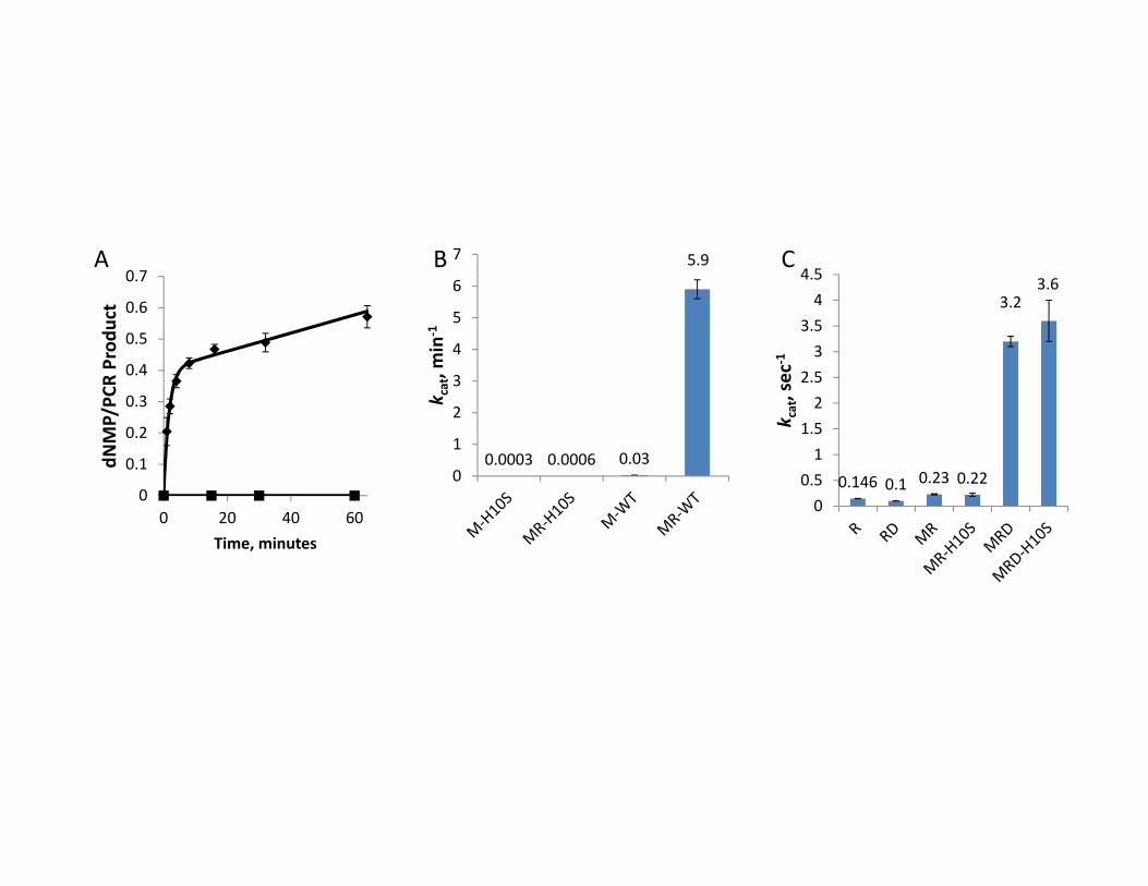
0.0003 0.0006 0.03
5.9
0
1
2
3
4
5
6
7
k cat, m
in‐1
0.146 0.1 0.23 0.22
3.23.6
00.51
1.52
2.53
3.54
4.5
k cat, sec
‐1
0
0.1
0.2
0.3
0.4
0.5
0.6
0.7
0 20 40 60
dNMP/PC
R Prod
uct
Time, minutes
A B C