copyright © 2005 pearson education, inc. publishing as benjamin cummings a tour of the cell
TRANSCRIPT

Copyright © 2005 Pearson Education, Inc. publishing as Benjamin Cummings
A Tour of the Cell

Copyright © 2005 Pearson Education, Inc. publishing as Benjamin Cummings
INTRODUCTION TO THE CELL
Using light microscopes, scientists came up with THE CELL THEORY:
1. All living things are made of cells2. All cells come from pre-existing cells3. Cells are the basic units of structure and function
in living things.

Copyright © 2005 Pearson Education, Inc. publishing as Benjamin Cummings
Sizes and shapes of cells

Human height
Length of somenerve andmuscle cells
Chicken egg
Frog egg
Most plant andanimal cells
Nucleus
Most bacteria
Mitochondrion
Mycoplasmas(smallest bacteria)
Ribosome
Viruses
Proteins
Lipids
Small molecules
Atoms
Un
aid
ed
ey
e
Lig
ht
mic
r osc
op
e
El e
ct r
on
mic
r os
co
pe
Cell sizes vary with
their function

Copyright © 2005 Pearson Education, Inc. publishing as Benjamin Cummings
Limitations to Cell Size
• Cells divide rather than grow indefinitely.
1. DNA “Overload”
• Stores information that controls the cells function.
• As a cell increases in size, it does not make extra copies of DNA.
• The larger a cell becomes, the more demands the cell places on its DNA.

Copyright © 2005 Pearson Education, Inc. publishing as Benjamin Cummings
2: Exchanging Materials
• Cell membrane regulates what enters and leaves.
• Rate of exchange depends on surface area of the cell.
• Rate at which products are used and produced depends on the cells volume.
• As the cell increases, its volume increases faster than its surface area.

Copyright © 2005 Pearson Education, Inc. publishing as Benjamin Cummings
Cell Surface Area & Volume
• Decrease in ratio of surface area to volume Makes it difficult for the cell to move needed materials in and waste products out.

Copyright © 2005 Pearson Education, Inc. publishing as Benjamin Cummings
VIRUSES
• Contain either DNA or RNA• Proteins attached to outer envelope• No nucleus• No ribosomes• No cell membrane or cell wall

Copyright © 2005 Pearson Education, Inc. publishing as Benjamin Cummings
Very small…….. (smaller than bacteria)
Size of Viruses

Copyright © 2005 Pearson Education, Inc. publishing as Benjamin Cummings
Viral Reproduction
•Reproduce by inserting genetic material into their host cell.
•Turn their host cell into a virus making factory
• Antibiotics do not work on viruses
• Vaccines can be made for viruses
• Viruses mutate easily and often

Copyright © 2005 Pearson Education, Inc. publishing as Benjamin Cummings
Assignment
• Compare and contrast a Virus, Animal Cell, Bacterial Cell, Plant Cell. (Structural features, Methods of Reproduction, Size, Examples of each)
• Based on your comparisons, would you classify viruses as living or nonliving? Explain your reasoning in paragraph form (A minimum of 5 complete sentences.)

Copyright © 2005 Pearson Education, Inc. publishing as Benjamin Cummings
Cell Types
There are two kinds of cells
– Prokaryotic (bacteria, archaea)
– Eukaryotic (protists, plants, fungi, animals)
All cells share some common features
– Plasma membrane
– DNA
– ribosomes

Nucleoidregion
Prokaryotic cell
Nucleus
Co
lor i
z ed
TE
M 1
5, 0
00
Eukaryotic cellOrganelles

Copyright © 2005 Pearson Education, Inc. publishing as Benjamin Cummings
PROKARYOTIC CELLS
Usually relatively small & simple cells
• Do not have a membrane-bound nucleus
• DNA is coiled into a nucleoid region in the cytoplasm
• Cytoplasm includes ribosomes
• Plasma membrane
• Complex cell wall
• Capsule, pili, prokaryotic flagella in some forms

Copyright © 2005 Pearson Education, Inc. publishing as Benjamin Cummings
Bacteria Shapes

Prokaryoticflagella
Ribosomes
Capsule
Cell wall
Plasmamembrane
Nucleoid region (DNA)
Pili
Prokaryotic Cell

Copyright © 2005 Pearson Education, Inc. publishing as Benjamin Cummings
EUKARYOTIC CELLS
• Partitioned into functional compartments
• Organelles are able to maintain their own environments.
• Larger than prokaryotic cells
• Distinguished by a True Nucleus

Copyright © 2005 Pearson Education, Inc. publishing as Benjamin Cummings
ANIMAL CELLS
– Are bounded by the plasma membrane alone
– Lack a cell wall
– Contain centrioles and lysosomes
– Often have flagella

Roughendoplasmicreticulum
Smoothendoplasmicreticulum
Nucleus
Flagellum
Lycosome
Centriole
Not in mostplant cells
Peroxisome
Microtubule
Intermediatefilament
Microfilament
Cytoskeleton
Golgiapparatus
Ribosomes
Plasma membrane
Mitochondrion

Copyright © 2005 Pearson Education, Inc. publishing as Benjamin Cummings
PLANT CELLS
– Are bounded by both a plasma membrane and a rigid cellulose cell wall
– Have a central vacuole and chloroplasts
– Usually lack centrioles, lysosomes, and flagella

Not inanimalcells
Golgiapparatus
Nucleus
Centralvacuole
Chloroplast
Cell wall
Mitochondrion
Peroxisome
Plasma membrane
Rough endoplasmicreticulum
Smooth endoplasmicreticulum
Ribosomes
Microtubule
Intermediatefilament
Microfilament
Cytoskeleton

Copyright © 2005 Pearson Education, Inc. publishing as Benjamin Cummings
Assignment
• Create 2 Venn diagram comparing the following cell structures:
1.Eukaryotic vs Prokaryotic Cell
2.Plant vs. Animal Cell
• Draw and label a plant and animal cell.
Copyright © 2005 Pearson Education, Inc. publishing as Benjamin Cummings
Nucleus
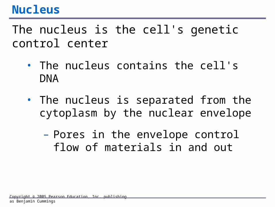
The nucleus is the cell's genetic control center
• The nucleus contains the cell's DNA
• The nucleus is separated from the cytoplasm by the nuclear envelope
– Pores in the envelope control flow of materials in and out

Chromatin
Nucleolus
Pore
Nucleus
Two membranesof nuclear envelope
Roughendoplasmicreticulum
Ribosomes

Copyright © 2005 Pearson Education, Inc. publishing as Benjamin Cummings
Endomembrane System
Overview: Many cell organelles are connected through the endomembrane system
• The endomembrane system is a collection of membranous organelles
– Divide the cell into compartments
– Work together in the synthesis, storage, and export of molecules

Copyright © 2005 Pearson Education, Inc. publishing as Benjamin Cummings
Smooth E.R.
• Lacks attached ribosomes
– Synthesizes lipids
– Processes materials such as toxins and drugs in liver cells
– Stores and releases calcium ions in muscle cells

Copyright © 2005 Pearson Education, Inc. publishing as Benjamin Cummings
Rough E.R.
• Contains ribosomes
• Manufactures membranes
• Modifies and packages proteins that will be
- Transported to other organelles
- Secreted by the cell

Smooth ER
Rough ER
Nuclearenvelope
Ribosomes
Smooth ER Rough ER
TE
M 4
5,0
00

Copyright © 2005 Pearson Education, Inc. publishing as Benjamin Cummings
Golgi Apparatus (Golgi Bodies)
• Consists of stacks of flattened membranous sacs
• Receives and modifies substances manufactured by ER
• Ships modified products to other organelles or the cell surface

Golgiapparatus
“Receiving” side ofGolgi apparatus
Transportvesiclefrom ER
New vesicleforming
“Shipping”side of Golgiapparatus Transport
vesicle fromthe Golgi
Golgi apparatus
TE
M 1
30,0
00

Copyright © 2005 Pearson Education, Inc. publishing as Benjamin Cummings
Lysosomes
• Sacs of enzymes that form from the Golgi apparatus
• Function in digestion within a cell
• Destroy bacteria that have been ingested into white blood cells
• Recycle damaged organelles

LE 4-10b
Lysosome
Nucleus
TE
M 8
,50
0

Lysosome containingtwo damaged organelles
TE
M 4
2,5
00
Mitochondrion fragment
Peroxisome fragment

Copyright © 2005 Pearson Education, Inc. publishing as Benjamin Cummings
Vacuoles
• General maintenance of the cell
• Plant cells contain a large central vacuole
– Has lysosomal and storage functions
• Some protists have contractile vacuoles
– Pump excess water out of cell

Centralvacuole
Nucleus
Chloroplast
Co
lori
zed
TE
M 8
,700

Nucleus
LM
650
Contractilevacuoles

Transport vesiclefrom ER to GolgiRough ER
Nucleus
Smooth ER Nuclear envelope Golgi apparatus
Lysosome
Vacuole
Plasmamembrane
Transport vesicle fromGolgi to plasma membrane

ChloroplastStroma
Inner and outermembranes
Granum
Intermembranespace
TE
M 9
,750
Chloroplasts convert solar energy to chemical energyAre found in plants and some protists

Copyright © 2005 Pearson Education, Inc. publishing as Benjamin Cummings
MITOCHONDRIA
• Found in nearly all eukaryotic cells
• Are the “powerhouses” of the cell
– Carry out cellular respiration
– Harvest chemical energy from food

Mitochondrion
Intermembranespace
Outermembrane
Innermembrane
Cristae
Matrix TE
M 4
4,88
0

Copyright © 2005 Pearson Education, Inc. publishing as Benjamin Cummings
THE CYTOSKELETON AND RELATED STRUCTURES
The cell's internal skeleton helps organize its structure and activities
• The cytoskeleton is network of three types of protein fibers
– Microfilaments
– Intermediate filaments
– Microtubules

Copyright © 2005 Pearson Education, Inc. publishing as Benjamin Cummings
Cilia/Flagella
• Eukaryotic cilia and flagella are locomotor appendages that protrude from certain cells
• Move when microtubules bend
• Move whole cells or materials across the cell surface


Copyright © 2005 Pearson Education, Inc. publishing as Benjamin Cummings
Bell Ringer: What regulates what enters and leaves the cell?
The Working Cell The Working Cell

Copyright © 2005 Pearson Education, Inc. publishing as Benjamin Cummings
CELL MEMBRANE
Controls what moves in and out of the cell
Semi-permeable: “picky/selective” regarding what it lets in and out.
Made of two layers of phospholipids: LIPID BILAYER
Outsideof cell

Copyright © 2005 Pearson Education, Inc. publishing as Benjamin Cummings
PHOSPHOLIPID BILAYER
Membrane phospholipids form a bilayer
• Phospholipids are the main structural components of membranes
– 2 nonpolar hydrophobic fatty acid "tails”
– 1 phosphate group attached to the hydrophilic glycerol "head“
• Proteins and other molecules are embedded in a framework of phospholipids

Copyright © 2005 Pearson Education, Inc. publishing as Benjamin Cummings
Hydrophilicheads
Hydrophobictails
Water
Water

Copyright © 2005 Pearson Education, Inc. publishing as Benjamin Cummings
Membranes are selectively permeable
• Small molecules, and hydrophobic (non-polar) molecules can easily pass through the membrane.
• Large or hydrophilic (polar) molecules cannot get through without help.

Copyright © 2005 Pearson Education, Inc. publishing as Benjamin Cummings
Extracellularmatrix
Glycoprotein
Carbohydrate
Plasmamembrane
Microfilamentsof cytoskeleton
Phospholipid
Cholesterol
Proteins
Cytoplasm
Glycolipid

Copyright © 2005 Pearson Education, Inc. publishing as Benjamin Cummings
Bell Ringer
• If I were to spray an air freshener in the front of the classroom, would someone in the back of the room eventually smell it? Explain why.

Copyright © 2005 Pearson Education, Inc. publishing as Benjamin Cummings
PASSIVE TRANSPORT
• Molecule diffuses down a concentration gradient (from high to low)
1. Diffusion is the tendency for particles to spread out evenly in an available space
– From an area of high concentration to an area of low concentration
– O2 and CO2 diffuse easily

Copyright © 2005 Pearson Education, Inc. publishing as Benjamin Cummings
EquilibriumMolecules of dye Membrane

Copyright © 2005 Pearson Education, Inc. publishing as Benjamin Cummings
Transport proteins may facilitate diffusion across membranes
2. Facilitated diffusion
– Transport proteins help substances diffuse down a concentration gradient
Solutemolecule
Transportprotein

Copyright © 2005 Pearson Education, Inc. publishing as Benjamin Cummings
3. Osmosis is the diffusion of water across a
membrane– From an area of low solute concentration– To an area of high solute concentration– Until the solution is equally concentrated
• Direction of movement is determined by the difference in total solute concentration

Copyright © 2005 Pearson Education, Inc. publishing as Benjamin Cummings
• Osmoregulation is the control of water balance.
– Isotonic solution: solute concentration is the same in the cell and in the solution.
• No osmosis occurs
• Animal cell volume remains constant; plant cell becomes flaccid (not firm)
Water Balance

Copyright © 2005 Pearson Education, Inc. publishing as Benjamin Cummings
– Hypotonic solution: solute concentration is greater in the cell than in the solution
• Cell gains water through osmosis
• Animal cell lyses; plant cell becomes swollen
– Hypertonic solution: solute concentration is lower in the cell than in the solution
• Cell loses water through osmosis
• Animal and plant cell become shriveled.

Copyright © 2005 Pearson Education, Inc. publishing as Benjamin Cummings
Isotonic solution Hypotonic solution Hypertonic solution
H2O H2O
(1) Normal (2) Lysed
H2O
H2O H2O H2O
Animalcell
Plantcell
(4) Less Firm (5) Swollen (6) Shriveled (plasmolyzed)
(3) Shriveled
Plasmamembrane
H2O
H2O

Copyright © 2005 Pearson Education, Inc. publishing as Benjamin Cummings
Active Transport
Cells expend energy for active transport
• Cells move solutes against a concentration gradient
– Transport proteins move solute molecules across the membrane
Solute binding Phosphorylation Transport Protein reversion
ATPADP
P Proteinchanges shape
PPhosphatedetaches
P

Copyright © 2005 Pearson Education, Inc. publishing as Benjamin Cummings
Exocytosis & Endocytosis
1. A vesicle may fuse with the membrane and expel its contents outside the cell (exocytosis)
2. Membranes may fold inward, enclosing material from the outside (endocytosis)