coral reefs: research methods d. r. stoddart and r. e ...concentrated [37-40 per cent) formaldehyde...
TRANSCRIPT

21
Coral Reefs: Research MethodsD. R. Stoddart and R. E. Johannes, Eds.Monographs on Oceanographic Methodology 5UNESCO 1978
Sponges in coral reefs
K. Rutzler l
INTRODUCTION
Sponges are sessile aquatic metazoans, bounded by pinacoderm and containingchoanocyte chambers. Choanocytes generate a waterflow from small ostia,through an incurrent and excurrenl aquiferous system to larger oscula. Mostsponges are massive (crust-, cushion-, fan-, tree- or cup-shape) without distinctsymmetry. The mesohyle (between pinacodcrm and choanodcrm) contains avariety of cell types, collagen and related products and, usually, an inorganicskeleton of silicon dioxide or calcium carbonate. Sexually produced larvae aremostly free-swimming and fundamental (0 lhe distribution of the adults. Allsponges are active filter feeders, and some use symbioses with bacteria andalgae to supplement their energy requirements. Many species are tolerant toepi- and endobiotic organisms (for recent summaries on sponge biology see:Fry, 1970 and Brien el a.L. 1973; for terminology see: Borojevic el al.• 1968).
Sponges are an important component of all coral reef communities. Theirbiomass and range of ecological tolerance frequently exceeds that of the reefbuilding coral species (Fig. lao Ib). They cause considerable impact on tbeirenvironment by effectively filtering large quantities ofwater (Reiswig, 1917a, b),by destroying the reef framework (Goreau and Hartman, 1963; Riitzler, 1975),by competition for space (Goreau and Hartman, 1966; Riitzler, 1970, 1975;Sara, 1970; Glynn, 1973}and by serving as food source and shelter for numerousfishes and invertebrates (Randall and Hartman, 1968; Tyler and Bohlke, 1972;Rutzler. 1976). Nevertheless, due to taxonomical problems and to difficultiesin quantitative assessment, quantitative studies of reef sponges are rare.
Although many ecological sponge studies have been made in non-reefenvironments most of the techniques for collecting, processing for systematicstudy, biomass determination and quantitative evaluation can be applied tocoral reefs.
I. Department of Invertebr'ate Zoology, National Museum of Natural History, SmithsonianInstitution, Washington. D.C. 20560. U.S.A.
299

Coral reefs: research methods
Figure ISpongt:$ in a Caribbean coral reef: a. Massive, tubular and whip-shapedspecimens at the edge of the fore-reef slopoe, Carrie Bow Cay, Belize, 2Q mdeep. A: Age/as sp.. H: Halic/O/ln ,,,hem (Pallas). M: Myca/e sp., V.c.:Verongil' rnu/i[ornris (Carter), V.g.: Vl'rO/lgin gigunlM (Hyatt). X:XeSIQS!'(lIIl/in sp.; b. Diver with large XeslOsporrgia sp., 1.4 m width of field:e, Incrusting-burrowing Arrthosginrella ~arj(ws (Duchassaing andMichelotti). 2S cm width of field.
COLLECTING, PRESERVATION AND PROCESSING FOR SYSTEMATIC
STUDY
The rollowing is a summary or procedures which can easily be rollowed by
300

Sponges in coral reefs
non·specialized field workers. The resulting data and preparations will notonly be the basis for ecological analysis but also an invaluable help for conducting and accelerating identification or systematic study (see also: Laubenfels, 1953; Hartman, 1964; Rubio, 1973).
Habitat data. Most reef surveys will be accomplished by wading, skin orscuba diving, or from submersible vessels. These techniques pennit detaileddata to be collected by direct observation. The following data should berecorded as completely as possible: date; exact locality (use reliable map andbearings); depth (from mean sea level); substratum (nature and inclination);light (estimate exposure to maximum available light in a given depth); visibility(estimate of suspended materials); exposure (to currents, wave action, and tofalling dry-intertidal); sediments (possibility of being buried); community(classify according to predominant organisms); photograph (of habitat andspecimen in situ).
Specimen data. The entire specimen should be removcd from the substratum,including basal membrane (using a sharp knife). Particularly thin incrustingand burrowing fonns should be taken with the substratum (using a rockhammer, hammer and chisel). Leave in fresh seawater until ready for fixation.Record the following data: shape (e.g. incrusting, massive; amorphous,ramose, cylindrical, tubiform, vasiform); size (surface area covered, diamehT,height); colour (use colour chart, if possible2
); consistency (e.g. hard brittle,soft elastic, compressible; note mucus production if present); surface (texture;structures like conuli, dilated subsurface channels, embedded sediments);apertures (distribution and size of expanded oscula and pori); photographs(colour, total views and close·ups of surface details-submerged in 'pan ofclean seawater, or in air after removal of excess water).
Fixation. Specimens are fixed (separately) in 10 per cent formalin-seawater(concentrated [37-40 per cent) formaldehyde solution: seawater = I :9). Toneutralize and buffer add 20 g Methenamine (C6 Hn N4 ) to each litre of finalsolution. To insure good fixation for histological purposes representative slices(about 2 cm\ including some surface area) should be cut from large specimensand fixed separately. Large specimens can be air-dricd after a small portion hasbeen fixed. For the study of sponges with well-developed spongin skeleton, it isuseful to cut a similar slice (about I cm thick) before fixation and let it maceratein fresh water (repeated rinsing) and dry. This will facilitate later observationof the skeleton architecture. Formalin-seawater is the best all-purpo$c fixativefor marine spongcs but it can be replaced by others for specific purposes. Notbefore 2 days and not after 3 weeks the specimens should be transferred into75 per cent ethyl alcohol (at least I change). Prolonged preservation of spongesin fonnalin frequently causes maceration of the tissue. Notes should be made
2. A useful and ine:\pensive colour chart is published by the Royal Horticultural Society, London.
301

Coral reefs: research methods
of any colour changes of the fixing and preservation fluids due to exudationsfrom the sponges. With each specimen a water- and alcohol-proof label mustbe enclosed, showing, specimen number (matched with data sheet); possibly adescriptive field name; locality and depth; collector and date collected ; remarks,if applicable (colour change, etc.).
Thick sections. These serve for microscopic study of the skeleton architecture(Figs. 2a, 2b), i.e. the three-dimensional structure of the spongin fibre network,the position of spicules in relation to each other, to the fibre network or toother morphological features (e.g. ectosome, choanosome differentiation). The
Figure 2Microscope preparations for systematic study: a. Spongin fibres cleanedfrom cellular tissue (maceration) of Verongiafts/u/aris (Pallas), 1.2 mmwidth of field; b, Thick ~tion (200 Jim) of Age/as coni/era (Schmidt)showing arrangement of spicules, partially embedded in spongin fibres500 11m width of field; c, Isolated spicules of Geodia neplUni (Sollas), 300 Jimwidth of field; d, Polished thin section of C/iona lampa Laubenfels showingchoanocyte chamber, safranin-crystal violet stain, 60 ",m width of field.
302

Sponges in coral reefs
following steps should be taken: transfer a representative piece of the spongeinto 96 per cent ethyl alcohol for hardening. Cut with razor blade several slicesperpendicular to the surface, as thin as possible (0.2-0.5 mm is usually possibleand sufficient). Even thicker sections are permissible (and sometimes necessary)when macerated spongin skeletons are cut. Make sure both ectosomal andchoanosomal portions are present on the section. Then make several tangentialsections from the surface down to the choanosome. In some species the ectosome detaches easily and can be peeled off with fine pointed forceps. Stain inbasic fuchsin or safranin dissolved in 96 per cent ethyl alcohol (10 seconds tose'(eral minutes). Solution can be up to saturation, depending on how readilythe sections accept the stain. Stain and subsequent dehydrating and clearingfluids can be kept in small petri dishes. Transfer of sections can be done byspatula or forceps. Dehydrate in two changes, one of 96 per cent and the otherof 100 per cent ethyl alcohol (30 seconds minimum in each). Observe understereo microscope the extent to which stain is washed out. If section is understained go back to fuchsin and extend staining time; if overstained extendwashing time in 96 per cent alcohol. Dehydratc and c1car in saturated solutionof phenol in xylene. Transfer to pure xylene and change once. Mount on slidewith Canada balsam or similar medium. Thick irregular sections do not holdthe cover glass parallel to the slide and balsam tends to run oul. This can beavoided by cutting strips of cardboard or short lengths of nylon fishing line(thickness adjusted to section) to support the cover glass. Use small leadweights to press down the cover slips during drying (in oven at 37°C). A labelshould show specimen number and locality. Near the sections (outside coverglass) the cutting direction can be marked: 1. perpendicular, ¢ ~angcntial.
Spicule mounts. Whereas thc thick sections serve mainly for higher classificationof sponges, study of the type, shape and size of isolated spicules (Fig. 2c) isimportant for species determination (except in those groups where properspicules are lacking). For temporary mounts, fragments of the sponge (fromthe ectosomal as well as the choanosomal region) are placed on a microscopeslide and a few drops of sodium hypochlorite are added. Arter disintegrationof the sort parts a cover slip is added. For permanent mounts take the followingsteps: dissolve soft parts of the sponge fragments in a test tube using coldsodium hypochlorite or boiling concentrated nitric acid (except for calcareoussponges). Wash spicules with t~p water by filling the test tube and shaking.Await settling of spicules on the bottom (at least I hour) or accelerate thisprocess by centrifuging. Decant carefully. Wash spicules twice in 96 per centalcohol (as above, settling time at least 30 minutes). Decant carefully. Shakeand pour sediment on microscope slide. Allow to dry, then add a few drops ofxylene, mounting mcdium, cover slip and label.
Histological sections. These are routinely used for the study ofchoanocytes andchoanocyte chambers, the location of very small microscleres. spongin traces,reproductive cells and, of course, all other histological observations. The
303

Coral reefs: research methods
techniques arc similar to those which can be found in any general manual ortextbook of histological and staining methods. Some special practices areworth pointingoul. Calcareoussponges which are usually packed with relativelylarge spicules can be easily decalcified in 5 per cent nitric acid with subsequentneutralization in 5 per cent sodium sulphate solution (24 hours) and rinsing intap water (24 hours). The same should be done with all sponges which have largeamounts of calcareous particles incorporated in tissue or spongin fibres. Thesubstratum of burrowing (boring) sponges must be removed in the same way:to keep the sponge from falling apart, preliminary embedding in 12 per centgelatine and decalcification in a cool place are advantageous. Gelatine embedding is useful for all sponges to be cut with a cryostat microtome. Anexcellent embedding medium for sectioning at room temperature is polyesterwax (N. W. Riser, personal communication). Mallory's triple stain is wellsuited for routine purposes and for detection of small traces of spongin.
Wet-ground and polished thick and thin sections of epoxy embeddedmaterial can be effectively used when demineralization is not desirable andserial sections arc not needed (Fig. 2d). The methods arc the same as applied topalaeontological preparations, except that tissue is dehydrated in a gradedalcohol scries and propylene oxide before embedding in epoxy resin. Toluidinblue or, fora double stain, saJranin followed by crystal violct (I percent aqueoussolution, 5-10 mins. at 600 e) give excellent staining results (sec also: Rtitzler,1974).
DETERMINATION OF RIOMASS
Several methods have been used to express the quantitative importance ofsponges within a community. Each has its merits in the context of a particularstudy and no absolute value judgments are possible. Detailed description ofthe method used is important to make the results repeatable. Sara (1966)pointed out two most desirable attributes of a quantitative method, ease ofapplication under difficult field conditions and suitability for statistical elaboration.
Number of individuals. Porifcrologists generally agree that a sponge massbounded by pinacocytes is an individual rather than a colony (Hartman andReiswig, 1973). Nevertheless, counts of specimens alone are not very usefulfrom a quantitative point of view. The size range, even of sexually matureindividuals within a given species can amount to two orders of magnitude(Figs. Ib, Ie).
Surface area. This measure has been used in various ways. laborel and Vacelet(1958) adopted and modified the Braun-Blanquet phytosociology methodwhere surface area covered by organisms is estimated in percent of the samplesquare to evaluate abundance. The values obtained were combined with an
304

Sponges in coral reefs
index of sociability which also distinguishes whether one and the same areacoverage is caused by many small or one large individual. Vasseur (1964)employed this method for different taxa, including sponges and introduceddesignations for reduced vitality and epizoism.
Russ and Ruttier (1959), Sara (1961), Sara and Siribelli (1960, 1962) andRuttier (196530 b) used specimen numbers and area of projected surfacecoverage (in em2
) to study quantitative distribution ofpredominantly encrustingMediterranean sponges. A similar method was employed by Storr (1964) forreef biota in the Bahamas; however, there is but little information on sponges.Wiedenmayer (1977) studied the quantitative distribution of massive and erectsponges near Bimini (Bahamas). He too measured projected surface area butoriented the projection plane to obtain maximum area, depending on the growthform of each species. Surface values have been obtained by Riitzler (1965a, b)by counting I cm2 meshes of a overlaying grid drawn on a plexiglas plate or,more accurately, by planimetry of paper traces from projected photographs(Riitzler,1975).
Sara (1966) introduced a frame counting method by which frequency ofoccurrence of sponges in a 5 em grid are recorded. This technique also resultsin sponge coverage values.
Volume. When one is dealing with large, irregular massive, tree-shaped, tubularand cup-shaped sponges, the projected surface area becomes an inadequatemeasure for biomass. For this reason ROtzler (1972) determined the displacement volume of sponges sampled in a reef off Nossi-Be (Madagascar). Dripdried specimens were submerged in a container filled with seawater to the levelof an overflow tube. the amount of displaced water could be determined usinga graduated cylinder. This method, however, requires collecting ofall specimensto be measured. The displacement technique can also be used to estimate theinternal space system, available to an endobiotic fauna, in relation to the spongetissue volume (Riitzler, 1976). Whole sponges were enclosed tightly in thinplastic foil and submerged to measure total volume. From this the value of thedisplacement volume without wrapping can be subtracted to give the interstitial space volume.
A convenient volumetric field method was used by Reiswig (1973, 1974) ina Jamaica reef. Specimen size was determined by measuring the three axes ofeach specimen in situ. The net volume, excluding atrial cavity, where present,was estimated by applying the values of a predetermined regression curve.
In a comparable manner Dayton et al. (1974) converted linear measurements, taken from photographs of Antarctic sponges into wet weight values.The length (or diameter) to wet weight regression had been pre-determined foreach species.
Weight and calorific content. Allhough a common practice in ecologicalstudies, weight measurements have not been much used for sponges. Theprocedures are tedious and require equipment which is not usually available in
305

Coral reefs: research methods
the field. As with displacement volume determinations, specimens have to beremoved from the habitat. The method, however, if properly used, gives themost accurate measure of biomass.
Russ and Runler (1959) supplemented area coverage with wet-weight databut did not compensate for the varying amount of skeleton material (spicules)in different species. Rutzler (1975) determined wet- and dry-weight for 8species of burrowing sponges and related these data to the size of their papillarfields, a measure that can be readily determined in situ. In this example theweight data were important to compensate for specific differences in penetrationdepth and burrowing patterns. The spicule to soft tissue ratio was fairlyconstant among all species and could be neglected. Dry-weights proved to beslightly more repeatable than wet-weights.
If accurate biomass comparison of species with very different skeletoncontents is required, ash·free weights, or another measure that excludes hardtissue, e.g. organic carbon content, must be determined. Calculalion of energybudgets may require measurement of calorific content of tissues.
Randall and Hartman (1968) presented data on the relalion of ash contentto organic matter in 18 species of sponges which arc frequently eaten by WestIndian fishes. The content ranges from 0.5 per cent in ChOlldros;o to as muchas 70 per cent in Anthosigmella.
For comparative study of metabolism in 3 species ofJamaican reef spongesReiswig (1973, 1974) determined weight and calorific content of tissues. Dryweights were obtained by drying to constant weight at IOSoC, ash weights bycombustion at 5OOo_S1 O°C for 2 hours. Corrections were applied for water lossfrom siliceous spicules (Paine, 1964). The results (Reiswig, 1973) showedrelatively low ash values of 31 per cent for Myrole (poecilosclerida) and 34 percent for Verongia (Dictyoceratida) as opposed to 62 per cent for Tethya(Hadromerida). Calorific values per ml sponge, obtained by microbombcalorimetry were high in Verongia (407 cal/ml), only about halfof that amountin Mycale (200 cal/rol) and Tethya (214 cal/ml). In Mycale over 40 per cent ofthe calories are contajned in skeletal spongin.
Similar parameters (dry weight, ash-free dry weight. calorific content oftissue) were measured by Dayton et al. (1974) to quantify the standing crop ofan Antarctic sponge community.
Oxidizable carbon is considered a 'realistic measure of the energy storedin a crop' (Strickland and Parsons, 1968). In developing a preliminary modelof a coral reef ecosystem, the participants in a recent workshop meeting onGlover's Reef (Belize, Central America: 1971) have agreed to use carbon asthe universal measure for biomass and material flow (Macintyre el al.. 1974).The meeting also stimulated preliminary determinations of wet-weight- dryweight: carbon ratios in Caribbean reef sponges (Riitzler, unpublished).Specimens of ten species of sponges belonging to six orders were collected inBermuda. Two samples of each species were cleaned from sediments andsymbionts under a stereo microscope. Wet-weight was obtained afier drainingoff interstitial water and quick superficial blotting. Dry-weight was determined
306
Sponges in coral reefs
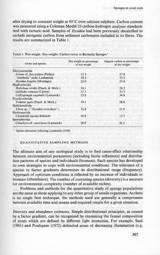
after drying to constant weight at 95°C over calcium sulphate. Carbon contentwas measured using a Coleman Model 33 carbon.hydrogen analyser standardized with tartaric acid. Samples of Dy"sidea had been previously decalcified toexclude inorganic carbon from sediment carbonates included in its fibres" Theresults are summarized in Table I.
TABLE 1. Wet-weight: Dry-weight: Carbon ratios in Bermuda Sponges'
Order and $p«ie~Dry_weigbt as percentage
of wet·weightOrpnle c:arb<m as percentage
of dry.we;gbt
Dictyoceratida/rcinia cf.jascicu/a/a (Pallas)""lanrhclla"' ardis LaubenfelsDysidcafragi/is (Montagu)
HaploscleridaHaliclona viridis (Ouch. & Mich.)Gel/iodes ram()$a (Carter)Callyspong;a vagina/is (Lamarck)
PoeciloscleridaTedania ;gnis (Ouch. & Mich.)
HalichondridaU/OS(1 sp. (""Dysidca crawshayi"")
HadromcridaChondrilla nucula Schmidt
SpirophoridaCinachyra cr. caocrnosa (Lamarck)
'. Species .110<:8t;on follow;ng LaubenfelS (195(1).
13.3 27.819.3 33.522.0 23.6
24.1 24.217.1 33.313.8 34.0
19.1 28.0
32.4 21.9
16.9 13.7
26.9 26.1
QUANTITATIVE SAMPLING METHODS
The ultimate aim of any ecological study is to find cause-effect relationshipbetween environmental parameters (including biotic influences) and distribution patterns of species and individuals (biomass). Each species has developedits own strategies to cope with environmental conditions. The tolerance of aspecies to factor gradients determines its distributional range (frequency).Approach of optimum conditions is reflected by an increase of individuals orbiomass (abundance). The number ofcoexisting species (diversity) is a measurefor environmental complexity (number of available niches).
Problems and methods for the quantitative study of sponge 'populationsare the same as those applying to any other group of sessile organisms. As thereis no single best technique, the methods used are generally a compromisebetween available time and means and required results for a given situation.
Diversity and abundance estimates. Simple distributional principles, as causedby a factor gradient, can be recognized by examining the faunal compositionof zones which are defined by different light intensities. For example, Sara(1961) and Pouliquen (1972) delimited areas of decreasing illumination (e.g.
307

Coral reefs: rescal"(;h methods
shaded, obscure and dark) in Mediterranean caves. They listed the spongespecies in each zone and determined their relative abundance (e.g. present, rare,abundant, very abundant). AJthough these data are not suitable for statisticalelaborations, they do represent a measure of diversity and permit comparisonswith other habitats.
In a similar manner, without having a uniform sample size for statisticaltreatment, Rutzler (I965b) determined the influence of substrata stability onthe diversity and growth of a sponge fauna inhabiting the lower surfaces ofloose rocks.
The majority ofquantitative sponge studies, however, arc based on quadratsampling.
Quadrat size und sampling area. Quadrat size and sampling area are usually notidentical. Choice of suitable quadrat size depends on the method of datacollecting, the complexity of the environment and the size range and densityof the organisms to be studied. If the entire sample is to be removed and broughtto the laboratory, if the factor gradients are steep (complex substrata configura.tion, heterogeneous microclimate), and if the organisms are comparatively smalland densely spaced, squares of f6 m2 (25 x 25 em) can be used (R uss andRuuler, 1959). For visual census and coarse area coverage estimates involvingvery large sponges in a homogeneous environment, quadrats of up to 100 m2
(10 X 10 m) have been considered necessary (Wiedenmayer, 1971). The bestsuitable sample size has to be found empirically for each application. If thequadrat is too small, distortion in statistical treatment can occur (Ryland,1973). If it is too large, environmental changes cannot be resolved.
For statistical reasons all sampling units should be of equal size. Forapplications where this is not possible, a method of conversion based oncomputer simulation has been introduced (Ryland, 1972). The sum of allsamples within a stratum (community) represents the sampling area. Itsminimum size can be determined by the species·area curve (Cain, 1938; Riedl,1953). Another, more recently introduced, method is based on binomialsampling theory (Dennison and Hay, 1967). The minimum proportionalpresence of a species within the study area must be found empirically. From agraph one can then determine the sampling area required for a desired proba·bility value for collecting efficiency.
Quadrat sampling and evaluation. The phytosociological quadrat method hasbeen applied by Laborel and Vacelet (1958) and Vasseur (1964) for the studyof sessile populations (including sponges) in a Mediterranean cave and inIndian Ocean reef habitats. Two coefficients, quantifying abundance andsociability, were estimated in situ for a number of species. The data are notstatistically usable but serve for recognition and comparison of characteristicplant and animal assemblages and their dependence upon such factors as light,hydrodynamics and silting.
The influence of intertidal exposure, light, water movement and sedimen·
308

Sponges ill coral reefs
tation on sponge distribution was determined by Russ and Rutzler (1959) andRiHzler (I 965a, 1972) for Mediterranean caves and for an Indian Oceanfringing reef. Transects were selected which represented profiles through factorgradients (e.g. in a cave, from light to dark, from ceiling to bottom). Along.these transects, quadrat samples (-hm2 ori m2) were marked in fixed dislances.Most samples were removed entirely (including burrowed portions of thesubstratum) and taken to the laboratory for species and biomass determinations. Some quantitative estimates were made in situ and from photographs.Where transects were meaningless, random samples had to be chosen withinareas of apparently uniform environmental conditions. Sample data were thenarranged in groups corresponding to different intensities of a particular parameter. Care was taken to eliminate counteracting factors (e.g. samples representing a light gradient were chosen from vertical substrata with averagehydrodynamic exposure, to avoid influences from sediments and excess watermovement). Indices of homogeneity (Riedl, 1953) were calculated to determinefaunal similarities between samples assigned to one microclimatic regime, andto compare the community structure of different environments.
Although this method is objective and suitable for statistical treatment,it is tedious and not suitable for large scale application.
With these difficulties in mind Sara (1966, 1970) introduced a quick quadratcounting method for the quantitative study of Mediterranean shallow-watersponges. A 25 em x 25 em frame was subdivided with nylon filament to forma grid of25 meshes (5 em x 5 em). The frame was placed on the substratum andthe number of meshes occupied by each species was counted. The frequency ofcounts is correlated with the surface extension of the individuals. The effect ofthis method is similar to chain link counts which have been used for coraldiversity studies (Porter, 1972). Ruttier (1975) adopted the frame countingmethod for large scale distributional studies of burrowing sponges in Bermudareefs. It was modified to express estimated coverage values (in cm2) inside themeshes and calibrated for the surface area-biomass ratio of each species.
SUMMARY AND CONCLUSIONS
Synopsis. Compared with other important reef organisms, sponges have beengreatly neglected in quantitative studies. The principal reasons for this aretaxonomic problems and difficulties in quantification, due to great variabilityin shape and size.
Identification of sponge taxa is much facilitated if guidelines for properpreservation, data collecting and micro·anatomical preparation are followed.
Specimen counts have limited quantitative application. Biomass estimatesin the field can be based on projected surface area or on measurement of thethree main axes of a specimen. It is necessary, however, to correct the data forvariations in skeleton contents, or tissue porosity, by submitting subsamplcs of
309

Coral reefs: research methodoi
each species to laboratory analysis. In this manner, quick and reliable estimatesof weight, volume or energy content can be obtained, without need to removethe entire sample.
Quadrat sampling along transects and, at random, with defined zones hasbeen used for quantitative sponge studies in rocky sublittoral and coral reefareas. Quadrat size and sample area vary with size classes and distributionalcharacteristics of the fauna. Phytosociological techniques arc best suited touncover characteristic organism assemblages and interactions. Other, morcobjective methods, involving counts of species presences, or biomass measurements, can be evaluated statistically and have been applied to reveal relationships between factor complexes and faunal distribution patterns.
Methods adopted for I.M.S. WE. Reef Projecl. The current Investigations ofMarine Shallow Water Ecosystems (I.M.S.W.E., Smithsonian lnstitution)Coral Reef Project is conducting quantitative studies on reef biota off CarrieBow Cay, Belize.
The zoning of the reef complex has been mapped, using a permanent maintransect. This transect starts in the Thalassia zone of the lagoon and extends,perpendicular to the direction of the barrier reef to the 45 m depth mark onthe fore-reef slope. Habitats, characterized by dominant organisms (e.g.Millepora zone, Tllrbinaria zone) or geological features (e.g. rubble and pavement zone) have been and are still being mapped and their position fixed inrelation to the main transect.
Principal organism assemblages in each habitat are quantified usingauxiliary traDsect counts (crossing habitat boundaries) and random quadratcounts (within habitats). (Figs. 3c, 3d). Chain transects (mainly in coral dominated habitats) are compared with quadrat transects (quadrat positioned every2 m on the transect). Quadrats vary in size from 0.5 m x 0.5 m to 2 m x 2 m,depending on the size classes of organisms. Area coverage of each species(dimension of main axes, for large sponges) is determined in situ (Figs. 3a, 3b),subsamples are taken to the laboratory for biomass assessment (wet weight,displacement volume). Substratum complexity is taken into consideration andsample size is corrected for functional surface area (Dahl, 1973).
Each habitat is subdivided into strata, which are defined on the basis ofmicroclimatic conditions and substrata properties. For example, colonized(unburied) lower surfaces of rocks, seagrass rhizomes or leaves, interstitialspaces or burrows in dead coral, sand or rubble are each considered a stratumand sampled separately, after its proportional extension within the habitat hadbeen Doted. Volume samples replace surface area counts, where appropriate(e.g. rubble, sand). Interstitial organisms are extracted after anaesthetizingwith magnesium chloride or by oxygen depletion (Kirsteuer, 1967).
Environmental para~eters applying to each stratum arc measured orestimated at the time of sampling. Whenever possible, these factors are monitored or checked over long periods of time to learn about intensity means andeXlremes.
310

Sponges in coral reefs
Figure 3Quantitative methods applied during coral reef study at Carrie Bow Cay,Belize: a, Diver holding 25 x 25 em frame to record height and width oflarge tubular VerOl1gill sp.: b, Frame on top of tube duster of Age/as sp,:c, Frame overlaying Agaricia-Poriles patch (spur and groove system) forpoint counting at the intcrs<xtions of the filament grid; d, Situation like in c.but frame placed on the pavement with some Agaricia, gorgonians andgreen algae.
311

Coral reefs: researeh methods
A suitable method of multivariate analysis will be applied (R. G. Dorney,peTS. carnm.), as soon as sufficient amount of data has been processed.
ACKNOWLEDGEMENT"
This work was supported by the Smithsonian Research Foundation, by IheSmithsonian Environmenlal Programme and by a grant from the ExxonCorporation. It is Contribution No. 21, Investigations of Marine SballowWaterEcosystems Projcct, Smithsonian InSlitution.
ReferencesBo..OJEVlC, R.; FKY. W. G.; JONES, W. C; Ltvl, c.; RAswo",.. R.; SAJl.-\, M.; VACELE'T, J. 1968.
Mise au point ..etuelle de la tenn.inologie des esponges (A Reasst5Smcnt of the TermilKllogy forSponges]. Bull. Mru. NaIl. Hut. Nat. vol. 39, p. 1224-35.
BRIEN, P.; LtVl, C.; SARA. M.; TuZfl. 0.; VAC£LE'T. J. 1973. Spongiaires. In: P.-B. Grasse (cd.).TraUbit' Zooi09~: vol. 3 (I), Masson, Paris, (716 pl.
CAIN, S. A. 1938. The species area curve. Am. Midi. Nat., vol. 19, p. 573-81.DAHL., A. L. 1973. Surface arca in ecological analysis: Quantification of benthic coral·reef algae.
Mur. Bial.. vol. 23, p. 239--49.DAYTON, P. K.; ROllILUARD, G. A.; PAIN!!, R. T.; DAYTON, L. 8.,1974. 8iologic:alaccommodation
in the benthic community at McMurdo Sound, Antarctica. Etaf. Monogr., vol. 44, p. 105-28.DENNISON, J. M.; HAY, W. W. 1967. Estimating the needed sampling area for subaquatic l:tologic
studies. J. Pa/eontol.. vol. oil, p. 706-8.FRY. w. G. (cd.). 1970. Thebiolo!/)' ofllie Poriferu, Symp. zool. Soc. London, vol. 25, London
and New York, Academic Press. (512 p.).vLYNN, P. W. 1973. Aspects 01" the ecology of coral reefs in the western Atlantic region. In: O. A.
Jones and R, Endean (eds). Biology and geolagy of toral reefs, vol. I: Biology. London andNew York. Academic Press, p. 271-324.
QoREAU, T. F.; HAJtTMAN, W. D. 1963. Boring sponges as controlling factors in the formation andmaintenance of coral reefs. In: R. F. Sognnaes (cd.). Mechtlllums of hard tWIN thslfUetion.Am. Assoc. Adv. Sci.. Publ. 75, p. 25-54.
----. 1966. Sponge: Effect on Ihe form of rec:fcorals. Scimce. vol. lSI, p. 343-44.HAaTltAl'l, w. D. 1964. Phylum Porifent. In: R. I. Smith (cd.). K~ysto marine fnPerlebrOlts of the
Woods Hole regio'l. Systematics-Ecology Program, Marine BioloJic:al Laboratory, WoodsHole:, Massachll5ClU. p. 1-7.
--; REIsWIG, H. M. 1973. The indlvldualily ofspongcs. In: R. S. Boardman. A. H. Cheethamand W. A. Oliver, Jr. (cds). Animal ta/all~s, lkrdopmml andfunctiQfl thrawgh tim~. Stroudsburg, Pennsylvania, Dowden, Hutchinson aDd Ross, p. 567-84.
KIRSTEUER, E. 1967. Marine benthonic nemerteans: How to COlltICl and preserve them. Am.Museum NavilaltS, 2290, p. 1-10.
LABORE!., J.; VAC'ELE'T, J. 1958. ~tude des pcuplements d'une grolle sous·marine du golfe deMarseille. Bull. 1r1S1. Oewn" vol. 1120, p. 1-20. .
LAVBEN~liLS, M. W. DIl. 1950. The Porifera of the Bermuda Arehipelago. Trans. zoo/. SOt. Lend.vol. 27, p. 1-154.
--. 1953. A guide to the sponges of Eastern North Americ:a. The Marine Laboratory. Universityof Miami, Spedal Public:ation. University of Miami Press (32 p.).
MACINTYRE,I. G.; SMITH, S. V.; ZIEMAN, Jr. J. C, 1974. Carbon nux through acoral·reefercoSY1tem:A conceptual model, J. Gtol.. vol. 82, p. 161-71.
PAINE, R. T. 1964. Ash and c:aloric determinations of sponge and opisllwbranch tissues. &0109)'.vol. 45, p. 384-87,
Po.TU, J. W. 19n. &ology ..nd species diversity of coral reefs 00 opposite sides of the isthmus ofPaDama. In: M. L. Jones (cd.). The Panamit:: biota: Some observations prior to a SClI·leveicanal. Bull, bioi. Soc. Wash .. vol. 2, p. 89-116.
312

Sponges in coral reefs
Poul..lQUeS. L. 1972. Les spongiaires des grotles $Ous·marines de la region de Marseilk: &ologieet systematique. Titlrys, \'ol 3, p. 717-58.
RA....DALI.. J. E.; HARTMAN, W. D. 1968. Sponge·feeding fishes of the West Indies. Mar. Bioi.. vol.3, p. 216-2.5.
REISWIG, H. M. 1971a. In situ pumping activity of tropical Demospongiae, Mar. Bioi.. vol. 38-.50.--. 1971b. Particle feeding in natural population of three marine demosponges. Bioi. Bull. Mar.
8iol. Lab. Woods Hole. Mass.. vol. 141, p. 568-91.--. 1973. Population dynamics of lhree Jamaican Dcmospongiae. Bull. mar. Sci., vol. 23. p.
191-226.--. 1974. Water transport, respiralion and energetics of three tropical marine sponges. J. txp.
mar. Biol. £COl.. vol. 14, p. 231-49.RIEOL, R. 1953. Quantitativ okologische Methoden mariner Turbcllarienforschung. (Jslt". Zool.
Zeitschr., vol. 4. p. 108-4.5.RUBJ6, M. 1973. Recolea::i6n y primera descripci6n de esponjas: fijaci6n, eonscrvaci6n y prepara·
cion. 11Int. y Cim6D. vol. 3, p. 37-48.Russ. K.; RiJTz.lla, K. 1959. Zur Kenntnis der SCh.....mm(auna unteneeiscber Hohlen. PubbI.
SUI:. Zoo/. Napoli. \'01. 30 (Suppl.), p. 756-87.ROTz1.u., K. 196.5a. Systematik und Okologie der PorifereD aus Litoral·Schattengebieten der
Nordadria. Z. Morph. (Jkol. Tiere. vol. 55, p. 1-82.--. 19Mb. Subslratstabilil.at im marinen Benthos als okologischer Faktor, dargestellt am Beispiel
adriatischer Porifera. Int. Rell. gesaml. Hydrobiol., vol. SO, p. 281-92.--. 1970. Spatial competition among Porifera: Solution by epizoism. Ol!COlogia (Bcrl.), vol. 5.
p.85-9.5.--. 1972. Principles of sponge distribution in Indo·Pacific coral reefs. In: PrO(;. Symp. Corals and
Coral Rf!tjs.I969. Mar. BioI. AS$OC.lndia, p. 31's-32.--. 1974. The burrowing sponges of Bermuda. Smithsonian Conlr. Zoo/., vol. 165, p. 1-32.--. 197.5. The role of burrowing sponges in bioerosion. Oeoo1O!J1a (Berl.). vol. 19, p. 203-16.--. 1976. Ecology of Tunisian eommemalsponges. Tbhys, vol. 7, p. 249-64.RYlAND, J. S. 1972. The analysis of pattern in communities of bryowa. I. Discrete Ampling
methods. J. up. nwu. Bioi. £col. vol. 8. p. 2n-97.--. 1973. The analysis ofspatial distribution patterns. In: G. P. Larwood (ed.). Li~i"f1 rmdjossil
ll,,·ozoa: RUe,.1 arkan«J in research. London and New York. Academic Press, p. 16S-72.SARA, M. 1961. La fauna di Poriferi delle groue delle i$Ole Tremili. Studio ecologico e sistematico.
Auh. 2001. Ilal.. vol. 46, p. I-59.--. 1966. Studio quantitativo della distribu~ione dei Poriferi in ambienti superficiali della
Riviera Ligure di Levantf';. Arch. Oc~a"ogr. Limnol.. vol. 14, p. 36.5-86.--. 1970. Competition and co-operation in sponge populations. In: W. G. Fry (cd.). The biology
o/the Porifera, Symp. ~ool. Soc. London, vol. 25, p. 273-84. London and New York, AcademicPress.
--; SlJ./BELU, L. 1960. La fauna di Poriferi delle "scx:clJe" del Golfo di Napoli. I. La $COCa dellaGaiola. Anmr. Isf. Mus. 2001. UnifJ. Napoli. vol. 12. p. 1-93.
--; StJ./BEU.t, L. 1962. La fauna di Porum delle "secche" del Golfo di Napoli. II. La seee:a diBenda Pa/umno. AI\IIII./JI. Mus. Zool. Unil1. N"poIi, vol. 14, p. 1-62.
S~ J. F. 1964. Ecology aDd oceanography of the coral·red" tract, Abaro Island, Bahamas.Geo1. Soc. Am. Spec. Paper. 79. p. 1-98.
SWCJ:.LANO, J. D. H.; PAJ15Ol'.'$, T. R. 1968. A practical handbook of seawater ana/'J5is. Bull. FisJI.Res. Bd Can .. Ottawa, Qucc:n's Printer, no. 167 (311 po).
TYLER, J. C.: BOIILKE, J. E. 1972. Records of sponge-dwelling fishes, primarily of the Caribbean.Bull. mar. Set.. vol. 22, p. 601-42.
VASSEUR, P. 1964. Contribution ll. J'etude bionomique des pcuplcmenu 5Ciaphiles infraliuoraux desubstrat dur dans les r~ifs de Tultar (Madagascar). Ree. Trav. Sin mar. £lIdaume, Supp.HoTS Sl!rie 2, p. 1-77.
WIEDENMAYEll., F. 1977. A monograph of the shallow_water sponges of tbe Western Bahamas.Bascl, Birkhil,lSCf. (in PI"C55).
313