coronary arterial bk channel dysfunction exacerbates mice · coronary arterial bk channel...
TRANSCRIPT

Draft
Coronary Arterial BK Channel Dysfunction Exacerbates
Ischemia Reperfusion-Induced Myocardial Injury in Diabetic Mice
Journal: Applied Physiology, Nutrition, and Metabolism
Manuscript ID apnm-2016-0048.R2
Manuscript Type: Article
Date Submitted by the Author: 10-May-2016
Complete List of Authors: Lu, Tong; Mayo Clinic Minnesota, Cardiovascular Diseases
Jiang, Bin; the First Affiliated Hospital of Soochow University, Cardiology; Mayo Clinic Minnesota, Cardiovascular Diseases Wang, Xiao-Li; Mayo Clinic Minnesota, Cardiovascular Diseases Lee, Hon-Chi; Mayo Clinic Minnesota, Cardiovascular Diseases
Keyword: BK channel, angiotensin II type 1 receptor, vascular smooth muscle cell, myocardial ischemia-reperfusion injury, diabetes
https://mc06.manuscriptcentral.com/apnm-pubs
Applied Physiology, Nutrition, and Metabolism
Draft
1
Coronary Arterial BK Channel Dysfunction Exacerbates Ischemia Reperfusion-Induced
Myocardial Injury in Diabetic Mice
Tong Lu, Bin Jiang, Xiao-Li Wang, Hon-Chi Lee
Corresponding Authors: Tong Lu, M.D., Ph.D., Department of Cardiovascular Diseases, Mayo
Clinic, 200 First Street SW, Rochester, MN 55905, USA. Tel: 507-255-9653; Fax: 507-538-
6418; E-mail: [email protected]
Bin Jiang, MD., PhD., Department of Cardiovascular Diseases, Mayo Clinic, 200 First Street
SW, Rochester, MN 55905, USA. Department of Cardiology, the First Affiliated Hospital of
Soochow University, 96 Shixin Street, Soochow, Jiangsu 215006, P. R. China. Email:
Xiao-Li Wang, MD., PhD., Department of Cardiovascular Diseases, Mayo Clinic, 200 First
Street SW, Rochester, MN 55905, USA. Email: [email protected]
Hon-Chi Lee, MD., PhD., Department of Cardiovascular Diseases, Mayo Clinic, 200 First Street
SW, Rochester, MN 55905, USA. Email: [email protected]
Page 1 of 34
https://mc06.manuscriptcentral.com/apnm-pubs
Applied Physiology, Nutrition, and Metabolism

Draft
2
Abstract
The large conductance Ca2+
-activated K+ (BK) channels, abundantly expressed in coronary
artery smooth muscle cells (SMCs), play a pivotal role in regulating coronary circulation. A large
body of evidence indicates that coronary arterial BK channel function is diminished in both type
1 and type 2 diabetes. However, the consequence of coronary BK channel dysfunction in
diabetes is not clear. We hypothesized that impaired coronary BK channel function exacerbates
myocardial ischemia reperfusion (I/R) injury in streptozotocin (STZ)-induced diabetic mice.
Combining patch-clamp techniques and cellular biological approaches, we found that diabetes
facilitated the colocalization of Angiotensin II (Ang II) type 1 receptors (AT1R) and BK channel
α-subunits (BK-α), but not BK channel β1-subunits (BK-β1), in the caveolae of coronary SMCs.
Such caveolae compartmentalization in vascular SMCs not only enhanced Ang II-mediated
inhibition in BK-α, but also produced a physical disassociation between BK-α and BK-β1,
leading to increased infarct size in diabetic hearts. Most importantly, genetic ablation of caveolae
integrity or pharmacological activation of coronary BK channels protected the cardiac function
of diabetic mice from experimental I/R injury in both in vivo and ex vivo preparations. Our
results demonstrate a vascular ionic mechanism underlying the poor outcome of myocardial
injury in diabetes. Hence, activation of coronary BK channels may serve as a therapeutic target
for diabetic cardiovascular complications.
Key words: BK channel, angiotensin II type 1 receptor, vascular smooth muscle cell, myocardial
ischemia-reperfusion injury, diabetes.
Page 2 of 34
https://mc06.manuscriptcentral.com/apnm-pubs
Applied Physiology, Nutrition, and Metabolism

Draft
3
Introduction
Diabetes mellitus has become an epidemic worldwide. The global prevalence of diabetes is
estimated to be 9% among adults according to the World Health Organization global status
report. Diabetes is associated with a 2- to 4-fold increase in the risks of cardiovascular diseases
which remain the leading cause of death in both men and women with diabetes (Snell-Bergeon &
Wadwa, 2012). A large body of clinical evidence has shown that the poor prognosis of acute
myocardial infarction and cardiac sudden death in patients diagnosed with diabetes is attributed
to compromised coronary blood flow and increased susceptibility of myocardial ischemia
(Kurisu et al., 2003; Cubbon et al., 2007; Yeung et al., 2012). Despite advances in the
management of diabetes, the cardiovascular morbidity and mortality in diabetic patients remain
twice that of non-diabetic patients (Wackers, 2005; Norhammar et al., 2007). However, the
molecular mechanisms that underlie the poor cardiovascular outcome in diabetic patients are not
fully understood.
Because of high unitary conductance and high density in coronary smooth muscle cells
(SMCs), the large conductance Ca2+
-activated K+ (BK) channels play a negative feedback role in
the regulation of vascular tone and cardiac perfusion. Activation of vascular BK channels by
elevated intracellular free Ca2+
concentrations gives rise to the generation of spontaneous
transient outward currents that hyperpolarize the membrane potentials, shut off the voltage-
dependent Ca2+
channels in vascular SMCs and lead to vascular relaxation (Nelson et al., 1995;
Jaggar et al., 1998). Functional vascular BK channels are octameric complexes with four pore-
forming α subunits (BK-α, encoded by Slo1 gene) and four regulatory β1 or γ1 subunits (BK-β1
or BK-γ1) (Tanaka et al., 1997; Evanson et al., 2014). In diabetes, however, the BK channel
Page 3 of 34
https://mc06.manuscriptcentral.com/apnm-pubs
Applied Physiology, Nutrition, and Metabolism

Draft
4
function is diminished in many vascular beds including those of coronary arteries (Lu et al.,
2010; Zhang et al., 2010; Wang et al., 2012; Yi et al., 2014). Whether coronary BK channel
dysfunction contributes to worsened outcome of cardiovascular diseases with diabetes has not
been studied.
Caveolae are flask-shaped nonclathrin-coated plasma membrane structures that contain the
signature protein, caveolin (Cav). Three Cav isoforms are known: Cav-1, Cav-2 and Cav-3. Cav-
1 and Cav-2 are present in vascular endothelial cells and SMCs, while Cav-3 is primarily
expressed in cardiac and skeletal muscles (Krajewska & Maslowska, 2004). The N-terminus of
Cav-1 contains an important functional structure: the Cav scaffolding domain, which is essential
for interaction with other proteins containing the Cav binding motifs (ΦXΦXXXXΦ or
ΦXXXXΦXXΦ, where Φ represents an aromatic amino acid and X is any amino acid)
(Krajewska & Maslowska, 2004). Such Cav binding motifs are present in human BK-α (1102-
YNMLCFGIY-1110) and human AT1R (304-FLGKKFKRY-312), but not in human BK-β1 or
BK-γ1. Caveolae are membrane microdomains that assemble signaling molecules including
those of Ang II (Ushio-Fukai & Alexander, 2006). Two types of Ang II receptors are expressed
in the heart: type 1 (AT1R) and type 2 (AT2R) with AT1R being the dominant isoform that
mediates the major cardiovascular effects of Ang II (Dasgupta & Zhang, 2011). We have
demonstrated that Ang II promoted in AT1R trafficking into the caveolae of vascular SMCs,
forming the BK channel-AT1R-caveolae microdomain complexes. Importantly, such caveolae
compartmentation was enriched in diabetic vessels, potentiating the inhibitory effect of Ang II on
BK channels in the vicinity and promoting coronary vasoconstriction (Lu et al., 2010). In
addition, it has been reported that caveolae facilitated direct coupling between BK channels and
Page 4 of 34
https://mc06.manuscriptcentral.com/apnm-pubs
Applied Physiology, Nutrition, and Metabolism

Draft
5
L-type Ca2+
channels in vascular SMCs to regulate vascular tone (Suzuki et al., 2013). However,
the role of BK channel-AT1R-caveolae microdomain complexes in cardiovascular diseases,
particularly with diabetes, has not been established. In this study, we found that dysregulation of
coronary smooth muscle BK channel activity contributes to exacerbated ischemia/reperfusion
(I/R) injury in diabetic mouse hearts, due to an increase of BK chanel-AT1R-caveolae
microdomain compartmentation in coronary SMCs. Activation of coronary BK channels and
suppression of AT1R-caveolae signaling minimized myocardial I/R injury in diabetic mice.
Hence, protection of coronary BK channel function may serve as a novel molecular target for the
treatment of cardiovascular complications in diabetes.
Materials and methods
Diabetic mouse model Male Cav-1 null mice (Cav-1-/-
, Cav1tm1M1s
/J) and wild type (wt)
control mice (C57BL/6J) were purchased from the Jackson Laboratory at 4 weeks of age. Mice
received a STZ injection (100 mg/kg body weight, ip.) and those with blood glucose higher than
300 mg/dl were considered diabetic. Eight weeks after developing hyperglycemia, mice and age-
matched controls were sacrificed and used for experiments. All animal protocols were approved
by the Mayo Clinic Institute Animal Care and Use Committee (Rochester, MN).
Coronary SMC isolation and BK channel current recording Mouse coronary SMCs were
enzymatically isolated for whole-cell K+
current recordings as we have previously described (Lu
et al., 2010; Lu et al., 2012). The pipette solution contained (in mM): KCl 140, MgCl2 0.5,
Na2ATP 5.0, Na2GTP 0.5, HEPES 10.0, EGTA 1.0, CaCl2 0.465 (~0.2 µM free Ca2+
) and pH at
7.38. The bath solution contained (in mM): NaCl 145, KCl 5.6, MgCl2 1.0, CaCl2 1.0, HEPES
10.0, glucose 5.0 and pH at 7.40. BK currents were defined by its sensitivity to 0.1 µM
Page 5 of 34
https://mc06.manuscriptcentral.com/apnm-pubs
Applied Physiology, Nutrition, and Metabolism

Draft
6
iberiotoxin (IBTX, a membrane impermeable BK channel-specific blocker) and were obtained
by subtraction of the IBTX-insensitive component from the total K+ currents. All patch clamp
experiments were conducted at room temperature (22 °C).
Sucrose gradient density centrifugation The cellular distributions of caveolae-targeted BK
channels and AT1R in mouse aortic SMCs were determined by sucrose density gradient
fractionation as previously described (Wang et al., 2005; Lu et al., 2010). Cells were
homogenized in 500 mM Na2CO3 with 2% protease inhibitors (v/v), and then centrifuged at
5,000 rpm at 4 °C for 10 min. One ml of the supernatant were adjusted to 40% sucrose-MBS (2-
[N-morpholino]-ethanesµfonic acid) by adding 1 ml of 80% sucrose-MBS, placed to the bottom
of a 6-ml ultracentrifuge tube, layered with a 2-ml discontinuous sucrose gradient (40%, 30%
and 5%), and centrifuged at 32,000 rpm at 4 °C for 20 h. Eight fractions of 1.25 ml each were
collected and analyzed by immunoblotting.
Co-immunoprecipitation (Co-IP) and immunoblotting Co-IP and immunoblotting were
performed as we have previously reported (Lu et al., 2010; Lu et al., 2012). Mouse aortas were
incubated with 200 µl RIPA buffer (in mM): Tris 50, NaCl 150, NaF2 1, EDTA 1, EGTA 1,
NaVO4 1, and 1% Triton X-100 (pH 7.5) and 1 µl protease inhibitor on ice for 30 min,
homogenized and then centrifuged at 2000 rpm at 4 °C for 10 min. The supernatant (200 µg
protein in 200 µl) was incubated with 4 µg anti-Cav-1 antibody (Santa Cruz Biotechnology, Inc.,
catalog number: sc-7875) at 4 °C overnight. Incubation with same amount of IgG severed as a
negative control. The samples were then incubated with 20 µl Protein G Plus-Agarose (Santa
Cruz Biotechnology, Inc.) at 4 °C for 2 h. After centrifugation at 1000 rpm for 7 min and washed
twice with RIPA/protease inhibitor buffer, the immunoprecipitates were collected and eluted
Page 6 of 34
https://mc06.manuscriptcentral.com/apnm-pubs
Applied Physiology, Nutrition, and Metabolism

Draft
7
from Agarose with 30 µl SDS-PAGE loading buffer per tube, resolved by polyacrylamide gel
electrophoresis and blotted against rabbit anti-BK-α antibodies (1:200, custom made) and rabbit
anti-AT1R antibody (1:200, Santa Cruz Biotechnology, Inc., catalog number sc-1173). Custom-
made rabbit polyclonal anti-BK-α and anti-BK-β1 antibodies against rat BK α-subunit peptide
(KTKEAQKINNGSSQADGTLKPVDE) and rat BK-β1 peptide
(HTEDTRDQNQQCSYIPRNL) were made in Mayo Clinic Antibody Core Facility as we have
previously published (Wang et al., 2005;Lu et al., 2010;Zhang et al., 2010;Yi et al., 2014).
Optical density of the bands was analyzed using Scion Image software (Scion Corp.).
Confocal immunofluorescence microscopy Freshly isolated mouse coronary SMCs were fixed
with 4% formaldehyde on glass slides for 30 min and permeated with 0.1% Triton X-100 in PBS
for 2 min. After incubation with 10% normal goat serum in PBS for 1 h, cells were incubated
with monoclonal anti-Cav-1 antibody (1:200) plus either polyclonal anti-BK-α (1:100) or anti-
AT1R (1:100) antibodies. Cells were washed with PBS and the fluorescein isothiocyanate-
conjugated goat anti-mouse secondary antibody (1:1000) or Texas Red-conjugated goat-anti-
rabbit secondary antibody (1:500) was applied for 1 h. After washing with PBS, a coverslip was
applied to the glass slide with a drop of ProLong Gold antifade reagent (Molecular Probes-
Invitrogen Co., catalog number 36934). Cells were visualized using a confocal laser microscope
(LSM 510, Zeiss) with a 60X water immersion lens (Wang et al., 2005; Lu et al., 2010).
Quantitative correlation of the fluorescence intensities of two images was analyzed using ImageJ
JaCop (Bolte & Cordelieres, 2006; Schneider et al., 2012).
HEK293 cell line stalely expressing hSlo gene and site-directed mutagenesis 1 µg hSlo
cDNA/pTracer-CMV2 (accession no. NM_001014797) was transfected into HEK293 cells using
Page 7 of 34
https://mc06.manuscriptcentral.com/apnm-pubs
Applied Physiology, Nutrition, and Metabolism
Draft
8
Effectene Transfection Reagent kit (Qiagen Co.). After 48-h transfection, the cells expressing
hSlo gene at high levels were selected by growth in 10% FBS culture medium (Gibco-Invitrogen
Ltd.) containing 1.0 mg/ml Geneticin (Gibco-Thermo Fisher Scientific Inc.) for four weeks. Only
the transfected cells (hSlo-HEK293 cell line) with resistance to Geneticin survived in long-term
cultures. Human EGFP-tagged Cav-1 cDNA/pcDNA3.1 (accession no. NM_001753) was
transiently co-transfected with human AT1R cDNA/pcDNA3.1 (accession no. NM_000685) in
the hSlo-HEK293 cells. The transfected cells were detected by the presence of strong green
fluorescent protein under ultraviolet microscopy (IX70, Olympus) 48 h after transfection. The
AT1R F309A mutation was generated by PCR (forward: 5’-
GGCTTTCTGGGGAAAAAAGCTAAAAGATATTTTCTCCAGC-3’; reward: 5’-
GCTGGAGAAAATATCTTTTAGCTTTTTTCCCCAGAAAGCC-3’) according to the
manufacture’s protocol of the QuickChange site-directed mutagenesis kit (Stratagene, Inc.). The
construct orientation and correctness of the mutation were verified by DNA sequencing (DNA
Facility Core of Mayo Clinic, Rochester, MN).
Ex vivo myocardial I/R studies Isolated mouse hearts were rapidly mounted on the cannulus
of a modified Langendorff apparatus and perfused with Tyrode’s solution (in mM): NaCl 119,
KCl 4.8, KH2PO4 1.2, MgSO4 1.2, CaCl2 1.0, NaHCO3 24.9, glucose 10.0, pyruvate 5.0, heparin
1200 U/L, pH=7.4), equilibrated with 95% O2-5% CO2, using a peristaltic pump (P720, Instech
Laboratories Inc.) at 3.0 mL/min at 37 °C. The left atrium was cut open, and a balloon catheter
was inserted through the mitral valve into the left ventricular chamber. The balloon catheter was
connected to a pressure transducer (Gould Inc.) for monitoring left ventricular pressure. A
bipolar electrode was placed on the ventricular epicardium for pacing (350 beats/min) and for
Page 8 of 34
https://mc06.manuscriptcentral.com/apnm-pubs
Applied Physiology, Nutrition, and Metabolism

Draft
9
monitoring heart rate. Hearts were paced at 350 beats/min throughout all experiments. The left
ventricular developed pressure (LVDP) was calculated as the left ventricular systolic pressure
minus the left ventricular diastolic pressure before the onset of experimental ischemia and at the
end of reperfusion as previously described (Gelpi et al., 2002). The ischemia-reperfusion
protocols (Gelpi et al., 2002) for isolated hearts were conducted as follows: (i) Control group: 30
min of normal perfusion with Tyrode’s solution, then 40 min of global ischemia (no flow),
followed by 60 min of reperfusion with Tyrode’s solution; (ii) Ang II group: 30 min of perfusion
with 2 µM Ang II, then 40 min of global ischemia, followed by 60 min of perfusion with 2 µM
Ang II; (iii) Ang II+Losartan (an AT1R specific blocker) group: 30 min of perfusion with 2 µM
Ang II and 10 µM Losartan, then 40 min of global ischemia, followed by 60 min of perfusion
with 2 µM Ang II and 10 µM Losartan; (iv) Ang II+NS1619 (a BK channel activator) group: 30
min of perfusion with 2 µM Ang II and 10 µM NS1619, then 40 min of global ischemia,
followed by 60 min of perfusion with 2 µM Ang II and 10 µM NS1619; (v) IBTX (a membrane
impermeable BK channel-specific blocker) group: 30 min of perfusion with 0.2 µM IBTX, then
40 min of global ischemia, followed by 60 min of perfusion with 0.2 µM IBTX; (vi)
IBTX+NS1619: 30 min of perfusion with 0.2 µM IBTX and 10 µM NS1619, then 40 min of
global ischemia, followed by 60 min of perfusion with 0.2 µM IBTX and 10 µM NS1619.
At the end of the I/R procedure, the heart was perfused with 1% 2,3,5-triphenyltetrazolium
chloride (TTC) in 0.1 M phosphate buffer (pH 7.4) at 37 °C for 10 min (Gao et al., 2002). Viable
myocardium containing dehydrogenase enzymes was stained brick red by reacting with TTC,
whereas infarct tissue remained unstained (white color) because of the lack of the enzymes. After
Page 9 of 34
https://mc06.manuscriptcentral.com/apnm-pubs
Applied Physiology, Nutrition, and Metabolism

Draft
10
incubation overnight with 10% formaldehyde, the heart was cut into 6 to 8 slices (each 0.1 mm
thick) and the infarct weight was calculated as:
A1W1+A2W2+··· ···+A7W7+A8W8, where A is the area of infarct as a percentage of the total
area of the slice and W is the weight of the respective section. TTC-stained area and TTC
negative area were measured digitally using ImageJ software (Schneider et al., 2012). The
cardiac infarct size (ΣAW) was presented as a percentage of the total weight of the heart.
In vivo myocardial I/R studies The surgical in vivo ischemia-reperfusion procedure was
performed as previously described (Suzuki et al., 2002). Mice were anesthetized with
pentobarbital (60 mg/kg, ip.) and anesthesia was maintained by 1.5% isoflurane with O2 through
a vaporizer (Midmark Co.). Anesthetized mice were placed on a heating plate (at 37 °C) to keep
the body temperature stable. Mice were tracheotomized, intubated, and ventilated with a small
animal ventilator (Kent Scientific Co.). The tidal volume and ventilation rate were 0.5 to 0.6 ml
and 100 to 120 strokes per min, respectively. Thoracotomy was performed through the third left
intercostal space, and the pericardium was opened. A 7-0 silk suture was passed around the left
anterior descending coronary artery (LAD) using a tapered needle. Electrocardiogram (ECG)
limb leads were placed on the fore- and hind-limbs to record cardiac ECG throughout the
experiment. A balloon catheter was inserted into the femoral artery to monitor systemic blood
pressure. Acute myocardial ischemia was induced by ligation of the LAD for 40 min, followed
by a 60-min of reperfusion achieved by releasing the ligation. After reperfusion, the heart was
rapidly excised and mounted on the cannulus connected to a syringe. 1% TTC (10 ml, at 37 °C)
was injected into the coronaries retrogradely through the aorta. Then, the heart was cut into 6 to
8 transverse slices for infarct size measurements.
Page 10 of 34
https://mc06.manuscriptcentral.com/apnm-pubs
Applied Physiology, Nutrition, and Metabolism
Draft
11
Statistical analysis Data were presented as mean±S.E.M. Statistical analysis was performed
using SigmaStat 3.5 software (Systat Software, Inc.). One way ANOVA followed by Tukey’s test
was employed to compare data from multiple groups. Student’s t test was used to compare data
between two groups. Paired t test was used to compare data from same subjects before and after
treatment. Statistical significance was defined as p<0.05.
Results
Myocardial I/R injury studies in vivo In vivo myocardial I/R injury was accomplished by a 40-
min period of LAD ligation followed by a 60-min period of reperfusion (Fig. 1A). Myocardial
infarct size was 20.2±2.7% (n=5) in control wt mice but 42.2±5.6% in diabetic wt mice (n=5,
p=0.007 vs controls). However, the infarct size was 24.9±4.6% in non-diabetic Cav-1-/-
mice
(n=5, p=N.S. vs non-diabetic wt mice) and was 27.0±1.5% in diabetic Cav-1-/-
mice (n=5, p=0.69
vs non-diabetic Cav-1-/-
mice) (Fig. 1B). There was 55.3% reduction in diabetic effects on I/R-
induced cardiac infarct size in Cav-1-/-
mice. Hence, the absence of caveolae integrity reduced the
myocardial injury and infarct size produced by I/R in diabetic mice to the level of non-diabetic
mice.
Myocardial I/R injury studies ex vivo To determine the role of coronary BK channel function
on I/R-induced myocardial infarction, we performed ex vivo myocardial I/R injury studies in
Langendorff-perfused mouse hearts subjected to 30 min of pretreatment with saline (control) or
chemicals (Ang II, IBTX or NS1619), followed by 40 min of global ischemia and 60 min of
reperfusion with saline or chemicals. In non-diabetic wt mouse hearts, infarct size was
44.1±2.3% (n=8) under control conditions but increased to 80.6±4.3% (n=7) after pretreatment
with 2 µM Ang II (p=5.1x10⁻⁶
vs non-diabetic controls). The effect of Ang II on myocardial
Page 11 of 34
https://mc06.manuscriptcentral.com/apnm-pubs
Applied Physiology, Nutrition, and Metabolism

Draft
12
infarct size was antagonized by 10 µM Losartan (49.0±4.6%, n=8, p=2.4x10⁻⁴
vs Ang II and
p=0.35 vs non-diabetic controls) and by 10 µM NS1619 (49.0±7.1%, n=7, p=0.002 vs Ang II;
p=0.47 vs non-diabetic controls). Pretreatment with 0.2 µM IBTX also increased the I/R-induced
infarct size (72.5±7.6%, n=7, p=0.003 vs non-diabetic controls), but the effect of IBTX was not
salvaged by the presence of 10 µM NS1619 (69.4±1.8%, n=7, p=0.72 vs IBTX treatment) (Fig.
2). These results indicate that sarcolemmal BK channels of coronary SMCs are critically
involved in I/R-induced myocardial injury since the protective effect of NS1619 was abolished
by the membrane-impermeable IBTX. In addition, diabetic wt mice had a myocardial infarct size
(90.7±4.0%, n=9, p=1.3x10⁻³
vs non-diabetic controls) as large as those of non-diabetic wt mice
with Ang II or IBTX pretreatment (p=0.28 vs Ang II treatment; p=0.81 vs IBTX treatment).
Strikingly, pretreatment with 10 µM NS1619 dramatically reduced I/R-induced infarct size
(27.7±2.3%, n=8, p=3.1x10⁻⁶
vs diabetes) in wt diabetic mouse hearts, to a level even smaller
than those of non-diabetic control mice (p=1.4x10⁻⁴
) (Fig. 2), suggesting that the NS1619 effects
on cardiac infarct size is more complicated than our original thoughts.
In non-diabetic Cav-1-/-
mouse hearts, the infarct size (28.8±2.4%, n=7) induced by I/R
procedure was about half that of non-diabetic wt controls (p=5.6x10⁻⁴
). Pretreatment and
reperfusion with 2 µM Ang II produced significantly larger infarcts (46.7±3.6%, n=7, p=0.003 vs
non-diabetic Cav-1-/-
controls), similar to those of non-diabetic wt mice without treatment. IBTX
pretreatment also greatly augmented the infarct size (84.9±3.9%, n=7, p=2.2x10⁻⁷
vs non-diabetic
Cav-1-/-
controls), which was twice that of Ang II-treated Cav-1-/-
mice (p=1.0x10⁻⁴
) and
comparable to that of non-diabetic wt with IBTX treatment (p=0.23) (Fig. 3). It is worth noting
that the effect of IBTX on I/R-induced myocardial infarction was independent of Cav-1
Page 12 of 34
https://mc06.manuscriptcentral.com/apnm-pubs
Applied Physiology, Nutrition, and Metabolism

Draft
13
expression in coronary SMCs, suggesting that AT1R-caveolae signaling is the upstream of
coronary BK channel modulation. Furthermore, the presence of diabetes significantly enlarged
I/R-induced myocardial infarct size in Cav-1-/-
mice (44.7±6.0%, n=9, p=0.040 vs non-diabetic
Cav-1-/-
controls), but it was smaller than that of diabetic wt mice (p=5.8x10⁻⁴
) and IBTX-treated
non-diabetic Cav-1-/-
mice (p=2.9x10⁻⁴
) (Fig. 3). In addition, pretreatment with 10 µM NS1619
significantly minimized myocardial infarct size in diabetic Cav-1-/-
mice to 23.6±5.4% (n=7,
p=0.023 vs diabetic Cav-1-/-
mice), similar to that of non-diabetic Cav-1-/-
mice without treatment
(Fig. 3). The changes in LVDP of wt and Cav-1-/-
mice before and after I/R procedure are
summarized in Table 1.
Colocalization of BK-α and AT1R in the caveolae of coronary SMCs of mice We determined
the cellular distribution of BK-α, BK-β1 and AT1R in cultured mouse coronary SMCs by
sucrose density gradient and immunoblot analysis. As shown in Fig. 4A, BK-α and AT1R
proteins were abundantly found in the low buoyant density, caveolae-rich membrane fractions
(No. 2 to 4) and in whole cell lysates, while BK-β1 protein expressions in those caveolae-rich
membrane fractions were very little, compared to those in the cell lysate and the heavy density
fractions (No. 7 to 8). Confocal immunofluorescence imaging of freshly isolated mouse coronary
SMCs also showed that the signals of BK-α with Cav-1, and BK-α with AT1R were overlapped
(Fig. 4B). Pixel-wise correlation between the staining intensities of two images was estimated
using the ImageJ JaCop software, with the slope (Pearson correlation coefficient, γ) of 0.77
between BK-α with Cav-1 signals, and 0.82 between BK-α with AT1R signals respectively.
Note: γ is from 1 to -1, with 1 standing for complete positive correlation and -1 for negative
Page 13 of 34
https://mc06.manuscriptcentral.com/apnm-pubs
Applied Physiology, Nutrition, and Metabolism

Draft
14
correlation, with 0 standing for no correlation). In addition, co-IP studies detected both BK-α
and AT1R in the immunoprecipitates pulled down by anti-Cav-1 antibody from the cell lysates
of mouse aortas, suggesting there was physical interaction of Cav-1, BK-α and AT1R. Moreover,
the colocalization of BK-α and AT1R in caveolae microdomain complexes was enriched in
diabetic mouse vessels, compared to that of non-diabetic controls (Fig. 4C).
Cav binding motif of AT1R is essential for Ang II-mediated BK channel inhibition We have
reported that pretreatment of Ang II facilitated AT1R trafficking into the caveolae of aortic
SMCs of rats and enhance its effect on BK channel inhibition, and ablation of Cav-1 preserved
BK channel activity in diabetic vessels (Lu et al., 2010). To further delineate the molecular
mechanism underlying BK channel dysregulation by caveolae-targeting, we generated an AT1R
mutation in the Cav binding motif by substituting the phenylalanine residue at 309 with an
alanine (AT1R F309A) and examined the effects of Ang II on hSlo-HEK293 cell line 48 h after
co-transfection of Cav-1 wt cDNAs with AT1R wt or the AT1R F309A mutant. Fig. 5A shows
the time course of hSlo currents recorded at +100 mV in hSlo-HEK293 cell line transiently co-
transfected with Cav-1 wt and AT1R wt cDNAs after exposure to Ang II (2 µM), Losartan (10
µM) and IBTX (0.1 µM). Ang II suppressed about 40% of hSlo currents and the Ang II effect
was reversible upon washing out and blocked by Losartan (Fig. 5B). Ang II had no effect on
hSlo currents stably expressed in HEK293 cells after co-transfection with AT1R F309A and
Cav-1 wt cDNAs, while BK channel expression remained intact as shown by sensitivity to IBTX
(Fig. 6A). Fig. 6B shows whole-cell hSlo currents tracings and the current-voltage relationships
(I-V curve) at baseline and after exposure to 2 µM Ang II. A critical substitution in the Cav
binding motif in AT1R completely abolished the inhibitory effect of Ang II on hSlo, revealing
Page 14 of 34
https://mc06.manuscriptcentral.com/apnm-pubs
Applied Physiology, Nutrition, and Metabolism

Draft
15
the functional consequences of physical interaction between AT1R and Cav-1. Our results
demonstrate that caveolae in vascular SMCs is a central platform for Ang II biological effects on
BK channel regulation.
Discussion
The clinical outcome of patients with acute coronary events is dependent on the maintenance of
effective myocardial perfusion (Wackers, 2005; Esposito et al., 2014). Studies over the last
couple of decades improved our understanding on the pathological mechanisms of myocardial
I/R injury which involve myocardial factors and vascular factors (Yellon & Hausenloy, 2007).
The vascular factors are attributed to microvessel obstruction and blood flow deficiency in
previously ischemic region of the heart after coronary angioplasty, contributing to the “no-
reflow” phenomenon (Hoffmann et al., 2003). It is well known that pathological and functional
changes in diabetic coronary endothelium contribute to exacerbated I/R-induced myocardial
injury, including increased platelet activation and clot coagulation, as well as aggravated
inflammatory response (Hausenloy & Yellon, 2013). In this study, we have further demonstrated
that coronary smooth muscle BK channel activity is also important for the I/R-induced
myocardial infarction, particularly in diabetic hearts. Activation of AT1R by Ang II and
suppression of BK channels by IBTX enlarged myocardial infarct size in non-diabetic mice,
similar to the effects of diabetes. Blockade of AT1R by Losartan and activation of BK channel
by NS1619 reduced Ang II-induced myocardial infarct size and improved cardiac contractility.
More importantly, the infarct size in diabetic hearts was reduced by NS1619 to a level even
smaller than that in diabetic controls and in Ang II-treated mice, suggesting coronary BK
channel dysfunction may play a fundamental role in the outcome of myocardial I/R injury,
particularly in diabetes. It has been reported that NS1619 protects the heart by activating
Page 15 of 34
https://mc06.manuscriptcentral.com/apnm-pubs
Applied Physiology, Nutrition, and Metabolism

Draft
16
myocardial mitochondrial BK channels (Singh et al., 2013), cardiac neuron BK channels
(Wojtovich et al., 2013) and other non-selective ion channels (Cancherini et al., 2007). Indeed,
activation of mitochondrial BK channels has protective effects on cardiac I/R injury (Sakamoto
et al., 2008; Borchert et al., 2011; Singh et al., 2013; Testai et al., 2013). We believe that
activation of sarcolemma BK channels in coronary SMCs is also critical in reducing I/R-induced
myocardial infarct size in diabetic mice, based on following reasons: First, the effects of NS1619
on cardiac protection were abolished by IBTX which is membrane impermeable, indicating the
involvement of sarcolemmal BK channels rather than mitochondrial BK channels. It is worth
noting that BK channels are not found in the sarcolemma of cardiac myocytes (Singh et al.,
2012) and are poorly expressed in the most freshly isolated vascular endothelial cells (Feletou,
2009). Second, significant reduction in myocardial infarct size was associated with ablation of
the Cav-1 gene that is expressed in the vasculature, but not in myocardium which expresses Cav-
3. A recent study also showed that forskolin-induced myocardial infarct size reduction was
blocked by IBTX, demonstrating the protective role of vascular BK channels in whole heart I/R
injury (Heinen et al., 2014).
The molecular mechanisms underlying BK channel regulation by caveolae-targeting could be
complicated. It has been reported that inhibition of BK channel activity by Ang II is mediated
through posttranslational modification of BK-α protein (Lu et al., 2010) or a direct interaction
between AT1R and BK-α proteins (Zhang et al., 2014). In addition, binding of BK-α to Cav
scaffolding domain results in a decrease of BK channel current density in cultured endothelial
cells (Wang et al., 2005; Riddle et al., 2011). Interestingly, we found that there is an increase of
colocalization of BK-α and AT1R, but not BK-β1, in the caveolae of diabetic vascular SMCs.
Page 16 of 34
https://mc06.manuscriptcentral.com/apnm-pubs
Applied Physiology, Nutrition, and Metabolism
Draft
17
Such cellular distribution of protein profiles will greatly suppress BK-α activity not only through
proximity with AT1R and its downstream signaling, but also through a physical dissociation with
BK-β1. This contention is supported by the finding that with the loss of caveolae integrity,
coronary BK channel activity is preserved and I/R-induced myocardial infarct size in diabetic
mice is attenuated.
Ang II plays an essential role in the regulation of cardiovascular homeostasis. AT1R and its
associated signaling proteins, such as G-proteins, non-phagocytic NAD(P)H oxidases (NOXs)
and c-Src kinase, are colocalized in the caveolae of vascular SMCs (Ushio-Fukai & Alexander,
2006; Lu et al., 2010). Upon stimulation by Ang II, AT1R translocates into the caveolae,
activates protein kinase C and NOXs, promotes reactive oxygen species (ROS) generation
(Touyz & Schiffrin, 2000) and leads to the redox-mediated protein modification of BK channels
(Lu et al., 2010). It is known that development of diabetic vascular dysfunction is associated
with augmented Ang II signaling and increased ROS production (Van Linthout et al., 2008;
Giacco & Brownlee, 2010). However, the roles of the renin-angiotensin system in cardiac I/R
injury remain controversial. Autoradiography and immunohistochemistry experiments found that
AT1R expression in the infarct area of rats was upregulated after myocardial I/R (Higuchi et al.,
2010). AT1R antagonists preserved cardiac function against I/R injury (Flynn & Akers, 2003;
Han et al., 2013). In contrast, some studies suggested that Ang II had protective effects on
cardiac I/R injury through Ang II initiated myocardial preconditioning as a result of increase in
mitochondrial ROS generation with activation of JNK and p38 MAP kinase pathways (Kimura et
al., 2005; Das et al., 2006). Reasons for the discrepancy among these studies are unclear, but
they may reflect the differences in the animal species used and experimental conditions
employed. In particular, the range of Ang II concentration for vasoconstriction studies was
Page 17 of 34
https://mc06.manuscriptcentral.com/apnm-pubs
Applied Physiology, Nutrition, and Metabolism

Draft
18
varied from 10-12
to 10-4
M and the efficiency of Ang II was vascular bed dependent (Pallone,
1994; Batenburg et al., 2004; Patzak et al., 2005; Park et al., 2012). Since there was a 10-fold
increase of Ang II production in cardiomyocytes of STZ-induced diabetic rats (Singh et al.,
2008), we applied Ang II in 10-6
M range to Langendorff-perfused hearts to mimic a gain of
activity of renin-angiotensin in diabetic hearts as we have published (Lu et al., 2010). We found
that the worsening of I/R-induced myocardial injury by Ang II was caveolae-dependent and
mediated through the inhibition of coronary BK channel function. In Cav-1-/-
hearts, the I/R-
induced myocardial infarct size produced by Ang II was about 40% smaller than that in wt mice,
suggesting the role of caveolae in mediating the detrimental effects of Ang II. However, the
effect of IBTX on I/R-induced myocardial infraction was similar between Cav-1-/-
and wt mice,
indicating that coronary BK channels are the downstream of AT1R signaling.
Our study has potential limitations. First, cardiovascular complications are commonly
associated with type 2 diabetes mellitus. However, STZ injection usually induces type 1 diabetes
rather than type 2 diabetes. Use of high fat-diet or genetic induced type 2 diabetic mouse models
will have a better clinical relevant. Second, the maximal efficacy of Ang II is about in 10-7
~10-8
M range. We applied a relative high Ang II concentration to isolated perfusion hearts to mimic
an increase of Ang II production in diabetes, although we did not measure serum Ang II level in
STZ diabetic mice. However, we have shown deleterious roles of caveolae and diabetes in
myocardial I/R study in vivo, similar to the results observed from ex vivo study. Hence, we
believe that conclusions derived from these experiments are valid.
In summary, our study demonstrated a novel mechanism by which the receptor-channel-
caveolae microdomain complexes in coronary SMCs suppressed BK channel function and
exacerbated myocardial infarction in diabetic mice. Protection and activation of coronary BK
Page 18 of 34
https://mc06.manuscriptcentral.com/apnm-pubs
Applied Physiology, Nutrition, and Metabolism

Draft
19
channel function may provide a new strategy in the prevention and the treatment of
cardiovascular complications in diabetes.
Conflict of interest: The authors declare that they have no conflict of interests in the contents of
this article.
Acknowledgement
This work was supported by grants from the American Diabetes Association (1-12-BS-119 and
1-16-IBS-195), and from the National Institutes of Health (HL-74180 and HL-080118).
Author contributions: T. L. deigned research, performed research, analyzed data and wrote
paper; B. J. and X-L.W. performed experiments and analyzed results. H-C.L. discussed and
prepared the manuscript.
Footnotes: Ang II, Angiotensin II; AT1R, Ang II type 1 receptor; BK channel, the large
conductance Ca2+
-activated K+ channel; BK-α, BK channel α subunit; Cav, Caveolin; Co-IP, co-
immunoprecipitation; ECG, Electrocardiogram; IBTX, iberiotoxin; IPs, immunoprecipitates; I/R,
ischemia/reperfusion; SMC, smooth muscle cell; STZ, streptozotocin; TTC, 2,3,5-
triphenyltetrazolium chloride; wt, wild type.
Page 19 of 34
https://mc06.manuscriptcentral.com/apnm-pubs
Applied Physiology, Nutrition, and Metabolism
Draft
20
References
Batenburg, W.W., Garrelds, I.M., Bernasconi, C.C., Juillerat-Jeanneret, L., van Kats, J.P.,
Saxena, P.R., et al. 2004. Angiotensin II type 2 receptor-mediated vasodilation in human
coronary microarteries. Circulation 109: 2296-2301. PMID:15117835
Bolte, S., and Cordelieres, F.P. 2006. A guided tour into subcellular colocalization analysis in
light microscopy. J. Microsc. 224: 213-232. PMID:17210054
Borchert, G.H., Yang, C., and Kolar, F. 2011. Mitochondrial BKCa channels contribute to
protection of cardiomyocytes isolated from chronically hypoxic rats. Am. J. Physiol. 300: H507-
513. PMID:21112954
Cancherini, D.V., Queliconi, B.B., and Kowaltowski, A.J. 2007. Pharmacological and
physiological stimuli do not promote Ca2+
-sensitive K+ channel activity in isolated heart
mitochondria. Cardiovasc. Res. 73: 720-728. PMID:17208207
Cubbon, R.M., Wheatcroft, S.B., Grant, P.J., Gale, C.P., Barth, J.H., Sapsford, R.J., et al. 2007.
Temporal trends in mortality of patients with diabetes mellitus suffering acute myocardial
infarction: a comparison of over 3000 patients between 1995 and 2003. Eur. Heart J. 28: 540-
545. PMID:17289742
Das, S., Engelman, R.M., Maulik, N., and Das, D.K. 2006. Angiotensin preconditioning of the
heart: evidence for redox signaling. Cell Biochem. Biophys. 44: 103-110. PMID:16456238
Dasgupta, C., and Zhang, L. 2011. Angiotensin II receptors and drug discovery in cardiovascular
disease. Drug Discov. Today 16: 22-34. PMID:21147255
Esposito, M., Shah, N.N., Korabathina, R., Pan, C., Paruchuri, V., Finley, J., et al. 2014.
Quantitative assessment of myocardial perfusion using time-density curve analysis after elective
percutaneous coronary intervention. J. Invasive Cardiol. 26: 60-63. PMID:24486662
Evanson, K.W., Bannister, J.P., Leo, M.D., and Jaggar, J.H. 2014. LRRC26 is a functional BK
channel auxiliary gamma subunit in arterial smooth muscle cells. Circ. Res. 115: 423-431.
PMID:24906643
Feletou, M. 2009. Calcium-activated potassium channels and endothelial dysfunction:
therapeutic options? British J. Pharmacol. 156, 545-562. PMID:19187341
Flynn, J.D., and Akers, W.S. 2003. Effects of the angiotensin II subtype 1 receptor antagonist
losartan on functional recovery of isolated rat hearts undergoing global myocardial ischemia-
reperfusion. Pharmacotherapy 23: 1401-1410. PMID:16420386
Page 20 of 34
https://mc06.manuscriptcentral.com/apnm-pubs
Applied Physiology, Nutrition, and Metabolism
Draft
21
Gao, F., Yue, T.L., Shi, D.W., Christopher, T.A., Lopez, B.L., Ohlstein, E.H., et al. 2002. p38
MAPK inhibition reduces myocardial reperfusion injury via inhibition of endothelial adhesion
molecule expression and blockade of PMN accumulation. Cardiovasc. Res. 53: 414-422.
PMID:11827692
Gelpi, R.J., Morales, C., Cohen, M.V., and Downey, J.M. 2002. Xanthine oxidase contributes to
preconditioning's preservation of left ventricular developed pressure in isolated rat heart:
developed pressure may not be an appropriate end-point for studies of preconditioning. Basic
Res. Cardiol. 97: 40-46. PMID:11998976
Giacco, F., and Brownlee, M. 2010. Oxidative stress and diabetic complications. Circ. Res. 107:
1058-1070. PMID:21030723
Han, J., Park, S.J., Thu, V.T., Lee, S.R., Long le, T., Kim, H.K., et al. 2013. Effects of the novel
angiotensin II receptor type I antagonist, fimasartan on myocardial ischemia/reperfusion injury.
Int. J. Cardiol. 168: 2851-2859. PMID:23642815
Hausenloy, D.J., and Yellon, D.M. 2013. Myocardial ischemia-reperfusion injury: a neglected
therapeutic target. J. Clin. Invest. 123: 92-100. PMID:23281415
Heinen, A., Strothoff, M., Schmidt, A., Stracke, N., Behmenburg, F., Bauer, I., et al. 2014.
Pharmacological options to protect the aged heart from ischemia and reperfusion injury by
targeting the PKA-BKCa signaling pathway. Exp. Gerontol. 56: 99-105. PMID:24727217
Higuchi, T., Fukushima, K., Xia, J., Mathews, W.B., Lautamaki, R., Bravo, P.E., et al. 2010.
Radionuclide imaging of angiotensin II type 1 receptor upregulation after myocardial ischemia-
reperfusion injury. J. Nucl. Med. 51: 1956-1961. PMID:21078800
Hoffmann, R., Haager, P., Lepper, W., Franke, A., and Hanrath, P. 2003. Relation of coronary
flow pattern to myocardial blush grade in patients with first acute myocardial infarction. Heart
89: 1147-1151. PMID:12975402
Jaggar, J.H., Wellman, G.C., Heppner, T.J., Porter, V.A., Perez, G.J., Gollasch, M., et al. 1998.
Ca2+
channels, ryanodine receptors and Ca2+
-activated K+ channels: a functional unit for
regulating arterial tone. Acta Physiol. Scand. 164: 577-587. PMID:9887980
Kimura, S., Zhang, G.X., Nishiyamam, A., Shokoji, T., Yao, L., Fan, Y.Y., et al. 2005. Role of
NAD(P)H oxidase- and mitochondria-derived reactive oxygen species in cardioprotection of
ischemic reperfusion injury by angiotensin II. Hypertension 45: 860-866. PMID:15824196
Krajewska, W.M., and Maslowska, I. 2004. Caveolins: structure and function in signal
transduction. Cell Mol. Biol Lett. 9: 195-220. PMID:15213803
Kurisu, S., Inoue, I., Kawagoe, T., Ishihara, M., Shimatani, Y., Nishioka, K., et al. 2003.
Diabetes mellitus is associated with insufficient microvascular reperfusion following
Page 21 of 34
https://mc06.manuscriptcentral.com/apnm-pubs
Applied Physiology, Nutrition, and Metabolism

Draft
22
revascularization for anterior acute myocardial infarction. Intern. Med. 42: 554-559.
PMID:12879945
Lu, T., Chai, Q., Yu, L., d'Uscio, L.V., Katusic, Z.S., He, T., et al. 2012. Reactive oxygen
species signaling facilitates FOXO-3a/FBXO-dependent vascular BK channel beta1 subunit
degradation in diabetic mice. Diabetes 61: 1860-1868. PMID: 22586590
Lu, T., Zhang, D.M., Wang, X.L., He, T., Wang, R.X., Chai, Q., et al. 2010. Regulation of
coronary arterial BK channels by caveolae-mediated angiotensin II signaling in diabetes mellitus.
Circ. Res. 106: 1164-1173. PMID:20167931
Nelson, M.T., Cheng, H., Rubart, M., Santana, L.F., Bonev, A.D., Knot, H.J., et al. 1995.
Relaxation of arterial smooth muscle by calcium sparks. Science 270: 633-637. PMID:7570021
Norhammar, A., Lindback, J., Ryden, L., Wallentin, L., and Stenestrand, U. 2007. Improved but
still high short- and long-term mortality rates after myocardial infarction in patients with diabetes
mellitus: a time-trend report from the Swedish Register of Information and Knowledge about
Swedish Heart Intensive Care Admission. Heart 93: 1577-1583. PMID:17237125
Pallone, T.L. 1994. Vasoconstriction of outer medullary vasa recta by angiotensin II is
modulated by prostaglandin E2. The Am. J. Physiol. 266: F850-857. PMID:8023965
Park, Y., Prisby, R.D., Behnke, B.J., Dominguez, J.M., II, Lesniewski, L.A., Donato, A.J., et al.
2012. Effects of aging, TNF-alpha, and exercise training on angiotensin II-induced
vasoconstriction of rat skeletal muscle arterioles. J. Appl. Physiol. 113: 1091-1100.
PMID:22923503
Patzak, A., Bontscho, J., Lai, E., Kupsch, E., Skalweit, A., Richter, C.M., et al. 2005.
Angiotensin II sensitivity of afferent glomerular arterioles in endothelin-1 transgenic mice.
Nephrol. Dial. Transplant. 20: 2681-2689. PMID:16188896
Riddle, M.A., Hughes, J.M., and Walker, B.R. 2011. Role of caveolin-1 in endothelial BKCa
channel regulation of vasoreactivity. Am. J. Physiol. Cell Physiol. 301: C1404-1414.
PMID:21900688
Sakamoto, K., Ohya, S., Muraki, K., and Imaizumi, Y. 2008. A novel opener of large-
conductance Ca2+
-activated K+ (BK) channel reduces ischemic injury in rat cardiac myocytes by
activating mitochondrial KCa channel. J. Pharmacol. Sci. 108: 135-139. PMID:18757135
Schneider, C.A., Rasband, W.S., and Eliceiri, K.W. 2012. NIH Image to ImageJ: 25 years of
image analysis. Nat. Methods 9: 671-675. PMID:22930834
Singh, H., Lu, R., Bopassa, J.C., Meredith, A.L., Stefani, E., and Toro, L. 2013. MitoBKCa is
encoded by the KCNMA1 gene, and a splicing sequence defines its mitochondrial location. Proc.
Natl. Acad. Sci. USA. 110: 10836-10841. PMID:23754429
Page 22 of 34
https://mc06.manuscriptcentral.com/apnm-pubs
Applied Physiology, Nutrition, and Metabolism

Draft
23
Singh, H., Stefani, E., and Toro, L. 2012. Intracellular BKCa (iBKCa) channels. J. Physiol. 590:
5937-5947. PMID:22930268
Singh, V.P., Baker, K.M., and Kumar, R. 2008. Activation of the intracellular renin-angiotensin
system in cardiac fibroblasts by high glucose: role in extracellular matrix production. Am. J.
Physiol. 294: H1675-1684. PMID:18296558
Snell-Bergeon, J.K., and Wadwa, R.P. 2012. Hypoglycemia, diabetes, and cardiovascular
disease. Diabetes Technol. Ther. 14 Suppl 1: S51-58. PMID:22650225
Suzuki, M., Sasaki, N., Miki, T., Sakamoto, N., Ohmoto-Sekine, Y., Tamagawa, M., et al. 2002.
Role of sarcolemmal KATP channels in cardioprotection against ischemia/reperfusion injury in
mice. J. Clin. Invest. 109: 509-516. PMID:11854323
Suzuki, Y., Yamamura, H., Ohya, S., and Imaizumi, Y. 2013. Caveolin-1 Facilitates the Direct
Coupling between large conductance Ca2+
-activated K+ (BKCa) and Cav1.2 Ca
2+ channels and
their clustering to regulate membrane excitability in vascular myocytes. J. Biol. Chem. 288:
36750-36761. PMID:24202214
Tanaka, Y., Meera, P., Song, M., Knaus, H.G., and Toro, L. 1997. Molecular constituents of
maxi KCa channels in human coronary smooth muscle: predominant alpha + beta subunit
complexes. J. Physiol. 502 ( Pt 3): 545-557. PMID:9278907
Testai, L., Martelli, A., Marino, A., D'Antongiovanni, V., Ciregia, F., Giusti, L., et al. 2013. The
activation of mitochondrial BK potassium channels contributes to the protective effects of
naringenin against myocardial ischemia/reperfusion injury. Biochem. Pharmacol. 85: 1634-1643.
PMID:23567997
Touyz, R.M., and Schiffrin, E.L. 2000. Signal transduction mechanisms mediating the
physiological and pathophysiological actions of angiotensin II in vascular smooth muscle cells.
Pharmacol. Rev. 52: 639-672. PMID:11121512
Ushio-Fukai, M., and Alexander, R.W. 2006. Caveolin-dependent angiotensin II type 1 receptor
signaling in vascular smooth muscle. Hypertension 48: 797-803. PMID:17015782
Van Linthout, S., Spillmann, F., Riad, A., Trimpert, C., Lievens, J., Meloni, M., et al. 2008.
Human apolipoprotein A-I gene transfer reduces the development of experimental diabetic
cardiomyopathy. Circulation 117: 1563-1573. PMID:18332268
Wackers, F.J. 2005. Diabetes and coronary artery disease: the role of stress myocardial perfusion
imaging. Cleve. Clin. J. Med. 72: 21-25, 29-33. PMID:15691055
Page 23 of 34
https://mc06.manuscriptcentral.com/apnm-pubs
Applied Physiology, Nutrition, and Metabolism

Draft
24
Wang, R.X., Shi, H.F., Chai, Q., Wu, Y., Sun, W., Ji, Y., et al. 2012. Molecular mechanisms of
diabetic coronary dysfunction due to large conductance Ca2+
-activated K+ channel impairment.
Chin. Med. J. (Engl.) 125: 2548-2555. PMID:22882938
Wang, X.L., Ye, D., Peterson, T.E., Cao, S., Shah, V.H., Katusic, Z.S., et al. 2005. Caveolae
targeting and regulation of large conductance Ca2+
-activated K+ channels in vascular endothelial
cells. J. Biol. Chem. 280: 11656-11664. PMID:15665381
Wojtovich, A.P., Nadtochiy, S.M., Urciuoli, W.R., Smith, C.O., Grunnet, M., Nehrke, K., et al.
2013. A non-cardiomyocyte autonomous mechanism of cardioprotection involving the SLO1 BK
channel. PeerJ 1, E48. PMID:23638385
Yellon, D.M., and Hausenloy, D.J. 2007. Myocardial reperfusion injury. New Eng. J. Med. 357:
1121-1135. PMID:17855673
Yeung, C.Y., Lam, K.S., Li, S.W., Lam, K.F., Tse, H.F., and Siu, C.W. 2012. Sudden cardiac
death after myocardial infarction in type 2 diabetic patients with no residual myocardial
ischemia. Diab. Care 35: 2564-2569. PMID:22875229
Yi, F., Wang, H., Chai, Q., Wang, X., Shen, W.K., Willis, M.S., et al. 2014. Regulation of large
conductance Ca2+
-activated K+ (BK) channel beta1 subunit expression by muscle RING finger
protein 1 in diabetic vessels. J. Biol. Chem. 289: 10853-10864. PMID:24570002
Zhang, D.M., He, T., Katusic, Z.S., Lee, H-C., and Lu, T. 2010. Muscle-specific F-box only
proteins facilitate BK channel beta-1 subunit downregulation in vascular smooth muscle cells of
diabetes mellitus. Circ. Res. 107: 1454-1459. PMID:20966391
Zhang, Z., Li, M., Lu, R., Alioua, A., Stefani, E., and Toro, L. 2014. The angiotensin II type 1
receptor (AT1R) closely interacts with large conductance voltage- and Ca2+
-activated K+ (BK)
channels and inhibits their activity independent of G-protein activation. J. Biol. Chem. 289:
25678-25689. PMID:25070892
Page 24 of 34
https://mc06.manuscriptcentral.com/apnm-pubs
Applied Physiology, Nutrition, and Metabolism

Draft
25
Table 1. Left ventricular developed pressure (LVDP, mm Hg) of Langendorff-perfused mouse hearts before after
ischemia/reperfusion procedure
LVDP was significantly decreased in each group after ischemia reperfusion (I/R) procedure. NS1619, losartan and caveolin-1
knockout (Cav-1-/-
) counteract the exacerbated effects of diabetes, Ang II and IBTX on LVDP after I/R. Data are presented as
mean±S.E.M. *: p<0.05 vs before I/R; �: p<0.05 vs wt controls after I/R; ‡: p<0.05 vs Cav-1-/-
control mice after I/R.
Mice
Treatment
WT, Ctrl WT, Ang
II
WT,
IBTX
WT, Ang
II,
NS1619
WT, Ang
II,
Losartan
WT,
IBTX,
NS1619
WT, STZ WT,
STZ,
NS1619
Cav-1-/-,
Ctrl
Cav-1-/-,
Ang II
Cav-1-/-,
IBTX
Cav-1-/-,
STZ
Cav-1-/-,
STZ,
NS1619
Before I/R 86.2±7.3 91.3±8.1 86.2±3.2 90.1±5.6 81.9±7.5 83.6±5.6 82.6±5.3 90.5±4.2 83.2±4.9 72.2±8.7 76.4±5.0 78.8±3.4 86.6±4.0
After I/R 53.4±4.1* 34.1±2.6* � 34.2±3.9* � 48.1±4.7* 47.4±7.5* 52.9±3.0* 30.1±5.3* � 52.9±3.1* 55.2±4.1* 49.2±5.3* 38.7±5.9* ‡ 48.1±4.2* 49.1±4.3*
n 8 7 7 7 8 7 9 8 7 6 6 9 7
Page 25 of 34
https://mc06.manuscriptcentral.com/apnm-pubs
Applied Physiology, Nutrition, and Metabolism

Draft
Figure Captions
Figure 1. In vivo myocardial I/R injury studies in wt and Cav-1-/- mice. (A) Myocardial
infarction of mice was induced by a 40-min occlusion of the left anterior descending (LAD)
coronary artery followed by a 60-min period of blood reflow. Body surface ECG and femoral
artery blood pressure were recorded in diabetic wt mice throughout I/R procedure. Selected ECG
recordings before and after 40 min of LAD occlusion are expanded to show S-T segment
elevations (arrows) and pathological Q waves (asterisks) during blood reflow. (B) Representative
photographs showing the healthy tissue and the infarct areas in myocardial slices. Diabetic wt
mice (STZ) had a significant increase in I/R-induced myocardial infarct size, compared to that of
non-diabetic wt controls. However, diabetes has no effect on infarct size in Cav-1-/-
mice. Group
data with statistical analyses are shown in the bar graphs. ∗: p<0.05 vs wt controls; �: p<0.05 vs
wt STZ.
Figure 2. Ex vivo myocardial I/R injury studies in wt mice. Representative photographs of
myocardial slices from wt control and diabetic mice under different experimental conditions as
indicated. Isolated hearts were pretreated with chemicals for 30 min, followed by 40 min of
global ischemia and then 60 min of reperfusion with the same chemicals. Treatment with Ang II
(2 µM) and IBTX (0.2 µM) produced a significantly larger infarct size, with values similar to
that in diabetes (STZ). The effects of Ang II on I/R-induced infarct size were reduced by
NS1619 (10 µM) and Losartan (10 µM). However, the effect of IBTX was not altered by
NS1619. Group data with statistical analysis are shown in the bar graphs. *: p<0.05 vs controls;
�: p<0.05 vs STZ; ‡: p<0.05 vs Ang II+NS1619.
Figure 3. Ex vivo myocardial I/R injury studies in Cav-1-/- mice. Representative photographs
of myocardial slices from Cav-1-/-
mice under different experimental conditions as indicated.
Page 26 of 34
https://mc06.manuscriptcentral.com/apnm-pubs
Applied Physiology, Nutrition, and Metabolism

Draft
Myocardial infarct size induced by I/R injury was increased by Ang II (2 µM) and with diabetes,
but the lesions were about 40% smaller than those in wt mice. Infarct size in Cav-1-/-
mice
doubled after pretreatment with IBTX, comparable to those with Ang II and with diabetes (STZ),
suggesting the BK channel activity is important in determining infarct size and the absence of
Cav-1 is protective against I/R-induced injury. Group data with statistical analysis are shown in
the bar graphs. ∗: p<0.05 vs controls; �: p<0.05 vs Ang II and STZ; ‡: p<0.05 vs STZ.
Figure 4. Colocalization of BK-αααα and AT1R, but not BK-ββββ1, in the caveolae of vascular
SMCs. (A) Western blots showing BK-α and AT1R proteins were present in the low-buoyant
density, caveolae-rich membrane fractions (fraction No. 2 to 4) and in cell lysates of mouse
aortas. There were very little protein expression of BK-β1 in the caveolae-rich membrane
fractions, compared to that of cell lysate and the heavy density fractions (No. 7 to 8). (B) Freshly
isolated coronary SMCs of mice were incubated with anti-Cav-1, anti-BK-α and anti-AT1R
antibodies, and the primary antibodies were detected by fluorescein isothiocyanate-conjugated
goat anti-mouse secondary antibody or with Texas Red-conjugated goat-anti-rabbit secondary
antibody. Fluorescence images show the overlap of the signals between BK-α (red) and Cav-1
(green) (upper panel), as well as between AT1R (red) and BK-α (green) (lower panel). Pixel-by-
pixel colocalization was estimated using a linear equation with the slope (Pearson correlation
coefficient, γ) of 0.77 and 0.82 respectively. (C) Western blotting analysis shows the pulldowns
of anti-Cav-1 antibody from two pairs of control and STZ-induced diabetic mouse aorta
homogenates blotted against anti-BK-α and anti-AT1R antibodies. BK-α and AT1R proteins
were markedly enriched in the pulldowns of diabetic mouse aortas. Group data with statistical
significance are illustrated in the bar graphs.
Page 27 of 34
https://mc06.manuscriptcentral.com/apnm-pubs
Applied Physiology, Nutrition, and Metabolism

Draft
Figure 5. Effects of Ang II on HEK293 cells stably expressing hSlo. (A) hSlo currents were
continuously elicited at +100 mV from a holding potential of -60 mV at 10-s intervals in
HEK293 cells stably expression hSlo gene (hSlo-HEK293 cell line), 48 h after transient
transfection with Cav-1 and AT1R wt cDNAs. The time course (left panel) and representative
tracings (right panel) of hSlo currents in response to Ang II (2 µM), Losartan (10 µM) and IBTX
(0.1 µM) are shown. Ang II inhibited the hSlo currents by 50% and the effect of Ang II was
blocked by Losartan. (B) The I-V curves of hSlo currents recorded from hSlo-HEK293 cell line
48 h after co-expression with AT1R wt cDNA at baseline and exposure to 2 µM Ang II (left) or
2 µM Ang II + 10 µM Losartan (right) show that the inhibitory effects of Ang II on hSlo currents
were blocked by Losartan. *: <0.05 (n=5 for both groups).
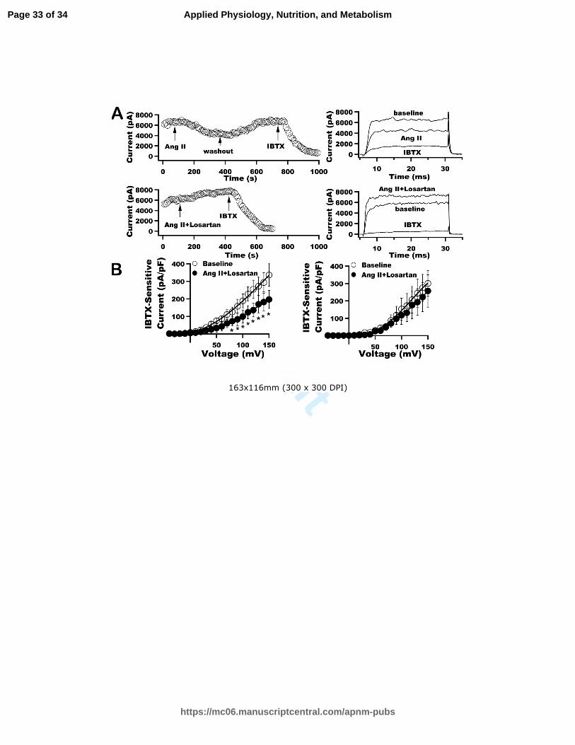
Figure 6. Mutation in the Cav-1 binding motif in AT1R abolished the Ang II effect on hSlo
currents. (A) Time course of the effects of Ang II (2 µM), IBTX (0.1 µM) and wash out of
chemicals on hSlo currents stably expressed in HEK293 cells, 48 h after co-transfection with
Cav-1 wt and AT1R F309A mutant cDNAs. The Ang II did not inhibit hSlo channels which
were inhibited by IBTX, indicating that hSlo channels were present and active but the AT1R
mutant was unable to mediate the inhibitory effects of Ang II on BK channels. (B) Whole-cell
hSlo currents recorded from hSlo-HEK293 cells co-expressing Cav-1 and AT1R F309A (left
column) and their I-V relationships (right column) elicited from -40 mV to +150 mV at a holding
potential of -60 mV before and after exposure to Ang II. Ang II failed to inhibit the hSlo
channels.
Page 28 of 34
https://mc06.manuscriptcentral.com/apnm-pubs
Applied Physiology, Nutrition, and Metabolism

Draft
255x325mm (300 x 300 DPI)
Page 29 of 34
https://mc06.manuscriptcentral.com/apnm-pubs
Applied Physiology, Nutrition, and Metabolism

Draft
254x330mm (300 x 300 DPI)
Page 30 of 34
https://mc06.manuscriptcentral.com/apnm-pubs
Applied Physiology, Nutrition, and Metabolism

Draft
254x338mm (300 x 300 DPI)
Page 31 of 34
https://mc06.manuscriptcentral.com/apnm-pubs
Applied Physiology, Nutrition, and Metabolism

Draft
256x322mm (300 x 300 DPI)
Page 32 of 34
https://mc06.manuscriptcentral.com/apnm-pubs
Applied Physiology, Nutrition, and Metabolism
Draft
163x116mm (300 x 300 DPI)
Page 33 of 34
https://mc06.manuscriptcentral.com/apnm-pubs
Applied Physiology, Nutrition, and Metabolism

Draft
152x116mm (300 x 300 DPI)
Page 34 of 34
https://mc06.manuscriptcentral.com/apnm-pubs
Applied Physiology, Nutrition, and Metabolism