correlation of hypoxia and pro-senescence protein ... · chemiluminescence western blotting kit...
TRANSCRIPT
J. Math. Fund. Sci., Vol. 50, No. 1, 2018, 59-71 59
Received September 30th, 2016, Revised September 27th, 2017, Accepted for publication October 16th. 2017. Copyright © 2018 Published by ITB Journal Publisher, ISSN: 2337-5760, DOI: 10.5614/j.math.fund.sci.2018.50.1.5
Correlation of Hypoxia and Pro-senescence Protein Expression in Green Sea Turtle (Chelonia mydas) Lung
Epithelial and Dermal Fibroblast Cell Culture
Anggraini Barlian & Yemima Dani Riani Biology Study Program, School of Life Sciences and Technology,
Institut Teknologi Bandung, Jalan Ganesha 10, Bandung 40132, Indonesia E-mail: [email protected]
Abstract. Recent studies have shown hypoxia-induced gene expression correlated with cellular senescence. HIF-1α (hypoxia-inducible factor 1-alpha), p53, and pRB were induced under hypoxia and correlated with cellular senescence. The localization and expression of HIF-1α, p53, and pRB in Chelonia mydas lung epithelial and dermal fibroblast cell cultures were analyzed under normoxic and hypoxic conditions (at 4 and 24 hours). Human dermal fibroblast was used for comparison purposes. Protein localization was analyzed with immunocytochemistry, while protein expression was analyzed with the Western blot and enhanced chemiluminescence (ECL) method. HIF-1α, p53, and pRB were localized in the nuclei of the C. mydas cell cultures treated with hypoxia. The C. mydas lung epithelial cell cultures had a higher increase of HIF-1α expression than the human dermal fibroblast cell culture. The hypoxic conditions did not affect p53 expression significantly in C. mydas lung epithelial and dermal fibroblast cell cultures. Meanwhile, pRB expression changed significantly under hypoxia in the C. mydas dermal fibroblast cells. Expression of p53 and pRB in the human cell cultures was higher than in the C. mydas cell cultures. This research suggests that C. mydas and human cell cultures have different pro-senescence protein expression responses under hypoxic conditions.
Keywords: Green sea turtle; hypoxia; immunocytochemistry; pro-senescence protein; Western blotting.
1 Introduction
C. mydas is a long-living species that can live up to 150 years and is suspected to have negligible senescence [1]. A previous study has shown that C. mydas does not undergo telomere shortening and telomerase activity decline in higher culture passage numbers. Apart from this, C. mydas was also hypothesized to overcome aging with elevated antioxidant enzyme activity as found in higher passage numbers of C. mydas dermal fibroblast cultures [2].
Hypoxia is one of the stresses that can lead to cell cycle arrest, cell death, and apoptosis. Even though it breathes using lungs, C. mydas can dive and migrate under water for 40 minutes. C. mydas was hypothesized to have high hypoxic
60 Anggraini Barlian & Yemima Dani Riani
tolerance like other Testudinae, such as Chrysemys picta and Trachemys scripta. Under hypoxic conditions, C. picta and T. scripta can increase the expression of antioxidant enzymes, heat shock proteins, and anti-apoptotic proteins [3]. These proteins play a role in regulating the cell cycle. Thus, C. mydas perhaps also has a way to regulate hypoxic stress and this regulation could correlate with its negligible senescence, which hypothetically happens very slowly in C. mydas.
HIF-1α (hypoxia-inducible factor-1 alpha) is the master regulator of hypoxic conditions in cells. HIF-1α activation under hypoxia will induce the transcription of some essential genes to help cell survival. Some of the genes that HIF-1α regulates are glycolysis enzymes to produce ATP without oxydative phosphorylation, ion transporters to increase acidic pH caused by anaerobic metabolism, and cell cycle regulator proteins to arrest the cell cycle so the cell can concentrate on overcoming the stress [4].
p53 and pRB are well-known cell cycle regulators and pro-senescence proteins. These two proteins work upstream of the senescence pathway and are regulated under hypoxia [5]. p53 is induced by elevating ROS under hypoxia and is also upregulated indirectly by HIF-1α [6]. pRB is also upregulated under hypoxia and it can bind directly to HIF-1α and act as HIF-1α co-activator [7]. In that sense, the expression of HIF-1α and pro-senescence proteins in C. mydas cells under hypoxic conditions is an interesting object of study because it may have a correlation with the aging process of C. mydas. The objective of the present research was to analyze the localization and expression of HIF-1α, p53, and pRB in C. mydas lung epithelial and dermal fibroblast cells under normoxia and at 4 and 24 hours of hypoxia. Human dermal fibroblast cells treated under the same conditions were used for comparison purposes.
2 Materials and Methods
2.1 Cell Culture
C. mydas epithelial lung and dermal fibroblast cell cultures were established from C. mydas embryos aged 2-3 weeks. Human dermal fibroblast was established from the foreskin of 4-5 year old human subjects. The cultures were made using the primary explant method. The C. mydas and human cell cultures were maintained in Leibovitz-15 medium and DMEM-low glucose (with supplementation of penicillin/streptomycin and 10% FBS) respectively. The cells were incubated at 27 °C (BlueM) for the C. mydas cultures, while the human dermal fibroblast cells were incubated at 37 °C with 5% CO2 (Heraeus). Subculture was done with trypsin 0.5% and EDTA 0.2%.
Correlation of Hypoxia and Pro-senescence Protein 61
2.2 Hypoxic Treatment
The cells for immunocytochemistry were plated on a cover glass and put in 6-well plates (Nunc) with media, while the cells for Western blot and ECL (enhanced chemiluminescence) analysis were plated on a 75-cm2 flask (Nunc). Plates and flasks were put inside an inflatable hypoxic chamber [8] filled with gas mixture (0.5% O2, 5% CO2, 94.5% N2) and then incubated at 27 °C and 37 °C for the C. mydas and the human cells respectively. The cells for immunocytochemistry were incubated for 24 hours, while the cells for Western blot and ECL analysis were incubated for 4 and 24 hours.
2.3 Immunocytochemistry
After hypoxic treatment, the cells were fixed with 100% methanol and then permeabilized with 0.25% Triton X-100 (Sigma) in PBS. Blocking was performed with 1% BSA in PBST. The cells were double stained with mouse monoclonal anti-HIF-1-alpha (ab1, Abcam) and rabbit polyclonal anti-p53 (ab4060, Abcam) or mouse monoclonal anti-HIF-1-alpha (ab1, Abcam) and rabbit polyclonal anti-Rb (ab6075, Abcam) primary antibodies overnight at 4 °C. Secondary antibodies used were goat polyclonal anti-Rabbit IgG-H&L FITC (ab6717, Abcam) and mouse monoclonal anti-Mouse IgG H&L Alexa Fluor 647 (ab150127, Abcam). The nuclei were stained with DAPI 1ug/mL. The cover glasses were mounted with glycerol. Analysis was performed with a confocal microscope (Nikon A1RSi).
2.4 Western Blot and Enhanced Chemiluminescence Analysis
Cytoplasmic and nuclear proteins from cells treated under normoxia and hypoxia were harvested on ice using lysis buffer A (10 mM HEPES pH 7.9, 10 mM KCl, 0.1 mM EDTA pH 8, 0.1 mM EGTA pH 8, NP40 10%, protease inhibitors) and lysis buffer C (20 mM HEPES pH 7.9, 400 mM NaCl, 1 mM EDTA pH 8, 1 mM EGTA pH 8, 25% Glycerin, protease inhibitors) respectively. HIF-1α was analyzed from nuclear extract, while p53 and pRB were analyzed from cytoplasmic extract. The internal control used was actin from cytoplasmic extract. SDS-PAGE was performed with 10% acrylamide gel and proteins were blotted onto PVDF membranes (Roche, 16114600). The membranes were incubated in specific mouse monoclonal anti-HIF-1-alpha (ab1, Abcam), rabbit polyclonal anti-p53 (ab4060, Abcam), or rabbit polyclonal anti-Rb (ab6075, Abcam) primary antibodies seperately overnight at 4 °C. Secondary antibodies used were anti-mouse IgG-POD/anti-rabbit IgG POD (Roche). Detection was performed with the ECL method using a BM Chemiluminescence Western Blotting Kit (Mouse/Rabbit) (Roche, 13222400). The membranes were exposed to an X-ray film in a Kodak X-Omat casette. Western blotting data analysis was performed using ImageJ software. The
62 Anggraini Barlian & Yemima Dani Riani
region of interest was drawn surrounding the bands and then the mean intensity was measured. The intensity of each sample was normalized to the actin bands of the respective samples. Statistical analysis of significant differences between normoxic and 4/24 hours hypoxic treatment was done using one-way ANOVA (p = 0.05), while the statistical significance between each cell line was analyzed using two-way ANOVA
3 Results and Discussion
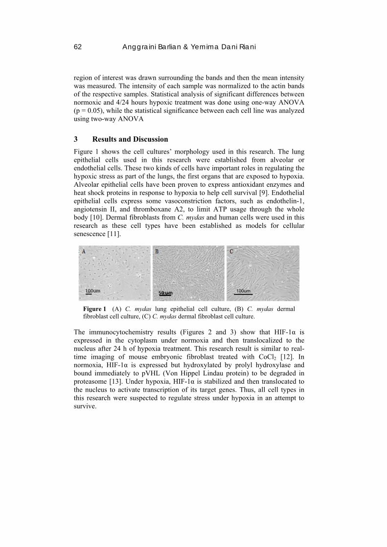
Figure 1 shows the cell cultures’ morphology used in this research. The lung epithelial cells used in this research were established from alveolar or endothelial cells. These two kinds of cells have important roles in regulating the hypoxic stress as part of the lungs, the first organs that are exposed to hypoxia. Alveolar epithelial cells have been proven to express antioxidant enzymes and heat shock proteins in response to hypoxia to help cell survival [9]. Endothelial epithelial cells express some vasoconstriction factors, such as endothelin-1, angiotensin II, and thromboxane A2, to limit ATP usage through the whole body [10]. Dermal fibroblasts from C. mydas and human cells were used in this research as these cell types have been established as models for cellular senescence [11].
Figure 1 (A) C. mydas lung epithelial cell culture, (B) C. mydas dermal fibroblast cell culture, (C) C. mydas dermal fibroblast cell culture.
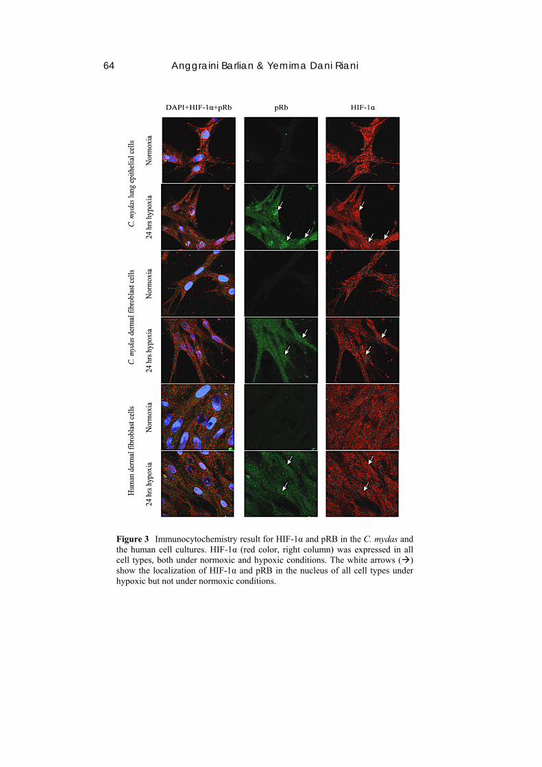
The immunocytochemistry results (Figures 2 and 3) show that HIF-1α is expressed in the cytoplasm under normoxia and then translocalized to the nucleus after 24 h of hypoxia treatment. This research result is similar to real-time imaging of mouse embryonic fibroblast treated with CoCl2 [12]. In normoxia, HIF-1α is expressed but hydroxylated by prolyl hydroxylase and bound immediately to pVHL (Von Hippel Lindau protein) to be degraded in proteasome [13]. Under hypoxia, HIF-1α is stabilized and then translocated to the nucleus to activate transcription of its target genes. Thus, all cell types in this research were suspected to regulate stress under hypoxia in an attempt to survive.
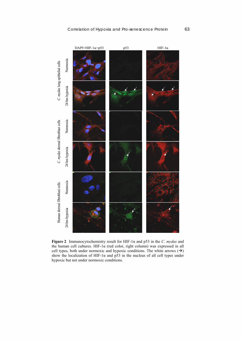
Correlation of Hypoxia and Pro-senescence Protein 63
Figure 2 Immunocytochemistry result for HIF-1α and p53 in the C. mydas and the human cell cultures. HIF-1α (red color, right column) was expressed in all cell types, both under normoxic and hypoxic conditions. The white arrows () show the localization of HIF-1α and p53 in the nucleus of all cell types under hypoxic but not under normoxic conditions.
64 Anggraini Barlian & Yemima Dani Riani
Figure 3 Immunocytochemistry result for HIF-1α and pRB in the C. mydas and the human cell cultures. HIF-1α (red color, right column) was expressed in all cell types, both under normoxic and hypoxic conditions. The white arrows () show the localization of HIF-1α and pRB in the nucleus of all cell types under hypoxic but not under normoxic conditions.
Correlation of Hypoxia and Pro-senescence Protein 65
Compared to the research performed by Uchida, et al. [14] under the same oxygen concentration, the HIF-1α expression level in the nuclei of the human lung epithelial cells increased after 4 hours of hypoxia but decreased to the same level as in normoxia after 12 hours of hypoxia. In this research, HIF-1α was still abundant in the nuclei and appeared more significantly after 24 hours of hypoxia than under normoxia. It is suspected that HIF-1α localization in the nuclei of C. mydas lung epithelial cells is more stable than in human lung epithelial cells.
Localization of p53 and pRB in the nucleus was also found in cells under hypoxia but were hardly found under normoxia. In that sense, p53 has a role as transcription factor of p21, the cell-dependent kinase inhibitor, so p21 can arrest the cell cycle in G1 [15]. Furthermore, p21 can induce pRB hypophosphorylation, in which state pRB will translocate to the nucleus and is then bound to E2F, the transcription factor of some cyclins and cdks [16]. In conclusion, p53 and pRB translocation to the nucleus perhaps leads to cell cycle arrest in C. mydas cells the same as in human dermal fibroblast cells.
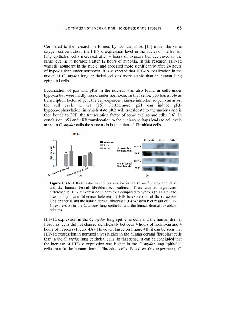
Figure 4 (A) HIF-1α ratio to actin expression in the C. mydas lung epithelial and the human dermal fibroblast cell cultures. There was no significant difference in HIF-1α expression in normoxia compared to hypoxia (p > 0.05) and also no significant difference between the HIF-1α expression of the C. mydas lung epithelial and the human dermal fibroblast. (B) Western blot result of HIF-1α expression in the C. mydas lung epithelial and the human dermal fibroblast cultures.
HIF-1α expression in the C. mydas lung epithelial cells and the human dermal fibroblast cells did not change significantly between 4 hours of normoxia and 4 hours of hypoxia (Figure 4A). However, based on Figure 4B, it can be seen that HIF-1α expression in normoxia was higher in the human dermal fibroblast cells than in the C. mydas lung epithelial cells. In that sense, it can be concluded that the increase of HIF-1α expression was higher in the C. mydas lung epithelial cells than in the human dermal fibroblast cells. Based on this experiment, C.
66 Anggraini Barlian & Yemima Dani Riani
mydas lung epithelial cells possibly can overcome stress caused by hypoxia better than human dermal fibroblast cells.
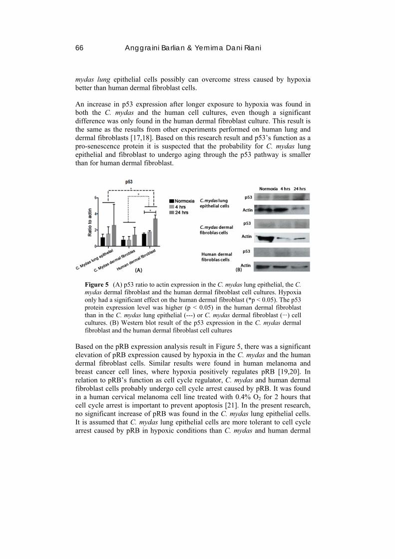
An increase in p53 expression after longer exposure to hypoxia was found in both the C. mydas and the human cell cultures, even though a significant difference was only found in the human dermal fibroblast culture. This result is the same as the results from other experiments performed on human lung and dermal fibroblasts [17,18]. Based on this research result and p53’s function as a pro-senescence protein it is suspected that the probability for C. mydas lung epithelial and fibroblast to undergo aging through the p53 pathway is smaller than for human dermal fibroblast.
Figure 5 (A) p53 ratio to actin expression in the C. mydas lung epithelial, the C. mydas dermal fibroblast and the human dermal fibroblast cell cultures. Hypoxia only had a significant effect on the human dermal fibroblast (*p < 0.05). The p53 protein expression level was higher (p < 0.05) in the human dermal fibroblast than in the C. mydas lung epithelial (---) or C. mydas dermal fibroblast (‧‧‧) cell cultures. (B) Western blot result of the p53 expression in the C. mydas dermal fibroblast and the human dermal fibroblast cell cultures
Based on the pRB expression analysis result in Figure 5, there was a significant elevation of pRB expression caused by hypoxia in the C. mydas and the human dermal fibroblast cells. Similar results were found in human melanoma and breast cancer cell lines, where hypoxia positively regulates pRB [19,20]. In relation to pRB’s function as cell cycle regulator, C. mydas and human dermal fibroblast cells probably undergo cell cycle arrest caused by pRB. It was found in a human cervical melanoma cell line treated with 0.4% O2 for 2 hours that cell cycle arrest is important to prevent apoptosis [21]. In the present research, no significant increase of pRB was found in the C. mydas lung epithelial cells. It is assumed that C. mydas lung epithelial cells are more tolerant to cell cycle arrest caused by pRB in hypoxic conditions than C. mydas and human dermal
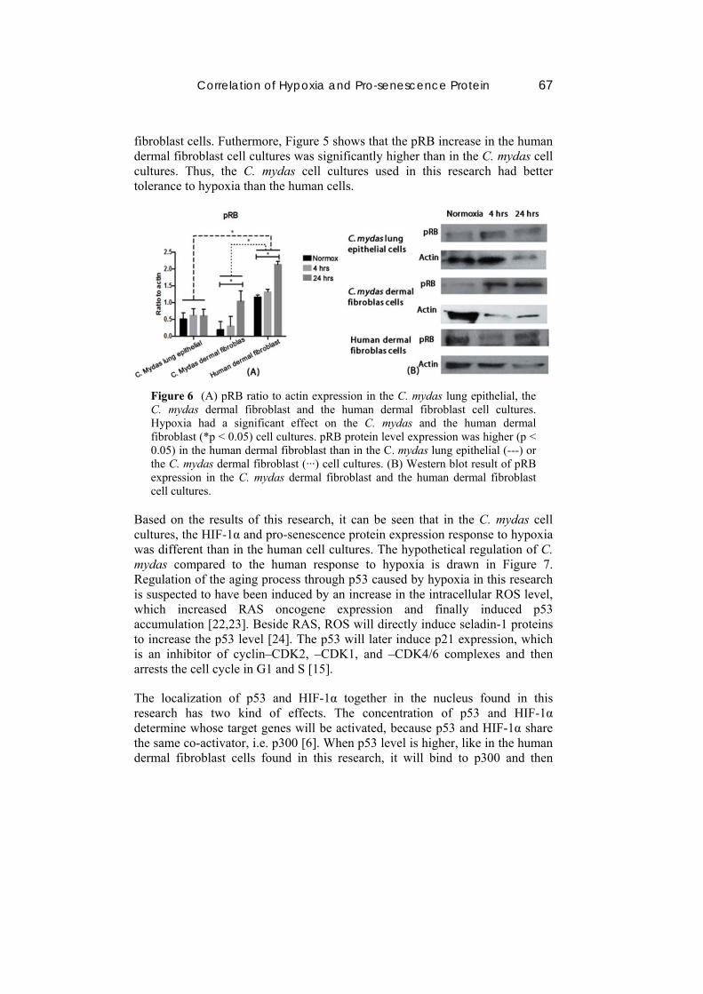
Correlation of Hypoxia and Pro-senescence Protein 67
fibroblast cells. Futhermore, Figure 5 shows that the pRB increase in the human dermal fibroblast cell cultures was significantly higher than in the C. mydas cell cultures. Thus, the C. mydas cell cultures used in this research had better tolerance to hypoxia than the human cells.
Figure 6 (A) pRB ratio to actin expression in the C. mydas lung epithelial, the C. mydas dermal fibroblast and the human dermal fibroblast cell cultures. Hypoxia had a significant effect on the C. mydas and the human dermal fibroblast (*p < 0.05) cell cultures. pRB protein level expression was higher (p < 0.05) in the human dermal fibroblast than in the C. mydas lung epithelial (---) or the C. mydas dermal fibroblast (‧‧‧) cell cultures. (B) Western blot result of pRB expression in the C. mydas dermal fibroblast and the human dermal fibroblast cell cultures.
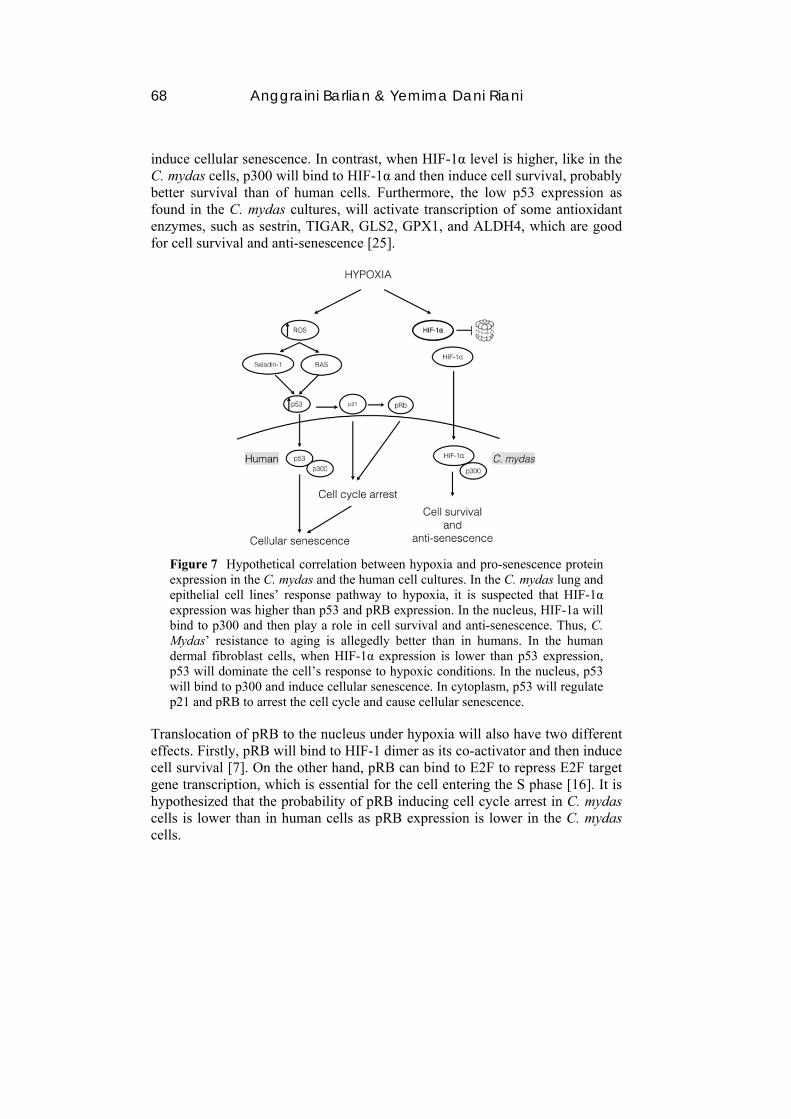
Based on the results of this research, it can be seen that in the C. mydas cell cultures, the HIF-1α and pro-senescence protein expression response to hypoxia was different than in the human cell cultures. The hypothetical regulation of C. mydas compared to the human response to hypoxia is drawn in Figure 7. Regulation of the aging process through p53 caused by hypoxia in this research is suspected to have been induced by an increase in the intracellular ROS level, which increased RAS oncogene expression and finally induced p53 accumulation [22,23]. Beside RAS, ROS will directly induce seladin-1 proteins to increase the p53 level [24]. The p53 will later induce p21 expression, which is an inhibitor of cyclin–CDK2, –CDK1, and –CDK4/6 complexes and then arrests the cell cycle in G1 and S [15].
The localization of p53 and HIF-1α together in the nucleus found in this research has two kind of effects. The concentration of p53 and HIF-1α determine whose target genes will be activated, because p53 and HIF-1α share the same co-activator, i.e. p300 [6]. When p53 level is higher, like in the human dermal fibroblast cells found in this research, it will bind to p300 and then
68 Anggraini Barlian & Yemima Dani Riani
induce cellular senescence. In contrast, when HIF-1α level is higher, like in the C. mydas cells, p300 will bind to HIF-1α and then induce cell survival, probably better survival than of human cells. Furthermore, the low p53 expression as found in the C. mydas cultures, will activate transcription of some antioxidant enzymes, such as sestrin, TIGAR, GLS2, GPX1, and ALDH4, which are good for cell survival and anti-senescence [25].
Figure 7 Hypothetical correlation between hypoxia and pro-senescence protein expression in the C. mydas and the human cell cultures. In the C. mydas lung and epithelial cell lines’ response pathway to hypoxia, it is suspected that HIF-1α expression was higher than p53 and pRB expression. In the nucleus, HIF-1a will bind to p300 and then play a role in cell survival and anti-senescence. Thus, C. Mydas’ resistance to aging is allegedly better than in humans. In the human dermal fibroblast cells, when HIF-1α expression is lower than p53 expression, p53 will dominate the cell’s response to hypoxic conditions. In the nucleus, p53 will bind to p300 and induce cellular senescence. In cytoplasm, p53 will regulate p21 and pRB to arrest the cell cycle and cause cellular senescence.
Translocation of pRB to the nucleus under hypoxia will also have two different effects. Firstly, pRB will bind to HIF-1 dimer as its co-activator and then induce cell survival [7]. On the other hand, pRB can bind to E2F to repress E2F target gene transcription, which is essential for the cell entering the S phase [16]. It is hypothesized that the probability of pRB inducing cell cycle arrest in C. mydas cells is lower than in human cells as pRB expression is lower in the C. mydas cells.
Correlation of Hypoxia and Pro-senescence Protein 69
4 Conclusion
Localization of HIF-1α, p53, and pRB under hypoxic conditions (0.5% O2) was found in C. mydas lung epithelial and C. mydas dermal fibroblast cells. Localization of HIF-1α, p53, and pRB under hypoxic conditions (0.5% O2) also occurred in the human dermal fibroblast cells. Furthermore, hypoxic treatment for 4 hours and 24 hours caused increased HIF-1α, p53, and pRB expression in C. mydas, although not significantly. HIF-1α expression was higher in the C. mydas than in the human cell cultures. In contrast, p53 and pRB expression was higher in the human than in the C. mydas cell cultures.
Acknowledgments
This research was funded by ITB Innovative Research 2014 and Nikon Singapore Ltd.
References
[1] Finch, C.E., Update on Slow Aging and Negligible Senescence, Gerontology, 55(3), pp. 307-313, 2009. DOI:10.1159/000215589
[2] Barlian, A., Riani, Y.D., Christina, C., Lestari, A. & Fajriah, N., Molecular Study of Aging in Green Sea Turtle: Telomere, Telomerase and Antioxidant Enzyme, In New Hope in Aging Medicine, 2013.
[3] Krivoruchko, A. & Storey, K.B., Forever Young: Mechanism of Natural Anoxia Tolerance and Potential Links to Longevity, Oxid. Med. Cell Longev., 3(3), pp. 186-198, 2010. DOI:10.4161/oxim.3.3.12356
[4] Koh, M.Y., Spivak-Kroizman, T.R. & Powis, G., HIF-1 Regulation: Not So Easy Come Easy Go, Trends Biochemical Sci., 33(11), pp. 526-534, 2008. DOI:10.1016/j.tibs.2008.08.002
[5] Campisi, J., Senescent Cells, Tumor Supression, and Organismal Aging: Good Citizens, Bad Neighbors, Cell., 120(4), pp. 513-522, 2005. DOI:10.1016/j.cell.2005.02.003
[6] Sermeus, A. & Michiels, C., Reciprocal Influence of the p53 and the Hypoxic Pathways, Cell Death Dis., 2(5), e164, 2011. DOI:10.1038/ cddis.2011.48
[7] Budde, A., Marra, N.M., Petersen, G. & Brune, B., Retinoblastoma Susceptibility Gene Product pRB Activates Hypoxia-inducible Factor-1 (HIF-1), Oncogene, 24(10), pp. 1082-1088, 2005. DOI:10.1038/ sj.onc.1208369
[8] Wang, R., Jin, F., & Zhong, H., A Novel Experimental Hypoxia Chamber for Cell Culture, Am. J. Cancer Res., 4(1), pp.53-60, 2014. PMCID: PMC3902232
70 Anggraini Barlian & Yemima Dani Riani
[9] Jain, M. & Sznajder, J.I., Effects of Hypoxia on the Alveolar Epithelium, Proc. Am. Thorac. Soc., 2(3), pp. 202-205, 2005. DOI: 10.1513/pats. 200501-006AC
[10] Pak, O., Aldashev, A., Welsh, D. & Peacock, A., The Effects of Hypoxia on the Cells of the Pulmonary Vasculature, Eur. Respir. J., 30(2), pp. 364-372, 2007. DOI:10.1183/09031936.00128706
[11] Hayflick, L. & Moorhead, P.S., The Serial Cultivation of Human Diploid Cell Strains, Exp. Cell. Res., 25(3), pp. 585-621, 1961. DOI:10.1016/ 0014-4827(61)90192-6
[12] Moroz, E., Carlin, S., Dyomina, K., Burke, S., Thaler, H.T. & Blasberg, R., Real Time Imaging of HIF-1α Stabilization and Degradation, 4(4), e5077, 2009. doi: 10.1371/journal.pone.0005077
[13] Semenza, G. & Wang, G.A., Nucleur Factor Induced by Hypoxia via de Novo Protein Synthesis to the Human Erythropoietin Gene Enhancer at A Site Required for Transcriptional Activation, Mol. Cell. Biol., 12(12), pp. 5447-5454, 1992.
[14] Uchida. T., Rossignol, F., Matthay, M.A., Mounier, R., Couette, S. & Clottes, E., Prolonged Hypoxia Differentially Regulates HIF1α and HIF2α Expression in Lung Epithelial Cells, J. Biol. Chem., 279, pp. 14871-14878, 2004. DOI:10.1074/jbc.M400461200
[15] Rufini, A., Tucci, P., Celardo, I. & Melino, G., Senescence and Aging : the Critical Roles of p53, Nature, 32, pp. 5129-5143, 2013. DOI:10.1038/ onc.2012.640
[16] Gianciti, C. & Giordano, A., RB and Cell Cycle Progression, Nature, 25, pp. 5220-5227, 2005. doi:10.1038/sj.onc.1209615
[17] Graeber, T.G., Peterson, J.F., Tsai, M., Monica, K., Fornace, A.J. & Giaccia, A.J., Hypoxia Induces Accumulation of p53 Protein, but Activation of A G1-phase Checkpoint by Low Oxygen Conditions is Independent of p53 Status, Cell. Mol. Biol., 14(9), pp. 6264-6277, 1994.
[18] Mizuno, S., Bogaard, H.J., Voelkel, N.F., Umeda, Y., Kadowaki, M. & Ameshima, S., Hypoxia Regulates Human Lung Fibroblast Proliferation via p53 Dependent and Independent Pathways, Respiratory Res., 10(17), pp. 14870-14878, 2009. DOI:10.1186/1465-9921-10-17
[19] Danielsen, T., Hvidsten, M., Stokke, T. & Rofwstad, E.K., Hypoxia Induces p53 Accumulation in the S-phase and Accumulation of Hypophosphorylated Restinoblastoma Protein in all Cell Cycle Phase of Human Melanoma Cells, British Journal of Cancer, 78(12), pp. 1547-1558, 1998.
[20] Amellem, O., Sandvik, J.A., Stokke, T. & Pettersen, E.O., The Retinoblastoma Protein-associated Cell Cycle Arrest in S-phase under Moderate Hypoxia is Disrupted in Cells Expressing HPV18 E7 Oncoprotein, British Journal of Cancer, 77(6), pp. 862-872, 1998.
Correlation of Hypoxia and Pro-senescence Protein 71
[21] Amellem, O. & Pettersen, E.O., Cell Inactivation and Cell Cycle Inhibition as Induced by Extreme Hypoxia, The Possible Role of Cell Cycle Arrest as A Protection Against Hypoxia-induced Lethal Damage, Cell Prolif., 24(2), pp. 127–141, 1991. DOI:10.1111/j.1365-2184.1991. tb01144.x
[22] Chandel, N.S., McClintock, D.S., Feliciano, C.E., Wood, T.M., Melendez, J.A. & Rodriguez, A.M., Reactive Oxygen Species Generated at Mitochondrial Complex III Stabilize Hypoxia-inducible Factor-1 alpha during Hypoxia: A Mechanism of O2 Sensing, J. Biol. Chem., 275, pp. 25130-25138, 2005. DOI: 10.1074/jbc.M001914200
[23] Guzy, R.D., Hoyos, B., Robin, E., Chen, H., Liu, L. & Mansfield, K.D., Mitochondrial Complex III is Required for Hypoxia-induced ROS Production and Cellular Oxygen Sensing, Cell Metabol, 1(6), pp. 401-408, 2005. DOI:10.1016/j.cmet.2005.05.001
[24] Wu, C., Miloslavskaya, I., Demontis, S., Maestro, R. & Galaktionov, K., Regulation of Cellular Response to Oncogenic and Oxidative Stress by Seladin-1, Nature, 432, pp. 640-645, 2004. DOI:10.1038/nature03173
[25] Feng, Z. , Lin, M. & Wu, R., The Regulation of Aging and Longevity: A New and Complex Network, Genes. & Cancer, 2(4), pp. 443-452, 2011. DOI:10.1177/1947601911410223