correlations between mri white matter lesion location …bradd/smith_neurol_2011.pdfcorrelations...
TRANSCRIPT

DOI 10.1212/WNL.0b013e318217e7c8 2011;76;1492Neurology
E.E. Smith, D.H. Salat, J. Jeng, et al.executive function and episodic memory
Correlations between MRI white matter lesion location and
April 30, 2013This information is current as of
http://www.neurology.org/content/76/17/1492.full.html
located on the World Wide Web at: The online version of this article, along with updated information and services, is
Neurology. All rights reserved. Print ISSN: 0028-3878. Online ISSN: 1526-632X.since 1951, it is now a weekly with 48 issues per year. Copyright © 2011 American Academy of
® is the official journal of the American Academy of Neurology. Published continuouslyNeurology

Correlations between MRI white matterlesion location and executive function andepisodic memory
E.E. Smith, MD, MPHD.H. Salat, PhDJ. Jeng, MSC.R. McCreary, PhDB. Fischl, PhDJ.D. Schmahmann, MDB.C. Dickerson, MDA. Viswanathan, MD,
PhDM.S. Albert, PhDD. Blacker, MD, ScDS.M. Greenberg, MD,
PhD
ABSTRACT
Objectives: MRI white matter hyperintensity (WMH) volume is associated with cognitive impair-ment. We hypothesized that specific loci of WMH would correlate with cognition even after ac-counting for total WMH volume.
Methods: Subjects were identified from a prospective community-based study: 40 had normalcognition, 94 had mild impairment (defined here as a Clinical Dementia Rating [CDR] score of 0.5without dementia), and 11 had mild Alzheimer’s dementia. Factor analysis of a 22-item neuropsy-chological battery yielded 4 factors (episodic memory, executive function, spatial skills, and gen-eral knowledge). MRI WMH segmentation and analysis was performed using FreeSurfer software.
Results: Higher WMH volume was independently associated with lower executive function andepisodic memory factor scores. Voxel-based general linear models showed loci where WMH wasstrongly inversely associated with specific cognitive factor scores (p � 0.001), controlling forage, education, sex, APOE genotype, and total WMH volume. For episodic memory, clusters wereobserved in bilateral temporal-occipital and right parietal periventricular white matter, and theleft anterior limb of the internal capsule. For executive function, clusters were observed in bilat-eral inferior frontal white matter, bilateral temporal-occipital and right parietal periventricularwhite matter, and the anterior limb of the internal capsule bilaterally.
Conclusions: Specific WMH loci are closely associated with executive function and episodic mem-ory, independent of total WMH volume. The anatomic locations suggest that WMH may causecognitive impairment by affecting connections between cortex and subcortical structures, includ-ing the thalamus and striatum, or connections between the occipital lobe and frontal or parietallobes. Neurology® 2011;76:1492–1499
GLOSSARYAD � Alzheimer disease; CADASIL � cerebral autosomal dominant arteriopathy with stroke and ischemic leukoencephalop-athy; CDR � Clinical Dementia Rating; DSM-IV � Diagnostic and Statistical Manual of Mental Disorders, 4th edition; FA �fractional anisotropy; MCI � mild cognitive impairment; PDW � proton density-weighted; SPGR � spoiled gradient recalled;T1W � T1-weighted; T2W � T2-weighted; TE � echo time; TR � repetition time; WMH � white matter hyperintensity.
There is growing recognition that ischemic brain lesions are a significant contributor to cogni-tive impairment and that many cases of dementia are mixed, with a cerebrovascular compo-nent.1 Ischemic white matter lesions, seen on MRI as white matter hyperintensity (WMH),have previously been associated with decreased performance on neuropsychological testing,2,3
and risk of mild cognitive impairment4 and dementia.5 It has been hypothesized that WMHinterfere with cognitive processing by impairing the speed or fidelity of signal transmissionthrough affected areas.6 The direct evidence to support this hypothesis is scant, however.6
We reasoned that if WMH impair white matter function, then clinical impairments should beassociated with WMH in discrete loci involving white matter tracts connecting cortical networksserving those clinical functions. For example, WMH in specific occipito-parietal periventricular
From the Department of Clinical Neurosciences (E.E.S.), Hotchkiss Brain Institute (E.E.S., C.R.M.), and Department of Radiology (E.E.S., C.R.M.),University of Calgary, Canada; Martinos Biomedical Imaging Center (D.H.S., J.J., B.F., B.C.D.), Department of Neurology (J.D.S., B.C.D., A.V.,S.M.G.), Department of Psychiatry (D.B.) and Gerontology Research Unit (D.B.), Massachusetts General Hospital, Boston; and Department ofNeurology (M.S.A.), Johns Hopkins University, Baltimore, MD.
Study funding: Supported by the National Institute on Aging (P01 AG04953, R01 AG26484) and the National Institute of Neurological Disordersand Stroke (R01 NS062028).
Disclosure: Author disclosures are provided at the end of the article.
Supplemental data atwww.neurology.org
Address correspondence andreprint requests to Dr. Eric E.Smith, Department of ClinicalNeurosciences, Foothills MedicalCentre, Room C1212, Calgary,AB, T2N 2T9, [email protected]
1492 Copyright © 2011 by AAN Enterprises, Inc.

regions have previously been associated with gaitimpairment.7 Similarly, specific regions of ab-normal fractional anisotropy (FA) have beenidentified in the frontal white matter that areassociated with executive dysfunction in the he-reditary small vessel disease cerebral autosomaldominant arteriopathy with stroke and ischemicleukoencephalopathy (CADASIL).8
In order to better understand the relationshipbetween WMH location and cognitive impair-ment, we determined the correlation betweenWMH involvement in specific regions and neu-ropsychological test performance in subjectsparticipating in a prospective study of cognitivedecline.
METHODS Subject population. Subjects were drawn from
a longitudinal prospective cohort study of predictive factors for tran-
sition to dementia. The details of study subject recruitment and
assessment have been previously published.9 Study subjects were re-
cruited through community advertising seeking older individuals
with and without memory impairment. To be included in the
study, all participants had to be 65 or older and to have an infor-
mant as a collateral source of information. At entry to the parent
study, persons with dementia, major vascular risk factors (atrial fi-
brillation or diabetes mellitus requiring insulin), history of stroke, or
clinical diagnosis of cognitive impairment due to medical disorderssuch as hypothyroidism were excluded.
A semi-structured interview by an experienced clinician wasused to evaluate the subjects at study entry and at each subse-quent year, as previously described,9,10 to generate a Clinical De-mentia Rating (CDR) score.11 For subjects who developeddementia during the study, the underlying disorder was diag-nosed by consensus of the investigators using all available studyinformation. Dementia was defined using criteria from theDSM-IV,12 probable Alzheimer disease (AD) was diagnosed us-ing the National Institute of Neurological and CommunicativeDisorders and Stroke–Alzheimer’s Disease and Related Disor-ders Association criteria,13 and vascular dementia was diagnosedaccording to the National Institute of Neurological Disordersand Stroke–Association Internationale pour la Recherche enl’Enseignement en Neurosciences criteria.14
Those with mildly impaired cognition at the time of MRI, de-fined as a CDR score of 0.5 without dementia, are defined here asmild cognitive impairment (MCI).4,15 The distribution of CDRsum of boxes scores among the mildly impaired subjects was broad(see table 1). At the mild end of the spectrum, many subjects wouldnot meet psychometric cutoffs commonly used to select MCI sub-jects in epidemiologic studies and clinical trials,16 and could be con-sidered early MCI. The subjects at the more impaired end of thespectrum (i.e., CDR sum of boxes �2) are comparable to MCIsubjects recruited in these settings, based on likelihood of progres-sion to a diagnosis of AD.9 We use MCI here to refer to the entiregroup of mildly impaired subjects.
Neuropsychological assessments. A 22-item neuropsycho-logical battery was administered to all study subjects.17 The me-
Table 1 Study population characteristicsa
CharacteristicNormal cognition(n � 40), %
MCI(n � 96), %
Mild dementia(n � 11), % p Value
Age, y 71.2 � 4.74 72.9 � 5.5 74.1 � 6.9 0.15
Female sex 65 61 45 0.50
Education, y 16.0 � 2.9 15.6 � 2.8 13.9 � 3.7 0.12
Hypertension 35 38 45 0.83
Cardiovascular disease 8 14 0 0.38
Diabetes 3 5 27 0.02
Current smoker 8 0 0 0.05
Past smoker 60 58 45 0.71
>1 APOE �4 allele 25 27 36 0.73
>1 APOE �2 allele 25 9 18 0.04
CDR sum of boxes 0 (0, 0) 1 (1, 2) 5 (4.5, 5) �0.001
WMHr, % 0.82 (0.60, 1.09) 0.97 (0.76, 1.38) 1.13 (0.93, 1.48) 0.07
>1 lacunar lesion 18 19 27 0.71
Neuropsychological test factor score
Episodic memory 0.16 � 0.68 �0.67 � 1.03 �1.70 � 1.30 �0.001
Executive function 0.07 � 0.86 �0.33 � 0.94 �0.66 � 0.96 0.02
Spatial skills 0.39 � 0.82 �0.07 � 1.01 �0.84 � 1.27 �0.001
General knowledge 0.45 � 0.85 �0.01 � 1.01 �0.15 � 0.98 0.03
Abbreviations: CDR � Clinical Dementia Rating; MCI � mild cognitive impairment; WMHr � total white matter hyperinten-sity volume, expressed as percentage of intracranial volume.a Values are percentages, mean � SD, or median (25th, 75th percentile). p Values are by Fisher exact test, testing forsignificant differences across all 3 groups.
Neurology 76 April 26, 2011 1493

dian time between MRI and neuropsychological testing was 76days (interquartile range 19–219 days). Details of the battery,including the individual test items, have been previously de-scribed (table e-1 on the Neurology® Web site at www.neurology.org).17,18 A factor analysis of the neuropsychological measuresyielded a 4-factor representation of the overall test battery: 1)general knowledge, 2) episodic memory, 3) spatial skill, and 4)executive function.17 The factor scores were standardized to havezero mean and unit variance among the cognitively normal studysubjects, adjusted for age and education.
MRI measurements. Subjects underwent MRI on either of 21.5-T scanners (Signa, General Electric Medical Systems, Mil-waukee, WI).4 A sagittal localizer, coronal T1-weighted (T1W)spoiled gradient recalled (SPGR), and dual echo sequence, yield-ing proton density-weighted (PDW) and T2-weighted (T2W)images of the whole head, was performed. T1W SPGR sequenceparameters were as follows: repetition time (TR) 35 msec, echotime (TE) 5 msec, flip angle 45 degrees, 1.5-mm slice thicknesswith no interslice gap. Dual echo sequence parameters were asfollows: TR 3,000 msec, TE1 30 msec (for PDW images), TE280 msec (for T2W images), and slice thickness 3 mm inter-leaved. The matrix was 256 � 192, and field of view 24 cm, forall sequences.
Previous work in this study population has shown thattotal WMH volume was associated with the risk of progres-sion from normal cognition to MCI.4 To take advantage ofadvances in computer processing, we have revised our meth-ods for WMH segmentation, using custom-designed algo-rithms implemented in the FreeSurfer image analysis suite(http://surfer.nmr.mgh.harvard.edu/). Briefly, this processingincludes motion correction, removal of nonbrain tissue using ahybrid watershed/surface deformation procedure, automatedTalairach transformation, segmentation of the subcortical whitematter and deep gray matter volumetric structures, intensity nor-malization, tessellation of the gray matter white matter bound-ary, automated topology correction, and surface deformationfollowing intensity gradients to optimally segment borders at thelocation where the greatest shift in intensity defines the transi-tion to the other tissue class.19-23 The structural segmentationswere used to identify regions where WMH was possible whileexcluding regions where WMH does not occur (that is, corticaland subcortical gray matter structures). Final algorithm-generated WMH maps were visually inspected to identify obvi-ous errors. No manual corrections were necessary; however, 13scans could not be processed because of motion or other artifact.The accuracy of the automated FreeSurfer algorithm was verifiedby comparison with 10 scans where WMH was independentlymanually segmented by an experienced rater blinded to the auto-mated algorithm results; the intraclass correlation coefficient was0.91, indicating excellent agreement.
There were 160 consecutive subjects who underwent MRI aspart of the study and had no history of stroke prior to MRIscanning. Exclusion of 13 subjects whose scan data could not beprocessed left 147 for analysis. The MRI was performed afterstudy entry in 65/147 (44%); dementia had developed in 11subjects during the preceding median 4.8 years of follow-up (in-terquartile range 2.9 to 9.1 years). The cause of dementia wasprobable AD in all 11 subjects.
Statistical analysis. Total WMH volume was expressed as apercentage of the intracranial volume (WMHr), as in other studies,to account for differences in subject head size.4 Continuous variableswere normally distributed, with the exception of WMH, which wasright-skewed and therefore analyzed using nonparametric tests.
There were no missing data. Characteristics associated with the neu-ropsychological factor scores or WMHr were determined by t test,Wilcoxon rank sum test, Pearson correlation coefficient, or Spear-man correlation coefficient as appropriate. To determine the inde-pendent predictors of each of the factor scores, variables associatedwith the factor score in univariate analysis (p � 0.20) were enteredinto a linear regression model with backward elimination of nonsig-nificant variables.
Regional associations between WMH frequency and neuro-psychological factor scores were tested using voxel-wise generallinear models, implemented in FreeSurfer v4.0.1. We chose toperform MRI voxel-wise analyses of the executive function andepisodic memory factor scores only, because those domains havebeen most consistently associated with WMH in the previousliterature. The spatial skills and general knowledge factor scoreswere not analyzed further because cognitive function in thesedomains has been less closely associated with WMH-associatedcognitive dysfunction in prior studies, and because these factorscores were not associated with total WMH volume in initialanalyses. Univariate associations between neuropsychologicalfactor score (as the dependent variable) and the voxel-specificpresence or absence of WMH (as the independent variable) wereassessed. Because age and years of education were also associatedwith neuropsychological test performance, we next constructedwhole-brain voxel-wise multivariable models additionally con-trolling for these variables. Finally, in order to explore whetherthe association of WMH regions with neuropsychological testperformance was independent of total WMH volume, final fullyadjusted models were created that additionally controlled forWMHr as a covariate.24 In all models we adjusted for multiplecomparisons using the False Discovery Rate method with an � of0.05.25
Standard protocol approvals, registrations, and patientconsents. The study was approved by the Institutional ReviewBoard of Massachusetts General Hospital. Written informedconsent was obtained from all study participants.
RESULTS There were 147 subjects who met inclu-sion criteria and were included in the analysis. Therewere 40 subjects with normal cognition (CDR � 0),96 subjects with MCI (CDR 0.5 without dementia),and 11 subjects with mild dementia due to probableAD (all had CDR 1.0). Characteristics according toclinical diagnosis are given in table 1. There was atrend toward higher WMHr in those with MCI ormild dementia (p � 0.07). Age (p � 0.001) andhypertension (p � 0.01) were the only other charac-teristics associated with WMHr.
Univariate analyses showed that worse scores forexecutive function and episodic memory were associ-ated with increased age (p � 0.04 for both compari-sons), fewer years of education (p � 0.005 for bothcomparisons), and the presence of 1 or 2 APOE �4alleles (p � 0.03 for both comparisons). Female sexwas associated with higher scores in episodic memory(p � 0.001). Higher WMHr was correlated withlower scores on episodic memory (r � �0.28, p �0.001) and executive function (r � �0.28, p �0.001). Fully adjusted models of the relationship be-tween factor scores and WMHr are given in table 2.
1494 Neurology 76 April 26, 2011
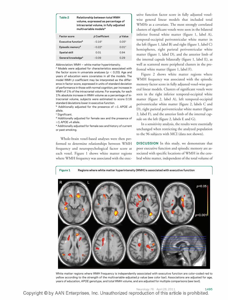
Whole-brain voxel-based analyses were then per-formed to determine relationships between WMHfrequency and neuropsychological factor score ateach voxel. Figure 1 shows white matter regionswhere WMH frequency was associated with the exec-
utive function factor score in fully adjusted voxel-wise general linear models that included totalWMHr as a covariate. The most strongly correlatedclusters of significant voxels were seen in the bilateralinferior frontal white matter (figure 1, label A),temporal-occipital periventricular white matter inthe left (figure 1, label B) and right (figure 1, label C)hemispheres, right parietal periventricular whitematter (figure 1, label D), and the anterior limb ofthe internal capsule bilaterally (figure 1, label E), aswell as scattered more peripheral clusters in the pre-frontal white matter (figure 1, label F).
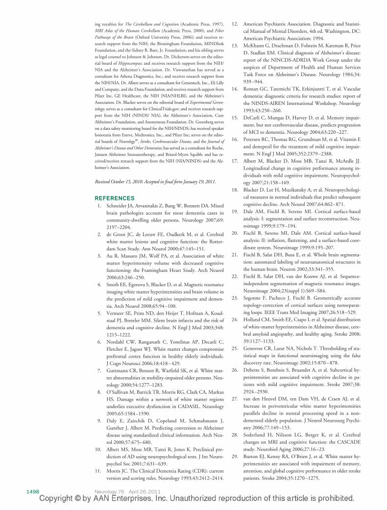
Figure 2 shows white matter regions whereWMH frequency was associated with the episodicmemory factor score in fully adjusted voxel-wise gen-eral linear models. Clusters of significant voxels wereseen in the right inferior temporal-occipital whitematter (figure 2, label A), left temporal-occipitalperiventricular white matter (figure 2, labels C andD), right parietal periventricular white matter (figure2, label F), and the anterior limb of the internal cap-sule on the left (figure 2, labels E and G).
In a sensitivity analysis, the results were essentiallyunchanged when restricting the analyzed populationto the 96 subjects with MCI (data not shown).
DISCUSSION In this study, we demonstrate thatpoor executive function and episodic memory are as-sociated with specific locations of WMH in the cere-bral white matter, independent of the total volume of
Table 2 Relationship between total WMHvolume, expressed as percentage ofintracranial volume, in fully adjustedmultivariable modelsa
Factor score � Coefficient p Value
Executive functionb �0.16c 0.05c
Episodic memoryd �0.22c 0.01c
Spatial skill 0.01 0.94
General knowledgee �0.09 0.29
Abbreviation: WMH � white matter hyperintensity.a Models were adjusted for characteristics associated withthe factor score in univariate analyses ( p � 0.20). Age andyears of education were covariates in all the models. Themodel WMH � coefficient may be interpreted as the differ-ence in factor score, expressed in units of standard deviationof performance in those with normal cognition, per increase inWMH of 1% of the intracranial volume. For example, for each1% absolute increase in WMH volume as a percentage of in-tracranial volume, subjects were estimated to score 0.16standard deviations lower in executive function.b Additionally adjusted for the presence of �1 APOE �4allele.c Significant.d Additionally adjusted for female sex and the presence of�1 APOE �4 allele.e Additionally adjusted for female sex and history of currentor past smoking.
Figure 1 Regions where white matter hyperintensity (WMH) is associated with executive function
White matter regions where WMH frequency is independently associated with executive function are color-coded red toyellow according to the strength of the multivariable-adjusted p value (see color bar). Associations are adjusted for age,years of education, APOE genotype, and total WMH volume, and are adjusted for multiple comparisons (see text).
Neurology 76 April 26, 2011 1495

WMH. These observations support the hypothesisthat WMH causes vascular cognitive impairment bydisrupting cortical connections mediated by specificwhite matter tracts. We find that WMH are associ-ated with memory impairment as well as executivedysfunction, which has also been observed in otherstudies.2
Most previous studies have correlated cognitiveperformance with global WMH volume. Relativelyfew studies have examined relationships between thetopography of WMH frequency and the resultingcognitive deficits, and these clinical-anatomic corre-lation studies have generally divided the white matterinto large regions of interest, such as entire lobes orperiventricular vs subcortical regions.2,6,26-30 This ap-proach does not take into account the fact that suchlarge areas will contain multiple white matter tractscoursing to many destinations, however. Despite thislimitation, some previous studies have demonstratedclinical-anatomic correlations between WMH andcognitive function. In general, periventricular WMHare more closely associated with cognitive impair-ment or cognitive decline, possibly suggesting a rolefor dysfunction of long association tracts.2,26-28 Onestudy of stroke patients suggests that frontal WMHis associated with executive dysfunction and tempo-ral WMH is associated with memory impairment.29
Another study found that left dorsolateral prefrontalcortex WMH is associated with decreased perfor-mance on a working memory task.6 In a study using a
visual rating scale to grade severity of WMH along awhite matter pathway of interest, lesions in cholin-ergic pathways were shown to be associated withmemory impairment.30
An advantage of the present study is the use ofsmaller regions of interest, namely the individualMRI voxels, to perform regional clinical-anatomiccorrelations with higher spatial resolution. Using thistechnique we were able to show strong regional cor-relations between WMH frequency and dysfunctionin specific cognitive domains. We also controlled fortotal WMH volume when determining regions ofcorrelation, allowing us to determine that these re-gional correlations are specific to that anatomic areaand are independent of total lesion volume. Becausethe frequency of WMH at any location is partly cor-related with total lesion volume, relationships be-tween regional WMH frequency and cognition areconfounded by total lesion volume, and may not bevalid unless this confounding is considered. By cova-rying for total lesion volume in our analyses, we canconclude with reasonable certainty that the observedrelationships between regional WMH frequency andcognitive functions are equally true in those with lowtotal WMH volume and high total WMH volume.
In this study, we were unable to directly identifythe white matter tracts traveling though the regionswhere associations were seen between WMH fre-quency and cognition. We did not perform diffusiontensor imaging, which can be used to map white
Figure 2 Regions where white matter hyperintensity (WMH) is associated with episodic memory
White matter regions where WMH frequency is independently associated with episodic memory are color-coded red toyellow according to the strength of the multivariable-adjusted p value (see color bar). Associations are adjusted for age,years of education, APOE genotype, and total WMH volume, and are adjusted for multiple comparisons (see text).
1496 Neurology 76 April 26, 2011

matter tracts. This notwithstanding, we believe it isreasonable to speculate on which tracts might be in-volved based on tract-tracing studies in monkeys31
and diffusion tensor imaging studies in monkeys32
and humans33-35 that provide insights into the ana-tomic locations of cerebral white matter tracts. Thelabeled WMH regions associated with impaired exec-utive function in figure 1 may involve the followingfiber tracts: the uncinate fasciculus, linking rostraltemporal with orbital and medial prefrontal cortices(figure 1, label A); the inferior longitudinal fascicu-lus, linking rostral and caudal regions of the ventralvisual steam (figure 1, labels B and C); fronto-occipital fibers (figure 1, label C); superior longitudi-nal fasciculus that links parietal and temporal regionswith the prefrontal cortex and that is thought to playa role in spatial processing and spatial attention (fig-ure 1, label C); cingulum bundle, relevant for moti-vation and behavioral control (figure 1, label D);anterior limb of the internal capsule and, on the left,genu of the internal capsule and striatal fibers cours-ing in the bundle of Muratoff to the caudate nucleus(figure 1, label E); and multiple types of fibers enter-ing and leaving the cortex (figure 1, label F). Theinvolvement of the anterior limb of the internal cap-sule is particularly interesting because it conveys in-formation between prefrontal cortex and the medialdorsal and anterior thalamic nuclei, and the prefron-topontine fibers destined for the cerebellar posteriorlobe—subcortical areas now known to be relevant forexecutive function.36,37 The labeled WMH regionsassociated with impaired episodic memory (figure 2)may involve the following fiber tracts: fronto-occipital fibers and the inferior longitudinal fascicu-lus (figure 2, labels A and D); rostral and caudallimbs of the arc of the cingulum bundle (figure 2,labels B, C, and F); anterior limb and genu of theinternal capsule (figure 2, label E); and bundle ofMuratoff (figure 2, label G). The cingulum bundle isimplicated in the motivational and emotional aspectsof behavior, and it is also thought to contribute todifferent aspects of mnemonic processing by virtue ofits connections with the hippocampus, parahip-pocampal regions, and retrosplenial cortex, in addi-tion to its connections with the parietal and frontallobes.31 The finding that WMH in the left anteriorlimb of the internal capsule, but not the right, wasassociated with worse episodic memory may reflectthe predominant weighting of verbal memory, as op-posed to visuospatial memory, in the factor score.
A limitation is that our findings were derivedfrom a population that mostly consisted of subjectswith mildly impaired cognition and may not be validin other populations with different distributions ofcognitive impairment. The distribution of cognitive
impairment of our subjects reflects the original de-sign of the study, with intentional enrichment of thestudy population for subjects with MCI.9 A sensitiv-ity analysis showed that the cluster locations wereessentially unchanged when the analyzed populationwas restricted to those with MCI, suggesting that thestudy findings are robust for this group. Furtherstudies with larger sample sizes will be needed to con-firm whether our findings are relevant to patientswith overall normal cognition or dementia. Anotherlimitation is that our analysis of the neuropsycholog-ical data did not permit investigation of componentsof memory such as encoding or retrieval, which maydepend on different anatomic pathways; future stud-ies will be needed to address this.
Our findings suggest that WMH location shouldbe considered in subsequent studies that attempt todetermine the relevance of WMH to cognitive func-tion. Such studies should attempt to confirm theclinical relevance of WMH in the regions identifiedin our work. Future studies could also incorporatequantitative imaging markers of tissue microstruc-ture such as measures of water proton diffusion,which seems to offer additional clinically relevant in-formation beyond that conferred by the presence orabsence of T2 hyperintensity.38 White matter diffu-sivity reflects microstructural changes that can becaused by processes other than WMH, and has beenobserved in AD (presumably as a consequence ofneurodegeneration with secondary alteration ofwhite matter integrity)39 as well as in normal aging.40
The optimal way to integrate and interpret WMHand diffusion information is a critical subject for fu-ture studies. Ultimately, a better understanding ofWMH location and its significance could inform theindividual clinical evaluation of patients with cogni-tive impairment, by allowing the discrimination ofclinically relevant patterns of WMH from relatively“benign” patterns of WMH sometimes seen in cog-nitively normal individuals.
AUTHOR CONTRIBUTIONSStatistical analysis was conducted by Dr. Eric Smith and Dr. David Salat.
DISCLOSUREDr. Smith has received speaker honoraria from the BMJ Group and
QuantiaMD; has received an honorarium from the Canadian Conference
on Dementia; has served on a scientific advisory board for Genentech,
Inc.; serves as an Assistant Editor for Stroke; and receives research support
from the NIH, the Alberta Heritage Foundation for Medical Research,
Canadian Institutes for Health Research, Heart and Stroke Foundation of
Canada, Canadian Stroke Network, and the Hotchkiss Brain Institute.
Dr. Salat receives research support from the NIH/NINR. J. Jeng and Dr.
McCreary report no disclosures. Dr. Fischl serves on the editorial board of
NeuroImage; has served as a consultant for Cephalon, Inc.; and receives
research support from the NIH (NCRR/NIBIB/NIA/NINDS) and the
Ellison Medical Foundation. Dr. Schmahmann serves on the editorial
board of The Cerebellum; is listed as author on a pending patent re: Trans-
cranial magnetic stimulation applied to the cerebellum; receives publish-
Neurology 76 April 26, 2011 1497
ing royalties for The Cerebellum and Cognition (Academic Press, 1997),
MRI Atlas of the Human Cerebellum (Academic Press, 2000), and Fiber
Pathways of the Brain (Oxford University Press, 2006); and receives re-
search support from the NIH, the Birmingham Foundation, MINDlink
Foundation, and the Sidney R. Baer, Jr. Foundation; and his sibling serves
as legal counsel to Johnson & Johnson. Dr. Dickerson serves on the edito-
rial board of Hippocampus; and receives research support from the NIH/
NIA and the Alzheimer’s Association. Dr. Viswanathan has served as a
consultant for Athena Diagnostics, Inc.; and receives research support from
the NIH/NIA. Dr. Albert serves as a consultant for Genentech, Inc., Eli Lilly
and Company, and the Dana Foundation; and receives research support from
Pfizer Inc, GE Healthcare, the NIH (NIA/NHLBI), and the Alzheimer’s
Association. Dr. Blacker serves on the editorial board of Experimental Geron-
tology; serves as a consultant for ClinicalTrials.gov; and receives research sup-
port from the NIH (NINDS/ NIA), the Alzheimer’s Association, Cure
Alzheimer’s Foundation, and Anonymous Foundation. Dr. Greenberg serves
on a data safety monitoring board for the NIH/NINDS; has received speaker
honoraria from Esteve, Medtronics, Inc., and Pfizer Inc; serves on the edito-
rial boards of Neurology®, Stroke, Cerebrovascular Disease, and the Journal of
Alzheimer’s Disease and Other Dementias; has served as a consultant for Roche,
Janssen Alzheimer Immunotherapy, and Bristol-Myers Squibb; and has re-
ceived/receives research support from the NIH (NIA/NINDS) and the Alz-
heimer’s Association.
Received October 15, 2010. Accepted in final form January 19, 2011.
REFERENCES1. Schneider JA, Arvanitakis Z, Bang W, Bennett DA. Mixed
brain pathologies account for most dementia cases incommunity-dwelling older persons. Neurology 2007;69:2197–2204.
2. de Groot JC, de Leeuw FE, Oudkerk M, et al. Cerebralwhite matter lesions and cognitive function: the Rotter-dam Scan Study. Ann Neurol 2000;47:145–151.
3. Au R, Massaro JM, Wolf PA, et al. Association of whitematter hyperintensity volume with decreased cognitivefunctioning: the Framingham Heart Study. Arch Neurol2006;63:246–250.
4. Smith EE, Egorova S, Blacker D, et al. Magnetic resonanceimaging white matter hyperintensities and brain volume inthe prediction of mild cognitive impairment and demen-tia. Arch Neurol 2008;65:94–100.
5. Vermeer SE, Prins ND, den Heijer T, Hofman A, Koud-staal PJ, Breteler MM. Silent brain infarcts and the risk ofdementia and cognitive decline. N Engl J Med 2003;348:1215–1222.
6. Nordahl CW, Ranganath C, Yonelinas AP, Decarli C,Fletcher E, Jagust WJ. White matter changes compromiseprefrontal cortex function in healthy elderly individuals.J Cogn Neurosci 2006;18:418–429.
7. Guttmann CR, Benson R, Warfield SK, et al. White mat-ter abnormalities in mobility-impaired older persons. Neu-rology 2000;54:1277–1283.
8. O’Sullivan M, Barrick TR, Morris RG, Clark CA, MarkusHS. Damage within a network of white matter regionsunderlies executive dysfunction in CADASIL. Neurology2005;65:1584–1590.
9. Daly E, Zaitchik D, Copeland M, Schmahmann J,Gunther J, Albert M. Predicting conversion to Alzheimerdisease using standardized clinical information. Arch Neu-rol 2000;57:675–680.
10. Albert MS, Moss MB, Tanzi R, Jones K. Preclinical pre-diction of AD using neuropsychological tests. J Int Neuro-psychol Soc 2001;7:631–639.
11. Morris JC. The Clinical Dementia Rating (CDR): currentversion and scoring rules. Neurology 1993;43:2412–2414.
12. American Psychiatric Association. Diagnostic and Statisti-cal Manual of Mental Disorders, 4th ed. Washington, DC:American Psychiatric Association; 1994.
13. McKhann G, Drachman D, Folstein M, Katzman R, PriceD, Stadlan EM. Clinical diagnosis of Alzheimer’s disease:report of the NINCDS-ADRDA Work Group under theauspices of Department of Health and Human ServicesTask Force on Alzheimer’s Disease. Neurology 1984;34:939–944.
14. Roman GC, Tatemichi TK, Erkinjuntti T, et al. Vasculardementia: diagnostic criteria for research studies: report ofthe NINDS-AIREN International Workshop. Neurology1993;43:250–260.
15. DeCarli C, Mungas D, Harvey D, et al. Memory impair-ment, but not cerebrovascular disease, predicts progressionof MCI to dementia. Neurology 2004;63:220–227.
16. Petersen RC, Thomas RG, Grundman M, et al. Vitamin Eand donepezil for the treatment of mild cognitive impair-ment. N Engl J Med 2005;352:2379–2388.
17. Albert M, Blacker D, Moss MB, Tanzi R, McArdle JJ.Longitudinal change in cognitive performance among in-dividuals with mild cognitive impairment. Neuropsychol-ogy 2007;21:158–169.
18. Blacker D, Lee H, Muzikansky A, et al. Neuropsychologi-cal measures in normal individuals that predict subsequentcognitive decline. Arch Neurol 2007;64:862–871.
19. Dale AM, Fischl B, Sereno MI. Cortical surface-basedanalysis: I: segmentation and surface reconstruction. Neu-roimage 1999;9:179–194.
20. Fischl B, Sereno MI, Dale AM. Cortical surface-basedanalysis: II: inflation, flattening, and a surface-based coor-dinate system. Neuroimage 1999;9:195–207.
21. Fischl B, Salat DH, Busa E, et al. Whole brain segmenta-tion: automated labeling of neuroanatomical structures inthe human brain. Neuron 2002;33:341–355.
22. Fischl B, Salat DH, van der Kouwe AJ, et al. Sequence-independent segmentation of magnetic resonance images.Neuroimage 2004;23(suppl 1):S69–S84.
23. Segonne F, Pacheco J, Fischl B. Geometrically accuratetopology-correction of cortical surfaces using nonseparat-ing loops. IEEE Trans Med Imaging 2007;26:518–529.
24. Holland CM, Smith EE, Csapo I, et al. Spatial distributionof white-matter hyperintensities in Alzheimer disease, cere-bral amyloid angiopathy, and healthy aging. Stroke 2008;39:1127–1133.
25. Genovese CR, Lazar NA, Nichols T. Thresholding of sta-tistical maps in functional neuroimaging using the falsediscovery rate. Neuroimage 2002;15:870–878.
26. Debette S, Bombois S, Bruandet A, et al. Subcortical hy-perintensities are associated with cognitive decline in pa-tients with mild cognitive impairment. Stroke 2007;38:2924–2930.
27. van den Heuvel DM, ten Dam VH, de Craen AJ, et al.Increase in periventricular white matter hyperintensitiesparallels decline in mental processing speed in a non-demented elderly population. J Neurol Neurosurg Psychi-atry 2006;77:149–153.
28. Soderlund H, Nilsson LG, Berger K, et al. Cerebralchanges on MRI and cognitive function: the CASCADEstudy. Neurobiol Aging 2006;27:16–23.
29. Burton EJ, Kenny RA, O’Brien J, et al. White matter hy-perintensities are associated with impairment of memory,attention, and global cognitive performance in older strokepatients. Stroke 2004;35:1270–1275.
1498 Neurology 76 April 26, 2011

30. Bocti C, Swartz RH, Gao FQ, Sahlas DJ, Behl P, BlackSE. A new visual rating scale to assess strategic white mat-ter hyperintensities within cholinergic pathways in demen-tia. Stroke 2005;36:2126–2131.
31. Schmahmann JD, Pandya DN. Fiber Pathways of theBrain. New York: Oxford University Press; 2006.
32. Schmahmann JD, Pandya DN, Wang R, et al. Associationfibre pathways of the brain: parallel observations from dif-fusion spectrum imaging and autoradiography. Brain2007;130:630–653.
33. Mori S, Oishi K, Jiang H, et al. Stereotaxic white matteratlas based on diffusion tensor imaging in an ICBM tem-plate. Neuroimage 2008;40:570–582.
34. Catani M, Thiebaut de Schotten M. A diffusion tensorimaging tractography atlas for virtual in vivo dissections.Cortex 2008;44:1105–1132.
35. Makris N, Worth AJ, Sorensen AG, et al. Morphometry ofin vivo human white matter association pathways with
diffusion-weighted magnetic resonance imaging. AnnNeurol 1997;42:951–962.
36. Schmahmann JD. Vascular syndromes of the thalamus.Stroke 2003;34:2264–2278.
37. Schmahmann JD, Pandya DN. Disconnection syndromesof basal ganglia, thalamus, and cerebrocerebellar systems.Cortex 2008;44:1037–1066.
38. Viswanathan A, Patel P, Rahman R, et al. Tissue micro-structural changes are independently associated with cog-nitive impairment in cerebral amyloid angiopathy. Stroke2008;39:1988–1992.
39. Salat DH, Tuch DS, van der Kouwe AJ, et al. White mat-ter pathology isolates the hippocampal formation in Alz-heimer’s disease. Neurobiol Aging 2008;31:244–256.
40. Salat DH, Tuch DS, Greve DN, et al. Age-related alter-ations in white matter microstructure measured bydiffusion tensor imaging. Neurobiol Aging 2005;26:1215–1227.
Historical Abstract: January 23, 2001
HIV-ASSOCIATED NEUROLOGIC DISEASE INCIDENCE CHANGES: MULTICENTER AIDS COHORT STUDY, 1990–1998
N. Sacktor, R.H. Lyles, R. Skolasky, C. Kleeberger, O.A. Selnes, E.N. Miller, J.T. Becker, B. Cohen, J.C. McArthur,and the Multicenter AIDS Cohort Study
Neurology 2001;56:257–260
This study examined the temporal trends in the incidence rates of HIV dementia, cryptococcal meningitis, toxoplasmosis, progressivemultifocal leukoencephalopathy, and CNS lymphoma from January 1990 to December 1998 in the Multicenter AIDS Cohort Study.The incidence rates for HIV dementia, cryptococcal meningitis, and lymphoma decreased following the introduction of highly activeantiretroviral therapy (HAART). The proportion of new cases of HIV dementia with a CD4 count in a higher range (i.e., 201 to 350)since 1996 may be increasing.
Free Access to this article at www.neurology.org/content/56/2/257
Comment from Richard M. Ransohoff, MD, Associate Editor: This report showed that AIDS-related dementia was reducedwith introduction of active antiretroviral therapies, illuminating its pathogenesis.
Neurology 76 April 26, 2011 1499

DOI 10.1212/WNL.0b013e318217e7c8 2011;76;1492Neurology
E.E. Smith, D.H. Salat, J. Jeng, et al.episodic memory
Correlations between MRI white matter lesion location and executive function and
April 30, 2013This information is current as of
ServicesUpdated Information &
http://www.neurology.org/content/76/17/1492.full.htmlincluding high resolution figures, can be found at:
Supplementary Material
.DC1.htmlhttp://www.neurology.org/content/suppl/2011/04/24/76.17.1492Supplementary material can be found at:
References
1http://www.neurology.org/content/76/17/1492.full.html#ref-list-at:This article cites 38 articles, 15 of which can be accessed free
Citations
urlshttp://www.neurology.org/content/76/17/1492.full.html#related-This article has been cited by 2 HighWire-hosted articles:
Subspecialty Collections
http://www.neurology.org/cgi/collection/volumetric_mriVolumetric MRI
http://www.neurology.org/cgi/collection/mriMRI
http://www.neurology.org/cgi/collection/memoryMemory
mpairmenthttp://www.neurology.org/cgi/collection/mci_mild_cognitive_iMCI (mild cognitive impairment)
s_dementiahttp://www.neurology.org/cgi/collection/all_cognitive_disorderAll Cognitive Disorders/Dementiafollowing collection(s):This article, along with others on similar topics, appears in the
Permissions & Licensing
http://www.neurology.org/misc/about.xhtml#permissionstables) or in its entirety can be found online at: Information about reproducing this article in parts (figures,
Reprints http://www.neurology.org/misc/addir.xhtml#reprintsus
Information about ordering reprints can be found online: