cover page - core.ac.uk · op gezag van de rector magnificus prof. mr. p.f ... suffocation and...
TRANSCRIPT

Cover Page
The handle http://hdl.handle.net/1887/19952 holds various files of this Leiden University dissertation.
Author: Vonk, Freek Jacobus Title: Snake evolution and prospecting of snake venom Date: 2012-09-06

Snake Evolution and
Prospecting of Snake Venom
Freek Jacobus Vonk

To my family and (snake) friends, with special thanks to my parents…..
Freek Jacobus Vonk
Snake Evolution and Prospecting of Snake Venom
Dissertation Leiden University
Cover photo: a wild king cobra (Ophiophagus hannah), Indonesia (Java). By Freek Vonk
Printed by: Wöhrmann Print Services, Zutphen.
Copyright © 2012 by Freek J. Vonk, Leiden, The Netherlands. All rights reserved.

Snake Evolution and Prospecting of
Snake Venom
Proefschrift
ter verkrijging van
de graad van Doctor aan de Universiteit Leiden,
op gezag van de Rector Magnificus Prof. Mr. P.F. van der Heijden,
volgens besluit van het College voor Promoties
te verdedigen op donderdag 6 september 2012
klokke 10:00 uur
door
Freek Jacobus Vonk
Geboren te Dordrecht,
Nederland 24 februari, 1983.

Promotiecommissie
Promotor Prof. Dr. Michael K. Richardson
Overige leden Prof. Dr. Carel J. ten Cate
Prof. Dr. Herman P. Spaink
Dr. Bart Vervust (University of Antwerp)
Prof. Dr. Rob E. Poelman (Leiden University Medical Centre)
Prof. Dr. Peter G.L. Klinkhamer
The work described in this thesis was supported by a Toptalent grant from the Netherlands Organization
for Scientific Research under grant number 021.002.034 and funding from the Naturalis Biodiversity
Center (Leiden).

Contents
Chapter 1 Introduction 1
Chapter 2 Snake Venom: from Fieldwork to the
Clinic.
3
Chapter 3 Evolutionary Origin and Development
of Snake Fangs
25
Chapter 4 Axial Patterning in Snakes and
Caecilians: Evidence for an Alternative
Interpretation of the Hox Code.
39
Chapter 5 Massive Evolutionary Expansion of
Venom Genes in the King Cobra
60
Chapter 6 An Efficient Analytical Platform for
On-line Microfluidic Profiling of
Neurotoxic Snake Venoms Towards
Nicotinic Receptor Like Affinity
78
Chapter 7 Summary of Thesis and Discussion 112
Samenvatting van Thesis en Discussie 116
References 120
Curriculum vitae 139
Acknowledgements 142


1
Chapter 1: Introduction
Snakes are limbless predators that have evolved two different ways of incapacitating prey: suffocation
and envenomation. The evolution of these life-history strategies lie in close relation with the evolution of
body-elongation and limblessness. Taken together, these adaptations underlie the massive radiation of
snakes which occurred after the K-T boundary and extinction of the dinosaurs, presumably because
multiple niches became available and many of their predators got extinct. In this thesis, I examine the
evolution of these adaptations. A general question to address would be: what underlies these adaptations
in snakes and how do they correspond with their ecology? In addition to these evolutionary questions, I
also provide a summary in chapter one on the current status of using snake venom toxins in drug
discovery and design, because the evolution of venom is closely related to the evolution of a serpentine
body form and fangs, but also to the question of “why” snake venom is so interesting for the
pharmacological prospector. The high potency and specifity of snake venom toxins is generated by
millions of years of evolution and strong selective pressures, so it is my believe that the different chapters
in my thesis are tightly bound together. It is believed that it was Aristotle who first suggested – on the
basis of symptoms observed in snakebite victims – that snakes may also be used to cure certain diseases
(it was not yet known that venom was responsible for the observed effects). And in 1835 venom was
already (although unsuccessfully) used to treat rabies. This field of “bioprospecting” has accelerated
especially in the last decade, mainly because the development of many new sequencing and screening
techniques. In chapter two, we go into the development and evolution of the different snake venom-
conducting fangs. This has been a matter of great controversy among scientists for the last century and
many different hypotheses have been proposed. This was particularly because the two groups of snakes
with fangs in the front of their upper jaw (Elapidae and Viperidae), have only relatively recently in the
90s been shown not to form a monophyletic group, hinting towards an independent origin and evolution
of front fangs. By using modern techniques such as in situ hybridization of snake embryos, and careful
histological analysis we were able to reconstruct the evolutionary history and make a solid hypothesis for

2
the origin and evolution of fangs. In the third chapter of this thesis we look at the evolution of body-
elongation and deregionalization in snakes, since that presumably has also been a major driver of venom
evolution by providing the ability for snakes to coil up and act as coiled spring that can strike and
envenomate prey at a distance. Since Hox determine the basic structure of an embryo we have performed
many in situ hybridisations in snake embryos and carefully compared the expression profiles of different
Hox genes. For the fourth chapter of this thesis we sequenced a draft genome of the largest venomous
snake in the world, the king cobra (Ophiophagus hannah) and annotated many of its venom genes. We
wanted to find out whether we could find evidence of gene duplication and / or gene modification of
physiological genes for use in the venom gland. The birth-and-death model of gene evolution is the
canonical framework for venom evolution, but was never actually thoroughly studies due to lack of
genomic resources. Our genomic sequences let us study the king cobra venom genes in relation to its
‘normal’ physiological genes. In the fifth chapter, we have set out to develop a new efficient and rapid
method of analysing snake venom mixtures and performing a biological assay at the same time. We used
the acetylcholine binding protein (AChBP) as biological target, because acetylcholine receptors are also
often involved in neurological disorders, such as Alzheimer’s and Myasthenia gravis. In the last chapter
of this thesis, I discuss this work and try to put it into broader perspective. The field of evolutionary
genomics, molecular evolution as well as bioprospecting is huge and broad. However, I strongly believe
that working on the edge of different scientific disciplines will provide you with a better understanding of
nature and allows you to answer more questions.
3
Chapter 2: Snake venom: from fieldwork to the clinic.
Vonk FJ, Jackson K, Doley R, Madaras F, Mirtschin PJ and N Vidal.
This chapter has been published as 'cover article' in BioEssays 33, 269-79 (2011).

4
Abstract
Snake venoms are recognized here as a grossly under-explored resource in pharmacological
prospecting. Discoveries in snake systematics demonstrate that former taxonomic bias in research
has led to the neglect of thousands of species of potential medical use. Recent discoveries reveal an
unexpectedly vast degree of variation in venom composition among snakes, from different species
down to litter mates. The molecular mechanisms underlying this diversity are only beginning to be
understood. However, the enormous potential that this resource represents for pharmacological
prospecting is clear. New high-throughput screening systems offer greatly increased speed and
efficiency in identifying and extracting therapeutically useful molecules. At the same time a global
biodiversity crisis is threatening the very snake populations on which hopes for new venom-derived
medications depend. Biomedical researchers, pharmacologists, clinicians, herpetologists and
conservation biologists must combine their efforts if the full potential of snake venom derived
medications is to be realized.

5
Introduction
Snakes are represented on earth today by some 3150 species 1 . Of these the vast majority (ca. 2700
species, see Fig. 1) represent a single massive diversification event which occurred after the K-T
boundary and extinction of the dinosaurs. This large and relatively recent group is known as Caenophidia
or “advanced snakes” and characterized by the possession of a venom-delivery system or components of
such a system 2. Snakes traditionally considered venomous are the 600 or so species with tubular front
fangs, muscularized venom glands, and a bite significantly dangerous to humans (Viperidae, Elapidae and
Atractaspidinae) - including well-known examples as the cobras, seasnakes, vipers and rattlesnakes. The
remaining caenophidians were traditionally classified as “Colubridae”, meaning snakes with a venom
gland whose venom poses no danger to humans, and who lack the fangs at the front of the mouth for
injecting it. The “Colubridae” has been shown to be paraphyletic, and most of its subfamilies have
recently been elevated to a familial rank in order to reflect their evolutionary distinctiveness (Fig. 1) 1,3.
“Colubrid” snakes have been largely neglected in venom research, because of the sole fact that bites have
not been perceived as of medical importance, except few species as the African boomslang (Dispholidus
typus) and twigsnakes (Thelotornis spp.) and the Asian yamakagashi (Rhabdophis tigrinus). However,
during the last few years, there has been a trend towards studying the venoms of these harmless venomous
snakes to, primarily, increase our understanding of venom evolution. It became evident that these
harmless snakes do secrete a strong acting venom with powerful toxins, although in a significantly lower
amount and without an efficient injection mechanism 2,4. This influenced the ongoing quest for venom
molecules that may be useful in fighting human disease - a snake need not be dangerous to humans for its
venom to still have a profound effect upon the human body. This field of biodiscovery is now rapidly
emerging, especially due to the development of modern high-throughput screening assays that allow rapid
identification of potential therapeutic agents.

6
Figure 1 | Phylogenetic tree showing the distribution of 28 snake venom protein families among advanced snakes, with the
number of currently known species of each family and the number of red listed species. Families in grey used to make up the old
traditional “Colubridae”. Phylogeny based on 1,5.
Snake venom contains a mixture of powerful proteins and peptides that have evolved to be targeted to
receptors, ion channels or enzymes 6, in addition to some carbohydrates, nucleosides, lipids, and metal
ions, whose functions are not all known 7,8. They interact with a wide variety of mammalian proteins and
can disrupt the central and peripheral nervous systems, the blood coagulation cascade, the cardiovascular
and neuromuscular systems, and homeostasis in general. These venom proteins act with great precision –
different toxins recognize different subtypes of certain receptors with only subtle differences – and are
very biologically active. The precision and power with which they work lies right at the centre of why
they form such a valuable resource to biochemists, biomedical researchers, evolutionary biologists and
others.

7
Several major human drugs or diagnostics have been developed based on snake venom
components. In addition, some fundamental biological processes have been revealed by using toxins as
probes to study cells and their receptors. Here we review the state of this field and emphasize that the
emerging field of high-throughput screening assays applied to a wide range of unstudied venoms, has the
potential to provide a solid basis for the discovery of new lead compounds for new drugs.
Forces driving evolutionary divergence in venom composition: diet, phylogeny,
biogeography and ontogeny
Only in relatively recent years has the remarkable variability of venom composition at the genus, species,
subspecies, population and even individual levels been fully appreciated, and the underlying ecological
forces and mechanisms of mutation at the molecular level started to be identified and understood.
Variation in venom composition has far-reaching implications. For the treatment of snakebite, for
example, the importance of using pooled venom (mixtures of venom from several individuals of the same
species representing different ages and geographical origins) has been emphasized in the production of
antivenom to – in some cases - produce a serum that will be maximally effective against a bite from any
snake of that species 9. However, it should be noted that many antivenoms provide excellent – sometimes
even better – cross reactions against antigens not even included in the original mixture, a phenomenon
which is not yet well understood.
Since different species of prey differ in their physiological reaction to venom molecules, it
follows that venom composition is linked to diet and varies from the species level all the way to
individuals. In saw-scaled vipers (Echis), venom from those species that feed on arthropods was highly
toxic to scorpions 10. By contrast, scorpions were unaffected by venom from those species that feed on
mammals 10. Phylogenetic analysis revealed repeated instances of co-evolution of venom composition
with prey preference. Other examples of variation in venom composition, at the individual to the species
level, correlated with diet have been found in coral snakes (Micrurus) 11, rattlesnakes (Crotalus and

8
Sistrurus) 9,12-14, Malayan pitvipers (Calloselasma) 15, lanceheads (Bothrops) 16,17, puff adders (Bitis
arietans) 18, and others.
Specificity of venom to prey type has also been found by studies approaching the question by
analyzing the composition of the venom. A single species of coral snake (Micrurus s. surinamensis),
distinctive within its genus in feeding on fish, was found to have venom containing neurotoxins lethal to
fish and not known from the venom of other Micrurus species 11. Significant differences in the protein
composition were also found in the venoms of three subspecies of the rattlesnake Sistrurus catenatus,
attributable to dietary differences between the subspecies 19. Of the 11 protein families represented in the
venom of this species, variation between the subspecies was found in all protein families, but variation
was greater in some than in others. The metalloproteinases proved to be the most conserved while the
PLA2s were the most divergent. PLA2s are often associated with neurotoxins – i.e the neurotoxin
molecules also have PLA2 activity (e.g. notexin, taipoxin, crotoxin, β-Bungarotoxin etc.), so it could be
that the neurotoxins vary more to accommodate the different prey types. Some toxins may assist in
prey breakdown (digestion) but others – like neurotoxins causing paralysis or haemotoxins causing rapid
low blood pressure or circulation – are perhaps more critical, they may determine whether the snake gets
the meal in the first place.
In addition to diet, other variables such as phylogeny, biogeography and ontogeny may also be
forces driving the evolutionary divergence of venom proteins (these factors may well be correlated with
diet in many cases) – and perhaps just even separation time between populations due to genetic drift 20.
Although most studies documenting variation attribute it to a single factor, considering these categories in
isolation may result in an incomplete understanding of the true scenario. For example, individuals of the
common lancehead (Bothrops asper) from two different localities and of different ages displayed
significant differences in venom proteome 21. Variation in symptoms of bite victims from individuals of
this species varying in age or geographic origin has been observed though never formally documented.
Ontogenetic differences in venom compositions are most probably explained by ontogenetic shifts in diet

9
22. The venom of neonates was found to differ significantly from that of adults, with a trend toward
increasing complexity with age 22. In the proteomes of neonate and adult individuals of the rattlesnake
Crotalus simus the venom of adults and neonates had only 50% of their proteome in common, with a
trend from neurotoxic changing to hemorrhagic properties from neonate to adult 23.
While ontogenetic variation certainly, and biogeographical variation in some cases, may well be
explained by variation in prey type, observations of variation in the venom proteome of male versus
female neonates from a single litter of the South American viper Bothrops jararaca 24 are puzzling and
underscore just how much remains to be learned about the evolutionary forces driving variation in venom
composition.
For the pharmacological prospector, variation is right at the heart of the potential goldmine of
molecules that snake venom represents since a high degree of variation in venom composition increases
the number of potentially useful novel molecules. Recognizing the crucial biomedical significance of
variation in venom underscores the importance of efforts by those field biologists working with snakes to
understand and conserve biological diversity, from the species down to the individual level. This
emphasizes the importance of fieldwork in biodiscovery and the collection of venom samples from a wide
range of species and specimens of different geographical localities and ages (Fig. 2).

10
Figure 2 | Due to the enormous diversity in venom composition and the accelerated evolution of different isoforms of each of the
toxin types in Fig. 1, fieldwork is essential to obtain samples from a wide range of species and specimens of different
geographical localities and ages to exploit the full pharmacological potential of venom. A: a rare species of “colubrid”, the green
tree snake (Dipsadoboa viridis), in the Republic of Congo (Africa), photo by K.J. B: one of the authors (F.J.V.) with a wild king
cobra (Ophiophagus hannah) – the longest venomous snake in the world - on the island of Java in Indonesia, photo by Smarley.
C. The same author obtaining a venom sample from an Australian King brown (Pseudechis rossignoli), photo by H-W Herrmann.
D. one of the authors (K.J.) removing a watersnake (Grayia ornata), mimic of the venomous Water cobra (Naja annulata) from a
net in the Republic of Congo.

11
Sources of toxin diversity: multiple splicing, exon insertion, exon switching, post-
translational modification and domain switching
Snake toxin genes are the result of gene-duplications of normal body proteins that are subsequently
selectively expressed in the venom gland 25, often followed by accelerated point mutations in the protein
coding regions 26. Gene duplication creates redundancy and allows the duplicated gene to escape the
pressures of negative selection and acquire new functions through accelerated adaptive molecular
evolution 26. Acetylcholinesterase is the only currently known exception, because both the toxin and the
normal enzyme are encoded by the same gene but differentially expressed using alternative splicing 27.
The molecular mechanisms that cause accelerated evolution – a bias towards nucleotide
mutations that lead to amino acid changes (nonsynonymous substitutions) compared to those that don’t
(synonymous substitutions) – are currently not understood. When the first snake genome becomes
available, this may shed light upon the molecular mechanisms that operate on the venom genes.
Interestingly, nucleotide sequences appear to determine the accelerated rate of point mutations 27. Specific
triplets were found to be more ‘stable’ with regards to mutations than others, and the stable triplets were
found to be higher in abundance in venom introns while the non-stable triplets were found higher in
abundance in venom exons. There also appears to be a bias for transversions over transitions in nucleotide
substitutions in some toxins 28.
There are several mechanisms by which molecular diversity of venom toxins is generated. First,
alternative splicing allows multiple different functional proteins to be created using the same exons (Fig.
3a) 29,30 as it may cause binding to different receptors 31 or change of target altogether. Second, exons may
be inserted into existing genes - as in denmotoxin (Fig. 3b), a three-finger toxin in the venom of the
“colubrid” mangrove catsnake (Boiga dendrophila) with bird-specific activity 32. Third, part of the intron
may be retained in the mRNA due to error in splicing (Fig. 3c). Also, a recent discovered mechanism
termed accelerated segment switch in exons to alter targeting (ASSET) may play an important role in

12
generating the molecular diversity in snake venom molecules 33 (Fig. 3d). During ASSET certain parts of
exons are changed through accelerated segment switch and generate a functionally new toxin with a
conserved structural fold. Sometimes, synergistic action between two different toxins may enhance their
potency 34,35.
Post-translational modifications such as disulphide bridge formation and proteolysis play
important roles in structural modification and acquisition of new functional sites. Both covalent and non-
covalent interactions between similar and dissimilar proteins can form complexes that may exhibit a much
higher pharmacological activity compared to the individual components. Sometimes formation of
hetero/homo dimeric or trimeric complexes may lead to recognition of new targets as protein-protein
interaction in complexes may expose critical amino acid residues that were otherwise buried in monomers
28.
New interaction sites are also formed in certain proteins to exhibit higher pharmacological potencies
through domain swapping. In domain swapping, exchange of identical structural elements takes place
between two or more molecules to form dimers or oligomers. For example, with the exchange of domains
by α and β subunits of IX/X binding protein, isolated from the venom of the Okinawa habu
(Protobothrops flavoviridis) 36, the hinge region forms a concave structure between the subunits and
provides a new functional site in the heterodimer 28. Without this swapping the loop would fold back and
the ligand binding site would be absent.

13
Figure 3 | Some molecular mechanisms by which the diversity of toxin proteins is achieved. A: normal and alternative splicing.
B: insertion of exon, as in denmotoxin 32. C: intron retention due to error in splicing. D: accelerated segment switch in exons to
alter targeting (ASSET) sas in a 3FTX from the venom of the rattlesnake Sistrurus catenatus 33. Colored segments (red, purple,
pink, yellow) represent exchanges of segments.

14
Snake venom enzymes and toxins
Of the following enzymes, some have currently been described in all snake venoms, and others from only
a limited number of species: PLA2s, metalloproteinases, serine proteases, acetylcholinesterases, LAOs,
and hyaluronidases (Fig. 1). Two important and diverse families are the PLA2s and the
metalloproteinases. PLA2 enzymes exhibit a wide variety of pharmacological effects in prey/victims and
are therefore interesting for the pharmacological prospector 37. Catalytically inactive Lys49 PLA2 homologs
– found in many viperid venoms and being strongly myotoxic – work through a mechanism independent of
hydrolysis 38. Furthermore, although different in their pharmacological and enzymatic activity, the PLA2-like
and PLA2 toxins are highly similar in sequence and structure and differ only for the substation of Asp-49
with Lys or Ser 39 - an example of how puzzling the mechanisms of toxicity can be. Some of the neurotoxic
PLA2 are either homo/heterodimeric complexes. For example, β–bungarotoxin contains a covalently linked
kunitz-type serine protease inhibitor which is involved in the blocking activity 40.
SVMPs are responsible for major local symptoms in snakebite - causing hemorrhage, edema,
hypotension, hypovolemia, inflammation and necrosis. They are divided into three groups (P-I to P-III)
based on the presence of other domains in the mature protein 41. PI SVMP’s have a prodomain and a
single metalloproteinase domain that overall causes less hemorrhagic action then the other types, but still
displays a variety of biological activities. The PII SVMP’s are composed of a metalloproteinase and
disintegrin domain, along with a prodomain.
Non-enzymatic venom proteins include 3FTXs, Kunitz-type serine protease inhibitors,
sarafotoxins, CRISP, disintegrins, C-type lectins, waprins, veficolins and vespryns. 3FTXs have three
beta-stranded loops resembling three-fingers and are mainly found in the venoms of elapids, some
“colubrids” 32,42 and, in low quantity, in some viperids 43,44. The structure is stabilized by four conserved
disulphide bridges 45,46. However, these structurally related molecules differ greatly in their biological
functions 47,48. Functional characterization of this family of proteins has contributed significantly to our

15
understanding of the mechanisms of venom toxicity and of normal physiological processes 48. For
example, characterization of α-bungarotoxin, a three-finger neurotoxin found in the venom of the banded
krait (Bungarus multicinctus), enabled the isolation of the human nAChR 49 and contributed to our
understanding of myasthenia gravis 50. Significant contributions have been made in determining the
distribution of specific receptors or ion channels in particular tissues or cells, identification of subtypes of
receptors, imaging receptor trafficking 51-53 as well as in the development of therapeutic agents for
treatment of adrenomyeloneuropathy and multiple sclerosis 54.
Snake venom kunitz-type serine protease inhibitors like dendrotoxin, calcicludine and the B chain
of β-bungarotoxin, act as Ca2+ and K+ channel blockers respectively 55-57. Textilinin, a kunitz-type serine
protease inhibitor from the venom of the highly dangerous Australian brown snake (Pseudonaja textilis)
is a reversible plasmin inhibitor and has promising potential for development of anti-bleeding agent 58.
Waprins have only recently been identified in snake venom and show homology to whey acidic
proteins 59. So far their functions are not really understood except for omwaprin, isolated from the Inland
taipan (Oxyuranus microlepidotus), which possesses selective antimicrobial activities 60, useful for
developing potential antibiotics.
CRISP toxins have molecular weights of 20-30 kDa and 16 conserved cysteine residues.
Functional characterization of some CRISP has revealed that they are involved in blocking cyclic
nucleotide-gated ion channels and block potassium-stimulated smooth muscle contraction 61. Therefore in
addition to their involvement in disrupting the normal physiological functions of victims they can be used
for studying ion channel chemistry. A CRISP toxin from the venom of the “colubrid” snake the Patagonia
green racer (Philodryas patagoniensis) was recently shown to cause damage to the murine gastrocnemius
muscle, an action never before shown for any CRISP toxin 62 – showing the potential “colubrid” venoms
have to find new toxins.

16
Snaclecs comprise two subclasses of protein, C-type lectins (CTLs) and C-type lectin-related
proteins (CLRPs), found in venoms of most families of advanced snakes. Snake venom CTLs are
involved in hemagglutinating and platelet aggregation activities during envenomation 63-66. The CLRPs
are involved in anticoagulant, procoagulant and agonist/antagonist of platelet activation 67,68.
Pharmacological characterization reveals that they either enhance or inhibit the function of coagulation
factors which underscores their potential in drug discovery for blood related diseases. Their immaculate
specificity also helps in understanding platelet physiology.
Disintegrins are a class of non-enzymatic that bind to integrins
expressed on platelets and other vascular endothelial cells as well as some tumor cells 69 – they are an
important class of cell surface receptors that are critically involved in cell–cell and cell–matrix
interactions and are therefore good candidates to understand the interaction between extracellular matrix
and cells 70. In addition to their role in antiplatelet activity, these molecules are used in the diagnosis of
cardiovascular diseases as well as serving as prototypes for therapeutic molecules in treatment of cancer.
The medicinal use of snake venom
A large number of venom proteins affect the haemostatic system 71 and can have procoagulant,
anticoagulant, fibrinolytic or platelet active activities. Ancrod (Arvin) from the venom of the Malayan
pitviper (Calloselasma rhodostoma), Batroxobin (Reptilase) from the common lancehead (Bothrops
atrox), Crotalase from the Eastern diamondback rattlesnake (Crotalus adamanteus) have all been used as
defibrinogenating agents for several clinical conditions including deep vein thrombosis, myocardial
infarction, pulmonary embolus, and many others 72. Venoms with anticoagulant properties are extensively
studied for possible medical applications. The drug Aggrastat (tirobifan) was developed from a compound
in the venom of the saw-scaled viper (Echis carinatus), and is used as an anti-platelet drug (glycoprotein
IIb/IIIa inhibitors) 73 and given to those with unstable angina (Fig. 4). Many venoms with procoagulant
properties find application in the diagnosis of clotting abnormalities, and most of the clotting pathways
can be assayed by some venom component. For example “Reptilase time” (B. atrox) assays for thrombin

17
inhibitors 74, “Ecarin” (E. carinatus) and “taipan time” (O. scutellatus) assays for phrothrombin, and
Russell’s viper (Daboia russelii) venom assays for factor X and for monitoring anticoagulant therapy 75.
Figure 4 | Mechanisms of action of the anticoagulant Aggrastat developed from a compound in the venom of the Indian saw-
scaled viper (Echis carinatus). A: aggregated platelets by formation of fibrinogen bridges between the glycoprotein IIb/IIIa
receptors. B: glycoprotein IIB / IIIA receptor antagonists like Aggrastat prevent platelet aggregation, and are mainly used in
patients with acute coronary syndromes.
A number of snake venoms create a transient condition of depressed blood pressure in
envenomed patients. ACE-inhibitors were developed from a bradykinin potentiating enzyme isolated
from the venom of the Brazilian pitviper (Bothrops jararaca) and approved in 1979 by the FDA 76 to treat
high-blood pressure and heart disease (Fig. 5). They work by blocking the switch between angiotensin-I
and angiotensin-II, the latter being a vasoconstrictor. These inhibitors are now prescribed worldwide and
have saved the lives of millions.
Many venoms have analgesic properties. Hannalgesin, derived from the venom of the king cobra
(Ophiophagus hannah, see Fig. 2b) is already in clinical trials 77. Promising toxins have been isolated
from the tropical rattlesnake (Crotalus durissuss terrificus) and some other related species. Compounds
derived from the Asiatic cobra (Naja kaouthia) are already in use in alternative medicine. Being more
18
powerful than morphine, cobra venom was used in the 1930’s for treating intractable pain in cancer
sufferers.
There is great deal of research currently been done into the anti-cancer properties of venoms. For
example malignant brain and spinal-cord tumors (gliomas) are not curable by surgery because they invade
the surrounding brain tissue without clear boundaries, making removal impossible. Disintegrins, like
contortrostatin from American copperhead (Agkistrodon contortrix) venom, prevent cells from sticking
together, and inhibit their interaction with surrounding tissue and result in a blockage of cell motility and
invasiveness 78.
It has been demonstrated that fibrin(ogen) plays separate and distinctive roles at different stages
of tumor growth and dissemination. At the primary site, fibrin deposition around the tumor could form a
protective barrier, but also limit tumor progression. On the other hand, fibrin deposits formed by
metastatic tumor cells may help disseminating these tumor cells 79. Anticoagulants and the removal of
fibrin could be an effective therapy. One of the earliest reports on the successful use of a venom
defibrinogenating enzyme was that of Wood and Hilgard 80.

19
Figure 5 | Mechanism of action of ACE inhibitors developed from a bradykinin potentiating enzyme isolated from the venom of
the Brazilian pitviper (Bothrops jararaca) and approved in 1979 by the FDA [75] to treat high-blood pressure and heart disease.
Angiotensin I causes vasoconstriction which raises the blood pressure.
Many cobra venoms contain Cobra Venom Factor (CVF), that activates and depletes the
mammalian immune-complement system 81. They are structural and functional analogues of the
mammalian serum complement factor C3 82. CVF is used as a tool to study various aspects of the
complement system and in this respect CVF is a uniquely useful venom component that has also been
found useful as an immuno suppressant agent in tissue transplantation and in cancer therapy 83.
Many venoms have antibacterial properties. For example, Stiles et al 84 found 2 antibacterial
bioactive L-amino acid oxidase components in King brown (Pseudechis australis) venom that were 70
and 17.5 times more effective in-vitro than tetracycline, a drug of choice for Aeromonas infections.

20
Antibacterial and antiparasitic effects of the venom of the Marajó lancehead (Bothrops marajoensis) were
shown to be caused by PLA2 and and L-amino acid oxidase toxins 85.
Antiviral activity has been demonstrated in several venoms, although no commercialization of any of
these compounds has yet taken place. The venom of the tropical rattlesnake (Crotalus durissus terrificus)
from Brazil has been shown to be active against the measles virus 86. The purified PLA2 venom
neurotoxin “taipoxin” from the coastal taipan (Oxyuranus scutellatus), Nigexine from the African black
necked cobra (Naja nigricolis) and a basic PLA2 from the Mozambique spitting cobra (Naja
mossambica) have been shown to have potent anti-viral activities against HIV-1 virus 87.
Myasthenia gravis, a chronic autoimmune disorder affecting 2 out of every 100,000 people, results
in progressive skeletal muscle weakness with rapid fatigue and loss of strength. It primarily affects
mastication, facial and swallowing muscles and in advanced cases, respiratory muscles. Autoimmune
antibodies destroy acetylcholine receptor sites at the neuromuscular junctions preventing nerve impulses
from reaching the muscles. A rapid, quantitative and sensitive radioimmunoassay using human
acetylcholine receptor, affinity labeled with purified neurotoxin, alpha-bungaratoxin, from the venom of
the Taiwan krait (Bungarus multicinctus) is used to diagnose the condition 88,89. Most venoms possessing
alpha neurotoxins are potential candidates for this purpose.
High-throughput screening to identify potential new medicines in venoms
A major obstacle in exploring snake venom for new leads into drug discovery is the low amount of
venom usually obtained during the process of milking (extracting venom from the venom glands of a live
snake, see Fig. 2c). In addition, it is difficult to extract venom from many of the “colubrid” snakes.
Because of this, venom research has focused mainly on those snakes that are dangerous to humans, but
this doesn’t necessarily reflect the potential of their venom for drug discovery. Some of the harmless
“colubrid” venom toxins have been shown to be as potent as those of some deadly elapids, only produced

21
in small amounts 4,90. For example, the venoms of the Patagonia green racer (Phildryas patagoniensis)
and Lichtenstein’s green racer (P. olfersii) have been shown to posses high proteolytic activity, degrading
fibrinogen and the vascular wall 91, and strong edematogenic and myotoxic activity 92, respectively. In
addition, a completely new family of toxins has recently been found in the venom of the Asian dog-faced
water snake (Cerberus rynchops). These toxins, called veficolins, may induce platelet aggregation and/or
initiate complement activation 93. Hence, the bias towards deadly snakes in exploring venoms for drug
discovery is completely unwarranted.
Alternatively with the use of molecular tools the venom gland transcriptome can be complied.
This approach not only allows us to understand the expression profile and the evolution of the venom
proteins 94, but also reveals the lowly expressed proteins which will expand our resource for
pharmaceutically active molecules.
Many of the medically significant venomous snakes produce hundred to a couple hundred
milligrams of dry venom in a single milking 95. Large species of elapids and viperids may produce from
several hundred mg’s up to more than a gram of venom in a single milking 95. The recently described
giant spitting cobra (Naja ashei) has been shown to produce the immense amount of 3 gm’s of weight of
dry venom 96, the largest amount ever collected during a single milking. The amount of venom that a
snake produces during milking is determined by the species, its geographic origin, its body size and
relative head size, and by the time of the year that it is milked, as well as by interactions among these
factors, body size being the primary factor 97.
When biomedical researchers are looking for lead therapeutic agents against diseases, they can
first look at those venoms of which the snakebite symptoms involve the same pathways as the disease, for
example, using venom that causes vasodilatation to look for potential blood pressure regulators. However,
snake venom is very complex and contains many different isoforms, some of which may only be present
in very low quantities in the venom. For example, nerve growth factor is only present in a concentration

22
of about 0.1-0.5% 98. A fraction that stimulates neurite outgrowth was first identified in tumour cell
extracts, but venom from cottonmouth vipers (Agkistrodon piscivorus), was then found to be up to 6,000
times more potent 99 – a discovery that allowed the authors to study mechanisms which regulate cell and
organ growth and for which they were awarded the Nobel prize in 1986. The problem is that venom
compounds that are present only in low concentrations don’t necessarily contribute to the bite
symptomology and may thus be easily overlooked, although they can be of high potential biomedical
interest. To exploit the full potential of snake venoms, a wide variety of them need to be fractionated and
all fractions tested separately in a high-throughput screen of interest. Mice have typically been used as
model organisms, in for example identifying genes involved in pain and anxiety, and in screening for
analgesic peptides 100. However, using mice is ethically controversial and low-throughput, screens also
require relatively large amounts of the precious venoms. Zebrafish (Danio rerio) are now more and more
used as model organisms to screen for new drugs, and this provides a significant window of opportunity
for using snake venom (or venom from other venomous animals) to screen. Zebrafish are cheaper to
maintain than rodents, their eggs and embryos are transparent so phenotypic changes can be visualized,
and they can be easily scaled up into high-throughput assays 101 that require minimal amounts of venom,
and automated imaging and analysis systems are available 102,103. Morpholino knockdowns can be
performed by injecting the yolk sac of embryos, allowing live imaging studies, and many disease-related
genes identified in humans have orthologs in zebrafish 101. Although zebrafish will never replace
mammalian models completely in the drug development pipeline, they do form a cost-effective bridge
between cell-based models on the one hand and rodent whole-organism models on the other 101.
Conservation of snakes
A recent report by conservation biologists 104 documented a disturbing trend - a global decline in non-
related snake species over the world during the same time period. Although they only looked at a small
number of snakes (17 populations of eight species), we need to keep in mind that any species of snake
that goes extinct may have held a new drug in its venom. Species having small home ranges, sedentary

23
habits and ambush feeding strategies are likely to be the most vulnerable as they rely on sites with
specific types of ground cover that are disrupted by anthropogenic activities, and as ambush foraging is
associated with a suite of life-history traits that involve low rates of feeding, growth and reproduction 105.
Current research on snake venoms is phylogenetically biased. Of the 1622 snake venom toxin
sequences available on Universal Protein Resource database, only 49 are sequenced from snakes that do
not possess any danger to humans. But with the high-throughput systems currently being developed this
may hopefully soon change.
Very little data actually exist on the true conservation status of snakes worldwide. Out of 2711
extant caenophidian species, 26 are CITES listed, i.e. less than 1%, while 967 are listed on the IUCN Red
List of Threatened Species, i.e. 36%. The main causes identified for their threatened status are caused by
humans: annual & perennial non-timber crops (272 sp.), logging & wood harvesting (179 sp.), housing &
urban areas (143 sp.), livestock farming & ranching (98 sp.), hunting & trapping (64 sp.) and natural
system modifications (57 sp.). Although the majority of the species listed have been categorized as Least
Concern (60%), this does not necessarily reflect their true status 104, and for another 22% of the species
the data are insufficient to categorize them accordingly. In addition, 87% of the species (837) have been
added to the Red List since 2007, highlighting the lack of proper attention in the years before.
If biogeography is considered, relatively well protected regions are North America (123 red listed
sp.), Meso America (325 sp.), the Philippines (84 sp.) and Europe (29 sp.). On the other hand, several
biodiversity hotspots such as South America (113 sp.), Sub-Saharan Africa (82 sp.), and the Caribbean
Islands (16 sp.), among others are neglected while these areas may represent the locations with the highest
potential for discovering snake species new to science, each representing new potential for advancing the
frontiers of clinical medicine.

24
Conclusions and future perspectives
While the use of snake venom for medicinal purposes dates back to ancient times, the past few decades
have seen several new drugs derived from components of snake venom becoming available to patients
worldwide. Here, we have reviewed recent advances in areas ranging from evolutionary to molecular
biology that taken altogether indicate new possibilities for contributions to clinical medicine and medical
research from snake venom. First, advances in snake systematics demonstrate a high degree of
phylogenetic bias in previous molecular prospecting in snake venom, increasing the number of species
from which medically useful molecules may be obtained. Secondly, discoveries in snake venom
composition have revealed an unexpectedly high degree of variation of snake venoms of the same species
linked to variables such as diet, geographical distribution, ontogeny and others. These two factors
increase the potential pool that snake venom represents for pharmacological prospecting. Finally, new and
innovative techniques in high throughput systems offer increased speed and efficiency in identifying and
extracting desirable molecules from snake venom. In conclusion, we emphasize the challenges faced by
the conservation of snake biodiversity, since it is ultimately on this that hopes for the development of new
therapeutic agents from snake venom depends.
Acknowledgments We are grateful to M. K. Richardson, R. M. Kini and M. Goyffon, for help in
criticallyr eading and commenting on the manuscript. This work was supported by a TopTalent grant
from the Netherlands Organization for Scientific Research (NWO; F.J.V.), the Leiden University Fund
(F.J.V.) and two grants from the The Herpetofauna Foundation (F.J.V.).

25
Chapter 3: Evolutionary Origin and Development of Snake Fangs
Vonk FJ, Admiraal JF, Jackson K, Reshef R, de Bakker MAG, Vanderschoot K, van den Berge
I, van Atten M, Burgerhout E, Beck A, Mirtschin PJ, Kochva E, Witte F, Fry BG, Woods A and
MK Richardson.
This chapter has been published as 'cover article' in Nature 454, 630-633 (2008).
All supplementary material can be found here:
http://www.nature.com/nature/journal/v454/n7204/suppinfo/nature07178.html

26
Many advanced snakes use fangs — specialised teeth associated with a venom gland 106-108
— to
introduce venom during prey capture or defence. Various front- and rear-fanged groups are
recognised, according to whether fangs are positioned anterior (e.g. cobras and vipers) or posterior
(e.g. grass snakes) in the upper jaw 109-111
. A fundamental controversy in snake evolution is whether
or not front and rear fangs share the same evolutionary and developmental origin 107,109-114
. Solving
this controversy would point to a major evolutionary transition that underlies the massive radiation
of advanced snakes, and to the underlying developmental events. We examine this issue by
visualising the tooth-forming epithelium in the upper jaw of 96 snake embryos, covering eight
species. We use the sonic hedgehog (shh) gene as a marker 115-118
, and reconstruct the development
in 3D in 41 of these. Here we show that front fangs develop from the posterior end of the upper jaw,
and we reveal a striking similarity in morphogenesis between front and rear fangs that argue for
their homology. In front-fanged snakes, the anterior part of the upper jaw lacks shh expression,
and ontogenetic allometry displaces the fang from its posterior developmental origin to its adult
front position — suggesting a posterior evolutionary origin. In rear-fanged snakes, the fangs
develop from an independent posterior dental lamina and retain their posterior position. In light of
our findings, we suggest a possible new model for the evolution of snake fangs; a posterior sub-
region of the tooth-forming epithelium became developmentally uncoupled from the remaining
dentition. This would have allowed the posterior teeth to evolve independently and in close
association with the venom gland, becoming highly modified in different lineages. This
developmental event could have facilitated the massive radiation of advanced snakes in the
Cenozoic era, resulting in the spectacular diversity of snakes seen today 5,107,119
.

27
Many advanced snakes (Caenophidia, sensu ref. 22) use venom, with or without constriction, to subdue
their prey 5,107. Their venom-delivery system includes a post-orbital venom gland associated with
specialised venom-conducting fangs108. Fangs can occupy various positions on the upper jaw, but are
always located on the maxilla and never on any other tooth-bearing bone 120 (Fig. 6C). Viperidae (vipers
& pitvipers), Atractaspis (Lamprophiidae, sensu ref. 22), and Elapidae (cobras and relatives) have tubular
front fangs (Fig. 6B,C). The remaining lineages are all non-front-fanged and called either ‘non-fanged’
(no distinguishable enlarged posterior tooth) or rear-fanged 107,120 (Fig. 6B,C). Rear fangs can be solid,
slightly or deeply grooved, but never tubular 120.
There has been active debate concerning the evolutionary origin of these different fang types, and
their relationships to the simple, unmodified teeth of non-fanged 121 basal snakes 107,109-114,121 such as
pythons and boas (Boidae). Proposed hypotheses include: (i) front-fanged snakes form a monophyletic
group and their fangs are derived from rear-fangs 113,122,123 ; (ii) elapid fangs are derived from front teeth
and viperid fangs from rear fangs 124,125 ; (iii) elapid and viperid fangs are both derived from rear-fangs 112
; and (iv) front and rear fangs have independent origins 107. Establishing the origin and evolutionary
transformation series between these dentition types requires a robust phylogeny to map the characters
onto. Since recent molecular phylogenies of caenophidians place the front-fanged Viperidae as relatively
basal and the front-fanged Elapidae as more recently derived 1,107 (Fig. 6A), the current evidence seems to
support the ‘independent-origin’ hypothesis 107.

28
Figure 6 | Adult maxillary dentition mapped onto a molecular snake phylogeny to show relative positions of the various fang
types. a, Phylogeny from ref. 16. b, c, Adult skulls (Supplementary Table 4): lateral views (b); palate, schematic ventral views (c;
maxilla coloured, fangs circled). Asterisks indicate species studied by electron microscopy (Supplementary Fig. 5,
Supplementary Table 3). The evolutionary changes leading from an unmodified maxillary dentition to the different fang types in
advanced snakes are indicated at the nodes: (1) continuous maxillary dental lamina, no specialized subregions—ancestral
condition for advanced snakes; (2) evolution of posterior maxillary dental lamina—developmental uncoupling of posterior from
anterior teeth; (3) starting differentiation of the posterior teeth with the venom gland; (4) loss of anterior dental lamina and
development of front fangs.

29
To date, this issue has not been examined using a molecular developmental approach, not least
because of the difficulty of obtaining snake embryos of all the different species. Development of fangs
and venom glands has been studied before in viperids 126,127, Natrix 128,129 (Natricidae), Spalerosophis 130
,
Thamnophis and Telescopus 106,108 (Colubridae). Those morphological studies identified a common
primordium of the venom gland and fangs 106,126,128, but did not visualise the odontogenic band (tooth-
forming epithelium: a band of epithelial tissue that invaginates and forms a dental lamina). Therefore, no
conclusions could be drawn about the origin and evolutionary transformation series of the fangs.
Here, we have carried out in situ hybridisation of the sonic hedgehog (shh) gene in 96 snake
embryos of multiple stages, and reconstructed the development of the maxillary dentition in 3D in 41 of
these through serial sections. We use nine advanced snake species — comprising two front-, one rear- and
one ‘non-fanged’ lineage. As outgroup we included the non-fanged waterpython (Liasis mackloti:
Boidea), which is basal to advanced snakes (Fig. 6). Shh is expressed in the odontogenic band in different
vertebrate species 115-117. By visualising this band, we aim to find evidence for the ancestral condition of
the maxillary dentition. A list of all material studied can be found in Supplementary Tables 1-4. We map
our characters onto the recently published, robust molecular phylogeny of advanced snakes obtained by
Vidal et. al. 2007 (ref. 22, and Fig. 6A).
In the waterpython (L. mackloti), shh expression reveals one continuous maxillary odontogenic
band (Fig. 7a). As confirmed by serial sections of embryos ranging from young to old, this band
invaginates to form one dental lamina — a single continuous, invaginating epithelium that will develop a
row of teeth (Fig. 8a & Fig. 9j-l). This is consistent with a recent morphological study of Python sebae 131.
The odontogenic band, with its associated lamina, appears along the entire rostrocaudal extent of the
upper jaw — from the premaxilla to the mandibular articulation (Fig. 7a). This suggests that the ancestral
condition for the maxillary dentition of advanced snakes is one dental lamina that appears along the entire
rostrocaudal extent of the upper jaw, lacking specialised sub-regions.

30
The early odontogenic band in the non-front-fanged grass snake Natrix natrix (Natricidae) and rat
snake Elaphe obsoleta (Colubridae) is similar to that of the waterpython (Fig. 7c & Suppl. Fig. 2f,i)
However, we show for the first time that there are two dental laminae which invaginate separately (Fig.
8b,e & Fig. 9a-c & Suppl. Fig. 8e-g) and fuse during development (Fig. 9c & Suppl. Fig. 3h-i). The
anterior lamina bears only teeth (Fig. 9c & Suppl. Fig. 3f) and is similar in development to the one in the
waterpython (Fig. 9j,k&l). The posterior lamina, however, bears teeth and forms the common primordium
with a post-orbital gland (Fig. 8b & Fig. 9b-c & Suppl. Fig. 3i). These develop into the rear fangs and
venom gland in the grass snake, and probably represent the first differentiation of the posterior teeth with
a venom gland in the rat snake. The latter observation is consistent with a recent MRI and histology study
132, showing the presence of a small gland in rat snakes. To verify that the anterior and posterior dental
laminae are truly developmentally independent, we ablated the primordium of the anterior lamina in
isolated developing upper jaws of the dice snake Natrix tessellate 133 (Suppl. Fig. 3m-o) 134. After
cultivation under the yolk sac membrane, we found that the posterior lamina, with its venom gland
and fangs, developed normally in the absence of the anterior lamina (Suppl. Fig. 4), showing that they are
developmentally independent.

31
Figure 7 | Shh expression in the embryonic snake palate, showing the posterior developmental origins of front fangs. a–d, Palate,
ventral view: top, anterior; scale bar, 0.5 mm; dotted lines, upper jaw (posterior margin of premaxilla to attachment of the
mandible); boxes, schemes of maxillary odontogenic band (purple, shh expression; grey, no shh expression). Positions of fangs in
b–d were identified histologically (Fig. 3, Supplementary Fig. 3). The odontogenic band in the front-fanged species is located
posterior in the upper jaw (b, d). In the non-fanged outgroup (a) and the rear-fanged Natrix (c), the odontogenic band extends
along the entire upper jaw. f, fang; mx, maxillary odontogenic band; pa, palatine odontogenic band; pt, pterygoid odontogenic
band. e, Ontogenetic allometry in the fang in the front-fanged Causus displaces the fang along the upper jaw (Supplementary
Figs 5–9, Supplementary Tables 5–9). Scale bars, 1 mm. We note the change in relative size of the upper jaw subregions: i,
anterior; ii, fang; iii, posterior. d.a.o., days after oviposition.

32
Figure 8 | Sections of the shh in situ hybridizations of the embryonic upper jaw in five snake species, showing the posterior and
anterior dental laminae. a–c, e–f, Sagittal sections, anterior to the left, of L. mackloti (Boidae) 22 d.a.o. (a), N. natrix (Natricidae)
22 d.a.o. (b), Calloselasma rhodostoma (Viperidae) 8 d.a.o. (c), N. natrix 22 d.a.o. (e), Naja siamensis (Elapidae) 23 d.a.o. (f). d,
transverse section, medial to the left, of Trimeresurus hageni (Viperidae) 8 d.a.o. The posterior maxillary dental laminae in b and
e are similar in morphogenesis to the maxillary dental laminae in all front-fanged species examined (c, d, f; see also Fig. 4).
Arrowheads, shh expression; amdl, anterior maxillary dental lamina; dr, dental ridge; e, eye; f, fang; mdl, maxillary dental
lamina; pa, palatine dental lamina; pmdl, posterior maxillary dental lamina; t, tooth bud; vd, primordium of venom gland; scale
bars, 300 mm.
In the five front-fanged species examined (Viperidae and Elapidae), the maxillary odontogenic
band is found in the posterior part of the upper jaw (Fig. 7b,d & Suppl. Fig. 2a,d,g). There is no shh
expression nor dental lamina in the anterior region (verified by histology, data not shown). By contrast, in
the waterpython, grass and rat snake, the odontogenic band and associated dental laminae appear along
the entire rostrocaudal extent of the upper jaw. We find that, during development, the ‘rear’ fang is
displaced to its adult ‘front’ position by ontogenetic allometry (Suppl. Figs. 6 & 7 and Suppl. Table 6 for
statistical analyses); suggesting a posterior evolutionary origin of the front fangs. Histology shows that
although the odontogenic band invaginates normally and forms one dental lamina (in contrast to the non-
front-fanged snakes examined above), in all front-fanged species the fangs develop from the
posteriormost part of this lamina and there are no developing teeth in the anterior part (Fig 8f & Fig. 9d-

33
h). This apparently toothless part of the dental lamina has been described before only in Vipera aspis
(Viperidae) and termed the ‘dental ridge’ 128. We show it here for the first time in Elapidae (Fig. 8f &
Suppl. Fig. 3j-l). The fact that viperids and elapids share; (i) the ‘dental ridge’ and (ii) a posterior
developmental origin for their front fangs is interesting, since they are phylogenetically not closely related
(Fig. 6A).
Figure 9 | (page 34) Schematic three-dimensional reconstructions showing the similarity in morphogenesis between the rear and
front fangs. Derived from serial sections (Fig. 8, Supplementary Fig. 3); materials analysed are listed in Supplementary Tables 1,
2, 4. Left-hand side of the upper jaw is depicted, and only epithelial components are shown. Purple, shh expression; grey, tooth
buds; green, unspecialized maxillary dental lamina; orange, specialized maxillary dental lamina that bears fangs. The specialized
dental lamina is dilated into a bifurcated epithelial sac, the lateral part giving rise to the venom duct and venom gland by growing
rostrad (see also Fig. 3b–d, f), then turning caudad to reach the post-orbital region (as previously described for vipers 126,127,
Natrix 128,129 and Spalerosophis 130. In Elaphe obsoleta (a–c) and Natrix natrix (data not shown), fangs develop rostrally and
caudally alongside the base of the venom duct; in Naja siamensis (d–f) and Trimeresurus hageni (g–i) the rostral part regresses,
remaining visible only as the dental ridge, whereas in b and c this part bears fangs and fuses with the anterior dental lamina. The
unspecialized dental lamina in E. obsoleta (a–c) and the outgroup Liasis mackloti (j–l) starts developing anterior and grows
caudad.

34

35
Because Elapidae and Viperidae do not form a monophyletic group (Fig. 1A), the ‘dental ridge’,
the posterior developmental origin of the fangs, and the allometric growth in both lineages may still
reflect convergent evolution. However, our 3D reconstructions reveal that there is a striking similarity in
morphogenesis of all front and rear fangs examined (Fig. 9b-i), despite the large variation in adult
morphology. The toothless ‘dental ridge’ seen in elapids and viperids is similar to the part of the posterior
dental lamina that fuses with the anterior dental lamina in the grass snake and the rat snake (Fig. 9).
Although developmental similarity is not conclusive proof of structural homology, this is especially
interesting in light of the posterior developmental origin of the front fangs in both elapids and viperids
mentioned above. These results are difficult to reconcile with the ‘independent-origin’ hypothesis, but are
consistent with the hypothesis that elapid and viperid front fangs, and the posterior dental lamina in non-
front-fanged snakes, represent homologous structures.
Our results suggest a possible model for the evolution of snake fangs. A posterior sub-region of
the ancestral tooth-forming epithelium became developmentally uncoupled from the remaining dentition,
resulting in a posterior and anterior dental lamina that are developmentally independent (Suppl. Fig. 1).
This condition is retained in the non-front-fanged snakes, such as the grass and rat snake. This model
would imply that the front-fanged elapids and viperids have independently and secondarily lost the
anterior dental lamina (Fig. 6), which is supported by the lack of shh expression anterior in their upper
jaws.
Since obtaining developmental data for each non-front-fanged advanced snake lineage is
impracticable, convergence cannot be ruled out completely. We have, therefore, examined the adult
maxillary tooth morphologies through scanning electron microscopy in the waterpython, the grass and rat
snake, and a further wide range of non-front-fanged advanced snake species (Fig. 6A). We aimed to find
differences in the maxillary dentition, which might suggest the presence of two maxillary dental laminae
in additional lineages. Our results show, indeed, that there is a consistent difference in anterior versus

36
posterior tooth morphologies in additional advanced snake lineages (Suppl. Fig. 5d-x, and Suppl. Table
3). In contrast, the maxillary teeth from the Boidae do not show a morphological difference between
anterior and posterior teeth (Suppl. Fig. 5f). These results suggest the possible presence of two dental
laminae in other non-front-fanged advanced snake lineages, and provides additional support to our
proposed model.
The developmental uncoupling of the posterior from the anterior tooth region represents a
possible mechanism that could have allowed the posterior teeth to evolve independently and in close
association with the venom gland. Subsequently, it could became modified and form the fang-gland
complex — an event which underlies the massive radiation of advanced snakes during the Cenozoic era
107,119,132.

37
Methods
Snake embryos. Snake eggs and embryos were acquired in accordance with local and international
regulations from European and Australian breeders and zoos. Eggs were incubated at 30°C and embryos
fixed in 4% paraformaldehyde in PBS at 4°C overnight. They were dehydrated through graded methanols
and stored at -18°C.
In situ hybridization. The RNA probe is based on the partial PCR product of SHH using the cDNA of a
1-day-old Rhombic Night Adder (Causus rhombeatus) embryo as template (GeneBank accession number
EU236145). Hybridization was performed according to standard protocols. In all species examined, the
odontogenic band (tooth-forming epithelium) always expressed shh (Fig. 2 & Suppl. Fig 2). This shows
that, as in other vertebrate groups 115-117, shh is also a marker for odontogenic epithelium in snakes. A list
of embryos studied can be found in Suppl. Table. 1.
Histology. Embryos where dehydrated through ethanols or methanols, cleared in HistoClear or
tetrahydronaphthalene, and embedded in paraffin. Sections were cut at 5-7 microns and counterstained
using Neutral Red or H+E. A list of embryos studied can be found in Suppl. Table. 2.
3D modeling. Schematic 3D models were drawn from analyses of the serial sections of the embryos
using a Nikon Eclipse E800 microscope. All models were drawn using Adobe Illustrator and Adobe
Photoshop.
Scanning electron microscopy. The maxillary bone on one side was dissected out of the specimen,
allowed to dry and mounted on a stub, using double-sided tape, with teeth pointing upwards. Specimens
were sputter-coated with gold, and examined using a JEOL JSM-T300 scanning electron microscope, at
an acceleration voltage of 15kV. A list of specimens examined with their museum numbers can be found
in Suppl. Table. 3.

38
Ablation experiment. Was performed as previously described 133.
Acknowledgements We thank the following persons and institutions who helped us or contributed
material used in this study: J. W. Arntzen, M. Brittijn, M. de Boer, R. van Deutekom, N. Dunstan, K. Van
Egmond, P. L. Y. Fung, I. Gavrilov, W. Getreuer, J. Hanken, E. Heida, I. Ineich, T. de Jong, K. V.
Kardong, M. Lautenbach, J. Losos, D. Millar, C. Pepermans, J. M. Richman, J. Rosado, R. de Ruiter, P.
Schilperoord, M. M. Smith, S. Soubzmaigne, N. Vidal, E. M. Wielhouwer, J. Woltering, National
Museum of Natural History Naturalis Leiden, Reptilezoo Serpo, Muséum National d'Histoire Naturelle
Paris, AQIS, DEH, APCG and DWLBC (Australia). This work received funding from the following
sources: a Toptalent grant from the Netherlands Organization for Scientific Research (NWO; F.J.V.), a
Smart Mix grant from the Dutch government (M.K.R.), a Valorisation grant from the Dutch Technology
Foundation (STW; M.K.R., F.J.V., B.G.F.), the Curatoren fund (F.J.V.), the LUSTRA fund (F.J.V.), the
Australian Research Council and the Australian Academy of Science (B.G.F.), DEST-ISL (B.G.F.),
Whitman College (K.J.), a NWO visitors grant (M.K.R., B.G.F.) and the Leiden University Fund (F.J.V.).
Author Contributions F.J.V.: study concept and design; embryo and skull collection, acquisition and
processing; probe synthesis; in situ hybridizations; histology; figures; paper. J.F.A.: three-dimensional
reconstructions, figures. K.J.: scanning electron microscopy. R.R.: study concept, ablation experiment,
histology. E.K.: study concept, embryological data. K.V., I.v.d.B., M.v.A.: in situ hybridizations,
histology. M.A.G.d.B.: probe design, in situ hybridizations. A.B.: Naja embryo collection. P.J.M.: supply
of Naja material. B.G.F.: study concept, supply of Causus material. A.W.: provision of laboratory space.
E.B., F.W.: morphometric analyses. M.K.R. (project leader): study concept and design, provision of
funding and laboratory space.
39
Chapter 4: Axial Patterning in Snakes and Caecilians: Evidence for an Alternative
Interpretation of the Hox Code
Woltering JM, Vonk FJ, Müller H, Bardine N, Tuduce IL, de Bakker MA, Knöchel W, Sirbu IO,
Durston AJ and MK Richardson.
This chapter has been published in Dev Biol 332, 82-89 (2009).
Supplementary Material can be found here: http://bit.ly/HVisC0
Part of this chapter has been published in:
Vonk FJ & Richardson MK. Serpent clocks tick faster. Nature 17, 282-3 (2008).

40
It is generally assumed that the characteristic deregionalized body plan of species with a snake-like
morphology evolved through a corresponding homogenization of Hox gene expression domains
along the primary axis. Here, we examine the expression of Hox genes in snake embryos and show
that a collinear pattern of Hox expression is retained within the paraxial mesoderm of the trunk.
Genes expressed at the anterior and most posterior, regionalized, parts of the skeleton correspond
to the expected anatomical boundaries. Unexpectedly however, also the dorsal (thoracic),
homogenous rib-bearing region of trunk, is regionalized by unconventional gradual anterior limits
of Hox expression that are not obviously reflected in the skeletal anatomy. In the lateral plate
mesoderm we also detect regionalized Hox expression yet the forelimb marker Tbx5 is not
restricted to a rudimentary forelimb domain but is expressed throughout the entire flank region.
Analysis of several Hox genes in a caecilian amphibian, which convergently evolved a
deregionalized body plan, reveals a similar global collinear pattern of Hox expression. The
differential expression of posterior, vertebra-modifying or even rib-suppressing Hox genes within
the dorsal region is inconsistent with the homogeneity in vertebral identity. Our results suggest that
the evolution of a deregionalized, snake-like body involved not only alterations in Hox gene cis-
regulation but also a different downstream interpretation of the Hox code.

41
The patterning and morphogenesis of the vertebrate skeleton during embryonic development is regulated
by Hox genes. In tetrapods, the Hox gene family consists of 39 closely related homeobox genes consisting
of 13 paralogous gene groups organized in 4 clusters (named A, B, C and D) (reviewed in 135. These
genes are expressed in collinear spatial domains along the antero-posterior body axis and act as effectors
of vertebral identity. The sequential expression pattern of different Hox genes is often referred to as “the
Hox code” 136 which, combined with different inductive properties of the genes, provides positional
information for the antero-posterior regionalization of the axial skeleton. Typically, Hox genes form
nested expression series and the domains of anterior genes overlap with those of more posterior genes.
Where several genes are co-expressed, posterior genes act dominantly over anterior genes, a form of
phenotypic suppression called “posterior prevalence” 137. The evolutionary conservation of the Hox
patterning system is reflected in the fact that expression boundaries of Hox genes correlate with the same
anatomical transitions in different vertebrate species 138,139 and changes in Hox expression are often
suggested to underlie macro-evolutionary modifications of the bodyplan140.
Evolution has produced an astonishing diversity of body shapes adapted to different lifestyles. An
important contributor to the shape of animals with backbones is the number of bones (vertebrae) that
make up this structure. Some animals have gone to extremes (Fig. 10), none more so than snakes, which
have more vertebrae than any other living animal — often more than 300, with some species having more
than 500. One of the most striking modifications of the vertebrate axial skeleton is the evolution of a
‘snake-like’, elongated body plan as adopted by at least 7 extant taxa of reptiles, fish and amphibians
independently141 (Fig. 10). The evolutionary transition to a snake-like body plan is accompanied by a
profound deregionalization of the axial skeleton which includes loss or reduction of the limbs and limb
girdles, as well as sternal elements 141,142. These species have a greatly increased number of
vertebrae141,143,144 and, with the exception of the atlas, all of the pre-cloacal vertebrae are usually rib
bearing. In contrast to non-elongated species, the dorsal (in squamates the rib-bearing region of the trunk
skeleton is termed ‘dorsal’ instead of ‘thoracic’) region of the skeleton is extremely homogeneous with

42
few or no differences between ribs and vertebrae along the antero-posterior axis. It has been hypothesized
that this peculiar “deregionalized” limbless anatomy results from a corresponding homogenization of Hox
expression domains along the snake’s primary axis145. In this study we set out to analyse Hox expression
in detail, including previously unexamined posterior paralog members in the corn snake (Pantherophis
guttatus) and to a lesser extent, because of material scarcity, in a caecilian amphibian (Ichthyophis cf.
kohtaoensis), which evolved a comparable body plan independently.
Figure 10 | Body elongation, including increased numbers of vertebrae and a longer backbone, has evolved independently many
times among vertebrates. a, Eastern glass lizard (Ophisaurus ventralis). b, European eel (Anguilla anguilla). c, Sao Tome
caecilian ((Schistometopum thomense). d, Malayan pit viper (Calloselasma rhodostoma). Photos not to scale. (a,b,d, F.J.V. &
M.K.R.; c, S. Blair Hedges.)

43
Results
Spatially collinear Hox expression in the snake somitic mesoderm
The axial skeleton of the corn snake consists of a total of 308 vertebrae (Fig. 11) and the anatomical
subdivisions are listed in Table 1. Characteristic features include the complete absence of limb girdles,
sternal elements, forelimbs and hindlimbs and the presence of homogeneous ribs on all but the 3 most
anterior pre-cloacal vertebrae.
Figure 11 | Alcian blue staining of an advanced corn snake embryo to show the skeletal anatomy. The axiak skeleton consists of
308 vertebrae; the atlas (At), the axis (Ax), 1 additional cervical vertebrae (C3) and 231 dorsal vertebrae (D), 4 cloacal vertebrae
(CL) and 70 caudal vertebrae (CD). The first doral, cloacal and caudal vertebrae are indicated. SB; skull bone, H; hyoid.

44
We examined the expression of a panel of Hox genes in tailbud stage embryos (see
Supplementary Material, Fig. S1). For all Hox genes examined, we observe, as reported previously145, a
strong homogenous expression in the posterior part of the trunk, but also a clear spatially collinear
expression in the anterior part of their expression domains (Fig. 12 A-T and Table 1).
Table 1 | Corn snake axial formula and anterior somitic Hox expression limits
Indicated here are the vertebral types, their position and number, and whether they are rib-bearing, the lymphapophyses of the
snake cloacal vertebrae are indicated as forked ribs. Anterior somatic Hox expression boundaries are given below. Where Hox
expression boundaries coincide with an anatomical transition this is indicated. A pre-vertebrae range is given in case of a diffuse
expression limit.

45
The anterior expression boundaries of the most anterior and most posterior Hox genes examined,
HoxA3, HoxB4 and HoxC13, coincide with homologous morphological boundaries in the mouse and
chicken 4, 12 (Fig. 12 A, B, S, T and Table 1). They are located at the atlas, axis and cloaca respectively
and are thus expressed at expected positions within regionalized parts of the snake trunk.
Figure 12 | Hox gene expression in corn snake embryos. All embryos are at the second day after oviposition. Hox gene names
are indicated in the upper right corner of each picture. (A-E,J,K). Expression of HoxA3, HoxB4, HoxC5, HoxA6, HoxB6, HoxB7
and HoxB8. These anterior genes have sharp anterior expression boundaries in the somitic mesoderm indicated with a black
arrowhead and a number referring to the pre-vertebra level. Red arrowheads indicate the anterior expression boundaries in the
lateral plate mesoderm and blue arrowheads indicate the anterior limit of expression in the neural tissue. In some panels an
example of spinal ganglion expression is indicated with a green arrowhead. For these Hox genes additional expression data is
provided in the supplementary data (Fig.S2). (F-I, L-O) Expression of HoxC6, HoxA7, HoxC8 and HoxB9. These anterior genes

46
have gradual anterior expression limits in the somitic mesoderm which are difficult to visualize exactly in the whole mount views
(F,H,L,N,O). In these whole mounts a black asterisk is placed in the anterior somitic domain where no expression is detected and
a white asterisk is placed in the posterior domain where the genes are expressed robustly. The expression limits for these genes
are indicated more precisely in flat mounts of the anterior trunk region (G,H,M,O). In these images anterior expression limits in
the somitic mesoderm are indicated with a black arrowhead and a number indicating the pre-vertebra level. This somitic domain
is indicated again more posteriorly in a region of more robust expression with a white arrowhead and the indication of pre-
vertebra level. In some panels an example of spinal ganglion expression is indicated with a green arrowhead and the anterior start
of the neural expression domain is indicated with a blue arrowhead. For these Hox genes additional expression data is provided in
the supplementary material (Fig.S3-6). (P-Q) HoxC10 expression in whole mount (p) and flatmount of trunk/tail transition (Q).
HoxC10 is expressed in the somotic mesoderm anterior to the cloaca (indicated “C”) and cloacal vertebrae (indicated with black
arrowheads and “CL”) anterior to approximately pre-vertebra 200 (indicated with a black arrowhead and number) in a region of
the trunk that will give rise to rib bearing vertebrae. Expression in pronephric vesicles is indicated with a black arrowhead and
“P”. Additional HoxC10 expression data is provided in the supplementary material (Fig.S7). (R,S) Expression of HoxC13 in
whole mount (R) and flat mount of trunk/tail transition (S). HoxC13 is expressed in the somitic mesoderm up till approximately
the cloaca (indicated “C”), expression indicated with a black arrowhead. The anterior limit of neural expression is indicated with
a blue arrowhead.
The dorsal region in snakes extends for most of the body length and has remarkable similar
vertebrae. Despite this very poor regionalization, we do detect collinear expression in the dorsal region of
the Hox genes belonging to paralogous groups 5-9. These genes are differentially expressed in the trunk
and have been experimentally linked to the patterning of the thoracic and lumbar region in mouse and
chicken 138,146-148 (Fig 12 C-O, Supplementary Material Fig S2, 3 and Table 1). The anterior boundaries of
the most anteriorly expressed of these paralogous genes are sharply demarcated (HoxC5, HoxA6 and
HoxB6) or decrease in intensity over several somites (HoxB7 and HoxB8). Genes expressed more
posteriorly (HoxC6, HoxA7, HoxC8 and HoxB9) show unconventional gradual anterior limits of
expression, slowly fading out over the course of approximately 10 somites, but being completely absent
from the anterior part of the trunk paraxial mesoderm. We analyzed expression of several genes at earlier
stages (~150 somites: HoxA6, HoxB6, HoxC6 and HoxB9; data not shown) or later stages (~ 5 days:

47
HoxC8 and HoxB9; data not shown) and detected expression in the same pattern as observed in the stages
shown in Fig. 12.
Snakes lack a lumbar region and rib-bearing vertebrae are present up to the cloaca. Genes from
the Hox-10 group suppress thoracic and induce lumbar fate in the mouse and expression of these genes is
normally absent in the thoracic mesoderm138,149-151. In the corn snake by contrast, we detect expression of
HoxC10 within the rib-bearing part of the trunk (Fig. 12 Q, R, Fig. S4) and this expression thus conflicts
with its presumed function as a rib suppressor (at least in snakes). As it has been suggested that Hox-10
genes exert their rib suppressing activity in the pre-somitic mesoderm 150, we analyzed embryos at earlier
stages and we also detected HoxC10 expression in the pre-somitic mesoderm of rib-bearing somites
(Supplementary Material Fig. S4).
Contrary to expectations, therefore, we find that the somitic mesoderm, which will give rise to the
dorsal (thoracic) homogeneous rib-bearing part of the skeleton, is regionalized by Hox genes that are
known to possess vertebra-modifying or even rib-suppressing activity in the mouse. Despite this
regionalization, the dorsal skeleton shows no or very little regionalisation of vertebral anatomy.

48
Comparison of Hox expression at the cervico-dorsal transition in snake and lizard embryos
Snakes have no well-defined transition from trunk to neck (cervico-dorsal) which in other squamates is
marked by the position of the forelimb girdle (squamates often have rib-bearing vertebrae in the neck
region hence presence of ribs is not a useful anatomical marker)140,142. The expression boundaries of
HoxC5, HoxA6, HoxB6, HoxC6 and HoxB8 are located around the cervico-dorsal (thoracic) transition in
mouse and chicken138,148,152,153 and expression of the three Hox-6 paralogs coincides at the level of 1st
thoracic vertebra138,153. In the corn snake HoxA6 is expressed as far anterior as pre-vertebra 4, HoxB6 as
far as pre-vertebra 3 while HoxC6 is expressed gradually up to approximately pre-vertebra 11 (Fig. 13
and Supplementary Material Fig. S2). The expression of these genes thus seems shifted relative to each
other, possibly in relation to the deregionalization of the snake body plan. However, due to lack of Hox
expression data from non-snake squamates these differences in expression could also represent a
synapomorphic squamate character. Therefore, we investigated the expression boundaries of hoxC5,
HoxC6, HoxB6 and HoxB8 in the bearded dragon lizard (Pogona vitticeps) and of HoxA6 in the green
anole lizard (Anolis carolinensis) (Fig. 14). These species have 4 rib-less neck vertebrae and the forelimbs
are positioned between vertebra 6 and 7 (data not shown). HoxC5 and HoxB8 have anterior expression
boundaries at the level of the 6th pre-vertebra, HoxA6 is expressed at the level of 5th pre-vertebra (the first
rib-bearing vertebra), HoxC6 at the 7th pre-vertebra and HoxB6 at the 3rd pre-vertebra (Fig. 14 A-E). This
shows that a relatively normal cervico-dorsal transition, although slightly elongated, is patterned in snakes
(Fig. 14 F). Interestingly, the expression of HoxA6 corresponds to the transition from rib-bearing to rib-
less vertebrae at pre-vertebra level 4 in the snake and pre-vertebra 5 in the anole lizard. However, all the
other skeletal anatomical features associated with this transition are absent in snakes.

49
Figure 13 | Schematic visualization of the approximate anterior mesodermal limits of Hox gene expression in the mouse and
snake. Schematic drawing of a mouse and a snake skeleton, with indications of the approximate anterior boundaries of Hox gene
expression, demonstrating a conservation of global collinear Hox expression zones in the snake despite absence of a
correspondingly regionalized skeleton. Colours indicate the predominant anatomic region the genes are associated with. For
precise levels of Hox expression zones in the snake, see Table 1, for the exact mouse expression data, refer to the cited literature.
For the mouse, expression data from 138 were used for HoxB4, HoxC5, HoxC6 and HoxB7. HoxA7 is according to 154, HoxA3
according to139, HoxC13 according to155, HoxA6 according to153, HoxB6 according to152, HoxB8 and HoxC8 according to148, and
HoxC10 according to151. The relative position of HoxB9 is mapped according to its expression at the lumbar/thoracic transition in
chicken138. In the mouse HoxB9 expression appears however to be dynamically shifting along this axis, with at earlier stages
more anterior and at later stages more posterior expression than indicated here 146,156. Genetically the Hox-9 genes have been
demonstrated to be involved in the patterning of the thorax as well as the suppression of anterior thoracic fate in the lumbar
region 146,156, therefore the gene is indicated in the mouse as being both thoracic and lumbar.

50
Conservation of global collinear Hox expression domains in a caecilian amphibian
Caecilian amphibians have independently evolved a snake like postcranial skeleton. We analysed Hox
gene expression in embryos from a caecilian (Ichthyophis cf. kohtaoensis) which has a skeleton consisting
of 126 vertebrae with the atlas being the only pre-cloacal rib-less vertebra (Fig. 15, axial formula given in
Table 2). We examined expression of HoxC5, HoxC6, HoxB8, HoxC8 and HoxC13 and analyzed their
expression in Brauer stage 26 embryos (Supplementary Material Fig. S1). The global expression patterns
of these genes are comparable to snakes and they exhibit spatially collinear expression within the trunk
(Fig. 16, Supplementary Material Fig. S5 & Table 2). Similarly to snakes, the expression domains of
HoxC5, HoxC6, HoxB8 and HoxC8 are not linked to morphology in the axial skeleton in the dorsal region
and HoxC13 expression corresponds to the transition into the caudal region.
Figure 14 | Hox gene expression in lizard embryos and comparison of snake and lizard cervico-dorsal transition Hox patterning.
(A-E) Expression of HoxC6, HoxB6 and HoxB8 in embryos of bearded dragon lizard and HoxA6 in the green anole lizard.
Expression in the somitic mesoderm is indicated with black arrowheads and numbers referring to the pre-vertebra levels. Anterior
boundaries of neural expression are indicated with a blue arrowhead. Hox genes are indicated in the upper right corner of each
image. In the panel for HoxB8 (D) an example of spinal ganglion expression is indicated with a green arrowhead. (f) Schematic
visualization of Hox expression patterns in the cervico-dorsal transition in lizard and corn snake. The arrow indicates the position
of the transition between cervical and dorsal region in the lizard. Despite the absence of most of the morphological skeletal

51
features corresponding to the cervico-dorsal transition, a regionalized pattern of Hox expression is preserved. Note that HoxA6
expression corresponds to the start of the rib-bearing region in both snakes and lizards.
Figure 15 | Alcian blue - Alazarin red staining of an advanced caecilian embryo to show the skeletal anatomy. The axial skeleton
consists of 126 vertebrae, the atlas (At), 121 cervical / dorsal vertebrae and 5 caudal vertebrae. Each tenth vertebra is marked
with an asterisk. C; cloaca.

52
Table 2 | Caecilian axial formula and anterior somitic Hox expression limits.
In
dicating here are the vertebral types, their position and number, and whether they are rib-bearing. Anterior somitic Hox
expression boundaries are given below. Where Hox expression boundaries coincide with an anatomical transition this is
indicated.

53
Figure 16 | (page 52) Hox gene expression in Brauer stage 26 caecilian embryos. (A-E) Shown are flat mounted embryos with
the yolk removed (for whole mount unprocessed example see Supplementary Material Fig. S1). Hox genes are indicated on the
left. Anterior boundaries Hox gene expression in the somitic mesoderm is indicated with black arrowheads and pre-vertebrae
levels are indicated with numbers. The anterior boundary of neural expression is indicated with a blue arrowhead and in the panel
for HoxC8 (d) an example of spinal ganglia expression is indicated with a green arrowhead. (G) Close up of the tail plus posterior
trunk region of HoxC13 expression, “C” indicates the position of the cloaca. (H-L) Fluorescent immune staining with the somite
marker MF-20 antibody together with Hox gene in situ hybridization. In the brightfield close ups of head plus anterior trunk, the
anterior Hox gene expression boundaries in the somites are indicated with black arrowheads and numbers indicating the pre-
vertebra level. In the fluorescent microscopy views of the same samples showing the somite specific staining of the MF-20
antibody, the anterior level of Hox expression is marked with a white arrowhead and a white number referring to the somite
number.
Hox and Tbx5 expression in the snake lateral plate mesoderm
In addition to the homogenous, extended rib cage, another typical feature of snake-like species is the lack
of limbs. Hox genes are involved in limb bud induction and it has been shown that the forelimb buds
develop at the anterior boundary of Hox-9 gene expression in the chick lateral plate mesoderm157. In the
corn snake, we do observe collinear expression of HoxA, HoxB and HoxC genes within the lateral plate
somatopleure (Fig. 12, Supplementary Material Fig. S2, 3 Fig. 16 A, D; lateral plate expression of all
“thoracic genes” investigated, except HoxB4, HoxB6 and HoxA3, was verified by cryosectioning:
Supplementary Material Fig. S2, 3 and data not shown). In general the axial level of Hox gene expression
coincides or is located very near the axial level of the somitic boundary. For HoxC6 and HoxC8, which
have very gradual anterior limits in the somites, we observe similar gradual anterior limits in the lateral
plate (Supplementary Material Fig. S3 and data not shown).
HoxB9 is expressed in the lateral plate mesoderm up to the same axial level as in the somitic
mesoderm (Fig. 17 A, D). This position could thus correspond to a vestigial forelimb domain. We
investigated the expression of the forelimb specific marker Tbx5 158,159 in the corn snake to see if any such
domain could be detected at the molecular level. Unexpectedly however, Tbx5 is expressed throughout
the entire antero-posterior extent of the somatopleure (Fig. 17 B, E and Supplementary Material Fig. S6).

54
Thus, despite regionalized collinear Hox gene expression in the lateral plate mesoderm, no forelimb buds
are induced in the snake and the forelimb gene Tbx5 shows a very strong deregionalized expression that
seems independent from the Hox expression pattern in the lateral plate (Fig. 17 F).
Figure 17 | Expression of HoxB9 and Tbx5 in the snake lateral plate mesoderm. All tissue shown is from embryos at the second
day after oviposition. (A) HoxB9 is expressed in the snake with an anterior limit in the lateral plate (red arrowhead) and somitic
mesoderm (black arrowhead) around pre-vertebra 17. The anterior limit of neural tube expression is marked with a blue
arrowhead and an example of spinal ganglion expression is marked with a green arrowhead. (B) The forelimb marker Tbx5 is
expressed throughout the antero-posterior extent of the pre-cloacal lateral plate somatopleure in the corn snake. Expression starts
more anterior than HoxB9 at the head-trunk transition (marked with a red asterisk). The general lateral plate expression domain is
indicated with red arrowheads. (c) To determine the position of the lateral somitic frontier in the transversal sections
immunostaining with MF20 antibody specific for somitic mesoderm was performed. The cryosection shown was taken in the
anterior trunk. The lateral somitic frontier is indicated with a yellow arrowhead. (D) HoxB9 expression in transversal cryosection
taken around pre-vertebra 40 shows expression in lateral plate and somitic mesoderm. (E) Tbx5 expression in transversal wax
section taken at the mid-axial level shows Tbx5 expression in the lateral plate mesoderm. (F) Schematic comparison of HoxB9
and Tbx5 expression in the lateral plate. In the chicken HoxB9 and Tbx5 are expressed mutually exclusive at these stages of
development with Tbx5 being anterior and HoxB9 posterior. In the snake, Tbx5 expression starts more anterior than HoxB9 but
overlaps posteriorly throughout the pre-cloacal region of the trunk with HoxB9 expression. Thus, despite the “normal” collinear
pattern of HoxB9 expression, Tbx5 is not regionalized to a forelimb domain but expressed more posterior than expected. Lateral
plate, somitic, spinal ganglion and neural tube staining as well as the position of the lateral somitic frontier are indicated with

55
different colour arrowheads. Additional snake HoxB9 expression data is provided in the Supplementary Material (Fig. S5) and
additional chicken and snake Tbx5 data in Fig.S8.
Discussion
Snakes put their long bodies to good use when moving through dense vegetation, along tree branches or
in water, which they do with incredible grace. Constricting snakes use their coils to suffocate prey,
whereas venomous snakes are like a coiled spring that can strike prey at a distance and with terrifying
speed. Snakes are among the most successful of vertebrate groups, both in terms of number of species and
geographic distribution160. Elongated bodies have certainly contributed to this success. Model species
(such as mice) will always be the mainstay of research because scientists can engineer changes in their
genomes and look for effects on the body plan under controlled conditions161. Biologists are nevertheless
increasingly considering non-model organisms. In snakes, natural selection has done the genetic
engineering for us, and so we can study them as experiments performed by evolution. Investigating
snakes hand-in-hand with model species will provide a holistic view of evolution and development. Snake
embryos are not easy to work with in the lab because it is difficult to open their eggs without damaging
the embryo, and obtaining eggs from snakes is not always easy. But if these technical challenges are
overcome, and snake genome sequences become widely available, a new era of 'evo-devo' research may
open up.
We have described a collinear pattern of Hox expression along the primary axis in snake and
caecilian embryos. At the anterior and posterior regionalized parts of the axis we find that Hox gene
expression coincides with expressed anatomical transition. Genes, such as HoxA3, HoxB4, HoxA6, and
HoxC13 have anterior expression boundaries corresponding to clear morphological transitions in the axial
skeleton (atlas, axis, rib-bearing region and dorsal-caudal transition).
We however also detect collinear Hox expression boundaries within the dorsal homogenous rib-
bearing part of the skeleton that are not correlated with transitions in the morphology of the axial
skeleton. Genes such as HoxC6, HoxA7, HoxC8, HoxB9 and HoxC10, that normally act to modify the

56
axial skeleton, display regionalized expression in “silent” domains (i.e. not reflected in anatomy),
suggesting that they have lost their ancestral effects on vertebral regionalisation. The diffuse anterior
expression boundaries of these genes in the snake also may reflect loss of involvement in the
regionalization of the axial skeleton.
Although snakes lack a neck region, as defined in squamates by the positioning of the forelimb
girdle, the comparison Hox gene expression in lizards indicates that a relative normal neck region is
patterned. Interestingly, HoxA6 expression coincides with the beginning of the rib-bearing region in both
lizards and snake, and our observations do not rule out the possibility that a single Hox gene is
responsible for rib formation. Apparently, though, the mechanism of posterior prevalence, according to
which more posterior Hox genes modify these ribs or suppress rib formation completely, is not
operational.
The snake lateral plate mesoderm shows a collinear pattern of Hox expression as has been
implicated in the positioning of the limbs in chicken. No limb buds are present in corn snakes however,
and neither is any molecular trace of a discrete forelimb. In contrast to the potential upstream
regionalizing signal provided by differential Hox expression (especially HoxB9), the expression of Tbx5 is
highly deregionalized.
In summary, our data suggest that the axial expression patterns of Hox genes in the somitic and
lateral plate mesoderm of the snake and caecilian do not induce the same downstream responses as they
do in the mouse. And so, while some ancestral features of the ‘Hox code’ are retained in the two long-
bodied lineages studied here, they result in a remarkably different phenotype from that seen in the mouse.
We provide evidence that, besides changes in expression domains through alterations in cis-
regulation, alterations in downstream gene interpretation may also play an important role in the evolution
of a snake-like body plan. These changes in downstream response to the Hox pattern could, in principle,
occur at two levels. First, the response of downstream genes could be altered through changes in cis-

57
regulatory sequences leading to a different response to Hox genes. Alternatively, changes in Hox coding
sequences could lead to the differential activation of target genes. The contribution of coding sequence
evolution to evolutionary modifications has received ample attention recently 162,163 and it has been shown
that for instance changes in Ubx coding sequence were essential for changes in invertebrate
bodyplans164,165. The possibility exists that in snakes and caecilians a similar mechanism is involved in
changes in their body plan. Transgenic gain-of-function assays in mouse should reveal whether snake and
caecilian Hox genes do indeed have different inducing properties from those of their mouse orthologs.

58
Material & Methods
Corn snake and bearded dragon embryos were obtained from private breeders in the Netherlands and
caecilian embryos were collected from the wild in Thailand, in accordance with local and international
regulations. Alizarin red and alcian blue staining was carried out according to standard protocols, and
embryos were cleared in methyl salicylate.
Genes were cloned by PCR from Patherophis spiloides cDNA or Pantherophis guttatus genomic DNA
and caecilian genomic DNA and are deposited in genebank under accession numbers GQ176238-
GQ176263.
Immunostaining and histology was carried out according to standard procedures. The MF-20 antibody
was obtained from the Developmental Studies Hybridoma Bank, University of Iowa. In situ hybridization
was carried out according to standard protocols. Pre-vertebral axial formulae were determined by somite
counting and, if visible in the in situ hybridization wholemounts, the position of the first spinal ganglion.
In corn snakes and bearded dragons, we used the amniote standard formula where the 1st pre-vertebra
forms from somite 5-6. In caecilian we determined (by Alcian blue staining and the position of the first
spinal ganglion) that the 1st vertebra forms from somites 3-4, which is the same as in Xenopus laevis.
Acknowledgements We thank the following persons who helped us or contributed material used in this
study: M. de Boer, R. van Deutekom, W. Himstedt, R. Kraan, S. Kuperus, A. Kupfer, T. de Jong, M.
Lautenbach, J. Nabhitabhata, P. Schilperoord and P. Tschops. This work received funding from the
following sources: an NWO grant 811.38.006 (JMW & AJD) , the EU network of excellence grant
LSHM-CT-2003-504468: ‘Cells Into Organs’ (JMW & AJD), an NWO Top Talent grant (FJV), the
Smart Mix program from the Dutch government (MKR), the Valorisation grant from The Technology
Foundation STW (MKR, FJV), Leiden University Fund (FJV), Curatoren fund (FJV), LUSTRA fund
(FJV), Natural History Museum, Department of Zoology studentship (HM). JMW wishes to thank and
acknowledge Denis Duboule for support during the revision period of this work.

59
Author contributions JMW - study concept and design, writing manuscript, experimental work,
collection of embryonic material, project leader; FJV study concept and design, writing manuscript,
experimental work, collection of embryonic material; HM - writing manuscript, experimental work,
collection of embryonic material; NB - experimental work; IT- experimental work; MAGdB -
experimental work; WPK- provision lab space and reagents; IOS- provision lab space and reagents; AJD -
- study concept and design, writing manuscript, provision lab space and reagents; MKR; - study concept
and design, writing manuscript, collection of embryonic material, provision lab space and reagents.

60
Chapter 5: Massive Evolutionary Expansion of Venom Genes in the
King Cobra Genome
Vonk FJ, Henkel CV, Casewell NV, Kini RM, Kerkkamp HM, Wuster W, Castoe TA, Ribeiro
JMC, Spaink HP, Jansen HJ, Hyder SA, Arntzen JW, Pollock DD, van den Thillart GEEJ,
Boetzer M, Pirovano W, Dirks RP and MK Richardson.

61
Snake venom is a complex mixture of proteins and peptides evolved to immobilize prey and deter
predators166
. The rapid evolution of venom toxins is part of a predator-prey ‘arms race’ that
represents a classic model for studying molecular evolution13,25,27,167,168
. Snake toxins are thought to
evolve from normal physiological proteins through gene duplication and recruitment to the venom
gland169-172
. However, in the absence of genomic resources, these hypotheses remain speculative.
Using Illumina sequencing technology we have produced a draft genome of an adult male
Indonesian king cobra (Ophiophagus hannah), and have deep-sequenced its venom gland
transcriptome. Comparative genomics revealed evidence of tandem duplication of genes encoding
physiological L-amino acid oxidase, cysteine-rich secretory proteins and metalloproteinases,
followed by recruitment through selective expression in the venom gland. By contrast, nerve growth
factor toxins appear to have evolved by duplication and dual recruitment, while hyaluronidase and
phospholipase B evolved by recruitment of existing physiological genes without further duplication,
similar to acetylcholinesterase173
. We also identify 21 different three-finger toxin (3FTX) genes in
the genome, suggesting a massive expansion of this family. We find a significant variation in the
expression levels of these 3FTX genes in the venom. These data show that venom proteins originate
and evolve through multiple distinct mechanisms. These sequences provide a valuable resource for
studying rapid evolution of gene sequences and the evolution of recruitment of genes to different
tissues.

62
Many advanced snakes (Caenophidia174) use venom, with or without constriction, to subdue their prey.
Venom is produced in a post-orbital venom gland that probably evolved from an ancestral gland in the
posterior part of the mouth2. There is evidence that the evolutionary history of venom in reptiles may be
traced as far back as Triassic lizards175. The pharmacologically-active proteins and peptides in venom
target a wide variety of proteins including receptors, ion channels and enzymes176. Their actions include
disruption of the central and peripheral nervous systems, the blood coagulation cascade, the
cardiovascular and neuromuscular systems, and homeostasis. Only in recent years has the remarkable
variability of venom composition at the taxonomic, population, and individual levels been fully
appreciated, and a start made to investigate the underlying ecological drivers and molecular
mechanisms176.
It is thought that toxin gene families have evolved through duplication of normal physiological
genes170-172,177, followed by recruitment and expression in the venom gland. However, this hypothesis has
not been verified with genomic data. Therefore, we have produced a draft genome of an adult male
Indonesian king cobra (Ophiophagus hannah) and deep-sequenced the transcriptome of its venom gland
using Illumina technology. The sequence data were first assembled de novo into contigs, which were
subsequently oriented and merged in scaffolds (see Methods summary). Haploid genome size was
estimated using flow cytometry to be around 1.36-1.59 Gbp. Our assembled draft has an N50 contig size
of 6533 bp, and an N50 scaffold size of 242 Kbp. The contigs sum to 1.34 Gbp, and the scaffolds (which
contain gaps) to 1.59 Gbp.
Mitochondrial genome phylogeny confirms that the male specimen we used for genome
sequencing clusters in the Ophiophagus group with other king cobras (Fig. 18). Using Augustus gene
prediction178, and our transcriptome data we estimate that the king cobra has approximately 22,183
protein-coding genes (data not shown). Although some of the predicted genes will be either part of a gene
that spans multiple scaffolds, or will represent mispredictions, the values suggest that the total number of
genes in snakes and other amniotes is similar179-181.

63
Figure 18 | Mitochondrial genome phylogeny showing that our male Indonesian king cobra (Ophiophgus hannah) groups with
the king cobra from China (Yi Chuan. 2010 Jul;32(7):719-25). Note that the Naja naja is in fact a taxonomy error because the
sample was taken from a Naja atra (Wolfgang Wuster, pers comm).
We identified 17 different toxin families in the venom gland transcriptome by blasting against
reference sequences (from www.ncbi.nlm.nih.gov, and see Fig. 19) and annotated nine of them in the
genome (Figs. 20-25). These nine include: three-finger toxins (3FTXs), L-amino acid oxidase (LAAO),
phospholipase A2 (PLA2), phospholipase-B (PLB), cysteine-rich secretory protein (CRISP),
metalloproteinases (ADAM), nerve growth factor (NGF), hyaluronidase (HYA), cobra venom factor
(CVF). Three of these (NGF, PLB and CVF) have not previously been reported in king cobra venom.
Figure 19 | (page 64) Relative abundance of the venom toxins in the venom gland transcriptome. The percentages are calculated
based on the expression value of the transcripts sequenced from the venom gland transcriptome. The most abundant family is the
three�finger toxins (31,54% of all toxin transcripts identified), represented in the genome by at least 21 different isoforms (see
also Fig. 20).

64

65
Proteins in two of these families (3FTX and PLA2), are known to exhibit a wide variety of toxic
and pharmacological effects including neurotoxicity, cardiotoxicity and hemolysis182,183. We find
evidence for massive expansion in the genome in both these families. We found seven different exons-2
that belong to PLA2 (Fig. 20). These genomic sequences do not contain premature stop codons or
frameshifts indicating that they do not contain pseudogenes.
Figure 20 | Massive expansion of PLA2 genes. Alignment of multiple PLA2 genomic hits. Note no stopcodons or frameshifts in
the sequences.
3FTXs are three-exon genes, of which the second exon is most readily identified. We found 21 of
these exons-2 in the genome (Fig. 21). However, some of these are on small contigs and covered by
relatively many sequencing reads, indicative of high copy numbers. Therefore, the actual diversity of full-
length 3FTX genes may be even higher. Most exons-2 are expressed in the venom gland, although the

66
Figure 21 | Unrooted phylogenetic tree constructed from all different exon 2 sequences of the three finger toxin genes. Isoform
19 contains a premature stop codon, thus most likely is a pseudogene. Green circles indicate relative expression levels (on a
logarithmic scale), blue circles apparent genomic copy numbers, both based on local coverage by venom.
| |
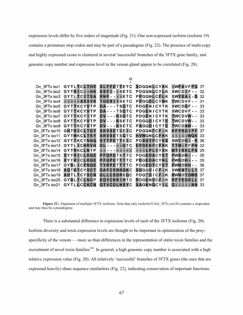
67
expression levels differ by five orders of magnitude (Fig. 21). One non-expressed isoform (isoform 19)
contains a premature stop codon and may be part of a pseudogene (Fig. 22). The presence of multi-copy
and highly expressed exons is clustered in several 'successful' branches of the 3FTX gene family, and
genomic copy number and expression level in the venom gland appear to be correlated (Fig. 20).
Figure 22 | Alignment of multiple 3FTX isoforms. Note that only isoform19 (Gn_3FTx iso19) contains a stopcodon and may thus be a pseudogene.
There is a substantial difference in expression levels of each of the 3FTX isoforms (Fig. 20).
Isoform diversity and toxin expression levels are thought to be important in optimization of the prey-
specificity of the venom — more so than differences in the representation of entire toxin families and the
recruitment of novel toxin families184. In general, a high genomic copy number is associated with a high
relative expression value (Fig. 20). All relatively ‘successful’ branches of 3FTX genes (the ones that are
expressed heavily) share sequence similarities (Fig. 22), indicating conservation of important functions.

68
Reptile venom CRISPs act as regulators of several types of ion channels185. We find three CRISP
genes in tandem in the king cobra genome (Fig. 23), only two which are represented in our venom gland
transcriptome (Fig. 23). Together with the comparative genomic data (Fig. 23), this is consistent with an
evolutionary scenario in which the two venom genes have been derived by tandem duplication from the
non-venom expressed (physiological) CRISP gene.
Figure 23 | Comparative genomic architecture of the CRISP genes. a, chicken (Gallus gallus); b, anole lizard (Anolis
carolinensis); and c, king cobra (Ophiophagus hannah). Chick and Anolis sequences are from www.ensembl.org. The exploded
views show scale diagrams of the exons and introns. Scale bar refers to the exploded views. NNN, unresolved sequence. In the

69
Anolis genome we annotated three CRISP genes with different orientations. Based on the relative sizes of the second introns the
two ‘venom’ CRISP genes are comparable to isoform 3 in Anolis. In chicken we could only find one CRISP gene.
Figure 24 | The scaffold containing three CRISP genes with different isoform transcripts (see Figure 23c) mapped on as follows:
a) isoform 1; b) isoform 2; c) isoform 3. As can be seen, only the first two isoforms are expressed in the venom gland; d)

70
alignment of the three CRISP genes with reference sequences showing that our identified genes belong to the CRISP family.
Isoform1 is opharin and isoform 2 is ophanin.
Venom metalloproteinases belong to the ADAM family and target various stages of blood
coagulation and platelet aggregation and are responsible for hemorrhage186. We also find three ADAM
genes in tandem, only one of which was expressed in the venom gland transcriptome (Fig. 25).
Figure 25 | Scaffold containing three ADAM genes; b) isoform 1; c) isoform 2; d) isoform 3. Only isoform 2 is expressed in the
venom gland (data not shown). Amino acid alignments of these three metalloproteinase genes with the single transcriptome
sequence (not shown) shows that one gene is identical and confirms its expression. Isoform 1 has a longer C-terminal tail. In O.
hannah isoform 2 is expressed in the venom gland, while in Naja atra (a different elapid snake) isoform 3 appears to be
expressed, since we find that N. atra metalloproteinase sequence is more similar to isoform 3 than isoform 2
LAAO produces H2O2 during oxidation of amino acids leading to cytotoxicity and inhibition of
platelet aggregation (and is responsible for the yellow color of the venoms)187. We find two LAAO genes
on two different scaffolds (Fig. 26 a). Based on the mapping of venom gland transcriptome reads (not
shown), only one LAAO gene appears to be expressed in the venom gland; the other is presumably the

71
non-venom, physiological gene. To the best of our knowledge, non-venom LAAO proteins have not been
found in reptiles before, although they are found widely among vertebrates.
Figure 26 | a, Genomic architecture of l amino acid oxidase (LAAO) genes in the chicken and king cobra. b, scheme of the
genomic context of the hyaluronidase genes in the mouse (Mus musculus) and king cobra. Mouse genomic sequences from
www.ensembl.org. Scale bar refers to the exploded views. NNN, unresolved sequence.
|

72
The role of venom NGF is not clear98. We find two different NGF genes, both of which are
encoded by a single exon; and both of them are expressed in the venom gland (Fig. 27). Presumably, one
or both of these has duplicate functions (in both venom-gland and in other tissues). Venom hyaluronidase
plays a key role as the venom ‘spreading factor’, making tissue more permeable188. We annotated two
hyaluronidase genes in the king cobra genome, both lie downstream of the WASL gene, and we find the
same arrangement in the mouse genome (Fig. 26 b). Only the gene corresponding to HYALP1 is
expressed in the venom gland (data not shown), which is interesting because in the mouse this gene
appears to be inactive189. This synteny is consistent with a scenario in which the duplication of the
hyaluronidase gene took place long before one of the copies was recruited to the venom gland.

73
Figure 27 | Mapping of the transcriptome reads onto the two scaffolds containing two NGF genes shows that both of these genes
are expressed in the venom gland; a) isoform 1; b) isoform 2; c) Alignment of the two NGF genes with reference sequences
showing that our identified genes belong to the NGF gene family (data not shown).

74
Recently, PL-B was also found to be expressed in the venom gland190 but its role in toxicity is yet
unclear. We could only find one PL-B gene (Figure 28). This indicates that an existing PL-B gene was
recruited to the venom gland. Thus HYA, NGF and PL-B genes appear to be recruited for expression in
the venom gland without gene duplication being involved. In the case of the Asian krait (Bungarus
fasciatus) Acetylcholinesterase toxin, it was shown that both the neuronal and the venom enzymes are
encoded by the same gene, although alternatively spliced173.
Figure 28 | Scheme of the genomic synteny of the PL-B genes in the Anolis, Gallus and king cobra. Anolis, Gallus genomic
sequences from www.ensembl.org. Mapping of the transcriptome reads onto one scaffolds containing the PL-B gene shows that
this gene is expressed in the venom gland (data not shown). This synteny is consistent with the scenario of recruitment of the
existing PLBD1 into the venom gland during snake evolution. The alignment of the PL-B gene with reference sequences showing
that our identified genes belong to the PL-B gene family (data not shown).

75
It has been shown, in the case of factor X toxin in the rough-scaled snake (Tropidechis carinatus),
that a specific insertion in the promoter region of the toxin was responsible for the selective recruitment to
the venom gland169. We have scanned all our scaffolds for this sequence but could not find anything
similar. This suggests that that the specific insertion is not a universal feature of toxin gene recruitment,
and that several distinct mechanisms are responsible for the origin and recruitment of venom proteins.
Conclusion and discussion
Comparative genomics has revealed flexible mechanisms of mutation and recruitment of venom genes.
We found evidence of tandem duplication of genes encoding physiological L-amino acid oxidase,
cysteine-rich secretory proteins and metalloproteinases, followed by recruitment through selective
expression in the venom gland. By contrast, nerve growth factor toxins appear to have evolved by
duplication and dual recruitment, while hyaluronidase and phospholipase B evolved by recruitment of
existing physiological genes without further duplication. We also identify 21 different three-finger toxin
(3FTX) genes in the genome, suggesting a massive expansion of this family. We find a significant
variation in the expression levels of these 3FTX genes in the venom. Our data therefore shows that venom
proteins originate and evolve through multiple distinct mechanisms.
The king cobra genome is an important resource for studying molecular evolution. The powerful
combination of genomics with transcriptomics here used lead to the identification of toxin genes
previously unknown in the king cobra. Because of the massive functional diversity known to exist not
only between toxins but also their isoforms176, functional studies of these new toxins could prove to be of
great interest.

76
Methods summary
king cobra tissue acquisition and processing. All animal procedure complied with local ethical
approval. Genome sequencing was done on a blood sample obtained from an adult male king Cobra from
a captive specimen that originated from Bali, Indonesia. Blood was obtained by caudal puncture and
frozen in liquid nitrogen. The venom gland and other tissue samples were dissected from a freshly
euthanized second adult male specimen and stored in RNAlater.
Sequencing and assembly. We used a whole-genome shotgun sequencing strategy and Illumina
Genome Analyser sequencing technology. Two paired-end and four mate pair libraries were constructed
with insert sizes of up to 15K nucleotides. In total, we generated 41.2 Gbp (approximately 28x genome
coverage) of sequence data for contig building, 21.1 Gbp for scaffolding, and 1.7 Gbp for the
transcriptome. We built contigs from the short reads using the CLC bio de novo assembler (CLC bio,
Aarhus, Denmark) and oriented these contigs using SSPACE. A more extensive methods section is
included in the supplementary information.
Annotation and gene prediction. Gene prediction was carried out automatically using Augustus
software178, using venom gland transcripts as hints. Further extensive manual annotation was performed
to establish the intron-exon boundaries.
Acknowledgements We thank Austin Hughes, thank Daniëlle de Wijze and Yuki Minegishi for
discussions. This was funded by The Netherlands Centre for Biodiversity Naturalis and the Smart Mix
Programme of the Netherlands Ministry of Economic Affairs and the Netherlands Ministry of Education,
Culture and Science. Jeroen Admiraal helped with illustrations.
Author contributions F.J.V., study concept and design, tissue preparation, flow cytometry, sequence
analysis, writing of manuscript. C.V.H., genome assembly and analysis, transcriptome analysis,
preparation of figures, writing of manuscript. R.M.K., analysis of venom genes, comparative genomics.
H.M.IJ.K, sequence analysis, comparative genomics, drawing of figures. H.P.S,, study concept and
design, genome and transcriptome sequencing, sequence analysis. H.J.J. genome and transcriptome

77
sequencing. S.A.H. sequence analysis, comparative genomics, drawing of figures. P.A. study design and
financing. G.E.E.J.M.vd.T, sequencing facilities, M.B. and W.P., assembly, . R.P.H.D., sequence analysis
M.K.R., project leader, study concept and design, sequence analysis, preparation of figures and
manuscript.

78
Chapter 6: An Efficient Analytical Platform for On-line Microfluidic Profiling of
Neurotoxic Snake Venoms Towards Nicotinic Receptor Like Affinity
Heus F, Vonk FJ, Bruyneel B, Smit AB, Lingeman H, Richardson MK, Niessen W and J Kool.

79
The highly variable toxic peptides in snake venoms target a myriad of neurotoxic and hemotoxic
receptors and enzymes and comprise highly interesting candidates for drug discovery. Venom
profiling studies are normally directed either at identification of venom peptides or at biological
activity using elaborate and complicated workflows employing relatively large amounts of precious
venom. We have developed a microfluidic on-line screening approach utilizing nano-LC–MS with
parallel bioaffinity detection, which enables efficient correlation of bioaffinity to identity of the
venom peptides. Using the acetylcholine binding protein (AChBP) as biological target, we evaluated
our technology with high-affinity α-bungarotoxin and hemotoxic/proteolytic Vipera ammodytes
venom spiked with α-bungarotoxin. The methodology was applied to profile the venoms of
Dendroaspis jamesoni kaimosae, Naja annulifera and N. nivea, which took less than two hours per
venom. The data yielded over 20 AChBP ligands of which the corresponding accurate masses were
used to retrieve information from literature regarding their function and targeting specificity. We
found that from these 20 ligands, 13 were previously reported on, while information on the others
could not be found. Our data demonstrated that nano-LC-MS coupled to an on-line biochemical
assay is a promising analytical approach for rapid identification of bioactive peptides in snake
venoms.

80
Introduction
Recent advances in scientific understanding of diseases have raised hopes of an increase in the number of
novel drugs reaching the market. Further, many potentially disease-related orphan receptors are now
being investigated. It is therefore disappointing to realise that the number of drugs reaching the market is
actually dwindling191. One of the solutions to this problem is the exploration of natural sources, such as
animal venoms, as to increase the numbers of potentially new lead compounds. Why? Possible reasons:
venoms are highly potent because they are under extreme strong natural selection176,192. Venoms often
contain multiple novel compounds, free from IP restrictions. They have also provided many new drugs or
even new classes of drugs, in the past.
There is huge variability in snake venom composition15,184,193 due to an accelerated form of
mutation and evolution25,33,192,194,195 that is currently not understood at the molecular level. Venomous
snakes differ in their venom composition not only between species (interspecific) but also within a
species (intraspecific). In order to exploit this massive natural resource, several challenges have to be
resolved. These include bio-analytical challenges such as: 1) the complex nature of the samples and the
intrinsically time-consuming process of elucidation, 2) the occurrence of synergistic actions between
components in a complex natural extract, and 3) sourcing adequate quantities. For these and other
reasons, pharmaceutical companies scaled-down or even abandoned their natural extract drug discovery
pipelines. Nevertheless, many drugs approved during the last decades are based on natural products 196.
Another potential reason for abandoning natural extract product pipelines is that most of them focused on
plant and fungal derived bioactives197, because these mostly small molecule compounds fitted well in
their small molecule drug discovery pipelines. Traditional problems with peptide and protein derived
drugs are amongst others due to issues with (oral) drug administration, bodily distribution (e.g. limited
crossing of the blood-brain barrier) and immunogenicity issues. Advanced formulation methodologies,
administration routes198-201, protein engineering and production techniques202-204,204, as well as current
knowledge and available technologies dealing with immunogenicity and immunotoxicity205-209 have now
brought these biopharmaceuticals back into consideration as candidate drugs.

81
Great advances have been made in protein-based pharmaceuticals during the last ten years210-213.
In this regard, venom peptides also gained interest. A couple successful examples include: (i) Prialt
(Ziconotide), a non-opioid painkiller derived from the neurotoxic venom of a predatory cone snail (Conus
Textilus) and used to treat severe chronic pain by injection directly into the spinal fluid 214; (ii) Byetta
(Exenatide), a drug against diabetes TypeII derived from the venom of the Gila monster (Heloderma
suspectum)215; ACE inhibitors used for the treatment of hypertension and derived from a compound in the
venom of the South American Lancehead snake (Bothrops jararaca)76; and the antiplatelet drug
Integrillin (Eptifibatide) derived from a compound the venom of the Saw-scaled viper (Echis carinatus)
and used to reduce the risk of acute cardiac ischemic events in patients with unstable angina. In addition
to the drugs that have been derived from animal venoms, there have also been some successful
applications in diagnostics like the detection of lupus anticoagulants (abnormally high risk of blood
clotting ) using Taipan venom (Oxyuranus scutulatus)216. Some snake toxins have also been used to probe
receptors. The deciphering of the molecular structure of neurons was made possible by the use of
bungarotoxin from the venom of the banded krait (Bungarus multicinctus) and led to the discovery of a
receptor for acetylcholine217: the acetylcholine receptor.
One of the specific difficulties of effective natural extract screening is the accurate and rapid
bioactivity/identity correlation of bioactives with current traditional effect directed analysis (EDA)
methodologies218. EDA approaches are today’s standard for bioactive mixture analysis and are widely
applied in environmental219-221 and natural product screening222-224, as well as animal venom profiling225.
In this approach, bioactive compounds are separated and subsequently fractionated, e.g., with liquid
chromatography (LC), into a number of fractions, after which each fraction is tested for a specific
bioactivity. Ultimately, the bioactives are analysed with mass spectrometry (MS) to identify them.
However, EDA studies often fail to identify the bioactive compounds, because even after repeated
fractionation, biologically active fractions often remain too complex for chemical identification. This
might result in wrong interpretation and erroneous identification of bioactives, especially for those
bioactives with high affinity but low abundance. These are usually present in the same fraction with high-

82
abundance non-bioactive compounds, which impairs their detection. Some snake toxins are also only
present in very low amounts. Nerve Growth Factor toxins (NGF) – for example – are only present in 0.1-
0.5% in snake venom and are easily missed out. But NGF from the venom from cottonmouth vipers
(Agkistrodon piscivorus) allowed the authors to study the mechanisms regulating cell and organ growth99,
for which they were awarded the Nobel prize in 1986. Hence it is crucial to take into account these low
abundance bioactives in order to make sure the full potential of venoms are realized.
Furthermore, the freeze-drying (lyophilisation) of the chromatographic carrier fluid from the
fractionated sample may deactivate toxins, by altering the tertiary structure of proteins by (for instance)
unfolding, which might lead to false negatives. Additionally, the EDA workflow requires large amounts
of sample, which is difficult in the case of the very precious venoms. Even though some snake species
produce relatively large amounts of venom, most don’t166. Most snakes produce less than a milligram of
venom per milking226, severely hampering efficient venom profiling.
Even though some snake venom proteomes have been completely elucidated and sequenced,
many toxins have unknown functions and protein targets. Of these “orphan toxins’, only a hypothesised
functionality is known by sequence homologies with toxins having known bioactivities. And based on the
large functional diversification followed my only minor mutation of some of the toxin families47, it is
difficult to estimate function based on sequence similarity. These toxins are often only classified broadly
as cytotoxic, cardiotoxic or weak-neurotoxic227, including a myriad of different (unknown) receptors and
proteins recognized or bound.
Due to current advances in analytical strategies and microfluidics technologies, effective
screening for some pharmaceutical targets is currently possible with so-called on-line high-resolution
screening (HRS) approaches 228-232: chemical analysis and biological screening are integrated in a single
instrument platform by post-LC continuous-flow biochemical detection and parallel MS analysis232.

83
Analyzing natural extracts with a HRS platform can reveal in a single measurement the number
and chemical nature of the majority of bioactive compounds, with their affinity towards the target
receptor analysed 233-236. Unfortunately, as stated above, the often yields in animal venoms obtained
during a single milking are not compatible with the larger amounts required for traditional on-line HRS
approaches. We recently developed a miniaturized HRS methodology for analysis of small compound
libraries where nano-LC was hyphenated on-line to a microfluidic biochemical detector237. This
methodology however, although it would in theory allow for analysis of intrinsically low venom amounts,
is unsuited for profiling snake venoms as the complexity in this case overwhelms the analytical system.
The main issue here is that the system is only able to assess bioaffinity in one run therefore necessitating a
second nano-LC-MS analysis for peptide identification. Now, the main bottleneck becomes apparent as
repeatability of nano-LC systems is by far not sufficient for the demanded precision in retention time of
eluting peptides. Therefore, the bioactivity of peptides can never be correlated to their identity. For
identification purposes, a nano-electrospray MS (nano-ESI-MS) has to be placed in parallel via a post-
column split as is traditionally done in on-line HRS approaches232. Performing post-column splits after
nano-LC, however, is very tedious, error prone and unstable. Furthermore, often the split and the flows
cannot be operated in a controlled manner. Therefore, this endeavour has been avoided by the analytical
community. Nano-LC-MS is also the analytical technique of choice in proteomics approaches due to its
mass-sensitive detection and low sample consumption, and well suited as most snake venoms comprise
predominantly peptides and proteins. Many (snake) venomics studies nowadays comprise nano-LC-MS in
their analytical strategies7.
During the last ten years, drug discovery directed at the α7 nicotinic acetylcholine receptor (α7
nAChR) experienced a great leap by using the acetylcholine binding protein238,239. Ls-AChBP is a stable
structural homologue of the extracellular ligand binding domain of the α7 nAChR. It was first crystallized
and validated as model for nAChRs, especially the α7 nAChR, by us240 in 2001 and has often been used
as nAChR model since then. In this regard, it is used as mimicking drug target for example to identify
nAChR ligands241,242, and very well suited for HRS approaches, as was recently demonstrated by us243.

84
Subsequently, we developed a miniaturized screening format for bioaffinity profiling of small molecular
ligands towards the Ls-AChBP237. This system, however, only allowed bioaffinity profiling and did not
involve parallel compound identification by MS.
In the current work, the miniaturized HRS approach was applied to efficient venom profiling of
mainly snake venoms from species belonging to the Elapidae (cobras, mambas and relatives). The HRS
system comprises a post-column nano-flow split which directs the chromatographic effluent to mass
spectrometric and on-line biochemical detection allowing for simultaneous correlation of separated
bioactive venom toxins with their corresponding accurate mass. This approach may direct subsequent
targeted purification (with straightforward LC-MS). The analytical approach led to the identification of at
least 20 bioactive peptides with Ls-AChBP affinity and assigned accurate mass, in the venoms of the
species examined: the Mozambique spitting cobra (Naja mossambica); the black-tailed Jameson’s mamba
(Dendroaspis jamesoni kaimosae); the banded cobra (Naja annulifera); and the Cape cobra (Naja nivea).
From horned viper (Vipera ammodytes) venom - a member of the Viperidae family known for having
mainly hemotoxic/proteolytic peptides - also a low-affinity bioactive peptide was discovered.
Experimental
Chemical and biological reagents
Ls-AChBP (from snail species Lymnaea stagnalis) was expressed from Baculovirus using the pFastbac I
vector in Sf9 insect cells and purified from the medium as described by (ref 244). The fluorescent-tracer
ligand DAHBA, (E)-3-(3-(4-diethylamino-2-hydroxybenzylidene)-3,4,5,6-tetrahydropyridin-2-
yl)pyridine, was synthesized in-house243. NaCl, trizma base, HCl, formic acid (FA), DL–dithiothreitol,
guanidine-HCl, rhodamine-110, 5% dimethyldichlorosilane (DMDCS) and α-bungarotoxin (α-BTX) were
purchased from Sigma-Aldrich, (Zwijndrecht, The Netherlands). KH2PO4, Na2HPO4 and NH4HCO3 were
obtained from Riedel-de Haën (Seelze, Germany). I-Nicotine (99.0%) was purchased from Janssen
Chimica (Beerse, Belgium). Enzyme-linked immunosorbent assay (ELISA) blocking reagent (ELISA BR)
was purchased from Hoffmann-La Roche (Mannheim, Germany). ULC/MS grade methanol (MeOH;

85
99.98%) and acetonitrile (ACN; 99.97%) were purchased from Biosolve (Valkenswaard, The
Netherlands). HPLC grade water was produced using a Milli-Q purification system from Millipore
(Amsterdam, The Netherlands).
Instrumentation
Microfluidic confocal fluorescence detection system: The complete system consisted of a nano-LC unit,
an MS, an on-line chip functioning as biochemical reactor, where the LC flow and a bioassay solution
were mixed, and a microfluidic LED-based LIF detector as described by (ref 237). The complete HRS
platform is schematically shown in Fig. 29.
Nano-LC and solvent delivery system: The Ultimate nano-LC system with a Famos autosampler was
from LC Packings (Amsterdam, The Netherlands). The system was operated at 400 nL/min. Mobile phase
solvent A consisted of water and 0.1% Tri Fluoro Acetic acid (TFA) and solvent B of ACN and 0.1%
TFA. Sample volumes of 500 nL were injected into the analytical capillary column (150 mm × 75 µm
internal diameter (i.d.)) packed in-house with Aqua C18 particles (particle size 3 µm, 200 Å pore
diameter; Phenomenex, Torrance, CA, USA). For venom analysis, a 90-minute gradient elution was
applied running 5 min isocratic at 5% solvent B, then rising to 15% solvent B in 10 minutes, followed by
a rise towards 45% solvent B in 60 min, where after solvent B was set to reach 70% during 10 minutes,
running isocratically at 70% for 5 minutes. To prevent particulate matter to clog the capillary column, the
column inlet was connected to a low dead volume union containing a filter capsule (type M-572 and M-
128 respectively, Upchurch Scientific, Oak Harbor, WA, USA). The column exit was connected to a low
void volume T-connector where the effluent was split to the MS and the on-line bioassay by two pieces of
bare fused-silica (1,000 mm × 10 µm i.d.). All fused-silica capillaries used for connections to and from
the chip were purchased from BGB Analytik AG, Schloßböckelheim, Germany.
Microfluidic chip: The microfluidic chip and the chip holder (type 4515), produced by Micronit
Microfluidics, (Enschede, The Netherlands), was described in detail elsewhere237. The open tubular
microreactor had a total volume of 4 µL. The reactor had two inlets and one outlet. One inlet was used to

86
connect the nano-LC carrier flow; the other inlet to infuse the AChBP and tracer ligand DAHBA237,243 at a
flow rate of 5 µL/min by a Model 980532 syringe pump (Harvard Apparatus, Holliston, MA, USA). The
chip outlet was connected via a deactivated fused-silica to the microfluidic LIF detector, as described237.
Microfluidic LED-based LIF detector: The flow cell of the detector consisted of a 150 µm i.d. extended
light path “bubble-cell” with 50 µm i.d. connecting capillaries (G1600-64232, Agilent Technologies,
Amstelveen, The Netherlands). This bubble cell served as the actual flow-through detector cell. Light
emanating from a high-intensity LED passed a 465 nm single band pass filter, was collimated by a lens,
reflected by a dichroic mirror under 90° and focused into the centre of the bubble cell by 20× quartz
microscope objective. Emitted light passed the same dichroic mirror, a focusing lens, and a 520 nm single
band pass filter, after which it was detected by photomultiplier tube. A detailed description of this
detector can be found elsewhere237.
Nano-LC/MS: A Shimadzu (‘s Hertogenbosch, The Netherlands) ion trap – time-of-flight (IT-TOF)
hybrid mass spectrometer equipped with a Picoview nano-ESI source from New Objective (Woburn, MA,
USA) was operated in positive-ion mode. A 40 mm × 180 µm o.d. × 30 µm i.d. stainless steel emitter was
used (ES522, Proxeon/Thermo Scientific, Waltham, MA, USA), functioning as the spray needle. The
spray needle was connected to the nano-LC system via a 1,000 mm × 10 µm i.d. bare fused-silica
capillary by a low void volume connector (type P-720, Upchurch Scientific) which was integrated in the
nano-ESI source. The temperature of the heating block and curved desolvation line were set to 200°C.
The interface voltage was set at 1.7 kV, resulting in a current of ~32 µA.
Biochemical assay and samples
Biochemical assay: Fresh solutions of 5 nM Ls-AChBP and 15 nM DAHBA were made every day by
dissolving in a bioassay solution containing 1 mM KH2PO4, 3 mM Na2HPO4, 0.16 mM NaCl, 20 mM
trizma base/HCl at pH 7.5 and 400 µg/mL ELISA BR. The bioassay solution was housed in a 2.5 mL

87
syringe (type 1002LTN, Hamilton, Bonaduz, Switzerland) which was kept at 4 °C. The on-line bioassay
itself was performed at 22 °C.
Snake venoms: Lyophilized venoms samples of Naja mossambica, N. annulifera, Dendroaspis jamesoni
kaimosae and N. nivea were acquired as described by (ref 166). Vipera ammodytes and N. pallida venom
were purchased from Sigma Aldrich. About 2 to 3 mg of lyophilized crude venom sample was dissolved
in water/ACN/TFA 95:5:0.1% at a concentration of 10 mg/mL and subsequently centrifuged at 13,400
rpm for 10 min to remove particulate matter. Aliquots of these samples were stored at -20°C until further
use. Before analysis, 40 µM internal standard nicotine was added together with three peptides at 2 µM
each. The resulting venom solutions were directly injected in duplicate onto the nano-LC system for
parallel bioaffinity screening and MS identification. Samples were re-analyzed at lower or higher
concentrations whenever necessary. The nicotine served as an internal standard and for alignment
between the MS chromatograms and the bioassay readout. In the same way, the three reference peptides
served to align MS data between different runs, whenever necessary. These peptides were Met-
Enkephalin, Mca-Lys-Pro-Leu-Gly-Leu-Dap(Dnp)-Ala-Arg-NH2 (Bachem, Bubendorf, Switzerland) and
Enkephalin acetate salt hydrate (Sigma Aldrich).
Results and Discussion
This works describes the use of an efficient analytical platform for the profiling of neurotoxic snake
venoms. The technology is based on an on-line HRS approach in which the on-line biochemical assay
assesses bioactivity towards Ls-AChBP, mimicking predominantly neuronal nAChR binding. Parallel MS
analysis provides accurate masses of the bioactive peptides for identification purposes. We developed a
miniaturized nano-LC setup to overcome the main challenge of the limitation of the amount of venom
available per snake species, and to provide sufficient sensitivity. This miniaturized setup was developed
previously by using an on-line microfluidic chip (4 µL) and nano-LC237. In the current setup (Fig. 29), an
additional post-column split is introduced directing half of the 400 nL/min effluent from the nano-LC to

88
the nano-ESI interface. The other half is directed to the microfluidic chip where it is mixed continuously
with the bioassay solution, which is infused at 5 µL/min by a syringe pump. The outlet of the chip is
connected to the confocal LED-induced fluorescence detector, which monitors bioaffinity of the eluting
toxins after in-flow incubation. The bioassay is based on fluorescence enhancement in which the tracer
ligand DAHBA shows increased fluorescence yield upon binding to the AChBP. As an eluting peptide
competes with DABHA for Ls-AChBP binding, a negative peak in the signal is observed. Bioactives can
subsequently be purified by normal-bore LC(-MS) in a straightforward fashion for further
pharmacological studies on nAChRs.

89
Figure 29 | Schematic view of the complete analytical setup from 'milking' venom to our developed method. The bioassay
solution containing AChBP and the tracer molecule DAHBA were infused by a syringe pump at 5 µL/min and mixed with the
nano-LC effluent running at 400 nL/min, in the 4-µL open tubular reaction chamber of the microfluidic chip. The chip’s outlet
was hyphenated to the bubble cell capillary wherein light emanating from the high intensity LED was focused. Emitted light from
the AChBP/DABHA complex was subsequently detected by the photomultiplier tube. The setup is described in detail in the
“Experimental section”.

90
Method evaluation for the screening of snake venoms
In order to evaluate the applicability and performance of the miniaturized methodology for the screening
of bioactive peptides in snake venom, a standard addition study was performed first, to assess sensitivity,
reproducibility, correlation of peak shapes observed in MS and the biochemical assay, and (post-column)
band broadening. This was done by injecting 5 µg venom from the sand viper (Vipera ammodytes) in a
500 nL sample containing increasing amounts of α-bungarotoxin (α-BTX). Vipera ammodytes venom is
primarily hemotoxic, thus not containing abundant, highly active neurotoxic peptides (with probable
affinity towards Ls-AChBP). In this way, the highly active spiked α-BTX was studied in a complex
venom matrix without other significant bioactives. This allowed determination of whether the HRS
system is capable of pinpointing a single neurotoxic peptide amongst hundreds of other (mainly non-
neurotoxic) peptides. Furthermore, this part of the study gives information on the expected limits of
detection (LODs) for the venom toxins with respect to their biochemical and MS detection.

91
Figure 30 | The applicability and performance of the analytical setup was evaluated by injecting 5 µg of Vipera ammodytes (sand
viper) venom, spiked with increasing amounts of α-Bungarotoxin (α-BTX). The mass spectrometric data represents the data
obtained for the Vipera venom spiked with 250 ng α-BTX. Shown are the Total Ion Current (TIC) of the spiked V. ammodytes
venom and the Extracted Ion Currents (EICs) of α-BTX (isoforms 1 and 2 which both elute as two peaks, as also previously
reported in literature). Reference peptides allow for alignment with other chromatographic systems. The superimposed bioaffinity
data represent 5 injections of the V. ammodytes venom with 0 ng, 2 ng, 10 ng, 50 ng and 250 ng α-BTX. The venom analysis
without α-BTX (trace A) obtained a previously unreported V. ammodytes ligand as is marked by the green circle (corresponding
EIC is also shown). Nicotine was added for alignment of the mass spectral data with the bioaffinity data. This experiment was
repeated without V. ammodytes venom (data not shown) where 1 ng, 2 ng, 10 ng, 50 ng and 250 ng α-BTX were injected.
Chromatographic and mass spectrometric performance where comparable to this figure, while the bioaffinity data clearly showed
the absence of the V. ammodytes venom ligand binding to Ls-AChBP.

92
As can be seen in Fig. 30, α-BTX elutes from the reversed-phase LC column as two iso-forms in
4 peaks, a separation profile earlier reported by245,246. This profile can be easily mistaken for being 4
different peptide species, but due to correlating peak shapes between the biochemical and the MS
detection, misinterpretations like this can be avoided. The results depicted in Fig. 30 clearly emphasize
one of the main advantages of this methodology. In contrast to reversed-phase LC separations of reduced
and unfolded peptides in proteomics studies, the LC elution profiles of intact small proteins often do not
give single sharp peaks. Nevertheless, the bioaffinity can be correlated to corresponding accurate masses.
With the highest amount of spiked α-BTX (250 ng, absolute amount injected), the biochemical assay
became saturated, indicating that practically all DAHBA tracer ligand was displaced from Ls-AChBP
(Fig. 30, trace E). It was found that the LOD of the high-affinity ligand α-BTX (Kd of 8.59)240 was ~2 ng
(Fig. 30, Trace B) in the biochemical assay and ~25 times higher with MS.
Although the system showed to be very reproducible, sometimes, and especially when samples
were repeatedly measured over several days, retention times shifted slightly (data not shown). This is a
known issue with nano-LC systems and in our case did not impose serious problems as the biochemical
data and MS data were measured simultaneously and despite the shifting retention times, the correlation
remained accurate. Additionally, three reference peptides were introduced to enable alignment of
chromatograms from different runs, and to allow for convenient transfer of the methodology and samples
analyzed to other laboratories, if necessary. In addition, α-BTX was also analyzed separately without the
viper venom. This resulted in similar biochemical detection signals, while only α-BTX peaks were
observed in the LC-MS data (data not shown). These data confirm that the biochemical assay gives
similar results for α-BTX, whether present in a complex venom sample or injected separately, thus also
ruling out matrix effects. Finally, in these standard addition experiments, a viper venom peptide was
found with affinity for Ls-AChBP, as is observed in the in Fig. 30 trace A (encircled green), as discussed
below.

93
Snake venom analysis
Snake-venom proteomes consist of a complex cocktail of peptides, which during millions of years of
evolution, evolved into highly active ligands towards enzymes and receptors. In general, the Viperidae
family geared its venom to affect the function of various blood components, while the family Elapidae
(including several species from the Colubrid colubrinae subfamily) mainly developed a venom system
with neurotoxic and cardiotoxic peptides107,176 - although this is by no means a golden rule and there are
many exceptions to be found in both families. The three-finger toxins (3FTX) – a major and diverse
family of low-molecular weight (<10kDa) neurotoxins - share a common tertiary structure. This main
body is shaped by four disulfide bridges, truncating the amino acid sequence in such a way that three
loops are formed (hence the three “fingers”). All extant members have retained their peculiar “three-
fingered” molecular scaffold whilst exhibiting a diversity of different functions47. However, despite
retaining the common three-finger motif, members of this family have since evolved a myriad of different
functions182. These include effects on: platelet function247, different receptors associated with
neurotransmission248-254; ion channels255-257,257, viability of cardimyocytes258 and red bloods cells259,
mitosis and apoptosis260; and effects on the cell membrane261,262. The different 3FTX members also vary
considerably in binding affinity for the different receptors263 and are important research ligands for
studying receptors264-266. Homologous 3FTXs are found in all advanced snakes: Elapidae35 (cobras and
relatives), Viperidae194 (vipers and pitvipers), and others4,227,267. Not only their tertiary structure, but also
their chain length is similar. Type I (or short-chain) neurotoxins and cardiotoxins usually contain 60 to 62
amino acid residues, while the Type II (or long-chain) neurotoxins comprise 70 to 80 amino acids182.
Furthermore, these peptides show 40 – 50% amino acid sequence homology. The target affinities of these
peptide toxins are ‘tuned’ by mutations in the extremities of the three loops, as this is where interactions
with protein targets take place. For the neurotoxic peptides, the amino acids in the ‘fingertips’ of Loops I
and II are most determining. Type I and Type II neurotoxins bind competitively to (postsynaptic)
nicotinic acetylcholine receptors (nAChRs). These neurotoxins are commonly known as α-neurotoxins (α-
NTXs)49,225. Besides an elongated c-terminus, the Type II α-NTXs differ from Type I by an additional

94
disulfide bridge at the extremity of Loop II. This difference causes the Type I α-NTXs to mainly target
muscle α1 nAChRs, while Type II α-NTXs also target α7 nAChRs. The cardiotoxic (CTX) peptides are
essentially cytotoxic peptides as they disturb membrane structures of a range of excitable and non-
excitable cells268. Most likely, similar to the neurotoxins, there is a wide range of receptors and
(ion)channels targeted by the CTX peptides268-270. Although mainly α-neurotoxins from Elapidae have
been associated with nAChR binding, toxins from the Viperidae have also been reported to show (low)
nAChR affinity. In these venoms, phospholipases A2 A (PLA2s) rather than 3Ftxs are responsible for the
neurotoxic effects. This family of toxins typically contains seven disulfide bridges and a peptide-chain
length of 115 to 139 amino acids. It was reported that crude viperid venoms also block nAChRs (and
voltage-gated Ca2+ channels) in isolated identified neurons of L. stagnalis271. Subsequently, they showed
that an isolated PLA2 called Bitanarin has affinity for human α7 and Torpedo californica nAChRs, as
well as for the AChBP (from L. stagnalis). This novel ligand is structurally similar to (smaller) PLA2s
and shows PLA2 like activity, although it has 14 disulfide bridges and a peptide-chain length of 138
amino acids271. Table 3 provides a summary of all peptides, which were mass-correlated with literature
via Uniprot. Most toxins have cysteine bridges and we measured the intact peptides. Since Uniprot and
most literature give nominal masses from the reduced peptides, the known difference in molecular mass
(2 Da per cysteine bridge) was taken into account when the number of cysteine bridges was known. In our
case, most of the known bioactive peptides found had 4 or 5 cysteine bridges, implying a molecular mass
difference of 8 or 10 Da, respectively. The toxin nomenclature used for the identified toxins, by
correlation of nominal masses of bioactive peptides found per snake with literature, is done according to
(ref 227), and/or given as Uniprot identifier for easy reference.
2

95
Table 3 | The table on the next page shows the accurate mass of the most abundant charge state of each bioactive peptide, the
corresponding nominal peptide mass, the toxin family the peptide belongs to (when identified and known), the corresponding
accession number and their possible mechanism of action. All peptide masses reported in the table and in the following
discussion are nominal masses and are correlated with masses found in Uniprot by searching the venom proteomes of the
respective snakes profiled. Peptides not correlated in this way are referred to as ‘peptide #’.

96

97
Results
Sand viper (Vipera ammodytes)
As discussed in the previous section, a viper ligand was detected from a 5 µg total venom sample. Figure
S1 shows both the mass spectrum and the biochemical detection signal of this analysis. Upon increasing
concentrations of α-BTX, this viper bioactive is quickly masked by the highly active α-BTX standard.
Table 1 additionally shows mass spectrometric information of this ligand, which could not been identified
and is called peptide 1.
Figure 31 | The ligand (peptide 1) obtained from the 5 µg Vipera ammodytes (sand viper) venom analysis. The most abundant
charge state corresponds to total peptide mass of 13857 Da. Reported Viperidae neurotoxic phospholipases in this mass range
typically contain 7 disulphide bridges. Hence the nominal mass of this ligand is probably 13871.

98
Mozambique spitting cobra (Naja mossambica)
The analysis of Naja mossambica venom is a good example how neurotoxic ligands can be screened by
the miniaturized on-line HRS methodology, effectively narrowing down the number of probable and
possible ligands. When correlating the extracted ion chromatograms (EIC) with the biochemical detection
signal of a 5 µg Naja mossambica venom analysis, three strong binders and one weaker binder were
detected. Their nominal masses correlate with peptides P01469, P01470, P01467 and P25517 respectively
(Fig. 32).
Figure 32 | 5 µg injection of Naja mossambica (Mozambique spitting cobra) venom. Three strong binders and one weaker binder
were detected (P01469, P01470, P01467 and P25517 respectively) which were preciously reported as Type IA cytotoxins, a
group consisting of cytotoxins and cardiotoxins.

99
The first three of these peptides (P01469, P01470 and P01467) share a >90% homology, whereas
P25517 has a ~80% homology with the very high affinity ligands as deduced from a Blast search in
Uniprot. These toxins have been classified by (ref 227) by homology to the group Type IA cytotoxins, a
group of cardiotoxic and cytotoxic toxins. Interestingly, these four particular toxins have also been found
to form a clade separate from the Type IA cytotoxins227, including being separated from other African
cobra species because of their amino acid sequence divergence level. Furthermore, these authors
suggested comparative assaying to determine differences in potency or specificity between these toxins
and the Type IA group of cytotoxins227 – that normally exhibit cadiotoxic and cytotoxic effects. Indeed,
this proved to be a valid suggestion as we found these toxins showed relatively high affinities to the
model Ls-AChBP target and are thus likely to be neurotoxic. This, to our knowledge, has not been
reported before. These toxins, together with the highly homologous toxin P01468 from the venom of the
Naja pallida (closely related to N. mossambica) that is the only other found in the clade of (ref 227) could
form a new group of so-called Type IV α-neurotoxins because of their affinity/functionality but being
distinctly different in their sequence than, for instance, the Type I α-neurotoxins. So far, predominantly
3FTX-type cardiotoxins have been reported in comprehensive venomics studies on the composition of the
Naja mossambica venom272. In addition, two Type I α1 nAChR ligands have been reported (NMM I and
NMM III), one of which was also observed in our LC-MS analysis. It has been shown that this peptide
(‘NMM I’ or ‘P01431’ 62 amino acids (reduced) molar mass 7081 Da, most abundant charge state m/z
1415,635 ), binds exclusively to muscle type α1-nAChRs273,274. In our analysis, this peptide did not show
affinity towards the AChBP (Fig. 32).
Initially, 5 µg of venom was injected for analysis to a) also observe low-affinity and/or low-
abundant binders, and b) to provide sufficient sensitivity in MS. This, however, resulted in severe peak
broadening for the high-affinity and/or high-abundant bioactive peptides, as can be seen in Fig. 32.
Subsequently, lower venom concentrations were injected and analysed. In this case, sharper peaks were

100
observed and correlations were more straightforward, which assisted in the total elucidation process (Fig.
33).
Figure 33 | For more straightforward correlation between the mass spectrometric data and the bioaffinity data, 1.5 µg Naja
mossambica (Mozambique spitting cobra) venom was injected, which obtained sharper peaks and less co-elution of toxins than
the 5 µg injection (Fig. 32). To demonstrate the effect of a chromatographic run in terms of selectivity, the pH of the reversed-
phase LC run was increased from pH 2.5 to pH 7.5 (data not shown). This resulted in an elution order change where P01469,
P01470, P01467 (at pH 2.5) shifted to P01469, P01467, P01470. As a result of shifting retention times another probable ligand
could be detected (peptide 2 (Table 3)) with a mass of 15.186.

101
In order to test P01469, P01470, P01467 and P25517 for α7-nAChR affinity in an off-line
radioligand assay, these toxins were separated and fractionated on a normal bore LC-column (4,6mm x
100mm column Symmetry Shield, Waters Etten-Leur, the Netherlands). For the ‘large bore’ LC
measurements, 2.5 mg venom was injected at 2.5 mL/min under the same gradient conditions. The
fractions were subsequently lyophilized, re-dissolved in water/ACN/TFA 95:5:0.1% and injected in the
on-line miniaturized assay to check for purity and activity. It was found that after fractionation,
lyophilisation and re-desolvation most activity was diminished (data not shown). Experiments were
carried out such as collecting the fractions in 50 mM ammonium bicarbonate to neutralize the TFA in the
LC carrier fluid, but this did not significantly increase activity.
Jameson’s mamba (Dendroaspis jamesoni kaimosae)
From a 5 µg venom injection of the Dendroaspis jamesoni kaimosae (Jameson’s mamba), eight ligands
were detected in the biochemical detection signal (Fig. 34). The first (weak) binder, eluting at 33 min, can
be mass-correlated to a peptide (P01417) first reported by (ref 275) as a (weak) nAChR neurotoxin based
on anti-venom affinity for the homologous cobra venom. This toxin was then classified as a Type I α-
neurotoxin227. The massive bioactive peak at 39 min is caused by two peptides (peptide 3 and peptide 4)
which could not be mass-correlated with literature. The fourth affinity peak at 44 min was attributed to
either co-eluting P25682 or peptide 5. P25682 has been classified an orphan toxin XIX (a group of
Dendroaspis and Bungarus toxins of unknown function / protein target)227. The onset of binding signal at
48 min is probably caused by peptide 6, whereas the remaining signal is caused by P01406, which has
been classified as an orphan group XI toxin (of unknown function/target)227. P01406 elutes in two forms,
also causing another binding signal at 55 min. The small binding signal at 62 min is caused by peptide 7,
whereas the last signal at 77 min can be attributed to P01418, which has been classified as a Type I α-
neurotoxin227.
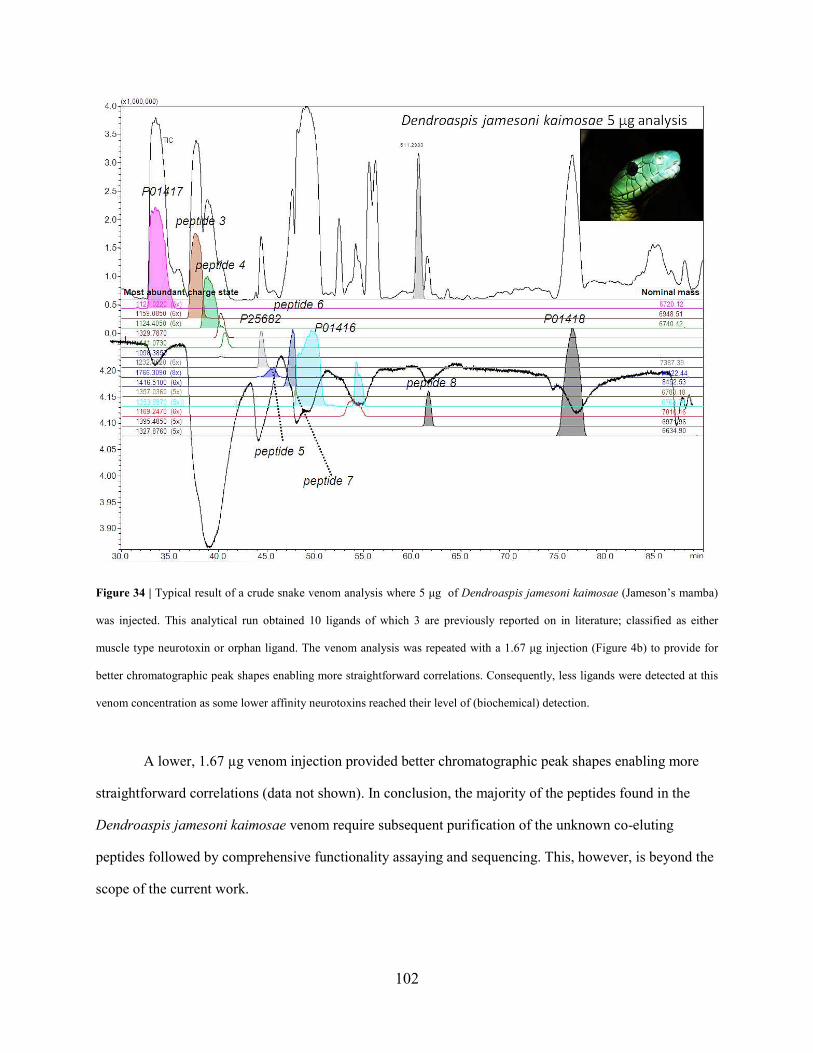
102
Figure 34 | Typical result of a crude snake venom analysis where 5 µg of Dendroaspis jamesoni kaimosae (Jameson’s mamba)
was injected. This analytical run obtained 10 ligands of which 3 are previously reported on in literature; classified as either
muscle type neurotoxin or orphan ligand. The venom analysis was repeated with a 1.67 µg injection (Figure 4b) to provide for
better chromatographic peak shapes enabling more straightforward correlations. Consequently, less ligands were detected at this
venom concentration as some lower affinity neurotoxins reached their level of (biochemical) detection.
A lower, 1.67 µg venom injection provided better chromatographic peak shapes enabling more
straightforward correlations (data not shown). In conclusion, the majority of the peptides found in the
Dendroaspis jamesoni kaimosae venom require subsequent purification of the unknown co-eluting
peptides followed by comprehensive functionality assaying and sequencing. This, however, is beyond the
scope of the current work.
103
African cobra (Naja annulifera)
From a 5 µg Naja annulifera venom injection, clearly three binding signals can be observed (Fig. 35).
The two binding signals eluting at ~ 65 min can be attributed to P62390 and P01461, first reported by (ref
276). The latter has been classified a Type IA cytotoxin227. P01461 sequence-wise is not part of the African
Spitting cobra’s ‘divergence clade’ mentioned above, although it shares 81% homology with Naja
mossambica’s P01467. The binding peak shows that possibly more Type IA cytotoxins exhibit neuronal
α-neurotoxicity. P6290 has not been classified by (ref 227), but by sequence similarity it mostly resembles
the ‘orphan group XV’ toxins (via Blast search in Uniprot), a group coined with only limited known
biochemical function besides probable ‘low-level cytotoxicity’ for Naja toxins227. P6290 shares, for
instance, 90% homology with P1541, a Naja kaouthia toxin classified as orphan group XV type227.
As the MS signals of these peptides are quite high, while the affinity signals are (relatively) low,
P62390 and P01461 are probably low-affinity binders. Based on the same criteria, the main ligand eluting
between 40 and 50 min is a low-abundant, high-affinity ligand. From the initial analysis at pH 2.5, two
co-eluting ligand peptides were identified, which could be mass-correlated with P25674 and P01399 from
Naja haje haje (Egyptian cobra). When repeated at pH 7.5, P25674 and P01399 showed improved
resolution (data not shown). In addition, the MS intensity of peptide P25674 increased, making
correlation with the bioaffinity signal more straightforward. P01399 was first reported by (ref 277) and
classified as a weak cytotoxin. A homologue of this peptide has also been found in the Naja sputatrix
(Indonesian spitting cobra (Q802B2); with 82% homology) and is reported as a weak (muscle type)
nAChR ligand278.

104
Figure 35 | Analysis of 5 µg Naja annulifera (African cobra) venom obtained three binding signals . The first, large binding
signal can be attributed to a highly active α-cobratoxin ligand P25674 (a toxin which has been reported in the venom of N. haje).
The two latter binding signals were correlated to P62390 and P01461 (previously classified as Type IA cytotoxins). As the α7
ligand P25674 co-elutes with toxin P01399, the analysis was repeated at pH 7.5 (data not shown). P25674 and P01399 showed
improved resolution and the MS intensity of peptide P25674 increased. Again this clearly shows the value of the higher pH
analysis ‘mode’. The analysis was thereafter repeated by injecting 1 µg and 0.2 µg venom (data not shown), which not only
allows for more precise correlation between the MS and bioaffinity data sets, but also demonstrates the repeatability of the
system.
The main neurotoxic ligand in the Naja annulifera venom, P25674, was identified by its
homologue reported in the Naja haje haje venom, which contains a peptide with an identical mass and has

105
been reported as a highly active α7 ligand279. As these cobra subspecies share many homologues toxin
peptides, it can be assumed that this particularly high-affinity peptide has been 100% conserved in
sequence in both species. Although it has to be taken into account that in the 70s and 80s many venoms
came from species that were mis-identified280, so it cannot be ruled out that the Naja haje haje venom
used in (ref 279) was in fact Naja annulifera venom. Lower amounts of venom injected (1 µg and 0.2 µg)
provided better chromatographic signals allowing for more reliable biochemical correlations (data not
shown). The caveat mentioned earlier and also the rationale for injecting a high amount of venom for
analysis: the major bioactive was not observed in MS anymore when looking at the amount of 0.2 µg
injected (data not shown). This can easily lead to misinterpretations. Therefore, in some cases like the one
described here, different concentrations need to be analysed in order to reach a high confidence regarding
correlation of bioactives to their identity. As one additional analytical run with a lower concentration
injected takes less than two hours, this is not a significant problem.
Cape cobra (Naja nivea)
Naja nivea is closely related to other African cobras such as the Naja haje and Naja annulifera, as can be
traced back to its venom, having a high-affinity α-cobratoxin and several low-affinity Ls-AChBP binders
(Fig. 36). At least five ligands could be detected from analyzing 5, 1 and 0.2 µg crude venom samples.
The high-affinity binder at 39 min, P01390, is a Type II α-neurotoxin227. As due to column overloading a
small percentage of the total bioactive P01390 slowly elutes from the column in a tailing fashion, the Ls-
AChBP affinity of the remaining neurotoxins is somewhat harder to distinguish (see Fig. 36). This again
shows the potential problem of seemingly severe peak broadening when very high-abundant and/or high-
affinity ligands are present in a venom sample. It has to be noted that the bioaffinity signals are sigmodial
in their response and not linear. This results in seemingly very severe peak broadening in the bioaffinity
trace, while this is not observed in the parallel MS (or UV) response. This indicates that it is in fact not
severe peak broadening but due to an intrinsic characteristic of biochemical detection in general.

106
Figure 36 | Injection of 5 µg of crude Naja nivea (Cape cobra) venom obtained at least 5 ligands. The binding signal at 39
minutes can be attributed to the high-affinity α-cobratoxin P01390. As this strong binder elutes from the analytical column in a
very broad peak (and thereby overloading the bioaffinity signal) the remaining toxins are hard to distinguish (such as toxin
P25680). This was resolved by injecting smaller concentrations of venom, as seen in Figs. 37 and 38.
Closely eluting to the high affinity binder P01390 is P25680, an orphan group II toxin227. It could be a
low-affinity binder, but due to the high-binding signal of P01390 no conclusions can be drawn. The
bioactivity signal at 57 min can be attributed to P01456, classified as a Type IA cytotoxin227. The binding
profile at 64 min is caused by a peptide (peptide 9) could not be mass-correlated with literature data. At
67 min, the peptide is most probably binding to AChBP is P01463, which is classified as an α1 nAChR
ligand. But AChBP bioaffinity of the co-eluting peptide 8 cannot be ruled out. As also shown for the Naja
mossambica, Dendroaspis jamesoni and Naja annulifera venom analysis, analyzing lower concentrations

107
of the Naja nivea venom assisted us in more accurate mass-correlations of bioactives, thus facilitating
their identification.
Figure 37 | Injection of 5 µg of crude Naja nivea (Cape cobra) venom. From the lower concentration venom analysis it was
possible to correlate at least four other toxins, of which three are classified as Type IA cytotoxins (P01456, P01463 and P68419)
while the other two toxins are not reported on.
The data in these analyses are complementary: while the major bioactive peptides are readily
detected by MS upon a 5 µg injection, their detection becomes more difficult or even impossible at 1 µg
(Fig. 37) and 0.2 µg analysis (Fig. 38). However, in the biochemical detection, the smaller sample
injections result in increased chromatographic resolution and allow the minor bioactives to be analyzed
more clearly.

108
Figure 38 | Injection of 0.2 µg of crude Naja nivea (Cape cobra) venom increased chromatographic resolution and allowed minor
bioactives to be analyzed more clearly.

109
Discussion and perspectives
The AChBP is an established structural homologue of the α7 nAChR, a (potential) drug target for pain
syndromes, cognitive and neurodegenrative diseases. As Type II α-NTXs are high-affinity binders to the
α7 nAChR, AChBP is a valid and very useful water-soluble target model to screen for high-affinity
binders in these venoms. Snake venoms might contain potential biopharmaceuticals. In case of neurotoxin
venoms, particular interest is in targeting nAChRs, e.g., the α7 nAChR. Besides discovery of such
potential biopharmaceuticals, screening of venoms may also yield valuable tool compounds with high
affinity towards specific ion channels, which can be used as tools for biological and pharmacological
studies281,282. Typical examples of such snake toxin peptides are the α-bungarotoxins and α-
cobratoxins240,283,284. This study showed that screening of snake venoms using the HRS approach with the
Ls-AChBP yields not only many high-affinity peptide/protein ligands, but also low-affinity Ls-AChBP
ligands, which were reported upon in the literature as Type I α-NTXs, cardiotoxins and PLA�s. In
addition, several other bioactive peptide ligands were found that were previously unreported or reported
as orphan-ligands, of which the biological function is presumed, only based on sequence homology.
The data obtained from screening Naja mossambica venom clearly shows that ligands still can be
identified even though the peptides are closely eluting in relatively broad peaks. For the screening of
Dendroaspis jamesoni kaimosae venom, this is shown again, but here the number of ligands found is
substantially higher, which makes ligand identification more complex. Here, low-affinity binders are first
identified by injecting a higher concentration sample, followed by an analysis of more diluted sample(s)
to distinguish ligands with high affinity from low-affinity ligands. In the third snake venom profiled, a
strategy is shown in which co-eluting peptides from Naja haje annulifera venom (of which one has a very
high affinity towards Ls-AChBP) are correlated with the biochemical detection signal with higher
confidence by means of changing the selectivity of the reversed-phase LC separation followed by
reanalysis of the sample. This strategy, where an additional chromatographic run was performed at a high

110
pH, narrowed possible ligands significantly. The last example showed how a serial dilution analysis of
Naja Nivea venom enabled the detection of 5 possible AChR ligands.
As described in the method evaluation, the structure of venom peptides is highly conserved in
their evolutionary trail causing their chromatographic behaviour also to be similar. Besides poor
separation because of many iso-toxins166, also bad chromatographic peak-shapes pose an analytical
challenge. Even though these characteristics result in often challenging chromatographic separations,
peptide ligands could be identified by direct correlation of the biochemical detection signal with the EICs
from MS as obtained from the IT-TOF MS measurements. Once correlated, these masses give an idea of
the identity of peptides (e.g. Type 1 or Type 2). As the correlated masses found are exact (measurements
are performed by an accurate and high resolution MS), these masses furthermore provide for a
straightforward and targeted fractionation approach. After chromatographic purification, a validation can
be performed by re-analyzing the purified sample, followed by further structural elucidation by e.g.
tryptic digestion and novo-sequencing, Edman degradation and receptor binding and activation studies.
This study described the evaluation, validation and demonstration of the potential of our
miniaturized on-line HRS approach for comprehensive profiling of snake venoms with respect to nAChR
affinity. Snake venoms of Vipera ammodytes, Naja mossambica, Dendroaspis jamesoni kaimosae, N.
annulifera and N. nivea were analysed yielding data on both the accurate molecular mass and identity of
the peptides present and their individual bioaffinity towards the AChBP.
Conclusion
As is well known, animal venoms provide for numerous ligands towards a myriad of receptors, enzymes
and ion channels and therefore have the intrinsic potential to modulate biological and pharmaceutically
relevant signal cascades. This study describes the application of a miniaturized on-line analytical
methodology where neurotoxic (Elapidae) snake venoms are screened for toxins with Ls-AChBP affinity,
mimicking binding to neuronal nAChRs, especially α7 nAChR. Prior to snake profiling, a post-column
nano-split was developed and implemented followed by evaluation and validation of the methodology.

111
The methodology proved to be suitable for profiling of snake venoms. By correlating bioactive peptides
with their mass identified we were able to identify bioactives. In many cases, toxins described in literature
are only correlated to a possible function by amino acid homology only.
We found 11 peptides that were not previously correlated with neuronal nAChR affinity and
therefore are interesting molecules for further studies on their biological interactions with nAChR. As
discussed, many of the toxins sequenced and described in literature are classified by function only by
their homologies towards toxins of which the protein target / biological function is known. Although
subsequent in-depth studies of the toxins described in this study for nAChR binding are needed, their
affinities towards the Ls-AChBP give a good hint of (one of) their function(s): i.e. targeting the nAChRs
to causing immobilization of the prey animal. Besides these orphan toxins described, also 10 unknown
proteins were found, which are in need of additional sequencing and functional assaying. Even though
only the long neurotoxins (Type II) have been correlated with the neuronal α7 nAChR, most of the
peptides that were found in this study were short neurotoxins (60 to 64) amino acids. All short
neurotoxins described in literature are 6700-6900 Da. Four previously sequenced toxins were found in
Naja mossambica venom. Although these toxins were classified as cardiotoxins, they did show significant
binding to Ls-AChBP. However, further functionality assaying is required to validate human (neuronal)
activity. Attempts to do this by fractionation failed as most activity was lost in the lyophilisation
procedure. This shows the value of our ‘on-line’ method as no additional lyophilisation process is
required. Therefore, probably, our methodology suffers less from false negatives. Venom analysis from
Dendroaspis jamesoni kaimosae yielded two ligands known as α1 nAChR ligands, while two other
binders are classified as orphan ligands. Further studies are required to validate these results, as for the six
other (possible) neuronal nicotinic acetylcholine receptor ligands found in this venom proteome, which, to
this date, are not described in literature. The main binder in the Naja annulifera proteome was attributed
to a known α7 nAChR binder, of a closely related Naja haje haje, while two other distinct ligands are
correlated to toxins described in literature as cardiotoxin only. The analysis of this particular venom
shows the benefit of analysing at both a high and low pH, as this caused selectivity changes in the

112
reversed phase LC separation leading to more straightforward correlation of the bioactivity and the mass
spectrometric detection of a low abundant, high affinity ligand. The Naja nivea venom proteome shows
four distinct binding ‘curves’ which can be attributed to five ligands; the largest signal is caused by the a
known α7 ligand, while the three other curves can be attributed to tree ligands previously described as
cardiotoxin and two unknown peptides, of which one co-elutes with one of the cardiotoxins.
The rapid and high resolution screening of these five venom proteomes shows the potential of the
here described microfluidic on-line screening technique. Not only did the application of this technique
show the analysis speed, it also demonstrates the advantage of the direct post-column analysis, as
fractionation and lyophilisation causes the deactivation of toxins, which in previous runs did show
significant signals in the on-line Ls-AChBP assay. Furthermore, by the coupling of a miniaturized
separation technique (nano-LC) to a microfluidic on-line assay and sensitive fluorescence detection, we
showed that multiple, sensitive and robust analyses are possible with only a minimal amount of (the very
precious) venom required. In the future, it may be possible to combine these technologies with the
emerging microfluidic whole-animal assay systems such as those being developed in our lab for zebrafish
embryos285.
Acknowledgements
This work was financially supported with an ECHO grant of the Dutch Scientific Society NWO
(OND1331288 ECHO number 700.56.043), and a TopTalent grant from NWO. ABS was partially funded
by a grant from the European Union Seventh Framework Programme under grant agreement n°
HEALTH-F2-2007-202088 (Structure, function and disease of Cys-loop receptors, "NeuroCypres"
project).
113
Chapter 7: Summary and Discussion
Snakes are a fascinating and intriguing group of vertebrates that have gone to extremes in adaptation.
Their venom system belongs to one of the most sophisticated natural weapons systems in the natural
world. It is composed of a venom gland, duct and fang, and in some phylogenetic groups an accessory
venom gland. In Chapter 2 we have elucidated the evolution of the different types of snake fangs. There
are three distinct groups of snake fangs found. The first is a rear-fanged type, where the fangs are
positioned in the rear of the upper jaw and this fang-type is found in most snake species. Then there are
two types of front-fangs found. The first is found in vipers and pitvipers, where the fangs are often large
and lie recumbent in the upper jaw. They are mobile and can move back and forth. The second type is the
one found in cobras and mambas and relatives and is smaller and immobile. Because these two groups do
not form a monophyletic group, the evolution of these different fang types has always been subject to
debate. We have collected many different snake embryos of all three groups and looked at the
development of the dentition using the Sonic hedgehog gene as a marker. Using in situ hybridization
followed by careful 3D reconstruction we could follow the development of the fang. Our results showed a
great amount of similarity in morphogenesis of the fang of the three different groups of snakes. The two
front-fangs start right at the base of the rear of the upper jaw in development, just as the rear-fang. The
only difference is that the two front-fangs move forward in development by ontogenetic allometry
(pushing the fang forward due to rapid growth of the upper jaw behind the fang) to its adult front position.
Also, we showed that the rear-fang develops from its own dental lamina instead of with the same dental
lamina as the front 'normal' teeth. It was this posterior dental lamina that showed great similarity to the
dental laminae in the two front-fang snake groups. Our results thus suggest a scenario in which all three
fang-types are homologous and descendent from a rear-fanged ancestor (by means of uncoupling the
posterior from the anterior teeth), after which the two front-fanged groups moved their rear-fang forward
during evolution. This probably allowed them to feed on larger and more dangerous prey items as a quick

114
'snap-and-release' was made possible by the front-fangs compared to the 'bite-and-hold-on' that is only
possible with rear-fangs.
Another extreme of adaptation in snakes is their body elongation. Snakes have elongated bodies
with numerous vertebrae, some even up to 500 of them. They have lost most clear morphological
boundaries in their adult bodies. This has been of major importance in the evolution of the venom-
delivery system, as snakes use their elongated bodies as a coiled spring that can strike prey at a distance
and with terrifying speed, injecting the immobilizing venom. Without elongated bodies able to strike and
bite, their venom-delivery systems (especially the fangs) would probably have evolved in a completely
different manner. In chapter 3 we have analyzed and examined the expression of Hox genes in different
snake embryos from different stages to examine the evolution of s serpentine body form. Hox genes
determine the basis structure of animals, and it was assumed that the species with a snake-like body
evolved such a body by a homogenization of the Hox expression domains along the primary axis.
However, in our extensive analysis using in situ hybridizations we have found - on the contrary - that
snakes do retain a collinear expression of the Hox genes in the developing embryo. Some Hox genes
boundaries correspond to expected anatomical boundaries (the most anterior and posterior Hox genes),
but many do not. The dorsal (thoracic), homogenous rib-bearing region of trunk has also retained
regionalized Hox expression that is not obviously reflected in the anatomy. Our results suggest that the
evolution of a deregionalized, serpentine body involved not only alterations in Hox gene cis-regulation
but also a different downstream interpretation of the Hox code.
The snake venom secretions themselves consist of a complex mixture of proteins and pepties that
have evolved to target important physiological pathways and immobilize prey items. These toxins are of
major evolutionary and biomedical interest. From an evolutionary perspective, they undergo an
accelerated form of evolution. The genes undergo accelerated evolution due to a larger amount of
mutation in coding regions than in non-coding regions, while in almost all other known genes in the
natural world this is the other way around - except the genes in the immune system. This allows fast

115
evolution and adaptation towards ever changing prey and prey physiology (evolution of resistance). In an
analogous manner, our immune systems must fight ever changing pathogens. This arms-race is the
driving force behind diversity. From a biomedical perspective, this diversity is right at the base of why
snake venoms form such a potential goldmine for new pharmaceuticals. In chapter 4 we have sequenced
the king cobra (Ophiophagus hannah) genome to look at the evolution (and recruitment) of the venom
genes. They are thought to have evoloved from normal, physiological genes by means of gene duplication
followed by selective expression in the venom gland. However, in the absence of genomic resources these
hypotheses remain mainly speculative. Our results show that venom genes evolve through distinct
mechanisms. L-amino acid oxidase, cysteine-rich secretory proteins and metalloproteinases, evolved by
tandem-duplication of ancestral physiological genes, followed by recruitment through selective
expression in the venom gland. By contrast, nerve growth factor toxins appear to have evolved by
duplication and dual recruitment, while hyaluronidase and phospholipase B evolved by recruitment of
existing physiological genes without further duplication. The massive toxin families the 3FTXs and the
PLAs have undergone extensive duplications that have resulted in (at least) 21 copies in the genome.
These results show a much more dynamic evolutionary history in venom genes as previously assumed. It
is yet unclear what is causing the recruitment into the venom gland, and we are currently working on
answering this question.
The enormous diversity in venom genes, caused by the different ways of evolution and
recruitment as shown in chapter 5, makes it a challenge to identify bioactives for a particular receptor in
order to either study the venom for evolutionary studies, or to find new pharmaceuticals. In addition to
this, the extreme low venom yield that most snakes have make it a very precious sample. For most
traditional methods of evaluating a sample with mass spec and bioactivity studies you need a relatively
large amount of venom, which is a major challenge for many species. We have developed in chapter 6 a
new method to study the make-up of the venom and - at the same time and in the same run - look for
bioactives that bind to acetylcholine binding protein. One test used up less than 5 ug of venom and took

116
less than 2 hours. We found 21 bioactives of which 7 were never reported on before. This technique
includes a microfluidic on-line screening approach utilizing nano-LC–MS with parallel bioaffinity
detection using the acetylcholine binding protein (AChBP). The system comprises a post-column nano-
flow split which directs the chromatographic effluent to mass spectrometric and on-line biochemical
detection allowing for simultaneous correlation of separated bioactive venom toxins with their
corresponding accurate mass.
In conclusion, in this thesis I have shown that snakes have undergone multiple changes in their genome
and embryonic development that has provided them with the variation to which natural selection could
act. This thesis provides evidence for the variable mechanisms of venom gene evolution, which
presumably is much more flexible than previously thought. But it also underscores the potential use of the
many different types of snake venom toxins that could be screened for use against human disorders. And
most of all, I hope I have contributed towards the fact that snakes are just an incredibly interesting group
of vertebrates from both the perspective of ecology and life-style, as well as from a genomic and
molecular perspective. They are, and will always be, my first and true love.

117
Samenvatting en Discussie
Slangen zijn een fascinerende en intrigerende groep gewervelde dieren die tot extremen zijn gegaan in
evolutionaire adaptatie. Het gifsysteem is bijvoorbeeld een van de meest geavanceerde biowapensystemen
uit de natuur. Het omvat een gifklier, een gifkanaaltje, een giftand, en in sommige fylogenetische groepen
een ‘accessory gland’. In hoofdstuk 3 hebben we een hypothese opgesteld voor de evolutionaire
oorsprong van de verschillende typen giftanden. We kunnen drie typen van elkaar onderscheiden. De
eerste is een kleine, onbeweeglijke giftand die achter in de bek staat, en dit type giftand vinden we bij de
meeste slangensoorten. Dan zijn er twee verschillende typen van giftanden die voorin de bek staan. Het
eerste type is te vinden bij de familie Viperidae, dat bestaat uit adders en ratelslangen (groefkopadders).
Hierbij zijn de giftanden vaak groot en liggen ze horizontaal in de bovenkaak. Ze zijn beweeglijk en
kunnen op en neer bewegen door een scharnier. Het tweede type is te vinden in de familie van de
Elapidae, de cobra’s, mamba’s en verwanten. Hier is de giftand vaak kleiner en onbeweeglijk. Omdat
deze twee families geen monofyletische groep vormen, is de evolutie van deze giftanden onderwerp
geweest van discussie. Om dit te onderzoeken hebben wij een grote hoeveelheid slangen embryo’s
verzameld van verschillende soorten, behorend tot alle drie de giftand typen. Hierbij hebben we de
ontwikkeling van het gebit gevolgd met het gen sonic hedgehog als marker. Door middel van in situ
hybridisatie en 3D reconstructie konden we de algehele ontwikkeling van de giftand in kaart brengen. De
resultaten waren verbluffend: er was een grote mate van gelijkenis te vinden in de ontwikkeling van alle
drie de typen. Bij de Elapidae en de Viperidae begon de ontwikkeling van de giftanden in de embryonale
ontwikkeling bij beiden achter in de bek, net als bij de overige slangen. Het grote verschil is echter dat de
giftanden van de Elapidae en de Viperidae tijdens de ontwikkeling naar voren werden geduwd door
ontogenetische allometrie, doordat de kaak achter de giftand sneller groeide dan het gedeelte ervoor. Bij
achtertand giftige slangen ontstaat de giftand vanuit zijn eigen onafhankelijke dental laminae (embryonaal
primordium), en deze vertoont grote gelijkenis met de ontwikkeling van de dental laminea van de
giftanden van de Elapidae en de Viperidae. Onze resultaten suggereren een scenario waarin alle drie de

118
giftanden homoloog zijn aan elkaar, afkomstig van een giftand die achterin de bek stond. Hierna hebben
de Elapidae en de Viperidae onafhankelijk van elkaar de giftand naar voren geschoven in de evolutie.
Vermoedelijk kwam dit door het bijkomstig evolutionair voordeel dat deze slangen grotere en
gevaarlijkere prooien aan konden door middel van een snelle “bijt en laat los” methode. De slangen met
hun giftand achter in de bek kunnen dit niet, en moeten hun prooi vasthouden om gif in de wond te
kauwen. Dit is onmogelijk bij gevaarlijke prooien zoals grote ratten.
Slangen hebben verlengde lichamen met tot 500 wervels. Ze hebben de duidelijke morfologische grenzen
van hun lichaam verloren die veel andere dieren (inclusief mensen) wel hebben. Het verlenging van het
lichaam is van groot belang geweest voor de evolutie van het gif systeem, omdat slangen hun lange
lichamen kunnen gebruiken als een springveer om prooien met grote snelheid te bijten, en gif te
injecteren. Zonder deze lange lichamen om te bijten, zal het gifsysteem – en zeker de giftanden -
waarschijnlijk op een hele andere manier zijn geëvolueerd. In hoofdstuk 4 hebben we de expressie van
Hox genen bekeken in verschillende slangen embryo’s van verschillende stadia om de evolutie van een
‘slangachtig lichaam’ te bestuderen. Hox genen bepalen de lichaamsvorm van dieren. Het was algemeen
aangenomen dat dieren met een ‘slangachtig lichaam’ dit geëvolueerd hadden door een homogene
expressie van Hox genen langs de primaire lichaamsas, in tegenstelling tot de altijd zo karakteristieke
colineaire (regionaliserende) expressie. Echter, in onze experimenten met behulp van in situ hybridisatie
hebben wij juist het tegenovergestelde gevonden. Slangen hebben wel de colineaire expressie van Hox
genen behouden in hun embryonale ontwikkeling. De grenzen van de expressie van sommige Hox genen
corresponderen met de te verwachtte anatomische grenzen (de most anteriore en posteriore Hox genen),
maar bij veel andere Hox genen is dat niet het geval. Het dorsale (thoracische), homogene rib-dragende
deel van de slang heeft ook de regionaliserende Hox expressie patronen behouden. Deze grenzen zijn
echter niet terug te vinden in de anatomie van een slang. Onze resultaten suggereren daarom dat de
evolutie van een gederegionaliseerde, ‘slangachtig lichaam’ niet alleen veranderingen in de expressie van
Hox genen zelf verreist maar ook een andere downstream interpretatie van de Hox code.

119
Slangengif bestaat uit een complex mengsel van verschillende eiwitten en peptiden die geëvolueerd zijn
om prooien te immobiliseren door bepaalde belangrijke fysiologische doelen aan te vallen. Deze
gifstoffen zijn zeer interessant voor de evolutionaire en biomedische wetenschap. Ze ondergaan
bijvoorbeeld een vorm van ‘versnelde evolutie’, door een groter aantal mutaties in de coderende stukken
DNA (exons), in relatie tot de mutaties in de niet-coderende stukken DNA (introns). In de meeste genen
is dit juist andersom. Eenzelfde patroon vinden we in de genen die betrokken zijn bij immuunsystemen.
Wij moeten ons wapenen tegen alsmaar veranderende pathogenen, maar op een analoge wijze moeten
slangen zich wapenen tegen alsmaar veranderende prooien die resistentie ontwikkelen. Deze
wapenwedloop is de drijvende kracht achter de diversiteit in gif. Vanuit een biomedisch oogpunt, zijn
deze stoffen interessant omdat ze een grote variatie vertonen en daarmee een potentiële goudmijn vormen
voor nieuwe farmaceutische stoffen. In hoofdstuk 5 hebben we het genoom van de koningscobra
(Ophiophagus hannah) in kaart gebracht. We hebben gekeken naar de oorsprong, evolutie en werving van
gifgenen in de gifklier. Er werd gedacht dat gifgenen volledig evolueren door middel van de duplicatie
van normale, fysiologische genen gevolgd door werving van de gifklier. Maar omdat er nog geen
slangengenoom beschikbaar was, bleef dit speculatie. Onze resultaten laten zien dat gifgenen evolueren
door meerder verschillende mechanismen, en dat de evolutie van gifgenen dus flexibeler is dan dat
voorheen werd gedacht. L-amino acid oxidase, cysteine-rich secretory proteins en metalloproteinases zijn
bijvoorbeeld geëvolueerd door tandem duplicatie van fysiologische genen, gevolgd door werving door de
gifklier. Maar, nerve growth factor toxins zijn geëvolueerd door duplicatie waarbij beide kopieën door de
gifklier zijn gerekruteerd, terwijl bij hyaluronidase en phsopholipase B geëvolueerd zijn door werving van
al bestaande genen zonder duplicatie. De three-finger toxins en phospholipases A2 hebben beiden veel
duplicaties ondergaan dat heeft geresulteerd in meerdere kopieën in het genoom. De resultaten laten zien
dat de evolutionaire geschiedenis van gifgenen dynamischer is dan tot nu toe werd aangenomen. Hoe de
gifgenen door de gifklier worden gerekruteerd is nog onbekend, maar deze vraag zal hopelijk in de nabije
toekomst beantwoord kunnen worden door zorgvuldige analyse van de promotor regio’s in de gifgenen.

120
De enorme variatie binnen gifstoffen maken het een uitdaging om stoffen te vinden die aan een bepaalde
interessante receptor aanhechten. Dit kan zowel belangrijk zijn voor evolutionaire studies als voor de
zoektocht naar nieuwe farmaceutische interessante stoffen. Het feit dat slangen vaak maar een hele kleine
hoeveelheid gif produceren maakt dit niet makkelijker. Bij de meeste traditionele methodes waarbij een
monster wordt geanalyseerd met massaspectrometrie gevolgd door een zoektocht naar een bepaalde
bioactieve stoffen is een relatieve grote hoeveelheid van het monster nodig, wat een uitdaging is bij veel
soorten slangen. In hoofdstuk 6 hebben we een nieuwe methode ontwikkeld waarbij we in een run zowel
de samenstelling van het gif via massaspectrometrie kunnen bepalen, als ook bioactieve stoffen kunnen
vinden die hechten aan de zogenaamde acetylcholine binding protein (een analoog van de acetylcholine
receptor, die vermoedelijk betrokken is bij verscheidene menselijke aandoeningen). Voor het uitvoeren
van een enkele test hebben we minder dan 5ug gif nodig, en minder dan twee uur tijd. We hebben 21
bioactieve stoffen gevonden, waarvan 7 nog niet waren gerapporteerd. Deze techniek omvat een
microfluidics on-line screening dat een nano-LC-MS gebruikt, met daaraan gekoppeld een bioaffinity
detectie met de acteylcholine binding protein. Hierdoor kan het systeem (dat gebruik maakt van een post-
column nano-flow split) gelijktijdig de gescheiden bioactieve gifstoffen correleren aan hun bepaalde
massa.
Ik heb in dit proefschrift laten zien dat slangen verscheidene veranderingen in hun genoom en embryonale
ontwikkelingen hebben ondergaan die ze de variatie hebben gegeven waarop natuurlijke selectie druk kon
uitoefenen. Ik heb in dit proefschrift bewijs geleverd dat gifgenen op verschillende manieren evolueren,
en daarin meer flexibel zijn dat gedacht werd. Ik onderstreep ook de potentie die de verschillende stoffen
in het slangengif hebben voor de farmaceutische wetenschap. Maar bovenal, heb ik hopelijk bijgedragen
aan het feit slangen een waanzinnig interessante groep gewervelde dieren zijn vanuit zowel het
perspectief van ecologie, alsook genomics en evolutie. Ze zullen altijd mijn eerste en grote liefde blijven!

121
Reference List
1 Vidal,N. et al. (2007) The phylogeny and classification of caenophidian snakes inferred from seven
nuclear protein-coding genes. C. R. Biol. 330, 182-187
2 Vonk,F.J. et al. (2008) Evolutionary origin and development of snake fangs. Nature 454, 630-633
3 Lawson,R. et al. (2005) Phylogeny of the Colubroidea (Serpentes): new evidence from mitochondrial
and nuclear genes. Mol. Phylogenet. Evol. 37, 581-601
4 Fry,B.G. et al. (2003) Isolation of a neurotoxin (alpha-colubritoxin) from a nonvenomous colubrid:
evidence for early origin of venom in snakes. Journal of molecular evolution 57, 446-452
5 Fry,B.G. et al. (2008) Evolution of an arsenal: structural and functional diversification of the venom
system in the advanced snakes (Caenophidia). Mol. Cell Proteomics. 7, 215-246
6 Calvete,J.J. (2009) Venomics: digging into the evolution of venomous systems and learning to twist
nature to fight pathology. J Proteomics. 72, 121-126
7 Fox,J.W. and Serrano,S.M. (2008) Exploring snake venom proteomes: multifaceted analyses for
complex toxin mixtures. Proteomics. 8, 909-920
8 Georgieva,D. et al. (2008) Proteome analysis of snake venom toxins: pharmacological insights. Expert.
Rev. Proteomics. 5, 787-797
9 Gibbs,H.L. et al. (2009) Snake population venomics: proteomics-based analyses of individual variation
reveals significant gene regulation effects on venom protein expression in Sistrurus rattlesnakes. J. Mol.
Evol. 68, 113-125
10 Barlow,A. et al. (2009) Coevolution of diet and prey-specific venom activity supports the role of
selection in snake venom evolution. Proc. Biol. Sci. 276, 2443-2449
11 Olamendi-Portugal,T. et al. (2008) Proteomic analysis of the venom from the fish eating coral snake
Micrurus surinamensis: novel toxins, their function and phylogeny. Proteomics. 8, 1919-1932
12 Oguiura,N. et al. (2009) Intraspecific variation of the crotamine and crotasin genes in Crotalus
durissus rattlesnakes. Gene 446, 35-40
13 Gibbs,H.L. and Mackessy,S.P. (2009) Functional basis of a molecular adaptation: prey-specific toxic
effects of venom from Sistrurus rattlesnakes. Toxicon 53, 672-679
14 Salazar,A.M. et al. (2009) Venom variation in hemostasis of the southern Pacific rattlesnake (Crotalus
oreganus helleri): isolation of hellerase. Comp Biochem. Physiol C. Toxicol. Pharmacol. 149, 307-316
15 Daltry,J.C. et al. (1996) Diet and snake venom evolution. Nature 379, 537-540

122
16 Gutierrez,J.M. et al. (2010) Impact of regional variation in Bothrops asper snake venom on the design
of antivenoms: integrating antivenomics and neutralization approaches. J. Proteome. Res. 9, 564-577
17 Salazar,A.M. et al. (2007) A comparative analysis of the clotting and fibrinolytic activities of the snake
venom (Bothrops atrox) from different geographical areas in Venezuela. Thrombosis research 120, 95-
104
18 Currier,R.B. et al. (2010) Intra-specific variation in venom of the African Puff Adder (Bitis arietans):
Differential expression and activity of snake venom metalloproteinases (SVMPs). Toxicon 55, 864-873
19 Sanz,L. et al. (2006) Venom proteomes of closely related Sistrurus rattlesnakes with divergent diets. J.
Proteome. Res. 5, 2098-2112
20 Williams,V. et al. (1988) Variation in venom proteins from isolated populations of tiger snakes
(Notechis ater niger, N. scutatus) in South Australia. Toxicon 26, 1067-1075
21 Alape-Giron,A. et al. (2009) Studies on the venom proteome of Bothrops asper: perspectives and
applications. Toxicon 54, 938-948
22 Antunes,T.C. et al. (2010) Comparative analysis of newborn and adult Bothrops jararaca snake
venoms. Toxicon
23 Calvete,J.J. et al. (2010) Snake venomics of the Central American rattlesnake Crotalus simus and the
South American Crotalus durissus complex points to neurotoxicity as an adaptive paedomorphic trend
along Crotalus dispersal in South America. J Proteome. Res. 9, 528-544
24 Menezes,M.C. et al. (2006) Sex-based individual variation of snake venom proteome among eighteen
Bothrops jararaca siblings. Toxicon 47, 304-312
25 Kini,R.M. and Chan,Y.M. (1999) Accelerated evolution and molecular surface of venom phospholipase
A2 enzymes. Journal of molecular evolution 48, 125-132
26 Calvete,J.J. (2009) Venomics: digging into the evolution of venomous systems and learning to twist
nature to fight pathology. J. Proteomics. 72, 121-126
27 Kini,R.M. and Chinnasamy,A. (2010) Nucleotide sequence determines the accelerated rate of point
mutations. Toxicon 56, 295-304
28 Doley,R. and Kini,R.M. (2009) Protein complexes in snake venom. Cell Mol. Life Sci. 66, 2851-2871
29 Fujimi,T.J. et al. (2003) Molecular evolution and diversification of snake toxin genes, revealed by
analysis of intron sequences. Gene 313, 111-118
30 Tamiya,T. et al. (1999) Complete nucleotide sequences of cDNAs encoding long chain alpha-
neurotoxins from sea krait, Laticauda semifasciata. Toxicon 37, 181-185
31 Servent,D. et al. (1997) Only snake curaremimetic toxins with a fifth disulfide bond have high affinity
for the neuronal alpha7 nicotinic receptor. J. Biol. Chem. 272, 24279-24286

123
32 Pawlak,J. et al. (2006) Denmotoxin, a three-finger toxin from the colubrid snake Boiga dendrophila
(Mangrove Catsnake) with bird-specific activity. J. Biol. Chem. 281, 29030-29041
33 Doley,R. et al. (2009) Role of accelerated segment switch in exons to alter targeting (ASSET) in the
molecular evolution of snake venom proteins. BMC. Evol. Biol. 9, 146
34 Chang,C.C. et al. (1973) Studies of the presynaptic effect of -bungarotoxin on neuromuscular
transmission. J. Pharmacol. Exp. Ther. 184, 339-345
35 Habermann,E. and Breithaupt,H. (1978) Mini-review. The crotoxin complex--an example of
biochemical and pharmacological protein complementation. Toxicon 16, 19-30
36 Atoda,H. and Morita,T. (1989) A novel blood coagulation factor IX/factor X-binding protein with
anticoagulant activity from the venom of Trimeresurus flavoviridis (Habu snake): isolation and
characterization. J. Biochem. (Tokyo) 106, 808-813
37 Kini,R.M. (1997) Phospholipase A2 A Complex Multifunctional Protein Puzzle. In Venom
Phospholipase A2 Enzymes: Structure, Function and Mechanism (Kini,R.M., ed), pp. 1-28, John Wiley &
Sons
38 Bonfim,V.L. et al. (2009) Toxicity of phospholipases A2 D49 (6-1 and 6-2) and K49 (Bj-VII) from
Bothrops jararacussu venom. Cell Biol. Toxicol. 25, 523-532
39 de Paula,R.C. et al. (2009) Structural and pharmacological features of phospholipases A2 from snake
venoms. Protein Pept. Lett. 16, 899-907
40 Benishin,C.G. (1990) Potassium channel blockade by the B subunit of beta-bungarotoxin. Mol.
Pharmacol. 38, 164-169
41 Hite,L.A. et al. (1994) cDNA sequences for four snake venom metalloproteinases: structure,
classification, and their relationship to mammalian reproductive proteins. Arch. Biochem. Biophys. 308,
182-191
42 Pawlak,J. et al. (2009) Irditoxin, a novel covalently linked heterodimeric three-finger toxin with high
taxon-specific neurotoxicity. FASEB J. 23, 534-545
43 Jiang,M. et al. (1987) Isolation and pharmacological characterization of a new alpha-neurotoxin
(alpha-AgTx) from venom of the viper Agkistrodon halys (Pallas). Toxicon 25, 1019-1022
44 Pahari,S. et al. (2007) The venom gland transcriptome of the Desert Massasauga Rattlesnake
(Sistrurus catenatus edwardsii): towards an understanding of venom composition among advanced
snakes (Superfamily Colubroidea). BMC. Mol. Biol. 8, 115
45 Menez,A. (1998) Functional architectures of animal toxins: a clue to drug design? Toxicon 36, 1557-
1572
46 Tsetlin,V. (1999) Snake venom alpha-neurotoxins and other 'three-finger' proteins. Eur. J. Biochem.
264, 281-286

124
47 Kini,R.M. (2002) Molecular moulds with multiple missions: functional sites in three-finger toxins. Clin.
Exp. Pharmacol. Physiol 29, 815-822
48 Kini,R.M. and Doley,R. (2010) Structure, function and evolution of three-finger toxins - Mini proteins
with multiple targets. Toxicon
49 Nirthanan,S. and Gwee,M.C. (2004) Three-finger alpha-neurotoxins and the nicotinic acetylcholine
receptor, forty years on. J. Pharmacol. Sci. 94, 1-17
50 Chu,N.S. (2005) Contribution of a snake venom toxin to myasthenia gravis: the discovery of alpha-
bungarotoxin in Taiwan. J. Hist Neurosci. 14, 138-148
51 Mulugeta,E. et al. (2003) Loss of muscarinic M4 receptors in hippocampus of Alzheimer patients.
Brain Res. 960, 259-262
52 Sekine-Aizawa,Y. and Huganir,R.L. (2004) Imaging of receptor trafficking by using alpha-bungarotoxin-
binding-site-tagged receptors. Proc. Natl. Acad. Sci. U. S. A 101, 17114-17119
53 Tsetlin,V. et al. (2007) Detection of alpha7 nicotinic acetylcholine receptors with the aid of antibodies
and toxins. Life Sci. 80, 2202-2205
54 Reid,P.F. (2007) Alpha-cobratoxin as a possible therapy for multiple sclerosis: a review of the
literature leading to its development for this application. Crit Rev. Immunol. 27, 291-302
55 Gilquin,B. et al. (1999) Conformational and functional variability supported by the BPTI fold: solution
structure of the Ca2+ channel blocker calcicludine. Proteins 34, 520-532
56 Harvey,A.L. and Karlsson,E. (1980) Dendrotoxin from the venom of the green mamba, Dendroaspis
angusticeps. A neurotoxin that enhances acetylcholine release at neuromuscular junction. Naunyn
Schmiedebergs Arch. Pharmacol. 312, 1-6
57 Joubert,F.J. and Taljaard,N. (1979) Snake venoms. The amino-acid sequence of protein S2C4 from
Dendroaspis jamesoni kaimosae (Jameson's mamba) venom. Hoppe Seylers. Z. Physiol Chem. 360, 571-
580
58 Millers,E.K. et al. (2009) Crystal structure of textilinin-1, a Kunitz-type serine protease inhibitor from
the venom of the Australian common brown snake (Pseudonaja textilis). The FEBS journal 276, 3163-
3175
59 Torres,A.M. et al. (2003) Identification of a novel family of proteins in snake venoms. Purification and
structural characterization of nawaprin from Naja nigricollis snake venom. J. Biol. Chem. 278, 40097-
40104
60 Nair,D.G. et al. (2007) Antimicrobial activity of omwaprin, a new member of the waprin family of
snake venom proteins. Biochem. J. 402, 93-104
61 Shikamoto,Y. et al. (2005) Crystal structure of a CRISP family Ca2+ -channel blocker derived from
snake venom. Journal of molecular biology 350, 735-743

125
62 Peichoto,M.E. et al. (2009) Purification and characterization of a cysteine-rich secretory protein from
Philodryas patagoniensis snake venom. Comp Biochem. Physiol C. Toxicol. Pharmacol. 150, 79-84
63 Clemetson,K.J. et al. (1998) Snake venom C-type lectins as tools in platelet research. Platelets. 9, 165-
169
64 Clemetson,K.J. et al. (2001) Multifunctional snake C-type lectins affecting platelets. Haemostasis 31,
148-154
65 Hirabayashi,J. et al. (1991) Complete primary structure of a galactose-specific lectin from the venom
of the rattlesnake Crotalus atrox. Homologies with Ca2(+)-dependent-type lectins. J. Biol. Chem. 266,
2320-2326
66 Lin,L.P. et al. (2007) Cloning, expression and characterization of two C-type lectins from the venom
gland of Bungarus multicinctus. Toxicon 50, 411-419
67 Drickamer,K. (1993) Recognition of complex carbohydrates by Ca(2+)-dependent animal lectins.
Biochem. Soc Trans. 21, 456-459
68 Morita,T. (2004) C-type lectin-related proteins from snake venoms. Curr. Drug Targets. Cardiovasc.
Haematol. Disord. 4, 357-373
69 Gould,R.J. et al. (1990) Disintegrins: a family of integrin inhibitory proteins from viper venoms. Proc.
Soc. Exp. Biol. Med. 195, 168-171
70 Lin,E. et al. (2010) The disintegrin contortrostatin in combination with docetaxel is a potent inhibitor
of prostate cancer in vitro and in vivo. Prostate 70, 1359-1370
71 Markland,F.S., Jr. (1998) Snake venom fibrinogenolytic and fibrinolytic enzymes: an updated
inventory. Registry of Exogenous Hemostatic Factors of the Scientific and Standardization Committee of
the International Society on Thrombosis and Haemostasis. Thrombosis and haemostasis 79, 668-674
72 Markland,F.S. (1998) Snake venoms and the hemostatic system. Toxicon 36, 1749-1800
73 Hartman,G.D. et al. (1992) Non-peptide fibrinogen receptor antagonists. 1. Discovery and design of
exosite inhibitors. J. Med. Chem. 35, 4640-4642
74 Funk,C. et al. (1971) Reptilase-R--a new reagent in blood coagulation. Br. J. Haematol. 21, 43-52
75 Madaras,F. et al. (1981) Automated estimation of factor Xa using the chromogenic substrate S-2222.
Haemostasis 10, 271-275
76 Cushman,D.W. et al. (1979) Development of specific inhibitors of angiotensin I converting enzyme
(kininase II). Fed. Proc 38, 2778-2782
77 Pu,X.C. et al. (1995) A novel analgesic toxin (hannalgesin) from the venom of king cobra
(Ophiophagus hannah). Toxicon 33, 1425-1431

126
78 Trikha,M. et al. (1994) Contortrostatin, a snake venom disintegrin, inhibits beta 1 integrin-mediated
human metastatic melanoma cell adhesion and blocks experimental metastasis. Cancer Res. 54, 4993-
4998
79 DeWys,W.D. et al. (1976) Effect of defibrination on tumor growth and response to chemotherapy.
Cancer Res. 36, 3584-3587
80 Wood S Jr and Hilgard,P.H. (1973) Arvin-induced hypofibrinogenemia and metastasis formation from
blood-borne cancer cells. Johns. Hopkins. Med. J. 133, 207-213
81 Eggertsen,G. et al. (1981) Molecular characterization of the complement activating protein in the
venom of the Indian cobra (Naja N. siamensis). Mol. Immunol. 18, 125-133
82 Vogt,W. (1990) Snake venom constituents affecting the complement system. In Medical Use of Snake
Venom Proteins (Stocker,K.F., ed), pp. 79-96, CRC Press
83 Vogel,C.-W. (1987) Immunoconjugates. Antibody Conjugates in Radioimaging and Therapy of Cancer,
Oxford University Press
84 Stiles,B.G. et al. (1991) Antibacterial effects of different snake venoms: purification and
characterization of antibacterial proteins from Pseudechis australis (Australian king brown or mulga
snake) venom. Toxicon 29, 1129-1141
85 Costa Torres,A.F. et al. (2010) Antibacterial and antiparasitic effects of Bothrops marajoensis venom
and its fractions: Phospholipase A2 and L-amino acid oxidase. Toxicon 55, 795-804
86 Petricevich,V.L. and Mendonca,R.Z. (2003) Inhibitory potential of Crotalus durissus terrificus venom
on measles virus growth. Toxicon 42, 143-153
87 Fenard,D. et al. (1999) Secreted phospholipases A(2), a new class of HIV inhibitors that block virus
entry into host cells. J. Clin. Invest 104, 611-618
88 Chu,N.S. (2005) Contribution of a snake venom toxin to myasthenia gravis: the discovery of alpha-
bungarotoxin in Taiwan. Journal of the history of the neurosciences 14, 138-148
89 Vincent,A. (2002) Unravelling the pathogenesis of myasthenia gravis. Nat. Rev. Immunol. 2, 797-804
90 Zelanis,A. et al. (2010) Preliminary biochemical characterization of the venoms of five Colubridae
species from Brazil. Toxicon 55, 666-669
91 Acosta,O. et al. (2003) Hemorrhagic activity of the Duvernoy's gland secretion of the xenodontine
colubrid Philodryas patagoniensis from the north-east region of Argentina. Toxicon 41, 1007-1012
92 Acosta de,P.O. et al. (2003) Edematogenic and myotoxic activities of the Duvernoy's gland secretion
of Philodryas olfersii from the north-east region of Argentina. Biocell 27, 363-370
93 Ompraba,G. et al. (2010) Identification of a Novel Family of Snake Venom Proteins Veficolins from
Cerberus rynchops Using a Venom Gland Transcriptomics and Proteomics Approach. J Proteome. Res.

127
94 Junqueira-de-Azevedo,I.L. et al. (2006) Lachesis muta (Viperidae) cDNAs reveal diverging pit viper
molecules and scaffolds typical of cobra (Elapidae) venoms: implications for snake toxin repertoire
evolution. Genetics 173, 877-889
95 Mirtschin,P.J. et al. (2006) Venom yields from Australian and some other species of snakes.
Ecotoxicology. 15, 531-538
96 Wuster,W. and Broadley,D.G. (2007) Get an eyeful of this: a new species of giant spitting cobra from
eastern and north-eastern Africa (Squamata: Serpentes: Elapidae: Naja). Zootaxa 1532, 51-68
97 Mirtschin,P.J. et al. (2002) Influences on venom yield in Australian tigersnakes (Notechis scutatus)
and brownsnakes (Pseudonaja textilis: Elapidae, Serpentes). Toxicon 40, 1581-1592
98 Kostiza,T. and Meier,J. (1996) Nerve growth factors from snake venoms: chemical properties, mode
of action and biological significance. Toxicon 34, 787-806
99 Cohen,S. and Levi-Montalcini,R. (1956) A Nerve Growth-Stimulating Factor Isolated from Snake
Venom. Proceedings of the National Academy of Sciences of the United States of America 42, 571-574
100 Callahan,B.L. et al. (2008) Modulation of mechanical and thermal nociceptive sensitivity in the
laboratory mouse by behavioral state. J. Pain 9, 174-184
101 Brittijn,S.A. et al. (2009) Zebrafish development and regeneration: new tools for biomedical
research. Int. J Dev. Biol. 53, 835-850
102 Vogt,A. et al. (2010) Development of automated imaging and analysis for zebrafish chemical
screens. J. Vis. Exp.
103 Vogt,A. et al. (2009) Automated image-based phenotypic analysis in zebrafish embryos. Dev. Dyn.
238, 656-663
104 Reading,C.J. et al. (2010) Are snake populations in widespread decline? Biology Letters
105 Shine,R. et al. (2002) Reproductive isolating mechanisms between two sympatric sibling species of
sea snakes. Evolution Evolution 56, 1655-1662
106 Kochva,E. (1987) The origin of snakes and evolution of the venom apparatus. Toxicon 25, 65-106
107 Vidal,N. (2002) Colubroid systematics: Evidence for an early appearance of the venom apparatus
followed by extensive evolutionary tinkering. Journal of Toxicology-Toxin Reviews 21, 21-41
108 Kochva,E. (1978) Oral Glands of the Reptilia. In Biology of the Reptilia (Gans,C. and Gans,K.A., eds),
Academic press
109 Jackson,K. (2003) The Evolution of Venom-delivery Systems in Snakes. Zoological Journal of the
Linnean Society 137, 337-354
110 Kardong,K.V. (1979) Protovipers and the Evolution of Snake Fangs. Evolution Evolution 33, 433-443

128
111 Jackson,K. (2007) The evolution of venom-conducting fangs: insights from developmental biology.
Toxicon 49, 975-981
112 Jackson,K. (1995) Evicence from tooth surface morphology for a posterior maxillary origin of the
proteroglyph fang. Amphibia - Reptilia 16, 273-288
113 Kardong,K. (1980) Evolutionary Patterns in Advanced Snakes. American Zoologist 20, 269-282
114 Kardong,K. (1982) The Evolution of the Venom Apparatus in Snakes from Colubrids to Viperids &
Elapids. Memorias do Instituto Butantan 46, 105-118
115 Cobourne,M.T. and Sharpe,P.T. (2005) Sonic hedgehog signaling and the developing tooth. Current
topics in developmental biology 65, 255-287
116 Fraser,G.J. et al. (2006) Gene deployment for tooth replacement in the rainbow trout
(Oncorhynchus mykiss): a developmental model for evolution of the osteichthyan dentition. Evol. Dev. 8,
446-457
117 Fraser,G.J. et al. (2006) Developmental and evolutionary origins of the vertebrate dentition:
molecular controls for spatio-temporal organisation of tooth sites in osteichthyans. J. Exp. Zool. B Mol.
Dev. Evol. 306, 183-203
118 Yamanaka,A. et al. (2007) Development of heterodont dentition in house shrew (Suncus murinus)
1. Eur J Oral Sci 115, 433-440
119 Kuch,U. et al. (2006) Snake fangs from the Lower Miocene of Germany: evolutionary stability of
perfect weapons. Die Naturwissenschaften 93, 84-87
120 Young,B. (1996) Dentitional surface features in snakes (Reptilia: Serpentes). Amphibia - Reptilia 17,
261-276
121 Frazzetta,T.H. (1966) Studies on the morphology and function of the skull in the Boidae (Serpentes).
II. Morphology and function of the jaw apparatus in Phython sebae and Python molurus. J. Morphol.
118, 217-295
122 Bogert,C.M. (1943) Dentitional phenomena in cobras and other elapids with notes on adaptive
modifications of fangs. Bulletin American Museum of Natural History 285-357
123 Cope,E.D. (1900). In US National Museum Annual Report for 1898, US Govt Print. Off.
124 Boulenger,G.A. (1896) Remarks on the Dentition of Snakes and on the Evolution of the Poison-fangs.
Proceedings of the Zoological Society of London, 614-616
125 Anthony,J. (1955) Essai sur l'evolution anatomique de l'appareil venimeux des Ophidiens. Ann. Sci.
Nat. Zool. Biol. Anim. II, 7-53
126 Shayer-Wollberg,M. and Kochva,E. (1967) Embryonic development of the venom apparatus in
Causus rhombeatus (Viperidae, Ophidia). herpetologica 23, 249-259
129
127 Kochva,E. (1963) Development of the venom gland and trigeminal muscles in Vipera palaestinae.
Acta Anat. 52, 49-89
128 Ovadia,M. (1984) Embryonic Development of Duvernoy's Gland in the Snake Natrix tesselata
(Colubridae). Copeia 2, 516-521
129 Gygax,P. (1971) Entwicklung, bau und funktion der giftdrusse (Duvernoy's gland) von Natrix
tesselata. Acta Trop. 28, 226-274
130 Ovadia,M. (1985) Embryonic Development of Duvernoy's Gland in the Snake Spalerosophis cliffordi
(Colubridae). Copeia 1, 101-106
131 Buchtova,M. et al. (2007) Embryonic development of Python sebae - II: Craniofacial microscopic
anatomy, cell proliferation and apoptosis. Zoology. (Jena) 110, 231-251
132 Fry,B.G. et al. (2008) Evolution of an arsenal: structural and functional diversification of the venom
system in the advanced snakes (Caenophidia). Mol. Cell Proteomics. 7, 215-246
133 Reshef,R. (1995) Ontogenetic mechanisms of teeth and oral gland development in snake - a possible
contribution to the understanding of evolutionary processes in reptiles, Tel Aviv University
134 Reshef,R. et al. (1995) Snake yolk sac as a site for in vivo organ incubation: a new method in the
research of snake embryo development. J. Exp. Zool. 270, 538-546
135 Pearson,J.C. et al. (2005) Modulating Hox gene functions during animal body patterning. Nat. Rev.
Genet. 6, 893-904
136 Kessel,M. and Gruss,P. (1991) Homeotic transformations of murine vertebrae and concomitant
alteration of Hox codes induced by retinoic acid. Cell 67, 89-104
137 Duboule,D. and Morata,G. (1994) Colinearity and functional hierarchy among genes of the homeotic
complexes. Trends Genet. 10, 358-364
138 Burke,A.C. et al. (1995) Hox genes and the evolution of vertebrate axial morphology. Development
(Cambridge, England) 121, 333-346
139 Gaunt,S.J. (1994) Conservation in the Hox code during morphological evolution. Int. J. Dev. Biol. 38,
549-552
140 Caroll,R.L. et al. (2001) From DNA to Diversity, Blackwell Science, London
141 Caldwell,M.W. (2003) "Without a leg to stand on": on the evolution and development of axial
elongation and limblessness in tetrapods. (40 edn) pp. 573-588
142 Caroll,R.L. (2003) Vertebrate Paleontology and Evolution.
143 Gomez,C. et al. (2008) Control of segment number in vertebrate embryos. Nature 454, 335-339
144 Vonk,F.J. and Richardson,M.K. (2008) Developmental biology: Serpent clocks tick faster. Nature 454,
282-283

130
145 Cohn,M.J. and Tickle,C. (1999) Developmental basis of limblessness and axial patterning in snakes.
Nature 399, 474-479
146 McIntyre,D.C. et al. (2007) Hox patterning of the vertebrate rib cage. Development (Cambridge,
England) 134, 2981-2989
147 Chen,F. et al. (1998) Analysis of Hoxa7/Hoxb7 mutants suggests periodicity in the generation of the
different sets of vertebrae. Mech. Dev. 77, 49-57
148 van den Akker,E. et al. (2001) Axial skeletal patterning in mice lacking all paralogous group 8 Hox
genes. Development (Cambridge, England) 128, 1911-1921
149 Wellik,D.M. and Capecchi,M.R. (2003) Hox10 and Hox11 genes are required to globally pattern the
mammalian skeleton. Science (New York, N. Y 301, 363-367
150 Carapuco,M. et al. (2005) Hox genes specify vertebral types in the presomitic mesoderm. Genes
Dev. 19, 2116-2121
151 Hostikka,S.L. and Capecchi,M.R. (1998) The mouse Hoxc11 gene: genomic structure and expression
pattern. Mech. Dev. 70, 133-145
152 Becker,D. et al. (1996) Conserved regulatory element involved in the early onset of Hoxb6 gene
expression. Dev. Dyn. 205, 73-81
153 Toth,L.E. et al. (1987) Region-specific expression of mouse homeobox genes in the embryonic
mesoderm and central nervous system. Proc. Natl. Acad. Sci. U. S. A 84, 6790-6794
154 Puschel,A.W. et al. (1990) Position-specific activity of the Hox1.1 promoter in transgenic mice.
Development (Cambridge, England) 108, 435-442
155 Godwin,A.R. and Capecchi,M.R. (1998) Hoxc13 mutant mice lack external hair. Genes Dev. 12, 11-20
156 Chen,F. and Capecchi,M.R. (1997) Targeted mutations in hoxa-9 and hoxb-9 reveal synergistic
interactions. Dev. Biol. 181, 186-196
157 Cohn,M.J. et al. (1997) Hox9 genes and vertebrate limb specification. Nature 387, 97-101
158 Rallis,C. et al. (2003) Tbx5 is required for forelimb bud formation and continued outgrowth.
Development (Cambridge, England) 130, 2741-2751
159 Gibson-Brown,J.J. et al. (1998) Involvement of T-box genes Tbx2-Tbx5 in vertebrate limb
specification and development. Development (Cambridge, England) 125, 2499-2509
160 Greene,H.W. (1997) Snakes : the evolution of mystery in nature, University of California Press
161 Mogil,J.S. et al. (2006) Screening for pain phenotypes: analysis of three congenic mouse strains on a
battery of nine nociceptive assays. Pain 126, 24-34
162 Hoekstra,H.E. and Coyne,J.A. (2007) The locus of evolution: evo devo and the genetics of
adaptation. Evolution Evolution 61, 995-1016

131
163 Wagner,G.P. and Lynch,V.J. (2008) The gene regulatory logic of transcription factor evolution.
Trends Ecol. Evol. 23, 377-385
164 Galant,R. and Carroll,S.B. (2002) Evolution of a transcriptional repression domain in an insect Hox
protein. Nature 415, 910-913
165 Ronshaugen,M. et al. (2002) Hox protein mutation and macroevolution of the insect body plan.
Nature 415, 914-917
166 Vonk,F.J. et al. (2011) Snake venom: From fieldwork to the clinic: Recent insights into snake biology,
together with new technology allowing high-throughput screening of venom, bring new hope for drug
discovery. Bioessays 33, 269-279
167 Zupunski,V. et al. (2003) Adaptive evolution in the snake venom Kunitz/BPTI protein family. FEBS
letters 547, 131-136
168 Kordis,D. and Gubensek,F. (2000) Adaptive evolution of animal toxin multigene families. Gene 261,
43-52
169 Reza,M.A. et al. (2007) Structure of two genes encoding parallel prothrombin activators in
Tropidechis carinatus snake: gene duplication and recruitment of factor X gene to the venom gland. J
Thromb Haemost 5, 117-126
170 Fry,B.G. (2005) From genome to "venome": molecular origin and evolution of the snake venom
proteome inferred from phylogenetic analysis of toxin sequences and related body proteins. Genome
research 15, 403-420
171 Kwong,S. et al. (2009) The recruitment of blood coagulation factor X into snake venom gland as a
toxin: the role of promoter cis-elements in its expression. Thromb. Haemost. 102, 469-478
172 Minh Le,T.N. et al. (2005) Gene duplication of coagulation factor V and origin of venom prothrombin
activator in Pseudonaja textilis snake. Thrombosis and haemostasis 93, 420-429
173 Cousin,X. et al. (1998) Identification of a novel type of alternatively spliced exon from the
acetylcholinesterase gene of Bungarus fasciatus. Molecular forms of acetylcholinesterase in the snake
liver and muscle. J. Biol. Chem. 273, 9812-9820
174 Vidal,N. et al. (2007) The phylogeny and classification of caenophidian snakes inferred from seven
nuclear protein-coding genes. C. R. Biol 330, 182-187
175 Fry,B.G. et al. (2006) Early evolution of the venom system in lizards and snakes. Nature 439, 584-588
176 Vonk,F.J. et al. (2011) Snake venom: From fieldwork to the clinic. Bioessays 33, 269-279
177 Reza,M.A. et al. (2005) Gene structures of trocarin D and coagulation factor X, two functionally
diverse prothrombin activators from Australian rough scaled snake. Pathophysiology of haemostasis and
thrombosis 34, 205-208

132
178 Stanke,M. et al. (2006) Gene prediction in eukaryotes with a generalized hidden Markov model that
uses hints from external sources. BMC. Bioinformatics. 7, 62
179 Warren,W.C. et al. (2008) Genome analysis of the platypus reveals unique signatures of evolution.
Nature 453, 175-183
180 Li,R. et al. (2010) The sequence and de novo assembly of the giant panda genome. Nature 463, 311-
317
181 Wallis,J.W. et al. (2004) A physical map of the chicken genome. Nature 432, 761-764
182 Kini,R.M. and Doley,R. (2010) Structure, function and evolution of three-finger toxins - Mini proteins
with multiple targets. Toxicon
183 R.Manjunatha Kini (1997) Venom Phospholipase A2 Enzymes, John Wiley & Sons, Inc.
184 Casewell,N.R. et al. (2009) Comparative venom gland transcriptome surveys of the saw-scaled
vipers (Viperidae: Echis) reveal substantial intra-family gene diversity and novel venom transcripts. BMC.
Genomics 10, 564
185 Gibbs,G.M. and O'Bryan,M.K. (2007) Cysteine rich secretory proteins in reproduction and venom.
Soc. Reprod. Fertil. Suppl 65, 261-267
186 Moura-da-Silva,A.M. et al. (2007) Importance of snake venom metalloproteinases in cell biology:
effects on platelets, inflammatory and endothelial cells. Curr. Pharm. Des 13, 2893-2905
187 Du,X.Y. and Clemetson,K.J. (2002) Snake venom L-amino acid oxidases. Toxicon 40, 659-665
188 Kemparaju,K. and Girish,K.S. (2006) Snake venom hyaluronidase: a therapeutic target. Cell Biochem.
Funct. 24, 7-12
189 Reitinger,S. et al. (2008) High-yield recombinant expression of the extremophile enzyme, bee
hyaluronidase in Pichia pastoris. Protein Expr. Purif. 57, 226-233
190 Chatrath,S.T. et al. (2011) Identification of novel proteins from the venom of a cryptic snake
Drysdalia coronoides by a combined transcriptomics and proteomics approach. J. Proteome. Res. 10,
739-750
191 Li,J.W. and Vederas,J.C. (2009) Drug discovery and natural products: end of an era or an endless
frontier? Science (New York, N. Y 325, 161-165
192 Deshimaru,M. et al. (1996) Accelerated evolution of crotalinae snake venom gland serine proteases.
FEBS letters 397, 83-88
193 Calvete,J.J. et al. (2007) Snake Venomics of Bitis Species Reveals Large Intragenus Venom Toxin
Composition Variation: Application to Taxonomy of Congeneric Taxa. Journal of proteome research
194 Doley,R. et al. (2008) Accelerated exchange of exon segments in Viperid three-finger toxin genes
(Sistrurus catenatus edwardsii; Desert Massasauga). BMC. Evol. Biol. 8, 196

133
195 Doley,R. et al. (2008) Unusual accelerated rate of deletions and insertions in toxin genes in the
venom glands of the pygmy copperhead (Austrelaps labialis) from Kangaroo island. BMC. Evol. Biol. 8, 70
196 Harvey,A.L. (2008) Natural products in drug discovery. Drug Discov. Today 13, 894-901
197 Kapoor,V.K. (2010) Natural toxins and their therapeutic potential. Indian J. Exp. Biol. 48, 228-237
198 Hamidi,M. et al. (2007) Applications of carrier erythrocytes in delivery of biopharmaceuticals. J.
Control Release 118, 145-160
199 Bailon,P. and Won,C.Y. (2009) PEG-modified biopharmaceuticals. Expert. Opin. Drug Deliv. 6, 1-16
200 Takahashi,H. et al. (2007) Delivery of large biopharmaceuticals from cardiovascular stents: a review.
Biomacromolecules. 8, 3281-3293
201 Dinarvand,R. et al. (2004) Polymeric delivery systems for biopharmaceuticals. Biotechnol. Genet.
Eng Rev. 21, 147-181
202 Decker,E.L. and Reski,R. (2008) Current achievements in the production of complex
biopharmaceuticals with moss bioreactors. Bioprocess. Biosyst. Eng 31, 3-9
203 Wurm,F.M. (2007) Manufacturing of biopharmaceuticals and implications for biosimilars. Kidney
Blood Press Res. 30 Suppl 1, 6-8
204 Komarova,T.V. et al. (2010) Transient expression systems for plant-derived biopharmaceuticals.
Expert. Rev. Vaccines. 9, 859-876
205 Daniell,H. et al. (2009) Plant-made vaccine antigens and biopharmaceuticals. Trends Plant Sci. 14,
669-679
206 Kessler,M. et al. (2006) Immunogenicity of biopharmaceuticals. Nephrol. Dial. Transplant. 21 Suppl
5, v9-12
207 Bhogal,N. (2010) Immunotoxicity and immunogenicity of biopharmaceuticals: design concepts and
safety assessment. Curr. Drug Saf 5, 293-307
208 Bussiere,J.L. et al. (2009) Alternative strategies for toxicity testing of species-specific
biopharmaceuticals. Int. J. Toxicol. 28, 230-253
209 Giezen,T.J. et al. (2009) Pharmacovigilance of biopharmaceuticals: challenges remain. Drug Saf 32,
811-817
210 Reichert,J.M. (2000) New biopharmaceuticals in the USA: trends in development and marketing
approvals 1995-1999. Trends Biotechnol. 18, 364-369
211 Reichert,J.M. and Healy,E.M. (2001) Biopharmaceuticals approved in the EU 1995-1999: a European
Union-United States comparison. Eur. J. Pharm. Biopharm. 51, 1-7
212 Ponzio,T.A. et al. (2011) License Compliance Issues For Biopharmaceuticals: Special Challenges For
Negotiations Between Companies And Non-Profit Research Institutions. LES Nouv. 46, 216-225

134
213 Ratner,M. (2010) Pfizer stakes a claim in plant cell-made biopharmaceuticals. Nat. Biotechnol. 28,
107-108
214 Wallace,M.S. (2006) Ziconotide: a new nonopioid intrathecal analgesic for the treatment of chronic
pain. Expert. Rev. Neurother. 6, 1423-1428
215 Gallwitz,B. (2006) Exenatide in type 2 diabetes: treatment effects in clinical studies and animal study
data. Int. J. Clin. Pract. 60, 1654-1661
216 van Os,G.M. et al. (2011) Detection of lupus anticoagulant in the presence of rivaroxaban using
Taipan snake venom time. J. Thromb. Haemost. 9, 1657-1659
217 Magazanik,L.G. and Vyskocyl,F. (1972) [Mechanism of action of snake venom neurotoxins]. Zhurnal
evoliutsionnoi biokhimii i fiziologii 8, 555-557
218 Koehn,F.E. and Carter,G.T. (2005) The evolving role of natural products in drug discovery. Nat. Rev.
Drug Discov. 4, 206-220
219 Brack,W. (2003) Effect-directed analysis: a promising tool for the identification of organic toxicants
in complex mixtures? Anal. Bioanal. Chem. 377, 397-407
220 Brack,W. et al. (2007) Effect-directed analysis of key toxicants in European river basins a review.
Environ. Sci. Pollut. Res. Int. 14, 30-38
221 Houtman,C.J. et al. (2007) Biomonitoring of estrogenic exposure and identification of responsible
compounds in bream from Dutch surface waters. Environ. Toxicol. Chem. 26, 898-907
222 Yu,S. et al. (2010) Screening and isolation of a natural dopamine D1 receptor antagonist using cell-
based assays. J. Biotechnol. 145, 304-309
223 Nyila,M.A. et al. (2009) Bioactivities of Plectranthus ecklonii constituents. Nat. Prod. Commun. 4,
1177-1180
224 Ebada,S.S. et al. (2008) Methods for isolation, purification and structural elucidation of bioactive
secondary metabolites from marine invertebrates. Nat. Protoc. 3, 1820-1831
225 Fernandez,J. et al. (2011) Venomic and antivenomic analyses of the Central American coral snake,
Micrurus nigrocinctus (Elapidae). J. Proteome. Res. 10, 1816-1827
226 Hill,R.E. and Mackessy,S.P. (1997) Venom yields from several species of colubrid snakes and
differential effects of ketamine. Toxicon 35, 671-678
227 Fry,B.G. et al. (2003) Molecular evolution and phylogeny of elapid snake venom three-finger toxins.
Journal of molecular evolution 57, 110-129
228 Schebb,N.H. et al. (2009) Analysis of glutathione adducts of patulin by means of liquid
chromatography (HPLC) with biochemical detection (BCD) and electrospray ionization tandem mass
spectrometry (ESI-MS/MS). Anal. Bioanal. Chem. 394, 1361-1373

135
229 Kool,J. et al. (2007) Online biochemical detection of glutathione-S-transferase P1-specific inhibitors
in complex mixtures. J. Biomol. Screen. 12, 396-405
230 Kool,J. et al. (2007) Cytochrome P450 bio-affinity detection coupled to gradient HPLC: on-line
screening of affinities to cytochrome P4501A2 and 2D6. J. Chromatogr. B Analyt. Technol. Biomed. Life
Sci. 858, 49-58
231 Kool,J. et al. (2007) An on-line post-column detection system for the detection of reactive-oxygen-
species-producing compounds and antioxidants in mixtures. Anal. Bioanal. Chem. 388, 871-879
232 Kool,J. et al. (2011) Advances in mass spectrometry-based post-column bioaffinity profiling of
mixtures. Anal. Bioanal. Chem. 399, 2655-2668
233 de Jong,C.F. et al. (2006) High-performance liquid chromatography-mass spectrometry-based
acetylcholinesterase assay for the screening of inhibitors in natural extracts. J. Chromatogr. A 1112, 303-
310
234 van Elswijk,D.A. et al. (2004) Rapid dereplication of estrogenic compounds in pomegranate (Punica
granatum) using on-line biochemical detection coupled to mass spectrometry. Phytochemistry 65, 233-
241
235 Schobel,U. et al. (2001) High resolution screening of plant natural product extracts for estrogen
receptor alpha and beta binding activity using an online HPLC-MS biochemical detection system. J.
Biomol. Screen. 6, 291-303
236 Ingkaninan,K. et al. (2000) The application of HPLC with on-line coupled UV/MS-biochemical
detection for isolation of an acetylcholinesterase inhibitor from narcissus 'Sir Winston Churchill'. J. Nat.
Prod. 63, 803-806
237 Heus,F. et al. (2010) Development of a microfluidic confocal fluorescence detection system for the
hyphenation of nano-LC to on-line biochemical assays. Anal. Bioanal. Chem. 398, 3023-3032
238 Dutertre,S. et al. (2007) AChBP-targeted alpha-conotoxin correlates distinct binding orientations
with nAChR subtype selectivity. EMBO J. 26, 3858-3867
239 Smit,A.B. et al. (2006) Acetylcholine-binding proteins: functional and structural homologs of
nicotinic acetylcholine receptors. J. Mol. Neurosci. 30, 9-10
240 Smit,A.B. et al. (2001) A glia-derived acetylcholine-binding protein that modulates synaptic
transmission. Nature 411, 261-268
241 Edink,E. et al. (2011) Fragment growing induces conformational changes in acetylcholine-binding
protein: a structural and thermodynamic analysis. J. Am. Chem. Soc. 133, 5363-5371
242 de Kloe,G.E. et al. (2010) Surface plasmon resonance biosensor based fragment screening using
acetylcholine binding protein identifies ligand efficiency hot spots (LE hot spots) by deconstruction of
nicotinic acetylcholine receptor alpha7 ligands. J. Med. Chem. 53, 7192-7201

136
243 Kool,J. et al. (2010) Online fluorescence enhancement assay for the acetylcholine binding protein
with parallel mass spectrometric identification. J. Med. Chem. 53, 4720-4730
244 Celie,P.H. et al. (2004) Nicotine and carbamylcholine binding to nicotinic acetylcholine receptors as
studied in AChBP crystal structures. Neuron 41, 907-914
245 Chang,L. et al. (1999) Genetic organization of alpha-bungarotoxins from Bungarus multicinctus
(Taiwan banded krait): evidence showing that the production of alpha-bungarotoxin isotoxins is not
derived from edited mRNAs. Nucleic Acids Res. 27, 3970-3975
246 Fiordalisi,J.J. and Grant,G.A. (1993) Evidence for a fast-exchange conformational process in alpha-
bungarotoxin. Toxicon 31, 767-775
247 McDowell,R.S. et al. (1992) Mambin, a potent glycoprotein IIb-IIIa antagonist and platelet
aggregation inhibitor structurally related to the short neurotoxins. Biochemistry 31, 4766-4772
248 Marchot,P. et al. (1997) Expression and activity of mutants of fasciculin, a peptidic
acetylcholinesterase inhibitor from mamba venom. J. Biol. Chem. 272, 3502-3510
249 Marchot,P. et al. (1998) Inhibition of mouse acetylcholinesterase by fasciculin: crystal structure of
the complex and mutagenesis of fasciculin. Toxicon 36, 1613-1622
250 jas-Bailador,F. et al. (1998) Effects of alpha-erabutoxin, alpha-bungarotoxin, alpha-cobratoxin and
fasciculin on the nicotine-evoked release of dopamine in the rat striatum in vivo. Neurochem. Int. 33,
307-312
251 Carsi,J.M. et al. (1999) m2-toxin: A selective ligand for M2 muscarinic receptors. Mol. Pharmacol.
56, 933-937
252 Carsi,J.M. and Potter,L.T. (2000) m1-toxin isotoxins from the green mamba (Dendroaspis
angusticeps) that selectively block m1 muscarinic receptors. Toxicon 38, 187-198
253 Gong,N. et al. (1999) Postsynaptic short-chain neurotoxins from Pseudonaja textilis. cDNA cloning,
expression and protein characterization. Eur. J. Biochem. 265, 982-989
254 Gong,N. et al. (2000) Molecular cloning, characterization and evolution of the gene encoding a new
group of short-chain alpha-neurotoxins in an Australian elapid, Pseudonaja textilis. FEBS Lett. 473, 303-
310
255 Chaki,S. et al. (1992) Purification and partial characterization of K+ channel blockers from the venom
of Dendroaspis angusticeps. Neurochem. Int. 20, 553-558
256 Albrand,J.P. et al. (1995) NMR and restrained molecular dynamics study of the three-dimensional
solution structure of toxin FS2, a specific blocker of the L-type calcium channel, isolated from black
mamba venom. Biochemistry 34, 5923-5937
257 Kini,R.M. et al. (1998) Flanking proline residues identify the L-type Ca2+ channel binding site of
calciseptine and FS2. Biochemistry 37, 9058-9063

137
258 Jang,J.Y. et al. (1997) Comparison of the hemolytic activity and solution structures of two snake
venom cardiotoxin analogues which only differ in their N-terminal amino acid. Biochemistry 36, 14635-
14641
259 Fryklund,L. and Eaker,D. (1973) Complete amino acid sequence of a nonneurotoxic hemolytic
protein from the venom of Haemachatus haemachates (African ringhals cobra). Biochemistry 12, 661-
667
260 Choudhury,S.R. et al. (2006) Purification, crystallization and preliminary X-ray structural studies of a
7.2 kDa cytotoxin isolated from the venom of Daboia russelli russelli of the Viperidae family. Acta
Crystallogr. Sect. F. Struct. Biol. Cryst. Commun. 62, 292-294
261 Dubovskii,P.V. et al. (2005) Interaction of three-finger toxins with phospholipid membranes:
comparison of S- and P-type cytotoxins. Biochem. J. 387, 807-815
262 Ploug,M. and Ellis,V. (1994) Structure-function relationships in the receptor for urokinase-type
plasminogen activator. Comparison to other members of the Ly-6 family and snake venom alpha-
neurotoxins. FEBS letters 349, 163-168
263 Kornisiuk,E. et al. (1995) Binding of muscarinic toxins MTx1 and MTx2 from the venom of the green
mamba Dendroaspis angusticeps to cloned human muscarinic cholinoceptors. Toxicon 33, 11-18
264 Jerusalinsky,D. et al. (1997) Workshop: the use of muscarinic toxins in the study of muscarinic
receptors. Life Sci. 60, 1161-1162
265 Kanda,T. et al. (1983) Neuromuscular junctions of the posterior cricoarytenoid muscle in the cat.
Acta Otolaryngol. Suppl 393, 25-32
266 Unwin,N. (1993) Nicotinic acetylcholine receptor at 9 A resolution. J. Mol. Biol. 229, 1101-1124
267 Pawlak,J. et al. (2006) Denmotoxin, a three-finger toxin from the colubrid snake Boiga dendrophila
(Mangrove Catsnake) with bird-specific activity. The Journal of biological chemistry 281, 29030-29041
268 Kumar,T.K. et al. (1997) Snake venom cardiotoxins-structure, dynamics, function and folding. J.
Biomol. Struct. Dyn. 15, 431-463
269 Konshina,A.G. et al. (2011) Snake cytotoxins bind to membranes via interactions with
phosphatidylserine head groups of lipids. PLoS. One. 6, e19064
270 Dubovskii,P.V. et al. (2003) Interaction of the P-type cardiotoxin with phospholipid membranes. Eur.
J. Biochem. 270, 2038-2046
271 Vulfius,C.A. et al. (2011) An unusual phospholipase A(2) from puff adder Bitis arietans venom--a
novel blocker of nicotinic acetylcholine receptors. Toxicon 57, 787-793
272 Petras,D. et al. (2011) Snake venomics of African spitting cobras: toxin composition and assessment
of congeneric cross-reactivity of the pan-African EchiTAb-Plus-ICP antivenom by antivenomics and
neutralization approaches. J. Proteome. Res. 10, 1266-1280

138
273 Ackermann,E.J. and Taylor,P. (1997) Nonidentity of the alpha-neurotoxin binding sites on the
nicotinic acetylcholine receptor revealed by modification in alpha-neurotoxin and receptor structures.
Biochemistry 36, 12836-12844
274 Malany,S. et al. (2000) Orientation of alpha-neurotoxin at the subunit interfaces of the nicotinic
acetylcholine receptor. Biochemistry 39, 15388-15398
275 Strydom,A.J. (1973) Snake venom toxins. The amino acid sequences of two toxins from Dendroaspis
jamesoni kaimosae (Jameson's mamba) venom. Biochimica et biophysica acta 328, 491-509
276 Joubert,F.J. (1976) Snake venom toxins. The amino-acid sequences of three toxins (CM-8, CM-11
and CM-13a) from Naja haje annulifera (Egyptian cobra) venom. European journal of biochemistry / FEBS
64, 219-232
277 Joubert,F.J. (1975) The purification and amino acid sequence of toxin CM-13b from Naja haje
annulifera (Egyptian cobra) venom. Hoppe Seylers. Z. Physiol Chem. 356, 1901-1908
278 Jeyaseelan,K. et al. (2003) Structurally conserved alpha-neurotoxin genes encode functionally
diverse proteins in the venom of Naja sputatrix. FEBS Lett. 553, 333-341
279 Joubert,F. and Taljaard,N. (1978) Purification, some properties and the primary structures of three
reduced and S-carboxymethylated toxins (CM-5, CM-6 and CM-10a) from Naje haje haje (Egyptian
cobra) venom. Biochim. Biophys. Acta 537, 1-8
280 Pook,C.E. and McEwing,R. (2005) Mitochondrial DNA sequences from dried snake venom: a DNA
barcoding approach to the identification of venom samples. Toxicon 46, 711-715
281 Lewis,R.J. and Garcia,M.L. (2003) Therapeutic potential of venom peptides. Nat. Rev. Drug Discov. 2,
790-802
282 Mouhat,S. et al. (2008) Animal toxins acting on voltage-gated potassium channels. Curr. Pharm. Des
14, 2503-2518
283 Dutertre,S. et al. (2006) Isolation and characterisation of conomap-Vt, a D-amino acid containing
excitatory peptide from the venom of a vermivorous cone snail. FEBS Lett. 580, 3860-3866
284 Hansen,S.B. et al. (2002) Tryptophan fluorescence reveals conformational changes in the
acetylcholine binding protein. J. Biol. Chem. 277, 41299-41302
285 Wielhouwer,E.M. et al. (2011) Zebrafish embryo development in a microfluidic flow-through
system. Lab Chip. 11, 1815-1824

139
CV Freek Vonk
Born: 24th of February 1983 (Dordrecht, The Netherlands).
Voorbereidend Wetenschappelijk Onderwijs
Titus Brandsma College, Dordrecht, 1995-2001.
Doctoraal biologie 2001-2008
University of Leiden, Faculty of Science; May 24, 2008; Grade average: 8,8 (out of 10 points).
Work experience
Fieldwork
Led several research expeditions to collect rare and unique venom and genetic samples for the Leiden
University compound libraries. These include two long (four month) expeditions to Australia (2009 and
2010; to Indonesia and Malaysia (2008); India (2011); Israel (2008); Zambia (2010); Tanzania (2010);
and South-Africa (2011), and many other countries.
Freek has performed labwork in several foreign labs including The University of South-Australia (2005);
the Australian Venom Research Unit at the University of Melbourne (2006); the Population and
Evolutionary Genetics Unit, Museum Victoria (2006); and the University of Paris (2006).
2012-
2008 - 2012
Research Fellow, Naturalis Biodiversity Center
PhD project (Leiden University), promotor Prof. Dr. MK Richardson
June 2010 - Guest-researcher (Netherlands Centre for Biodiversity Naturalis)
2008 Doctoraal biologie (Leiden University)
2006 Internship (the Hubrecht laboratory, Utrecht)
2006 Internship (Australian Venom Research Unit, Melbourne University & Population and
Evolutionary Genetics Unit, Museum Victoria)
2002-2004 Guest-researcher (National Museum of Natural History Naturalis)

140
Academic activities
Freek gave over 50 (mainly invited) conference talks and seminars all over the world. He reviewed
scientific papers for the following journals: Nature Communications; Applied Herpetology; Contributions
to Zoology; Proc. R. Soc. B; European Journal of Biology; Journal of Experimental Zoology Part B:
Molecular and Developmental Evolution; and the Journal of Molecular Evolution. He is an editor of the
Journal of Venom Research and has been asked to review scientific (research) grants for the Research
Foundation Flanders (FWO), Belgium and the Sultan Qaboos University, Oman. Freek is a member of the
International Society for Toxinology.
Scholarships and prizes
Apart from the Toptalent grant that supported this study, Freek received several grants from various
sources, including the Naturalis Biodiversity center, the Leiden University Fund, the Gratama Stichting,
the Curatoren fund, STW, the LUSTRA fund, the Schure-Beijerinck-Popping Fonds (KNAW), and a
Rubicon grant from NWO, for a total of €410.000,-
Peer-reviewed publications
Published
• Vidal N, Marin J, Sassi J, Battistuzzi FU, Donnellan S, Fitch AJ, Fry BG, Vonk FJ, Rodriguez de
la Vega RC, Couloux R, & SB Hedges. Molecular Evidence for an Asian Origin of Monitor
Lizards Followed by Tertiary Dispersals to Africa and Australasia. Biol Lett. 2012 Jul 18
• Vonk FJ, Jackson K, Doley R, Madaras F, Mirtschin PJ & N Vidal. Snake Venom: from
Fieldwork to the Clinic. BioEssays 33, 269-79 (2011).
• Siang AS, Doley R, Vonk FJ, & RM Kini. Transcriptomic Analysis of the Venom Gland of the
Red-headed Krait (Bungarus flaviceps) Using Expressed Sequence Tags. BMC Molecular
Biology 11, 24 - 34 (2010).
• Brittijn SA, Duivesteijn SJ, Belmamoune M, Bertens LFM, Bitter W, de Bruijn JD, Champagne
D, Cuppen E, Flik G, Vandenbroucke-Grauls CM, Janssen RAJ, de Jong IM, de Kloet ER, Kros
A, Meijer AH, Metz JR, van der Sar A, Schaaf MJM, Schulte-Merker S, Spaink HP, Tak PP,
Verbeek FJ, Vervoordeldonk MJ, Vonk FJ, Witte F, Yuan H & MK Richardson. Zebrafish
Development and Regeneration: New Tools for Biomedical Research. Int. J. Dev. Biol. 53, 835-
850 (2009).
• Woltering JM, Vonk FJ, Muller H, Bardine N, Tuduce IL, de Bakker MAG, Knochel W, Sirbu
IO, Durston AJ & MK Richardson. Axial Patterning in Snakes and Caecilians: Evidence for an
Alternative Interpretation of the Hox Code. Dev Biol. 332, 82-9 (2009).
• Vonk FJ, Admiraal JF, Jackson K, Reshef R, de Bakker MAG, Vanderschoot K, van den Berge I,
van Atten M, Burgerhout E, Beck A, Mirtschin PJ, Kochva E, Witte F, Fry BG, Woods A & MK
Richardson. Evolutionary Origin and Development of Snake Fangs. Nature 454, 630-633 (2008).
• Vonk FJ & Richardson MK. Serpent Clocks Tick Faster. Nature 454, 282-283 (2008).
• Bertola LD, Ott EB, Griepsma S, Vonk FJ & CP Bagowski. Developmental Expression of the
Alpha-Skeletal Actin Gene. BMC Evol Biol. 8, 166 (2008).
• Vonk FJ & Wüster W. Roles of CITES in Protecting New Species. Science 313, 915-916. (2006).

141
• Fry BG, Vidal N, Norman JA, Vonk FJ, Scheib H, Ramjan SF, Kuruppu S, Fung K, Hedges SB,
Richardson MK, Hodgson WC, Ignjatovic V, Summerhayes V & E Kochva. Early Evolution of
the Venom-System in Lizards and Snakes. Nature 439, 584-588 (2006).
In preparation
• Casewell N, Wüster W, Vonk FJ, Harrison R, & BG Fry. Complex Cocktails: the Evolutionary
Novelty of Venoms. Invited review by Tre nds in Ecology & Evolution, under review.
• Heus F, Vonk FJ, Bruyneel B, Smit AB, Lingeman H, Richardson MK, Niessen W and J Kool.
An Efficient Analytical Platform for On-line Microfluidic Profiling of Neurotoxic Snake Venoms
Towards Nicotinic Receptor Like Affinity. Under review at Toxicon.
• Vonk FJ, Henkel CV, Casewell NV, Kini RM, Kerkkamp HM, Wuster W, Castoe TA, Ribeiro
JMC, Spaink HP, Jansen HJ, Hyder SA, Arntzen JW, Pollock DD, van den Thillart GEEJ,
Boetzer M, Pirovano W, Dirks RP and MK Richardson. Massive Evolutionary Expansion of
Venom Genes in the King Cobra Genome. To be submitted late 2012.
• Castoe TA, de Koning AJP, Hall K, Yokoyama KY, Gu W, Smith EN, Feschotte C, Uetz P, Ray
DA, Bogden R, Mackessy SP, Bronikowski AM, Vonk FJ, Henkel CV, Yandel M, Richardson
MK, Warren WC, Secor S, and DD Pollock. The Burmese Python genome and the extreme
adaptations of the trancriptome and proteome of snakes. To be submitted late 2012.

142
Acknowledgements
I would like to thank my PhD supervisor Professor Michael Richardson for his enthusiasm and support,
and especially endurance. I’d also like to thank my parents, who have supported me in all my sometimes
crazy and even dangerous endeavors, and without whom I could never ever have possibly finished this
thesis.
I thank the Netherlands Organization for Scientific Research NWO that has provided me with a Toptalent
grant so that I could do the work described in this thesis. This work was further supported thanks to the
funding by several additional grants from STW, the Leiden University Fund, the Gratama stichting and
the Naturalis Biodiversity Center. I am incredibly grateful to all of them.
Many thanks to all the people around me that that have given me so much joy and fruitful discussions in
the lab: Ilse, Bart, Eric, Shaukat, Merijn, Jacco, Joost, Harald, Jeroen, Frans, Chris, Hans, Janine, Asad,
Harald, Jeroen, Pim, Farooq, Corine, and many others.
And last but not least, thanks to my buddies Ferdy, Charles, Sumardi and Romilly for their support and
taking care of my animals when I was away on a fieldtrip.