covid-19 in africa: an ovarian victory?
TRANSCRIPT
REVIEW Open Access
COVID-19 in Africa: an ovarian victory?Osman A. Dufailu1†, Afrakoma Afriyie-Asante2†, Bernard Gyan3, David Adu Kwabena3,4, Helena Yeboah5,Frank Ntiakoh6 and Meshach Asare-Werehene7,8*
Abstract
Coronavirus disease 2019 (COVID-19) caused by Severe Acute Respiratory Syndrome Coronavirus 2 (SARS-CoV-2)mainly attacks the respiratory system and is characterized by pneumonia, cytokine storm, coagulation disorders andsevere immune downregulation. Although public health experts predicted worst outcomes in Africa, the incidence,hospitalization and mortality rates have been lower in Africa compared to other continents. Interestingly, lowerincidence and mortality rates have been observed in women from Africa compared to their cohorts from othercontinents. Also, in the US non-Hispanic Black females have lower COVID-19 and death rates compared to theirwhite counterparts. It’s unclear why this significant difference exists; however, the ovarian function, genetics andimmunological statuses could play a major role. Women of African descent have elevated levels of estrogencompared with Caucasians hence we anticipate that estrogen might offer some protection against the SARS-CoV-2infections. The racial differences in lifestyle, age and inaccessibility to contraceptive usage might also play a role.Here, we provide insight on how the high levels of estrogen in African women might contribute to the lower casesand fatalities in Africa. Specifically, estrogen might offer protection against COVID-19 by suppressing hyper-production of cytokines, promoting anti-inflammatory cytokines, stimulating antibody production and suppressingendoplasmic reticulum (ER) stress. This will as well provide useful information on how future pandemics could bemanaged using Africa as a case study.
Keywords: COVID-19, SARS-CoV-2, Africa, Ovary, Mortality rate, Estrogen, Pro-inflammation cytokine
Background of coronavirus disease 2019 (COVID-19)COVID-19 is caused by Severe Acute Respiratory Syn-drome Coronavirus 2 (SARS-CoV-2). This novel viruswas first identified in Wuhan, China to be causing anatypical form of pneumonia and has since spread tomost parts of the world due to the fluidity of the humanpopulace [1, 2]. The disease was declared a pandemic bythe world health organization (WHO) in March, 2020.SARS-CoV-2 is mainly transmitted via respiratory
droplets and aerosols although other studies have sug-gested potential fecal and airborne transmissions [3, 4].COVID-19 is characterized by hyper-production of in-flammatory cytokines (cytokine storm), down-regulatedimmune system, coagulation disorders, multiple organdysfunctions (MOD) and in severe cases neurologicalproblems [5–8]. Currently, the gold standard for the diag-nosis of COVID-19 is real time reverse transcriptase poly-merase chain reaction (rRT-PCR) assays of respiratorysamples obtained from nasopharyngeal and oropharyngealswabs although antibody detection in the serum could in-dicate exposure. Other samples that could be used are ob-tained from bronchoalveolar lavage, serum and sputum[9]. The treatment of COVID-19 is mainly supportive asthere is no world health organization (WHO) approvedprotocol for treatment. Clinical management currently in-clude the use of hydroxychloroquine-azithromycin
© The Author(s). 2021 Open Access This article is licensed under a Creative Commons Attribution 4.0 International License,which permits use, sharing, adaptation, distribution and reproduction in any medium or format, as long as you giveappropriate credit to the original author(s) and the source, provide a link to the Creative Commons licence, and indicate ifchanges were made. The images or other third party material in this article are included in the article's Creative Commonslicence, unless indicated otherwise in a credit line to the material. If material is not included in the article's Creative Commonslicence and your intended use is not permitted by statutory regulation or exceeds the permitted use, you will need to obtainpermission directly from the copyright holder. To view a copy of this licence, visit http://creativecommons.org/licenses/by/4.0/.The Creative Commons Public Domain Dedication waiver (http://creativecommons.org/publicdomain/zero/1.0/) applies to thedata made available in this article, unless otherwise stated in a credit line to the data.
* Correspondence: [email protected]†Osman A. Dufailu and Afrakoma Afriyie-Asante contributed equally to thiswork.7Departments of Cellular and Molecular Medicine and Obstetrics andGynecology, University of Ottawa, Ottawa, Ontario K1H 8M5, Canada8Chronic Disease Program, Ottawa Hospital Research Institute, The OttawaHospital, Ottawa, Ontario K1H 8L6, CanadaFull list of author information is available at the end of the article
Dufailu et al. Journal of Ovarian Research (2021) 14:70 https://doi.org/10.1186/s13048-021-00820-1

combination, analgesics, corticosteroids, remdesivir, rito-navir, rintatolimvir and other supportive measures such asoxygen therapy [2, 10]. At the time of writing this paper,biopharmaceutical companies such as Pfizer and Modernahave reported efficacy rates of 95 and 94.1% respectivelyfor their vaccines, findings that show significant potentialto protecting people from SARS-CoV-2 infection [11, 12].As at November, 8, 2020, 50,407, 819 cases of
COVID-19 have been recorded with 1,258,359 death and35,639,301 recoveries worldwide (https ://www.worldometers.info/). These indicate a case fatality rate(CFR) of 2.50% and case recovery rate (CRR) of 70.70%.In Africa, the number of cases, deaths and recoveries are1,882,911; 44,936, and 1,584,617 respectively. Thus, theCFR and CRR of the continent are 2.39 and 84.16%.Whilst the effect on the African economy is clear, thelower cases recorded in Africa remains unclear. Follow-ing the introduction of vaccines in America and Europe,Africa as at 31/03/2021, had recorded 4, 108, 596 caseswhilst Europe, America and Asia had recorded 41,506,917; 54, 659,860 and 23, 298, 845 cases respectively. Ofthese, the reported deaths in Africa, Europe, Americaand Asia are 109, 944; 920, 952; 1, 312, 918 and 376, 820respectively [13]. Although some experts attribute this tolow testing and poor reporting in Africa, this might notbe the exact case. Other factors such as genetics, sociallifestyle, experience from managing previous outbreakson the continent and strict safety protocols might beplaying a significant role. Similarly; the role of sex dis-parity on COVID-19 remains poorly defined. It’s unclearif estrogen could contribute to resistance in Africa espe-cially in females given that women of African descenthave higher estrogen levels and are also less infectedcompared to males. The review aims to decipher howthe ovary could potentially contribute to the low CFRand high CRR recorded in Africa compared to the globalvalues especially in females. This would help inform pol-icy decision in the management of COVID-19.
COVID-19 cases in AfricaAfrica is a continent of approximately 1.3 billion people[14]. As reports of coronavirus (COVID-19) emergedfrom Wuhan, China, in December 2019 [15], Africancountries due to their close ties with China and other af-fected countries started anticipating the introduction ofthe virus to the continent. Health experts feared andprojected a public health and economic catastrophe onthe continent [16]. Projections were based on social life-style, weak health care systems, fragile infrastructure, in-adequate availability of trained personnel, insufficientfunding, inefficient data transmission as well as reducedaccess to medical supplies and equipment in the contin-ent [14]. Interestingly, Africa is the last and least regionthe virus has affected as the pandemic spread across the
globe [17]. Ivory Coast [18], in early January, 2020,followed by other African countries worked towards pre-venting COVID-19 importation and containing onwardtransmission within countries. These included surveil-lance at airports, closure of borders, quarantine and iso-lation packages, awareness campaign and imposition ofcurfews. All these contributed to the limited spread ofthe virus in African countries [19].The first COVID-19 case in Africa was reported in
Egypt on 14th February, 2020 [17, 20]. Chronologically,Egypt was followed by Algeria, with its first case re-ported on 25th February, 2020, followed by Nigeria on27th of February, 2020 [21]. Most other African coun-tries including South Africa, Ghana, Morocco, Algeriaand Cameroon detected their first cases in March, 2020[22]. Most of the initial’s cases were imported cases fromEurope, which by 13th March, 2020 was the epicenter ofCOVID-19. This led to a surge in the number of casesin Africa and as of 18th April, 2020, 52 African countrieshad reported, 19,895 confirmed cases, while two coun-tries (Comoros and Lesotho) were virus-free [20, 22].However, by end of May, 2020, with the exception ofWestern Sahara, 54 of the 55 African Union MemberStates recorded a surge in coronavirus infection with ap-proximately 100,000 cases reported [17]. At that point,most countries had experienced managing importedcases and community transmission. Cases of COVID-19in Africa surpassed 200,000 by the second week in Juneand had escalated to 400,000 by 6th July [23]. Half of the500,000 cases reported in the continent were from SouthAfrica or Egypt [24]. In July 2020, the World Healthorganization voiced alarm at the spread of the pandemicin Africa stating that the surging numbers in South Af-rica could be a precursor for subsequent outbreaksacross the continent [24]. Five countries had made upover 75% of the total confirmed cases which hadexceeded a million by 6th August, 2020 [24]. These in-cluded South Africa, Egypt, Morocco, Ethiopia andNigeria [24]. As of 3rd September, 2020, the continenthad more than 1.2 million symptomatic cases [20].The highest number of confirmed cases in the African
continent as at November, 8, 2020, were detected in sixcountries; South Africa, Morocco, Egypt, Ethiopia,Tunisia and Libya with 734,175; 246,349; 108,754; 98,746; 66,334 and 66,444 cases respectively (Table 1). Con-trary, the lowest number of cases were found in SaoTome and Principe, Burundi, Comoros, Tanzania,Eritrea, Mauritius, Seychelles and Western Sahara withreported cases of 960, 606, 557, 509, 484, 453, 158 and10 respectively (Table 1). As of 8th November, 2020, thecollective confirmed cases in Africa had reached 1,866,132 representing ~ 4.2% of the global total [25]. How-ever, some experts challenge the true epidemiology ofthe pandemic as the exact case numbers are believed to
Dufailu et al. Journal of Ovarian Research (2021) 14:70 Page 2 of 13

Table 1 COVID-19 MORTALITY AND FATALITY IN AFRICA, WORLDOMETER (08/11/2020)
Country TotalCases
IncidenceRatea
TotalDeaths
MortalityRateb
FatalityRatec
TotalRecoveries
RecoveryRate
ActiveCases
Population
Africa 1,866,132
19.1 44,961 3.3 2.4 1,563,497 83.8 257,674 1,351,785,936
South Africa 734,175 65.2 19,749 33.2 2.7 675,593 92.0 38,833 59,573,601
Morocco 246,349 111.3 4127 11.1 1.7 200,954 81.6 41,268 37,065,690
Egypt 108,754 2.2 6343 6.2 5.8 100,106 92.0 2305 103,012,988
Ethiopia 98,746 33.7 1512 1.3 1.5 58,103 58.8 39,131 115,957,311
Tunisia 66,334 235.1 1721 14.5 2.6 36,727 55.4 27,886 11,862,542
Libya 66,444 389.7 915 13.3 1.4 38,624 58.1 26,905 6,904,355
Nigeria 63,731 1.3 1154 0.6 1.8 59,844 93.9 2733 207,922,463
Kenya 60,704 36.0 1093 2.0 1.8 40,131 66.1 19,480 54,186,063
Algeria 60,800 39.1 2024 4.6 3.3 41,510 68.3 17,266 44,129,712
Ghana 48,788 3.0 320 1.0 0.7 47,521 97.4 947 31,300,404
Cameroon 22,103 2.0 429 1.6 1.9 21,151 95.7 523 26,776,416
Ivory Coast 20,801 0.7 126 0.5 0.6 20,477 98.4 198 26,606,218
Madagascar 17,111 1.6 244 0.9 1.4 16,409 95.9 458 27,939,136
Zambia 16,819 3.3 349 1.9 2.1 15,862 94.3 608 18,562,871
Senegal 15,676 0.3 326 1.9 2.1 15,294 97.6 56 16,897,589
Uganda 13,852 13.0 131 0.3 0.9 7727 55.8 5994 46,240,646
Sudan 13,996 7.7 1115 2.5 8.0 9484 67.8 3397 44,207,179
Mozambique 13,485 6.7 99 0.3 0.7 11,275 83.6 2111 31,559,731
Namibia 13,143 56.0 133 5.2 1.0 11,578 88.1 1432 2,557,098
Guinea 12,363 11.6 73 0.6 0.6 10,751 87.0 1539 13,256,726
Angola 12,223 19.0 300 0.9 2.5 5626 46.0 6297 33,220,108
DRC 11,517 0.4 315 0.3 2.7 10,838 94.1 364 90,504,645
Cabo Verde 9224 136.4 100 17.9 1.1 8363 90.7 761 558,131
Gabon 9022 4.0 55 2.5 0.6 8878 98.4 89 2,244,088
Zimbabwe 8471 1.6 250 1.7 3.0 7983 94.2 238 14,939,395
Botswana 7835 96.0 27 1.1 0.3 5534 70.6 2274 2,368,293
Mauritania 7804 3.6 165 3.5 2.1 7469 95.7 170 4,692,224
Réunion 6264 95.5 27 3.0 0.4 5380 85.9 857 897,590
Eswatini 5976 13.3 117 10.0 2.0 5704 95.4 155 1,164,431
Malawi 5942 2.1 184 1.0 3.1 5346 90.0 412 19,302,215
Djibouti 5604 6.2 61 6.1 1.1 5481 97.8 62 993,078
Congo 5379 25.2 92 1.7 1.7 3887 72.3 1400 5,565,493
Rwanda 5208 1.7 36 0.3 0.7 4953 95.1 219 13,064,256
Equatorial Guinea 5092 2.7 85 6.0 1.7 4968 97.6 39 1,418,901
CAR 4866 59.3 62 1.3 1.3 1924 39.5 2880 4,859,344
Mayotte 4550 560.2 45 16.4 1.0 2964 65.1 1541 275,114
Somalia 4229 5.5 107 0.7 2.5 3247 76.8 875 16,047,330
Gambia 3684 1.5 121 5.0 3.3 3527 95.7 36 2,440,259
Mali 3657 3.4 137 0.7 3.7 2817 77.0 703 20,453,317
South Sudan 2943 14.2 59 0.5 2.0 1290 43.8 1594 11,240,267
Benin 2745 1.9 43 0.4 1.6 2466 89.8 236 12,233,806
Burkina Faso 2562 0.6 67 0.3 2.6 2366 92.3 129 21,102,610
Dufailu et al. Journal of Ovarian Research (2021) 14:70 Page 3 of 13
be significantly higher than the confirmed counts and at-tribute this to inadequate testing capacity for COVID-19in the continent. This might not be entirely true sinceAfrican countries have shown pro-active commitmentstoward the containment of the virus by implementinglockdowns at the early stages of the pandemic, imposingstrict safety protocols and establishing various testingcenters [13, 26]. Therefore, it is inappropriate to attri-bute the low number of cases to poor testing capacitywithout taking into consideration genetic, social lifestyle,environmental and other adaptability factors.According to the Africa Centre for Disease Control and
Prevention and the World health Organization, whilstother continents were dealing with a potential secondwave, a slight increase in SARS-CoV-2 infections in Africawas recorded. As at 8th November 2020, Africa had re-corded 252,718 active cases of COVID-19, with Moroccohaving the highest cases (42,708) followed by Ethiopia (38,386), South Africa (37,781), Libya, (27,069) Tunisia (21,143), Kenya (19,446) and Algeria (17,966) [27].
COVID-19 interventions and recovery in AfricaCOVID-19 case was first reported in Egypt on February,2020, which makes Africa the last continent to be hit byCOVID-19. With that, lessons were learnt from othercontinents on the pandemic, to act urgently on specificgaps and put in place stricter measures of detection, pre-vention, and control. Some of the strategic preventivemeasures deployed in Africa include complete and par-tial lockdowns, travel bans, closing of schools,
companies, and offices, ban on large gatherings (includ-ing religious, sports, social and other events), systematicquarantines, increased testing capacity and strict infec-tion control measures. The African task force for cor-onavirus (AFCOR) was established by Africa CDC towork with African Union Commission (AUC) and theWHO to manage the treatment of COVID-19 patientsas well as propose interventions [28].Other deployed measures included heightened sur-
veillance and rapid identification of suspected casesthrough laboratory testing, patient transfer and isola-tion, contact tracing, and follow-up of potential con-tacts, regional coordination and funding, infectionprevention and control (IPC), logistic mobilizationand control such as PPEs, points of entry (POE) man-agement, formation and deployment of rapid responseteams (RRT), risk communication and community en-gagement (RCCE), expert training, mobilization anddeployment [28]. The adoption and use of these vari-able technical and operational set of interventions iscountry specific. However, each country adheres tothe WHO International Health Regulations (IHR)Monitoring and Evaluation Framework (MEF) [28,29]. Efficient communication and timely disseminationof information through regional meetings and WHOdeveloped platforms such as incident managementsystem (IMS), Event Information System (EIS), Dis-ease Outbreak News, and External Situational Reportshave also played a key role in minimizing the devas-tating effects of COVID-19 in Africa [28, 29].
Table 1 COVID-19 MORTALITY AND FATALITY IN AFRICA, WORLDOMETER (08/11/2020) (Continued)
Country TotalCases
IncidenceRatea
TotalDeaths
MortalityRateb
FatalityRatec
TotalRecoveries
RecoveryRate
ActiveCases
Population
Togo 2460 8.2 57 0.7 2.3 1720 69.9 683 8,346,548
Guinea-Bissau 2414 25.7 42 2.1 1.7 1862 77.1 510 1,984,256
Sierra Leone 2373 6.1 74 0.9 3.1 1807 76.1 492 8,033,910
Lesotho 1967 41.8 44 2.0 2.2 1024 52.1 899 2,148,298
Chad 1538 0.5 99 0.6 6.4 1362 88.6 77 16,589,510
Liberia 1442 1.0 82 1.6 5.7 1310 90.8 50 5,099,231
Niger 1230 0.1 69 0.3 5.6 1143 92.9 18 24,507,853
Sao Tome andPrincipe
960 15.4 16 7.3 1.7 910 94.8 34 220,590
Burundi 606 0.8 1 0.0 0.2 511 84.3 94 12,013,565
Comoros 557 2.9 7 0.8 1.3 525 94.3 25 876,094
Tanzania 509 0.5 21 0.0 4.1 183 36.0 305 60,325,109
Eritrea 484 1.5 0 0.0 0.0 429 88.6 55 3,563,784
Mauritius 453 2.1 10 0.8 2.2 419 92.5 27 1,272,525
Seychelles 158 3.0 0 0.0 0.0 155 98.1 3 98,564
Western Sahara 10 0.2 1 0.2 10.0 0 0.0 1 602,465aincidence rate per 100,000; bmortality rate per 100,000 persons and cfatality rate expressed in percentagehttps://www.worldometers.info/Accessed on 8th November,2020
Dufailu et al. Journal of Ovarian Research (2021) 14:70 Page 4 of 13
Modern trends in laboratory testing of COVID-19in AfricaAccurate testing of COVID-19 is a crucial step in con-trolling the spread of SARS-CoV-2. This has necessitatedthe use of highly sensitive and specific tests that couldidentify the virus at the earliest exposure. The testingmethods deployed in Africa are the direct antigen detec-tion and indirect antibody testing [30]. The direct anti-gen detection involves the direct identification of SARS-CoV-2 nucleic acid (RNA) and antigens in nasopharyn-geal, oropharyngeal and sputum samples. RT-PCR andXpert SARS-CoV-2 are the two (2) most widely usedmolecular testing methods in Africa; with the latter be-ing a closed automated system which requires less so-phisticated biosafety protocols. The choice of themethods is largely influenced by test sensitivity and spe-cificity. A few serological tests have also been used as arapid testing alternative in the absence of molecularPCR testing. These serological tests that meet the WHOcriteria in terms of sensitivity and specificity have theadvantage of short turnaround time, easy to performwith little training and logistics, large community testingand low cost of testing. The indirect testing involves thetesting for antibodies in the blood of patients who havehad prior infection.A lot of African countries have advocated for the use
of these test kits as it will provide a general idea of thelevel of exposure and perhaps the level of immunity inhigh risk groups. It has however, not been accepted bypublic health authorities mainly due to high level of falsepositivity. Africa is faced with the challenge of mobiliz-ing enough resources for COVID-19 testing, thus, hasadopted innovative ways of managing the huge COVID-19 testing burden. This involves pooling together sam-ples from different individuals and testing them as if it’sone sample [31]. To ensure the quality and uniformity ofprotocols, Africa CDC developed guidelines and recom-mended 5–10 samples in a pool. However, the poolingefficiency is affected by sensitivity of RT-PCR assay, poolsize and prevalence of COVID-19 within the population.
COVID-19 fatality in AfricaThe WHO defines COVID-19 deaths for surveillancepurposes as any death which results from clinically com-patible illness in an individual with probable or con-firmed COVID-19 case, unless there is a clear evidenceof an alternative cause of death unrelated to COVID-19disease. Additionally, the individual should not have hadthe status of complete recovery from the time in be-tween diagnosis and death. Globally, the COVID-19 re-lated mortality rates may differ slightly mainly due tothe source of data, differences in the inclusion and ex-clusion criteria as well as the time interval for reportingboth cases and deaths by different countries. We present
here data from the worldometer on COVID-19 (Table 1)(https://www.worldometers.info/). At the onset of thepandemic, many experts predicted millions of COVID-19 deaths in Africa mainly because of poor health sys-tems, high illiteracy rates and poverty; however, Africahas recorded the lowest COVID-19 fatality. As of No-vember 6, 2020, the African continent has recorded atotal of 44,961 deaths as indicated by a lower case fatal-ity rate (CFR) of 2.4% in comparison to the global CFRdata of 2.6% (Table 1). The low COVID-19 deaths in theAfrican Region could be attributed to several reasons in-cluding Africa having a largely youthful population withmore than 60% below 25 years [32].Comorbidities such as hypertension and diabetes con-
tribute to the severity of COVID-19. In Gambia, al-though 6% of its populace are diagnosed with diabetesand 27% with hypertension, their mortality rate is lowercompared to that of Europe and the Americas [33].Other factors that may have influenced the low mortalityin Africa include pre-existing immunity or exposure tosimilar infections and virulence of the viral strain, genet-ics, timely interventions, experience from managing pre-vious pandemics and hormonal dynamics.
Sex disparities in COVID-19 cases and mortalityWomen are more likely to resist infectious diseases com-pared to men due to a perceived stronger immune systemwhich are efficient in eliminating pathogens [34]. During theSARS-CoV and Middle East Respiratory Syndrome (MERS)pandemics, lower case-fatalities were observed in womencompared to men [35]. This phenomenon has been repli-cated in the current COVID-19 pandemic where infectionand mortality rates are relatively lower in women comparedto men [9, 36–38]. It is also interesting to note that althoughwomen are generally at lower risk of COVID-19 infection,hospitalization and mortality, women in Africa have lowerincidence and mortality rates compared to women in otherparts of the world (https://globalhealth5050.org/).In our interrogation of confirmed COVID-19 cases
(rates per 100,000 women) between women across dif-ferent continents using the https://globalhealth5050.org/sex disaggregated data on COVID-19, we observed thatAfrican women had the lowest incidence rate (Fig. 1).Among African countries selected, Uganda had the low-est rates per 100,000 confirmed cases of 5.96 (Fig. 1). Onthe average, women from Africa had rates per 100,000confirmed cases of 92 compared to that of America(3035), Asia (289), Europe (4312) and Australia (116)(Fig. 1). Using the available data on death cases reportedby different countries into the database of https://globalhealth5050.org/, we compared the rates per 100,000 deaths of countries within our selected population(Fig. 2). Overall, women from Africa had the lowestmortality of 1.0 compared to America (67.0), Asia (6.0),
Dufailu et al. Journal of Ovarian Research (2021) 14:70 Page 5 of 13
Europe (113.0) and Australia (4.0) (Fig. 2). Despite thepoor economic status and health care systems in Africa,there have been an enviable incidence and low mortalityrates especially within the female population. It is yet tobe demonstrated why such disparities exist betweenwomen in Africa and those in the other parts of theworld. Further investigation into this protective mechan-ism will provide helpful information in the managementof COVID-19 worldwide, an approach that could be ex-tended to other viral infections. We thereforehypothesize that the difference in lifestyles, age and thefemale reproductive endocrine dynamics provide an im-mune protection against COVID-19 infection and sever-ity in African women.
Racial differences in COVID-19 cases and mortalityRacial disparities to COVID-19 infection remains under-studied. There is evidence on the role of race and ethni-city in COVID-19 infection. A number of studies
conducted in the USA reported that Blacks were moresusceptible to COVID-19 infection than their Whitecounterparts. However, after adjusting for socio-demographic factors, comorbidities and age in some ofthe studies, people of African descent had slightly betterprognosis [39–42]. In a study to assess the association ofrace and ethnicity with comorbidities and survival ofCOVID-19 patients at the Urban Medical Center in NewYork, non-Hispanic Black patients recorded a death rateof 17.2% whereas that of non-Hispanic patients was 20%.Additionally, a slightly improved survival was observedin the non-Hispanic Black population compared withtheir White counterparts [42]. In a COVID-19 race andethnicity disparity study in the US as reported by theCDC, Black (non-Hispanics) females between the ages15–19 years recorded 2915 cases per 100,000 while 4655were recorded for Whites (non-Hispanics) females. Forages between 20 and 25 years, 4316 cases per 100,000were recorded for Black (non-Hispanics) females
Fig. 1 Female confirmed COVID-19 cases (rates per 100,000). Females from African countries have lower confirmed cases of COVID-19 comparedto females in America, Asia, Europe and Australia
Fig. 2 Female COVID-19 Deaths (rates per 100,000). Females from African countries have lower death rate of COVID-19 compared to females inAmerica, Asia, Europe and Australia
Dufailu et al. Journal of Ovarian Research (2021) 14:70 Page 6 of 13
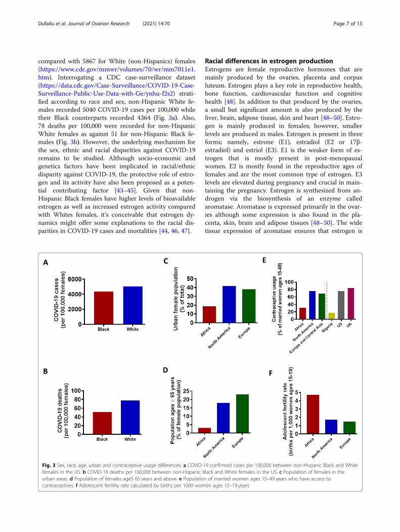
compared with 5867 for White (non-Hispanics) females(https://www.cdc.gov/mmwr/volumes/70/wr/mm7011e1.htm). Interrogating a CDC case-surveillance dataset(https://data.cdc.gov/Case-Surveillance/COVID-19-Case-Surveillance-Public-Use-Data-with-Ge/ynhu-f2s2) strati-fied according to race and sex, non-Hispanic White fe-males recorded 5040 COVID-19 cases per 100,000 whiletheir Black counterparts recorded 4364 (Fig. 3a). Also,78 deaths per 100,000 were recorded for non-HispanicWhite females as against 51 for non-Hispanic Black fe-males (Fig. 3b). However, the underlying mechanism forthe sex, ethnic and racial disparities against COVID-19remains to be studied. Although socio-economic andgenetics factors have been implicated in racial/ethnicdisparity against COVID-19, the protective role of estro-gen and its activity have also been proposed as a poten-tial contributing factor [43–45]. Given that non-Hispanic Black females have higher levels of bioavailableestrogen as well as increased estrogen activity comparedwith Whites females, it’s conceivable that estrogen dy-namics might offer some explanations to the racial dis-parities in COVID-19 cases and mortalities [44, 46, 47].
Racial differences in estrogen productionEstrogens are female reproductive hormones that aremainly produced by the ovaries, placenta and corpusluteum. Estrogen plays a key role in reproductive health,bone function, cardiovascular function and cognitivehealth [48]. In addition to that produced by the ovaries,a small but significant amount is also produced by theliver, brain, adipose tissue, skin and heart [48–50]. Estro-gen is mainly produced in females; however, smallerlevels are produced in males. Estrogen is present in threeforms; namely, estrone (E1), estradiol (E2 or 17β-estradiol) and estriol (E3). E1 is the weaker form of es-trogen that is mostly present in post-menopausalwomen. E2 is mostly found in the reproductive ages offemales and are the most common type of estrogen. E3levels are elevated during pregnancy and crucial in main-taining the pregnancy. Estrogen is synthesized from an-drogen via the biosynthesis of an enzyme calledaromatase. Aromatase is expressed primarily in the ovar-ies although some expression is also found in the pla-centa, skin, brain and adipose tissues [48–50]. The widetissue expression of aromatase ensures that estrogen is
Fig. 3 Sex, race, age, urban and contraceptive usage differences. a COVID-19 confirmed cases per 100,000 between non-Hispanic Black and Whitefemales in the US. b COVID-19 deaths per 100,000 between non-Hispanic Black and White females in the US. c Population of females in theurban areas. d Population of females ageS 65 years and above. e Population of married women ages 15–49 years who have access tocontraceptives. f Adolescent fertility rate calculated by births per 1000 women ages 15–19 years
Dufailu et al. Journal of Ovarian Research (2021) 14:70 Page 7 of 13

produced in a required amount detectable in the bloodfor clinical diagnosis and prognosis of diseaseconditions.With the decreased incidence, hospitalization and
mortality rates amongst African women compared tonon-African women (Figs. 1 and 2), we hypothesize thatestrogen production dynamics could play a central rolein the protection against COVID-19. Aromatase expres-sion, estrogen levels and activity amongst women fromdiverse race and ethnicity have been analyzed in recentstudies [47, 51–53]. Interestingly, estrogen levels as wellas activity have been shown to be 35% higher in non-Latina black women compared to Latinas and non-Latina white women [47]. In a separate study, aromataseactivity and estradiol levels were assessed betweenAfrican-American women and Caucasian women acrossthe menstrual cycle [51]. It was shown that although theAfrican-American and Caucasian women were of similarage (27.2 years) and body mass index (22.7 kg/m2), estra-diol levels were significantly elevated in the African-American women compared to their Caucasian counter-parts [51]. The differences were more pronounced in thelate follicular (225.2 ± 14.4 vs. 191.5 ± 10.2 pg/ml; P <0.02), midluteal (211.9 ± 22.2 vs.150.8 ± 9.9, P < 0.001)and late luteal (144.4 ± 13.2 vs. 103.5 ± 8.5, P < 0.01)phases suggesting an increased aromatase activity in theAfrican-American population [51]. Shaw et al. have alsodemonstrated that young African-American women havehigher levels of ovarian aromatase mRNA expression, es-tradiol and decreased androgen to estrogen ratio com-pared to young Caucasian women [52]. These studiesare consistent with others that have demonstrated in-creased levels of serum E1 and E2 in women of Africandescent compared to Caucasian women [53]. These sug-gest that the racial difference in aromatase expressioncould increase levels of estrogen production in womenof African descent, an outcome that could explain inpart why African-American women are associated withhigher incidence of estrogen-responsive pathologies suchas breast cancer, leiomyoma, increased bone density aswell as early puberty age [44, 46].Although racial differences could play a key role in es-
trogen levels, age, lifestyle, the use of contraceptives andpregnancy incidences could contribute to the differentialestrogen levels observed. Rural and urban lifestyles inthe selected geographic area could in part contribute tothe differential estrogen production. Cardiovascular dis-eases highly correlate with increased sedentary and un-healthy lifestyles; behaviors that are common in theurban areas. Estrogen levels are inversely associated withLDL and triglycerides but positively correlates with highHDL synthesis, circulatory indicators used in the moni-toring of cardiovascular diseases [54]. With only 18.6%of females residing in the urban areas of Africa
compared with 41.7 and 38% in North America and Eur-ope respectively (https://databank.worldbank.org/source/gender-statistics/Series/SP.URB.TOTL.FE.ZS) (Fig. 3c),there is likelihood that urban activities could influencetheir estrogen production. The population of women intheir premenopausal stages in Africa is considerablyhigher compared to their cohorts in the EU, Americaand Asia. Only 3% of females in Africa fall in the cat-egory of 65 years and above compared to that of NorthAmerica (18%) and Europe (23%) (https ://data .worldbank.org/indicator/SP.POP.65UP.FE.ZS) (Fig. 3d).With estrogen mostly produced in the reproductive ageof females, this might contribute to the high levels of es-trogen observed in women of Africa descent.Contraceptive use amongst women with diverse ethnic
backgrounds could potentially affect estrogen productionthus, contributing to the differential levels. Contracep-tives are known to directly act on ovaries resulting in asignificant down-regulation of estrogen synthesis [55]. InAfrica, only 31% of women ages 15–49 years have accessto the use of contraceptives compared with NorthAmerica (76%) and Europe and Central Asia combined(69%) (https://data.worldbank.org/indicator/SP.DYN.CONU.ZS) (Fig. 3e). In Nigeria, only 17% within thatage range have access to contraceptives compared withUK and US which are 84 and 76%, respectively (https://data.worldbank.org/indicator/SP.DYN.CONU.ZS) (Fig.3e). With only a small percentage of the reproductivewomen having access to contraceptives in Africa, thiscould in part explain why estrogen levels are higherin these population compared with their counterpartsfrom other ethnic backgrounds. The use of contracep-tives is directly reflected in the fertility rate recordedin each of the selected geographic areas. For instance,in Africa, 4.7 births per woman are recorded whereas1.7 and 1.5 are recorded in North America and Eur-ope respectively (https://data.worldbank.org/indicator/SP.DYN.TFRT.IN) (Fig. 3e). Circulatory levels of es-trogen are significantly high during pregnancy andthis coupled with the high fertility rate observed inwomen of African descent, could potentially contrib-ute to the reason why estrogen levels are high inthese population [56]. The mortality rate of pregnantwomen infected with SARS-CoV-2 (0.16%) is signifi-cantly lower compared with that the American femalepopulation (2.24%) suggesting the potential immuno-logical and protective functions of estrogen [57].Despite the interesting correlation discussed above, it’s
yet to be investigated if estrogen production is the cen-tral reason why lower incidence and case fatalities ofCOVID-19 are mostly seen in women of African des-cent. Here, we discuss the protective properties of estro-gen that could potentially explain why women of Africandescent have lower incidence and case fatalities of
Dufailu et al. Journal of Ovarian Research (2021) 14:70 Page 8 of 13

COVID-19, information that could contribute to thefight against COVID-19.
Potential anti-COVID-19 properties of estrogenCOVID-19 severity is characterized by cytokine storm,decreased immune function, coagulation dysfunction, in-creased ER stress, increased expression of ACE2 as wellas multiple organ dysfunctioning [5, 7, 58, 59]. Estrogenhas anti-inflammatory function, reduce ER stress, im-prove immune cell functions and decrease the expres-sion of ACE2 (Fig. 4). These suggest that increasedlevels of estrogen in African women could exert immuneprotection against COVID-19 infection, hospitalizationand mortality, a potential reason for the low incidence,hospitalization and mortality rates.
Anti-inflammatory function of estrogenEstrogen plays a key role in the physiological functioningof the immune system. Estrogen receptors (ER) areexpressed on CD4+ T cells, CD8+ T cells, B cells andmonocytes suggesting a direct action of estrogen onthese cells [60]. Increased levels of estrogen during ex-perimental mouse and human studies resulted in de-creased secretion of inflammatory mediators such as IL-1β, IL-6, CCL2, intercellular adhesion molecule-1(ICAM-1) and TNF-α by inhibiting the NF-kB signalingpathway [61–63]. These cytokines especially IL-6 are pri-marily involved in COVID-19-related cytokine storm, acondition that is detrimental to the organs of the pa-tients fueling poor patient outcomes [6, 64]. CCL2 is achemo-attractant for the migration of neutrophils and
monocytes to inflamed areas. Inhibiting their secretionthus, suppresses, alveolar edema, tissue-specific and sys-temic inflammation [63, 65]. E2 activates the signalingcascades in B cells leading to the secretion of antibodythat helps in the fight against pathogens [61, 62]. In-creased E2 production also promotes helper T cell type2 (Th2): type 1 (Th1) leading to the secretion of anti-inflammatory mediators such as IL-10, IFN-γ and IL-4[61, 62]. Although E2 suppresses the functions of Th17helper cells (decreased secretion of pro-inflammatory IL-7), the functions of regulatory T cells (Tregs) are pro-moted to aid in immune tolerance [61, 63]. Despite theanti-inflammatory properties of estrogen, pro-inflammatory cytokines could overwhelm estrogen func-tions. TNFα and IFNγ synergistically inhibit the expres-sion of estrogen in granulosa cells (GC) [66]. Similarmechanism has also been demonstrated using lipopoly-saccharides (LPS) [67]. IL-6 negatively regulates aroma-tase activity and estrogen production via MAKPsignaling pathway in human granulosa tumor cell line(KGN cells) [68]. This suggests that the interplay of cy-tokines and estrogen are key to controlling the hyper-inflammation in disease conditions such as COVID-19.Cumulatively, these anti-inflammatory functions of es-trogen could minimize COVID-19 severity in women,mechanisms that could partly explain why women of Af-rican descent are more resistant to COVID-19 complica-tions as demonstrated by their lower mortality rate.
Inhibition of ACE2 expressionACE2 receptor is expressed by the lung cells as well asthe upper respiratory tract and serves as an entry routefor SARS-COV-2 infection. Thus, increased expressionof this receptor is key to SARS-CoV-2 infection and sub-sequent COVID-19-related complications. Increasedproduction of estrogen inhibits the expression of ACE2receptors in bronchial epithelial cells, cardiocytes andkidney cells, a strategy that could help inhibit the entryand infection of SARS-CoV-2 [69]. It’s therefore possiblethat the elevated levels of estrogen minimize SARS-CoV-2 entry hence the lower infection rate observed inAfrican women.
Suppression of endoplasmic reticulum (ER) stressThe endoplasmic reticulum (ER) is burdened with viralreplication and protein translation (structural and non-structural proteins of coronavirus) when the host cellsare attacked by coronaviruses. These result in increasedlevel of stress in the ER by forming glycosylation, doublemembrane vesicle (DMV) and depleting ER membranelipids, activities that significantly affect the integrity ofthe ER [70]. SARS-CoV-2 infection of host cells pro-duces similar ER stress that contributes to the severity ofthe disease. Estrogen production lowers ER stress by
Fig. 4 Anti-COVID-19 functions of estrogen. Estrogen productionpromotes Th2/Th1, activates T regulatory cells and CD8+ T cells.However, ACE2 expression, ER stress, pro-inflammatory cytokines andTh17 helper cells are inhibited by estrogen
Dufailu et al. Journal of Ovarian Research (2021) 14:70 Page 9 of 13

activating the unfolded protein response (UPR) signalingpathway [70]. This results in the restoration of the ER in-tegrity in host cells as well as inhibits viral replication. Es-trogen has been shown to suppress the replication andtransmission of hepatitis B and influenza viruses minimizeoxidative stress in cardiocytes [70]. Currently, communitytransmission of SARS-CoV-2 in Africa is minimal [71].This could be explained in part by the elevated estrogenlevels in African women, thus, suppressing viral replica-tion and subsequently community transmission.Females infected with SARS-CoV-2 virus are reported
to have better prognosis compared with their malecounterparts. However, they exhibit severe outcomeswhen infected with influenza viruses [38, 69, 70, 72–75].Although SARS-CoV-2-mediated hyper-production ofcytokines and chemokines in females result in clearingthe viruses and improving patient survival faster thanthe males, a deleterious effect on pulmonary tissues areexhibited in females than males in situations of influenzainfections [76–78]. Additionally, males produce in-creased levels of amphiregulin (AREG) compared totheir female counterparts, a growth factor that promotesthe repair of damaged tissues in the lungs as well as re-covery [78, 79]. This suggests that host-mediated immu-nopathology plays a key role in influenza pathogenesisrather than viral titers, a condition that might explainwhy females are associated with severe influenza out-comes. Additionally, influenza pathogenesis significantlyreduces ovarian function thus, inhibiting estrogen pro-duction [77, 80]; an effect that has not been implicatedin SARS-CoV-2 infections. This suggests the relevanceof estrogen production and activity in both SARS-CoV-2and influenza viral infections.
Estrogen as a potential hormonal therapy for COVID-19To date, direct therapeutic options are limited to a mod-estly effective antiviral that remains inaccessible to mostpatients. Patients are managed using best supportive careincluding steroids, and in severe cases the use of mech-anical ventilation and extracorporeal membrane oxygen-ation (ECMO) [81–84]. Identifying novel treatmentmodalities will enable physicians effectively manageCOVID-19 patients effectively. One of the potentialtherapies being explored is estrogen since it has beenshown to inhibit the production of pro-inflammatory cy-tokines, suppress the expression of ACE2 mRNA, stimu-late antibody production, promote Th2/Th1 ratio andreduce ER stress [85]. In a retrospective study, the effectsof systemic hormone administration (estradiol therapy)in women against COVID-19 death were analyzed [45].Estradiol therapy significantly reduced the fatality riskfor post-menopausal women by > 50% lending to thestrength that prospective studies on the potential pro-tective role of estrogen should be investigated [45]. In a
cohort study, women with COVID-19 receiving hor-mone replacement therapy (HRT) showed a higher sur-vival rate supporting the protective effect of estrogen onCOVID-19 [86]. Currently, clinical trials are ongoing toinvestigate the therapeutic efficiency of estrogen therapyin COVID-19 disease. These include estradiol patch(NCT04359329) and Norelgesetromin 6mg / Ethinyl es-tradiol 0.60 mg (NCT04539626) which are being investi-gated in patients with COVID-19. In addition toestrogen, there are ongoing trials which are investigatingother potential COVID-19 treatments such as progester-one (NCT04365127), enzalutamide (NCT04456049,NCT04475601), nafamostat (NCT04418128) and tam-oxifen + isotretinoin (NCT04389580).
Future directions and summaryCOVID-19 is a major public health concern and effortsare being made to discover effective treatments, vaccinesand biomarkers to efficiently manage it. The incidenceand case fatality rates are significantly higher in malescompared to women and this is mostly attributed togenetic, hormonal and immunological differences be-tween men and women. Interestingly, women from Afri-can descent have lower COVID-19 incidence andmortality rates compared to non-African women. Givenestrogen levels and activity are higher in women of Afri-can descent, it’s conceivable that this hormone mightoffer some protection against COVID-19 by suppressinggreater production of cytokines, promoting anti-inflammatory cytokines, stimulating antibody productionand suppressing ER stress (Fig. 4). We acknowledge thatthese suggestions are correlative hence a more mechan-istic investigation is needed to substantiate this finding.Despite the promising findings discussed here, we alsoacknowledge the limitation of the number of sex-disaggregated data presented which are not reported bymost countries. Thus, we limited our selection to coun-tries that have all the parameters available. Also, we arenot able to analyze the data based on age groups andtotal tests done since these parameters are missing inthe dataset from most countries. Notwithstanding, theinformation presented here are compelling and warrantfurther investigation into why women of Africa descenthave lower incidence rate, hospitalization and reducedmortality rates compared to non-African women. Insummary, the greater production of estrogen in Africanwomen coupled with age, lifestyle, high fertility rate andcontraceptive inaccessibility might be the contributingfactors to resisting SARS-CoV-2 infection and minimiz-ing COVID-19 severity.
AcknowledgmentsNot applicable.
Dufailu et al. Journal of Ovarian Research (2021) 14:70 Page 10 of 13

Authors’ contributionsOsman A. Dufailu, Afrakoma Afriyie-Asante and Meshach Asare-Wereheneconceived and designed the study. Osman A. Dufailu, Afrakoma Afriyie-Asante, Meshach Asare-Werehene, Bernard Gyan, Frank Ntiakoh and DavidAdu Kwabena collected data for analyses. Osman A. Dufailu, AfrakomaAfriyie-Asante, Meshach Asare-Werehene, Bernard Gyan, David Adu Kwabena,Helena Yeboah and Frank Ntiakoh contributed contents to the manuscript.Osman A. Dufailu, Afrakoma Afriyie-Asante and Meshach Asare-Werehenedrafted, edited and revised the manuscript with scientific contributions fromall the authors. The author(s) read and approved the final manuscript.
FundingNot applicable.
Declarations
Ethics approval and consent to participateNot applicable.
Consent for publicationNot applicable.
Competing interestsThe authors have no conflict of interest to disclose.
Author details1Department of Microbiology, Faculty of Biosciences, University forDevelopment Studies, Box 1882, Nyankpala Campus, Tamale, Ghana.2Department of Biochemistry, Microbiology and Immunology, Faculty ofMedicine, University of Ottawa, Ottawa, Ontario K1H 8M5, Canada.3Department of Medical Diagnostics, College of Health and Well-Being,Kintampo, Ghana. 4School of Allied Health Sciences, University forDevelopment Studies, Tamale, Ghana. 5School of International Developmentand Global Studies, University of Ottawa, Ottawa, Ontario, Canada.6Department of Medical Laboratory, Effia-Nkwanta Regional Hospital,Sekondi, Western Region, Ghana. 7Departments of Cellular and MolecularMedicine and Obstetrics and Gynecology, University of Ottawa, Ottawa,Ontario K1H 8M5, Canada. 8Chronic Disease Program, Ottawa HospitalResearch Institute, The Ottawa Hospital, Ottawa, Ontario K1H 8L6, Canada.
Received: 1 December 2020 Accepted: 4 May 2021
References1. Rothan HA, Byrareddy SN. The epidemiology and pathogenesis of
coronavirus disease (COVID-19) outbreak. J Autoimmun. 2020;109:102433.https://doi.org/10.1016/j.jaut.2020.102433.
2. Costanzo M, De Giglio MAR, Roviello GN. SARS-CoV-2: recent reports onantiviral therapies based on Lopinavir/ritonavir, Darunavir/Umifenovir,Hydroxychloroquine, Remdesivir, Favipiravir and other drugs for thetreatment of the new coronavirus. Curr Med Chem. 2020;27(27):4536–41.https://doi.org/10.2174/0929867327666200416131117.
3. Jayaweera M, Perera H, Gunawardana B, Manatunge J. Transmission ofCOVID-19 virus by droplets and aerosols: a critical review on the unresolveddichotomy. Environ Res. 2020;188:109819. https://doi.org/10.1016/j.envres.2020.109819.
4. Zhang J, Wang S, Xue Y. Fecal specimen diagnosis 2019 novel coronavirus-infected pneumonia. J Med Virol. 2020;92(6):680–2. https://doi.org/10.1002/jmv.25742.
5. Tay MZ, Poh CM, Rénia L, MacAry PA, Ng LFP. The trinity of COVID-19:immunity, inflammation and intervention. Nat Rev Immunol. 2020;20(6):363–74. https://doi.org/10.1038/s41577-020-0311-8.
6. McGonagle D, et al. The Role of Cytokines including Interleukin-6 inCOVID-19 induced Pneumonia and Macrophage Activation Syndrome-Like Disease. Autoimmun Rev. 2020;19(6):102537. https://doi.org/10.1016/j.autrev.2020.102537.
7. Merad M, Martin JC. Pathological inflammation in patients with COVID-19: akey role for monocytes and macrophages. Nat Rev Immunol. 2020;(7):448.
8. Ozma MA, Maroufi P, Khodadadi E, Köse Ş, Esposito I, Ganbarov K, et al.Clinical manifestation, diagnosis, prevention and control of SARS-CoV-2(COVID-19) during the outbreak period. Infez Med. 2020;28(2):153–65.
9. Huang C, Wang Y, Li X, Ren L, Zhao J, Hu Y, et al. Clinical features ofpatients infected with 2019 novel coronavirus in Wuhan, China. Lancet.2020;395(10223):497–506. https://doi.org/10.1016/S0140-6736(20)30183-5.
10. Yang SS, Lipes J. Corticosteroids for critically ill COVID-19 patients withcytokine release syndrome: a limited case series. Can J Anaesth. 2020;67(10):1462–4. https://doi.org/10.1007/s12630-020-01700-w.
11. Baden LR, el Sahly HM, Essink B, Kotloff K, Frey S, Novak R, et al. Efficacy andsafety of the mRNA-1273 SARS-CoV-2 vaccine. N Engl J Med. 2021;384(5):403–16. https://doi.org/10.1056/NEJMoa2035389.
12. Polack FP, Thomas SJ, Kitchin N, Absalon J, Gurtman A, Lockhart S, et al.Safety and efficacy of the BNT162b2 mRNA Covid-19 vaccine. N Engl J Med.2020;383(27):2603–15. https://doi.org/10.1056/NEJMoa2034577.
13. Chitungo I, Dzobo M, Hlongwa M, Dzinamarira T. COVID-19: unpacking thelow number of cases in Africa. Public Health Pract. 2020;1:100038. https://doi.org/10.1016/j.puhip.2020.100038.
14. Lone SA, Ahmad A. COVID-19 pandemic - an African perspective. EmergMicrobe Infect. 2020;9(1):1300–8.
15. WJ G, et al. Clinical Characteristics of Coronavirus Disease 2019 in China. NEngl J Med. 2020;382(18):1708–20.
16. Why Sub-Saharan Africa needs a unique response to COVID-19. 2020;Available from: https://www.weforum.org/agenda/2020/03/why-sub-saharan-africa-needs-a-unique-response-to-covid-19/. Accessed 1 Nov 2020.
17. M, M.L, et al. COVID-19 in Africa: the spread and response. Nat Med. 2020;26(7):999–1003.
18. COVID-19 Government Measures Dataset. 2020. https://www.acaps.org/covid-19-government-measures-dataset. Accessed 1 Nov 2020.
19. JN, N. and M. W. Looming threat of COVID-19 infection in Africa: actcollectively, and fast. Lancet (London, England). 2020;395(10227):841–2.
20. M, M. COVID-19 in Africa: half a year later. Lancet Infect Dis. 2020;20(10):30708–8.
21. WHO:COVID-19 cases top 10 000 in Africa. 2020.22. @AfricaCDC, CDC. (2020). COVID-19 dashboard. 2020.23. @helenrsullivan, Global report: WHO warns of accelerating Covid-19
infections in Africa. 2020.24. Burke J. Total confirmed coronavirus cases in Africa pass 1 million; 2020.25. Coronavirus cases by country in Africa 2020 | Statista. 2020. https://www.sta
tista.com/statistics/1170463/coronavirus-cases-in-africa/. Accessed 1 Nov2020.
26. Haider N, et al. Lockdown measures in response to COVID-19 in nine sub-Saharan African countries. BMJ Glob Health. 2020;5(10):e003319.
27. Coronavirus active cases by country in Africa 2020 | Statista. 2020. https://www.statista.com/statistics/1170566/coronavirus-active-cases-in-africa/.Accessed 1 Nov 2020.
28. Lone SA, Ahmad A. COVID-19 pandemic - an African perspective. EmergMicrobes Infect. 2020;9(1):1300–8. https://doi.org/10.1080/22221751.2020.1775132.
29. Gilbert M, Pullano G, Pinotti F, Valdano E, Poletto C, Boëlle PY, et al.Preparedness and vulnerability of African countries against importations ofCOVID-19: a modelling study. Lancet. 2020;395(10227):871–7. https://doi.org/10.1016/S0140-6736(20)30411-6.
30. Tang YW, et al. Laboratory Diagnosis of COVID-19: Current Issues andChallenges. J Clin Microbiol. 2020;58(6):e00512–20.
31. Cherif A, Grobe N, Wang X, Kotanko P. Simulation of Pool testing to identifypatients with coronavirus disease 2019 under conditions of limited testavailability. JAMA Netw Open. 2020;3(6):e2013075. https://doi.org/10.1001/jamanetworkopen.2020.13075.
32. Massinga Loembé M, Tshangela A, Salyer SJ, Varma JK, Ouma AEO,Nkengasong JN. COVID-19 in Africa: the spread and response. Nat Med.2020;26(7):999–1003. https://doi.org/10.1038/s41591-020-0961-x.
33. Cham B, Scholes S, Ng Fat L, Badjie O, Mindell JS. Burden of hypertension inthe Gambia: evidence from a national World Health Organization (WHO)STEP survey. Int J Epidemiol. 2018;47(3):860–71. https://doi.org/10.1093/ije/dyx279.
34. Taneja V. Sex Hormones Determine Immune Response. Front Immunol.2018;9:1931. https://doi.org/10.3389/fimmu.2018.01931.
35. Karlberg J, Chong DS, Lai WY. Do men have a higher case fatality rate ofsevere acute respiratory syndrome than women do? Am J Epidemiol. 2004;159(3):229–31. https://doi.org/10.1093/aje/kwh056.
36. Mauvais-Jarvis F, Bairey Merz N, Barnes PJ, Brinton RD, Carrero JJ, DeMeo DL,et al. Sex and gender: modifiers of health, disease, and medicine. Lancet.2020;396(10250):565–82. https://doi.org/10.1016/S0140-6736(20)31561-0.
Dufailu et al. Journal of Ovarian Research (2021) 14:70 Page 11 of 13

37. Guan WJ, Ni ZY, Hu Y, Liang WH, Ou CQ, He JX, et al. Clinical characteristicsof coronavirus disease 2019 in China. N Engl J Med. 2020;382(18):1708–20.https://doi.org/10.1056/NEJMoa2002032.
38. Onder G, Rezza G, Brusaferro S. Case-fatality rate and characteristics ofpatients dying in relation to COVID-19 in Italy. Jama. 2020;323(18):1775–6.https://doi.org/10.1001/jama.2020.4683.
39. Rentsch CT, Kidwai-Khan F, Tate JP, Park LS, King JT, Skanderson M, et al.Patterns of COVID-19 testing and mortality by race and ethnicity amongUnited States veterans: a nationwide cohort study. PLoS Med. 2020;17(9):e1003379. https://doi.org/10.1371/journal.pmed.1003379.
40. Yehia BR, Winegar A, Fogel R, Fakih M, Ottenbacher A, Jesser C, et al.Association of Race with Mortality among Patients Hospitalized withCoronavirus Disease 2019 (COVID-19) at 92 US hospitals. JAMA Netw Open.2020;3(8):e2018039. https://doi.org/10.1001/jamanetworkopen.2020.18039.
41. Price-Haywood EG, Burton J, Fort D, Seoane L. Hospitalization and mortalityamong black patients and white patients with Covid-19. N Engl J Med.2020;382(26):2534–43. https://doi.org/10.1056/NEJMsa2011686.
42. Kabarriti R, Brodin NP, Maron MI, Guha C, Kalnicki S, Garg MK, et al. Associationof Race and Ethnicity with Comorbidities and survival among patients withCOVID-19 at an urban medical Center in New York. JAMA Netw Open. 2020;3(9):e2019795. https://doi.org/10.1001/jamanetworkopen.2020.19795.
43. McCoy J, Wambier CG, Vano-Galvan S, Shapiro J, Sinclair R, Ramos PM, et al.Racial variations in COVID-19 deaths may be due to androgen receptorgenetic variants associated with prostate cancer and androgenetic alopecia.Are anti-androgens a potential treatment for COVID-19? J Cosmet Dermatol.2020;19(7):1542–3. https://doi.org/10.1111/jocd.13455.
44. Pinheiro SP, et al. Racial differences in premenopausal endogenoushormones. Cancer Epidemiol Biomark Prev. 2005;14(9):2147–53. https://doi.org/10.1158/1055-9965.EPI-04-0944.
45. Seeland U, Coluzzi F, Simmaco M, Mura C, Bourne PE, Heiland M, et al.Evidence for treatment with estradiol for women with SARS-CoV-2 infection.BMC Med. 2020;18(1):369. https://doi.org/10.1186/s12916-020-01851-z.
46. Setiawan VW, Haiman CA, Stanczyk FZ, le Marchand L, Henderson BE.Racial/ethnic differences in postmenopausal endogenous hormones: themultiethnic cohort study. Cancer Epidemiol Biomark Prev. 2006;15(10):1849–55. https://doi.org/10.1158/1055-9965.EPI-06-0307.
47. Sanchez SS, Tachachartvanich P, Stanczyk FZ, Gomez SL, John EM, SmithMT, et al. Estrogenic activity, race/ethnicity, and indigenous Americanancestry among San Francisco Bay Area women. PLoS One. 2019;14(3):e0213809. https://doi.org/10.1371/journal.pone.0213809.
48. Cagnacci A, Soldani R, Carriero PL, Paoletti AM, Fioretti P, Melis GB. Effects oflow doses of transdermal 17 beta-estradiol on carbohydrate metabolism inpostmenopausal women. J Clin Endocrinol Metab. 1992;74(6):1396–400.https://doi.org/10.1210/jcem.74.6.1317387.
49. Zahid H, Simpson ER, Brown KA. Inflammation, dysregulated metabolismand aromatase in obesity and breast cancer. Curr Opin Pharmacol. 2016;31:90–6. https://doi.org/10.1016/j.coph.2016.11.003.
50. Nelson LR, Bulun SE. Estrogen production and action. J Am Acad Dermatol.2001;45(3 Suppl):S116–24. https://doi.org/10.1067/mjd.2001.117432.
51. Marsh EE, Shaw ND, Klingman KM, Tiamfook-Morgan TO, Yialamas MA, SlussPM, et al. Estrogen levels are higher across the menstrual cycle in African-American women compared with Caucasian women. J Clin EndocrinolMetab. 2011;96(10):3199–206. https://doi.org/10.1210/jc.2011-1314.
52. Shaw ND, Srouji SS, Welt CK, Cox KH, Fox JH, Adams JM, et al. Evidence thatincreased ovarian aromatase activity and expression account for higherestradiol levels in African American compared with Caucasian women. JClin Endocrinol Metab. 2014;99(4):1384–92. https://doi.org/10.1210/jc.2013-2398.
53. Woods MN, Barnett JB, Spiegelman D, Trail N, Hertzmark E, Longcope C,et al. Hormone levels during dietary changes in premenopausal African-American women. J Natl Cancer Inst. 1996;88(19):1369–74. https://doi.org/10.1093/jnci/88.19.1369.
54. Guetta V, Cannon RO 3rd. Cardiovascular effects of estrogen and lipid-lowering therapies in postmenopausal women. Circulation. 1996;93(10):1928–37. https://doi.org/10.1161/01.CIR.93.10.1928.
55. Mishell DR Jr, Thorneycroft IH, Nakamura RM, Nagata Y, Stone SC. Serumestradiol in women ingesting combination oral contraceptive steroids. Am JObstet Gynecol. 1972;114(7):923–8. https://doi.org/10.1016/0002-9378(72)90098-1.
56. Tulchinsky D, Hobel CJ, Yeager E, Marshall JR. Plasma estrone, estradiol,estriol, progesterone, and 17-hydroxyprogesterone in human pregnancy. I.
Normal pregnancy. Am J Obstet Gynecol. 1972;112(8):1095–100. https://doi.org/10.1016/0002-9378(72)90185-8.
57. Berhan Y. What immunological and hormonal protective factors lower therisk of COVID-19 related deaths in pregnant women? J Reprod Immunol.2020;142:103180. https://doi.org/10.1016/j.jri.2020.103180.
58. Liu J, Li S, Liu J, Liang B, Wang X, Wang H, et al. Longitudinal characteristicsof lymphocyte responses and cytokine profiles in the peripheral blood ofSARS-CoV-2 infected patients. EBioMedicine. 2020;55:102763. https://doi.org/10.1016/j.ebiom.2020.102763.
59. Shi Y, Wang Y, Shao C, et al. COVID-19 infection: the perspectives onimmune responses. Cell Death Differ; 2020;27:1451–4. https://doi.org/10.1038/s41418-020-0530-3.
60. Phiel KL, Henderson RA, Adelman SJ, Elloso MM. Differential estrogen receptorgene expression in human peripheral blood mononuclear cell populations.Immunol Lett. 2005;97(1):107–13. https://doi.org/10.1016/j.imlet.2004.10.007.
61. Straub RH. The complex role of estrogens in inflammation. Endocr Rev.2007;28(5):521–74. https://doi.org/10.1210/er.2007-0001.
62. Klein SL, Flanagan KL. Sex differences in immune responses. Nat RevImmunol. 2016;16(10):626–38. https://doi.org/10.1038/nri.2016.90.
63. Channappanavar R, Fett C, Mack M, ten Eyck PP, Meyerholz DK, Perlman S.Sex-based differences in susceptibility to severe acute respiratory syndromecoronavirus infection. J Immunol. 2017;198(10):4046–53. https://doi.org/10.4049/jimmunol.1601896.
64. Conti P, et al. Induction of pro-inflammatory cytokines (IL-1 and IL-6)and lung inflammation by Coronavirus-19 (COVI-19 or SARS-CoV-2):anti-inflammatory strategies. J Biol Regul Homeost Agents. 2020;34(2):327–31.
65. He L, Ding Y, Zhang Q, Che X, He Y, Shen H, et al. Expression of elevatedlevels of pro-inflammatory cytokines in SARS-CoV-infected ACE2+ cells inSARS patients: relation to the acute lung injury and pathogenesis of SARS. JPathol. 2006;210(3):288–97. https://doi.org/10.1002/path.2067.
66. Fukuoka M, Yasuda K, Fujiwara H, Kanzaki H, Mori T. Interactions betweeninterferon gamma, tumour necrosis factor alpha, and interleukin-1 inmodulating progesterone and oestradiol production by human luteinizedgranulosa cells in culture. Hum Reprod. 1992;7(10):1361–4. https://doi.org/10.1093/oxfordjournals.humrep.a137574.
67. Taylor CC, Terranova PF. Lipopolysaccharide inhibits in vitro luteinizinghormone-stimulated rat ovarian granulosa cell estradiol but notprogesterone secretion. Biol Reprod. 1996;54(6):1390–6. https://doi.org/10.1095/biolreprod54.6.1390.
68. Deura I, et al. Reduction of estrogen production by interleukin-6 in a humangranulosa tumor cell line may have implications for endometriosis-associated infertility. Fertil Steril. 2005;83(Suppl 1):1086–92. https://doi.org/10.1016/j.fertnstert.2004.12.014.
69. Groban L, Wang H, Sun X, Ahmad S, Ferrario CM. Is sex a determinant ofCOVID-19 infection? Truth or myth? Curr Hypertens Rep. 2020;22(9):62.https://doi.org/10.1007/s11906-020-01073-x.
70. Shabbir S, Hafeez A, Rafiq MA, Khan MJ. Estrogen shields women fromCOVID-19 complications by reducing ER stress. Med Hypotheses. 2020;143:110148. https://doi.org/10.1016/j.mehy.2020.110148.
71. Musa HH, Musa TH, Musa IH, Musa IH, Ranciaro A, Campbell MC. AddressingAfrica's pandemic puzzle: perspectives on COVID-19 transmission andmortality in sub-Saharan Africa. Int J Infect Dis. 2021;102:483–8. https://doi.org/10.1016/j.ijid.2020.09.1456.
72. Klein SL, Passaretti C, Anker M, Olukoya 'P, Pekosz A. The impact of sex,gender and pregnancy on 2009 H1N1 disease. Biol Sex Differ. 2010;1(1):5.https://doi.org/10.1186/2042-6410-1-5.
73. Serfling RE, Sherman IL, Houseworth WJ. Excess pneumonia-influenzamortality by age and sex in three major influenza A2 epidemics, UnitedStates, 1957-58, 1960 and 1963. Am J Epidemiol. 1967;86(2):433–41. https://doi.org/10.1093/oxfordjournals.aje.a120753.
74. Update. WHO-confirmed human cases of avian influenza a (H5N1) infection,November 2003-may 2008. Wkly Epidemiol Rec. 2008;83(46):415–20.
75. Kumar A, Zarychanski R, Pinto R, Cook DJ, Marshall J, Lacroix J, et al.Critically ill patients with 2009 influenza a(H1N1) infection in Canada. Jama.2009;302(17):1872–9. https://doi.org/10.1001/jama.2009.1496.
76. Hoffmann J, Otte A, Thiele S, Lotter H, Shu Y, Gabriel G. Sex differences inH7N9 influenza a virus pathogenesis. Vaccine. 2015;33(49):6949–54. https://doi.org/10.1016/j.vaccine.2015.08.044.
77. Robinson DP, Lorenzo ME, Jian W, Klein SL. Elevated 17β-estradiol protectsfemales from influenza a virus pathogenesis by suppressing inflammatory
Dufailu et al. Journal of Ovarian Research (2021) 14:70 Page 12 of 13

responses. PLoS Pathog. 2011;7(7):e1002149. https://doi.org/10.1371/journal.ppat.1002149.
78. Vermillion MS, Ursin RL, Kuok DIT, vom Steeg LG, Wohlgemuth N, Hall OJ,et al. Production of amphiregulin and recovery from influenza is greater inmales than females. Biol Sex Differ. 2018;9(1):24. https://doi.org/10.1186/s13293-018-0184-8.
79. Monticelli LA, Sonnenberg GF, Abt MC, Alenghat T, Ziegler CGK, Doering TA,et al. Innate lymphoid cells promote lung-tissue homeostasis after infectionwith influenza virus. Nat Immunol. 2011;12(11):1045–54. https://doi.org/10.1038/ni.2131.
80. Vermillion MS, Ursin RL, Attreed SE, Klein SL. Estriol reduces pulmonaryimmune cell recruitment and inflammation to protect female mice fromsevere influenza. Endocrinology. 2018;159(9):3306–20. https://doi.org/10.1210/en.2018-00486.
81. Esposito S, Noviello S, Pagliano P. Update on treatment of COVID-19:ongoing studies between promising and disappointing results. Infez Med.2020;28(2):198–211.
82. Ferner RE, Aronson JK. Chloroquine and hydroxychloroquine in covid-19.Bmj. 2020;369:m1432.
83. Gautret P, Lagier JC, Parola P, Hoang VT, Meddeb L, Mailhe M, et al.Hydroxychloroquine and azithromycin as a treatment of COVID-19: results ofan open-label non-randomized clinical trial. Int J Antimicrob Agents. 2020;56(1):105949. https://doi.org/10.1016/j.ijantimicag.2020.105949.
84. Tu YF, et al. A Review of SARS-CoV-2 and the Ongoing Clinical Trials. Int JMol Sci. 2020;21(7):2657.
85. Cattrini C, et al. Sex Hormones and Hormone Therapy during COVID-19Pandemic: Implications for Patients with Cancer. Cancers (Basel). 2020;12(8):2325.
86. Costeira R, et al. Estrogen and COVID-19 symptoms: associations in womenfrom the COVID Symptom Study. medRxiv. 2020; 2020.07.30.20164921.
Publisher’s NoteSpringer Nature remains neutral with regard to jurisdictional claims inpublished maps and institutional affiliations.
Dufailu et al. Journal of Ovarian Research (2021) 14:70 Page 13 of 13