craig strickland, ph.d. & 5 1 2017 winden rowe, ms...craig strickland, ph.d. & winden rowe,...
TRANSCRIPT
Craig Strickland, Ph.D. & Winden Rowe, MS
5_1_2017
1
The human brain is awesome. It functions 24 hours a day from the day we are
born and only stops when we are taking an exam or FALL IN LOVE!
Clinical Applications of Neuroscience
Craig Strickland, Ph.D. [email protected] Rowe, MS [email protected]
Overview of the Human Nervous System
Craig Strickland, Ph.D. & Winden Rowe, MS
5_1_2017
2
Identify things/events, etc. you think may change brain
structure or function
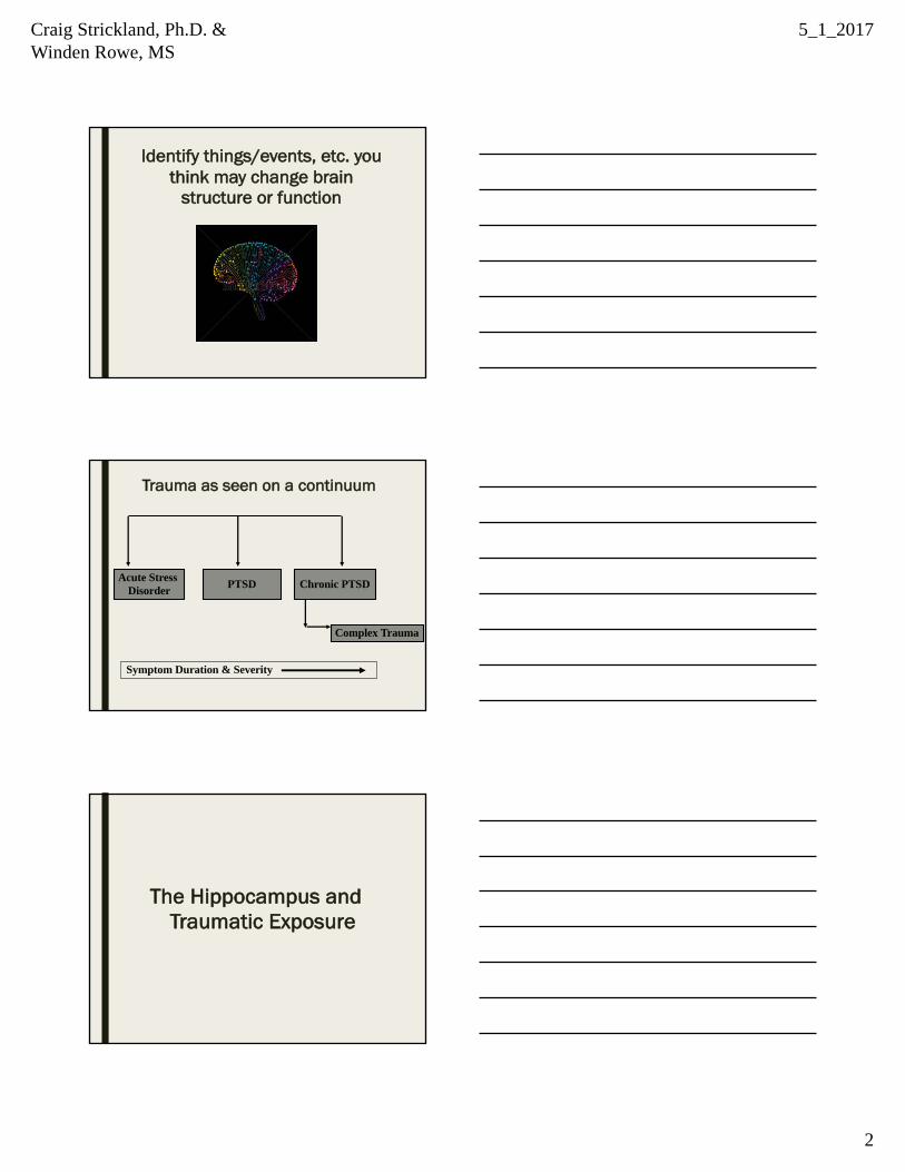
Trauma as seen on a continuum
Acute Stress Disorder PTSD Chronic PTSD
Complex Trauma
Symptom Duration & Severity
The Hippocampus and Traumatic Exposure

Craig Strickland, Ph.D. & Winden Rowe, MS
5_1_2017
3
CNS: Limbic System
■ Central role in emotional “modulation”: amygdala■ Connects brainstem & “higher” cortical structures■ Integrates emotion, memory and behavior■ Limbic system dysfunction & behavioral health
Hippocampus
Normal Functions of the Hippocampus
■ The hippocampus is a very complex structure
■ Is part of the Limbic System
■ Considered “transitional” tissue
■ Normal functions include (but may not be limited to)– Memory consolidation: works together with newer cortical brain
areas■ Integration of “emotional tone” with “higher” cognitive functions■ Cortex provides semantic influence to the more episodic (factual)
“hippocampal” memories– Behavioral inhibition– Inhibitory influence on brainstem activity
Craig Strickland, Ph.D. & Winden Rowe, MS
5_1_2017
4
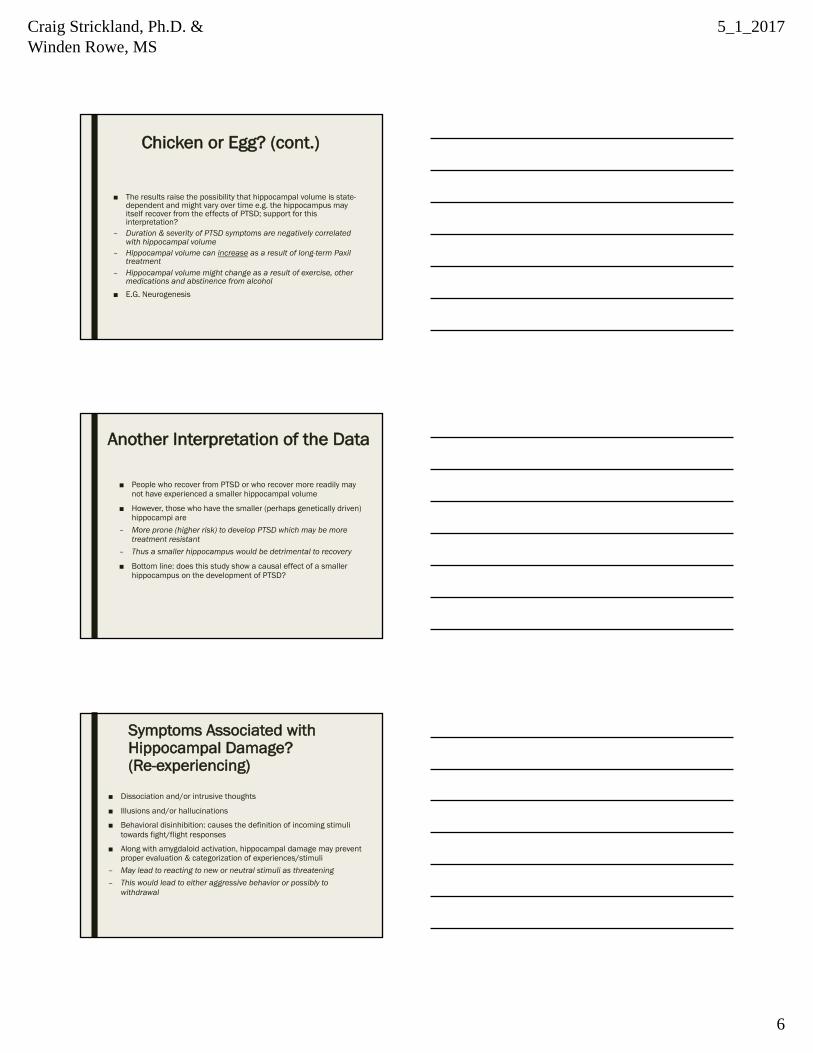
The Hippocampus & Trauma■ Original studies: decrease volume (size) of the hippocampus■ Vietnam vets: 8 to 26% reduction depending on the study■ 7% reduction in women with history of childhood sexual abuse■ 16% reduction in hippocampal volume in women with BPD (often associated
with a history of abuse)– Gilbertson, et. al. (2002): twin studies- Chicken/Egg question (image
from original study)
Chicken or Egg?
■ Apfel, et. al. (2011)– Gilbertson study did not contain true longitudinal data– This of course would be nearly impossible to do in this type
research– Apfel study looked at hippocampal volume in 244 male Gulf War
Veterans■ Study included those with current PTSD and those where the symptoms
of PTSD were remitted
Chicken or Egg? (cont.)
■ In addition to measuring symptoms of PTSD (using the Clinician Administered PTSD Scale (CAPS) assessment tool:
– Measured presence or absence of depression– Measured lifetime drinking history– History of other (non-combat related) stressors
■ Used structural MRI techniques to measure hippocampal volume

Craig Strickland, Ph.D. & Winden Rowe, MS
5_1_2017
5
Chicken or Egg? (cont.)
■ Ended up with four groups of subjects– Subjects with no traumatic exposure– Subjects with exposure but no PTSD– Subjects with a previous diagnosis of PTSD but have recovered– Subjects with chronic PTSD (lifetime and current)
■ The first two groups had identical hippocampal volume and were combined into one group for the further analysis
Chicken or Egg? (cont.)
■ Results– Subjects with current/lifetime PTSD■ Subjects with current/lifetime PTSD had smaller hippocampi by 5.1% than
those who had never developed PTSD■ 6.5% smaller hippocampi than those who had recovered from PTSD
– Note: there was no difference in hippocampal size between those who never had PTSD and those who had recovered from PTSD
■ Interpretations?
Chicken or Egg? (cont.)
■ The significant differences remained even after accounting for the following factors:
– Early life trauma– Current and lifetime alcohol use– Depression and treatment with antidepressants
■ Thus, a smaller hippocampus cannot be a vulnerability factor (e.g. a genetically driven cause) of PTSD; if this were the case, the group that recovered should have a hippocampal size that is smaller than those who never developed PTSD. This finding does NOT mean that hippocampal size is NOT related to the symptoms of PTSD
Craig Strickland, Ph.D. & Winden Rowe, MS
5_1_2017
6
Chicken or Egg? (cont.)
■ The results raise the possibility that hippocampal volume is state-dependent and might vary over time e.g. the hippocampus may itself recover from the effects of PTSD; support for this interpretation?
– Duration & severity of PTSD symptoms are negatively correlated with hippocampal volume
– Hippocampal volume can increase as a result of long-term Paxil treatment
– Hippocampal volume might change as a result of exercise, other medications and abstinence from alcohol
■ E.G. Neurogenesis
Another Interpretation of the Data
■ People who recover from PTSD or who recover more readily may not have experienced a smaller hippocampal volume
■ However, those who have the smaller (perhaps genetically driven) hippocampi are
– More prone (higher risk) to develop PTSD which may be more treatment resistant
– Thus a smaller hippocampus would be detrimental to recovery
■ Bottom line: does this study show a causal effect of a smaller hippocampus on the development of PTSD?
Symptoms Associated with Hippocampal Damage? (Re-experiencing)
■ Dissociation and/or intrusive thoughts
■ Illusions and/or hallucinations
■ Behavioral disinhibition: causes the definition of incoming stimuli towards fight/flight responses
■ Along with amygdaloid activation, hippocampal damage may prevent proper evaluation & categorization of experiences/stimuli
– May lead to reacting to new or neutral stimuli as threatening– This would lead to either aggressive behavior or possibly to
withdrawal
Craig Strickland, Ph.D. & Winden Rowe, MS
5_1_2017
7
The Limbic System and PTSD:
The Amygdala
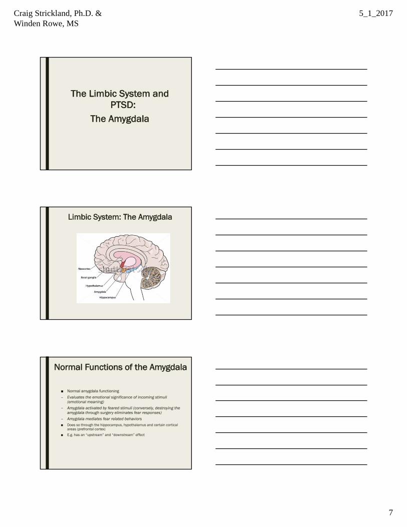
Limbic System: The Amygdala
Normal Functions of the Amygdala
■ Normal amygdala functioning– Evaluates the emotional significance of incoming stimuli
(emotional meaning)– Amygdala activated by feared stimuli (conversely, destroying the
amygdala through surgery eliminates fear responses)– Amygdala mediates fear related behaviors■ Does so through the hippocampus, hypothalamus and certain cortical
areas (prefrontal cortex)■ E.g. has an “upstream” and “downstream” effect
Craig Strickland, Ph.D. & Winden Rowe, MS
5_1_2017
8
Symptoms associated with Amygdaloid hyperactivity (hyperarousal)
■ Neutral stimuli are seen as fearful– Through connections with other limbic structures, enhanced
autonomic and neurohormonal responses■ Increased sympathetic nervous system activity■ Hypervigilance■ Exaggerated startle response■ Irritability or outbursts of anger
■ Destruction versus stimulating the amygdala
■ Increased Hypothalamic/Pituitary Adrenal axis activity (known as HPA)
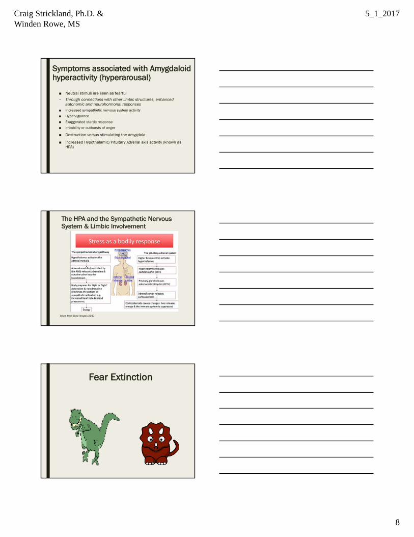
The HPA and the Sympathetic Nervous System & Limbic Involvement
Taken from Bing Images 2017
Fear Extinction
Craig Strickland, Ph.D. & Winden Rowe, MS
5_1_2017
9
Brain Areas Involved in Fear Extinction
■ Ventromedial Prefrontal Cortex (vmPFC)■ Hippocampus■ Amygdala■ Entorhinal Cortex■ Anterior Cingulate Cortex
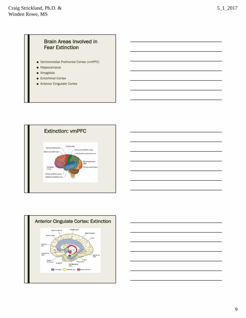
Extinction: vmPFC
Anterior Cingulate Cortex: Extinction

Craig Strickland, Ph.D. & Winden Rowe, MS
5_1_2017
10
Lack of ability to extinguish conditioned emotional response
■ Decrease in activity in the vmPFC and/or decrease in hippocampal and/or portions of anterior cingulate cortex (ACC)
■ In fact, direct correlation between ACC volume and treatment response to extinction therapy
■ Amygdala: amygdala activity should change as relationship between a stimulus and it’s consequence changes (e.g. CS+ or CS-) through learning
DISSOCIATION
Dissociative Phenomena (cont.)■ “Physiologically, they may respond as if they are being traumatized
again, but this may be dissociated from semantic knowledge” (van der Kolk, 1997)

Craig Strickland, Ph.D. & Winden Rowe, MS
5_1_2017
11
Dissociative Phenomena (van der Kolk)
■ Failure of left-hemisphere functioning at the time of the trauma (i.e. during extreme arousal)
– Decreased activation of Broca’s area (Broca’s area is involved in labeling emotions)
■ Cannot communicate what is going on, cannot label the internal state■ Thus, during extreme arousal/intense emotions, the individual cannot
“understand” what is going on– Left-hemisphere: also involved in sequencing events and categorizing
experiences. Dysfunction leads to:■ Trauma being seen as timeless■ Trauma being seen as “ego-alien”
SOCIAL ASPECTS OF TRAUMATIC EXPOSURE
Polyvagal Theory, Vagal Nerve Stimulation (VNS) and Trauma & Depression
PictureprovidedbyPeterJurek,MA,MShttp://www.peterjurek.com
Craig Strickland, Ph.D. & Winden Rowe, MS
5_1_2017
12
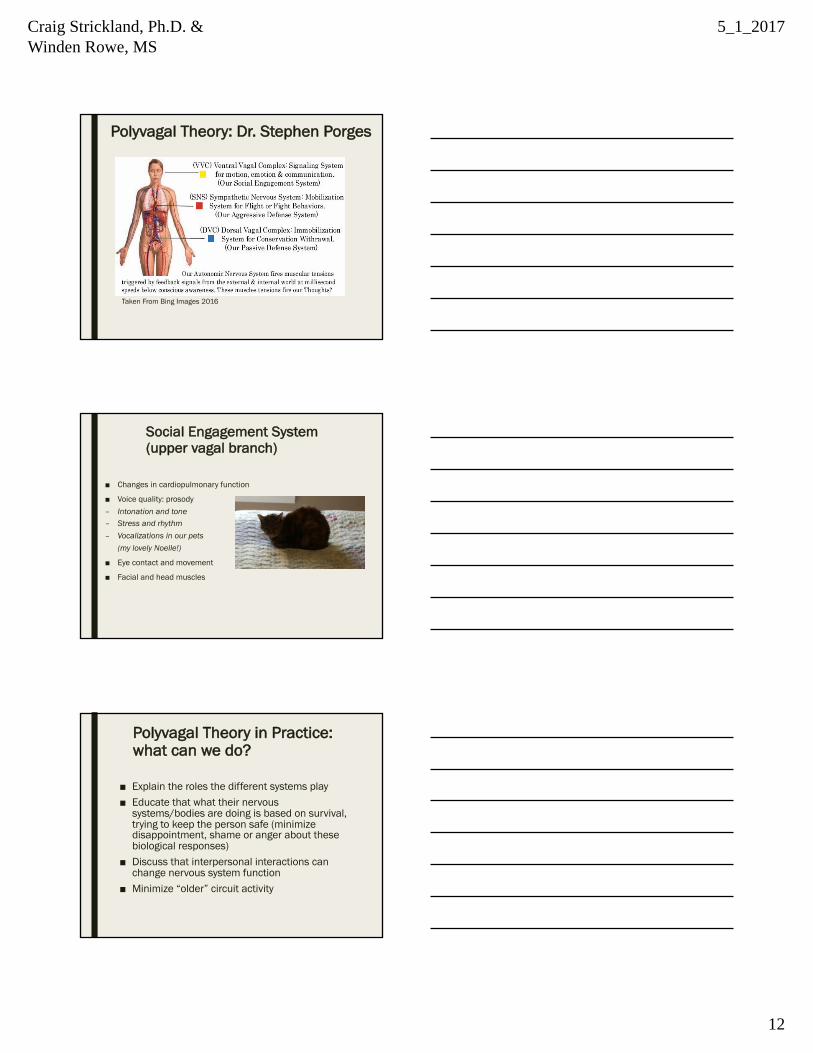
Polyvagal Theory: Dr. Stephen Porges
Taken From Bing Images 2016
Social Engagement System (upper vagal branch)
■ Changes in cardiopulmonary function
■ Voice quality: prosody– Intonation and tone– Stress and rhythm– Vocalizations in our pets
(my lovely Noelle!)
■ Eye contact and movement
■ Facial and head muscles
Polyvagal Theory in Practice: what can we do?
■ Explain the roles the different systems play■ Educate that what their nervous
systems/bodies are doing is based on survival, trying to keep the person safe (minimize disappointment, shame or anger about these biological responses)
■ Discuss that interpersonal interactions can change nervous system function
■ Minimize “older” circuit activity
Craig Strickland, Ph.D. & Winden Rowe, MS
5_1_2017
13
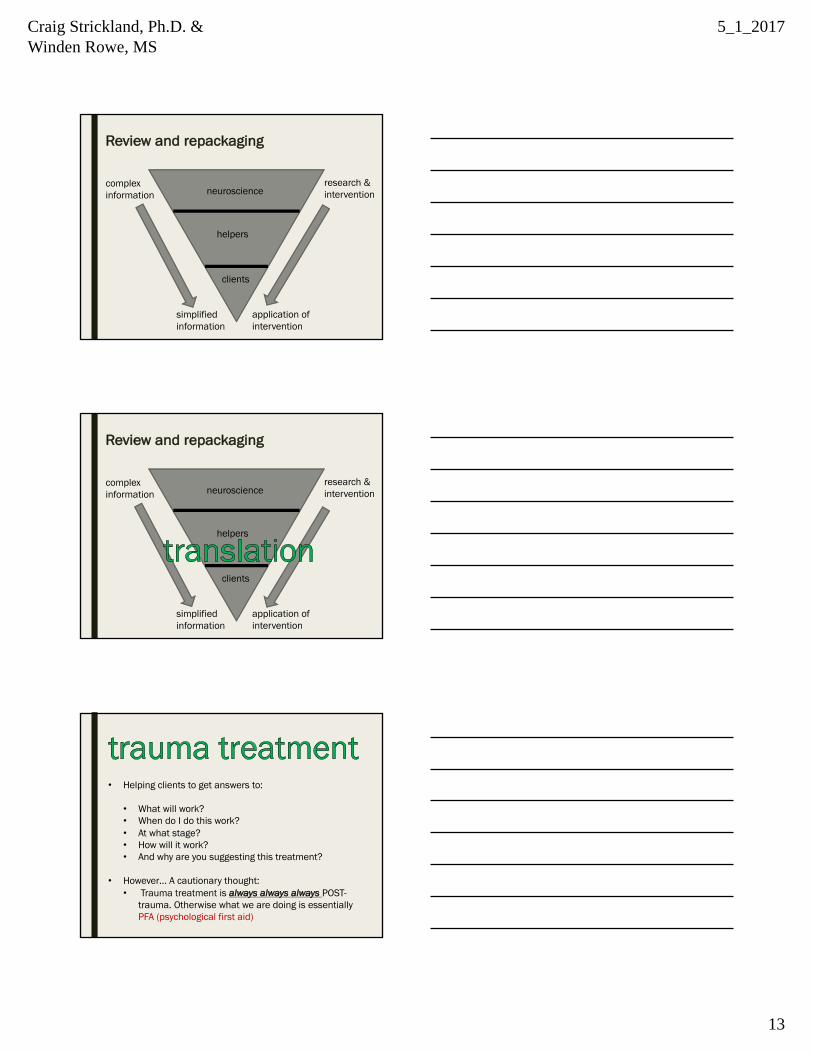
Review and repackaging
neuroscience
clients
helpers
simplifiedinformation
complexinformation
research &intervention
application ofintervention
Review and repackaging
neuroscience
clients
helpers
simplifiedinformation
complexinformation
application ofintervention
research &intervention
• Helping clients to get answers to:
• What will work?• When do I do this work? • At what stage?• How will it work?• And why are you suggesting this treatment?
• However… A cautionary thought:• Trauma treatment is always always always POST-
trauma. Otherwise what we are doing is essentially PFA (psychological first aid)
Craig Strickland, Ph.D. & Winden Rowe, MS
5_1_2017
14
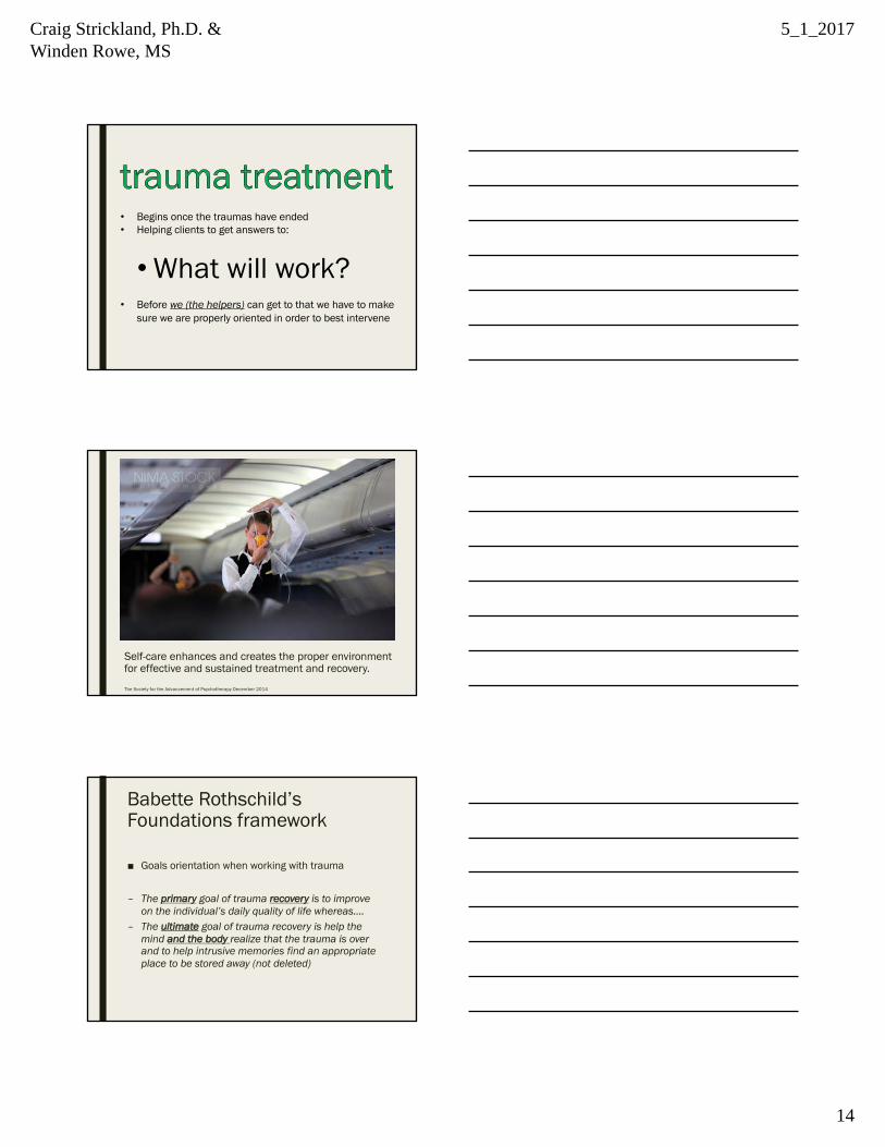
• Begins once the traumas have ended • Helping clients to get answers to:
•What will work?• Before we (the helpers) can get to that we have to make
sure we are properly oriented in order to best intervene
Self-care enhances and creates the proper environment for effective and sustained treatment and recovery.
The Society for the Advancement of Psychotherapy December 2014
Babette Rothschild’s Foundations framework
■ Goals orientation when working with trauma
– The primary goal of trauma recovery is to improve on the individual’s daily quality of life whereas….
– The ultimate goal of trauma recovery is help the mind and the body realize that the trauma is over and to help intrusive memories find an appropriate place to be stored away (not deleted)
Craig Strickland, Ph.D. & Winden Rowe, MS
5_1_2017
15
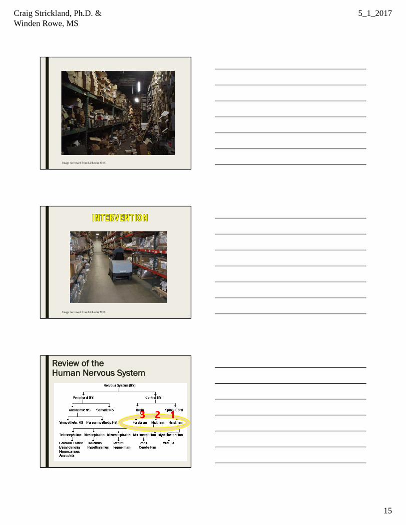
Image borrowed from Linkedin 2016
Image borrowed from Linkedin 2016
Review of the Human Nervous System
3 2 1

Craig Strickland, Ph.D. & Winden Rowe, MS
5_1_2017
16
Judith Herman’s Stages of Recovery for Trauma (complex)
1. Stabilization, safety (1- hindbrain)2. Remembering and mourning (2- midbrain)3. Meaning and reconnection (3- forebrain)
Stage 1 impact of trauma
■ Hindbrain - brain stem activity
– Instinctual reactions– Autonomic dysregulation■ Heartrate elevates■ Shallow breathing■ Slowed digestive processes ■ Hypertension■ Swallowing ■ Blood pressure
Stage 1 recovery & interventions
■ Brain stem responses - need to relearn regulation – CONTAINED■ Stabilization and Safety
– Yoga– Breath work– Grounding exercises– Tai chi– Simple sharing of daily events– Body scanning, mapping – Posturing exercises – SSRI and other pharma for short-term– Mindfulness of somatic expression– Considerations for early recovery

Craig Strickland, Ph.D. & Winden Rowe, MS
5_1_2017
17
■ The midbrain - limbic system
– Survival– Sensing and responding■ Fight, flight, freeze– Interpretation of information– Assessment of internal and external environments – Sequencing– Alarm on without switching off– Emotional tagging and rating
Image borrowed from Victoria Stillwell Online, 2016
Stage 2 impact of trauma
■ The midbrain- limbic system
■ Remembrance and Mourning - less containment
– Feeling identification– Guided imagery– Memory integration exercises– Timelining– Emotional responses to stress– EMDR (!)– TFCBT– DBT– Mindfulness (of thought, of feeling)– Early 12 Step recovery
Stage 2 recovery & interventions
Stage 3 impact of trauma
■ The forebrain
– Recalling facts and information– Accurate details of events– Verbal expression– Recognition of time– Understanding environment– Verbal recognition– Social isolation– Inability to ID accurate self-concept – Memory consolidation

Craig Strickland, Ph.D. & Winden Rowe, MS
5_1_2017
18
Stage 3 recovery & considerations■ The forebrain
■ Meaning and reconnection – open processing
– Cognitive processing– Self expression– Self actualization– Posttraumatic growth– Making connections– DBT– TFCBT– Mindfulness (open thought meditation)– 12 Step (later steps)
Image borrowed from Happy OnePlus Forums online, 2016
Siegel’s Hand Model of the Triune Brain
Optimal functioning incorporates all parts
Mindsight: Transform Your Brain with the New Science of Kindness, Daniel Siegel, Oneworld Publications, 2010
Craig Strickland, Ph.D. & Winden Rowe, MS
5_1_2017
19
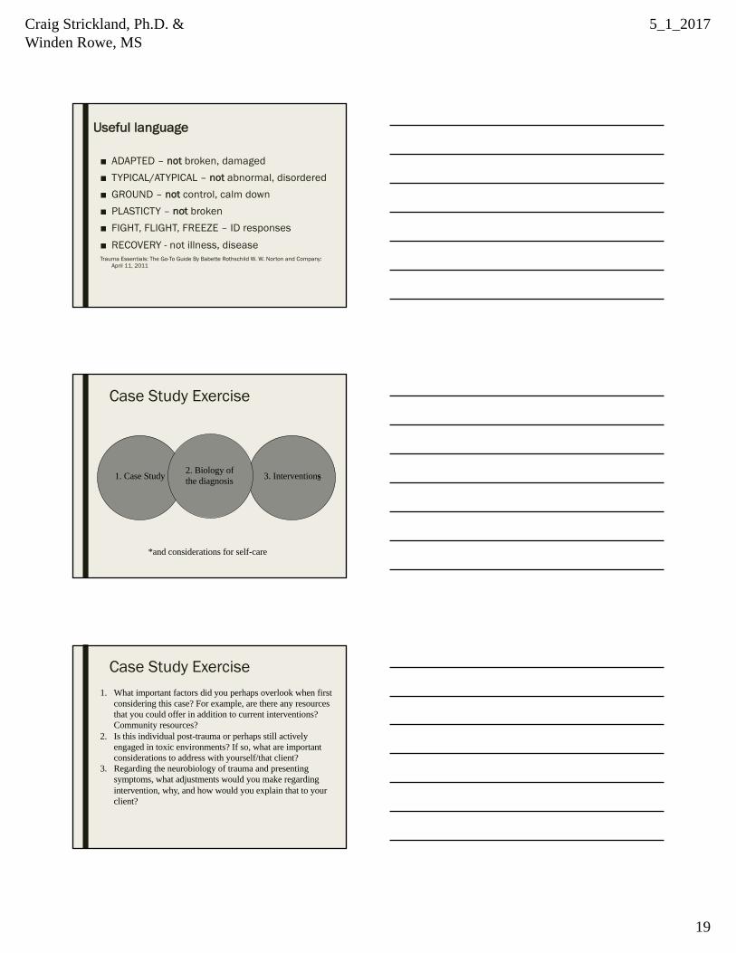
Useful language
■ ADAPTED – not broken, damaged ■ TYPICAL/ATYPICAL – not abnormal, disordered■ GROUND – not control, calm down ■ PLASTICTY – not broken ■ FIGHT, FLIGHT, FREEZE – ID responses■ RECOVERY - not illness, disease Trauma Essentials: The Go-To Guide By Babette Rothschild W. W. Norton and Company:
April 11, 2011
Case Study Exercise
3. Interventions1. Case Study 2. Biology of the diagnosis
*and considerations for self-care
Case Study Exercise 1. What important factors did you perhaps overlook when first
considering this case? For example, are there any resources that you could offer in addition to current interventions? Community resources?
2. Is this individual post-trauma or perhaps still actively engaged in toxic environments? If so, what are important considerations to address with yourself/that client?
3. Regarding the neurobiology of trauma and presenting symptoms, what adjustments would you make regarding intervention, why, and how would you explain that to your client?

Craig Strickland, Ph.D. & Winden Rowe, MS
5_1_2017
20
Case Study Exercise 4. What changes could you make in your language use in order
to best support the client’s self-concept and how they see the trauma and its impact?
5. And most of all, what are ways that you can engage in self-care before, during, and after treating this case?