creating an intraoperative mri suite for the … report creating an intraoperative mri suite for the...
TRANSCRIPT

CASE REPORT
Creating an Intraoperative MRI Suite for the MusculoskeletalTumor Center
Nathan W. Mesko MD, David M. Joyce MD, Hakan Ilaslan MD,
Michael J. Joyce MD
Received: 8 February 2015 / Accepted: 10 June 2015 / Published online: 17 July 2015
� The Association of Bone and Joint Surgeons1 2015
Abstract
Background Altered anatomy in a previously irradiated
surgical bed can make accurate localization of anatomic
landmarks and local recurrence nearly impossible. The use
of intraoperative MRI (iMRI) has been described in neu-
rosurgical settings, but to our knowledge, no such
description has been made regarding its utility for local
recurrence localization in sarcoma surgery.
Case Description A 58-year-old female presented after
previously undergoing two previous resection and rere-
section procedures of a myxoid liposarcoma located
adjacent to her proximal femoral vasculature. After post-
operative radiation therapy, she was referred to our
institution where she underwent two additional reexcisions
of local recurrences during a 3-year span, eventually
undergoing a regional rotational muscle flap for coverage.
Two years after her third reexcision procedure, she pre-
sented with two additional, nonpalpable surgical-bed local
recurrences. After converting an MRI bed and scanner to
allow for proximal thigh imaging in an iMRI surgical suite,
the patient underwent a successful resection that achieved
negative margins. To date, she remains without evidence of
disease at 37 months.
Literature Review Real-time iMRI in neurosurgical
studies has shown a high rate of residual disease leading to
immediate subsequent reexcision, thus lending to improved
rates of negative margin resection. To our knowledge, this
is the first example using iMRI technology to remove a
recurrent soft tissue sarcoma that otherwise was clinically
nonlocalizable.
Clinical Relevance The use of an iMRI surgical suite can
aid with identification of soft tissue nodules in conditions
such as an altered tumor bed from prior resection and
radiotherapy, which otherwise make recurrences difficult to
localize.A teamapproach between administration, surgeons,
and engineers is required to design and pragmatically
implement the use of an MRI-compatible table extension to
enhance existing iMRI surgical suite technology for
extremity sarcoma resection procedures.
Introduction
One of the challenges for the musculoskeletal oncologist is
accurate localization of masses deep to the fascia, a process
that can be rendered more difficult by obesity, skewed
anatomic landmarks, or abnormal tissue developing from
previous irradiation or surgery. One solution proposed is
the use of surgical navigation and CT-MRI fusion
Each author certifies that he or she, or a member of his or her
immediate family, has no funding or commercial associations (eg,
consultancies, stock ownership, equity interest, patent/licensing
arrangements, etc) that might pose a conflict of interest in connection
with the submitted article.
All ICMJE Conflict of Interest Forms for authors and Clinical
Orthopaedics and Related Research1 editors and board members are
on file with the publication and can be viewed on request.
Each author certifies that his or her institution approved the reporting
of this case report, that all investigations were conducted in
conformity with ethical principles of research, and that informed
consent for participation in the study was obtained.
The study was performed at Cleveland Clinic, Department of
Orthopaedic Surgery, Division of Musculoskeletal Oncology and
Trauma, Cleveland, OH, USA.
N. W. Mesko (&), D. M. Joyce, M. J. Joyce
Department of Orthopaedic Surgery, Cleveland Clinic, 9500
Euclid Ave. Crile Building, A-41, Cleveland, OH 44195, USA
e-mail: [email protected]
H. Ilaslan
Department of Diagnostic Radiology, Cleveland Clinic,
Cleveland, OH, USA
123
Clin Orthop Relat Res (2016) 474:1516–1522
DOI 10.1007/s11999-015-4412-9
Clinical Orthopaedicsand Related Research®
A Publication of The Association of Bone and Joint Surgeons®
technology [1, 5–7, 12, 13], but this technique comes with
inherent drawbacks. Although able to accurately locate a
tumor based on landmarks, navigation does not allow for
real-time confirmation of complete tumor excision. While
samples of tumor bed tissue can be sent for real-time frozen
section pathology review, and such analysis has shown
high accuracy [3], difficulties have been described in dis-
tinguishing among scar tissue, healthy tissue, and tumor
tissue in a surgical field previously altered by radiation or
surgery [10].
The use of intraoperative MRI (iMRI) technology is a
well-described neurosurgical technique that can enhance
the probability of complete tumor excision, with several
reports suggesting promising results for selected intracra-
nial procedures [4, 8, 11]. Although these real-time MRI
scans are characterized by small imaging fields, the visu-
alized portal can be adequate for tumor localization and
assessing adequacy of tumor resection in select orthopaedic
oncology procedures where surrounding anatomy cannot
be relied on to guide surgical resection, such as in the
context of distorted anatomic fields through prior serial
operations or radiation [9]. This may, in turn, allow the
surgeon to more accurately confirm a negative margin
resection with the use of real-time imaging, decreasing
dependency on difficult to interpret frozen section analyses.
To our knowledge, only one prior report of use of iMRI for
soft tissue sarcoma has been published [9], which intro-
duced this technique but did not discuss the technical and
practical aspects surrounding planning and performing the
resection.
We report our experience using a novel sarcoma
resection technique using iMRI technology, and provide a
step-by-step mechanism for converting a standard neuro-
surgery iMRI surgical suite into one that is pragmatic for
excision of difficult to locate soft tissue sarcomas in an
anatomically distorted field.
Case Report
A 58-year-old female had two margin-positive resections
(the index resection in February 2006, and the first reex-
cision attempt in September 2007) at an outside
musculoskeletal tumor center for a proximal and anterior
thigh low-grade myxoid lipsarcoma, located adjacent to the
femoral vasculature. After reexcision, she underwent 63Gy
adjuvant external beam radiation. In 2010, she presented to
our institution with a second local recurrence, noted to
have similar histologic features (Grade 2), and underwent a
second reexcision in 2010; she then had a subsequent
reexcision procedure 9 months later for a third recurrence
outside the prior reexcision fields. After this, she underwent
local soft tissue coverage using a rotational rectus
abdominis flap. Two years later (in 2012), after the four
previous operations and high-dose radiation, she had two
additional MRI-documented recurrences in distinctly sep-
arate ipsilateral thigh locations, each measuring
approximately 2 cm in diameter, and neither of which was
palpable. The surgical field in which the local recurrence
masses were found was characterized by substantial ana-
tomic distortion, fibrosis, and radiation changes, with the
femoral artery only localizable using Doppler ultrasound.
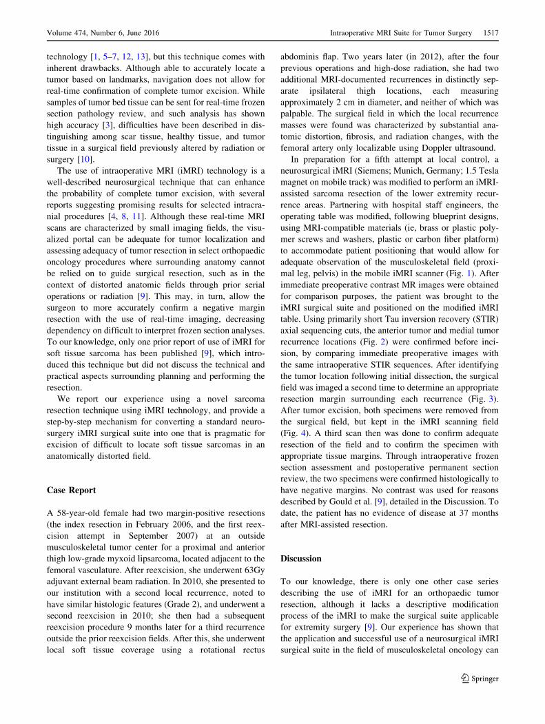
In preparation for a fifth attempt at local control, a
neurosurgical iMRI (Siemens; Munich, Germany; 1.5 Tesla
magnet on mobile track) was modified to perform an iMRI-
assisted sarcoma resection of the lower extremity recur-
rence areas. Partnering with hospital staff engineers, the
operating table was modified, following blueprint designs,
using MRI-compatible materials (ie, brass or plastic poly-
mer screws and washers, plastic or carbon fiber platform)
to accommodate patient positioning that would allow for
adequate observation of the musculoskeletal field (proxi-
mal leg, pelvis) in the mobile iMRI scanner (Fig. 1). After
immediate preoperative contrast MR images were obtained
for comparison purposes, the patient was brought to the
iMRI surgical suite and positioned on the modified iMRI
table. Using primarily short Tau inversion recovery (STIR)
axial sequencing cuts, the anterior tumor and medial tumor
recurrence locations (Fig. 2) were confirmed before inci-
sion, by comparing immediate preoperative images with
the same intraoperative STIR sequences. After identifying
the tumor location following initial dissection, the surgical
field was imaged a second time to determine an appropriate
resection margin surrounding each recurrence (Fig. 3).
After tumor excision, both specimens were removed from
the surgical field, but kept in the iMRI scanning field
(Fig. 4). A third scan then was done to confirm adequate
resection of the field and to confirm the specimen with
appropriate tissue margins. Through intraoperative frozen
section assessment and postoperative permanent section
review, the two specimens were confirmed histologically to
have negative margins. No contrast was used for reasons
described by Gould et al. [9], detailed in the Discussion. To
date, the patient has no evidence of disease at 37 months
after MRI-assisted resection.
Discussion
To our knowledge, there is only one other case series
describing the use of iMRI for an orthopaedic tumor
resection, although it lacks a descriptive modification
process of the iMRI to make the surgical suite applicable
for extremity surgery [9]. Our experience has shown that
the application and successful use of a neurosurgical iMRI
surgical suite in the field of musculoskeletal oncology can
Volume 474, Number 6, June 2016 Intraoperative MRI Suite for Tumor Surgery 1517
123

be successfully implemented. Although the comparative
rationale of iMRI benefit between neurosurgery and
orthopaedic oncology may, at first glance, appear dissimi-
lar (whereas the neurosurgeon is confronted with
preserving a maximum amount of neurologic tissue adja-
cent to the tumor, the orthopaedic oncologist’s dilemma is
in navigating a distorted surgical field to localize a tumor
that is not readily palpable as a mass), each situation can be
immensely enhanced with this emerging technique. In
defined and unique circumstances, however, we believe
this intraoperative technique could be advantageous in
navigating distorted anatomy incurred by a combination of
neoadjuvant treatments for orthopaedic tumor procedures.
Implementing a successful iMRI surgical suite for
extremity and pelvic tumor resections is a time-intensive
process, with the time commitment starting with initial
design and modification phases to real-time implementa-
tion lasting up to 3 months. Furthermore, the estimated cost
of table modification can range between USD 15,000 to
USD 20,000. Once the appropriate patient selection criteria
are met to determine the need for iMRI technology in
extremity sarcoma excision surgery (Table 1), careful
thought is required before implementing iMRI surgical
suite modification (Fig. 5). The neurosurgical iMRI surgi-
cal suite (the designated context in which most institutions
using iMRI technology have funded such capability) is
largely designed for intracranial cases, with a focal imag-
ing field centered over the brain. Because of this, special
considerations are required for each unique surgical loca-
tion, as operative table modifications may occur only in
Fig. 1A–H In our modified operative suite, (A) the table extension
(to accommodate the extremity) is made of nonmagnetic objects, such
as brass, polymers, and plastics. This modification can be removed
easily and does not effect the use for future neurosurgery cases. (B) Atechnician attaches the table modification that allows the patient’s
thigh or pelvis to enter the MRI scanner, with the patient in the supine
position on the (C) modified table. (D) The MRI scanner is in a room
adjacent to the operating room, and is moved to the operating room on
(E) ceiling tracks thru a connecting door, with (F) all personnel,
instruments, and equipment placed outside the gray peripheral line
(magnetic field range) on the floor. An instrument count is performed
before moving the instruments out of the magnetic field (any
instrument unaccounted for is a potential projectile), while positive
pressure flow prevents dirty airflow into the operating room suite
while the magnet moves into position. (G) The scanner moves over
the table extension, (H) encircling the extension where the operative
extremity is positioned. With each MRI scan the machine takes 15
minutes to move along the tracks (and a reciprocal 15 minutes to exit
the room), with 10 to 15 minutes used to count instruments with each
cycle. In this case, we performed three separate image acquisitions,
taking approximately 45 minutes for each cycle, accounting for 135
minutes of operating room time. In addition, the image acquisition
and interpretation took approximately 15 minutes each time. Use of
MRI for resection guidance accounted for nearly 3 hours of additional
operative time.
1518 Mesko et al. Clinical Orthopaedics and Related Research1
123

line with the longitudinal axis, so as to accommodate the
mobile iMRI machine tracking above the table (Fig. 1). In
addition to the modification process, preoperative training,
planning, and coordination dictates successful implemen-
tation of the iMRI surgical suite for specially modified
purposes (Fig. 6). During the actual excision procedure,
accuracy with intraoperative localization techniques is
paramount to limit the total number of sequences needed.
Vitamin E capsules are sealed in sterile plastic bags and
must be changed frequently owing to their limited half-life.
Sterility must be maintained through careful use of sterile
sheets covering the extremity with each magnet entry in the
room. Most important, efficiency is paramount during a
case using iMRI, as lengthy (15-minute) image acquisition
sequences and iMRI tracking over the table (30 minutes
with each acquisition) may create prolonged surgical times
and the inherent potential downstream associations, such as
infection, thromboembolic disease, or blood loss. The
added time to the procedure, including image acquisition,
image interpretation, and scanner mobilization, is in line
with previous descriptions [4, 9].
In 2002, Gould et al. described the use of iMRI for
resection of soft tissue sarcomas, although the three cases
chosen to highlight involved skin-based dermatofibrosar-
coma protuberans [9]. The reported benefits of iMRI were
those of improved soft tissue resolution, lack of ionizing
radiation, and multiplanar and postprocessing capabilities,
all thought to be beneficial when trying to gain negative
margins with resection of dermatofibrosarcoma protuber-
ans, a notoriously difficult low-grade sarcoma on which to
Fig. 2A–B Intraoperative axial STIR images through the right proximal thigh for localization show two small, hyperintense (A) anterior and (B)medial soft tissue masses (arrows) that are consistent with the known recurrent tumor. STIR = short Tau inversion recovery.
Fig. 3 An intraoperative axial STIR image through the right
proximal thigh shows the surgical flap (arrowheads) elevated over
the proximal recurrent soft tissue mass (arrows). STIR = short Tau
inversion recovery.
Fig. 4 A postoperative axial STIR image shows the resected tumor
(arrows at top right) without residual mass in the surgical bed. STIR =
short Tau inversion recovery.
Volume 474, Number 6, June 2016 Intraoperative MRI Suite for Tumor Surgery 1519
123
gain adequate margins. No contrast was used, as specific
axial STIR imaging sequences comparing preoperative MR
images with intraoperative and preresection imaging were
deemed adequate to determine the signal quality extent of
the tumor lesion. Furthermore, concern that contrast
washout may occur in a lengthy procedure, leaking into
surrounding tissue, was thought to make it potentially more
difficult to accurately confirm complete excision. Although
this technique allowed distinct recognition of malignant
tissue, no insight was provided regarding how to efficiently
run an intraoperative MRI suite for soft tissue sarcoma
resection. Similar to the methodology in the case series by
Gould et al. [9], our patient underwent iMRI assessment
after resection to evaluate margins on the resected speci-
men and the remaining excision bed. A recent animal study
[2] also showed efficacy of MRI technology in assessing
resection margins, studying resection specimen margins in
rats with intramuscular rhabdomyosarcoma. By comparing
lesion margins defined on 1.5T MRI versus histopathology
analysis, a high rate of agreement (Pearson coefficient 0.97,
average margin measured 0.3 mm in both analyses)
between the two techniques was shown, thus lending
indirect support to the utility of iMRI for use in the
assessment of margins [2]. iMRI is better described in
Table 1. Patient selection criteria for iMRI
Indications Contraindications
Primary lesion(s) or recurrent lesion(s) is deemed to be resectable in a
manner that will not render a functionless extremity or compromise
margins to a degree that amputation is best offered
Body habitus of the patient or the mass exceeds the limitations
of the iMRI table
The lesion(s) is not readily palpable, and reliable identification using
landmark-based imaging measurements is impossible because of
radiation changes, postsurgical changes, or a combination thereof
Previously implanted devices that are contraindicated in a
magnetic field (institution or MRI scanner-specific
protocols)
Tumor location (in relation to essential structures required for a
functional limb) does not allow for conventional, unguided wide
resection, including normal surrounding tissue, to ensure excision
of the lesion
Limited access to a musculoskeletal radiologist who is able to
aid with intraoperative imaging interpretation
Fig. 5 Initiating the process of converting a neurosurgical iMRI table
to being functionally acceptable for extremity tumor surgery requires
multidisciplinary teamwork, financial backing, and discussion with the
institutional engineers regarding the proper materials required to
proceed with successful operating room suite modification. IMRI =
intraoperative MRI; IMRIS = intraoperative MRI suite.
1520 Mesko et al. Clinical Orthopaedics and Related Research1
123

neurosurgical settings for pituitary macroadenoma and
temporal bone tumors, with suggested conclusions of
improved surgical performance and a higher rate of wide
margin resection [8, 12]. In two separate small population
prospective studies looking at 30 patients [4] and 73
patients [8] with pituitary macroadenomas, use of iMRI led
to an increased extent of tumor resection and higher nega-
tive margin resection rate. In both studies, less than 2.3 of
patients (59% and 66%) had a negative margin resection
denoted on the iMRI after the initial attempt at resec-
tion. Because of the high rate of residual tumor found on
intraoperative imaging, the authors [4, 8] described repeat
surgical resection as an effective way to decrease the
residual tumor volume retained—an afforded benefit owing
to real-time assessment with the patient under anesthesia. In
contrast, a 2014 randomized control trial comparing stan-
dard neuronavigation versus iMRI for resection of
glioblastoma showed no difference with respect to the
extent of resection, clinical performance, or overall survival
[11]. Despite this lack of observed difference, the authors
maintained that iMRI is useful in the appropriate situation.
Given the drawbacks of substantial operating time
(leading to higher rates of infection, blood loss, anesthesia
time) and expense, the use of this technology for orthopaedic
oncology should be limited. We believe this technology
should be used only in situations where localization of the
tumor through standard technique (referencing preoperative
study measurements with local anatomic landmarks) is
impossible because of fibrosis, prior muscle flaps, or other
anatomically distorting features. If landmarks are palpable
and the tumor can be clinically localized in a reliable fashion,
then standard techniques are much more fiscally responsible
and efficient. However, in situations such as with our patient,
the intraoperative MRI suite concept can be a powerful
instrument to aid the oncologic resection.
With multidisciplinary cooperation, creativity, and
resourcefulness among the orthopaedic and neurosurgery
surgical teams, the engineering department, and the insti-
tution’s administration, a neurosurgical iMRI surgical suite
can be modified to accomplish a musculoskeletal sarcoma
excision requiring pinpoint localization. Use of iMRI offers
a way to address a local soft tissue sarcoma recurrence in a
previously irradiated field with anatomic distortion. Intra-
operative MRI can benefit the surgeon and patient by
helping to assure a negative margin resection without
sacrificing vital structures.
An MRI safety training video, followed by review of wri�en material and a brief
examina�on, must be completed by each of the orthopaedic surgical team
Nursing team, scrub assistant, and surgeons
Determina�on of preopera�vely arranged protocols determining necessary protocols,
personnel, and instrumenta�on: The opera�ve suite is limited to only pre-selected, essen�al personnelSpecific instrument handling and count protocols must be implemented and prac�ced
Prac�ce “Dry Runs” prior to the surgical procedure, help determine areas of poten�al complica�on of process disrup�on:
Trouble shoo�ng specific an�cipated “scenarios”1) Malfunc�oning piece of equipment (e.g. Doppler Box)
2) The need to acquire a new instrument during the opera�on
Marking an�cipated anatomic loca�ons during image sequencing1) Placement of Vitamin E markers in a sterile envelope2) Vitamin E marker begins degrada�on/breakdown around 6 hours
Seamless transi�on between surgery and imaging1) Instruments/tables moved to peripheral “safe” loca�on during IMRI image acquisi�on to protect from effects of magne�c field
Planning for all available preopera�ve MRI imaging to be uploaded to the scanner
1) Preopera�ve imaging should be uploaded prior to surgery in order to allow for an intra- opera�ve , preincision MRI to be obtained to compare landmarks and anatomy on
the pre-opera�ve imaging versus real-�me intraopera�ve imaging (allowing the surgeon to account for differences in magnet strength)
IMRIS use for surgical sarcoma
resec�on
Fig. 6 After the iMRI surgical suite has been modified successfully,
training and preparation for the real surgery are needed, anticipating
for potential pitfalls or process adjustments intraoperative, and how to
properly deal with these situations. Each essential participant is
designated a specific role to help assure successful functioning of the
iMRI surgical suite. IMRI = intraoperative MRI.
Volume 474, Number 6, June 2016 Intraoperative MRI Suite for Tumor Surgery 1521
123
Acknowledgments We thank the Cleveland Clinic Engineering
Department for help with the design and manufacturing of the
intraoperative MRI suite operating table modification.
References
1. Aponte-Tinao L, Ritacco LE, Ayerza MA, Muscolo DL, Albergo
JI, Farfalli GL. Does intraoperative navigation assistance improve
bone tumor resection and allograft reconstruction results? Clin
Orthop Relat Res. 2015;473:796–804.
2. Bellanova L, Schubert T, Cartiaux O, Lecouvet F, Galant C,
Banse X, Docquier PL. MRI-based assessment of safe margins in
tumor surgery. Sarcoma. 2014;2014:686790. doi: 10.1155/2014/
686790. 2014 Feb 20. (Epub ahead of print]
3. Bhaker P, Mohan H, Handa U, Kumar S. Role of intraoperative
pathology consultation in skeletal tumors and tumor-like lesions.
Sarcoma. 2014:902104. doi: 10.1155/2014/902104. 2014 May
19. [Epub ahead of print]
4. Bohinski RJ, Warnick RE, Gaskill-Shipley MF, Zuccarello M,
van Loveren HR, Kormos DW, Tew JM Jr. Intraoperative mag-
netic resonance imaging to determine the extent of resection of
pituitary macroadenomas during transsphenoidal microsurgery.
Neurosurgery. 2001;49:1133–1143; discussion 1143–1144.
5. Cheong D, Letson GD. Computer-assisted navigation and mus-
culoskeletal sarcoma surgery. Cancer Control. 2011;18:171–176.
6. Cho HS, Oh JH, Han I, Kim HS. The outcomes of navigation-
assisted bone tumour surgery: minimum three-year follow-up. J
Bone Joint Surg Br. 2012;94:1414–1420.
7. Cho HS, Park IH, Jeon IH, Kim YG, Han I, Kim HS. Direct
application of MR images to computer-assisted bone tumor sur-
gery. J Orthop Sci. 2011;16:190–195.
8. Fomekong E, Duprez T, Docquier MA, Ntsambi G, Maiter D,
Raftopoulos C. Intraoperative 3T MRI for pituitary macroade-
noma resection: initial experience in 73 consecutive patients. Clin
Neurol Neurosurg. 2014;126:143–149.
9. Gould SW, Agarwal T, Benoist S, Patel B, Gedroyc W, Darzi A.
Resection of soft tissue sarcomas with intra-operative magnetic
resonance guidance. J Magn Reson Imaging. 2002;15:114–119.
10. Hohenberger P, Schwarzbach MHM. Management of locally
recurrent soft tissue sarcoma after prior surgery and radiation
therapy. Recent Results Cancer Res. 2009;179:271–283.
11. Kubben PL, Scholtes F, Schijns OE, ter Laak-Poort MP, Teernstra
OP, Kessels AG, van Overbeeke JJ, Martin DH, van Santbrink H.
Intraoperative magnetic resonance imaging versus standard neu-
ronavigation for the neurosurgical treatment of glioblastoma: a
randomized controlled trial. Surg Neurol Int. 2014;5:70.
12. Nemec SF, Donat MA, Mehrain S, Friedrich K, Krestan C,
Matula C, Imhof H, Czerny C. CT-MR image data fusion for
computer assisted navigated neurosurgery of temporal bone
tumors. Eur J Radiol. 2007;62:192–198.
13. Ritacco LE, Milano FE, Farfalli GL, Ayerza MA, Muscolo DL,
Aponte-Tinao LA. Accuracy of 3-D planning and navigation in
bone tumor resection. Orthopedics. 2013;36:e942–950.
1522 Mesko et al. Clinical Orthopaedics and Related Research1
123