creating order from chaos: cellular regulation by kinase...
TRANSCRIPT
PA53CH10-Scott ARI 5 December 2012 14:15
Creating Order from Chaos:Cellular Regulation by KinaseAnchoringJohn D. Scott,1,∗ Carmen W. Dessauer,2,∗
and Kjetil Tasken3,4,∗
1Howard Hughes Medical Institute and Department of Pharmacology, University ofWashington School of Medicine, Seattle, Washington 98195; email: [email protected] of Integrative Biology and Pharmacology, University of Texas Medical School,Houston, Texas 770303The Biotechnology Centre of Oslo and Centre for Molecular Medicine Norway, NordicEuropean Molecular Biology Laboratory Partnership, University of Oslo, N-0317 Oslo, Norway4Department of Infectious Diseases, Oslo University Hospital, N-0424 Oslo, Norway
Annu. Rev. Pharmacol. Toxicol. 2013. 53:187–210
First published online as a Review in Advance onOctober 8, 2012
The Annual Review of Pharmacology and Toxicologyis online at pharmtox.annualreviews.org
This article’s doi:10.1146/annurev-pharmtox-011112-140204
Copyright c© 2013 by Annual Reviews.All rights reserved
∗Each author contributed equally.
Keywords
cell signaling, compartmentalization, cAMP, A-kinase anchoring proteins,protein phosphorylation
Abstract
Second messenger responses rely on where and when the enzymes that prop-agate these signals become active. Spatial and temporal organization of cer-tain signaling enzymes is controlled in part by A-kinase anchoring proteins(AKAPs). This family of regulatory proteins was originally classified on thebasis of their ability to compartmentalize the cyclic adenosine monophos-phate (cAMP)-dependent protein kinase (also known as protein kinase A,or PKA). However, it is now recognized that AKAPs position G protein–coupled receptors, adenylyl cyclases, G proteins, and their effector proteinsin relation to protein kinases and signal termination enzymes such as phos-phodiesterases and protein phosphatases. This arrangement offers a simpleand efficient means to limit the scope, duration, and directional flow of in-formation to sites deep within the cell. This review focuses on the pros andcons of reagents that define the biological role of kinase anchoring insidecells and discusses recent advances in our understanding of anchored secondmessenger signaling in the cardiovascular and immune systems.
187
Ann
u. R
ev. P
harm
acol
. Tox
icol
. 201
3.53
:187
-210
. Dow
nloa
ded
from
ww
w.a
nnua
lrev
iew
s.or
gby
${i
ndiv
idua
lUse
r.di
spla
yNam
e} o
n 01
/09/
13. F
or p
erso
nal u
se o
nly.
PA53CH10-Scott ARI 5 December 2012 14:15
FOUNDING THIS FIELD
In the 1950s, Earl Sutherland defined how hormones stimulate the production of the secondmessenger 3′,5′-cyclic adenosine monophosphate (cAMP) by adenylyl cyclase (AC) (1, 2), andEdwin Krebs and Edmond Fischer demonstrated protein phosphorylation as a principal taskfor cAMP (3). The first defined recipient of this chemical message was protein kinase A (PKA,also known as the cAMP-dependent protein kinase), a heterotetrameric holoenzyme consist-ing of two regulatory (R) subunits that maintain two catalytic (C) subunits in an inhibited state(4, 5). When cAMP levels are low, PKA is dormant; however, when cAMP levels are elevated,two molecules of cAMP bind to each R subunit, thereby releasing the active C subunits. The Csubunits phosphorylate serine (S) or threonine (T) residues, typically within the sequence -R-R-X-S/T-X (6). The first evidence for localized cAMP signaling was shown in the late 1970s: Inheart, both prostaglandin E1 and epinephrine increased cAMP, yet only epinephrine increasedglycogen phosphorylase activity and cardiac contraction (7–9).
At approximately the same time, the PKA holoenzyme was discovered to exist in two forms: acytoplasmic type I PKA and an exclusively particulate type II PKA (10). Thus, it was postulatedthat activation of PKA was differentially regulated at organelles and on intracellular membranes.In 1982, Theurkauf & Vallee (11) provided evidence for the targeting of PKA subtypes when theyshowed that type II PKA is anchored to microtubules via its regulatory RII subunit interactionwith microtubule-associated protein MAP2. Slightly later, a second neuronal anchoring proteinwas identified as a protein contaminant that copurifies with RII subunits on cAMP-agarose (12).
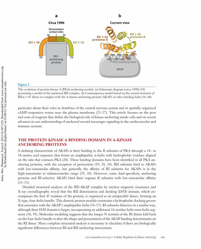
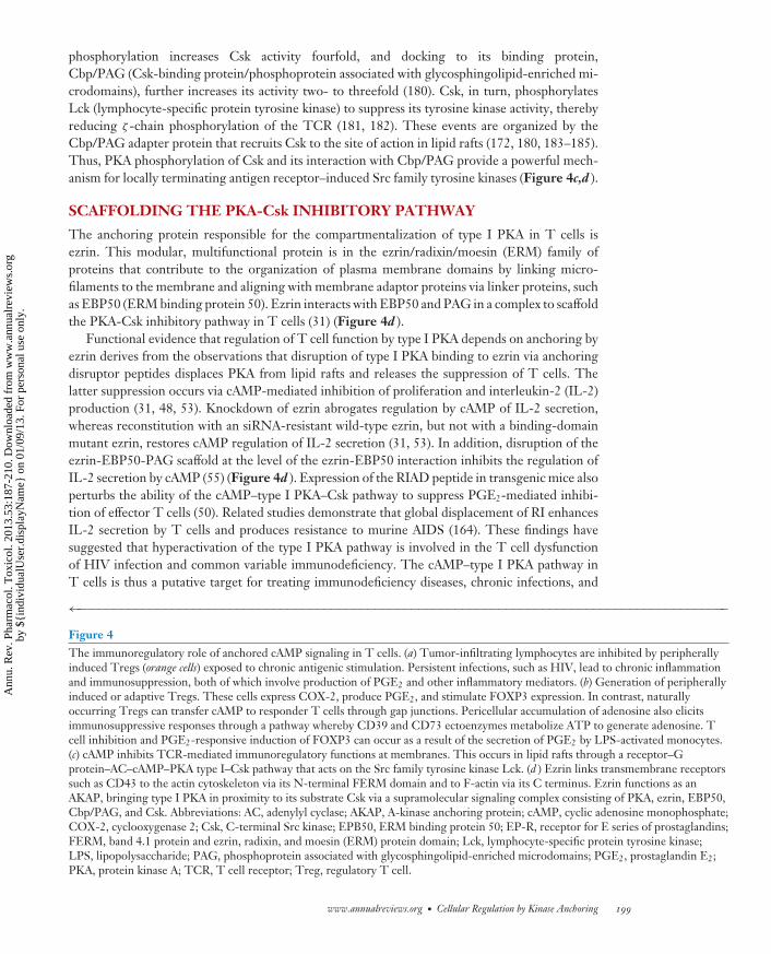
Subsequent technological advances, including protein-protein blotting using RII as the probe(RII overlay) and expression-cloning strategies, have uncovered many more of these anchoringproteins, which are now known as A-kinase anchoring proteins (AKAPs) (13–15). Since then,more than 50 human genes that encode AKAPs have been identified (16). Additionally, the adventof sophisticated molecular biology and genetic screening approaches permitted dramatic break-throughs in our understanding of the RII-AKAP interface and AKAP action (17). A survey of RIIdeletion mutants revealed that the first 79 residues were necessary and sufficient for AKAP binding(18), whereas analysis of the reciprocal binding surface on several anchoring proteins identifiedcommon helical regions (19). Mutagenesis of such a binding helix in human thyroid clone 31(Ht31, also known as AKAP-Lbc) dramatically reduced association with RII (20), providing thefirst evidence that AKAPs could interact with PKA via an amphipathic helix-binding motif (19).Proof of principle was provided when Ht31 peptides were used to uncouple PKA proximity toAMPA-type glutamate receptor ion channels in hippocampal neurons with concomitant effectson synaptic transmission (21). A simple model of this protein-protein interface put forward in theearly 1990s (Figure 1a) bears a striking resemblance to the three-dimensional structure of theRII-AKAP interface (Figure 1b) that was solved more than a decade later.
Much of our progress in understanding AKAP action can be traced through studies performedon a single molecule, the human anchoring protein AKAP79. Using this anchoring protein as thebait in a two-hybrid screen for additional neuronal binding partners identified the phosphatasecalcineurin, also known as protein phosphatase 2B (PP2B) (22), suggesting that AKAP79 associateswith multiple second messenger–regulated kinases and phosphatases. AKAP79 also interacts withisoforms of protein kinase C (PKC), thus creating macromolecular signaling complexes that canintegrate cAMP-, calcium-, phospholipid-, and calmodulin-dependent signals at defined intracel-lular loci (23). Subsequent studies have shown that AKAP79 and other anchoring proteins interactwith a plethora of other signaling proteins, such as G protein–coupled receptors, GTPases, phos-phatases, phosphodiesterases, and other kinases (24). Not surprisingly, as the scope of this field hasexploded in the past decade, investigators have written many excellent reviews about AKAPs, in
188 Scott · Dessauer · Tasken
Ann
u. R
ev. P
harm
acol
. Tox
icol
. 201
3.53
:187
-210
. Dow
nloa
ded
from
ww
w.a
nnua
lrev
iew
s.or
gby
${i
ndiv
idua
lUse
r.di
spla
yNam
e} o
n 01
/09/
13. F
or p
erso
nal u
se o
nly.
PA53CH10-Scott ARI 5 December 2012 14:15
30 30
79 79
1
Dimerizationcontact sites
Circa 1990
RII 1-45protomer A
RII 1-79
RII-anchoring
protein
RII 1-45protomer B
RII 1-79
PKA-binding helix
AKAP
Current view
a b
Figure 1The evolution of protein kinase A (PKA) anchoring models. (a) Schematic diagram (circa 1990) (18)presenting a model of the anchored RII complex. (b) Contemporary model based on the crystal structure ofRIIα 1-45 dimer in complex with the A-kinase anchoring protein (AKAP)–in silico binding helix (36, 48).
particular about their roles in dendrites of the central nervous system and in spatially organizedcAMP-responsive events near the plasma membrane (25–27). This article focuses on the prosand cons of reagents that define the biological role of kinase anchoring inside cells and on recentadvances in our understanding of anchored second messenger signaling in the cardiovascular andimmune systems.
THE PROTEIN KINASE A BINDING DOMAIN IN A-KINASEANCHORING PROTEINS
A defining characteristic of AKAPs is their binding to the R subunits of PKA through a 14– to18–amino acid sequence that forms an amphipathic α-helix with hydrophobic residues alignedon the side that contacts PKA (20). These binding domains have been identified in all PKA an-choring proteins, with the exception of pericentrin (19, 20, 28). RII subunits bind to AKAPswith low-nanomolar affinity, but generally the affinity of RI subunits for AKAPs is in thehigh-nanomolar to submicromolar range (29, 30). However, some dual-specificity anchoringproteins and RI-selective AKAPs bind their cognate R subunits with low-nanomolar affinity(31–33).
Detailed structural analyses of the RII-AKAP complex by nuclear magnetic resonance andX-ray crystallography reveal that the RII dimerization and docking (D/D) domain, which en-compasses the first 45 residues of the protein, is organized as an antiparallel dimer, forming anX-type, four-helix bundle. This dimeric protein module constrains a hydrophobic docking groovethat associates with the AKAP’s amphipathic helix (34–37). RI subunits dimerize in a similar way,although their D/D domain is larger, incorporating an additional 16-residue helix-turn-helix seg-ment (38, 39). Molecular modeling suggests that the longer N termini of the RI dimer fold backon the four-helix bundle to alter the shape and presentation of the AKAP binding determinants onthe RI dimer. More complete structural analysis is necessary to elucidate if there are biologicallysignificant differences between RI and RII anchoring interactions.
www.annualreviews.org • Cellular Regulation by Kinase Anchoring 189
Ann
u. R
ev. P
harm
acol
. Tox
icol
. 201
3.53
:187
-210
. Dow
nloa
ded
from
ww
w.a
nnua
lrev
iew
s.or
gby
${i
ndiv
idua
lUse
r.di
spla
yNam
e} o
n 01
/09/
13. F
or p
erso
nal u
se o
nly.
PA53CH10-Scott ARI 5 December 2012 14:15
PROBING FUNCTION WITH PKA ANCHORINGDISRUPTOR PEPTIDES
AKAP-derived peptides that bind to the D/D domains of PKA compete with AKAPs, therebyperturbing the subcellular location of the enzyme. These peptides have been important tools inthe study of the functional implications of PKA anchoring in cells. A 24–amino acid peptide derivedfrom AKAP-Lbc (originally named Ht31) was the first anchoring disruptor to be characterized(19). Validation of Ht31 action in cells was provided by studies in which the peptide was perfusedinto neurons or pancreatic β cells to uncouple aspects of synaptic transmission and insulin secretioncoupling, respectively (21, 40). Ht31 adducts have subsequently been used to demonstrate a rolefor PKA anchoring in numerous important signaling events, such as the modulation of L-typeCa2+ channels and excitation-contraction coupling in the heart, of sperm motility, and of fluidmovement in the lens of the eye (41–45). An advantage is that Ht31 has low-nanomolar affinityfor the type II PKA (Kd = 2.2 ± 0.03 nM) (19); however, a drawback is that this peptide alsoperturbs interactions between type I PKA and AKAPs (30).
Second-generation anchoring disruptor peptides were developed with the goal of discriminat-ing between RI and RII interactions with AKAPs. Bioinformatic scrutiny of RII binding domainsand peptide array–based optimization were combined to design AKAP–in silico (AKAP-IS), an an-choring disruptor with subnanomolar binding affinity for RII (46). In parallel, peptides patternedafter the PKA binding region of D-AKAP2 were developed to interfere with RI or RII interac-tions (47). Anchoring disruptors that are highly selective for either the anchored type I (the RIanchoring disruptor, also known as RIAD) or the type II (super-AKAP-IS) PKA holoenzymes weresubsequently developed (36, 48). These reagents have helped delineate effects regulated by typeI or type II PKA, such as cAMP-mediated immune regulation and steroidogenesis (49–52). Therecognition that dual-specificity AKAPs have additional determinants that secure the interactionwith the RI subunit led to the discovery of an RI specifier region (RISR) (53). A peptide derivedfrom an RISR disrupts type I PKA interaction with the AKAP ezrin independently of peptidesthat mimic the amphipathic helix (53). In summary, the toolbox of PKA anchoring disruptors isconsiderable and is frequently used as the first approach to define biological effects mediated byanchored PKA enzymes in cells.
Disruption of PKA anchoring has been used to assess the functional significance of localizedpools of PKA but is less useful in a therapeutic context. Furthermore, there would be merit indeveloping reagents that selectively target an individual AKAP. Another promising approach maybe to displace AKAP signaling complexes from their proximity to a particular PKA substrate. Forexample, peptides that block interaction between AKAP18δ and the PKA substrate phospholam-ban are equally as effective as the Ht31 peptide that disrupts PKA from the AKAP complex ininhibiting β-adrenergic receptor (βAR)-mediated stimulation of calcium reuptake by SERCA2(sarcoplasmic/endoplasmic reticulum Ca2+ pump 2) (54). Similarly, peptide-based disruption ofthe adapter protein EBP50 from ezrin disconnects PKA from Csk to reverse cAMP-mediatedinhibition of immune function (55).
LOCALIZED cAMP SIGNALING IN THE HEART
Efficient contraction of the heart requires coordinated handling of cAMP and Ca2+ signaling eventsin cardiomyocytes. This process, known as excitation-contraction coupling, occurs when an actionpotential triggers a transient rise in intracellular Ca2+ that drives contraction of cardiomyocytes(56). This process takes place in three phases. Phase 1 is initiated by the brief opening of voltage-gated L-type Ca2+ channels. Small amounts of Ca2+ enter regions of the myocyte where the
190 Scott · Dessauer · Tasken
Ann
u. R
ev. P
harm
acol
. Tox
icol
. 201
3.53
:187
-210
. Dow
nloa
ded
from
ww
w.a
nnua
lrev
iew
s.or
gby
${i
ndiv
idua
lUse
r.di
spla
yNam
e} o
n 01
/09/
13. F
or p
erso
nal u
se o
nly.
PA53CH10-Scott ARI 5 December 2012 14:15
junctional sarcoplasmic reticulum (SR) is nearby. In phase 2, this localized Ca2+ influx triggers thesynchronous activation of ryanodine-sensitive Ca2+ channels (i.e., ryanodine receptors, or RyRs)in the SR to produce a global Ca2+ transient. The concomitant activation of Ca2+-responsivecontractile proteins, such as cardiac troponin C, initiates contraction. Phase 3 requires terminationof SR Ca2+ release and the transport of Ca2+ back into the SR through the ATP-dependent Ca2+
pump SERCA2, which decreases [Ca2+]i and begins myocyte relaxation.Distinct AKAP complexes contribute to each phase of excitation-contraction coupling by op-
timizing the phosphorylation of ion channels and contractile proteins. These anchoring proteinsorganize cAMP effectors such as PKA, guanine exchange proteins activated by cAMP (EPACs),and cAMP-gated channels (HCN) in relation to ACs, the aforementioned enzymes that synthesizecAMP (57, 58). Although most AC isoforms are expressed in cardiac fibroblasts (59), the majorisoforms present in myocytes are AC5 and AC6 (59–61). Interestingly, these enzymes can exertcertain opposite functional effects on the heart. Overexpression of AC6 appears to be cardiopro-tective (62–64), whereas AC5 has been implicated in cAMP production arising from cardiac stress(65, 66). Moreover, deletion of AC6 reveals unique biological functions that are not duplicated byAC5, including reductions in PKA and Akt activity, phospholamban phosphorylation, and alteredleft ventricular contractile function (67). The distinctions between AC5 and AC6 action may, atleast in part, reflect their differential recruitment into macromolecular complexes.
In isolated cardiac myocytes, disruption of PKA anchoring results in impaired cAMP regulationof L-type Ca2+ channels (41, 68, 69), CFTR (cystic fibrosis transmembrane conductance regulator)chloride channels (70), and IK potassium currents (71). Similar approaches have implicated a rolefor anchored PKA phosphorylation of numerous targets in myocytes (72–74). At least 15 AKAPsare expressed in the heart, roles for which are discussed below. However, most of these anchoringproteins are not found solely in the heart (16, 25, 75, 76).
AKAPs IN CALCIUM-INDUCED CALCIUM RELEASE
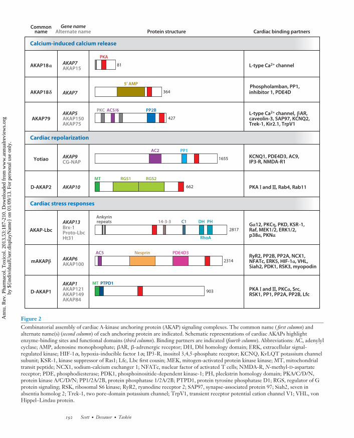
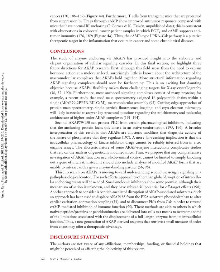
Sympathetic control of the heart through βAR stimulation increases the rate and force of con-traction and relaxation of cardiac muscle. As depicted in Figure 2, AKAP18 and AKAP79/150signaling complexes principally contribute to this vital process (77).
AKAP18α
Of the four AKAP7 gene transcripts, AKAP18α is the smallest and encodes an 81–amino acidprotein (42, 78–80) (Figure 2). Myristoylation and palmitoylation of N-terminal residues helptether AKAP18 (also known as AKAP15) to the inner face of the plasma membrane to facilitateβAR regulation of the L-type calcium channel CaV1.2 (42, 81). Physical association with AKAP18α
channels proceeds through a leucine zipper motif, thereby positioning PKA in proximity to itsphosphorylation target (82–84). Peptide-mediated disruption of the AKAP15/18-PKA-channelcomplex reduces stimulation of CaV1.2 currents by this kinase, a well-known modulator of Ca2+
channels (42, 82).The AKAP18δ variant, a longer transcript of the AKAP7 gene, acts as a scaffold to coordinate
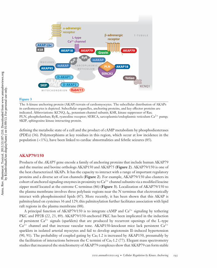
a crucial step for cardiac muscle relaxation, the βAR-promoted reuptake of calcium into theSR by SERCA2 (54) (Figures 2 and 3). Phosphorylation of membrane-bound phospholambanby PKA and/or CamKII (calmodulin-dependent protein kinase II) removes its inhibitory effecton SERCA2, thereby accelerating Ca2+ reuptake and myocyte relaxation. Curiously, AKAP18δ
contains a central phosphoesterase domain that binds 5′ AMP (36). Although AKAP18δ has nodetectable ligase or hydrolase activity, it may be a sensor of 5′ AMP, an important molecule in
www.annualreviews.org • Cellular Regulation by Kinase Anchoring 191
Ann
u. R
ev. P
harm
acol
. Tox
icol
. 201
3.53
:187
-210
. Dow
nloa
ded
from
ww
w.a
nnua
lrev
iew
s.or
gby
${i
ndiv
idua
lUse
r.di
spla
yNam
e} o
n 01
/09/
13. F
or p
erso
nal u
se o
nly.
PA53CH10-Scott ARI 5 December 2012 14:15
Calcium-induced calcium release
Cardiac repolarization
Cardiac stress responses
PKA
81 L-type Ca2+ channel
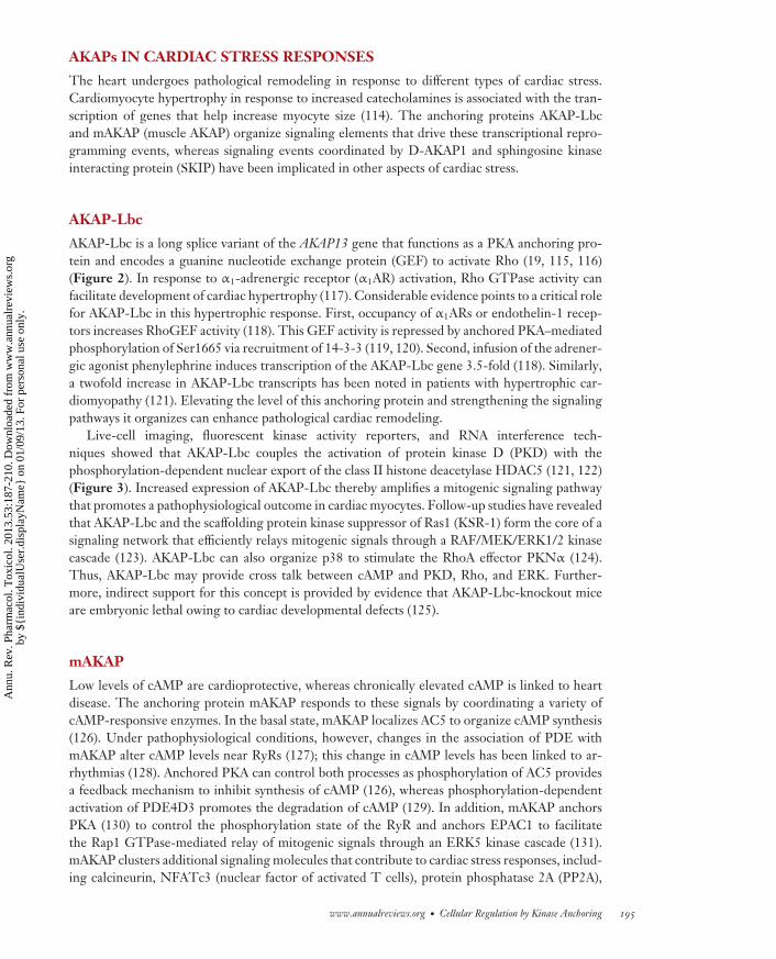
Commonname Protein structure Cardiac binding partners
Gene nameAlternate name
AKAP7AKAP15
5’ AMP
364 inhibitor 1, PDE4DPhospholamban, PP1,
AKAP7
PP2BPKC AC5/6
427 caveolin-3, SAP97, KCNQ2,Trek-1, Kir2.1, TrpV1
L-type Ca2+ channel, βAR,AKAP5AKAP150AKAP75
PP1AC2
1655 KCNQ1, PDE4D3, AC9,IP3-R, NMDA-R1
AKAP9CG-NAP
RGS2RGS1MT
662 PKA I and IIII, Rab4, Rab11AKAP10
14-3-3 C1 DH PH
RhoA
Ankyrinrepeats
2817Gα12, PKCη, PKD, KSR-1,Raf, MEK1/2, ERK1/2,p38α, PKNα
AKAP13Brx-1Proto-LbcHt31
PDE4D3NesprinAC5
2314RyR2, PP2B, PP2A, NCX1,NFATc, ERK5, HIF-1α, VHL,Siah2, PDK1, RSK3, myopodin
AKAP6AKAP100
MT PTPD1
903 PKA I and IIII, PKCα, Src,RSK1, PP1, PP2A, PP2B, Lfc
AKAP1AKAP121AKAP149AKAP84
AKAP18α
AKAP18δ
AKAP79
Yotiao
D-AKAP2
AKAP-Lbc
mAKAPβ
D-AKAP1
Figure 2Combinatorial assembly of cardiac A-kinase anchoring protein (AKAP) signaling complexes. The common name ( first column) andalternate name(s) (second column) of each anchoring protein are indicated. Schematic representations of cardiac AKAPs highlightenzyme-binding sites and functional domains (third column). Binding partners are indicated (fourth column). Abbreviations: AC, adenylylcyclase; AMP, adenosine monophosphate; βAR, β-adrenergic receptor; DH, Dbl homology domain; ERK, extracellular signal-regulated kinase; HIF-1α, hypoxia-inducible factor 1α; IP3-R, inositol 3,4,5-phosphate receptor; KCNQ, KvLQT potassium channelsubunit; KSR-1, kinase suppressor of Ras1; Lfc, Lbc first cousin; MEK, mitogen-activated protein kinase kinase; MT, mitochondrialtransit peptide; NCX1, sodium-calcium exchanger 1; NFATc, nuclear factor of activated T cells; NMDA-R, N-methyl-D-aspartatereceptor; PDE, phosphodiesterase; PDK1, phosphoinositide-dependent kinase-1; PH, pleckstrin homology domain; PKA/C/D/N,protein kinase A/C/D/N; PP1/2A/2B, protein phosphatase 1/2A/2B; PTPD1, protein tyrosine phosphatase D1; RGS, regulator of Gprotein signaling; RSK, ribosomal S6 kinase; RyR2, ryanodine receptor 2; SAP97, synapse-associated protein 97; Siah2, seven inabsentia homolog 2; Trek-1, two pore-domain potassium channel; TrpV1, transient receptor potential cation channel V1; VHL, vonHippel–Lindau protein.
192 Scott · Dessauer · Tasken
Ann
u. R
ev. P
harm
acol
. Tox
icol
. 201
3.53
:187
-210
. Dow
nloa
ded
from
ww
w.a
nnua
lrev
iew
s.or
gby
${i
ndiv
idua
lUse
r.di
spla
yNam
e} o
n 01
/09/
13. F
or p
erso
nal u
se o
nly.
PA53CH10-Scott ARI 5 December 2012 14:15
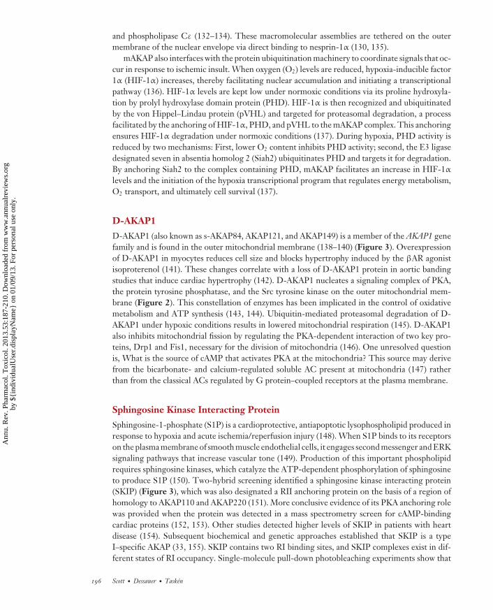
Gravin
T - T U B U L E
KCNQ1
β-adrenergicreceptor
α-adrenergicreceptor
N U C L E U SS A R C O P L A S M I C
R E T I C U L U M
M I T O C H O N D R I O N Rab4/11
L-typeCa2+ channel
AKAP-Lbc
AKAP18
AKAP95
AKAP79 AKAP79
D-AKAP1 Yotiao
D-AKAP2
KSR
RyRRyR mAKAP
mAKAP
SERCA2
SKIP
AKAP18PLN
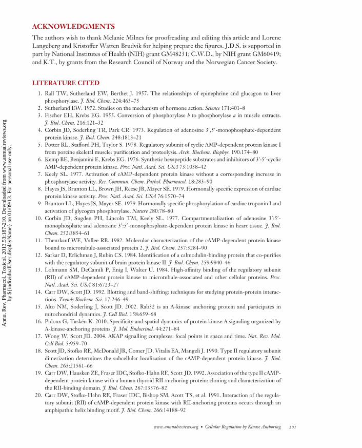
Figure 3The A-kinase anchoring protein (AKAP) terrain of cardiomyocytes. The subcellular distribution of AKAPsin cardiomyocytes is depicted. Subcellular organelles, anchoring proteins, and key effector proteins areindicated. Abbreviations: KCNQ, IKs potassium channel subunit; KSR, kinase suppressor of Ras;PLN, phospholamban; RyR, ryanodine receptor; SERCA, sarcoplasmic/endoplasmic reticulum Ca2+ pump;SKIP, sphingosine kinase interacting protein.
defining the metabolic state of a cell and the product of cAMP metabolism by phosphodiesterases(PDEs) (36). Polymorphisms at key residues in this region, which occur at low incidence in thepopulation (<1%), have been linked to cardiac abnormalities and febrile seizures (85).
AKAP79/150
Products of the AKAP5 gene encode a family of anchoring proteins that include human AKAP79and the murine and bovine orthologs AKAP150 and AKAP75 (Figure 2). AKAP79/150 is one ofthe best characterized AKAPs. It has the capacity to interact with a range of important regulatoryproteins and a diverse set of ion channels (Figure 2). For example, AKAP79/150 also clusters itscohort of anchored signaling enzymes in proximity to Ca2+ channel subunits via a modified leucinezipper motif located at the extreme C terminus (86) (Figure 3). Localization of AKAP79/150 tothe plasma membrane involves three polybasic regions near the N terminus that electrostaticallyinteract with phosphoinositol lipids (87). More recently, it has been shown that this AKAP ispalmitoylated on cysteines 36 and 129; this palmitoylation further facilitates association with lipidraft regions in the plasma membrane (88).
A principal function of AKAP79/150 is to integrate cAMP and Ca2+ signaling by tetheringPKC and PP2B (22, 23, 89). AKAP79/150-anchored PKC has been implicated in the inductionof persistent Ca2+ signals (sparklets) that are produced by recurrent openings of the L-typeCa2+ channel and that increase vascular tone. AKAP150-knockout mice lack persistent Ca2+
sparklets in isolated arterial myocytes and fail to develop angiotensin II–induced hypertension(90, 91). The probability of coupled gating by CaV1.2 is increased by AKAP150, presumably bythe facilitation of interactions between the C termini of CaV1.2 (77). Elegant mass spectrometrystudies that measured the stoichiometry of AKAP79 complexes show that AKAP79 can form stable
www.annualreviews.org • Cellular Regulation by Kinase Anchoring 193
Ann
u. R
ev. P
harm
acol
. Tox
icol
. 201
3.53
:187
-210
. Dow
nloa
ded
from
ww
w.a
nnua
lrev
iew
s.or
gby
${i
ndiv
idua
lUse
r.di
spla
yNam
e} o
n 01
/09/
13. F
or p
erso
nal u
se o
nly.
PA53CH10-Scott ARI 5 December 2012 14:15
homodimers (92). Subsequent evidence points to possible tetrameric assemblies or even hetero-oligomers with AKAP12 [e.g., gravin (93, 94)]. In ventricular cardiac myocytes, AKAP79/150has also been postulated to target β1/2ARs, AC5/6, PKA, and PP2B to a caveolin 3–associatedcomplex containing CaV1.2 (95, 96). AKAP79/150 also interacts with several classes of thechannel-associated MAGUK (membrane-associated guanylate kinase) scaffolding proteins (97).
Coupled gating of CaV1.2 channels occurs in this manner in smooth muscle from hypertensiveanimals and may increase myogenic tone and blood pressure. Moreover, AKAP79/150-anchoredPP2B modulates gene expression during hypertension by dephosphorylation of the transcriptionfactor NFATc3 (98, 99), thus expanding the number of potential binding partners that can berecruited to cardiac AKAP79/150-ion channel complexes.
AKAPs IN CARDIAC REPOLARIZATION
Yotiao
In humans, the sympathetic regulation of the cardiac action potential requires PKA-mediatedphosphorylation of the KCNQ1 subunit of the slowly activating delayed rectifier potassium chan-nel IKs (Figures 2 and 3). Yotiao is the smallest (250-kDa) transcript of the AKAP9 gene. Yotiaolocalizes to the plasma membrane, whereas longer splice variants (AKAP350 and AKAP450) lo-calize to the centrosome and the Golgi apparatus (100–102). Yotiao directs PKA, PP1, PDE4D3,and various ACs toward the α subunit (KCNQ1) of IKs (100, 103–105). PKA phosphorylates Ser27on KCNQ1 to modulate IKs and phosphorylates Ser43 of Yotiao to further enhance regulationby the sympathetic nervous system, whereas dephosphorylation of these sites and suppressionof IKs currents are facilitated by anchored PP1 (105–107). Mutations in KCNQ1 or Yotiao thatdisrupt this complex give rise to type 1 long-QT syndrome (LQT1), an inheritable, potentiallylethal arrhythmia syndrome (108, 109). These mutants eliminate cAMP-induced phosphoryla-tion of the channel subunit and the functional response of the IKs current to cAMP. Yotiaoanchors certain AC isoforms but surprisingly not AC5 or AC6 (104). Yotiao thus sets up severalfeedback loops by bringing together enzymes that participate in the control of cAMP availabil-ity and the reversible phosphorylation of IKs to modulate membrane repolarization and heartrate.
D-AKAP2
D-AKAP2 also contributes to the regulation of cardiac action potentials (Figure 2). A product ofthe AKAP10 gene, D-AKAP2 was so named because it can interact with type I or type II PKAsubunits. Genetic screens of more than 1,000 European-American individuals identified a single-nucleotide polymorphism (SNP) in D-AKAP2 that correlated with a decrease of the PR intervalin the electrocardiogram (110). This nonsynonymous SNP replaces Ile646 with Val in the PKAbinding domain of D-AKAP2. Biochemical analyses reveal that this amino acid change results ina threefold increased affinity of cAMP for the RIα subunit of PKA. Another study identified thesame Ile646Val substitution in a smaller cohort of patients at risk for sudden cardiac death be-cause of tachycardia and abnormal heart rate variability (111). Mouse models that phenocopy thesecardiac abnormalities have deletion of the PKA-anchoring region of D-AKAP2 (111). Althoughthe role for D-AKAP2 in cardiac rhythm is unknown, possible mechanisms include the localiza-tion of RI to the outer mitochondrial membrane and the association of the GTPases Rab4 andRab11 with the RGS domains of the anchoring protein to perturb trafficking of endocytic vesicles(110, 112, 113).
194 Scott · Dessauer · Tasken
Ann
u. R
ev. P
harm
acol
. Tox
icol
. 201
3.53
:187
-210
. Dow
nloa
ded
from
ww
w.a
nnua
lrev
iew
s.or
gby
${i
ndiv
idua
lUse
r.di
spla
yNam
e} o
n 01
/09/
13. F
or p
erso
nal u
se o
nly.
PA53CH10-Scott ARI 5 December 2012 14:15
AKAPs IN CARDIAC STRESS RESPONSES
The heart undergoes pathological remodeling in response to different types of cardiac stress.Cardiomyocyte hypertrophy in response to increased catecholamines is associated with the tran-scription of genes that help increase myocyte size (114). The anchoring proteins AKAP-Lbcand mAKAP (muscle AKAP) organize signaling elements that drive these transcriptional repro-gramming events, whereas signaling events coordinated by D-AKAP1 and sphingosine kinaseinteracting protein (SKIP) have been implicated in other aspects of cardiac stress.
AKAP-Lbc
AKAP-Lbc is a long splice variant of the AKAP13 gene that functions as a PKA anchoring pro-tein and encodes a guanine nucleotide exchange protein (GEF) to activate Rho (19, 115, 116)(Figure 2). In response to α1-adrenergic receptor (α1AR) activation, Rho GTPase activity canfacilitate development of cardiac hypertrophy (117). Considerable evidence points to a critical rolefor AKAP-Lbc in this hypertrophic response. First, occupancy of α1ARs or endothelin-1 recep-tors increases RhoGEF activity (118). This GEF activity is repressed by anchored PKA–mediatedphosphorylation of Ser1665 via recruitment of 14-3-3 (119, 120). Second, infusion of the adrener-gic agonist phenylephrine induces transcription of the AKAP-Lbc gene 3.5-fold (118). Similarly,a twofold increase in AKAP-Lbc transcripts has been noted in patients with hypertrophic car-diomyopathy (121). Elevating the level of this anchoring protein and strengthening the signalingpathways it organizes can enhance pathological cardiac remodeling.
Live-cell imaging, fluorescent kinase activity reporters, and RNA interference tech-niques showed that AKAP-Lbc couples the activation of protein kinase D (PKD) with thephosphorylation-dependent nuclear export of the class II histone deacetylase HDAC5 (121, 122)(Figure 3). Increased expression of AKAP-Lbc thereby amplifies a mitogenic signaling pathwaythat promotes a pathophysiological outcome in cardiac myocytes. Follow-up studies have revealedthat AKAP-Lbc and the scaffolding protein kinase suppressor of Ras1 (KSR-1) form the core of asignaling network that efficiently relays mitogenic signals through a RAF/MEK/ERK1/2 kinasecascade (123). AKAP-Lbc can also organize p38 to stimulate the RhoA effector PKNα (124).Thus, AKAP-Lbc may provide cross talk between cAMP and PKD, Rho, and ERK. Further-more, indirect support for this concept is provided by evidence that AKAP-Lbc-knockout miceare embryonic lethal owing to cardiac developmental defects (125).
mAKAP
Low levels of cAMP are cardioprotective, whereas chronically elevated cAMP is linked to heartdisease. The anchoring protein mAKAP responds to these signals by coordinating a variety ofcAMP-responsive enzymes. In the basal state, mAKAP localizes AC5 to organize cAMP synthesis(126). Under pathophysiological conditions, however, changes in the association of PDE withmAKAP alter cAMP levels near RyRs (127); this change in cAMP levels has been linked to ar-rhythmias (128). Anchored PKA can control both processes as phosphorylation of AC5 providesa feedback mechanism to inhibit synthesis of cAMP (126), whereas phosphorylation-dependentactivation of PDE4D3 promotes the degradation of cAMP (129). In addition, mAKAP anchorsPKA (130) to control the phosphorylation state of the RyR and anchors EPAC1 to facilitatethe Rap1 GTPase-mediated relay of mitogenic signals through an ERK5 kinase cascade (131).mAKAP clusters additional signaling molecules that contribute to cardiac stress responses, includ-ing calcineurin, NFATc3 (nuclear factor of activated T cells), protein phosphatase 2A (PP2A),
www.annualreviews.org • Cellular Regulation by Kinase Anchoring 195
Ann
u. R
ev. P
harm
acol
. Tox
icol
. 201
3.53
:187
-210
. Dow
nloa
ded
from
ww
w.a
nnua
lrev
iew
s.or
gby
${i
ndiv
idua
lUse
r.di
spla
yNam
e} o
n 01
/09/
13. F
or p
erso
nal u
se o
nly.
PA53CH10-Scott ARI 5 December 2012 14:15
and phospholipase Cε (132–134). These macromolecular assemblies are tethered on the outermembrane of the nuclear envelope via direct binding to nesprin-1α (130, 135).
mAKAP also interfaces with the protein ubiquitination machinery to coordinate signals that oc-cur in response to ischemic insult. When oxygen (O2) levels are reduced, hypoxia-inducible factor1α (HIF-1α) increases, thereby facilitating nuclear accumulation and initiating a transcriptionalpathway (136). HIF-1α levels are kept low under normoxic conditions via its proline hydroxyla-tion by prolyl hydroxylase domain protein (PHD). HIF-1α is then recognized and ubiquitinatedby the von Hippel–Lindau protein (pVHL) and targeted for proteasomal degradation, a processfacilitated by the anchoring of HIF-1α, PHD, and pVHL to the mAKAP complex. This anchoringensures HIF-1α degradation under normoxic conditions (137). During hypoxia, PHD activity isreduced by two mechanisms: First, lower O2 content inhibits PHD activity; second, the E3 ligasedesignated seven in absentia homolog 2 (Siah2) ubiquitinates PHD and targets it for degradation.By anchoring Siah2 to the complex containing PHD, mAKAP facilitates an increase in HIF-1α
levels and the initiation of the hypoxia transcriptional program that regulates energy metabolism,O2 transport, and ultimately cell survival (137).
D-AKAP1
D-AKAP1 (also known as s-AKAP84, AKAP121, and AKAP149) is a member of the AKAP1 genefamily and is found in the outer mitochondrial membrane (138–140) (Figure 3). Overexpressionof D-AKAP1 in myocytes reduces cell size and blocks hypertrophy induced by the βAR agonistisoproterenol (141). These changes correlate with a loss of D-AKAP1 protein in aortic bandingstudies that induce cardiac hypertrophy (142). D-AKAP1 nucleates a signaling complex of PKA,the protein tyrosine phosphatase, and the Src tyrosine kinase on the outer mitochondrial mem-brane (Figure 2). This constellation of enzymes has been implicated in the control of oxidativemetabolism and ATP synthesis (143, 144). Ubiquitin-mediated proteasomal degradation of D-AKAP1 under hypoxic conditions results in lowered mitochondrial respiration (145). D-AKAP1also inhibits mitochondrial fission by regulating the PKA-dependent interaction of two key pro-teins, Drp1 and Fis1, necessary for the division of mitochondria (146). One unresolved questionis, What is the source of cAMP that activates PKA at the mitochondria? This source may derivefrom the bicarbonate- and calcium-regulated soluble AC present at mitochondria (147) ratherthan from the classical ACs regulated by G protein–coupled receptors at the plasma membrane.
Sphingosine Kinase Interacting Protein
Sphingosine-1-phosphate (S1P) is a cardioprotective, antiapoptotic lysophospholipid produced inresponse to hypoxia and acute ischemia/reperfusion injury (148). When S1P binds to its receptorson the plasma membrane of smooth muscle endothelial cells, it engages second messenger and ERKsignaling pathways that increase vascular tone (149). Production of this important phospholipidrequires sphingosine kinases, which catalyze the ATP-dependent phosphorylation of sphingosineto produce S1P (150). Two-hybrid screening identified a sphingosine kinase interacting protein(SKIP) (Figure 3), which was also designated a RII anchoring protein on the basis of a region ofhomology to AKAP110 and AKAP220 (151). More conclusive evidence of its PKA anchoring rolewas provided when the protein was detected in a mass spectrometry screen for cAMP-bindingcardiac proteins (152, 153). Other studies detected higher levels of SKIP in patients with heartdisease (154). Subsequent biochemical and genetic approaches established that SKIP is a typeI–specific AKAP (33, 155). SKIP contains two RI binding sites, and SKIP complexes exist in dif-ferent states of RI occupancy. Single-molecule pull-down photobleaching experiments show that
196 Scott · Dessauer · Tasken
Ann
u. R
ev. P
harm
acol
. Tox
icol
. 201
3.53
:187
-210
. Dow
nloa
ded
from
ww
w.a
nnua
lrev
iew
s.or
gby
${i
ndiv
idua
lUse
r.di
spla
yNam
e} o
n 01
/09/
13. F
or p
erso
nal u
se o
nly.
PA53CH10-Scott ARI 5 December 2012 14:15
41% ± 10% of SKIP sequesters two RI dimers, whereas 59% of the anchoring protein bindsa single RI dimer (33). Proteomic and subcellular fractionation experiments show that the SKIPcomplex is enriched in cardiomyocytes. In these cells, SKIP associates with and facilitates thephosphorylation of a prominent PKA substrate, the coiled-coil helix protein ChChd3 (33, 156).
Other Cardiac AKAPs
Other AKAPs that have been identified in the heart include gravin, ezrin, AKAP95, BIG2,AKAP220, pericentrin, Rab32, and PI3K p110γ (15, 28, 157–162). Additional AKAPs such astroponin T, myospryn, synemin, and myomegalin are localized to sarcomeric structures in car-diac myocytes and are believed to play roles in cardiac contraction (124). However, physiologicalroles for these AKAPs in the heart and the signaling complexes that they assemble are currentlyunknown.
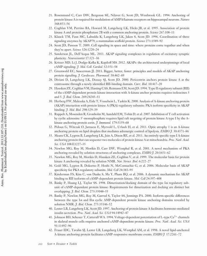
ANCHORED cAMP SIGNALS AND MODULATIONOF T CELL FUNCTION
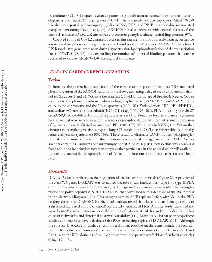
cAMP serves as a second messenger for a variety of immunoregulatory and inflamma-tory mediators—such as prostaglandin E2 (PGE2), catecholamines, serotonin, adenosine, andhistamine—that signal to effector T cells from monocytes, macrophages, and naturally occurringand peripherally induced regulatory T cells (Tregs) in settings such as inflammation, chronic in-fections, asthma, and cancer (Figure 4a,b). However, the spatial and temporal parameters thatunderlie such events are just beginning to be understood (163, 164). cAMP levels in effectorT cells are controlled in part by Tregs that suppress them. Naturally occurring Tregs have highcAMP levels in part due to FOXP3-dependent transcriptional downregulation of PDE3B (165).Cell-to-cell transmission of cAMP to suppress effector T cells proceeds via transfer through gapjunctions (166).
Pericellular accumulation of adenosine also elicits immunosuppressive responses. This is apathway whereby CD39 and CD73 ectonucleotidases on the surface of Tregs metabolize ATP togenerate adenosine (167), which activates A2A receptors to enhance intracellular cAMP synthesisand suppress effector T cells (167). In addition, continuous exposure of T cells to an antigenpromotes adaptive Tregs that express COX-2 (cyclooxygenase 2), leading to secretion of PGE2
(168–171). PGE2 stimulates FOXP3 expression in Tregs and inhibits effector T cell functionthrough mobilization of a PKA-Csk (C-terminal Src kinase) cascade (172) (Figure 4c). Type IPKA and PDE4 seem to be important for T cell receptor (TCR)-induced signaling and T cellfunction. Whereas TCR stimulation and concomitant PKA activation in lipid rafts inhibit proximalT cell signaling (173), CD28 costimulation favors the recruitment of a PDE4/β-arrestin signalingunit that derepresses cAMP inhibition as a prelude to the onset of the full T cell response. Thesefindings underscore the importance of enzyme anchoring and how the proximity of signalingenzymes with broad specificity can be used to drive specific, local cellular events.
In effector T cells, the cAMP pathway is involved in the regulation and modulation of immuneresponses that include antigen-induced proliferation and cytokine production (174, 175). cAMPalso induces a Treg response and suppression of T cells via several mechanisms (176). Numer-ous PKA targets intersect with mitogenic signaling pathways (164). Use of site-selective cAMPagonists with a preference for PKA isotypes and use of peptides that produce isoform-selectiveanchoring disruption have revealed (a) that an anchored pool of type I PKA in T, B, and nat-ural killer cells plays a dominant immunoregulatory role and (b) that PKA phosphorylation ofSer364 in Csk is the predominant inhibitory mechanism (31, 48, 50, 53, 175, 177–180). This
www.annualreviews.org • Cellular Regulation by Kinase Anchoring 197
Ann
u. R
ev. P
harm
acol
. Tox
icol
. 201
3.53
:187
-210
. Dow
nloa
ded
from
ww
w.a
nnua
lrev
iew
s.or
gby
${i
ndiv
idua
lUse
r.di
spla
yNam
e} o
n 01
/09/
13. F
or p
erso
nal u
se o
nly.
PA53CH10-Scott ARI 5 December 2012 14:15
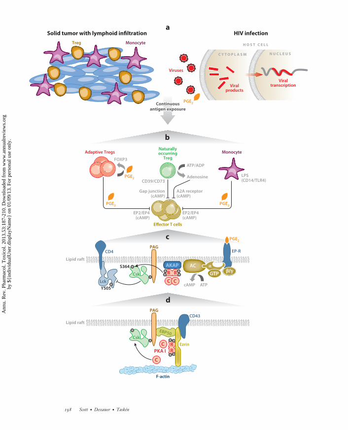
HIV infectionSolid tumor with lymphoid infiltration
Lipid raft
EP2/EP4
C Y T O P L A S M N U C L E U S
Viruses
Treg Monocyte
Monocyte
Viralproducts
Viraltranscription
Effector T cells
Adaptive TregsNaturallyoccurring
Treg
CD39/CD73
(cAMP)
Gap junction(cAMP)
A2A receptor(cAMP)
EP2/EP4(cAMP)
LPS(CD14/TLR4)
ATP/ADP
Adenosine
ATPcAMP
a
b
c
H O S T C E L L
PGE2
PGE2
PGE2 PGE2
PGE2
PAG
S364
Y505
Lck
Csk
EP-RCD4
ContinuousContinuousantigen exposureantigen exposure
Continuousantigen exposure
FOXP3
AKAP
R R
C C
GTP
ACβ/γ
α
Lipid raft
dPAG
PKA I
Csk
EBP50
Ezrin
F-actin
CD43
RR
C
C
198 Scott · Dessauer · Tasken
Ann
u. R
ev. P
harm
acol
. Tox
icol
. 201
3.53
:187
-210
. Dow
nloa
ded
from
ww
w.a
nnua
lrev
iew
s.or
gby
${i
ndiv
idua
lUse
r.di
spla
yNam
e} o
n 01
/09/
13. F
or p
erso
nal u
se o
nly.
PA53CH10-Scott ARI 5 December 2012 14:15
phosphorylation increases Csk activity fourfold, and docking to its binding protein,Cbp/PAG (Csk-binding protein/phosphoprotein associated with glycosphingolipid-enriched mi-crodomains), further increases its activity two- to threefold (180). Csk, in turn, phosphorylatesLck (lymphocyte-specific protein tyrosine kinase) to suppress its tyrosine kinase activity, therebyreducing ζ -chain phosphorylation of the TCR (181, 182). These events are organized by theCbp/PAG adapter protein that recruits Csk to the site of action in lipid rafts (172, 180, 183–185).Thus, PKA phosphorylation of Csk and its interaction with Cbp/PAG provide a powerful mech-anism for locally terminating antigen receptor–induced Src family tyrosine kinases (Figure 4c,d ).
SCAFFOLDING THE PKA-Csk INHIBITORY PATHWAY
The anchoring protein responsible for the compartmentalization of type I PKA in T cells isezrin. This modular, multifunctional protein is in the ezrin/radixin/moesin (ERM) family ofproteins that contribute to the organization of plasma membrane domains by linking micro-filaments to the membrane and aligning with membrane adaptor proteins via linker proteins, suchas EBP50 (ERM binding protein 50). Ezrin interacts with EBP50 and PAG in a complex to scaffoldthe PKA-Csk inhibitory pathway in T cells (31) (Figure 4d ).
Functional evidence that regulation of T cell function by type I PKA depends on anchoring byezrin derives from the observations that disruption of type I PKA binding to ezrin via anchoringdisruptor peptides displaces PKA from lipid rafts and releases the suppression of T cells. Thelatter suppression occurs via cAMP-mediated inhibition of proliferation and interleukin-2 (IL-2)production (31, 48, 53). Knockdown of ezrin abrogates regulation by cAMP of IL-2 secretion,whereas reconstitution with an siRNA-resistant wild-type ezrin, but not with a binding-domainmutant ezrin, restores cAMP regulation of IL-2 secretion (31, 53). In addition, disruption of theezrin-EBP50-PAG scaffold at the level of the ezrin-EBP50 interaction inhibits the regulation ofIL-2 secretion by cAMP (55) (Figure 4d ). Expression of the RIAD peptide in transgenic mice alsoperturbs the ability of the cAMP–type I PKA–Csk pathway to suppress PGE2-mediated inhibi-tion of effector T cells (50). Related studies demonstrate that global displacement of RI enhancesIL-2 secretion by T cells and produces resistance to murine AIDS (164). These findings havesuggested that hyperactivation of the type I PKA pathway is involved in the T cell dysfunctionof HIV infection and common variable immunodeficiency. The cAMP–type I PKA pathway inT cells is thus a putative target for treating immunodeficiency diseases, chronic infections, and
←−−−−−−−−−−−−−−−−−−−−−−−−−−−−−−−−−−−−−−−−−−−−−−−−−−−−−−−−−−−−−−−−−−−−−−−−−−−−−−−−−−−−−−−−−−Figure 4The immunoregulatory role of anchored cAMP signaling in T cells. (a) Tumor-infiltrating lymphocytes are inhibited by peripherallyinduced Tregs (orange cells) exposed to chronic antigenic stimulation. Persistent infections, such as HIV, lead to chronic inflammationand immunosuppression, both of which involve production of PGE2 and other inflammatory mediators. (b) Generation of peripherallyinduced or adaptive Tregs. These cells express COX-2, produce PGE2, and stimulate FOXP3 expression. In contrast, naturallyoccurring Tregs can transfer cAMP to responder T cells through gap junctions. Pericellular accumulation of adenosine also elicitsimmunosuppressive responses through a pathway whereby CD39 and CD73 ectoenzymes metabolize ATP to generate adenosine. Tcell inhibition and PGE2-responsive induction of FOXP3 can occur as a result of the secretion of PGE2 by LPS-activated monocytes.(c) cAMP inhibits TCR-mediated immunoregulatory functions at membranes. This occurs in lipid rafts through a receptor–Gprotein–AC–cAMP–PKA type I–Csk pathway that acts on the Src family tyrosine kinase Lck. (d ) Ezrin links transmembrane receptorssuch as CD43 to the actin cytoskeleton via its N-terminal FERM domain and to F-actin via its C terminus. Ezrin functions as anAKAP, bringing type I PKA in proximity to its substrate Csk via a supramolecular signaling complex consisting of PKA, ezrin, EBP50,Cbp/PAG, and Csk. Abbreviations: AC, adenylyl cyclase; AKAP, A-kinase anchoring protein; cAMP, cyclic adenosine monophosphate;COX-2, cyclooxygenase 2; Csk, C-terminal Src kinase; EPB50, ERM binding protein 50; EP-R, receptor for E series of prostaglandins;FERM, band 4.1 protein and ezrin, radixin, and moesin (ERM) protein domain; Lck, lymphocyte-specific protein tyrosine kinase;LPS, lipopolysaccharide; PAG, phosphoprotein associated with glycosphingolipid-enriched microdomains; PGE2, prostaglandin E2;PKA, protein kinase A; TCR, T cell receptor; Treg, regulatory T cell.
www.annualreviews.org • Cellular Regulation by Kinase Anchoring 199
Ann
u. R
ev. P
harm
acol
. Tox
icol
. 201
3.53
:187
-210
. Dow
nloa
ded
from
ww
w.a
nnua
lrev
iew
s.or
gby
${i
ndiv
idua
lUse
r.di
spla
yNam
e} o
n 01
/09/
13. F
or p
erso
nal u
se o
nly.
PA53CH10-Scott ARI 5 December 2012 14:15
cancer (170, 186–189) (Figure 4a). Furthermore, T cells from transgenic mice that are protectedfrom suppression by Tregs through cAMP show improved antitumor responses compared withmice that have normal RI anchoring (I. Cornez & K. Tasken, unpublished data); this is consistentwith observations in colorectal cancer patient samples in which PGE2 and cAMP suppress anti-tumor immunity (176, 189) (Figure 4a). Thus, the cAMP–type I PKA–Csk pathway is a putativetherapeutic target in the inflammation that occurs in cancer and some chronic viral diseases.
CONCLUSIONS
The study of enzyme anchoring via AKAPs has provided insight into the elaborate andelegant organization of cellular signaling cascades. In this final section, we highlight threefuture directions for AKAP research. First, although this field arose from the need to explainhormone action at a molecular level, surprisingly little is known about the architecture of themacromolecular complexes that AKAPs hold together. More structural information regardingAKAP signaling complexes should soon be forthcoming. This is an exciting but dauntingobjective because AKAPs’ flexibility makes them challenging targets for X-ray crystallography(36, 37, 190). Furthermore, most anchored signaling complexes consist of many proteins; forexample, a recent study that used mass spectrometry assigned 16 polypeptide chains within asingle (AKAP79-2PP2B-RII-CaM)2 macromolecular assembly (92). Cutting-edge approaches ofprotein mass spectrometry, single-particle fluorescence imaging, and cryo-electron microcopywill likely be needed to answer key structural questions regarding the stoichiometry and moleculararchitecture of higher-order AKAP complexes (191–194).
Second, AKAP79/150 can protect PKC from certain pharmacological inhibitors, indicatingthat the anchoring protein locks this kinase in an active conformation (195, 196). A broaderinterpretation of this result is that AKAPs are allosteric modifiers that shape the activity ofthe kinase or phosphatase that they regulate (197). A more far-reaching implication is that theintracellular pharmacology of kinase inhibitor drugs cannot be reliably inferred from in vitroenzyme assays. The allosteric nature of some AKAP-enzyme interactions complicates studiesthat rely on the analysis of genetically modified mice. Thus, we propose that any comprehensiveinvestigation of AKAP function in a whole-animal context cannot be limited to simply knockingout a gene of interest; instead, it should also include analysis of modified AKAP forms that areunable to interact with a given enzyme-binding partner (56, 96).
Third, research on AKAPs is moving toward understanding second messenger signaling in apathophysiological context. For such efforts, approaches other than global disruption of intracellu-lar anchoring events will be needed. Small-molecule inhibitors show some promise, although theirmechanism of action is unknown, and they have substantial potential for off-target effects (198).Another approach to consider is peptide-mediated disruption of AKAP-associated substrates. Suchan approach has been used to displace AKAP18δ from the PKA substrate phospholamban to altercardiac excitation-contraction coupling (54), and to disconnect PKA from Csk in order to reversecAMP-mediated inhibition of immune function (55). These methods are akin to others in whichnative peptides/proteins or peptidomimetics are delivered into cells as a means to overcome someof the limitations associated with the displacement of a full-length enzyme from its intracellularlocation. Thus, a new generation of AKAP-derived reagents that retrieve a small measure of orderfrom chaos may offer a therapeutic advantage.
DISCLOSURE STATEMENT
The authors are not aware of any affiliations, memberships, funding, or financial holdings thatmight be perceived as affecting the objectivity of this review.
200 Scott · Dessauer · Tasken
Ann
u. R
ev. P
harm
acol
. Tox
icol
. 201
3.53
:187
-210
. Dow
nloa
ded
from
ww
w.a
nnua
lrev
iew
s.or
gby
${i
ndiv
idua
lUse
r.di
spla
yNam
e} o
n 01
/09/
13. F
or p
erso
nal u
se o
nly.
PA53CH10-Scott ARI 5 December 2012 14:15
ACKNOWLEDGMENTS
The authors wish to thank Melanie Milnes for proofreading and editing this article and LoreneLangeberg and Kristoffer Watten Brudvik for helping prepare the figures. J.D.S. is supported inpart by National Institutes of Health (NIH) grant GM48231; C.W.D., by NIH grant GM60419;and K.T., by grants from the Research Council of Norway and the Norwegian Cancer Society.
LITERATURE CITED
1. Rall TW, Sutherland EW, Berthet J. 1957. The relationships of epinephrine and glucagon to liverphosphorylase. J. Biol. Chem. 224:463–75
2. Sutherland EW. 1972. Studies on the mechanism of hormone action. Science 171:401–83. Fischer EH, Krebs EG. 1955. Conversion of phosphorylase b to phosphorylase a in muscle extracts.
J. Biol. Chem. 216:121–324. Corbin JD, Soderling TR, Park CR. 1973. Regulation of adenosine 3′,5′-monophosphate-dependent
protein kinase. J. Biol. Chem. 248:1813–215. Potter RL, Stafford PH, Taylor S. 1978. Regulatory subunit of cyclic AMP-dependent protein kinase I
from porcine skeletal muscle: purification and proteolysis. Arch. Biochem. Biophys. 190:174–806. Kemp BE, Benjamini E, Krebs EG. 1976. Synthetic hexapeptide substrates and inhibitors of 3′:5′-cyclic
AMP-dependent protein kinase. Proc. Natl. Acad. Sci. USA 73:1038–427. Keely SL. 1977. Activation of cAMP-dependent protein kinase without a corresponding increase in
phosphorylase activity. Res. Commun. Chem. Pathol. Pharmacol. 18:283–908. Hayes JS, Brunton LL, Brown JH, Reese JB, Mayer SE. 1979. Hormonally specific expression of cardiac
protein kinase activity. Proc. Natl. Acad. Sci. USA 76:1570–749. Brunton LL, Hayes JS, Mayer SE. 1979. Hormonally specific phosphorylation of cardiac troponin I and
activation of glycogen phosphorylase. Nature 280:78–8010. Corbin JD, Sugden PH, Lincoln TM, Keely SL. 1977. Compartmentalization of adenosine 3′:5′-
monophosphate and adenosine 3′:5′-monophosphate-dependent protein kinase in heart tissue. J. Biol.Chem. 252:3854–61
11. Theurkauf WE, Vallee RB. 1982. Molecular characterization of the cAMP-dependent protein kinasebound to microtubule-associated protein 2. J. Biol. Chem. 257:3284–90
12. Sarkar D, Erlichman J, Rubin CS. 1984. Identification of a calmodulin-binding protein that co-purifieswith the regulatory subunit of brain protein kinase II. J. Biol. Chem. 259:9840–46
13. Lohmann SM, DeCamili P, Enig I, Walter U. 1984. High-affinity binding of the regulatory subunit(RII) of cAMP-dependent protein kinase to microtubule-associated and other cellular proteins. Proc.Natl. Acad. Sci. USA 81:6723–27
14. Carr DW, Scott JD. 1992. Blotting and band-shifting: techniques for studying protein-protein interac-tions. Trends Biochem. Sci. 17:246–49
15. Alto NM, Soderling J, Scott JD. 2002. Rab32 is an A-kinase anchoring protein and participates inmitochondrial dynamics. J. Cell Biol. 158:659–68
16. Pidoux G, Tasken K. 2010. Specificity and spatial dynamics of protein kinase A signaling organized byA-kinase-anchoring proteins. J. Mol. Endocrinol. 44:271–84
17. Wong W, Scott JD. 2004. AKAP signalling complexes: focal points in space and time. Nat. Rev. Mol.Cell Biol. 5:959–70
18. Scott JD, Stofko RE, McDonald JR, Comer JD, Vitalis EA, Mangeli J. 1990. Type II regulatory subunitdimerization determines the subcellular localization of the cAMP-dependent protein kinase. J. Biol.Chem. 265:21561–66
19. Carr DW, Hausken ZE, Fraser IDC, Stofko-Hahn RE, Scott JD. 1992. Association of the type II cAMP-dependent protein kinase with a human thyroid RII-anchoring protein: cloning and characterization ofthe RII-binding domain. J. Biol. Chem. 267:13376–82
20. Carr DW, Stofko-Hahn RE, Fraser IDC, Bishop SM, Acott TS, et al. 1991. Interaction of the regula-tory subunit (RII) of cAMP-dependent protein kinase with RII-anchoring proteins occurs through anamphipathic helix binding motif. J. Biol. Chem. 266:14188–92
www.annualreviews.org • Cellular Regulation by Kinase Anchoring 201
Ann
u. R
ev. P
harm
acol
. Tox
icol
. 201
3.53
:187
-210
. Dow
nloa
ded
from
ww
w.a
nnua
lrev
iew
s.or
gby
${i
ndiv
idua
lUse
r.di
spla
yNam
e} o
n 01
/09/
13. F
or p
erso
nal u
se o
nly.
PA53CH10-Scott ARI 5 December 2012 14:15
21. Rosenmund C, Carr DW, Bergeson SE, Nilaver G, Scott JD, Westbrook GL. 1994. Anchoring ofprotein kinase A is required for modulation of AMPA/kainate receptors on hippocampal neurons. Nature368:853–56
22. Coghlan VM, Perrino BA, Howard M, Langeberg LK, Hicks JB, et al. 1995. Association of proteinkinase A and protein phosphatase 2B with a common anchoring protein. Science 267:108–11
23. Klauck TM, Faux MC, Labudda K, Langeberg LK, Jaken S, Scott JD. 1996. Coordination of threesignaling enzymes by AKAP79, a mammalian scaffold protein. Science 271:1589–92
24. Scott JD, Pawson T. 2009. Cell signaling in space and time: where proteins come together and whenthey’re apart. Science 326:1220–24
25. Sanderson JL, Dell’Acqua ML. 2011. AKAP signaling complexes in regulation of excitatory synapticplasticity. Neuroscientist 17:321–36
26. Kritzer MD, Li J, Dodge-Kafka K, Kapiloff MS. 2012. AKAPs: the architectural underpinnings of localcAMP signaling. J. Mol. Cell. Cardiol. 52:351–58
27. Greenwald EC, Saucerman JJ. 2011. Bigger, better, faster: principles and models of AKAP anchoringprotein signaling. J. Cardiovasc. Pharmacol. 58:462–69
28. Diviani D, Langeberg LK, Doxsey SJ, Scott JD. 2000. Pericentrin anchors protein kinase A at thecentrosome through a newly identified RII-binding domain. Curr. Biol. 10:417–20
29. Hausken ZE, Coghlan VM, Hasting CAS, Reimann EM, Scott JD. 1994. Type II regulatory subunit (RII)of the cAMP-dependent protein kinase interaction with A-kinase anchor proteins requires isoleucines 3and 5. J. Biol. Chem. 269:24245–51
30. Herberg FW, Maleszka A, Eide T, Vossebein L, Tasken K. 2000. Analysis of A-kinase anchoring protein(AKAP) interaction with protein kinase A (PKA) regulatory subunits: PKA isoform specificity in AKAPbinding. J. Mol. Biol. 298:329–39
31. Ruppelt A, Mosenden R, Gronholm M, Aandahl EM, Tobin D, et al. 2007. Inhibition of T cell activationby cyclic adenosine 5′-monophosphate requires lipid raft targeting of protein kinase A type I by the A-kinase anchoring protein ezrin. J. Immunol. 179:5159–68
32. Pidoux G, Witczak O, Jarnaess E, Myrvold L, Urlaub H, et al. 2011. Optic atrophy 1 is an A-kinaseanchoring protein on lipid droplets that mediates adrenergic control of lipolysis. EMBO J. 30:4371–86
33. Means CK, Lygren B, Langeberg LK, Jain A, Dixon RE, et al. 2011. An entirely specific type I A-kinaseanchoring protein that can sequester two molecules of protein kinase A at mitochondria. Proc. Natl. Acad.Sci. USA 108:E1227–35
34. Newlon MG, Roy M, Morikis D, Carr DW, Westphal R, et al. 2001. A novel mechanism of PKAanchoring revealed by solution structures of anchoring complexes. EMBO J. 20:1651–62
35. Newlon MG, Roy M, Morikis D, Hausken ZE, Coghlan V, et al. 1999. The molecular basis for proteinkinase A anchoring revealed by solution NMR. Nat. Struct. Biol. 6:222–27
36. Gold MG, Lygren B, Dokurno P, Hoshi N, McConnachie G, et al. 2006. Molecular basis of AKAPspecificity for PKA regulatory subunits. Mol. Cell 24:383–95
37. Kinderman FS, Kim C, von Daake S, Ma Y, Pham BQ, et al. 2006. A dynamic mechanism for AKAPbinding to RII isoforms of cAMP-dependent protein kinase. Mol. Cell 24:397–408
38. Banky P, Huang LJ, Taylor SS. 1998. Dimerization/docking domain of the type Iα regulatory sub-unit of cAMP-dependent protein kinase: Requirements for dimerization and docking are distinct butoverlapping. J. Biol. Chem. 273:35048–55
39. Banky P, Newlon MG, Roy M, Garrod S, Taylor SS, Jennings PA. 2000. Isoform-specific differencesbetween the type Iα and IIα cyclic AMP-dependent protein kinase anchoring domains revealed bysolution NMR. J. Biol. Chem. 275:35146–52
40. Lester LB, Langeberg LK, Scott JD. 1997. Anchoring of protein kinase A facilitates hormone-mediatedinsulin secretion. Proc. Natl. Acad. Sci. USA 94:14942–47
41. Johnson BD, Scheuer T, Catterall WA. 1994. Voltage-dependent potentiation of L-type Ca2+ channelsin skeletal muscle cells requires anchored cAMP-dependent protein kinase. Proc. Natl. Acad. Sci. USA91:11492–96
42. Fraser IDC, Tavalin SJ, Lester LB, Langeberg LK, Westphal AM, et al. 1998. A novel lipid-anchoredA-kinase anchoring protein facilitates cAMP-responsive membrane events. EMBO J. 17:2261–72
202 Scott · Dessauer · Tasken
Ann
u. R
ev. P
harm
acol
. Tox
icol
. 201
3.53
:187
-210
. Dow
nloa
ded
from
ww
w.a
nnua
lrev
iew
s.or
gby
${i
ndiv
idua
lUse
r.di
spla
yNam
e} o
n 01
/09/
13. F
or p
erso
nal u
se o
nly.
PA53CH10-Scott ARI 5 December 2012 14:15
43. Vijayaraghavan S, Goueli SA, Davey MP, Carr DW. 1997. Protein kinase A-anchoring inhibitor peptidesarrest mammalian sperm motility. J. Biol. Chem. 272:4747–52
44. Fink MA, Zakhary DR, Mackey JA, Desnoyer RW, Apperson-Hansen C, et al. 2001. AKAP-mediatedtargeting of protein kinase A regulates contractility in cardiac myocytes. Circ. Res. 88:291–97
45. Gold MG, Reichow SL, O’Neill SE, Weisbrod CR, Langeberg LK, et al. 2012. AKAP2 anchors PKAwith aquaporin-0 to support ocular lens transparency. EMBO Mol. Med. 4:15–26
46. Alto NM, Soderling SH, Hoshi N, Langeberg LK, Fayos R, et al. 2003. Bioinformatic design of A-kinaseanchoring protein-in silico: a potent and selective peptide antagonist of type II protein kinase A anchoring.Proc. Natl. Acad. Sci. USA 100:4445–50
47. Burns-Hamuro LL, Ma Y, Kammerer S, Reineke U, Self C, et al. 2003. Designing isoform-specificpeptide disruptors of protein kinase A localization. Proc. Natl. Acad. Sci. USA 100:4072–77
48. Carlson CR, Lygren B, Berge T, Hoshi N, Wong W, et al. 2006. Delineation of type I protein kinaseA-selective signaling events using an RI anchoring disruptor. J. Biol. Chem. 281:21535–45
49. Torheim EA, Ndhlovu LC, Pettersen FO, Larsen TL, Jha AR, et al. 2009. Interleukin-10-secreting Tcells define a suppressive subset within the HIV-1-specific T-cell population. Eur. J. Immunol. 39:1280–87
50. Mosenden R, Singh P, Cornez I, Heglind M, Ruppelt A, et al. 2011. Mice with disrupted type I proteinkinase A anchoring in T cells resist retrovirus-induced immunodeficiency. J. Immunol. 186:5119–30
51. McKenzie AJ, Campbell SL, Howe AK. 2011. Protein kinase A activity and anchoring are required forovarian cancer cell migration and invasion. PLoS ONE 6:e26552
52. Di Benedetto G, Zoccarato A, Lissandron V, Terrin A, Li X, et al. 2008. Protein kinase A type I andtype II define distinct intracellular signaling compartments. Circ. Res. 103:836–44
53. Jarnaess E, Ruppelt A, Stokka AJ, Lygren B, Scott JD, Tasken K. 2008. Dual specificity A-kinase an-choring proteins (AKAPs) contain an additional binding region that enhances targeting of protein kinaseA type I. J. Biol. Chem. 283:33708–18
54. Lygren B, Carlson CR, Santamaria K, Lissandron V, McSorley T, et al. 2007. AKAP complex regulatesCa2+ re-uptake into heart sarcoplasmic reticulum. EMBO Rep. 8:1061–67
55. Stokka AJ, Mosenden R, Ruppelt A, Lygren B, Tasken K. 2010. The adaptor protein EBP50 is importantfor localization of the protein kinase A-Ezrin complex in T-cells and the immunomodulating effect ofcAMP. Biochem. J. 425:381–88
56. Bers DM. 2008. Calcium cycling and signaling in cardiac myocytes. Annu. Rev. Physiol. 70:23–4957. Sadana R, Dessauer CW. 2009. Physiological roles for G protein-regulated adenylyl cyclase isoforms:
insights from knockout and overexpression studies. NeuroSignals 17:5–2258. Willoughby D, Cooper DMF. 2007. Organization and Ca2+ regulation of adenylyl cyclases in cAMP
microdomains. Physiol. Rev. 87:965–101059. Ostrom RS, Naugle JE, Hase M, Gregorian C, Swaney JS, et al. 2003. Angiotensin II enhances adeny-
lyl cyclase signaling via Ca2+/calmodulin: Gq-Gs cross-talk regulates collagen production in cardiacfibroblasts. J. Biol. Chem. 278:24461–68
60. Iwatsubo K, Minamisawa S, Tsunematsu T, Nakagome M, Toya Y, et al. 2004. Direct inhibition oftype 5 adenylyl cyclase prevents myocardial apoptosis without functional deterioration. J. Biol. Chem.279:40938–45
61. Okumura S, Kawabe J, Yatani A, Takagi G, Lee MC, et al. 2003. Type 5 adenylyl cyclase disruptionalters not only sympathetic but also parasympathetic and calcium-mediated cardiac regulation. Circ. Res.93:364–71
62. Guellich A, Gao S, Hong C, Yan L, Wagner TE, et al. 2010. Effects of cardiac overexpression of type 6adenylyl cyclase affects on the response to chronic pressure overload. Am. J. Physiol. Heart Circ. Physiol.299:H707–12
63. Takahashi T, Tang T, Lai NC, Roth DM, Rebolledo B, et al. 2006. Increased cardiac adenylyl cyclaseexpression is associated with increased survival after myocardial infarction. Circulation 114:388–96
64. Gao MH, Tang T, Guo T, Miyanohara A, Yajima T, et al. 2008. Adenylyl cyclase type VI increases Aktactivity and phospholamban phosphorylation in cardiac myocytes. J. Biol. Chem. 283:33527–35
65. Yan L, Vatner DE, O’Connor JP, Ivessa A, Ge H, et al. 2007. Type 5 adenylyl cyclase disruption increaseslongevity and protects against stress. Cell 130:247–58
www.annualreviews.org • Cellular Regulation by Kinase Anchoring 203
Ann
u. R
ev. P
harm
acol
. Tox
icol
. 201
3.53
:187
-210
. Dow
nloa
ded
from
ww
w.a
nnua
lrev
iew
s.or
gby
${i
ndiv
idua
lUse
r.di
spla
yNam
e} o
n 01
/09/
13. F
or p
erso
nal u
se o
nly.
PA53CH10-Scott ARI 5 December 2012 14:15
66. Hu CL, Chandra R, Ge H, Pain J, Yan L, et al. 2009. Adenylyl cyclase type 5 protein expression duringcardiac development and stress. Am. J. Physiol. Heart Circ. Physiol. 297:H1776–82
67. Tang T, Gao MH, Lai NC, Firth AL, Takahashi T, et al. 2008. Adenylyl cyclase type 6 deletion decreasesleft ventricular function via impaired calcium handling. Circulation 117:61–69
68. Burton KA, Johnson BD, Hausken ZE, Westenbroek RE, Idzerda RL, et al. 1997. Type II regulatorysubunits are not required for the anchoring-dependent modulation of Ca2+ channel activity by cAMP-dependent protein kinase. Proc. Natl. Acad. Sci. USA 94:11067–72
69. Gao TY, Yatani A, Dell’Acqua ML, Sako H, Green SA, et al. 1997. cAMP-dependent regulation ofcardiac L-type Ca2+ channels requires membrane targeting of PKA and phosphorylation of channelsubunits. Neuron 19:185–96
70. Kockskamper J, Sendhoff K, Erlenkamp S, Bordusa F, Cerovsky V, Glitsch HG. 2001. Differences inthe protein-kinase-A-dependent regulation of CFTR Cl− channels and Na+-K+ pumps in guinea-pigventricular myocytes. Pflug. Arch. 441:807–15
71. Potet F, Scott JD, Mohammad-Panah R, Escande D, Baro I. 2001. AKAP proteins anchor cAMP-dependent protein kinase to KvLQT1/IsK channel complex. Am. J. Physiol. Heart Circ. Physiol.280:H2038–45
72. Fink MA, Zakhary DR, Mackey JA, Desnoyer RW, Apperson-Hansen C, et al. 2001. AKAP-mediatedtargeting of protein kinase A regulates contractility in cardiac myocytes. Circ. Res. 88:291–97
73. McConnell BK, Popovic Z, Mal N, Lee K, Bautista J, et al. 2009. Disruption of protein kinase Ainteraction with A-kinase-anchoring proteins in the heart in vivo: effects on cardiac contractility, proteinkinase A phosphorylation, and troponin I proteolysis. J. Biol. Chem. 284:1583–92
74. Patel HH, Hamuro LL, Chun BJ, Kawaraguchi Y, Quick A, et al. 2010. Disruption of protein kinaseA localization using a trans-activator of transcription (TAT)-conjugated A-kinase-anchoring peptidereduces cardiac function. J. Biol. Chem. 285:27632–40
75. Diviani D, Scott JD. 2001. AKAP signaling complexes at the cytoskeleton. J. Cell Sci. 114:1431–3776. Malbon CC, Tao J, Wang HY. 2004. AKAPs (A-kinase anchoring proteins) and molecules that compose
their G-protein-coupled receptor signalling complexes. Biochem. J. 379:1–977. Navedo MF, Cheng EP, Yuan C, Votaw S, Molkentin JD, et al. 2010. Increased coupled gating of L-type
Ca2+ channels during hypertension and Timothy syndrome. Circ. Res. 106:748–5678. Gray PC, Johnson BD, Westenbroek RE, Hays LG, Yates JR III, et al. 1998. Primary structure and
function of an A kinase anchoring protein associated with calcium channels. Neuron 20:1017–2679. Trotter KW, Fraser IDC, Scott GK, Stutts MJ, Scott JD, Milgram SL. 1999. Alternative splicing regu-
lates the subcellular localization of A-kinase anchoring protein 18 isoforms. J. Cell Biol. 147:1481–9280. Henn V, Edemir B, Stefan E, Wiesner B, Lorenz D, et al. 2004. Identification of a novel A-kinase
anchoring protein 18 isoform and evidence for its role in the vasopressin-induced aquaporin-2 shuttlein renal principal cells. J. Biol. Chem. 279:26654–65
81. Hulme JT, Lin TW, Westenbroek RE, Scheuer T, Catterall WA. 2003. β-Adrenergic regulation requiresdirect anchoring of PKA to cardiac CaV1.2 channels via a leucine zipper interaction with A kinase-anchoring protein 15. Proc. Natl. Acad. Sci. USA 100:13093–98
82. Hulme JT, Ahn M, Hauschka SD, Scheuer T, Catterall WA. 2002. A novel leucine zipper targets AKAP15and cyclic AMP-dependent protein kinase to the C terminus of the skeletal muscle Ca2+ channel andmodulates its function. J. Biol. Chem. 277:4079–87
83. Hulme JT, Westenbroek RE, Scheuer T, Catterall WA. 2006. Phosphorylation of serine 1928 in thedistal C-terminal domain of cardiac CaV1.2 channels during β1-adrenergic regulation. Proc. Natl. Acad.Sci. USA 103:16574–79
84. Fuller MD, Emrick MA, Sadilek M, Scheuer T, Catterall WA. 2010. Molecular mechanism of calciumchannel regulation in the fight-or-flight response. Sci. Signal. 3:ra70
85. Gretarsdottir S, Thorleifsson G, Reynisdottir ST, Manolescu A, Jonsdottir S, et al. 2003. The geneencoding phosphodiesterase 4D confers risk of ischemic stroke. Nat. Genet. 35:131–38
86. Oliveria SF, Dell’Acqua ML, Sather WA. 2007. AKAP79/150 anchoring of calcineurin controls neuronalL-type Ca2+ channel activity and nuclear signaling. Neuron 55:261–75
87. Dell’Acqua ML, Faux MC, Thorburn J, Thorburn A, Scott JD. 1998. Membrane-targeting sequenceson AKAP79 bind phosphatidylinositol-4, 5-bisphosphate. EMBO J. 17:2246–60
204 Scott · Dessauer · Tasken
Ann
u. R
ev. P
harm
acol
. Tox
icol
. 201
3.53
:187
-210
. Dow
nloa
ded
from
ww
w.a
nnua
lrev
iew
s.or
gby
${i
ndiv
idua
lUse
r.di
spla
yNam
e} o
n 01
/09/
13. F
or p
erso
nal u
se o
nly.
PA53CH10-Scott ARI 5 December 2012 14:15
88. Delint-Ramirez I, Willoughby D, Hammond GVR, Ayling LJ, Cooper DMF. 2011. Palmitoylationtargets AKAP79 protein to lipid rafts and promotes its regulation of calcium-sensitive adenylyl cyclasetype 8. J. Biol. Chem. 286:32962–75
89. Bauman AL, Soughayer J, Nguyen BT, Willoughby D, Carnegie GK, et al. 2006. Dynamic regulationof cAMP synthesis through anchored PKA-adenylyl cyclase V/VI complexes. Mol. Cell 23:925–31
90. Tunquist BJ, Hoshi N, Guire ES, Zhang F, Mullendorff K, et al. 2008. Loss of AKAP150 perturbsdistinct neuronal processes in mice. Proc. Natl. Acad. Sci. USA 105:12557–62
91. Navedo MF, Nieves-Cintron M, Amberg GC, Yuan C, Votaw VS, et al. 2008. AKAP150 is required forstuttering persistent Ca2+ sparklets and angiotensin II–induced hypertension. Circ. Res. 102:e1–11
92. Gold MG, Stengel F, Nygren PJ, Weisbrod CR, Bruce JE, et al. 2011. Architecture and dynamics of anA-kinase anchoring protein 79 (AKAP79) signaling complex. Proc. Natl. Acad. Sci. USA 108:6426–31
93. Gao S, Wang H-y, Malbon C. 2011. AKAP5 and AKAP12 form homo-oligomers. J. Mol. Signal. 6:394. Gao S, Wang H-y, Malbon C. 2011. AKAP12 and AKAP5 form higher-order hetero-oligomers. J. Mol.
Signal. 6:895. Fraser IDC, Cong M, Kim J, Rollins EN, Daaka Y, et al. 2000. Assembly of an A kinase-anchoring
protein–β2-adrenergic receptor complex facilitates receptor phosphorylation and signaling. Curr. Biol.10:409–12
96. Nichols CB, Rossow CF, Navedo MF, Westenbroek RE, Catterall WA, et al. 2010. Sympathetic stim-ulation of adult cardiomyocytes requires association of AKAP5 with a subpopulation of L-type calciumchannels. Circ. Res. 107:747–56
97. Colledge M, Dean RA, Scott GK, Langeberg LK, Huganir RL, Scott JD. 2000. Targeting of PKA toglutamate receptors through a MAGUK-AKAP complex. Neuron 27:107–19
98. Nieves-Cintron M, Amberg GC, Navedo MF, Molkentin JD, Santana LF. 2008. The control of Ca2+
influx and NFATc3 signaling in arterial smooth muscle during hypertension. Proc. Natl. Acad. Sci. USA105:15623–28
99. Nieves-Cintron M, Amberg GC, Nichols CB, Molkentin JD, Santana LF. 2007. Activation of NFATc3down-regulates the β1 subunit of large conductance, calcium-activated K+ channels in arterial smoothmuscle and contributes to hypertension. J. Biol. Chem. 282:3231–40
100. Westphal RS, Tavalin SJ, Lin JW, Alto NM, Fraser IDC, et al. 1999. Regulation of NMDA receptorsby an associated phosphatase-kinase signaling complex. Science 285:93–96
101. Schmidt PH, Dransfield DT, Claudio JO, Hawley RG, Trotter KW, et al. 1999. AKAP350, a multiplyspliced protein kinase A-anchoring protein associated with centrosomes. J. Biol. Chem. 274:3055–66
102. Witczak O, Skalhegg BS, Keryer G, Bornens M, Tasken K, et al. 1999. Cloning and characterizationof a cDNA encoding an A-kinase anchoring protein located in the centrosome, AKAP450. EMBO J.18:1858–68
103. Terrenoire C, Houslay MD, Baillie GS, Kass RS. 2009. The cardiac IKs potassium channel macromolec-ular complex includes the phosphodiesterase PDE4D3. J. Biol. Chem. 284:9140–46
104. Piggott LA, Bauman AL, Scott JD, Dessauer CW. 2008. The A-kinase anchoring protein Yotiao bindsand regulates adenylyl cyclase in brain. Proc. Natl. Acad. Sci. USA 105:13835–40
105. Marx SO, Kurokawa J, Reiken S, Motoike H, D’Armiento J, et al. 2002. Requirement of a macromolecularsignaling complex for β adrenergic receptor modulation of the KCNQ1-KCNE1 potassium channel.Science 295:496–99
106. Kurokawa J, Motoike HK, Rao J, Kass RS. 2004. Regulatory actions of the A-kinase anchoring proteinYotiao on a heart potassium channel downstream of PKA phosphorylation. Proc. Natl. Acad. Sci. USA101:16374–78
107. Chen L, Kurokawa J, Kass RS. 2005. Phosphorylation of the A-kinase-anchoring protein Yotiao con-tributes to protein kinase A regulation of a heart potassium channel. J. Biol. Chem. 280:31347–52
108. Chen L, Marquardt ML, Tester DJ, Sampson KJ, Ackerman MJ, Kass RS. 2007. Mutation of an A-kinase-anchoring protein causes long-QT syndrome. Proc. Natl. Acad. Sci. USA 104:20990–95
109. Lu JT, Kass RS. 2010. Recent progress in congenital long QT syndrome. Curr. Opin. Cardiol. 25:216–21110. Kammerer S, Burns-Hamuro LL, Ma Y, Hamon SC, Canaves JM, et al. 2003. Amino acid variant
in the kinase binding domain of dual-specific A kinase-anchoring protein 2: a disease susceptibilitypolymorphism. Proc. Natl. Acad. Sci. USA 100:4066–71
www.annualreviews.org • Cellular Regulation by Kinase Anchoring 205
Ann
u. R
ev. P
harm
acol
. Tox
icol
. 201
3.53
:187
-210
. Dow
nloa
ded
from
ww
w.a
nnua
lrev
iew
s.or
gby
${i
ndiv
idua
lUse
r.di
spla
yNam
e} o
n 01
/09/
13. F
or p
erso
nal u
se o
nly.
PA53CH10-Scott ARI 5 December 2012 14:15
111. Tingley WG, Pawlikowska L, Zaroff JG, Kim T, Nguyen T, et al. 2007. Gene-trapped mouse embryonicstem cell-derived cardiac myocytes and human genetics implicate AKAP10 in heart rhythm regulation.Proc. Natl. Acad. Sci. USA 104:8461–66
112. Wang L, Sunahara RK, Krumins A, Perkins G, Crochiere ML, et al. 2001. Cloning and mitochondriallocalization of full-length D-AKAP2, a protein kinase A anchoring protein. Proc. Natl. Acad. Sci. USA98:3220–25
113. Eggers CT, Schafer JC, Goldenring JR, Taylor SS. 2009. D-AKAP2 interacts with Rab4 and Rab11through its RGS domains and regulates transferrin receptor recycling. J. Biol. Chem. 284:32869–80
114. Frey N, Olson EN. 2003. Cardiac hypertrophy: the good, the bad, and the ugly. Annu. Rev. Physiol.65:45–79
115. Zheng Y, Olson MF, Hall A, Cerione RA, Toksoz D. 1995. Direct involvement of the small GTP-bindingprotein Rho in lbc oncogene function. J. Biol. Chem. 270:9031–34
116. Diviani D, Soderling J, Scott JD. 2001. AKAP-Lbc anchors protein kinase A and nucleates Gα12-selective Rho-mediated stress fiber formation. J. Biol. Chem. 276:44247–57
117. Sah VP, Hoshijima M, Chien KR, Brown JH. 1996. Rho is required for Gαq and α1-adrenergic receptorsignaling in cardiomyocytes: dissociation of Ras and Rho pathways. J. Biol. Chem. 271:31185–90
118. Appert-Collin A, Cotecchia S, Nenniger-Tosato M, Pedrazzini T, Diviani D. 2007. The A-kinase an-choring protein (AKAP)-Lbc-signaling complex mediates α1 adrenergic receptor-induced cardiomy-ocyte hypertrophy. Proc. Natl. Acad. Sci. USA 104:10140–45
119. Diviani D, Abuin L, Cotecchia S, Pansier L. 2004. Anchoring of both PKA and 14-3-3 inhibits theRho-GEF activity of the AKAP-Lbc signaling complex. EMBO J. 23:2811–20
120. Jin J, Smith FD, Stark C, Wells CD, Fawcett JP, et al. 2004. Proteomic, functional, and domain-basedanalysis of in vivo 14-3-3 binding proteins involved in cytoskeletal regulation and cellular organization.Curr. Biol. 14:1436–50
121. Carnegie GK, Soughayer J, Smith FD, Pedroja BS, Zhang F, et al. 2008. AKAP-Lbc mobilizes a cardiachypertrophy signaling pathway. Mol. Cell 32:169–79
122. Carnegie GK, Smith FD, McConnachie G, Langeberg LK, Scott JD. 2004. AKAP-Lbc nucleates aprotein kinase D activation scaffold. Mol. Cell 15:889–99
123. Smith FD, Langeberg LK, Cellurale C, Pawson T, Morrison DK, et al. 2010. AKAP-Lbc enhancescyclic AMP control of the ERK1/2 cascade. Nat. Cell Biol. 12:1242–49
124. Cariolato L, Cavin S, Diviani D. 2011. A-kinase anchoring protein (AKAP)-Lbc anchors a PKN-basedsignaling complex involved in α1-adrenergic receptor-induced p38 activation. J. Biol. Chem. 286:7925–37
125. Mayers CM, Wadell J, McLean K, Venere M, Malik M, et al. 2010. The Rho guanine nucleotide exchangefactor AKAP13 (BRX) is essential for cardiac development in mice. J. Biol. Chem. 285:12344–54
126. Kapiloff MS, Piggott LA, Sadana R, Li J, Heredia LA, et al. 2009. An adenylyl cyclase-mAKAPβsignalingcomplex regulates cAMP levels in cardiac myocytes. J. Biol. Chem. 284:23540–46
127. Dodge KL, Khouangsathiene S, Kapiloff MS, Mouton R, Hill EV, et al. 2001. mAKAP assembles aprotein kinase A/PDE4 phosphodiesterase cAMP signaling module. EMBO J. 20:1921–30
128. Marx SO, Reiken S, Hisamatsu Y, Jayaraman T, Burkhoff D, et al. 2000. PKA phosphorylation dissociatesFKBP12.6 from the calcium release channel (ryanodine receptor): defective regulation in failing hearts.Cell 101:365–76
129. Carlisle Michel JJ, Dodge KL, Wong W, Mayer NC, Langeberg LK, Scott JD. 2004. PKA-phosphorylation of PDE4D3 facilitates recruitment of the mAKAP signalling complex. Biochem. J.381:587–92
130. Kapiloff MS, Schillace RV, Westphal AM, Scott JD. 1999. mAKAP: an A-kinase anchoring proteintargeted to the nuclear membrane of differentiated myocytes. J. Cell Sci. 112:2725–36
131. Dodge-Kafka KL, Soughayer J, Pare GC, Carlisle Michel JJ, Langeberg LK, et al. 2005. The pro-tein kinase A anchoring protein mAKAP coordinates two integrated cAMP effector pathways. Nature437:574–78
132. Zhang L, Malik S, Kelley GG, Kapiloff MS, Smrcka AV. 2011. Phospholipase Cε scaffolds to muscle-specific A kinase anchoring protein (mAKAPβ) and integrates multiple hypertrophic stimuli in cardiacmyocytes. J. Biol. Chem. 286:23012–21
206 Scott · Dessauer · Tasken
Ann
u. R
ev. P
harm
acol
. Tox
icol
. 201
3.53
:187
-210
. Dow
nloa
ded
from
ww
w.a
nnua
lrev
iew
s.or
gby
${i
ndiv
idua
lUse
r.di
spla
yNam
e} o
n 01
/09/
13. F
or p
erso
nal u
se o
nly.
PA53CH10-Scott ARI 5 December 2012 14:15
133. Pare GC, Bauman AL, McHenry M, Carlisle Michel JJ, Dodge-Kafka KL, Kapiloff MS. 2005. ThemAKAP complex participates in the induction of cardiac myocyte hypertrophy by adrenergic receptorsignaling. J. Cell Sci. 118:5637–46
134. Dodge-Kafka KL, Bauman A, Mayer N, Henson E, Heredia L, et al. 2010. cAMP-stimulated proteinphosphatase 2A activity associated with muscle A kinase-anchoring protein (mAKAP) signaling complexesinhibits the phosphorylation and activity of the cAMP-specific phosphodiesterase PDE4D3. J. Biol. Chem.285:11078–86
135. Pare GC, Easlick JL, Mislow JM, McNally EM, Kapiloff MS. 2005. Nesprin-1α contributes to thetargeting of mAKAP to the cardiac myocyte nuclear envelope. Exp. Cell Res. 303:388–99
136. Heather LC, Clarke K. 2011. Metabolism, hypoxia and the diabetic heart. J. Mol. Cell. Cardiol. 50:598–605
137. Wong W, Goehring AS, Kapiloff MS, Langeberg LK, Scott JD. 2008. mAKAP compartmentalizesoxygen-dependent control of HIF-1α. Sci. Signal. 1:ra18
138. Feliciello A, Rubin CS, Avvedimento EV, Gottesman ME. 1998. Expression of a kinase anchor protein121 is regulated by hormones in thyroid and testicular germ cells. J. Biol. Chem. 273:23361–66
139. Huang LJ, Wang L, Ma Y, Durick K, Perkins G, et al. 1999. NH2-Terminal targeting motifs direct dualspecificity A-kinase–anchoring protein 1 (D-AKAP1) to either mitochondria or endoplasmic reticulum.J. Cell Biol. 145:951–59
140. Chen Q, Lin RY, Rubin CS. 1997. Organelle-specific targeting of protein kinase AII (PKAII): molecularand in situ characterization of murine A kinase anchor proteins that recruit regulatory subunits of PKAIIto the cytoplasmic surface of mitochondria. J. Biol. Chem. 272:15247–57
141. Abrenica B, AlShaaban M, Czubryt MP. 2009. The A-kinase anchor protein AKAP121 is a negativeregulator of cardiomyocyte hypertrophy. J. Mol. Cell. Cardiol. 46:674–81
142. Perrino C, Feliciello A, Schiattarella GG, Esposito G, Guerriero R, et al. 2010. AKAP121 downregulationimpairs protective cAMP signals, promotes mitochondrial dysfunction, and increases oxidative stress.Cardiovasc. Res. 88:101–10
143. Cardone L, Carlucci A, Affaitati A, Livigni A, DeCristofaro T, et al. 2004. Mitochondrial AKAP121binds and targets protein tyrosine phosphatase D1, a novel positive regulator of src signaling. Mol. Cell.Biol. 24:4613–26
144. Livigni A, Scorziello A, Agnese S, Adornetto A, Carlucci A, et al. 2006. Mitochondrial AKAP121 linkscAMP and src signaling to oxidative metabolism. Mol. Biol. Cell 17:263–71
145. Carlucci A, Adornetto A, Scorziello A, Viggiano D, Foca M, et al. 2008. Proteolysis of AKAP121 regulatesmitochondrial activity during cellular hypoxia and brain ischaemia. EMBO J. 27:1073–84
146. Kim H, Scimia MC, Wilkinson D, Trelles RD, Wood MR, et al. 2011. Fine-tuning of Drp1/Fis1availability by AKAP121/Siah2 regulates mitochondrial adaptation to hypoxia. Mol. Cell 44:532–44
147. Acin-Perez R, Salazar E, Kamenetsky M, Buck J, Levin LR, Manfredi G. 2009. Cyclic AMP producedinside mitochondria regulates oxidative phosphorylation. Cell Metab. 9:265–76
148. Means CK, Xiao CY, Li Z, Zhang T, Omens JH, et al. 2007. Sphingosine 1-phosphate S1P2 andS1P3 receptor-mediated Akt activation protects against in vivo myocardial ischemia-reperfusion injury.Am. J. Physiol. Heart Circ. Physiol. 292:H2944–51
149. Pitson SM, Moretti PA, Zebol JR, Lynn HE, Xia P, et al. 2003. Activation of sphingosine kinase 1 byERK1/2-mediated phosphorylation. EMBO J. 22:5491–500
150. Johnson KR, Becker KP, Facchinetti MM, Hannun YA, Obeid LM. 2002. PKC-dependent activationof sphingosine kinase 1 and translocation to the plasma membrane: extracellular release of sphingosine-1-phosphate induced by phorbol 12-myristate 13-acetate (PMA). J. Biol. Chem. 277:35257–62
151. Lacana E, Maceyka M, Milstien S, Spiegel S. 2002. Cloning and characterization of a protein kinase Aanchoring protein (AKAP)-related protein that interacts with and regulates sphingosine kinase 1 activity.J. Biol. Chem. 277:32947–53
152. Scholten A, Poh MK, van Veen TA, van Breukelen B, Vos MA, Heck AJ. 2006. Analysis of thecGMP/cAMP interactome using a chemical proteomics approach in mammalian heart tissue validatessphingosine kinase type 1-interacting protein as a genuine and highly abundant AKAP. J. Proteome Res.5:1435–47
www.annualreviews.org • Cellular Regulation by Kinase Anchoring 207
Ann
u. R
ev. P
harm
acol
. Tox
icol
. 201
3.53
:187
-210
. Dow
nloa
ded
from
ww
w.a
nnua
lrev
iew
s.or
gby
${i
ndiv
idua
lUse
r.di
spla
yNam
e} o
n 01
/09/
13. F
or p
erso
nal u
se o
nly.
PA53CH10-Scott ARI 5 December 2012 14:15
153. Scholten A, van Veen TA, Vos MA, Heck AJ. 2007. Diversity of cAMP-dependent protein kinase isoformsand their anchoring proteins in mouse ventricular tissue. J. Proteome Res. 6:1705–17
154. Aye TT, Soni S, van Veen TA, van der Heyden MA, Cappadona S, et al. 2012. Reorganized PKA-AKAPassociations in the failing human heart. J. Mol. Cell. Cardiol. 52:511–18
155. Kovanich D, van der Heyden MA, Aye TT, van Veen TA, Heck AJ, Scholten A. 2010. Sphingosinekinase interacting protein is an A-kinase anchoring protein specific for type I cAMP-dependent proteinkinase. ChemBioChem 11:963–71
156. Darshi M, Mendiola VL, Mackey MR, Murphy AN, Koller A, et al. 2010. ChChd3, an inner mitochon-drial membrane protein, is essential for maintaining crista integrity and mitochondrial function. J. Biol.Chem. 286:2918–32
157. Nauert JB, Klauck TM, Langeberg LK, Scott JD. 1997. Gravin, an autoantigen recognized by serumfrom myasthenia gravis patients, is a kinase scaffold protein. Curr. Biol. 7:52–62
158. Dransfield DT, Bradford AJ, Smith J, Martin M, Roy C, et al. 1997. Ezrin is a cyclic AMP-dependentprotein kinase anchoring protein. EMBO J. 16:35–43
159. Coghlan VM, Langeberg LK, Fernandez A, Lamb NJC, Scott JD. 1994. Cloning and characterizationof AKAP 95, a nuclear protein that associates with the regulatory subunit of type II cAMP-dependentprotein kinase. J. Biol. Chem. 269:7658–65
160. Li H, Adamik R, Pacheco-Rodriguez G, Moss J, Vaughan M. 2003. Protein kinase A-anchoring (AKAP)domains in brefeldin A-inhibited guanine nucleotide-exchange protein 2 (BIG2). Proc. Natl. Acad. Sci.USA 100:1627–32
161. Lester LB, Coghlan VM, Nauert B, Scott JD. 1996. Cloning and characterization of a novel A-kinaseanchoring protein: AKAP 220, association with testicular peroxisomes. J. Biol. Chem. 271:9460–65
162. Perino A, Ghigo A, Ferrero E, Morello F, Santulli G, et al. 2011. Integrating cardiac PIP3 and cAMPsignaling through a PKA anchoring function of p110γ. Mol. Cell 42:84–95
163. Hanoune J, Defer N. 2001. Regulation and role of adenylyl cyclase isoforms. Annu. Rev. Pharmacol.Toxicol. 41:145–74
164. Mosenden R, Tasken K. 2011. Cyclic AMP-mediated immune regulation—overview of mechanisms ofaction in T cells. Cell. Signal. 23:1009–16
165. Gavin MA, Rasmussen JP, Fontenot JD, Vasta V, Manganiello VC, et al. 2007. Foxp3-dependent pro-gramme of regulatory T-cell differentiation. Nature 445:771–75
166. Bopp T, Becker C, Klein M, Klein-Hessling S, Palmetshofer A, et al. 2007. Cyclic adenosine monophos-phate is a key component of regulatory T cell–mediated suppression. J. Exp. Med. 204:1303–10
167. Deaglio S, Dwyer KM, Gao W, Friedman D, Usheva A, et al. 2007. Adenosine generation catalyzed byCD39 and CD73 expressed on regulatory T cells mediates immune suppression. J. Exp. Med. 204:1257–65
168. Aandahl EM, Michaelsson J, Moretto WJ, Hecht FM, Nixon DF. 2004. Human CD4+ CD25+ regula-tory T cells control T-cell responses to human immunodeficiency virus and cytomegalovirus antigens.J. Virol. 78:2454–59
169. Aandahl EM, Quigley MF, Moretto WJ, Moll M, Gonzalez VD, et al. 2004. Expansion of CD7low andCD7negative CD8 T-cell effector subsets in HIV-1 infection: correlation with antigenic load and reversionby antiretroviral treatment. Blood 104:3672–78
170. Johansson CC, Bryn T, Aandahl EM, Areklett MA, Aukrust P, et al. 2004. Treatment with type-2 selectiveand non-selective cyclooxygenase inhibitors improves T-cell proliferation in HIV-infected patients onhighly active antiretroviral therapy. AIDS 18:951–52
171. Mahic M, Yaqub S, Johansson CC, Tasken K, Aandahl EM. 2006. FOXP3+CD4+CD25+ adaptive reg-ulatory T cells express cyclooxygenase-2 and suppress effector T cells by a prostaglandin E2-dependentmechanism. J. Immunol. 177:246–54
172. Torgersen KM, Vang T, Abrahamsen H, Yaqub S, Horejsi V, et al. 2001. Release from tonic inhibitionof T cell activation through transient displacement of C-terminal Src kinase (Csk) from lipid rafts.J. Biol. Chem. 276:29313–18
173. Abrahamsen H, Baillie G, Ngai J, Vang T, Nika K, et al. 2004. TCR- and CD28-mediated recruitmentof phosphodiesterase 4 to lipid rafts potentiates TCR signaling. J. Immunol. 173:4847–58
208 Scott · Dessauer · Tasken
Ann
u. R
ev. P
harm
acol
. Tox
icol
. 201
3.53
:187
-210
. Dow
nloa
ded
from
ww
w.a
nnua
lrev
iew
s.or
gby
${i
ndiv
idua
lUse
r.di
spla
yNam
e} o
n 01
/09/
13. F
or p
erso
nal u
se o
nly.
PA53CH10-Scott ARI 5 December 2012 14:15
174. Aandahl EM, Moretto WJ, Haslett PA, Vang T, Bryn T, et al. 2002. Inhibition of antigen-specific Tcell proliferation and cytokine production by protein kinase A type I. J. Immunol. 169:802–8
175. Skalhegg BS, Tasken K, Hansson V, Huitfeldt HS, Jahnsen T, Lea T. 1994. Location of cAMP-dependent protein kinase type I with the TCR-CD3 complex. Science 263:84–87
176. Yaqub S, Henjum K, Mahic M, Jahnsen FL, Aandahl EM, et al. 2008. Regulatory T cells in colorectalcancer patients suppress anti-tumor immune activity in a COX-2 dependent manner. Cancer Immunol.Immunother. 57:813–21
177. Skalhegg BS, Landmark BF, Doskeland SO, Hansson V, Lea T, Jahnsen T. 1992. Cyclic AMP-dependentprotein kinase type I mediates the inhibitory effects of 3′,5′-cyclic adenosine monophosphate on cellreplication in human T lymphocytes. J. Biol. Chem. 267:15707–14
178. Levy FO, Rasmussen AM, Tasken K, Skalhegg BS, Huitfeldt HS, et al. 1996. Cyclic AMP-dependentprotein kinase (cAK) in human B cells: co-localization of type I cAK (RIα2C2) with the antigen receptorduring anti-immunoglobulin-induced B cell activation. Eur. J. Immunol. 26:1290–96
179. Torgersen KM, Vaage JT, Levy FO, Hansson V, Rolstad B, Tasken K. 1997. Selective activation ofcAMP-dependent protein kinase type I inhibits rat natural killer cell cytotoxicity. J. Biol. Chem. 272:5495–500
180. Vang T, Abrahamsen H, Myklebust S, Horejsi V, Tasken K. 2003. Combined spatial and enzymaticregulation of Csk by cAMP and protein kinase A inhibits T cell receptor signaling. J. Biol. Chem.278:17597–600
181. Vang T, Torgersen KM, Sundvold V, Saxena M, Levy FO, et al. 2001. Activation of the COOH-terminalSrc kinase (Csk) by cAMP-dependent protein kinase inhibits signaling through the T cell receptor.J. Exp. Med. 193:497–507
182. Torgersen KM, Vang T, Abrahamsen H, Yaqub S, Tasken K. 2002. Molecular mechanisms for proteinkinase A-mediated modulation of immune function. Cell. Signal. 14:1–9
183. Brdicka T, Pavlistova D, Leo A, Bruyns E, Korinek V, et al. 2000. Phosphoprotein associated withglycosphingolipid-enriched microdomains (PAG), a novel ubiquitously expressed transmembrane adap-tor protein, binds the protein tyrosine kinase Csk and is involved in regulation of T cell activation.J. Exp. Med. 191:1591–604
184. Kawabuchi M, Satomi Y, Takao T, Shimonishi Y, Nada S, et al. 2000. Transmembrane phosphoproteinCbp regulates the activities of Src-family tyrosine kinases. Nature 404:999–1003
185. Takeuchi S, Takayama Y, Ogawa A, Tamura K, Okada M. 2000. Transmembrane phosphoprotein Cbppositively regulates the activity of the carboxyl-terminal Src kinase, Csk. J. Biol. Chem. 275:29183–86
186. Aandahl EM, Aukrust P, Muller F, Hansson V, Tasken K, Froland SS. 1999. Additive effects of IL-2and protein kinase A type I antagonist on function of T cells from HIV-infected patients on HAART.AIDS 13:F109–14
187. Aukrust P, Aandahl EM, Skalhegg BS, Nordoy I, Hansson V, et al. 1999. Increased activation of proteinkinase A type I contributes to the T cell deficiency in common variable immunodeficiency. J. Immunol.162:1178–85
188. Holm AM, Aukrust P, Aandahl EM, Muller F, Tasken K, Froland SS. 2003. Impaired secretion of IL-10by T cells from patients with common variable immunodeficiency—involvement of protein kinase Atype I. J. Immunol. 170:5772–77
189. Brudvik KW, Henjum K, Aandahl EM, Bjørnbeth BA, Tasken K. 2012. Regulatory T-cell-mediatedinhibition of antitumor immune responses is associated with clinical outcome in patients with livermetastasis from colorectal cancer. Cancer Immunol. Immunother. 61:1045–53
190. Gold MG, Smith FD, Scott JD, Barford D. 2008. AKAP18 contains a phosphoesterase domain thatbinds AMP. J. Mol. Biol. 375:1329–43
191. Robinson CV, Sali A, Baumeister W. 2007. The molecular sociology of the cell. Nature 450:973–82192. Heck AJ. 2010. At the interface of interaction proteomics and structural biology. Mol. Cell. Proteomics
9:1633193. Gonen T, Cheng Y, Sliz P, Hiroaki Y, Fujiyoshi Y, et al. 2005. Lipid-protein interactions in double-
layered two-dimensional AQP0 crystals. Nature 438:633–38194. Jain A, Liu R, Ramani B, Arauz E, Ishitsuka Y, et al. 2011. Probing cellular protein complexes using
single-molecule pull-down. Nature 473:484–88
www.annualreviews.org • Cellular Regulation by Kinase Anchoring 209
Ann
u. R
ev. P
harm
acol
. Tox
icol
. 201
3.53
:187
-210
. Dow
nloa
ded
from
ww
w.a
nnua
lrev
iew
s.or
gby
${i
ndiv
idua
lUse
r.di
spla
yNam
e} o
n 01
/09/
13. F
or p
erso
nal u
se o
nly.
PA53CH10-Scott ARI 5 December 2012 14:15
195. Hoshi N, Langeberg LK, Gould CM, Newton AC, Scott JD. 2010. Interaction with AKAP79 modifiesthe cellular pharmacology of PKC. Mol. Cell 37:541–50
196. Hoshi N, Langeberg LK, Scott JD. 2005. Distinct enzyme combinations in AKAP signalling complexespermit functional diversity. Nat. Cell Biol. 7:1066–73
197. Prince JT, Ahn NG. 2010. The case of the disappearing drug target. Mol. Cell 37:455–56198. Christian F, Szaszak M, Friedl S, Drewianka S, Lorenz D, et al. 2011. Small molecule AKAP-protein ki-
nase A (PKA) interaction disruptors that activate PKA interfere with compartmentalized cAMP signalingin cardiac myocytes. J. Biol. Chem. 286:9079–96
210 Scott · Dessauer · Tasken
Ann
u. R
ev. P
harm
acol
. Tox
icol
. 201
3.53
:187
-210
. Dow
nloa
ded
from
ww
w.a
nnua
lrev
iew
s.or
gby
${i
ndiv
idua
lUse
r.di
spla
yNam
e} o
n 01
/09/
13. F
or p
erso
nal u
se o
nly.