cryotherapy: biochemical alterations involved in …...cryotherapy as cold-water immersion is...
TRANSCRIPT
Cryotherapy: biochemical alterations involved inreduction of damage induced by exhaustive exercise
A.B.V. Furtado1, D.D. Hartmann2, R.P. Martins2, P.C. Rosa2, I.K. da Silva2, B.S.L. Duarte1,L.U. Signori1, F.A.A. Soares2 and G.O. Puntel1,2,3
1Centro de Ciências da Saúde, Programa de Pós-graduacão em Reabilitacão Funcional, Universidade Federal de Santa Maria,Santa Maria, RS, Brasil
2Centro de Ciências Naturais e Exatas, Programa de Pós-Graduacão em Ciências Biológicas, Bioquímica Toxicológica,Universidade Federal de Santa Maria, Santa Maria, RS, Brasil
3Centro de Ciências da Saúde, Departamento de Morfologia, Universidade Federal de Santa Maria, Santa Maria, RS, Brasil
Abstract
When exercises are done in intense or exhaustive modes, several acute biochemical mechanisms are triggered. The use ofcryotherapy as cold-water immersion is largely used to accelerate the process of muscular recovery based on its anti-inflammatoryand analgesic properties. The present study aimed to study the biochemical effects of cold-water immersion treatment in micesubmitted to exercise-induced exhaustion. Swiss albino mice were divided into 4 treatment groups: control, cold-water immer-sion (CWI), swimming exhaustive protocol (SEP), and SEP+CWI. Treatment groups were subdivided into times of analysis:0, 1, 3, and 5 days. Exhaustion groups were submitted to one SEP session, and the CWI groups submitted to one immersionsession (12 min at 12°C) every 24 h. Reactive species production, inflammatory, cell viability, and antioxidant status wereassessed. The SEP+CWI group showed a decrease in inflammatory damage biomarkers, and reactive species production,and presented increased cell viability compared to the SEP group. Furthermore, CWI increased acetylcholinesterase activityin the first two sessions. The present study showed that CWI was an effective treatment after exercise-induced muscle damage.It enhanced anti-inflammatory response, decreased reactive species production, increased cell viability, and promoted redoxbalance, which could decrease the time for the recovery process.
Key words: Exercise-induced damage; Muscular damage; Reactive species; Cold-water immersion; Therapeutic cold
Introduction
Chronic adaptations generated by regular physicalexercise are well known for their capability to improvehealth and quality of life. In the long-term perspective, phys-ical exercise causes regulation of metabolism and anti-oxidant status (1). However, when exercises are donein intense or exhaustive modes, several acute effectsare triggered, including excessive inflammation, hormonalchanges, and high production of reactive oxygen species,which may lead to oxidative stress, tissue damage, proteinoxidation, lipid peroxidation, and DNA damage (1,2). Theseimbalances in the oxidative process during muscle contrac-tion can contribute to decreases in contractile force, lead-ing to exercise-induced exhaustion and consequentlyincreased susceptibility to muscle damage (3).
It is well known that exercise-induced muscle damagegenerates an inflammatory response, which is followedby the muscle recovery phase (4). There are numerousmethods used by sports medicine that aim to acceleratethe muscle recovery process, such as cryotherapy (5).
Although it is frequently used as a recovery method, con-troversies remain around the real benefits of this treat-ment. Previous researchers have shown that individualssubmitted to cryotherapy have higher levels of antiox-idants (6), decrease in oxidative stress (7), and lowerlevels of inflammation biomarker levels and mitochondrialdysfunction (8). On the other hand, other studies have prov-en that cryotherapy is not effective in muscle damageor inflammation biomarkers (9), or in decreasing oxidativestress induced by lesions (10).
One of the most popular methods of cryotherapy iscold-water immersion (CWI), which is known for its anti-inflammatory and analgesic effects obtained through extremeor moderate exposure of body segments in water below15°C (11). Additionally, recent studies have shown thatCWI is more efficient than other forms of recovery becauseit causes local vasoconstriction that leads to the reduc-tion of fluid propagation in the interstitial space. Hence,this method favors the reduction of muscle damage,
Correspondence: G.O. Puntel: <[email protected]>
Received April 29, 2018 | Accepted August 7, 2018
Braz J Med Biol Res | doi: 10.1590/1414-431X20187702
Brazilian Journal of Medical and Biological Research (2018) 51(11): e7702, http://dx.doi.org/10.1590/1414-431X20187702ISSN 1414-431X Research Article
1/8

acute inflammation (12), muscle tissue temperature,venous O2 saturation, plasma myoglobin concentration,and swelling (13).
Despite extensive research on CWI, the results are stillcontroversial, which is explained by the diversity of theprotocols used in research (5,11,14,15). Such diversitypromotes different physiological and biochemical mech-anisms triggered by CWI that are not clearly described.In view of this, further studies are important, and thenevidence-based guidelines can be developed (6). In apreview study by our group that evaluated the use ofcryotherapy in muscle contusion, the use of low tempera-tures modulated biochemical response (7). Now, we testeda different protocol of muscle lesions induced by exerciseand cryotherapy, used here as a continuous treatmentwith cold-water immersion to test the effect in most of thebiochemical markers tested in our first study. Our swim-ming exhaustive protocol was designed to induce similarbiochemical alterations as a skeletal injury model and thebiochemical markers were selected taking into accountthis context. Considering this, the aim of this study was toestablish which biochemical changes are induced by CWItreatment in mice submitted to exercise-induced exhaus-tion after muscle damage. Our hypothesis was that theCWI protocol would reduce the inflammatory process andreactive species formation, and would increase antiox-idant status and cellular viability after exercise-inducedmuscle damage compared to passive recovery.
Material and Methods
Animals and reagentsAdult male Swiss albino mice weighing 30–50 g were
used in this study. During the experimental protocol,animals were kept in cages of 10 animals each, with foodand water ad libitum. Mice were maintained in a room withcontrolled temperature and a 12-h light/dark cycle. Theroom where the experimental procedures occurred hadcontrolled temperature of 22°C±2. Assay reagents werepurchased from Sigma (USA) and biochemical kits wereobtained from the standard commercial supplier Labtest(Brazil). All the procedures were in accordance with theguidelines of the Committee on Care and Use of Experi-mental Animal Resources of the Universidade Federal deSanta Maria, Brazil (UFSM; #4185290915).
Experimental groupsThe animals (n=80) were randomized and divided
into four homogeneous groups: 1) control: animals werenot submitted to either protocol of muscle damage (swim-ming exhaustion protocol, SEP) or treatment (CWI);2) CWI: animals were submitted only to the CWI protocol;3) exhaustion: animals were submitted only to SEP;4) SEP+CWI: animals were submitted to both protocols
All groups were subdivided into four different times ofanalysis: 0, 1, 3, and 5 days. The aim was to observe the
evaluation of biomarkers in different periods after exhaus-tion protocol and single or repeated sessions of CWI treat-ment. The subgroup sample sizes were calculated by apower analysis based on Puntel et al. (8) and determinedthat four animals would provide a statistical power of 95%at an alpha level of 5%.
Water adaptationAll animals were adapted to the water before the
beginning of the experiment. The adaptation consisted ofkeeping the animals walking in shallow water at 31°C for20 min for 7 days. The aim was to adapt the animals to thewater environment without promoting physical training.Warm water was used because, as shown in our results,cold-water immersion could promote biochemical changesat the first contact.
Swimming exhaustion protocolThe SEP and SEP+CWI groups were submitted to
the SEP according to the method proposed by Huang (16)with some modifications. It consisted of a swimming exer-cise in a tank with controlled temperature (31°C) carryingconstant loads of 10% of the body weight (17) that werefixed on mice tails. Exhaustion was characterized by theanimal losing the coordinated movements and not return-ing to the surface within 7 s. The animals were submittedto the exercise only once on day 0.
Cold-water immersionAfter the exhaustion protocol, the animals of SEP+
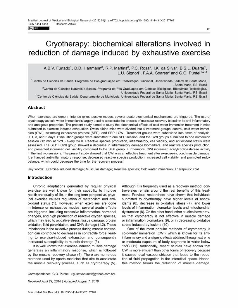
CWI were immediately put in a tank with controlled tem-perature (12°C) for 12 min following the protocol previouslydescribed by Machado et al. (15). The water depth wascontrolled so that the animal had the whole body sub-merged (except the head) without needing to swim tostay on the surface, that is, with the four paws resting onthe floor of the tank. The animals of the CWI group weresubmitted to the same protocol. Both groups repeatedthe protocol every 24 h for 5 days (Figure 1). To minimizesignificant variations in the time scale of the endogenoustemperature, after the cold-water immersion the animalswere dried with towels and then re-allocated in the cages.This procedure was done at room temperature in order notto interfere with the physiological response to immersion.
Tissue samplingThe animals were euthanized and blood was collected
by heart puncture in previously heparinized syringe. Bothgastrocnemius muscles were quickly removed, weighed,and placed on ice. Skeletal muscle tissue samples werehomogenized within 10 min in 10 volumes of cold Tris10 mM (pH 7.4) and centrifuged at 4000 g for 10 min at 4°Cto yield the low-speed supernatant fraction (S1) that wasused for different biochemical assays in all trials. Wholeblood samples were centrifuged at 1500 g for 10 min at4°C for plasma separation for biochemical analyses.
Braz J Med Biol Res | doi: 10.1590/1414-431X20187702
Cryotherapy: biochemical alterations induced by exhaustive exercise 2/8
Damage markers in skeletal muscle and plasmaOxidized dichlorofluoresceine reactive species (DCF-
RS) levels. DCF-RS levels were measured according toPerez-Severiano (19) with some modifications. Aliquotsof skeletal muscle homogenate (50 mL) were added to amedium containing Tris-HCl buffer (10 mM; pH 7.4) anddichloro-dihydro-fluorescein diacetate (1 mM). The medi-um was then incubated in the dark for 1h until fluores-cence measurement (excitation at 488nm and emission at525 nm; both slit widths used were at 1.5 nm). DCF-RSlevels were determined using a standard curve of DCFand the results were corrected by mg of protein.
Acetylcholinesterase (AChE) activity. AChE activitywas estimated in skeletal muscle by the Ellman method (21)using a plate containing acetylcholine iodide (ATC) used assubstrate and etopropazine as butyrylcholinesterase (BChE)inhibitor. Data were corrected by protein content and arereported in mmol of ATC hydrolyzed �min–1 �mL–1.
Creatine kinase (CK). CK activity was measured spec-trophotometrically in plasma samples by standard bio-logical kits (Labtest, Brazil).
Cell viability and antioxidant markers in skeletalmuscle
Measurement of methyltetrazolium (MTT) reductionlevels. Aliquots of skeletal muscle homogenate (90 mL)were added to a medium containing 1 mg/mL of MTT andwere incubated in the dark for 60 min at 37°C. Then, 900 mLof DMSO was added. Formazan levels were measuredspectrophotometrically at 570 nm and 630 nm and resultswere corrected by the protein content as proposed byMosmann (18).
Non-protein thiol (–SH) levels. Non-protein –SH levelswere determined according to the method proposed byEllman (20) with some modifications. Samples of skeletalmuscle homogenate (500 mL) were precipitated with 5%trichloroacetic acid (250 mL) and subsequently centrifugedat 1800 g for 10 min at 4°C.
The supernatant fraction (300 mL) was then added toa reaction medium containing TFK (0.5 mM, pH 7) andDTNB (20 mM). Non-protein –SH levels were measuredspectrophotometrically at 412 nm. Results were calculatedin relation to a standard curve constructed with GSH
at known concentrations and corrected by the proteincontent.
Protein quantificationThe protein content was estimated by the Bradford
method (22) using bovine serum albumin (BSA) as thestandard.
Statistical analysisGraphpad Prism 6 (USA) was used for all analyses.
Data are reported as means±SD and variations betweeninterventions are reported as mean differences (MD) and95% confidence intervals (95%CI). Significance wasassessed by two-way analysis of variance (ANOVA)followed by Tukey’s post hoc test. Statistical significancewas set at Po0.05. The main effects (effect size) weretested to reveal the size of the effect and complementP value (21) and are presented only when interactionsbetween SEP and SEP+CWI were significant.
Results
Time to exhaustionThe mean swimming time of the exhaustive protocol
was 343.3 s (±121.1).
Damage markers in skeletal muscle and plasmaDCF-RS levels in skeletal muscle tissue are reported
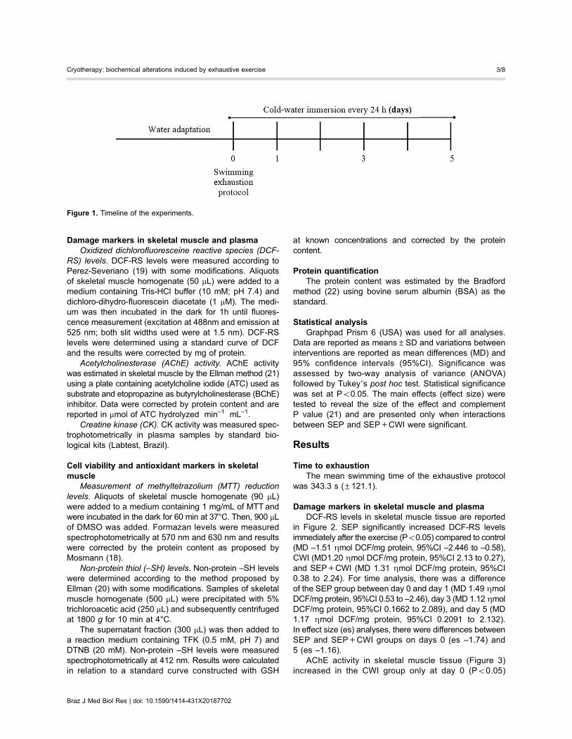
in Figure 2. SEP significantly increased DCF-RS levelsimmediately after the exercise (Po0.05) compared to control(MD –1.51 Zmol DCF/mg protein, 95%CI –2.446 to –0.58),CWI (MD1.20 Zmol DCF/mg protein, 95%CI 2.13 to 0.27),and SEP+CWI (MD 1.31 Zmol DCF/mg protein, 95%CI0.38 to 2.24). For time analysis, there was a differenceof the SEP group between day 0 and day 1 (MD 1.49 ZmolDCF/mg protein, 95%CI 0.53 to –2.46), day 3 (MD 1.12 ZmolDCF/mg protein, 95%CI 0.1662 to 2.089), and day 5 (MD1.17 Zmol DCF/mg protein, 95%CI 0.2091 to 2.132).In effect size (es) analyses, there were differences betweenSEP and SEP+CWI groups on days 0 (es –1.74) and5 (es –1.16).
AChE activity in skeletal muscle tissue (Figure 3)increased in the CWI group only at day 0 (Po0.05)
Figure 1. Timeline of the experiments.
Braz J Med Biol Res | doi: 10.1590/1414-431X20187702
Cryotherapy: biochemical alterations induced by exhaustive exercise 3/8
compared to the control group (MD –0.39� 10–3 mmol ACThydrolyzed �min–1 �mL–1, 95% CI 0.7480� 10–3 to –0.05�10–8.100–5) and the SEP group (MD 0.361�10–3 mmolACT hydrolyzed �min–1 �mL–1, 95% CI 1.25� 10–8 to0.70� 10–3) and on day 1 (Po0.001) compared to all othergroups (control, MD 0.0005188� 10–3 mmol ACT hydro-lyzed �min–1 �mL–1, 95% CI 0.86 to 0.17; SEP, MD 0.54,95%CI 0.19 to 0.88; SEP+CWI, MD 0.43� 10–3 mmolACT hydrolyzed �min–1 �mL–1, 95%CI 8.7e-005 –0.78�10–3 mmol ACT hydrolyzed �min–1 �mL–1). On time analy-sis, there were differences in CWI between days 0 and3 (MD 0.0004859, 95%CI 0.0001362–0.0008355), 0 and5 (MD 0.000417, 95%CI 6731e-005–0.0007667), 1 and 3(MD 0.0005891, 95%CI 0.0002394–0.0009388), 1 and 5(MD 0.0005203, 95%CI 0.0001706–0.0008699). In effectsize analyses, there were differences between SEP andSEP+CWI groups on all days, however, at immediate timeand on day 1, AChE increased in the SEP+CWI group (es1.70; 3.16), and on days 3 and 5 the opposite occurred,AChE was increased in the SEP group (es –0.89; –1.64).
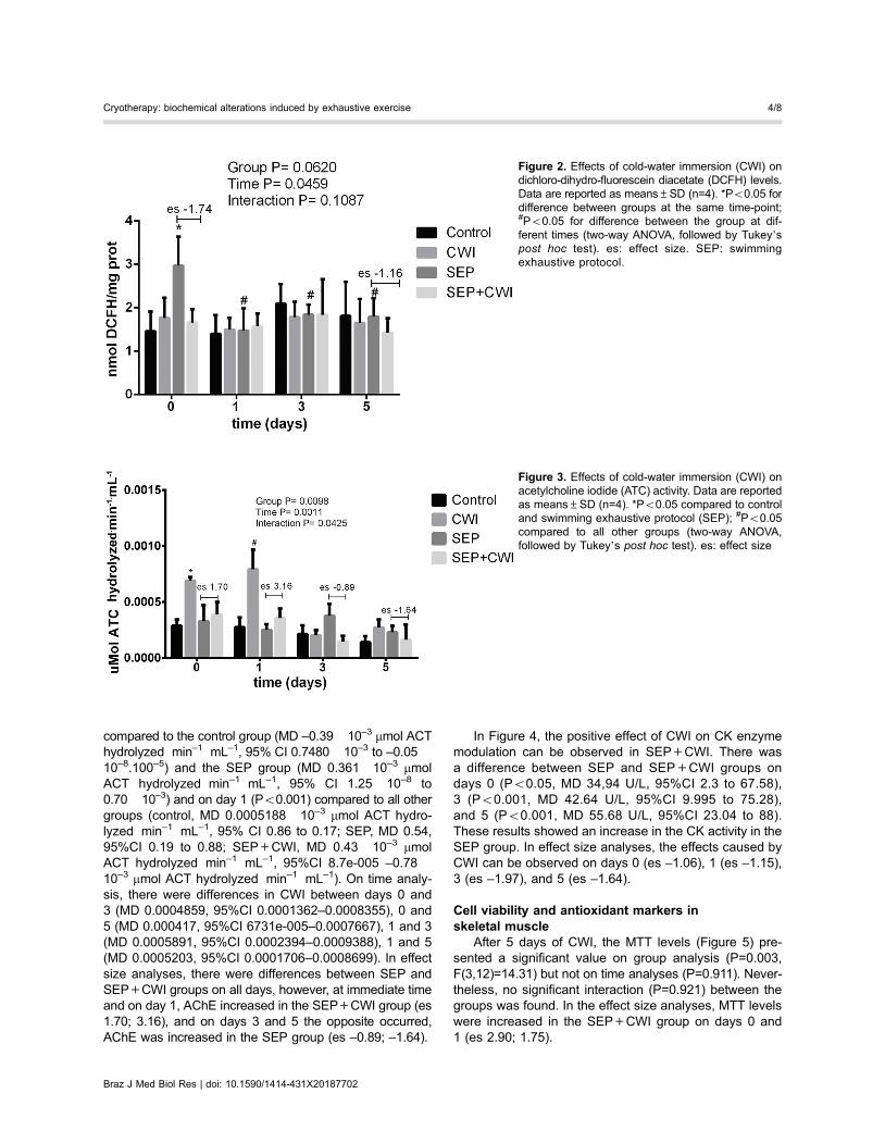
In Figure 4, the positive effect of CWI on CK enzymemodulation can be observed in SEP+CWI. There wasa difference between SEP and SEP+CWI groups ondays 0 (Po0.05, MD 34,94 U/L, 95%CI 2.3 to 67.58),3 (Po0.001, MD 42.64 U/L, 95%CI 9.995 to 75.28),and 5 (Po0.001, MD 55.68 U/L, 95%CI 23.04 to 88).These results showed an increase in the CK activity in theSEP group. In effect size analyses, the effects caused byCWI can be observed on days 0 (es –1.06), 1 (es –1.15),3 (es –1.97), and 5 (es –1.64).
Cell viability and antioxidant markers inskeletal muscle
After 5 days of CWI, the MTT levels (Figure 5) pre-sented a significant value on group analysis (P=0.003,F(3,12)=14.31) but not on time analyses (P=0.911). Never-theless, no significant interaction (P=0.921) between thegroups was found. In the effect size analyses, MTT levelswere increased in the SEP+CWI group on days 0 and1 (es 2.90; 1.75).
Figure 2. Effects of cold-water immersion (CWI) ondichloro-dihydro-fluorescein diacetate (DCFH) levels.Data are reported as means±SD (n=4). *Po0.05 fordifference between groups at the same time-point;#Po0.05 for difference between the group at dif-ferent times (two-way ANOVA, followed by Tukey’spost hoc test). es: effect size. SEP: swimmingexhaustive protocol.
Figure 3. Effects of cold-water immersion (CWI) onacetylcholine iodide (ATC) activity. Data are reportedas means±SD (n=4). *Po0.05 compared to controland swimming exhaustive protocol (SEP); #Po0.05compared to all other groups (two-way ANOVA,followed by Tukey’s post hoc test). es: effect size
Braz J Med Biol Res | doi: 10.1590/1414-431X20187702
Cryotherapy: biochemical alterations induced by exhaustive exercise 4/8
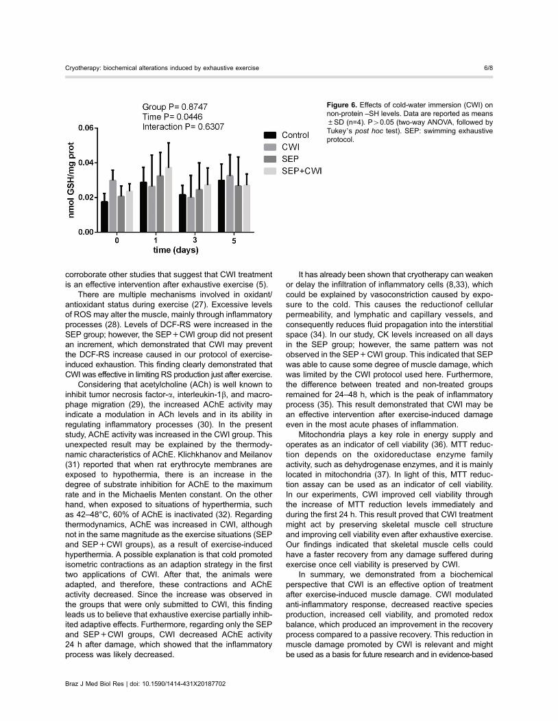
Non-protein –SH levels (Figure 6) presented a significantvalue on time (P=0.0446, F(3,36)=2.970), but not on groupanalyses (0.8747) or interaction (0.6307). When Tukey’spost hoc test was done, no significant difference was found.
Discussion
In the current study, biochemical changes inducedby CWI treatment in mice submitted to the SEP protocolwere evaluated. Our study is the first to demonstratean increase in ACHe activity in the first two sessions ofCWI treatment and a decrease afterwards, indicating amodulation of the inflammation system. To the best of ourknowledge, this is the first study to demonstrate the use ofCWI for five days consecutively after a single exhaustiveexercise session, which depicts a significant alteration inmuscle recovery.
The most popular reason for using CWI is that itinduces vasoconstriction, which leads to venous returnincrease, metabolite removal (24), and decreased muscleoxygen saturation (14), restricting the infiltration of inflam-matory cells into the muscle (25). These mechanisms pro-tect the uninjured tissue from enzymatic reactions triggeredby exercise-induced damage (26). The proposed exhaus-tive protocol was able to evoke alterations in biochemicalmarkers that clearly indicate muscle damage, such asincreased CK activity and DCF-RS production, althoughthe protocol did not lead to excessive damage, which maybe equivalent to real sportive situations. Our data demon-strated that CWI treatment after an intense exercise boutwas capable of decreasing reactive oxygen species (ROS)formation and damage, as well as increasing cellular via-bility. The results supported the hypothesis that CWI isbetter than passive recovery after exhaustive exercise and
Figure 4. Effects of cold-water immersion (CWI) oncreatine kinase (CK) activity. Data are reported asmeans±SD (n=5). *Po0.05 compared to the othergroups (two-way ANOVA, followed by Tukey’s posthoc test). es: effect size; SEP: swimming exhaustiveprotocol.
Figure 5. Effects of cold-water immersion (CWI) onMTT levels. Data are reported as means±SD (n=4).P40.05 (two-way ANOVA, followed by Tukey’s posthoc test). es: effect size; SEP: swimming exhaustiveprotocol.
Braz J Med Biol Res | doi: 10.1590/1414-431X20187702
Cryotherapy: biochemical alterations induced by exhaustive exercise 5/8
corroborate other studies that suggest that CWI treatmentis an effective intervention after exhaustive exercise (5).
There are multiple mechanisms involved in oxidant/antioxidant status during exercise (27). Excessive levelsof ROS may alter the muscle, mainly through inflammatoryprocesses (28). Levels of DCF-RS were increased in theSEP group; however, the SEP+CWI group did not presentan increment, which demonstrated that CWI may preventthe DCF-RS increase caused in our protocol of exercise-induced exhaustion. This finding clearly demonstrated thatCWI was effective in limiting RS production just after exercise.
Considering that acetylcholine (ACh) is well known toinhibit tumor necrosis factor-a, interleukin-1b, and macro-phage migration (29), the increased AChE activity mayindicate a modulation in ACh levels and in its ability inregulating inflammatory processes (30). In the presentstudy, AChE activity was increased in the CWI group. Thisunexpected result may be explained by the thermody-namic characteristics of AChE. Klichkhanov and Meilanov(31) reported that when rat erythrocyte membranes areexposed to hypothermia, there is an increase in thedegree of substrate inhibition for AChE to the maximumrate and in the Michaelis Menten constant. On the otherhand, when exposed to situations of hyperthermia, suchas 42–48°C, 60% of AChE is inactivated (32). Regardingthermodynamics, AChE was increased in CWI, althoughnot in the same magnitude as the exercise situations (SEPand SEP+CWI groups), as a result of exercise-inducedhyperthermia. A possible explanation is that cold promotedisometric contractions as an adaption strategy in the firsttwo applications of CWI. After that, the animals wereadapted, and therefore, these contractions and AChEactivity decreased. Since the increase was observed inthe groups that were only submitted to CWI, this findingleads us to believe that exhaustive exercise partially inhib-ited adaptive effects. Furthermore, regarding only the SEPand SEP+CWI groups, CWI decreased AChE activity24 h after damage, which showed that the inflammatoryprocess was likely decreased.
It has already been shown that cryotherapy can weakenor delay the infiltration of inflammatory cells (8,33), whichcould be explained by vasoconstriction caused by expo-sure to the cold. This causes the reductionof cellularpermeability, and lymphatic and capillary vessels, andconsequently reduces fluid propagation into the interstitialspace (34). In our study, CK levels increased on all daysin the SEP group; however, the same pattern was notobserved in the SEP+CWI group. This indicated that SEPwas able to cause some degree of muscle damage, whichwas limited by the CWI protocol used here. Furthermore,the difference between treated and non-treated groupsremained for 24–48 h, which is the peak of inflammatoryprocess (35). This result demonstrated that CWI may bean effective intervention after exercise-induced damageeven in the most acute phases of inflammation.
Mitochondria plays a key role in energy supply andoperates as an indicator of cell viability (36). MTT reduc-tion depends on the oxidoreductase enzyme familyactivity, such as dehydrogenase enzymes, and it is mainlylocated in mitochondria (37). In light of this, MTT reduc-tion assay can be used as an indicator of cell viability.In our experiments, CWI improved cell viability throughthe increase of MTT reduction levels immediately andduring the first 24 h. This result proved that CWI treatmentmight act by preserving skeletal muscle cell structureand improving cell viability even after exhaustive exercise.Our findings indicated that skeletal muscle cells couldhave a faster recovery from any damage suffered duringexercise once cell viability is preserved by CWI.
In summary, we demonstrated from a biochemicalperspective that CWI is an effective option of treatmentafter exercise-induced muscle damage. CWI modulatedanti-inflammatory response, decreased reactive speciesproduction, increased cell viability, and promoted redoxbalance, which produced an improvement in the recoveryprocess compared to a passive recovery. This reduction inmuscle damage promoted by CWI is relevant and mightbe used as a basis for future research and in evidence-based
Figure 6. Effects of cold-water immersion (CWI) onnon-protein –SH levels. Data are reported as means±SD (n=4). P40.05 (two-way ANOVA, followed byTukey’s post hoc test). SEP: swimming exhaustiveprotocol.
Braz J Med Biol Res | doi: 10.1590/1414-431X20187702
Cryotherapy: biochemical alterations induced by exhaustive exercise 6/8

clinical practice. The comparison of methodologies infuture studies is necessary to conclude which are the mosteffective.
Acknowledgments
The authors express their gratitude to PPGRF-UFSMand PPGBTOX- UFSM. Financial support was given byCAPES, CNPq, and FAPERGS/CNPq – PRONEM. D.D.
Hartmann received a fellowship from Coordenacão deAperfeicoamento de Pessoal de Nível Superior (CAPES/132018/2016-0). R.P. Martins received a fellowship fromConselho Nacional de Desenvolvimento Científico e Tecno-lógico (CNPq/134650/2016-6). I.K. da Silva received a fellow-ship from Fundacão de Amparo à Pesquisa do Estadodo Rio Grande do Sul (FAPERGS/05442551/16-0). Addi-tional financial support was provided by FAPERGS/CNPq -PRONEM.
References
1. McArdle WD, Katch FI, Katch VL. Exercise Physiology:energy, nutrition and human performance. 7 ed. Rio deJaneiro: Guanabara Koogan; 2011.
2. Fittipaldi S, Dimauro I, Mercatelli N, Caporossi D. Role ofexercise-induced reactive oxygen species in the modulationof heat shock protein response. Free Radic Res 2014; 48:52–70, doi: 10.3109/10715762.2013.835047.
3. Ibrahim MY, Ashour OM. Changes in nitric oxide and freeradical levels in rat gastrocnemius muscle during contractionand fatigue. Clin Exp Pharmacol Physiol 2011; 38: 791–795,doi: 10.1111/j.1440-1681.2011.05603.x.
4. Smith C, Kruger MJ, Smith RM, Myburgh KH. The inflam-matory response to skeletal muscle injury: illuminating com-plexities. Sports Med 2008; 38: 947–969, doi: 10.2165/00007256-200838110-00005.
5. Pournot H, Bieuzen F, Duffield R, Lepretre PM, Cozzolino C,Hausswirth C. Short term eVects of various water immer-sions on recovery from exhaustive intermittent exercise. EurJ Appl Physiol 2011; 111: 1287–1295, doi: 10.1007/s00421-010-1754-6.
6. Bleakley CM, Davison GW. What is the biochemical andphysiological rationale for using cold-water immersion in sportsrecovery? A systematic review. Br J Sports Med 2010; 44:179–187, doi: 10.1136/bjsm.2009.065565.
7. Mila-Kierzenkowska C, Wozniak A, Wozniak B, Drewa G,Rakowski A, Jurecka A, et al. Whole-body cryostimulation inkayaker women: a study of the effect of cryogenic temp-eratures on oxidative stress after the exercise. J Sports MedPhys Fitness 2009; 49: 201–207.
8. Puntel GO, Carvalho NR, Amaral GP, Lobato LD, SilveiraSO, Daubermann MF, et al. Therapeutic cold: An effectivekind to modulate the oxidative damage resulting of a skeletalmuscle contusion. Free Radic Res 2011; 45: 125–138,doi: 10.3109/10715762.2010.517252.
9. Rowsell GJ, Coutts AJ, Reaburn P, Hill-Haas S. Effects ofcold-water immersion on physical performance betweensuccessive matches in high-performance junior male soccerplayers. J Sports Sci 2009; 27: 565–573, doi: 10.1080/02640410802603855.
10. Silva MA, Carvalho TR, Cruz AC, Jesus LR, Silva Neto LA,Trajano ET, et al. Effect of time-dependent cryotherapy onredox balance of quadriceps injuries. Cryobiology 2016; 72:1–6, doi: 10.1016/j.cryobiol.2016.01.001.
11. Bleakley CM, Bieuzen F, Davison GW, Costello JT. Whole-body cryotherapy: empirical evidence and theoretical per-spectives. Open Access J Sports Med 2014; 5: 25–36,doi: 10.2147/OAJSM.S41655.
12. Degroot DW RP, Thompson SM, Kenefick RW. Extremitycooling for heat stress mitigation in military and occupationalsettings. J Thermal Biol 2013; 38: 305–310, doi: 10.1016/j.jtherbio.2013.03.010.
13. Roberts LA, Nosaka K, Coombes JS, Peake JM. Cold waterimmersion enhances recovery of submaximal muscle functionafter resistance exercise. Am J Physiol Regul Integr CompPhysiol 2014; 307: R998–R1008, doi: 10.1152/ajpregu.00180.2014.
14. Hohenauer E, Costello JT, Stoop R, Kung UM, Clarys P,Deliens T, et al. Cold-water or partial-body cryotherapy?Comparison of physiological responses and recovery follow-ing muscle damage. Scand J Med Sci Sports 2018; 28:1252–1262, doi: 10.1111/sms.13014.
15. Machado AF, Ferreira PH, Micheletti JK, de Almeida AC,Lemes ÍR, Vanderlei FM, et al. Can water temperature andimmersion time influence the effect of cold water immersionon muscle soreness? A systematic review and meta-analysis.Sports Med 2016; 46: 503–514, doi: 10.1007/s40279-015-0431-7.
16. Huang CC, Hsu MC, Huang WC, Yang HR, Hou CC.Triterpenoid-rich extract from Antrodia camphorata improvesphysical fatigue and exercise performance in mice. EvidBased Complement Alternat Med 2012; 2012: 364741,doi: 10.1155/2012/364741.
17. Elikov AV. Oxidative balance in rats during adaptation toswimming load. Bull Exp Biol Med 2016; 162: 180–183,doi: 10.1007/s10517-016-3570-4.
18. Mosmann T. Rapid colorimetric assay for cellular growth andsurvival: application to proliferation and cytotoxicity assays.J Immunol Methods 1983; 65: 55–63, doi: 10.1016/0022-1759(83)90303-4.
19. Perez-Severiano F, Rodriguez-Perez M, Pedraza-ChaverriJ, Maldonado PD, Medina-Campos ON, Ortiz-Plata A,et al. S-Allylcysteine, a garlic-derived antioxidant, amelio-rates quinolinic acid-induced neurotoxicity and oxidativedamage in rats. Neurochem Int 2004; 45: 1175–1183, doi:10.1016/j.neuint.2004.06.008.
20. Ellman GL. Tissue sulfhydryl groups. Arch Biochem Biophys1959; 82: 70–77, doi: 10.1016/0003-9861(59)90090-6.
21. Ellman GL, Courtney KD, Andres V Jr, Feather-Stone RM.A new and rapid colorimetric determination of acetylcholi-nesterase activity. Biochem Pharmacol 1961; 7: 88–95,doi: 10.1016/0006-2952(61)90145-9.
22. Bradford MM. A rapid and sensitive method for the quanti-tation of microgram quantities of protein utilizing the principleof protein-dye binding. Anal Biochem 1976; 72: 248–254,doi: 10.1016/0003-2697(76)90527-3.
Braz J Med Biol Res | doi: 10.1590/1414-431X20187702
Cryotherapy: biochemical alterations induced by exhaustive exercise 7/8

23. Sullivan GM, Feinn R. Using effect size-or why the p valueis not enough. J Grad Med Educ 2012; 4: 279–282, doi:10.4300/JGME-D-12-00156.1.
24. Cochrane DJ. Alternating hot and cold water immersion forathlete recovery: a review. Phys Ther Sport 2004; 5: 26–32,doi: 10.1016/j.ptsp.2003.10.002.
25. Lee H, Natsui H, Akimoto T, Yanagi K, Ohshima N, Kono I.Effects of cryotherapy after contusion using real-time intravitalmicroscopy. Med Sci Sports Exerc 2005; 37: 1093–1098, doi:10.1249/01.mss.0000169611.21671.2e.
26. Merrick MA, Jutte LS, Smith ME. Cold modalities withdifferent thermodynamic properties produce different sur-face and intramuscular temperatures. J Athl Train 2003; 38:28–33.
27. King MA, Clanton TL, Laitano O. Hyperthermia, dehydration,and osmotic stress: unconventional sources of exercise-induced reactive oxygen species. Am J Physiol Regul IntegrComp Physiol 2016; 310: R105–R114, doi: 10.1152/ajpregu.00395.2015.
28. Vollaard NB, Shearman JP, Cooper CE. Exercise-inducedoxidative stress: myths, realities and physiological relevance.Sports Med 2005; 35: 1045–1062, doi: 10.2165/00007256-200535120-00004.
29. Borovikova LV, Ivanova S, Nardi D, Zhang M, Yang H,Ombrellino M, et al. Role of vagus nerve signaling in CNI-1493-mediated suppression of acute inflammation. AutonNeurosci 2000; 85: 141–147, doi: 10.1016/S1566-0702(00)00233-2.
30. Tagliari B, Tagliari AP, Schmitz F, da Cunha AA, Dalmaz C,Wyse AT. Chronic variable stress alters inflammatory and
cholinergic parameters in hippocampus of rats. NeurochemRes 2011; 36: 487–493, doi: 10.1007/s11064-010-0367-0.
31. Klichkhanov NK, Meilanov IS. Effect of hypothermia on kineticcharacteristics of acetylcholine esterase in rat erythrocytemembranes. Bull Exp Biol Med 2004; 138: 47–49, doi:10.1007/BF02694472.
32. Edwards JA, Brimijoin S. Thermal inactivation of the molecularforms of acetylcholinesterase and butyrylcholinesterase.Biochim Biophys Acta 1983; 742: 509–516, doi: 10.1016/0167-4838(83)90268-6.
33. Singh DP, Barani Lonbani Z, Woodruff MA, Parker TJ, SteckR, Peake JM. Effects of topical icing on inflammation,angiogenesis, revascularization, and myofiber regenerationin skeletal muscle following contusion injury. Front Physiol2017; 8: 93, doi: 10.3389/fphys.2017.00093.
34. Wilcock IM, Cronin JB, Hing WA. Water immersion: doesit enhance recovery from exercise? Int J Sports PhysiolPerform 2006; 1: 195–206, doi: 10.1123/ijspp.1.3.195.
35. Borghi SM, Zarpelon AC, Pinho-Ribeiro FA, Cardoso RD,Martins-Pinge MC, Tatakihara RI, et al. Role of TNF-alpha/TNFR1 in intense acute swimming-induced delayed onsetmuscle soreness in mice. Physiol Behav 2014; 128: 277–287, doi: 10.1016/j.physbeh.2014.01.023.
36. Echtay KS. Mitochondrial uncoupling proteins–what istheir physiological role? Free Radic Biol Med 2007; 43:1351–1371, doi: 10.1016/j.freeradbiomed.2007.08.011.
37. Bernas T, Dobrucki J. Mitochondrial and non-mitochondrialreduction of MTT: interaction of MTT with TMRE, JC-1, andNAO mitochondrial fluorescent probes. Cytometry 2002; 47:236–242, doi: 10.1002/cyto.10080.
Braz J Med Biol Res | doi: 10.1590/1414-431X20187702
Cryotherapy: biochemical alterations induced by exhaustive exercise 8/8