cryptococcal screening program training for laboratory workers
TRANSCRIPT
Cryptococcal Screening Program
Training Modules for Laboratory Workers
Version 5.6.13
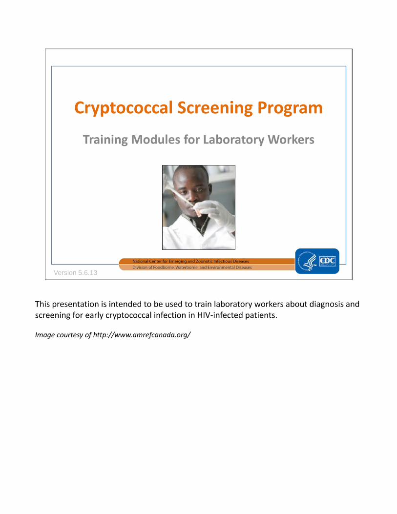
This presentation is intended to be used to train laboratory workers about diagnosis and screening for early cryptococcal infection in HIV-infected patients. Image courtesy of http://www.amrefcanada.org/

Clinical case scenario
• 27-year-old male with AIDS• CD4 count: 34 cells/mm3
• Presents to clinic to start anti-retrovirals• Complains of mild headache but is otherwise
well, started on anti-retrovirals• Two months later develops low grade fever,
nausea, confusion
First, we will describe a brief clinical case scenario to illustrate the public health importance of preventing cryptococcal infection. Imagine that a 27-year-old man with AIDS and a CD4 count of 34 comes to a clinic to start anti-retroviral therapy. He says he has a mild headache but no other symptoms. He begins taking anti-retroviral therapy; however, two months later, he starts feeling worse, and develops a fever, nausea, and confusion. You will see in a few slides that this is important because this is a common scenario among patients who develop cryptococcal meningitis.

Learning Objectives• Upon completion of this activity, participants
should be able to:– Understand the public health burden and high
mortality from cryptococcal meningitis– Identify the various diagnostic methods for
identifying Cryptococcus spp.– Describe the steps to perform latex agglutination
test– Describe the steps to perform a lateral flow assay– Understand the rationale for screening
The goals of this presentation are: to help you understand the large public health burden and high death rate from cryptococcal meningitis, to describe the different methods that are used to diagnose cryptococcal infection and describe the general steps to perform tests to diagnose cryptococcal disease, and to help you understand the rationale for screening for early cryptococcal infection in HIV-infected patients.
OverviewModule 1: What is Cryptococcus?Module 2: Signs & symptoms of cryptococcosisModule 3: Diagnosing cryptococcal disease
Module 3a: Diagnosis using latex agglutinationModule 3b: Diagnosis using lateral flow assay
Module 4: Preventing cryptococcal meningitisModule 5: Cryptococcal screening decision-making guide Module 6: Your role as a laboratory worker
This presentation contains six modules: “What is Cryptococcus?”, “signs and symptoms of cryptococcosis,” “diagnosing cryptococcal disease,” “preventing cryptococcal meningitis,” “cryptococcal screening decision-making guide,” and “your role as a laboratory worker.” NOTE: If you work in a setting where latex agglutination is not available or is not performed, you may skip module 3a. If you work in a setting where lateral flow assay is not available or is not performed, you may skip module 3b.

What is Cryptococcus?
• Fungus found in soil contaminated by bird droppings
• Fungal spores are inhaled from environment
• The fungus cannot spread from person to person
Cryptococcus spp.stained with India Ink
Module 1
Cryptococcus is a type of fungus that lives in soil, especially soil that is contaminated with large amounts of bird droppings. Some people inhale the spores from the environment and never get sick, but in people with weak immune systems, the fungus can cause an infection. The only way a person can get sick from this fungus is by directly inhaling the spores from the environment – the infection cannot spread from person to person. On the right is a picture of Cryptococcus stained with India Ink.

Cryptococcal infection
• Fungus can cause acute lung infection or no symptoms at all
• Incubation period unknown, may be dormant for many years
• Reactivation in immunosuppressed persons (HIV/AIDS, especially CD4 <100)
• Meningitis is the most common presentation
Module 1
Cryptococcal infection is associated with a range of illness. In some people, the fungus causes a lung infection similar to tuberculosis, or it can cause no symptoms at all. The incubation period is not known, but it is thought that the infection can remain dormant in the body for many years. In immunosuppressed people (people with weak immune systems), particularly HIV-infected people with CD4 counts under 100, the infection can reactivate and spread throughout the body. When this happens, the infection usually presents as meningitis, which is a swelling of the meninges, the tissues that protect the brain and spinal cord.

Death from cryptococcal meningitis
• Cryptococcal meningitis is a common cause of death among HIV/AIDS patients
• High mortality (30%-70% in sub-Saharan Africa) despite antiretroviral treatment (ART) and antifungal therapy
• In some areas of the world, it is estimated to cause more deaths than tuberculosis (TB)
Module 1
Cryptococcal meningitis is a common cause of death in HIV/AIDS patients. In low-resource settings, such as sub-Saharan Africa, one-third to over one-half of all patients with cryptococcal meningitis will die from it. There are several reasons that the death rate is so high in these areas of the world: first, patients often present with disease that is too advanced for treatment to be effective. Second, cryptococcal meningitis can occur even when patients with advanced HIV begin antiretroviral treatment. Third, the medications that are needed to treat cryptococcal meningitis are often expensive or not widely available. In some areas of the world, cryptococcal meningitis is estimated to cause more deaths than tuberculosis (TB).

Global burden of HIV-related cryptococcal meningitis
Park BJ, et al. AIDS 2009;23:525-30.
~1 million new cases per year~ 625,000 deaths per year
Module 1
This slide shows the global burden of HIV-related cryptococcal meningitis . Worldwide, there are approximately 1 million new cases and 625,000 deaths from cryptococcal meningitis each year. As you can see, sub-Saharan Africa accounts for the majority of cases (>70%) estimated annually worldwide, followed by East, South, and Southeast Asia.

Causes of death in sub-Saharan Africa (excluding HIV/AIDS)
Park BJ, et al. AIDS 2009;23:525-30. Module 1
This figure shows the leading causes of death in sub-Saharan Africa, not including HIV/AIDS, and deaths due to Cryptococcus is shown in orange.

Signs and symptoms of cryptococcal meningitis
• Headache• Fever• Change in mental status (ranging from confusion
to lethargy to coma)• Blurry vision (and other cranial nerve deficits)• Neck stiffness• Sensitivity to light• Nausea and vomiting• Seizures• Papilledema
Module 2
The most common signs and symptoms of cryptococcal meningitis are headache, fever, mental status changes, blurry vision, neck stiffness, sensitivity to light, nausea and vomiting, seizures, and papilledema (swelling of the optic nerve).

Clinical course of cryptococcal disease
Module 2
No symptoms or symptoms of lung infection
• Shortness of breath• Cough• Fever
Meningitis• Headache• Confusion or coma• Neck stiffness• Fever• Nausea, vomiting• Sensitivity to light
Infection spreads
Cryptococcal infection occurs after a person inhales the fungus from the environment, shown in the drawing on the left. In the lungs, the infection can cause shortness of breath, cough, and fever, or, in some people, it causes no symptoms at all. The infection can then spread from the lungs to other parts of the body, which typically presents as meningitis, shown in the drawing on the right.

Current diagnostic methods
• India Ink microscopy (CSF)• Culture (CSF or blood)• Antigen detection
– Latex agglutination (LA) (CSF or serum)– Enzyme immunoassay (EIA) (CSF or serum)– Lateral flow assay (LFA) (CSF or serum)
Module 3
There are three categories of methods that can be used to diagnose cryptococcal meningitis: India Ink microscopy, which can be used on cerebrospinal fluid (CSF); culture, which can be used on CSF or blood; and antigen detection. There are several methods to detect cryptococcal antigen in CSF or serum: latex agglutination (LA), enzyme immunoassay (EIA), and lateral flow assay (LFA). We will go into detail about each of these methods in the following slides.

Laboratory DiagnosisIndia Ink• Rapid diagnosis• “Halo”• 30-80%
sensitivity• CSF
MICROSCOPY1
Culture• Bird seed agar• Confirmatory test• CSF, blood
CULTURE2 SEROLOGY1 IMMUNO-CHROMATOGRAPHY3
Lateral Flow Assay• >95% sensitivity• CSF, Serum• Early diagnosis in
asymptomatic HIV + patients
1Heitman J, et al. Cryptococcus. Washington DC, USA: ASM Press, 2011. 2DoctorFungus.org, 3 CDC, unpublished data Module 3
Latex Agglutination and ELISA test
• >90% sensitivity• CSF, Serum• Early diagnosis in
asymptomaticHIV + patients
This slide gives an overview of how the different methods available to diagnose cryptococcal infection compare to each other. The first method, India Ink, shown in the first column, is a stain that can be performed quickly to visualize Cryptococcus cells under the microscope. It can be used on CSF; however, the sensitivity is often low. Culture, shown in the second column, is the gold standard for diagnosing cryptococcal infection and is typically performed on CSF or blood. Latex agglutination, enzyme immunoassay, and lateral flow assay, shown in the third and fourth columns, are all types of antigen detection tests. These tests can be used on CSF or serum and can be used for diagnosis of early, asymptomatic cryptococcal infection in HIV-infected patients. Latex agglutination and enzyme immunoassay are over 90% sensitive, while lateral flow assay is over 95% sensitive.

India Ink Microscopy• Biologic principle
– Cryptococcus yeast cell surrounded by a characteristic polysaccharide capsule
– Visualized microscopically, India Ink stains the background, revealing the extra cellular capsule
• Specimen type– Usually cerebrospinal fluid (CSF)
• Diagnosis– Pros: rapid– Cons: low sensitivity, technical subjectivity
Module 3
C. neoformans: India Ink preparation
cellcapsule
Cryptococcal yeast cells (indicated by the red arrow in the picture on the right) are surrounded by a characteristic polysaccharide capsule (indicated by the blue arrow). India Ink is a stain that reveals this extracellular capsule when visualized under the microscope. India Ink is usually performed on CSF to diagnose cryptococcal meningitis. The benefit of this test is that it can be performed quickly; however, the drawback is that it has low sensitivity, which can lead to missed diagnoses. Image courtesy of University of South Carolina School of Medicine - Dr Arthur DiSalvo’s lecture on Mycology

Culture
• Biologic principle – Isolation of Cryptococcus from specimen– Gold standard
• Specimen type– Blood or CSF
• Diagnosis– Pro: confirmatory testing– Cons: requires extensive laboratory infrastructure;
long incubation time; low sensitivity
Module 3
C. neoformans on a culture plate
Culture is the gold standard for diagnosing cryptococcal infection. It is typically performed on blood or CSF. The benefit of culture is that it gives a definitive diagnosis; however, there are several drawbacks: it requires extensive laboratory infrastructure, the fungus Cryptococcus has a long incubation time (at least 2 days, possibly weeks), and culture has low sensitivity.

Latex Agglutination (LA)• Biologic principle
– Anti-cryptococcal antibody-coated latex particles agglutinate with specimens containing cryptococcal antigen (CrAg)
• Specimen type– Serum or CSF– Process time: 20 min – 45 min
• Diagnosis– Pros: sensitive (>90%)– Cons: expensive, requires extensive laboratory
infrastructureModule 3a
NOTE: The next few slides will describe latex agglutination, a type of antigen detection test. If you are in a setting where latex agglutination is not available or not performed, you may skip the rest of module 3a. The latex agglutination test uses latex particles which are coated with anti-cryptococcal antibody. A visible reaction occurs when these particles come in contact with a patient sample that contains cryptococcal antigen. LA can be used on serum or CSF, and takes 20 to 45 minutes to perform. The major benefit of LA is that it is over 90% sensitive. However, it can be expensive and requires extensive laboratory infrastructure, including refrigeration. Image courtesy of Meridian Bioscience Inc.

LA Sample Pre-treatment
• CSF– 100°C for 5 min
• Serum1. Add serum to Pronase (reduces non-specific
interference) 2. Incubate serum/Pronase solution at 56°C for 15-30
minutes depending on manufacturer3. Terminate enzymatic digestion: add 1 drop of Pronase
inhibitor or boil the solution for 5 minutes
Module 3a
This slide describes how, in general, samples are pre-treated for latex agglutination after preparing the reagents (positive control, negative control, and pronase) according to the manufacturer’s instructions. Sample and reagent quantities and incubation times vary between different LA manufacturers, so you should refer to the specific instructions on the package insert when performing the test. For CSF, after centrifuging the sample and placing it into a sterile container, place the sample in a boiling water bath for 5 minutes, and then allow it to cool for 3 to 4 minutes before testing. For serum, add the sample to the prepared pronase, which is a reagent that reduces non-specific interference. Then incubate the serum/pronase solution at 56 degrees Celcius for 15 to 30 minutes depending on the manufacturer’s instructions. To terminate the reaction, either add a drop of Pronase inhibitor or boil the solution for 5 minutes depending on the manufacturer’s instructions.

LA Step by Step Procedure4. Add 25 µl of the following onto separate
rings of ring slide• Cryptococcus Antigen Positive Control• Cryptococcus Antigen Negative Control• Each treated serum or CSF specimen
5. Add 25 µl of Cryptococcal Latex to each ring. Using separate applicator sticks, thoroughly mix the contents of each ring
6. Rotate by hand or on a rotator set to 100 rpm (+/- 25) for 5 min at room temperature
7. Read and record the reactions immediately
Latex-Cryptococcus antigen detection system (IMMY) Module 3a
Ring slide for Latex Agglutination test
To perform the LA test, add 25 microliters of the pre-treated specimen and the positive and negative controls to the appropriate rings of the ring slide, using a new pipette tip for each reagent and specimen. Add 25 microliters of cryptococcal latex to each ring, and use a separate applicator stick to thoroughly mix the contents of each ring. Rotate the slide by hand or place it on a rotator set to approximately 100 rpm for 5 minutes. Read and record the reactions immediately. Image courtesy of IMMY Diagnostics

LA Interpretation of Results
Module 3a
Negative 1+ 2+ 3+ 4+
This slide shows how the results of a latex agglutination test are interpreted, on a scale ranging from negative to 4+.
• Negative (-), on the far left, is a homogeneous suspension of particles with no visible clumping.
• One plus (1+) is fine granulation against a milky background. • Two plus (2+) is small but definite clumps against a slightly cloudy background. • Three plus (3+) is large and small clumps against clear background. • Four plus (4+) is large clumps against a very clear background.
Depending on the test manufacturer, specimens that are positive at 1+ or 2+ should be titrated, and the results reported as the highest dilution showing a positive reaction.
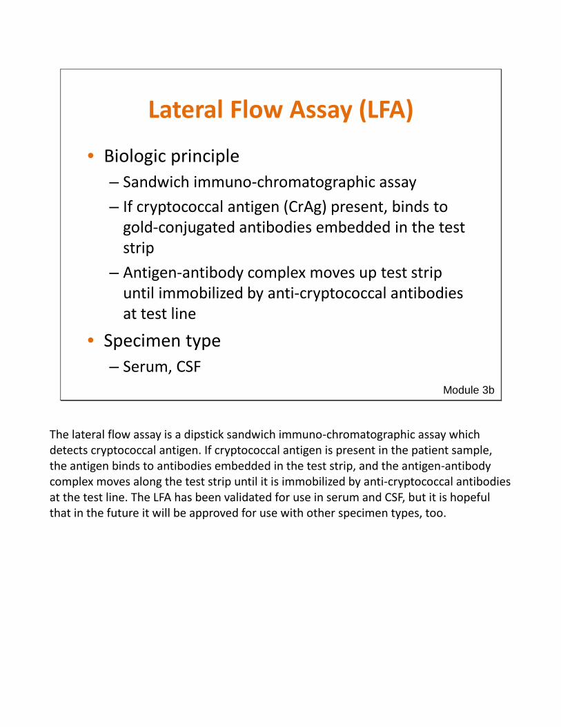
Lateral Flow Assay (LFA)
• Biologic principle – Sandwich immuno-chromatographic assay– If cryptococcal antigen (CrAg) present, binds to
gold-conjugated antibodies embedded in the test strip
– Antigen-antibody complex moves up test strip until immobilized by anti-cryptococcal antibodies at test line
• Specimen type– Serum, CSF
Module 3b
The lateral flow assay is a dipstick sandwich immuno-chromatographic assay which detects cryptococcal antigen. If cryptococcal antigen is present in the patient sample, the antigen binds to antibodies embedded in the test strip, and the antigen-antibody complex moves along the test strip until it is immobilized by anti-cryptococcal antibodies at the test line. The LFA has been validated for use in serum and CSF, but it is hopeful that in the future it will be approved for use with other specimen types, too.
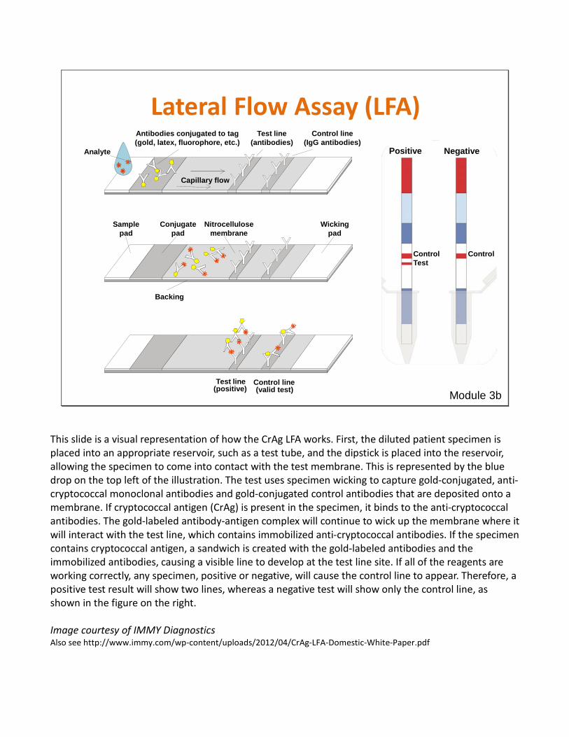
Lateral Flow Assay (LFA)Analyte
Antibodies conjugated to tag (gold, latex, fluorophore, etc.)
Test line (antibodies)
Control line(IgG antibodies)
Sample pad
Conjugate pad
Nitrocellulose membrane
Wicking pad
Test line(positive)
Control line(valid test)
Backing
Capillary flow
Module 3b
Positive Negative
ControlTest
Control
This slide is a visual representation of how the CrAg LFA works. First, the diluted patient specimen is placed into an appropriate reservoir, such as a test tube, and the dipstick is placed into the reservoir, allowing the specimen to come into contact with the test membrane. This is represented by the blue drop on the top left of the illustration. The test uses specimen wicking to capture gold-conjugated, anti-cryptococcal monoclonal antibodies and gold-conjugated control antibodies that are deposited onto a membrane. If cryptococcal antigen (CrAg) is present in the specimen, it binds to the anti-cryptococcal antibodies. The gold-labeled antibody-antigen complex will continue to wick up the membrane where it will interact with the test line, which contains immobilized anti-cryptococcal antibodies. If the specimen contains cryptococcal antigen, a sandwich is created with the gold-labeled antibodies and the immobilized antibodies, causing a visible line to develop at the test line site. If all of the reagents are working correctly, any specimen, positive or negative, will cause the control line to appear. Therefore, a positive test result will show two lines, whereas a negative test will show only the control line, as shown in the figure on the right. Image courtesy of IMMY Diagnostics Also see http://www.immy.com/wp-content/uploads/2012/04/CrAg-LFA-Domestic-White-Paper.pdf
Lateral Flow Assay (LFA)
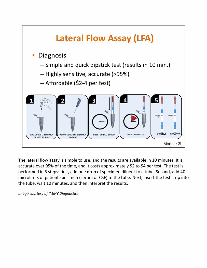
• Diagnosis– Simple and quick dipstick test (results in 10 min.)– Highly sensitive, accurate (>95%)– Affordable ($2-4 per test)
Module 3b
The lateral flow assay is simple to use, and the results are available in 10 minutes. It is accurate over 95% of the time, and it costs approximately $2 to $4 per test. The test is performed in 5 steps: first, add one drop of specimen diluent to a tube. Second, add 40 microliters of patient specimen (serum or CSF) to the tube. Next, insert the test strip into the tube, wait 10 minutes, and then interpret the results. Image courtesy of IMMY Diagnostics

CrAg LFA Lateral Flow Aassay
CrAg LALatex Agglutination
Affordable $2 - $4/Test $4/Test
Sensitive High Sensitivity for All 4 Serotypes
Slight weakness on Serotype C
Specific Very High Very High
User Friendly Low Training Threshold Requires Training, Subjective Reading
Rapid/Robust 10 minutes 35 minutes
Equipment-Free Pipettor or Capillary Tube Heat Block or Water Bath
DeliveredLightweight and Compact
Room Temperature StorageHumidity-Resistant Packaging
Bulky, Multiple Components
Refrigerated StorageCardboard Packaging
LFA vs. Latex Agglutination
Immy Diagnostics Module 3
This slide shows how various aspects of the lateral flow assay and the latex agglutination test compare. World Health Organization guidelines recommend that diagnostic tests in developing countries fit the ASSURED criteria: the test should be affordable, sensitive, specific, user-friendly, rapid/robust, equipment-free, and deliverable to end users. The CrAg lateral flow assay is more affordable and more sensitive than latex agglutination. Both tests are very specific, but the LFA is more user-friendly than LA, can be performed more quickly, requires less equipment, and does not require refrigeration.

Clinical case scenario revisited• 27-year-old male with AIDS
– CD4 34 cells/mm3
• Presents to clinic to start anti-retrovirals– Complains of mild headache, but is otherwise
well, so is started on anti-retrovirals– Two months later develops low grade fever,
nausea, confusion• Diagnosed with cryptococcal meningitis• Admitted to hospital for Amphotericin B
therapy, but subsequently diesCould this have been prevented?
Module 3
Returning to the clinical case scenario from the beginning of the presentation, you can now see that the patient had some of the typical signs and symptoms of cryptococcal meningitis. He was diagnosed with cryptococcal meningitis and admitted to hospital to receive amphotericin B, an intravenous antifungal medication, but subsequently died. Could this have been prevented, and how?
Why is preventing cryptococcal meningitis important?
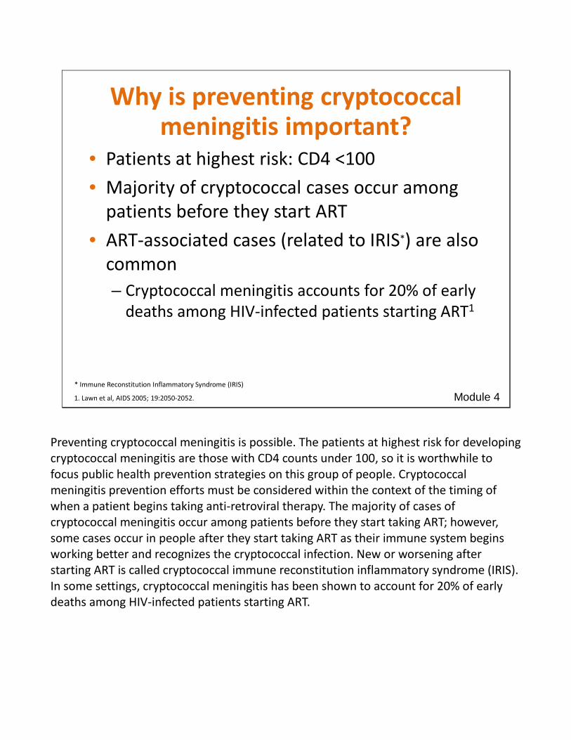
• Patients at highest risk: CD4 <100• Majority of cryptococcal cases occur among
patients before they start ART• ART-associated cases (related to IRIS*) are also
common– Cryptococcal meningitis accounts for 20% of early
deaths among HIV-infected patients starting ART1
1. Lawn et al, AIDS 2005; 19:2050-2052. Module 4* Immune Reconstitution Inflammatory Syndrome (IRIS)
Preventing cryptococcal meningitis is possible. The patients at highest risk for developing cryptococcal meningitis are those with CD4 counts under 100, so it is worthwhile to focus public health prevention strategies on this group of people. Cryptococcal meningitis prevention efforts must be considered within the context of the timing of when a patient begins taking anti-retroviral therapy. The majority of cases of cryptococcal meningitis occur among patients before they start taking ART; however, some cases occur in people after they start taking ART as their immune system begins working better and recognizes the cryptococcal infection. New or worsening after starting ART is called cryptococcal immune reconstitution inflammatory syndrome (IRIS). In some settings, cryptococcal meningitis has been shown to account for 20% of early deaths among HIV-infected patients starting ART.

Primary prophylaxis
• Treatment of all HIV-infected patients with low CD4 with low-dose fluconazole (200 mg/day) to prevent cryptococcal infection
• Limitations– Limited improvement in mortality– Cost– Concern for widespread fluconazole resistance– Drug toxicity– Interaction with TB and ART drugs– Use in women of child-bearing age
Module 4
There are two main strategies that could be used to prevent cryptococcal meningitis: prophylaxis and screening. We will describe primary prophylaxis on this slide, and then describe screening in the following slides. In a prophylaxis-based approach, all HIV/AIDS patients with a CD4 count under 100 would receive oral fluconazole; however, there is mixed evidence about whether this improves survival. In addition, this strategy could be quite expensive and could eventually contribute to fluconazole resistance. There is also concern about the effects that primary prophylaxis for cryptococcal infection would have on patients, including potential drug toxicity and interaction with other medications such as TB medications and ART. Fluconazole can also be dangerous for pregnant women.

Cryptococcal Antigen (CrAg)• Antigen test can detect CrAg in serum a median 22 days
(range 5-234) before symptoms of meningitis develop1
• Highly predictive of who is at risk for developing cryptococcal meningitis
• Possible to identify early cryptococcal disease, prevent progression to meningitis through early treatment
1. French et al., AIDS 2002 Module 4
Infection spreads+Serum CrAg but no
symptoms
Meningitis
As you learned in previous slides, there are several laboratory methods that can be used to detect cryptococcal antigen (CrAg), an indicator of infection. Most importantly, CrAg can be detected in serum a median of 22 days before symptoms of meningitis develop. The presence of CrAg in the serum is highly predictive of who will develop meningitis. This means that there is a window of time where cryptococcal disease can be identified early and then treated in order to prevent the early, asymptomatic infection from progressing to meningitis.

Cryptococcal screening1. Identify patients at risk (CD4 <100) 2. Test for cryptococcal antigenemia before
symptom onset3. Treat with oral fluconazole4. Prevent cryptococcal meningitis deaths
Module 4
Treatment+Serum CrAg but no
symptoms
Meningitis
1. French et al., AIDS 2002
In a screening strategy to prevent cryptococcal meningitis, HIV-infected patients with CD4 counts under 100 would receive an antigen test to screen for early cryptococcal infection before symptoms develop. Then, only the patients who test positive for early disease would get treatment with fluconazole, which can prevent the infection from developing into meningitis.
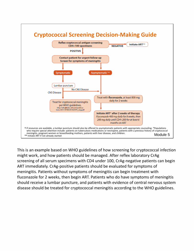
† If resources are available, a lumbar puncture should also be offered to asymptomatic patients with appropriate counseling. *Populations who require special attention include: patients on tuberculosis medications or nevirapine, patients with a previous history of cryptococcal meningitis, pregnant women or breastfeeding mothers, patients with liver disease, and children.
** Initiate ART if not already started
Cryptococcal Screening Decision-Making Guide
Module 5
This is an example based on WHO guidelines of how screening for cryptococcal infection might work, and how patients should be managed. After reflex laboratory CrAg screening of all serum specimens with CD4 under 100, CrAg-negative patients can begin ART immediately. CrAg-positive patients should be evaluated for symptoms of meningitis. Patients without symptoms of meningitis can begin treatment with fluconazole for 2 weeks, then begin ART. Patients who do have symptoms of meningitis should receive a lumbar puncture, and patients with evidence of central nervous system disease should be treated for cryptococcal meningitis according to the WHO guidelines.

What you can do as a laboratory worker
• Know the rationale for cryptococcal screening in HIV/AIDS patients
• Know how to perform latex agglutination and lateral flow assays
• Educate other laboratory workers about cryptococcal diagnostic methods and the rationale for screening
Module 6
As a laboratory worker, you can help reduce the public health burden of cryptococcal meningitis by doing three things: first, by knowing the rationale for targeted cryptococcal screening in HIV/AIDS patients. Second, by knowing how to perform the latex agglutination test and the lateral flow assay for cryptococcal infection, and third, by helping to educate other laboratory workers about these diagnostic methods and about cryptococcal screening in general.

For more information on these diagnostic tests and ordering, please contact:
Manufacturer Diagnostic test
Reference No
Telephone Website
Meridian Bioscience LA 140100 +1 800-543-
1980 www.meridianbioscience.com
Immy DiagnosticsLA
LFA
CR1004
CR2003
+1 800-654-3639 www.immy.com
Fumouze Diagnostics LA 501027 +33 149 68 42
65www.fumouze.com/ordering online: www.diagdirect.com/
Thermo scientific LA R30851501 +1 800-255-6730 www.thermoscientific.com

Studies on LFA performance data, 2011-2012
Table reference: Vijayan T, Chiller T, Klausner JD. Sensitivity and specificity of a new cryptococcal antigen lateral flow assay in serum and cerebrospinal fluid. Medical Laboratory Observer 2013; accessed online April 16, 2013. http://www.mlo-online.com/articles/201303/sensitivity-and-specificity-of-a-new-cryptococcal-antigen-lateral-flow-assay-in-serum-and-cerebrospinal-fluid.php

For more information, please contact the Centers for Disease Control and Prevention
1600 Clifton Road NE, Atlanta, GA 30333Telephone: 1-800-CDC-INFO (232-4636)/TTY: 1-888-232-6348
E-mail: [email protected] Web: http://www.cdc.gov
The findings and conclusions in this report are those of the authors and do not necessarily represent the official position of the Centers for Disease Control and Prevention.
These slides were developed in collaboration with Nicky Longley, University of Cape Town, St Georges Hospital London, and the Wellcome Trust