cryptococcosis presenting as cerebrovascular disease
TRANSCRIPT
Review began 11/03/2021 Review ended 11/07/2021 Published 11/10/2021
© Copyright 2021Tarhan et al. This is an open access articledistributed under the terms of the CreativeCommons Attribution License CC-BY 4.0.,which permits unrestricted use, distribution,and reproduction in any medium, providedthe original author and source are credited.
Cryptococcosis Presenting as CerebrovascularDiseaseBedirhan Tarhan , Yusuf Mehkri , Justin De Prey , Calvin Hu , Ibrahim S. Tuna , Hans Shuhaiber
1. Pediatric Neurology, University of Florida, Gainesville, USA 2. Neurosurgery, University of Florida, Gainesville, USA3. Neurology, University of Florida, Gainesville, USA 4. Radiology, University of Florida, Gainesville, USA
Corresponding author: Yusuf Mehkri, [email protected]
AbstractInfection plays a complex role in cerebrovascular disease and is believed to have both direct and indirectmechanisms on stroke pathogenesis. if not diagnosed and treated promptly, this may have devastatingconsequences. Management of infection-related strokes focuses on the treatment of the underlyinginfection with appropriate antimicrobial drugs and the prevention of medical complications. This can lead todevastating neurological deficits. We present two cases of cryptococcal meningoencephalitis that presentedwith an atypical cerebral infarction. A 55-year-old male with a history of unknown autoimmune diseasepresented with acute onset cognitive changes and no stroke-like symptoms. A 35-year-old male with nohistory of autoimmune disease or other existing immunodeficiency presented with breakthrough seizure along with stroke-like symptoms. Both patients developed multiple cerebral infarcts in multiple vascularterritories, with histologic and radiologic findings consistent with a central nervous system cryptococcosis.They were subsequently diagnosed with cryptococcal meningoencephalitis and started on the appropriateanti-fungal regimen with amphotericin B and flucytosine. Prior to discharge to an inpatient rehabilitationfacility, both patients were notably improved and near their neurologic baseline. It is important tounderstand the pathogenesis of cryptococcal infection in the central nervous system because it produces awide variety of clinico-radiographic features that can be overlooked. Clinicians should keep infection-mediated cerebral infarcts in mind, regardless of risk factors, in order to expedite antimicrobial therapy andminimize adverse events.
Categories: Neurology, Infectious DiseaseKeywords: cerebrovascular disease, stroke, central nervous system infection, cryptococcosis, cerebral infarction
IntroductionStroke is a leading cause of death and disability worldwide. Infection plays a complex role in cerebrovasculardisease and is believed to have both direct and indirect mechanisms on stroke pathogenesis. Given thecomplex mechanisms of infarction related to infection, it can be difficult for clinicians to appropriatelyattribute stroke to an underlying infection. Management of infection-related strokes focuses on treatment ofthe underlying infection with appropriate antimicrobial drugs and prevention of medical complications. Inthe appropriate clinical context, it is essential that health care providers consider infection-mediated strokeas delay in diagnosis and treatment can lead to poor outcomes with lasting residual neurologic deficits.Here, we present two cases of cryptococcal meningoencephalitis that presented with an atypical cerebralinfarction. Histologic and radiologic findings provide clues with regards to underlying pathogenesis.
Case PresentationCase 1A 55-year-old male with a history of type 1 diabetes mellitus (T1DM) and unspecified autoimmune diseasewho presented with acute onset of confusion as well as concrete visual hallucinations and behavioralchange. There were no reports of any headache, fever, or stroke-like symptoms. His only outpatientmedications were insulin and low-dose steroids.
The patient was initially admitted to an outside hospital where magnetic resonance imaging (MRI) of thebrain revealed multifocal areas of restricted diffusion with areas of corresponding T2 hyperintensities onfluid-attenuated inversion recovery (FLAIR) sequences (Figure 1). There was a concern for stroke in multiplevascular territories with concern for vasculitis. Initial workup was unremarkable, and the patient was startedon methylprednisolone for presumed primary central nervous system (CNS) vasculitis. He was transferred toour institution for further management by the Neurology service.
1 2 3 3 4 3
Open Access CaseReport DOI: 10.7759/cureus.19442
How to cite this articleTarhan B, Mehkri Y, De Prey J, et al. (November 10, 2021) Cryptococcosis Presenting as Cerebrovascular Disease. Cureus 13(11): e19442. DOI10.7759/cureus.19442

FIGURE 1: Axial DWI (A), ADC (B) and FLAIR (C) images demonstratingmultifocal areas of restricted diffusion (arrows in A and B) in differentvascular territories involving the left basal ganglia, bilateral medialfrontal lobes, posterior insula, left superior cerebellar peduncle andright cerebellum, with corresponding increased FLAIR signal in someareas (arrows in C).DWI: diffusion-weighted imaging; ADC: apparent diffusion coefficient; FLAIR: fluid-attenuated inversion recovery.
His initial neurologic exam was notable for encephalopathy, manifesting as inattention, disorientation toplace and time, and stupor. He was only able to follow simple appendicular commands. Cranial nerve examrevealed left lower facial droop. He had full strength in bilateral upper extremities and 4/5 strength inbilateral lower extremities. Initial differential diagnosis included autoimmune vasculopathies, primary CNSvasculitis, and infectious meningoencephalitis given his mental status changes, reported visualhallucinations, and multifocal strokes.
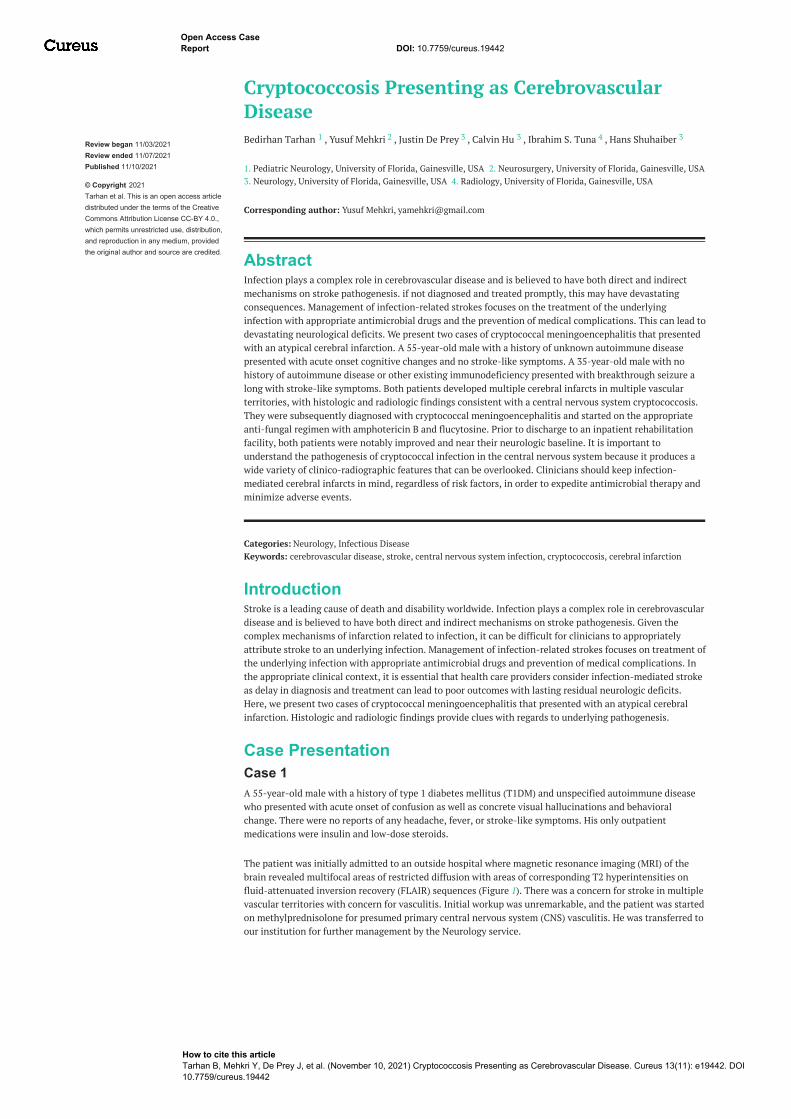
Steroids were initially held on admission to our institution until further workup could be performed.Extensive rheumatologic labs were ordered, and only rheumatoid factor and anti-CCP were found to bemildly elevated. A contrast-enhanced MRI of the brain demonstrated evolving areas of restricted diffusionwith multifocal new areas of restricted diffusion in multiple vascular territories (Figure 2). There was alsoincomplete suppression of CSF signal on FLAIR with multiple areas of abnormal leptomeningealenhancement, suggestive of a superimposed inflammatory process affecting the meninges (Figures 2, 3). Inaddition, there was abnormal vessel wall thickening and enhancement, particularly involving theintracranial carotid arteries as well as anterior cerebral arteries (ACA) and middle cerebral arteries (MCA)(Figure 3). Computed tomography angiography (CTA) of the head also demonstrated areas of vesselirregularity and multifocal areas of narrowing, particularly involving the ACA (Figure 4). On further reviewof the initial lab work and neuroimaging, it was felt that a primary CNS vasculitis was unlikely. Rather,findings were more suspicious for a meningoencephalitis complicated by acute stroke.
2021 Tarhan et al. Cureus 13(11): e19442. DOI 10.7759/cureus.19442 2 of 9
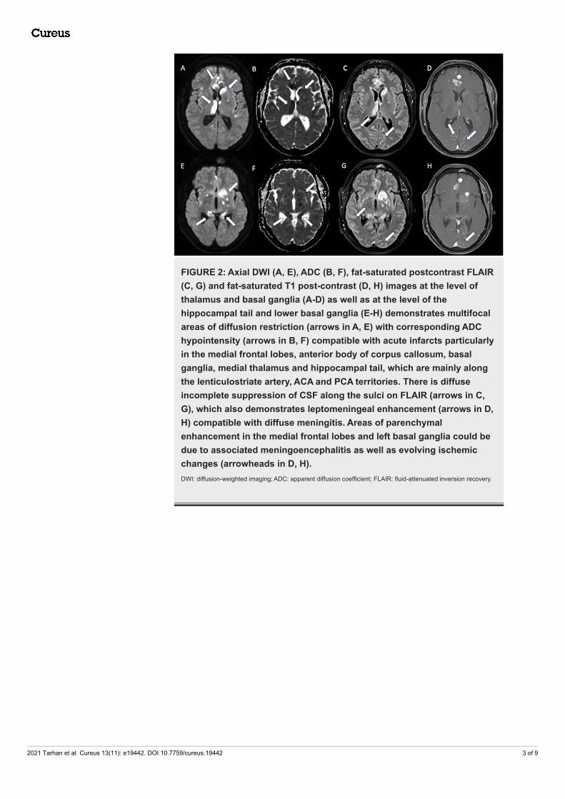
FIGURE 2: Axial DWI (A, E), ADC (B, F), fat-saturated postcontrast FLAIR(C, G) and fat-saturated T1 post-contrast (D, H) images at the level ofthalamus and basal ganglia (A-D) as well as at the level of thehippocampal tail and lower basal ganglia (E-H) demonstrates multifocalareas of diffusion restriction (arrows in A, E) with corresponding ADChypointensity (arrows in B, F) compatible with acute infarcts particularlyin the medial frontal lobes, anterior body of corpus callosum, basalganglia, medial thalamus and hippocampal tail, which are mainly alongthe lenticulostriate artery, ACA and PCA territories. There is diffuseincomplete suppression of CSF along the sulci on FLAIR (arrows in C,G), which also demonstrates leptomeningeal enhancement (arrows in D,H) compatible with diffuse meningitis. Areas of parenchymalenhancement in the medial frontal lobes and left basal ganglia could bedue to associated meningoencephalitis as well as evolving ischemicchanges (arrowheads in D, H).DWI: diffusion-weighted imaging; ADC: apparent diffusion coefficient; FLAIR: fluid-attenuated inversion recovery.
2021 Tarhan et al. Cureus 13(11): e19442. DOI 10.7759/cureus.19442 3 of 9
FIGURE 3: Axial postcontrast FLAIR (A-C) demonstrates diffuseincomplete suppression of CSF signal within the sulci basal cisternsparticularly in the interpeduncular cistern (arrowhead in A, B), whichalso demonstrates mild diffuse leptomeningeal enhancement(arrowheads in D, E). There is diffuse vessel wall thickening andenhancement particularly involving the petrous carotid arteries (arrowsin C, D), carotid terminus (arrows in E) as well as along the MCA andACA (arrows in D) compatible with vessel wall inflammation.FLAIR: fluid-attenuated inversion recovery; CSF: cerebrospinal fluid; MCA: middle cerebral artery; ACA: anteriorcerebral artery.
2021 Tarhan et al. Cureus 13(11): e19442. DOI 10.7759/cureus.19442 4 of 9

FIGURE 4: Superior view of 3D volume-rendered images from CTA (A)demonstrates diffuse vessel irregularity and multifocal areas ofnarrowing involving the bilateral ACA, MCA and distal PCA branches.Coronal MIP of contrast-enhanced CTA (B) demonstrates diffusenarrowing and irregularity of both MCA branches, right more than left(arrows in B). Overall findings are suggestive of meningoencephalitisassociated vasculitis with inflammation, spasms, constriction, andsubsequent thrombosis or necrotizing panarteritis.3D: three dimensional; CTA: computed tomography angiography; ACA: anterior cerebral artery; MCA: middlecerebral artery; PCA: posterior cerebral artery; MIP: maximum intensity projection.
A lumbar puncture (LP) was subsequently performed, which demonstrated a lymphocytic pleocytosis (WBC25 K/cumm, 80% lymphocytes), normal CSF glucose of 64 mg/dl, elevated protein of 104 mg/dl, andincreased opening pressure of 28 cm H20. Infectious studies (including Syphilis screen, bacterial cultures,fungal cultures, HSV PCR, and flow cytometry) and inflammatory markers (including oligoclonal bands andIgG index) were unremarkable; however, India ink was performed on the CSF, which revealed a smallnumber of encapsulated yeasts. He was subsequently diagnosed with cryptococcal meningoencephalitis andstarted on the appropriate anti-fungal regimen with amphotericin B and flucytosine.
He required daily LPs to ensure opening pressure remained less than 20 cm H2O. The patient was treatedwith four weeks of amphotericin B and flucytosine followed by eight weeks of fluconazole. Prior to dischargeto an inpatient rehabilitation facility, the patient’s mental status was notably improved and near hisneurologic baseline.
Case 2A 35-year-old male with a history of hyperlipidemia and seizure disorder presented to an outside hospitalfollowing a breakthrough seizure, where he was incidentally also found to have punctate areas of acutecerebral infarcts in multiple vascular territories. Additional workup revealed the presence of a left atrialthrombus and newly diagnosed atrial fibrillation. He was ultimately discharged to home on apixaban. Thepatient then re-presented a month later for evaluation of transient diplopia, expressive aphasia, daily righttemporal headaches, and right facial and left leg weakness. MRI of the brain showed new areas of diffusionrestriction in the left cerebellar hemisphere and left medial occipital lobe (Figure 5). CTA showed no signs ofcarotid occlusion or stenosis. The etiology of his multifocal strokes was thought to be related to his newlydiagnosed atrial fibrillation and left atrial thrombus.
2021 Tarhan et al. Cureus 13(11): e19442. DOI 10.7759/cureus.19442 5 of 9

FIGURE 5: Axial DWI (A, D), ADC (B, E) and FLAIR (C, F) imagesdemonstrating areas of restricted diffusion (arrows in A, B) in the leftcerebellum and left medial occipital lobe (arrows in D, E) withcorresponding increased FLAIR signal (arrows in C, F).DWI: diffusion-weighted imaging; ADC: apparent diffusion coefficient; FLAIR: fluid-attenuated inversion recovery.
The patient was then transferred to our hospital for further evaluation. His initial NIH stroke scale was 8(primary deficits were including unilateral facial palsy, bilateral lower extremity pronator drift and ataxia).Stroke labs, including lipid panel and hemoglobin A1C, were unremarkable. MRI of the brain with contrastshowed a new infarct in the splenium of the corpus callosum in addition to prominent generalizedmeningeal enhancement (Figure 6). MRI of the spine with contrast showed possible meningeal enhancementas well as punctate areas of encephalomalacia in the C3-4, C7, and T3 spinal levels. A bedside LP revealed amildly elevated opening pressure of 24 cm H20, lymphocytic pleocytosis (WBC 150 K/cumm, 61%lymphocytes), protein 170 mg/dl, hypoglycorrhachia of 15 mg/dl, and presence of cryptococcal antigen.Other notable CSF labs included the presence of 11 oligoclonal bands. He was diagnosed with cryptococcalmeningoencephalitis and started on a four-week course of amphotericin B and flucytosine. A repeat LP afterseveral days of treatment showed a normal opening pressure of 14 cm H20, mildly improved pleocytosis(WBC 130 K/cumm, 84% lymphocytes), protein 172 mg/dl, and glucose 14 mg/dl. He did not require anyadditional lumbar punctures, and his symptoms (including headaches and left lower extremity weakness)gradually improved. The patient was discharged to an inpatient rehabilitation facility prior to returninghome.
2021 Tarhan et al. Cureus 13(11): e19442. DOI 10.7759/cureus.19442 6 of 9

FIGURE 6: Axial DWI (A) and ADC (B) demonstrating a new area ofrestricted diffusion in the splenium. There is evolving FLAIRhyperintensity in the left cerebellum with multifocal areas of incompletesuppression of CSF signal on FLAIR (arrowheads in C and D). Multifocalareas of leptomeningeal enhancement (arrows in E, F) particularly in theposterior fossa.DWI: diffusion-weighted imaging; ADC: apparent diffusion coefficient; FLAIR: fluid-attenuated inversion recovery;CSF: cerebrospinal fluid.
Though our patient in Case 1 had a history of an unknown autoimmune disease, our patient in Case 2 had nohistory of autoimmune disease or other existing immunodeficiency. Both patients developed multiplecerebral infarcts in multiple vascular territories in the setting of cryptococcal meningoencephalitis, thoughour patient in Case 2 also had recently diagnosed with atrial fibrillation which further confounds theunderlying etiology of his strokes.
DiscussionCryptococcosis is an infectious disease with worldwide distribution and a wide array of clinicalpresentations caused by pathogenic encapsulated yeasts in the genus Cryptococcus. There are only twospecies commonly known to cause disease in humans, including C. neoformans and C. gattii. Theepidemiology of C. neoformans is well-characterized, and this organism causes disease in bothimmunocompromised and immunocompetent hosts [1,2].
C. neoformans disease is also likely to disseminate to the CNS. Clinical manifestations of CNScryptococcosis include a myriad of signs and symptoms, such as headache, fever, cranial neuropathies,altered mentation, lethargy, memory loss, and signs of meningeal irritation [1-4]. Symptoms usually developover a period of time; however, patients may occasionally present more acutely or lack typical features.
Cerebral infarction secondary to infection is a common complication, but the epidemiology of stroke infungal infection is unreported in the literature. The clinical and radiographical features of cerebralinfarction secondary to chronic fungal meningitis have been documented in case reports and case series [5-10].
CNS cryptococcosis produces a wide variety of clinico-radiographic features that may vary depending on theimmunological and HIV status of a patient [8].
2021 Tarhan et al. Cureus 13(11): e19442. DOI 10.7759/cureus.19442 7 of 9
Cryptococcal meningoencephalitis can have normal radiographic findings in 47% of cases by CT and 8% byMRI. Approximately 21-27% of cases have typical features of cryptococcal meningoencephalitis on MRI,which are contributory but not pathognomonic [11]. These findings may include,leptomeningeal/pachymeningeal enhancement, dilated perivascular spaces, miliary nodules, plexitis,cryptococcoma, hydrocephalus, and others either in isolation or with other MRI findings [7,8,12].
It is important to understand the pathogenesis of cryptococcal infection in the CNS. Cryptococcus canspread to the CNS hematogenously and penetrate the meningeal vessels, causing meningitis. From themeninges, it can directly invade the brain parenchyma by migrating to the perivascular Virchow-Robinspaces (VRS) in addition to the subarachnoid space. Subsequent activation of inflammatory cells within theVRS leads to dilation of these spaces and deposition of inflammatory material to form mucinouspseudocysts [13]. In neuroimaging studies, this is can be represented as punctate T2 hyperintensities withinthe basal ganglia, thalami, midbrain, and cerebellum. These subcortical areas are predominantly supplied bythe small perforating arteries of the brain. These imaging features are characteristic of cryptococcalinfection of the CNS, which generally produce minimal edema and enhancement [7].
While cerebrovascular involvement is a rare entity, cerebral infarctions associated with vasculitis or othersecondary complications is known to occur with cryptococcal meningoencephalitis. The presence of infarctwas also found to be associated with underlying diabetes and hypertension, and there is some data tosuggest higher morbidity and mortality in these patients. One study found that the incidence of cerebralinfarction secondary to cryptococcal meningitis in HIV-negative patients was roughly 4% [10]. Anotherstudy, which included both HIV-positive and HIV-negative patients, found the incidence to be 13%.Interestingly, the incidence of infarction was found to be higher in patients without HIV (78%) compared tothose with HIV (22%) [9]. According to the two largest published case series and numerous case reports,neurovascular involvement tends to present as multiple subcortical lacunar infarcts; however, majorvascular territories or brainstem involvement may be seen on rare occasions [8-10,14-19].
There are several theories to explain the possible underlying mechanism of vascular involvement. Proposedmechanisms include, direct invasion of arterial walls leading to arteritis, venous outflow obstruction,accelerated atherosclerosis through induction of cytokines, hypercoagulability as an indirect consequence ofacute infection, post-infectious infarction as a result in reduced cell-mediated immunity, among others[5,6,9,20]. Evidence of arteritis on angiography has also been observed in a patient with cryptococcalmeningitis [10].
ConclusionsIn patients with atypical presentations involving cerebral infarcts, it is important to keep infection on thedifferential diagnosis with regards to etiology. Our cases exemplify how cryptococcal meningoencephalitismay be present regardless of risk factors and immunocompetency. Overall, clinicians should keep infection-mediated cerebral infarcts in mind to expedite antimicrobial therapy and minimize adverse events.
Additional InformationDisclosuresHuman subjects: Consent was obtained or waived by all participants in this study. Conflicts of interest: Incompliance with the ICMJE uniform disclosure form, all authors declare the following: Payment/servicesinfo: All authors have declared that no financial support was received from any organization for thesubmitted work. Financial relationships: All authors have declared that they have no financialrelationships at present or within the previous three years with any organizations that might have aninterest in the submitted work. Other relationships: All authors have declared that there are no otherrelationships or activities that could appear to have influenced the submitted work.
References1. Maziarz EK, Perfect JR: Cryptococcosis. Infect Dis Clin North Am. 2016, 30:179-206.
10.1016/j.idc.2015.10.0062. Bennett JE, Dolin R, Blaser MJ: Cryptococcus neoformans and Cryptococcus gattii. In: Mandell, Douglas,
and Bennett’s principles and practice of infectious diseases, 8th Edition. Elsevier Saunders, Philadelphia;2015. 2934:48.
3. Casadevall A, Perfect JR: Cryptococcus neoformans. ASM Press, Washington, DC; 1998.4. Perfect JR, Casadevall A: Cryptococcosis. Infect Dis Clin North Am. 2002, 16:837-74. 10.1016/s0891-
5520(02)00036-35. Lan SH, Chang WN, Lu CH, Lui CC, Chang HW: Cerebral infarction in chronic meningitis: a comparison of
tuberculous meningitis and cryptococcal meningitis. QJM. 2001, 94:247-53. 10.1093/qjmed/94.5.2476. Leite AG, Vidal JE, Bonasser Filho F, Nogueira RS, Oliveira AC: Cerebral infarction related to cryptococcal
meningitis in an HIV-infected patient: case report and literature review. Braz J Infect Dis. 2004, 8:175-9.10.1590/s1413-86702004000200008
7. Xia S, Li X, Li H: Imaging characterization of cryptococcal meningoencephalitis . Radiol Infect Dis. 2016,3:187-91. 10.1016/j.jrid.2016.05.003
8. Duarte SB, Oshima MM, Mesquita JV, do Nascimento FB, de Azevedo PC, Reis F: Magnetic resonance
2021 Tarhan et al. Cureus 13(11): e19442. DOI 10.7759/cureus.19442 8 of 9
imaging findings in central nervous system cryptococcosis: comparison between immunocompetent andimmunocompromised patients. Radiol Bras. 2017, 50:359-65. 10.1590/0100-3984.2016.0017
9. Mishra AK, Arvind VH, Muliyil D, et al.: Cerebrovascular injury in cryptococcal meningitis . Int J Stroke. 2018,13:57-65. 10.1177/1747493017706240
10. Tjia TL, Yeow YK, Tan CB: Cryptococcal meningitis . J Neurol Neurosurg Psychiatry. 1985, 48:853-8.10.1136/jnnp.48.9.853
11. Katchanov J, Branding G, Jefferys L, Arastéh K, Stocker H, Siebert E: Neuroimaging of HIV-associatedcryptococcal meningitis: comparison of magnetic resonance imaging findings in patients with and withoutimmune reconstitution. Int J STD AIDS. 2016, 27:110-7. 10.1177/0956462415574633
12. Cheng YC, Ling JF, Chang FC, Wang SJ, Fuh JL, Chen SS: Radiological manifestations of cryptococcalinfection in central nervous system. J Chin Med Assoc. 2003, 66:19-26.
13. Fugate JE, Lyons JL, Thakur KT, Smith BR, Hedley-Whyte ET, Mateen FJ: Infectious causes of stroke. LancetInfect Dis. 2014, 14:869-80. 10.1016/S1473-3099(14)70755-8
14. Shimoda Y, Ohtomo S, Arai H, Ohtoh T, Tominaga T: Subarachnoid small vein occlusion due toinflammatory fibrosis-a possible mechanism for cerebellar infarction in cryptococcal meningoencephalitis:a case report. BMC Neurol. 2017, 17:157. 10.1186/s12883-017-0934-y
15. Saul RF, Gallagher JG, Mateer JE: Sudden hemiparesis as the presenting sign in cryptococcalmeningoencephalitis. Stroke. 1986, 17:753-4. 10.1161/01.str.17.4.753
16. Aharon-Peretz J, Kliot D, Finkelstein R, Ben Hayun R, Yarnitsky D, Goldsher D: Cryptococcal meningitismimicking vascular dementia. Neurology. 2004, 62:2135. 10.1212/01.wnl.0000127626.69822.67
17. Rosario M, Song SX, McCullough LD: An unusual case of stroke. Neurologist. 2012, 18:229-32.10.1097/NRL.0b013e31825bbf4d
18. Kang A, Haynor D: MR angiography of large-vessel intracranial stenosis after cryptococcal meningitis .Radiol Case Rep. 2011, 6:528. 10.2484/rcr.v6i4.528
19. Zhou W, Lai J, Huang T, Xu Y, Hu S: Cryptococcal meningitis mimicking cerebral infarction: a case report .Clin Interv Aging. 2018, 13:1999-2002. 10.2147/CIA.S181774
20. Miller EC, Elkind MS: Infection and Stroke: an Update on Recent Progress . Curr Neurol Neurosci Rep. 2016,16:2. 10.1007/s11910-015-0602-9
2021 Tarhan et al. Cureus 13(11): e19442. DOI 10.7759/cureus.19442 9 of 9