cryptococcus neoformans biofilm formation...
TRANSCRIPT

Cryptococcus neoformans BIOFILM FORMATION DEPENDS ON SURFACE SUPPORT 1
AND CARBON SOURCE AND REDUCES FUNGAL CELL SUSCEPTIBILITY TO 2
HEAT, COLD, AND ULTRAVIOLET LIGHT 3
4
Luis R. Martinez and Arturo Casadevall 5
6
7
Department of Microbiology and Immunology, 8
Albert Einstein College of Medicine, Bronx, New York 9
10
11
12
13
14
15
Address Correspondence: 16
17
Arturo Casadevall 18
19
Department of Medicine 20
21
Albert Einstein College of Medicine 22
23
1300 Morris Park Ave. 24
25
Bronx, NY 10461 26
27
Phone 718-430-2215 28
29
Fax 718-430-8968 30
31
E-mail: [email protected] 32
33
34
35
36
37
38
39
40
41
42
ACCEPTED
Copyright © 2007, American Society for Microbiology and/or the Listed Authors/Institutions. All Rights Reserved.Appl. Environ. Microbiol. doi:10.1128/AEM.02506-06 AEM Accepts, published online ahead of print on 18 May 2007
on October 1, 2018 by guest
http://aem.asm
.org/D
ownloaded from

ABSTRACT 43
The fungus Cryptococcus neoformans possess a polysaccharide capsule and can form 44
biofilms on medical devices. We describe the characteristics of C. neoformans biofilm 45
development using a microtiter plate model, microscopic examinations and a colorimetric 2, 3-46
bis (2-methoxy-4-nitro-5-sulfophenyl)-5-[(phenylamino) carbonyl]-2H-tetrazolium hydroxide 47
(XTT) reduction assay to observe the metabolic activity of cryptococci within a biofilm. Strong 48
correlation between XTT and CFU assays was demonstrated. Chemical analysis of the 49
exopolymeric material revealed sugar composition consisting predominantly of xylose, mannose, 50
and glucose, indicating the presence of other polysaccharides in addition to glucuroxylomannan. 51
Biofilm formation was affected by surface support differences, conditioning films on the surface, 52
characteristics of the medium, and properties of the microbial cell. A specific antibody to the 53
capsular polysaccharide of this fungus was used to stain the extracellular polysaccharide matrix 54
of the fungal biofilms using light and confocal microscopy. Additionally, the susceptibility of C. 55
neoformans biofilms and planktonic cells to environmental stress was investigated using XTT 56
reduction and CFU assays. Biofilms were less susceptible to heat, cold and UV light exposition 57
than their planktonic counterparts. Our findings demonstrate that fungal biofilm formation is 58
dependent on support surface characteristics and that growth in biofilm state makes fungal cells 59
less susceptible to potential environmental stresses. 60
61
62
63
64
65
ACCEPTED
on October 1, 2018 by guest
http://aem.asm
.org/D
ownloaded from

INTRODUCTION 66
Cryptococcus neoformans is an encapsulated yeast-like fungus that frequently causes life-67
threatening meningoencephalitis in immunocompromised individuals. Glucurunoxylomannan 68
(GXM) is the major component of the polysaccharide capsule. GXM is copiously shed in tissues 69
during cryptococcal infection and is believed to contribute to pathogenesis through many 70
deleterious effects on the host’s immunity (1, 16). GXM release is essential for cryptococcal 71
biofilm formation (9), a strategy that has been associated with chronic infections as a result of 72
acquired resistance to host immune mechanisms (8) and antimicrobial therapy (11). C. 73
neoformans forms biofilms on polystyrene plates (9) and prosthetic medical devices including 74
ventriculoatrial shunt catheters (17). In fact, the increasing use of ventriculoperitoneal shunts to 75
manage intracranial hypertension associated with cryptococcal meningoencephalities highlights 76
the importance of investigating the biofilm-forming properties of this organism. 77
Recent technological advances have provided evidence that biofilm formation represent 78
the most common mode of growth of microorganisms in nature, a state that presumably allows 79
microbial cells to both survive hostile environments and disperse to colonize new niches (5). C. 80
neoformans has several well-characterized virulence factors that allow the fungus to evade 81
multiple defenses and damage the host, including the polysaccharide capsule, the ability to grow 82
at mammalian temperatures, and melanin production. C. neoformans is frequently found in soil 83
contaminated with pigeon droppings (7) where the fungus may be in contact with many soil 84
predators and this interaction might have influenced the evolution of its virulence factors (15). 85
Biofilm formation is a major consideration in determining appropriate therapeutical 86
strategies against certain microbes. Biofilm formation is a well organized process that depends 87
on effects of the surface, conditioning films on the surface, characteristics of the medium, and 88
ACCEPTED
on October 1, 2018 by guest
http://aem.asm
.org/D
ownloaded from

properties of the microbial cell (3). In this study, we investigated the effect of these factors on 89
cryptococcal biofilm formation and compare the susceptibility to environmental stress of mature 90
C. neoformans biofilms and planktonic cells. Biofilm development and architecture were 91
explored under different conditions of growth using microscopy techniques. 92
93
MATERIALS AND METHODS 94
C. neoformans. C. neoformans strain B3501 (serotypes D) was acquired from the American 95
Type Culture Collection (Rockville, MD) and used in all experiments. This strain forms strong 96
biofilms on polystyrene surfaces (9). The cap59 gene deletion mutant (C536) of C. neoformans 97
B3501 was acquired from K. J. Kwon-Chung (Bethesda, MD). Yeasts were grown in Sabouraud 98
dextrose broth (Difco Laboratories, Detroit, Mich.) for 24 h at 30°C in a rotary shaker at 150 rpm 99
(to early stationary phase). 100
Biofilm formation. C. neoformans cells were collected by centrifugation, washed twice with 101
phosphate-buffered saline (PBS), counted using a hemacytometer, and suspended at 107 cells/ml 102
in minimal media (20 mg/ml thiamine, 30 mM glucose, 26 mM glycine, 20 mM MgSO4 7H2O 103
and 58.8 mM KH2PO4). Then, 100 µl of the suspension was added into individual wells of 104
polystyrene 96-well plates (Fisher, MA) and incubated at 37°C without shaking. Biofilms were 105
formed for 48 h. Following the adhesion stage, the wells containing C. neoformans biofilms 106
were washed three times with 0.05% Tween 20 in Tris-buffered saline (TBS) to remove non-107
adhered cryptococcal cells using a microtiter plate washer (Skan Washer 400, Molecular devices, 108
VA). Fungal cells that remained attached to the plastic surface were considered true biofilms. All 109
assays were carried out in six wells. 110
ACCEPTED
on October 1, 2018 by guest
http://aem.asm
.org/D
ownloaded from
Measurement of biofilm metabolic activity by XTT-reduction assay and CFUs. A 111
semiquantitative measurement of C. neoformans biofilm formation was obtained from the 2, 3-112
bis (2-methoxy-4-nitro-5-sulfophenyl)-5-[(phenylamino) carbonyl]-2H-tetrazolium hydroxide 113
(XTT) reduction assay. For C. neoformans strains, 50 µl of XTT salt solution (1 mg/ml in PBS) 114
and 4 µl of menadione solution (1 mM in acetone; Sigma) were added to each well. Microtiter 115
plates were incubated at 37°C for 5 h. Fungal mitochondrial dehydrogenases activity reduces 116
XTT tetrazolium salt to XTT formazan resulting in colorimetric change that correlates with cell 117
viability (13). The colorimetric change was measured using a microtiter reader (Labsystem 118
Multiskan MS, Finland) at 492 nm. In all the experiments, microtiter wells containing heat-119
killed C. neoformans and minimal media alone were included as negative controls. For the 120
environmental stress experiments, the percentage of metabolic activity was determined by 121
measuring the optical density (OD) of biofilms and planktonic cells exposed to heat, cold or UV 122
light relative to unexposed biofilms or planktonic cells. 123
To determine the density of C. neoformans planktonic cells used for comparison to 124
biofilms we estimated cell numbers from the XTT reduction signal using a dose-response curve. 125
Briefly, cells of C. neoformans B3501 were grown in minimal media for 48 h at 30°C in a rotary 126
shaker at 150 rpm, collected by centrifugation, washed twice with PBS, counted using a 127
hemacytometer, and suspended at various densities (5 × 10⁶, 1 × 10⁷, 5 × 10⁷ cells/ml) in minimal 128
media. Then, 100 µl of each suspension was added into individual wells of polystyrene 96-well 129
plates to final densities of 5 × 10⁵, 1 × 10⁶, 5 × 10⁶ cells/ml. The viability was measured by XTT 130
reduction assay. 131
The effect of environmental stress for C. neoformans biofilms and planktonic cells was 132
compared by CFU killing assay. After exposure to environmental stress, C. neoformans biofilms 133
ACCEPTED
on October 1, 2018 by guest
http://aem.asm
.org/D
ownloaded from

were scraped from the bottom of the wells with a sterile 200 µl micropipette tip to dissociate 134
yeast cells. A volume of 100 µl of suspension containing dissociated cells was aspirated from the 135
wells, transferred to an Eppendorff tube with 900 µl of PBS and vortexed gently for 3 min. Then, 136
serial dilutions were performed and 100 µl of diluted suspension were plated on sabouraud 137
dextrose agar plates. The percent of CFU survival was determined by comparing stress exposed 138
C. neoformans biofilms and planktonic cells relative to unexposed fungal cells. 139
XTT and CFU relationship with cell viability on biofilms. C. neoformans biofilms were 140
formed over a series of time intervals (2, 4, 8, 24 and 48 h) on polystyrene microtiter plates. The 141
metabolic activity and fungal mass were measured by XTT reduction and CFU counts, 142
respectively. The relationship between XTT reduction and CFU assays to quantify cryptococcal 143
biofilm formation was evaluated. 144
Isolation of C. neoformans biofilm exopolymeric matrix. C. neoformans biofilms were 145
formed on a 96-well polystyrene plate for 48 h at 37 ºC. After removal of non-adherent cells by 146
washing with Endosafe® reagent water (Charles Rivers Laboratories, Charleston, SC), 147
cryptococcal biofilms were scraped from the bottom of the wells with a sterile 200-µl 148
micropipette tip to dissociate yeast cells and exopolymeric matrix. A volume of 100 µl of 149
suspension containing dissociated cells and matrix was aspirated from the wells, transferred to an 150
Eppendorf tube and vortexed vigorously for 5 min. Then, cryptococci and exopolymeric matrix 151
were separated by centrifugation at 2000 rpm for 5 min. The supernatant containing the 152
carbohydrates was collected and analyzed. 153
Glycosyl composition analysis of matrix material. Glycosyl composition analysis was 154
performed by combined gas chromatography/mass spectrometry (GC/MS) of the per-O-155
trimethylsilyl (TMS) derivatives of the monosaccharide methyl glycosides produced from the 156
ACCEPTED
on October 1, 2018 by guest
http://aem.asm
.org/D
ownloaded from

sample by acidic methanolysis. The analysis was conducted at Center for Plant and Microbial 157
Complex Carbohydrates (Athens, GA). Methyl glycosides were first prepared from dry sample 158
by methanolysis in 1 M HCl in methanol at 80 ºC (18-22 h), followed by re-N-acetylationwith 159
pyridine and acetic anhydride in methanol (for detection of amino sugars). The samples were 160
then per-O-trimethylsilylated by treatment with Tri-Sil (Pierce, Rockford, IL) at 80 ºC (0.5 h). 161
GC/MS analysis of the TMS methyl glycosides was performed on an HP 5890 GC interfaced to a 162
5970 MSD, using an All Tech EC-1 fused silica capillary column (30 m × 0.25 mm ID). 163
Adhesion assay. Adhesion of C. neoformans cells was determined by cell counts using a 164
PhotoZoom inverted light microscope (Cambridge Instrument, MA). The number of cryptococcal 165
cells attached to the bottom of each well was averaged per 40-power field. This assay was done 166
in triplicate. 167
C. neoformans capsular polysaccharide isolation. C. neoformans was grown in Sabouraud 168
dextrose broth for 7 days at 30°C in a rotary shaker at 150 rpm. The supernatant was collected 169
and filtered through a 0.45-µm membrane. Then, NaCH3CO2 was slowly added to produce a 170
10% weight (per volume) solution and the pH adjusted to 7.0. C. neoformans capsular 171
polysaccharide was precipitated by adding 2.5 volumes of 95 to 100% ethanol and incubated at 172
room temperature or 4°C. Afterwards the supernatant was discarded and the polysaccharide was 173
dissolved in distilled H2O. Finally, the concentration of polysaccharide was measured by the 174
phenol-sulfuric acid method. 175
C. neoformans capsular polysaccharide binding assay. C. neoformans GXM was serially 176
diluted on the plate and allowed to attach to the bottom of the wells for 2 h. Microtiter 177
polystyrene plates were blocked with 1% bovine serum albumin (BSA) in phosphate-buffered 178
saline to prevent non-antibody-specific binding and incubated for 1 h. Next, the IgG1 GXM 179
ACCEPTED
on October 1, 2018 by guest
http://aem.asm
.org/D
ownloaded from
binding MAb 18B7 (2 µg/ml) was added and the plate was incubated for 1 h. The ELISA was 180
completed by adding, in successive steps, 1 µg of alkaline phosphatase-labeled goat anti-mouse 181
IgG1/ml and 50 µl of p-nitrophenyl phosphate (5 mg/ml) in substrate buffer. Finally, the amount 182
of GXM bound to the bottom of the plate was measured using a microtiter reader at 405 nm. 183
Between every step, the wells were washed with 0.05% Tween 20 in Tris-buffered saline. All 184
incubations were done at 37°C for 1 to 2 h or at 4°C overnight. 185
Effect of physical properties on C. neoformans biofilm formation. 186
(i) Surface support effects on C. neoformans biofilm formation. C. neoformans B3501 strain 187
was grown as described above utilizing 96-wells microtiter plates made of different materials 188
such as polystyrene, polyvinyl, polycarbonate and glass to examine the ability of yeast cells to 189
adhere and form biofilms. Biofilms formed over a series of time intervals (2, 4, 8, 24 and 48 h). 190
(ii) Surface pre-conditioning. The effect of surface preconditioning on C. neoformans biofilm 191
formation was examined. Microtiter plates were preconditioned with PBS, fetal calf serum 20% 192
(FCS), artificial cerebrospinal fluid (25 mM NaCl, 2.5 mM KCl, 26 mM NaHCO3, 1.25 mM 193
NaH2PO4*H2O, 1 mM MgCl2, 11 mM glucose, 2 mM CaCl2), or bovine serum albumin 1% 194
(BSA). Yeasts grown on unconditioned surface were utilized as control. All the conditions were 195
prepared at 50% (v/v) in sterile PBS, and were individually dispensed (200 µl) into six wells of a 196
96- well microtiter plate. The plate was then incubated in the wells overnight at 4 °C. Wells were 197
then aspirated, and the adsorbed conditioning film washed once in sterile distilled H2O. C. 198
neoformans B3501 strain were washed in PBS and re-suspended at a concentration of 107 cells 199
per ml in minimal media, and dispensed (100 µl per well) into the 96-well microtiter plates and 200
incubated for 0.5, 2, 4, 8, 24 and 48 h at 37 °C. For each time point the wells were aspirated and 201
ACCEPTED
on October 1, 2018 by guest
http://aem.asm
.org/D
ownloaded from

washed three times in sterile PBS, and adhesion measured by the XTT reduction assay. 202
Experiments were performed twice with similar results. 203
(iii) Temperature. To examine the effect of temperature on C. neoformans biofilm formation, 204
B3501 strain was grown as described above at various temperatures (4, 23, 30, 37 and 45°C). 205
Biofilms formed over a series of time intervals (2, 4, 8, 24, 48 and 72 h). 206
(iv) pH. To evaluate the effect of pH on biofilm production by C. neoformans, B3501 strain was 207
grown as described above at distinct pHs (3, 4, 5, 6, 7, 8 and 9). Biofilms formed over a series of 208
time intervals (2, 4, 8, 24 and 48 h). 209
(v) Sugars. To determine the effect of sugars on C. neoformans biofilm formation, yeasts were 210
grown in minimal media containing 30 mM of glucose, galactose, maltose, lactose, sucrose or 211
mannose as a sugar source in 96-wells plates over a series of time intervals (2, 4, 8, 24 and 48 h). 212
Imaging of C. neoformans biofilms. 213
(i) Scanning electron microscopy (SEM). C. neoformans biofilms were grown on polyvinyl 214
catheters. Catheters were added to a C. neoformans culture grown in minimal media for 48 h in 215
a rotary shaker at 150 rpm. Then, catheters with biofilms were washed three times with PBS, 216
and transferred to another 6-well microtiter plate containing 2.5% glutaraldehyde and incubated 217
for 1 h at room temperature. The samples were serially dehydrated in alcohol, fixed in a critical-218
point drier (Samdri-790; Tousimis, Rockville, Md.), coated with gold-palladium (Desk-1; Denton 219
Vacuum, Inc., Cherry Hill, N.J.), and viewed with a JEOL (Tokyo, Japan) JSM-6400 scanning 220
electron microscope. Two separate sets of cultures were prepared. 221
(ii) Light microscopy. Capsular polysaccharide surrounding cryptococcal biofilms was stained 222
using the GXM-binding mAb 18B7. After biofilm formation, wells were blocked with PBS (1% 223
BSA). Next, the IgG1 GXM binding mAb 18B7 (2 µg/ml) was added and the plate was 224
ACCEPTED
on October 1, 2018 by guest
http://aem.asm
.org/D
ownloaded from

incubated for 1 h at 37 ˚C. Peroxidase-conjugated goat anti-mouse IgG1 (Fisher Scientific, 225
Orangeburg, NY) was diluted at 1:250 in PBS (1% BSA) and applied for 1 h at room 226
temperature (RT). Color was developed after 5 min incubation with DAB and 0.3% H2O2 at RT. 227
Microscopic examinations were performed with light microscopy using an Axiovert 200 M 228
inverted microscope (Carl Zeiss MicroImaging, NY). 229
(iii) Confocal microscopy (CM). C. neoformans biofilms were incubated for 45 min in 75 µl of 230
PBS containing the fluorescent stains FUN-1 (10 µM). Then, wells were blocked with PBS (1% 231
BSA) for 1 h. Next, the IgG1 GXM binding mAb 18B7 (2 µg/ml) was added and the plate was 232
incubated for 1 h. Fluorescein isothiocyanate (FITC) -conjugated goat anti-mouse IgG1 (Fisher 233
Scientific) at 1 µg/ml in PBS (1% BSA) and applied for 1 h. Between every step, the wells were 234
washed with 0.05% Tween 20 in Tris-buffered saline. All incubations were done at 37 or 4°C 235
overnight. FUN-1 (excitation wavelength = 470 nm and emission = 590 nm) is converted to 236
orange-red cylindrical intravacuolar structures by metabolically active cells, while mAb 18B7-237
FITC-GAM-IgG1-conjugated (excitation wavelength = 488 nm and emission = 530 nm) binds to 238
GXM and fluoresces green. Microscopic examinations of biofilms formed in microtiter plates 239
were performed with confocal microscopy using an Axiovert 200 M inverted microscope (Carl 240
Zeiss MicroImaging, NY). The objective used was a 40X (numerical aperture of 0.6). Depth 241
measurements were taken at regular intervals across the width of the device. To determine the 242
structure of the biofilms, a series of horizontal (xy) optical sections with a thickness of 1.175 µm 243
were taken throughout the full length of the biofilm. Confocal images of green (mAb 18B7-244
FITC-GAM-IgG1-conjugated) and red (FUN-1) fluorescence were recorded simultaneously 245
using a multichannel mode. Z-stack images and measurements were corrected utilizing Axio 246
Vision 4.4 software-deconvolution mode (Carl Zeiss MicroImaging, NY). 247
ACCEPTED
on October 1, 2018 by guest
http://aem.asm
.org/D
ownloaded from
Susceptibility of C. neoformans biofilms to environmental stress. 248
(i) Exposure of C. neoformans biofilms to heat. C. neoformans biofilms and planktonic cells in 249
96-wells microtiter plates were placed in an incubator and heated to 47 °C. This temperature had 250
previously been shown to be microbicidal to C. neoformans (12). After various time intervals 251
(30 and 60 min) of exposure to the heat, the metabolic activity and fungal mass of biofilms and 252
planktonic cells were measured using the XTT reduction and CFU assays. 253
(ii) Susceptibility of C. neoformans biofilms to cold. Cryptococcal biofilms and planktonic 254
cells were cooled or frozen by incubation at -80, -20 and 4 °C for 24 h. After the incubation, the 255
wells containing the biofilms and planktonic cells were thawed at room temperature and the 256
viability was measured by XTT reduction assay and CFUs. Control cells were plated at the same 257
time the experimental cells were cooled or frozen to avoid changes in number. 258
(iii) Effect of UV light irradiation on mature C. neoformans biofilms. The susceptibility of 259
C. neoformans biofilms and planktonic cells to irradiation by UV light was determined by 260
exposing fungal cells to various doses (100, 200, 300 and 400 µJ × 100/cm2) of UV light (254 261
nm) generated by a Stratalinker 1800 (Stratagene, La Jolla, CA). The viability of biofilms and 262
planktonic cells irradiated with UV light was measured by XTT reduction assay and CFUs and 263
compared them relative to non-irradiated cells. 264
Statistical analysis. All data were subjected to statistical analysis using Origin 7.0 (Origin Lab 265
Corporation, Northampton, MA). P values were calculated by t-test. P values of <0.05 were 266
considered significant. 267
268
RESULTS 269
ACCEPTED
on October 1, 2018 by guest
http://aem.asm
.org/D
ownloaded from

Correlation between XTT reduction and CFU killing assays. We investigated the correlation 270
between XTT reduction and CFU assays to monitor C. neoformans biofilm formation (Fig. 1). 271
Strong correlation between both parameters was demonstrated (R2=0.954, P=0.012). XTT 272
activity was linearly associated with CFU counts. Consequently, XTT colorimetric intensity 273
reflects fungal mass in the biofilm. 274
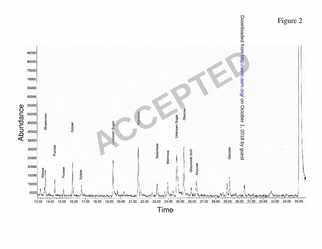
Glycosyl composition of exopolymeric matrix from biofilms of C. neoformans. Preparations 275
of cryptococcal biofilm matrix material from B3501 strain were analyzed for carbohydrate 276
composition by combined gas chromatography/mass spectrometry (Fig. 2). Glycosyl 277
composition of exopolymeric matrix isolated from biofilms is consistent with the presence of 278
GXM. However, we observed significant quantities of sugars not found in GXM such as glucose 279
(23.1%), ribose (4.5%) and fucose (7.4%; Table 1), implying that exopolymeric matrix of C. 280
neoformans biofilms includes polysaccharides other than GXM. 281
C. neoformans forms stronger biofilms on polyvinyl. The effect of the solid surface support 282
material in the ability of C. neoformans to adhere and form biofilms was investigated. 283
Experiments were carried on microtiter plates composed of polystyrene, polyvinyl, 284
polycarbonate and glass. C. neoformans B3501 strain grown in polyvinyl support formed 285
significantly stronger biofilms than cells grown in polystyrene, polycarbonate or glass support 286
(Fig. 3). C. neoformans B3501 strain formed biofilms in the following order: polyvinyl > glass > 287
polystyrene > polycarbonate. Fungal cells attached strongly to polyvinyl after 2 h incubation 288
(Fig. 3A). During early stage of biofilm formation (0 to 8 h), B3501 cells grown in polystyrene, 289
polycarbonate or glass showed a similar increase in metabolic activity (Fig. 3B). However, 290
yeasts grown in glass formed better biofilms than polystyrene and polycarbonate. 291
ACCEPTED
on October 1, 2018 by guest
http://aem.asm
.org/D
ownloaded from

Furthermore, we investigated whether there was a correlation between biofilm formation 292
and the ability of GXM to bind to a specific solid support material. Total capsular polysaccharide 293
of B3501 cells was isolated and added to polystyrene plates, and binding was detected with 294
MAbs. C. neoformans GXM adhered best to the polyvinyl and glass materials followed by 295
polystyrene and polycarbonate, respectively (Fig. 3C). Hence, the relative ability of C. 296
neoformans to form biofilms correlated with GXM binding to a specific material. 297
Influence of temperature on C. neoformans biofilm formation. The effect of temperature in 298
the kinetics of biofilm formation by the C. neoformans B3501 strain on the surface of 299
polystyrene microtiter plates was quantified (Fig. 4A). The metabolic activity displayed by C. 300
neoformans B3501 strain during the adhesion period (0 to 8 h) was similar at 23, 30 and 37 °C. 301
The kinetics of B3501 cells showed a rapid increase in cellular growth during the first 24 h and 302
then reached a plateau. C. neoformans B3501 strain formed similar and strong biofilms at 23, 30 303
and 37 °C (Fig. 4A). In contrast, B3501 strain did not form biofilms at 4 or 45°C. 304
Effects of surface conditioning on biofilm formation by C. neoformans. Cryptococcal 305
biofilm development of strain B3501 was tested on untreated or surfaces pre-conditioned with 306
different solutions. Stronger biofilm formation was observed in ACSF and PBS (Fig. 4B). After 307
2 h, C. neoformans cells grown on surfaces pre-conditioned with ACSF and PBS adhered rapidly 308
to the support surface forming strong biofilms. A similar increase in metabolic activity on 309
cryptococci grown on untreated or in a BSA conditioned surfaces was observed after 4 h 310
incubation. Fungi grown on pre-coated surfaces with FCS manifested a lower increase in 311
cellular metabolic activity after 8 h incubation than the growth in non-FCS coated surfaces. 312
After 48 h, C. neoformans B3501 strain formed biofilms on pre-conditioned surfaces in the 313
ACCEPTED
on October 1, 2018 by guest
http://aem.asm
.org/D
ownloaded from

following order: ACSF > PBS > untreated = BSA > FCS. Therefore, initial adhesion of C. 314
neoformans cells depended on the condition of the surface where the biofilm formed. 315
C. neoformans forms stronger biofilms at neutral pH. The role of pH in C. neoformans 316
biofilm formation was examined. C. neoformans B3501 strain formed significantly stronger 317
biofilms at slightly acidic and neutral pH conditions (pH 5 to 7) tested. Fungal cells grown in 318
neutral conditions attached significantly stronger to the polystyrene surface of microtiter plates 319
than yeast cells grown in acidic (pH 3 and 4) or alkaline (pH 8 and 9) environments (Fig. 5A). 320
Similarly, B3501 cells showed a significant increase in metabolic activity during the first 24 h 321
for cryptococci within biofilms grown at neutral milieu (Fig. 5B). 322
Since GXM release is necessary for cryptococcal biofilm formation, we purified GXM 323
from C. neoformans B3501 strain and investigated whether different pH conditions affect the 324
ability of GXM binding to plastic. GXM adhered best to the polystyrene material at neutral 325
conditions (Fig. 5C). Hence, the relative ability of C. neoformans to form biofilms on 326
polystyrene correlated at various pH correlated with GXM binding to that surface. 327
Effect of sugar type on C. neoformans biofilm formation. C. neoformans strain B3501 was 328
grown in the presence of different sugars to determine their effect in biofilm formation on 329
polystyrene plates. Strain B3501 was able to attach and form biofilms in the presence of glucose 330
or mannose (Fig. 6A), and form comparable biofilms with either sugar. During the adhesion 331
stage (0 to 4 h), fungal cells of B3501 strain grown in the presence of sucrose showed similar 332
metabolic activity than cells grown in the presence of mannose but after reaching a biofilm 333
maturity the metabolic activity decreased significantly (Fig. 6B). In contrast, no biofilm 334
formation was observed when the cells were grown in the presence of galactose, maltose or 335
lactose. 336
ACCEPTED
on October 1, 2018 by guest
http://aem.asm
.org/D
ownloaded from
SEM visualization of C. neoformans biofilms. Biofilm formation was monitored by SEM. 337
Catheters were incubated in a culture of C. neoformans in minimal medium on a rotary shaker 338
for 48 h. C. neoformans biofilms comprised a dense network of yeast cells strongly attached to a 339
piece of catheter that detached after SEM processing (Fig. 7A). Exopolymeric matrix was seen 340
surrounding cryptococcal cells. Closer image of cryptococcal biofilm showed fine 341
polysaccharide fibers from a yeast cell bound to most of the exopolymeric material (Fig. 7B). 342
Higher density collections of yeast cells had more extracellular matrix surrounding them 343
suggesting that close association helped to preserve the polysaccharide matrix from the 344
preparation effects of SEM (Fig. 7C). We evaluated whether the acapsular strain C536 could 345
form biofilm when grown with catheters but found no evidence of a biofilm (data not shown). 346
Distribution of GXM surrounding C. neoformans biofilms. The distribution of capsular 347
polysaccharide surrounding a C. neoformans biofilm was investigated by staining with specific 348
mAb to GXM and visualized by light microscopy. C. neoformans mature biofilms showed that 349
GXM is copiously released and profusely distributed through out the exopolymeric matrix (Fig. 350
8A). Individual fungal cells were not distinguishable due to encasement in extracellular capsular 351
polysaccharide material. A closer view of a biofilm displayed darker regions of mAb 18B7-352
stained GXM where yeast cells were densely packed within a thick layer of GXM (Fig. 8B). 353
CM of mature C. neoformans biofilms using a GXM specific mAb. A mature C. neoformans 354
biofilm was investigated using CM because this technique preserved the structural integrity of 355
biofilms. C. neoformans biofilm reconstruction was done by the compilation of a series of 356
individual xy sections taken across the z axis. Red color due to FUN-1 staining localized in 357
dense aggregates in the cytoplasm of metabolically active cells (Fig. 9A), while the intense green 358
fluorescence resulting from specific mAb 18B7-FITC-GAM-IgG1-conjugated bound to GXM 359
ACCEPTED
on October 1, 2018 by guest
http://aem.asm
.org/D
ownloaded from
(Fig. 9B). Orthogonal images of a mature C. neoformans biofilm in polystyrene 96-well plates 360
were analyzed to determine biofilm thickness and architecture. Vertical (xz) sectioning (side 361
view) of 3-D reconstructed images showed that mature C. neoformans biofilms consisted of a 362
highly organized architecture (~56 µm thick biofilm) with red spots representing metabolically 363
active cells interwoven with extracellular polysaccharide material (Fig. 9C). 364
C. neoformans cells in biofilms are less susceptible than planktonic cells to environmental 365
stress. The susceptibility of fungal biofilms to environmental harshness such as high and low 366
temperatures and UV light was investigated and compared with planktonic yeasts. XTT 367
reduction and CFU killing assays were utilized to quantify fungal metabolic activity and cellular 368
mass, respectively. 369
C. neoformans cells in biofilms were significantly more resistant to thermal stress than 370
their planktonic counterparts (Fig. 10A). The metabolic activity of biofilms was not affected 371
after exposures to relatively high temperature. However, planktonic cells showed a significant 372
reduction in metabolic activity after being exposed to 47 ºC for 30 (P=0.004) and 60 min 373
(P=00001), respectively. To confirm the results obtained by XTT reduction assay, the percent 374
survival of cells in biofilms or planktonic form was determined by counting the number of CFU 375
in wells treated with heat and comparing these to the number of colonies obtained from 376
unexposed cells (Fig. 10B). After 60 min exposure to heat, C. neoformans cells within biofilms 377
displayed approximately 50% survival whereas most of planktonic cells were killed. 378
C. neoformans biofilms were more resistant to cold than planktonic cells (Fig. 10C and 379
D). In both assays, biofilm and planktonic cells did not show statistical differences when yeast 380
cells were incubated at 4 ºC. Cryptococcal biofilms were less susceptible to damage by freezing 381
to -20 ºC than planktonic cells regardless of whether effect was was measured by XTT reduction 382
ACCEPTED
on October 1, 2018 by guest
http://aem.asm
.org/D
ownloaded from

(P=0.005) or CFU enumeration (P=0.0001). Moreover, biofilms and planktonic cells exhibited 383
approximately 80% reduction in viability after freezing at -80 ºC but survival of cells within 384
biofilms was significantly higher (P=0.004) than for planktonic cells. 385
Furthermore, cryptococcal biofilms were less susceptible to killing by UV irradiation 386
than planktonic cells as measured by CFU killing assay (Fig. 10E). There were no statistical 387
differences between biofilms or planktonic cells when fungal cells were irradiated with doses of 388
100 and 200 µJ×100/cm2. Conversely, after UV light irradiation of 300 µJ×100/cm2, the percent 389
of survival of planktonic cells was significantly reduced to approximately 80% versus 40% of 390
cryptococcal biofilms (P=0.008). In the XTT reduction assay for UV light susceptibility, there 391
was no statistical difference in metabolic activity of biofilms and planktonic cells (data not 392
shown). 393
394
DISCUSSION 395
Biofilm development by C. neoformans follows a standard sequence of events including 396
fungal surface attachment, microcolony formation, and matrix production (9). C. neoformans 397
biofilm development is dependent on the release of capsular polysaccharide to the solid surface 398
to create an exopolysaccharide matrix (9). Sugar composition analysis was remarkable for the 399
predominance of xylose, mannose, and glucose and the presence of several minor sugars not 400
found in either of the C. neoformans capsular polysaccharides GXM or galactoxylomannan 401
(GalXM). Since neither GXM nor GalXM contains glucose we infer that the exopolymeric 402
matrix is composed of different types of polysaccharides than used to assemble the capsule. 403
Detailed SEM imaging revealed that C. neoformans strongly attached to polyvinyl catheters 404
suggesting that the polysaccharide capsule conferred an advantage during the colonization 405
ACCEPTED
on October 1, 2018 by guest
http://aem.asm
.org/D
ownloaded from

process given that acapsular cryptococci did not adhere to the plastic material. Since GXM is the 406
major component of the extracellular polysaccharide surrounding fungal cells within a mature C. 407
neoformans biofilm, we utilized an IgG1 GXM-specific mAb 18B7 as a reagent to visualize the 408
exopolymeric matrix by light microscopy. A secondary IgG1 mAb-conjugated to horse 409
peroxidase was used to identify the mAb 18B7 bound to the biofilm matrix. C. neoformans 410
GXM was copiously released to the medium, accumulated, and encased fungal cells within an 411
exopolymeric matrix that could not be removed by shear forces product of an extensive and 412
tenacious washing. The use of specific mAbs is a simple and effective method to study microbial 413
biofilm exopolymeric matrix development by light microscopy. 414
Similarly, the combination of dye Fun-1 and mAb 18B7 binding and detection with 415
FITC-GAM-IgG1 were utilized to visualize mature cryptococcal biofilms using confocal 416
microscopy. MAb 18B7-FITC-GAM-IgG1-conjugated was used to stain the GXM in the 417
exopolymeric matrix and fluoresced green while Fun-1 entered yeast cells and fluoresced red 418
identifying metabolically active cryptococci. Mature C. neoformans biofilm displayed a 419
complex structure with internal regions of metabolically active cells interwoven with 420
extracellular polysaccharide material and interspersed with water channels. Our result suggests 421
that most of the extracellular polysaccharide comprising the matrix enclosing cryptococci within 422
a mature biofilm includes shed GXM and can be stained by specific mAb. Con A binds to 423
mannoproteins in the matrix of cryptococcal biofilms (8, 9, 11). Co-staining with Con A and 424
mAb 18B7 revealed different co-localization strongly suggesting that these compounds bind to 425
different moieties (10). 426
We compared XTT assay and CFU quantification in evaluating biofilm development of 427
C. neoformans. Traditionally, CFU determination was used to measure cell viability despite the 428
ACCEPTED
on October 1, 2018 by guest
http://aem.asm
.org/D
ownloaded from

laborious work involved and, difficulties in disrupting cell aggregates without affecting viability. 429
Recently, XTT reduction assay, which is based on metabolic activity rather than viability, has 430
been developed for biofilm quantification. The XTT reduction assay is a colorimetric method 431
that quantifies the number of living cells in a biofilm. Our data showed that the CFU assay 432
positively correlates with the XTT readings, suggesting that both methods can be reliably used 433
for quantification of cryptococcal biofilm mass. Nevertheless, we noted that for the experiments 434
where biofilms were exposed to heat and cold stress the reduction in CFU was disproportionately 435
greater than the reduction in XTT activity. This may reflect lethal damage to cells by heat and 436
cold stress without a concomitant temporally related reduction in enzymatic activity. Hence, the 437
slope of the correlation between CFU and XTT may be function of the type of experiment 438
performed. 439
Our studies revealed that the type of solid surface plays an important role in biofilm 440
formation. Surface support roughness robustly promotes microbial adhesion by diminishing 441
shear forces whereas physicochemical properties provide tenacious attachment of 442
microorganisms to a solid support (2). C. neoformans B3501 strain fairly adhered to all 443
materials tested, but formed strongest biofilms on polyvinyl support. The dynamic nature of the 444
capsule could allow C. neoformans cells to interact favorably with a variety of substrata and 445
promote fungal attachment. In contrast to many other microorganisms that preferably form 446
biofilms on hydrophobic surfaces, C. neoformans cells strongly attached to glass and formed a 447
mature biofilm. The polysaccharide capsule apparently plays an important role in the adhesion 448
of fungal cells to glass surface. For instance, the O antigen component of lipopolysaccharide 449
(LPS) has also been shown to confer hydrophilic properties to gram-negative bacteria (18). 450
ACCEPTED
on October 1, 2018 by guest
http://aem.asm
.org/D
ownloaded from

Artificial cerebrospinal fluid contains high concentrations of cations that could increase 451
the interactions of the microbe with support surface. When polystyrene material was conditioned 452
with ACSF, the C. neoformans B3501 strain attached tenaciously to the solid support. Surface 453
conditioning and the characteristics of the medium are important for microbial biofilm 454
development because these variables affect the rate and the extent of cryptococci attachment. For 455
example, Mittelman reported that a number of host fluids such blood, tears, urine, saliva, 456
intervascular fluid, and respiratory secretions influenced the attachment of bacteria to 457
biomaterials (14). Ventriculoperitoneal shunts in patients with cryptococcosis are constantly 458
irrigated by cerebrospinal fluid and may be highly susceptible to coating by cryptococcal 459
biofilms. Furthermore, this possibility increases taking in account that the cerebrospinal fluid is 460
in constant motion and this situation influences adhesion of microorganisms to biomaterials (3). 461
C. neoformans strain B3501 formed a strong biofilm in minimal medium supplemented 462
with either glucose or mannose as a carbon source. During initial attachment, fungal cells grown 463
in the presence of glucose, galactose, sucrose and mannose adhered similarly to polystyrene. 464
Conversely, galactose and sucrose did not stimulate C. neoformans biofilm maturation. These 465
results suggest that the carbon source available for nutrition can have an important effect on 466
biofilm maturation. For instance, sugar differences may affect fungal growth rate and the 467
expression of proteins involved in adhesion or matrix scaffolding (6). Furthermore, other 468
characteristics of the medium that may influence in microbial biofilm formation are temperature 469
and pH. High or low temperature affected C. neoformans biofilm formation. Previous studies 470
have shown that seasonal changes may affect the rates of attachment and biofilm formation (4). 471
C. neoformans was able to form biofilms at neutral pH conditions comparable to those that 472
would be expected in body fluids. This environment stimulates the ability of GXM to bind a 473
ACCEPTED
on October 1, 2018 by guest
http://aem.asm
.org/D
ownloaded from

solid support. However, lower or higher pH conditions did not allow C. neoformans to develop 474
mature biofilms, possibly by interfering with growth and/or influencing the charge of the GXM 475
that is conferred by the state of protonation of glucuronic acid groups. 476
The ability to form biofilms provides a fungus with survival advantages in the 477
environment, e.g., anchorage at a location where growth is favorable, protection from desiccation 478
or predation, and resistance to biocides and detergents. C. neoformans is found ubiquitously in 479
the environment in association with pigeon excreta (7) and is exposed to many environmental 480
changes. C. neoformans biofilms were significantly less susceptible to each of these stress 481
conditions than their planktonic counterparts. Biofilm establishment may provide protection 482
from environmental shifts due to facilitate cell to cell interactions and the exopolymeric matrix 483
may act as a shield against stress conditions. 484
In conclusion, our results indicate that biofilm development by C. neoformans depends on 485
various characteristics such as capsular production, physical properties of substrate material, 486
composition of the medium, etc. Characterization of microbial biofilms may be critically 487
important for designing of therapies against biofilm-related diseases. Furthermore, the utilization 488
of a GXM specific mAb to study the production of exopolymeric material by C. neoformans 489
during biofilm formation, add a potential and useful tool in biofilm-forming microbes research. 490
491
FIGURE LEGENDS 492
Figure 1. Correlation between XTT reduction and CFU assays for monitoring C. neoformans 493
biofilm formation. The R2 and P values for each regression are also indicated. 494
Figure 2. Gas chromatogram detection profile of the isolated matrix material from biofilms of 495
C. neoformans. 496
ACCEPTED
on October 1, 2018 by guest
http://aem.asm
.org/D
ownloaded from

Figure 3. Kinetics of C. neoformans biofilm formation in microtiter plates of different 497
materials, as determined by A. cell counts and B. XTT reduction assay. Each point represents 498
the average of six measurements. C. Binding of C. neoformans GXM to microtiter plates of 499
different materials. The inset diagram indicates the ELISA configuration used to detect the 500
polysaccharide bound to the bottom of the plate. AP, alkaline phosphatase. 501
Figure 4. Kinetics of C. neoformans biofilm formation A. grown at different temperatures and 502
B. in surface pre-conditioned polystyrene microtiter plates, as determined by XTT reduction 503
assay. Each point represents the average of six measurements. 504
Figure 5. Kinetics of C. neoformans biofilm formation in polystyrene microtiter plates growing 505
at various pH conditions, as determined by A. cell counts and B. XTT reduction assay. Each 506
point represents the average of six measurements. C. Binding of C. neoformans GXM to the 507
plastic surface of a microtiter plate at various pH conditions. The inset diagram indicates the 508
ELISA configuration used to detect the polysaccharide bound to the bottom of the plate. AP, 509
alkaline phosphatase. 510
Figure 6. Kinetics of C. neoformans biofilm formation in polystyrene microtiter plates using 511
different sugars as a carbon source in the medium, as determined by A. cell counts and B. XTT 512
reduction assay. Each point represents the average of six measurements. 513
Figure 7. Scanning electron microscopy images of a mature C. neoformans B3501 biofilm 514
formed on polyvinyl catheters in vitro revealed cryptococcal cells strong attachment to the 515
substrate. Scale bar: A. 10 µm, B. 1 µm and C. 5 µm. Black and white arrows denote 516
polysaccharide and polyvinyl substrate, respectively. 517
Figure 8. Light microscopy images of a mature C. neoformans biofilm stained exopolymeric 518
matrix with GXM specific mAb 18B7. Images of a mature biofilm shows capsular binding mAb 519
ACCEPTED
on October 1, 2018 by guest
http://aem.asm
.org/D
ownloaded from

18B7 binds and dark stains shed capsular polysaccharide. A. Picture was taken using a 10X 520
power field. Scale bar, 50 µm. B. Picture was taken using a 40X power field. Scale bar, 10 521
µm. Black and white arrows denote yeast cells and exopolymeric matrix, respectively. 522
Figure 9. Confocal microscopic image of a mature C. neoformans biofilm stained with GXM 523
specific mAb 18B7. Orthogonal image of a mature C. neoformans biofilm shows: A. 524
metabolically active (red, FUN-1-stained) C. neoformans cells; B. extracellular polysaccharide 525
material stained with capsular binding mAb 18B7 (green, GAM-γ1-FITC-stained); C. Merged 526
images A and B. Mature C. neoformans biofilm showed a complex structure with metabolically 527
active cells interwoven with extracellular polysaccharide material. The thickness of a mature 528
biofilm is approximately 56 µm. Pictures were taken using a 40X power field. Scale bar, 20 µm. 529
Figure 10. C. neoformans biofilms are less susceptible to environmental stress than planktonic 530
cells. (A-B) C. neoformans biofilms are less susceptible to heat (47 ºC) than planktonic cells. A. 531
The metabolic activity of C. neoformans biofilms and planktonic cells was measured using XTT 532
reduction assay and, for the treated group is shown here as a percentage of the activity measured 533
in unexposed. B. The percent survival of C. neoformans strain B3501 biofilms and planktonic 534
cells was determined by CFU and, for the treated group is shown here as a percentage of CFU 535
counted in unexposed. For graphs A and B, both cell types were exposed to 47 ºC for 30 and 60 536
min and its metabolic activity and CFUs were compared to fungal cells unexposed to heat as a 537
function of time. (C-D) C. neoformans biofilms are less susceptible than planktonic cells to cold. 538
C. The metabolic activity of C. neoformans biofilms and planktonic cells was measured using 539
XTT reduction assay and, for the treated group is shown here as a percentage of the activity 540
measured in unexposed. D. The percent survival of C. neoformans strain B3501 biofilms and 541
planktonic cells was determined by CFU and, for the treated group is shown here as a percentage 542
ACCEPTED
on October 1, 2018 by guest
http://aem.asm
.org/D
ownloaded from
of CFU counted in unexposed. For graphs C and D, both cell types were exposed to cold stress 543
for 24 h and its metabolic activity and CFUs were compared to fungal cells unexposed to cold. 544
E. C. neoformans biofilms are less susceptible to UV light exposure than planktonic cells. The 545
percent survival of C. neoformans strain B3501 biofilms and planktonic cells was determined by 546
CFU and, for the treated group is shown here as a percentage of the CFU counts unexposed to 547
UV light. For each graph, bars are the average of 6 measurements, and brackets denote standard 548
deviations. Asterisks denote p value significance calculated by t-test. 549
550
ACKNOWLEDGEMENTS 551
This work was supported by the following National Institutes of Health grants: HL59842-08, 552
AI33142-11, and AI33774-11. This work was supported in part by the Department of Energy-553
funded (DE-FG09-93ER-20097) Center for Plant and Microbial Complex Carbohydrates. 554
555
REFERENCES 556
1. Casadevall, A., and J.R. Perfect. 1998. Cryptococcus neoformans. ASM Press, 557
Washinghton, D.C. 558
559
2. Characklis, W. G., G. A. McFeters, and K. C. Marshall. 1990. Physiological ecology 560
in biofilm systems, vol. 90. John Wiley & Sons, New York. 561
562
3. Donlan, R. M. 2002. Biofilms: microbial life on surfaces. Emerg Infect Dis. 8:881. 563
564
4. Fera, P., M. A. Siebel, W. G. Characklis, and D. Prieur. 1989. Seasonal variations in 565
bacterial colonization of stainless steel, aluminum, and polycarbonate surfaces in 566
seawater flow system. Biofouling. 1:251. 567
568
5. Hall-Stoodley, L., J. W. Costerton, and P. Stoodley. 2004. Bacterial biofilms: from the 569
natural environment to infectious diseases. Nat Rev Microbiol. 2:95. 570
571
6. Jin, Y., L. P. Samaranayake, Y. Samaranayake, and H. K. Yip. 2004. Biofilm 572
formation of Candida albicans is variably affected by saliva and dietary sugars. Arch Oral 573
Biol. 49:789. 574
ACCEPTED
on October 1, 2018 by guest
http://aem.asm
.org/D
ownloaded from

575
7. Levitz, S. M. 1991. The ecology of Cryptococcus neoformans and the epidemiology of 576
cryptococcosis. Rev Infect Dis. 13:1163. 577
578
8. Martinez, L. R., and A. Casadevall. 2006. Cryptococcus neoformans cells in biofilms 579
are less susceptible to antimicrobial molecules produced by the innate immune system 580
than planktonic cells. Infect Immun. 74:7118. 581
582
9. Martinez, L. R., and A. Casadevall. 2005. Specific antibody can prevent fungal biofilm 583
formation and this effect correlates with protective efficacy. Infect Immun. 73:6350. 584
585
10. Martinez, L. R., E. Christaki, and A. Casadevall. 2006. Specific antibody to 586
Cryptococcus neoformans glucurunoxylomannan antagonizes antifungal action against 587
cryptococcal biofilms in vitro. J Infect Dis. 194:261. 588
589
11. Martinez, L. R. and A. Casadevall. 2006. Susceptibility of Cryptococcus neoformans 590
biofilms to antifungal agents in vitro. Antimicrob Agents Chemother. 50:1021. 591
592
12. Martinez L. R. , Garcia-Rivera J., and A. Casadevall. 2001. Cryptococcus 593
neoformans var. neoformans (serotype D) strains are more susceptible to heat than C. 594
neoformans var. grubii (serotype A) strains. J Clin Microb. 39:3365. 595
596
13. Meshulam T, L. S., Christin L, Diamond RD. 1995. A simplified new assay for 597
assessment of fungal cell damage with the tetrazolium dye, (2,3)-bis-(2-methoxy-4-nitro-598
5-sulphenyl)-(2H)-tetrazolium-5-carboxanilide (XTT). J Infect Dis. 172:1153. 599
600
14. Mittelman, M. W. 1996. Adhesion to biomaterials. Wiley-Liss, New York. 601
602
15. Steenbergen, J. N., H. A. Shuman, and A. Casadevall. 2001. Cryptococcus 603
neoformans interactions with amoebae suggest an explanation for its virulence and 604
intracellular pathogenic strategy in macrophages. Proc Natl Acad Sci U S A. 98:15245. 605
606
16. Vecchiarelli, A. 2000. Immunoregulation by capsular components of Cryptococcus 607
neoformans. Med Mycol. 38:407. 608
609
17. Walsh, T. J., R. Schlegel, M. M. Moody, J. W. Costerton, and M. Salcman. 1986. 610
Ventriculoatrial shunt infection due to Cryptococcus neoformans: an ultrastructural and 611
quantitative microbiological study. Neurosurgery. 18:373. 612
18. Williams, V. and M. Fletcher. 1996. Pseudomonas fluorescens adhesion and transport 613
through porous media are affected by lipopolysaccharide composition. Appl Environ 614
Microbiol. 62:100. 615
616
617
618
ACCEPTED
on October 1, 2018 by guest
http://aem.asm
.org/D
ownloaded from

TABLES 619
620
Table 1. Glycosyl composition analysis of exopolymeric matrix isolated from biofilms of C. 621
neoformans B3501. 622
623
Glycosyl residue Mass (µg) Mole% a
Ribose 0.04 4.5 Rhamnose 0.1 9.5 Fucose 0.1 7.4 Xylose 0.2 22.9 Glucuronic acid 0.1 6.0 Galacturonic acid 0.0 0.0 Mannose 0.2 20.0 Galactose 0.1 6.6 Glucose 0.2 23.1 N-Acetyl-Galactosamine 0.0 0.0 N-Acetyl-Glucosamine 0.0 0.0 Heptose 0.0 0.0 3-Deoxy-2-manno-2-Octulsonic acid 0.0 0.0
Sum of carbohydrates 1.04 100.0
624 aValues are expressed as mole percent of total carbohydrate. The total percent carbohydrate of 625
this sample is calculated to be 0.2%. 626
ACCEPTED
on October 1, 2018 by guest
http://aem.asm
.org/D
ownloaded from
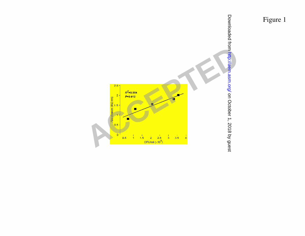
Figure 1
ACCEPTED on O
ctober 1, 2018 by guesthttp://aem
.asm.org/
Dow
nloaded from
Time
Abund
ance
Figure 2
ACCEPTED on O
ctober 1, 2018 by guesthttp://aem
.asm.org/
Dow
nloaded from

Figure 3
A B C
ACCEPTED on O
ctober 1, 2018 by guesthttp://aem
.asm.org/
Dow
nloaded from

Figure 4
BA
ACCEPTED on O
ctober 1, 2018 by guesthttp://aem
.asm.org/
Dow
nloaded from
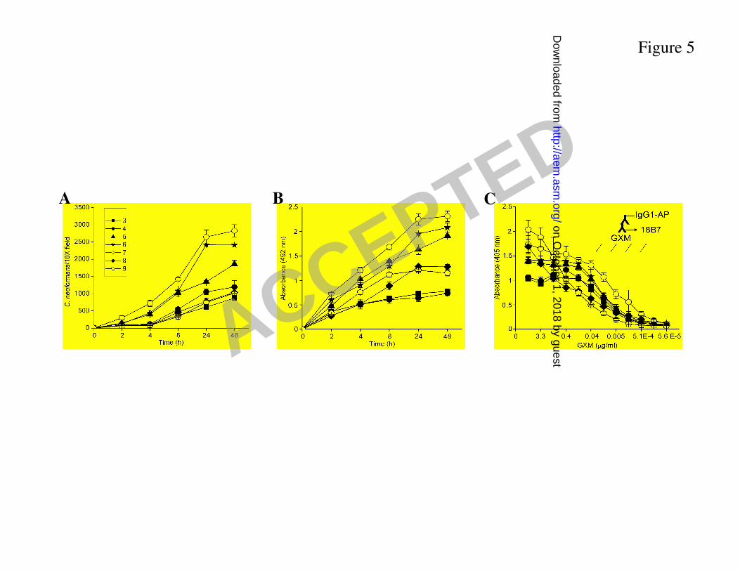
Figure 5
A B C
ACCEPTED on O
ctober 1, 2018 by guesthttp://aem
.asm.org/
Dow
nloaded from
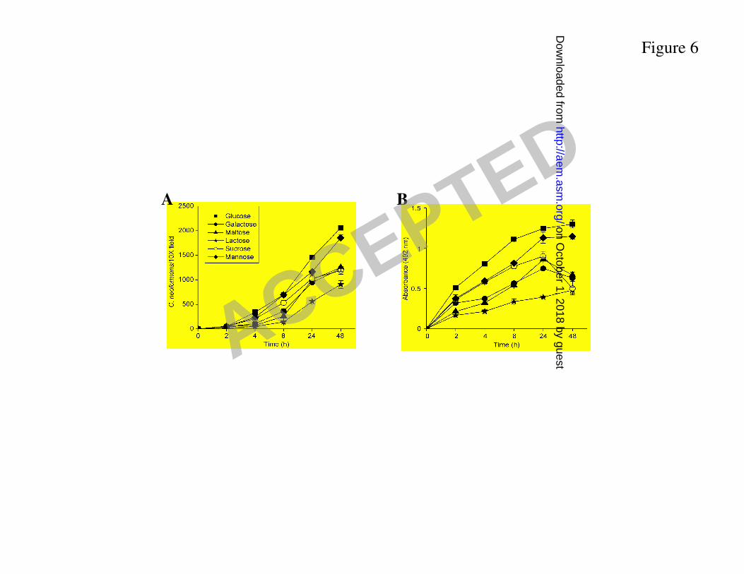
Figure 6
A B
ACCEPTED on O
ctober 1, 2018 by guesthttp://aem
.asm.org/
Dow
nloaded from

10 µm
A
1 µm
B
5 µm
C
Figure 7
ACCEPTED on O
ctober 1, 2018 by guesthttp://aem
.asm.org/
Dow
nloaded from

10 µm
B
50 µm
A
Figure 8
ACCEPTED on O
ctober 1, 2018 by guesthttp://aem
.asm.org/
Dow
nloaded from

A B C56
µm
Figure 9
ACCEPTED on O
ctober 1, 2018 by guesthttp://aem
.asm.org/
Dow
nloaded from

Figure 10
A B C
ED ACCEPTED on O
ctober 1, 2018 by guesthttp://aem
.asm.org/
Dow
nloaded from