crystal structure of the lytic chapkdomain of the endolysin lysk fromstaphylococcus...
TRANSCRIPT

Crystal structure of the lytic CHAPK domain of theendolysin LysK from Staphylococcus aureusbacteriophage KSanz-Gaitero et al.
Sanz-Gaitero et al. Virology Journal 2014, 11:133http://www.virologyj.com/content/11/1/133

Sanz-Gaitero et al. Virology Journal 2014, 11:133http://www.virologyj.com/content/11/1/133
RESEARCH Open Access
Crystal structure of the lytic CHAPK domain of theendolysin LysK from Staphylococcus aureusbacteriophage KMarta Sanz-Gaitero1, Ruth Keary2, Carmela Garcia-Doval1,3, Aidan Coffey2 and Mark J van Raaij1*
Abstract
Background: Bacteriophages encode endolysins to lyse their host cell and allow escape of their progeny. Endolysinsare also active against Gram-positive bacteria when applied from the outside and are thus attractive anti-bacterialagents. LysK, an endolysin from staphylococcal phage K, contains an N-terminal cysteine-histidine dependentamido-hydrolase/peptidase domain (CHAPK), a central amidase domain and a C-terminal SH3b cell wall-bindingdomain. CHAPK cleaves bacterial peptidoglycan between the tetra-peptide stem and the penta-glycine bridge.
Methods: The CHAPK domain of LysK was crystallized and high-resolution diffraction data was collected both froma native protein crystal and a methylmercury chloride derivatized crystal. The anomalous signal contained in thederivative data allowed the location of heavy atom sites and phase determination. The resulting structures werecompleted, refined and analyzed. The presence of calcium and zinc ions in the structure was confirmed by X-rayfluorescence emission spectroscopy. Zymogram analysis was performed on the enzyme and selected site-directedmutants.
Results: The structure of CHAPK revealed a papain-like topology with a hydrophobic cleft, where the catalytictriad is located. Ordered buffer molecules present in this groove may mimic the peptidoglycan substrate. Whencompared to previously solved CHAP domains, CHAPK contains an additional lobe in its N-terminal domain, with astructural calcium ion, coordinated by residues Asp45, Asp47, Tyr49, His51 and Asp56. The presence of a zinc ion inthe active site was also apparent, coordinated by the catalytic residue Cys54 and a possible substrate analogue.Site-directed mutagenesis was used to demonstrate that residues involved in calcium binding and of the proposedactive site were important for enzyme activity.
Conclusions: The high-resolution structure of the CHAPK domain of LysK was determined, suggesting the location ofthe active site, the substrate-binding groove and revealing the presence of a structurally important calcium ion. A zincion was found more loosely bound. Based on the structure, we propose a possible reaction mechanism. Future studieswill be aimed at co-crystallizing CHAPK with substrate analogues and elucidating its role in the complete LysK protein.This, in turn, may lead to the design of site-directed mutants with altered activity or substrate specificity.
Keywords: Bacteriophage, Calcium, Crystallography, Endolysin, Peptidoglycan, Protease, Staphylococcus, Zinc
* Correspondence: [email protected] de Estructura de Macromoleculas, Centro Nacional deBiotecnologia (CNB–CSIC), Calle Darwin 3, E-28049 Madrid, SpainFull list of author information is available at the end of the article
© 2014 Sanz-Gaitero et al.; licensee BioMed Central Ltd. This is an Open Access article distributed under the terms of theCreative Commons Attribution License (http://creativecommons.org/licenses/by/4.0), which permits unrestricted use,distribution, and reproduction in any medium, provided the original work is properly credited. The Creative Commons PublicDomain Dedication waiver (http://creativecommons.org/publicdomain/zero/1.0/) applies to the data made available in thisarticle, unless otherwise stated.

Sanz-Gaitero et al. Virology Journal 2014, 11:133 Page 2 of 10http://www.virologyj.com/content/11/1/133
Spanish abstract
Introducción: Los bacteriófagos codifican endolisinas para lisar sus bacterias hospedadoras y permitir la liberaciónde su progenie. Las endolisinas también son activas contra bacterias Gram positivas cuando se aplican desde elexterior, y por lo tanto, son consideradas agentes antibacterianos atractivos. LysK, una endolisina del fago K queinfecta estafilococos, contiene un dominio N-terminal amidohidrolasa/peptidasa dependiente de cisteína e histidina(CHAPK), un dominio amidasa central y un dominio C-terminal SH3b de unión a la pared bacteriana. CHAPK corta elpeptidoglicano bacteriano entre el tetrapéptido y los puentes pentaglicina.
Métodos: El dominio CHAPK de LysK fue cristalizado y se obtuvieron datos de difracción a alta resolución tanto deun cristal de proteína nativo como de un cristal derivado con cloruro de metilmercurio. La señal anómala presenteen los datos derivados permitió la localización de la posición de los átomos pesados y la determinación de la fase.Las estructuras resultantes se completaron, refinaron y analizaron. La presencia de iones de calcio y zinc en laestructura fue confirmada por espectroscopía de emisión de fluorescencia de rayos X. Se llevaron a cabo análisis dezimograma sobre la enzima nativa y sobre mutantes puntuales seleccionados.
Resultados: La estructura de CHAPK reveló una topología tipo papaína con un bolsillo hidrofóbico donde selocaliza la tríada catalítica. Moléculas de tampón ordenadas presentes en este hueco pueden mimetizar elsubstrato de peptidoglicano. Cuando se compara con dominios CHAP resueltos previamente, CHAPK contiene unlóbulo adicional en su dominio N-terminal, con un ión de calcio estructural, coordinado por los residuos Asp56,Asp45, Asp47. También se observa la presencia de un ión de zinc en el centro activo, coordinado con el residuocatalítico Cys54 y un posible análogo del substrato. Se usó mutagénesis dirigida para demostrar que los residuosinvolucrados en la unión a calcio y los presentes en el centro activo propuesto eran importantes para la actividadenzimática.
Conclusiones: Se determinó la estructura del dominio CHAPK de LysK a alta resolución, sugiriendo la localizacióndel centro activo, del bolsillo de unión al sustrato y revelando la presencia de un ión de calcio estructuralmenteimportante. Se encontró un ión de zinc unido más débilmente. Basándonos en la estructura, proponemos unposible mecanismo de reacción. Futuros estudios tendrán por objeto la cristalización de CHAPK con análogos delsustrato y la elucidación de su papel en la proteína LysK completa. Esto, a su vez, podría conducir al diseño demutantes puntuales con una actividad o especificidad de sustrato modificada.
BackgroundBacteriophage K is a virulent phage that infects a widerange of staphylococci. It belongs to the Myoviridaefamily of the Caudovirales order, with a genome of148,317 bp [1-3]. To allow its progeny to escape fromthe host cell (“lysis from within”), it encodes the endoly-sin LysK, a peptidoglycan hydrolase [4]. When appliedexogenously to the pathogen, LysK causes “lysis fromwithout” or exolysis [5]. Gram-positive endolysins arehighly specific [4], and no bacterial variants resistant totheir phage endolysins have been found despite the useof mutagenesis strategies to promote the chance ofresistance development [6]. LysK kills a wide range ofstaphylococci, including multi-drug-resistant Staphylo-coccus aureus (MRSA) [7].LysK contains three domains: an N-terminal cysteine-
histidine dependent amido-hydrolase/peptidase (CHAP)domain, a central amidase domain and a C-terminalSH3b cell wall-binding domain. The LysK amidase do-main cleaves peptidoglycan between N-acetylmuramicacid and L-alanine of the stem peptide, while theCHAP domain hydrolyzes it between the D-alanineof the tetra-peptide stem and the first glycine of the
penta-glycine cross-bridge [8]. A truncated enzymecalled CHAPK, containing only the first 165 aminoacids of LysK corresponding to the CHAP domain, alsoshowed exolytic activity [9]. CHAPK is able to lyse sev-eral staphyloccocal species, independently from theirorigin, their antibiotic resistance profile and their ability toproduce exopolysaccharides (associated with biofilm for-mation) [10,11]. It is also effective against other relatedgenera, such as Micrococcus or Streptococcus [7].In order to understand the reaction mechanism and
perhaps improve or alter the activity, we set out tosolve the structure of CHAPK. The CHAPK domainwas expressed in Escherichia coli, purified and crystal-lized. Although the crystallization procedure was notvery reproducible and crystals grew as inter-grownplates, a high-resolution dataset could be collectedfrom one of them, plus a dataset from a methylmercurychloride derivative of sufficient quality for structuresolution by single-wavelength anomalous dispersion[12]. This structure was refined against both the nativeand the derivative dataset. Here we present the high-resolution structure of the CHAPK domain solved byX-ray crystallography.

Sanz-Gaitero et al. Virology Journal 2014, 11:133 Page 3 of 10http://www.virologyj.com/content/11/1/133
Results and discussionOverall structureThe final models of the CHAPK enzyme contain aminoacids 2–165 for each of the four protein moleculespresent in the crystallographic asymmetric units, withgood crystallographic statistics and reasonable proteingeometry (Table 1). The models also contain metal ions,waters and other solvent molecules. For the native struc-ture, a calcium ion, a zinc ion and a 2-(N-morpholino)ethanesulfonic acid (MES) molecule have been modelledassociated with each of the protein chains, as discussedbelow. Other ordered solvent molecules have also beenmodelled in the asymmetric unit and consist of oneglycerol molecule, four putative sodium ions and 741water molecules. For the derivative structure, a calciumion and a 2-[4-(2-hydroxyethyl)piperazin-1-yl] ethane-sulfonic acid (HEPES) molecule have been modelledassociated with each of the protein chains, while Cys54is modelled as methylmercury-cysteine. In this case,ordered solvent molecules modelled in the unit cellinclude two glycerol molecules, ten additional putative
Table 1 Refinement and validation statistics for theCHAPK structure
Native Derivative
PDB code 4CSH 4CT3
Space group P1 P1
Cell edges (a, b, c, Å) 39.2, 61.5, 73.2 39.0, 61.5, 72.8
Cell angles (α, β, γ, º) 91.5, 98.7, 90.1 91.8, 98.7, 90.0
Resolution range used (Å) 32.9-1.79 (1.88-1.79)a 61.5-1.69 (1.78-1.69)
Multiplicity 2.0 (1.9) 3.4 (3.2)
Completeness 97.2 (94.3) 64.7 (10.4)
Mean <I/sigma(I)> 6.3 (2.9) 11.8 (1.8)
Rsym (%)b 9.1 (25.1) 6.0 (62.0)
Number of reflections used 59686 (8628) 46067 (1349)
Number of reflections usedfor R-free
2338 (112) 2431 (62)
R-factorc 0.175 (0.233) 0.181 (0.278)
R-free 0.201 (0.282) 0.224 (0.295)
Number of atoms(protein/water/other)
5286/741/66 5259/770/104
Average B-value/WilsonB-value (Å2)
18.4/14.5 22.2/14.9
Ramachandran statisticsd (%) 97.7/100.0 98.2/100.0
R.m.s. deviationse
(bonds, Å/angles, °)0.015/1.49 0.012/1.37
aValues in parentheses are for the highest resolution bin, where applicable.bRsym=ΣhΣi|Ihi-<Ih>|/ΣhΣi|Ihi|, where Ihi is the intensity of the ith measurement ofthe same reflection and <Ih> is the mean observed intensity for that reflection.cR=Σ||Fobs(hkl)|-|Fcalc(hkl)||/Σ|Fobs(hkl)|.dDetermined with MOLPROBITY. The percentages are indicated of residues infavoured and allowed regions of the Ramachandran plot, respectively.eProvided by REFMAC.
methylmercury ions, two putative chloride ions and770 waters. Despite the lower nominal resolution of thenative dataset when compared with the derivative (1.8vs. 1.7 Å), the general structural analyses describedbelow are done using the structure refined against thenative dataset, as that dataset is more complete (97.2vs. 64.7%), contains more measured reflections (62028vs. 48498) [10], and better maps with less non-interpretable noise peaks were obtained.The four CHAPK monomers do not form extensive
inter-monomer interfaces in the crystal, suggesting thatin solution the protein is monomeric. When the fourcrystallographically independent monomers are com-pared with each other, it is observed that they are verysimilar. While in part this is due to the use of localnon-crystallographic symmetry restraints in the refine-ment, the fact that including these restraints signifi-cantly improved correspondence of the model to thedata supports the similarity of the four crystallograph-ically independent protein chains. Chains A and B onone hand, and chains C and D on the other, can bemost reliably superposed, with root mean square differ-ences (r.m.s.d.) between C-alpha atoms of 0.07 and0.05 Å, respectively. The r.m.s.d. between chains A or Bon one hand and chains C or D on the other are 0.23-0.26 Å. The largest structural differences are concen-trated in residues 29–39 and 136–143, part of surfaceloops that interact with each other. These differencesbetween the monomers are likely caused by interactionwith neighbouring monomers in the crystal, i.e. differ-ent crystal contacts. The loop consisting of residues136 to 143 is right next to a putative substrate-bindinggroove, so it may be somewhat more flexible to allowaccess of the substrate and release of the cleavageproducts.The CHAPK protein consists of a single globular do-
main that contains two alpha-helices, two 310-helicesand six beta-strands (Figure 1A and B). The amino-terminal part of the protein consists of the two alpha-helices (I and II) interconnected by a long loop. Thislong loop borders a groove in the protein, at the bot-tom of which the catalytic site is located (see below).Another loop, containing a 310-helix, connects thisamino-terminal part of the protein to a six-strandedbeta-sheet that forms the carboxy-terminal part. Thesix beta-strands are arranged in an anti-parallel beta-sheet in the topology AFBCDE (Figure 1B). The struc-ture of CHAPK had previously been predicted by insilico modelling [13]. The six-stranded beta-sheet waspredicted well, but the amino-terminal alpha-heliceswere incorrectly placed and the calcium-binding loopbetween them was not present in the model. The mainchain atoms of the catalytic site residues were within2 Å of their predicted positions.
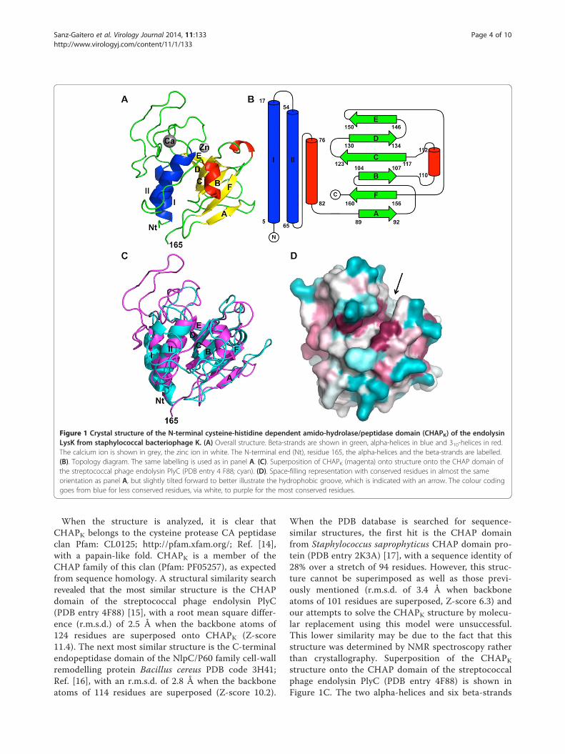
Figure 1 Crystal structure of the N-terminal cysteine-histidine dependent amido-hydrolase/peptidase domain (CHAPK) of the endolysinLysK from staphylococcal bacteriophage K. (A) Overall structure. Beta-strands are shown in green, alpha-helices in blue and 310-helices in red.The calcium ion is shown in grey, the zinc ion in white. The N-terminal end (Nt), residue 165, the alpha-helices and the beta-strands are labelled.(B). Topology diagram. The same labelling is used as in panel A. (C). Superposition of CHAPK (magenta) onto structure onto the CHAP domain ofthe streptococcal phage endolysin PlyC (PDB entry 4 F88; cyan). (D). Space-filling representation with conserved residues in almost the sameorientation as panel A, but slightly tilted forward to better illustrate the hydrophobic groove, which is indicated with an arrow. The colour codinggoes from blue for less conserved residues, via white, to purple for the most conserved residues.
Sanz-Gaitero et al. Virology Journal 2014, 11:133 Page 4 of 10http://www.virologyj.com/content/11/1/133
When the structure is analyzed, it is clear thatCHAPK belongs to the cysteine protease CA peptidaseclan Pfam: CL0125; http://pfam.xfam.org/; Ref. [14],with a papain-like fold. CHAPK is a member of theCHAP family of this clan (Pfam: PF05257), as expectedfrom sequence homology. A structural similarity searchrevealed that the most similar structure is the CHAPdomain of the streptococcal phage endolysin PlyC(PDB entry 4F88) [15], with a root mean square differ-ence (r.m.s.d.) of 2.5 Å when the backbone atoms of124 residues are superposed onto CHAPK (Z-score11.4). The next most similar structure is the C-terminalendopeptidase domain of the NlpC/P60 family cell-wallremodelling protein Bacillus cereus PDB code 3H41;Ref. [16], with an r.m.s.d. of 2.8 Å when the backboneatoms of 114 residues are superposed (Z-score 10.2).
When the PDB database is searched for sequence-similar structures, the first hit is the CHAP domainfrom Staphylococcus saprophyticus CHAP domain pro-tein (PDB entry 2K3A) [17], with a sequence identity of28% over a stretch of 94 residues. However, this struc-ture cannot be superimposed as well as those previ-ously mentioned (r.m.s.d. of 3.4 Å when backboneatoms of 101 residues are superposed, Z-score 6.3) andour attempts to solve the CHAPK structure by molecu-lar replacement using this model were unsuccessful.This lower similarity may be due to the fact that thisstructure was determined by NMR spectroscopy ratherthan crystallography. Superposition of the CHAPKstructure onto the CHAP domain of the streptococcalphage endolysin PlyC (PDB entry 4F88) is shown inFigure 1C. The two alpha-helices and six beta-strands

Sanz-Gaitero et al. Virology Journal 2014, 11:133 Page 5 of 10http://www.virologyj.com/content/11/1/133
of CHAPK superpose quite well with the backbone ofthe homologous structures, but the loops, including the310-helices, are very different.The globular CHAPK protein has a relatively long and
deep hydrophobic groove. When sequence conservationis mapped onto the surface, one notices that several resi-dues lining the groove are highly conserved (Figure 1D;the sequence alignment underlying this figure is inAdditional file 1: Table S1). In the native structure, aMES molecule is located in this groove (Figure 2, PDBentry 4CSH), while in the derivative structure a HEPESmolecule is present (PDB entry 4CT3). These moleculesmay well be mimicking the natural peptidoglycan sub-strate of the protein. Residues in the groove that mightcontact the peptidoglycan substrate are: Phe36, Asp47,Tyr49, Tyr50, Gln53 and Cys54 from the loop betweenhelices 1 and 2; Asp56 and Thr59 from helix 2; Arg71,Trp73 and Asn75 from the loop between helix 2 andbeta-strand A; Trp115 and His117 from the BC-loopand Asn136 and Trp137 from the DE-loop.
Bound metal ionsWhile building and refining the protein model, relativelystrong density peaks were observed near the terminalatoms of the side-chains of Cys54 and Asp56 in each ofthe four protein chains in the asymmetric unit, suggest-ing the presence of metal ions. X-ray fluorescence spec-troscopy is a powerful method to identify trace elementsin biological samples [18]. Therefore, we recorded an X-ray fluorescence spectrum from a frozen native CHAPKprotein crystal, which revealed significant amounts of
Figure 2 MES buffer molecule bound to the CHAPK enzymeputative substrate binding site. The CHAPK protein is shown intransparent surface and secondary structure cartoon representation;the calcium ion is also shown.
zinc and calcium (Figure 3A). Sulphur (from methionine,cysteine residues and buffer molecules) and chlorine(from the crystallization buffer) were also detected. Thepresence of trace amounts of titanium and copper islikely the result of interaction of the beam with certainbeamline or sample holder components not related tothe sample.The calcium ion is bound in the amino-terminal part
of the protein, involving residues of the long loopconnecting the first and second alpha-helices (residues17–54) and Asp56 in the second alpha-helix. It isbound in a monodentate way to the side chain of resi-dues Asp45 and Asp47 and in a bidentate way to bothoxygen atoms of the Asp56 side chain (Figure 3B).Additional ligands are the main chain oxygen atomsof Tyr49 and His51 and an ordered water molecule.The coordination is octahedral and almost exclusivelyinvolves carbonyl oxygen atoms, as expected for calcium.Experimentally determined metal ion-oxygen distancesare 2.3-2.5 Å, which is also consistent with usual calcium(II) coordination [19]. The occupancy of the calciumsite appears to be complete and the refined temperaturefactors of the calcium ions are very near those of thecoordinating atoms (the temperature factors for thecalcium ions vary between 10 and 12 Å2, while those forthe coordinating ligand atoms are between 7 and14 Å2). The calcium ion is near the proposed catalyticsite (Figure 2). We propose that the calcium ion plays astructural role, helping to maintain the structure of theamino-terminal domain and thus its catalytic residuesin the correct relative orientation. The calcium ionbinding loop also contains residues that may be incontact with the substrate and thus play a role in deter-mining substrate specificity. In the derivative proteinstructure, the calcium is present at the same occupancyand with the same coordinating ligands.In contrast to the tightly bound calcium ion, the zinc
ions appear to be bound more loosely and the derivativestructure shows they could be replaced by methylmer-cury ions upon soaking of the crystals with methylmer-cury chloride. Also, the occupancy appears to be lessthan unity, we estimate it to be around 0.67 based onrefinement runs performed at different occupancies.Finally, the resulting electron density around the zincions is somewhat ambiguous and we could not modelthe ligands without some remaining uncertainty. Thezinc ions are coordinated by the sulphydryl group ofCys54, the sulphate group of the bound MES and severalwater molecules (Figure 3C). It is also near the mainchain oxygen atom of Gly116. The coordination dis-tances for the zinc ion are not ideal; the zinc ion is tooclose to Cys54 and too far from the coordinating oxygenatoms. A report by another group showed that zinc ionsinhibit the LysK enzyme, while calcium ions have no

Figure 3 Presence of metal ions in the CHAPK crystal structure. A. X-ray fluorescence emission spectrum collected from a CHAPK crystalirradiated with monochromatic synchrotron radiation (12.7 KeV). B. Detail of the calcium ion coordination. Coordinating atoms are one Oδ atomof each of Asp45 and Asp47 residues, both Oδ atoms of Asp56, the main chain oxygen atoms of Tyr49 and His51 and an ordered water molecule(behind the calcium ion in this view). C. Detail of the zinc coordination. The zinc ion is sandwiched between Cys54 and the sulphate group ofthe MES ion, about 10 Å away from the calcium ion.
Sanz-Gaitero et al. Virology Journal 2014, 11:133 Page 6 of 10http://www.virologyj.com/content/11/1/133
effect on activity, but significantly enhance stability ofthe enzyme [20]. However, in this assay, metal ions werenot removed from the protein solution prior to testingtheir effects on the enzyme. Zinc ions may play a regula-tory role, and their binding near Cys54 suggests theymay regulate access of the substrate to the catalytic site.The importance of the calcium ion in relation to the
catalytic ability of CHAPK was investigated by creationof mutants containing a single amino acid change toalanine at each of the five residues involved in calciumcoordination. Zymogram analysis demonstrated thatmutation of residues Asp45, Asp47 and Asp56 resultedin the complete abolishment of the staphylolytic activ-ity of the enzyme (Figure 4). This result indicates thatthe coordinated calcium ion is essential for the catalyticmechanism of the enzyme and complements a previousstudy, which showed that the chelator EDTA was ableto reduce CHAPK activity by 99% [21]. While mutantHis51-Ala retained staphylolytic ability, activity of theenzyme was visibly reduced in comparison with theparental CHAPK. Mutation of Tyr49 to alanine did notappear to affect the staphylolytic ability of the enzymeas the clearing produced on a zymogram gel was compar-able to that seen for non-mutated CHAPK (Figure 4). Thefact that mutants His51-Ala and Tyr49-Ala retained
activity while the other mutants did not may be ex-plained by the fact that main chain oxygen atoms areinvolved in coordination as opposed to the side chainoxygens. Therefore these residues are more amenableto substitution without eliminating catalytic activity.
Catalytic centre and proposed reaction mechanismBy comparing the CHAPK protein with other proteinswith a similar function and structure (endolysins, CHAPdomains and others) and by doing an alignment be-tween them, we can deduce that the catalytic residuesare highly conserved. In the CHAP domain of Staphylo-coccus saprophyticus (PDB code 2K3A), the authorsdescribe the presence of a proteolytic triad formed byCys57, His109 and Glu126 [17], a catalytic triad alsofound in other members of the CA clan. In the streptococ-cal phage lysin PlyC (PDB code 4 F88), the catalytic resi-dues are Cys333 and His420 [15], while in NlpC/P60domain of lipoprotein SPR from E. coli (PDB code 3H41)the catalytic residues are Cys68, His119 and His339 [22].In CHAPK these residues correspond in the alignment toCys54 located in the second alpha-helix, His117 in beta-strand C and Glu134 in beta-strand D, making theseamino acids good candidates to form the catalytic triad ofthe enzyme (Figure 5). These hypothetical catalytic

Figure 4 Overexpression and activity of CHAPK mutants. A. Sodium dodecyl sulphate polyacryalamide electrophoresis gel of lysatescontaining over-expressed CHAPK and site-directed mutants. A control not expressing CHAPK is also included. B. Composite zymogram gel ofCHAPK, site-directed mutant CHAPK variants and negative control expression lysates.
Sanz-Gaitero et al. Virology Journal 2014, 11:133 Page 7 of 10http://www.virologyj.com/content/11/1/133
residues are close to the hydrophobic cleft, which supportsthe possibility that the catalytic part of the molecule is lo-cated in the hydrophobic groove. The predicted pKa ofHis117 is 9.3. This value contrasts with those of the rest ofhistidines in the protein: His51 (pKa 5.4), His91 (pKa 6.8)and His 157 (pKa 5.2). His117 may thus be protonated atphysiological pH.Mutation of the conserved Cys54 and His117 residues
to alanine resulted in complete elimination of staphylolyticactivity of the enzyme as demonstrated by zymographicanalysis, indicating an essential role of these residues andsupporting the hypothesis that they are part of the cata-lytic triad. Glu134 is believed to be the other residue ofthe catalytic triad, but is not as highly conserved as the
Figure 5 The proposed catalytic triad of the bacteriophage Kendolysin CHAP domain CHAPK. Cys54 (bottom), His117 (middle)and Glu134 (top) and the distances between them (in Å) are shown.
other two residues. When this residue was mutated to ala-nine, it was clear from zymogram results that, althoughthe catalytic activity was not completely eliminated, it wasstrongly reduced. In the absence of Glu134 perhaps an-other residue can take over its role.A likely mechanism of action, analogous to that of
other papain proteases [23,24], is the following: Glu134accepts a proton from the protonated imidazole groupof His117. His117 subsequently accepts a proton fromthe hydroxyl group of Cys54 (through its N-epsilon).The deprotonated Cys54 then performs a nucleophilicattack on the peptidic bond between D-Ala and Gly inthe staphylococcal peptidoglycan. As a result, a transa-cylation reaction between the enzyme and substrateoccurs, giving rise to an acyl-enzyme intermediate.This intermediate may be hydrolyzed to release the en-zyme and the cleaved peptidoglycan [25]. In the NlpC/P60 domain of lipoprotein SPR from E. coli, there is atyrosine residue (Tyr56) that has been reported to bevery conserved and which may modulate Cys nucleophi-licity or help in substrate binding [22]. In the case ofCHAPK, Tyr140 is located in an equivalent position, buthaving a different role, since its phenol group is pointingin the opposite direction. Cysteine proteases have an oxy-anion hole, which helps to stabilize the developing nega-tive charge during the formation of the acylenzymeintermediate [26]. Asn136, which is located in close prox-imity to the catalytic triad, is one residue hypothesizedto be involved in creating the oxyanion hole. Whenthis residue was mutated to an alanine, the activity ofthe enzyme was visibly reduced, but not completelyeliminated, supporting the aforementioned hypothesis.
Comparison with LysGH15 CHAP domain structureWhile this manuscript was under review, a paper de-scribing the structures of the CHAP domain (PDB entry4 OLK), amidase-2 domain (PDB entry 4OLS) and the

Sanz-Gaitero et al. Virology Journal 2014, 11:133 Page 8 of 10http://www.virologyj.com/content/11/1/133
SH3 domain (PDB entry 2MK5) of the endolysinLysGH15 from phage GH15 was published [27]. Thefirst two were solved by X-ray crystallography at 2.7and 2.2 Å resolution respectively, while the latter wassolved by NMR spectroscopy. Phages GH15 and Kshare 97% identity in 84% of their genomes (Genbankentries NC_019448 and NC_005880, respectively)[2,28]. The LysGH15 and LysK protein sequences arevirtually identical, with only four amino acid differ-ences in their 495-residue sequences. Of the differ-ences, two are in the CHAP domain: Val26 of CHAPKis an isoleucine in CHAPGH15 and Glu113 of CHAPKis a glutamine in CHAPGH15. The high sequence simi-larity means the enzymes are almost identical andexpected to share the same properties.When the crystal structures of the CHAP domains are
compared, it is notable the spacegroups and crystalpacking are very different, which suggests the protein isa monomer in solution and inter-monomer interactionsin the crystal are not likely to be biologically relevant.Given the almost identical sequences, it is not surprisingthat the monomer structures are highly similar; super-position of the two CHAP domains leads to an r.m.s.d.of 0.3 Å when 139 C-alpha atoms are superposed. Theonly significant difference in main-chain conformation ispresent in residues 109–116, which follow a differentpath in the two structures. This may indicate that thisloop, which is directed away from the active site, is flex-ible and of limited importance to the structure andactivity of the enzyme. The large side-chains of Tyr49,Trp73, Tyr140 and Tyr153, which are all on the surfaceof the protein, show different orientations.The higher resolution of the CHAPK structure when
compared to the CHAPGH15 structure (1.8 vs. 2.7 Å)should have led to more accurate placement of side-chain atoms and solvent molecules. In both structures, abuffer molecule occupies the groove that likely accom-modates the peptidoglycan substrate: a Bis-Tris molecule(2-[Bis(2-hydroxyethyl)amino]-2-(hydroxymethyl)-1,3-pro-panediol) in between the two monomers of the asymmet-ric unit of CHAPGH15 and a MES and HEPES molecule inthe case of the native and derivative structures of CHAPK,respectively. The calcium ion is in exactly the same pos-ition, as are its coordinating residues and the EF-hand-likedomain in which it is incorporated. No zinc ion was ob-served in the CHAPGH15 crystals.Gu et al. also performed site-directed mutagenesis stud-
ies [27], but on the intact LysGH15 enzyme, not on theisolated CHAPGH15 domain. As observed for CHAPK, itwas found that mutating the active site residue Cys54affected bacterial lysis activity strongly. Mutating thecalcium ion coordinating residues Asp45, Asp46 andAsp56 also diminished activity about ten-fold, while Tyr49and His51 seem less important, the same as we observed.
ConclusionsWe determined the structure of the CHAPK domain ofLysK at 1.8 Å resolution (1 Å = 0.1 nm). The structurehas the papain-type fold with a long loop between thetwo amino-terminal alpha-helices. The structure sug-gests the location of the active site near a hydrophobicgroove, with Cys54, His117 and Glu134 forming thecatalytic triad. The substrate most likely binds to thehydrophobic groove.A calcium ion was found tightly bound to the protein.
Its ligands are the side-chains of Asp45, Asp47 andAsp56, plus the backbone oxygens of Tyr49 and His51,all in the amino-terminal domain specific to CHAPK. Itlikely has a structural role, stabilizing the protein fold. Itmay also be involved in ensuring the correct location ofthe peptidoglycan inside the catalytic cleft or in thestabilization of the negative charge of the tetrahedralintermediate during catalysis. A zinc ion was also foundand is likely more loosely bound, as it is less buried, hasless protein ligands and could be exchanged for a meth-ylmercury ion upon derivatization. Its role, if any, maybe regulatory.Based on the structure, we propose a possible reaction
mechanism, involving all three residues of the likely cata-lytic triad. Future studies will include co-crystallizationwith peptidoglycan analogues and elucidating the role ofthe CHAPK domain in the complete LysK protein. Thismay allow site-directed mutation to modulate the pep-tidoglycan specificity and activity of both the CHAPKand LysK enzymes.
MethodsCHAPK was expressed, purified, crystallized and crystal-lographic data was collected as described [9,12]. Acomplete native dataset was collected to 1.8 Å resolutionwith good statistics. A dataset to 1.7 Å resolution, butwith inferior completeness, was also collected from amethylmercury chloride derivative at the Hg L-I edge[12]. However, this dataset allowed phase determinationby single anomalous dispersion (SAD) and automaticmodel building of four crystallographically independentprotein molecules in the P1 unit cell [12] (Table 1) usingthe ARP-WARP program [29]. The model was refinedagainst the derivative dataset and separately against thenative dataset. The models were completed and adjustedusing COOT [30] and refined with REFMAC5, usinglocal non-crystallographic symmetry restraints [31] andtaking care to select the same reflections for calculationof Rfree [32]. To confirm the presence of zinc and cal-cium ions in the sample, an X-ray fluorescence emissionspectrum was collected on a native protein crystal atESRF beamline ID23-1 [33]. Validation was performedwith MolProbity [34]. Refinement and validation statis-tics are shown in Table 1.
Sanz-Gaitero et al. Virology Journal 2014, 11:133 Page 9 of 10http://www.virologyj.com/content/11/1/133
Crystal contact analysis was done with PISA [35]; otheranalyses were performed with the CCP4 suite [36]. Struc-tural similarity analysis was performed with DALI [37]; forplotting a protein surface coloured according to amino acidconservation, CONSURF was used [38]. The pKa of se-lected residues in the protein structure was predicted withPROPKA [39]. The structural models and underlying datafiles have been submitted to the PDB (accession code4CSH for the native structure and 4CT3 for the derivative).PYMOL (Schrödinger LLC, Portland OR, USA) was usedfor making structure figures and TOPDRAW [40] to drawthe secondary structure diagram.CHAPK mutants were created using the QuikChange II
Site-Directed Mutagenesis Kit from Agilent (Santa ClaraCA, USA) as per the manufacturer’s instructions. Crudecell lysate was analyzed for over-expression using sodiumdodecyl sulphate gel electrophoresis and for ability tolyse Staphylococcus aureus cells using zymographic gelsas described previously [41].
Additional file
Additional file 1: Table S1. Sequence aligment underlining the colourcoding of Figure 1D.
Competing interestsThe authors declare that they have no competing interests.
Authors’ contributionsRK purified the protein and performed site directed mutagenesis andzymogram activity tests; MSG crystallized the protein. MSG and CGDcollected X-ray diffraction and fluorescence data. MSG, CGD and MJvRperformed structure solution and refinement. MSG, RK, AC and MJvRanalyzed the structure. MSG and MJvR drafted the first version of themanuscript. AC initiated the project, while AC and MJvR supervised it. Allauthors helped to write and improve the manuscript and approved thefinal version.
AcknowledgementsWe thank Jordi Benach (ALBA beamline BL13/XALOC), Max Nanao (ESRFbeamline ID23-2), Christian Perrin (ESRF), Christoph Mueller-Dieckmann(ESRF beamline ID23-1) and James Sandy (DLS beamline I02) for help withusing synchrotron data collection facilities. We acknowledge ALBA/CELLS(proposal number 2012010140), the European Synchrotron Radiation Facility(proposal number MX1477) and the Diamond Light Source (proposal numberMX3808), which contributed to the results presented here. The research leadingto these results has received funding from the European Community’s SeventhFramework Programme (FP7/2007-2013) under BioStruct-X grant agreement no.283570 and was sponsored by grant BFU2011-24843 (MJvR) from the SpanishMinistry of Economy and Competitiveness, a Masters fellowship (MSG) and anFPU Ph.D. fellowship (CGD) from the Spanish Ministry of Education, Culture andSports. We also acknowledge financial support from TSR-StrandIII:CRS/07/CR03and FIRM:08RDCIT600 of the Irish Department of Agriculture.
Author details1Departamento de Estructura de Macromoleculas, Centro Nacional deBiotecnologia (CNB–CSIC), Calle Darwin 3, E-28049 Madrid, Spain.2Department of Biological Sciences, Cork Institute of Technology,Bishopstown, Cork, Ireland. 3Current address: Department of Biochemistry,University of Zurich, Zurich, Switzerland.
Received: 28 April 2014 Accepted: 4 July 2014Published: 26 July 2014
References1. Rees PJ, Fry BA: The morphology of staphylococcal bacteriophage K and
DNA metabolism in infected Staphylococcus aureus. J Gen Virol 1981,53(Pt 2):293–307.
2. O’Flaherty S, Coffey A, Edwards R, Meaney W, Fitzgerald GF, Ross RP:Genome of staphylococcal phage K: a new lineage of Myoviridaeinfecting Gram-positive bacteria with a low G + C content. J Bacteriol2004, 186(9):2862–2871.
3. Gill JJ: Revised genome sequence of staphylococcus aureusbacteriophage K. Genome Announcements 2014, 2(1):e01173.
4. Loessner MJ: Bacteriophage endolysins - current state of research andapplications. Curr Opin Microbiol 2005, 8(4):480–487.
5. Ralston DJ, McIvor M: Lysis-from-without of Staphylococcus aureus strainsby combinations of specific phages and phage-induced lytic enzymes.J Bacteriol 1964, 88:676–681.
6. Loeffler JM, Nelson D, Fischetti VA: Rapid killing of Streptococcuspneumoniae with a bacteriophage cell wall hydrolase. Science 2001,294(5549):2170–2172.
7. O’Flaherty S, Coffey A, Meaney W, Fitzgerald GF, Ross RP: The recombinantphage lysin LysK has a broad spectrum of lytic activity against clinicallyrelevant staphylococci, including methicillin-resistant Staphylococcusaureus. J Bacteriol 2005, 187(20):7161–7164.
8. Becker SC, Dong S, Baker JR, Foster-Frey J, Protchard DF, Donovan DM: LysKCHAP endopeptidase domain is required for lysis of live staphylococcalcells. FEMS Microbiol Lett 2009, 294(1):52–60.
9. Horgan M, O’Flynn G, Garry J, Cooney J, Coffey A, Fitzgerald GF, Ross RP,McAuliffe O: Phage lysin LysK can be truncated to its CHAP domain andretain lytic activity against live antibiotic-resistant staphylococci.Appl Environ Microbiol 2009, 75(3):872–874.
10. Fenton M, Casey PG, Hill C, Gahan CG, Ross RP, McAuliffe O, O’Mahony J,Maher F, Coffey A: The truncated phage lysin CHAP(k) eliminatesStaphylococcus aureus in the nares of mice. Bioeng Bugs 2010,1(6):404–407.
11. Fenton M, Keary R, McAuliffe O, Ross RP, O’Mahony J, Coffey A:Bacteriophage-derived peptidase CHAPK eliminates and preventsstaphylococcal biofilms. Internat J Microbiol 2013, 2013:625341.
12. Sanz-Gaitero M, Keary R, Garcia-Doval C, Coffey A, van Raaij MJ:Crystallization of the CHAP domain of the endolysin from Staphylococcusaureus bacteriophage K. Acta Crystallogr Sect F Struct Biol Cryst Commun2013, 69(Pt 12):393–1396.
13. Fenton M, Cooney JC, Ross RP, Sleator RD, McAuliffe O, O’Mahony J,Coffey A: In silico modeling of the staphylococcal bacteriophage-derivedpeptidase CHAPK. Bacteriophage 2011, 1(4):198–206.
14. Punta M, Coggill PC, Eberhardt RY, Mistry J, Tate J, Boursnell C, Pang N,Forslund K, Ceric G, Clements J, Heger A, Holm L, Sonnhammer EL, Eddy SR,Bateman A, Finn RD: The Pfam protein families database. Nucl Acids Res2012, 40:D290–D301.
15. McGowan S, Buckle AM, Mitchell MS, Hoopes JT, Gallagher DT, HeselpothRD, Shen Y, Reboul CF, Law RHP, Fischetti VA, Whisstock JC, Nelson DC:X-ray crystal structure of the streptococcal specific phage lysin PlyC.Proc Natl Acad Sci U S A 2012, 109(31):12752–12757.
16. Xu Q, Abdubek P, Astakhova T, Axelrod HL, Bakolitsa C, Cai X, Carlton D,Chen C, Chiu HJ, Chiu M, Clayton T, Das D, Deller MC, Duan L, Ellrott K, FarrCL, Feuerhelm J, Grant JC, Grzechnik A, Han GW, Jaroszewski L, Jin KK, KlockHE, Knuth MW, Kozbial P, Krishna SS, Kumar A, Lam WW, Marciano D, MillerMD, et al: Structure of the c-D-glutamyl-L-diamino acid endopeptidaseYkfC from Bacillus cereus in complex with L-Ala-c-D-Glu: insights intosubstrate recognition by NlpC/P60 cysteine peptidases. Acta CrystallogrSect F Struct Biol Cryst Commun 2010, 66(Pt 10):1354–1364.
17. Rossi P, Aramini JMR, Xiao R, Chen CX, Nwosu C, Owens LA, Maglaqui M,Nair R, Fischer M, Acton TB, Honig B, Rost B, Montelione GT: Structuralelucidation of the Cys-His-Glu-Asn proteolytic relay in the secreted CHAPdomain enzyme from the human pathogen staphylococcus saprophyticus.Proteins 2009, 74(2):515–519.
18. Jones KW, Gordon BM, Hanson AL, Kwiatek WL, Pounds JG: X-ray fluorescencewith synchrotron radiation. Ultramicroscopy 1988, 24(2–3):313–328.
19. Harding MM: Small revisions to predicted distances around metal sites inproteins. Acta Crystallogr Sect D Biol Crystallogr 2006, 62(Pt 6):678–682.
20. Filatova LY, Becker SC, Donovan DM, Gladilin AK, Klyachko NL: LysK, theenzyme lysing staphylococcus aureus cells: specific kinetic features andapproaches towards stabilization. Biochimie 2010, 92:507–513.

Sanz-Gaitero et al. Virology Journal 2014, 11:133 Page 10 of 10http://www.virologyj.com/content/11/1/133
21. Fenton M, Ross RP, McAuliffe O, O’Mahony J, Coffey A: Characterization ofthe staphylococcal bacteriophage lysin CHAPK. J Appl Microbiol 2011,111:1025–1035.
22. Aramini JM, Rossi P, Huang YJ, Zhao L, Jiang M, Maglaqui M, Xiao R, Locke J,Nair R, Rost B, Acton TB, Inouye M, Montelione GT: Solution NMR structureof the NlpC/P60 domain of lipoprotein Spr from Escherichia coli:structural evidence for a novel cysteine peptidase catalytic triad.Biochemistry 2008, 47(37):9715–9717.
23. Shokhen M, Khazanov N, Albeck A: The mechanism of papain inhibitionby peptidyl aldehydes. Proteins 2011, 79:975–985.
24. Brömme D: Papain-like cysteine proteases. Curr Protoc Protein Sci 2001,21:21.2.1–21.2.14.
25. Lau EY, Bruice TC: Consequences of breaking the Asp-His hydrogen bondof the catalytic triad: effects on the structure and dynamics of the serineesterase cutinase. Biophys J 1999, 77(1):85–98.
26. Menard R, Storer AC: Oxyanion hole interactions in serine and cysteineproteases. Biol Chem Hoppe Seyler 1992, 373(7):393–400.
27. Gu J, Feng Y, Feng X, Sun C, Lei L, Ding W, Niu F, Jiao L, Yang M, Li Y, Liu X,Song J, Cui Z, Han D, Du C, Yang Y, Ouyang S, Liu ZJ, Han W: Structuraland biochemical characterization reveals LysGH15 as an unprecedented“EF-hand-like” calcium-binding phage lysin. PLOS Path 2014, 10(5):e1004109.
28. Gu J, Liu X, Lu R, Li Y, Song J, Lei L, Sun C, Feng X, Du C, Yu H, Yang Y,Han W: Complete genome sequence of staphylococcus aureusbacteriophage GH15. J Virol 2012, 86(16):8914–8915.
29. Langer G, Cohen SX, Lamzin VS, Perrakis A: Automated macromolecularmodel building for X-ray crystallography using ARP/wARP version 7. NatProtoc 2008, 3(7):1171–1179.
30. Emsley P, Cowtan K: Coot: model-building tools for molecular graphics.Acta Crystallogr Sect D Biol Crystallogr 2004, 60(Pt 12 Pt 1):2126–2132.
31. Murshudov GN, Skubak P, Lebedev AA, Pannu NS, Steiner RA, Nicholls RA,Winn MD, Long F, Vagin AA: Refmac5 for the refinement of macromolecularcrystal structures. Acta Crystallogr Sect D Biol Crystallogr 2011, 67(Pt 4):355–367.
32. Brünger A: Free R value: a novel statistical quantity for assessing theaccuracy of crystal structures. Nature 1992, 355(6359):472–475.
33. Leonard GA, Solé VA, Beteva A, Gabadinho J, Guijarro M, McCarthy J,Marrocchelli D, Nurizzo D, McSweeney S, Mueller-Dieckmann S: Onlinecollection and analysis of X-ray fluorescence spectra on themacromolecular crystallography beamlines of the ESRF. J Appl Crystallogr2009, 42:333–335.
34. Chen VB, Arendall WB 3rd, Headd JJ, Keedy DA, Immormino RM, Kapral GJ,Murray LW, Richardson JS, Richardson DC: MolProbity: all-atom structurevalidation for macromolecular crystallography. Acta Crystallogr Sect D BiolCrystallogr 2010, 66(Pt 1):12–21.
35. Krissinel E, Henrick K: Inference of macromolecular assemblies fromcrystalline state. J Mol Biol 2007, 372(3):774–797.
36. Winn MD, Ballard CC, Cowtan KD, Dodson EJ, Emsley P, Evans PR, KeeganRM, Krissinel EB, Leslie AG, McCoy A, McNicholas SJ, Murshudov GN, PannuNS, Potterton EA, Powell HR, Read RJ, Vagin A, Wilson KS: Overview of theCCP4 suite and current developments. Acta Crystallogr Sect D BiolCrystallogr 2011, 67(Pt 4):235–242.
37. Holm L, Rosenström P: Dali server: conservation mapping in 3D. NucleicAcids Res 2010, 38(Web Server issue):W545–W549.
38. Armon A, Graur AD, Ben-Tal N: ConSurf: an algorithmic tool for theidentification of functional regions in proteins by surface mapping ofphylogenetic information. Bioinformatics 2003, 19(1):163–164.
39. Rostkowski M, Olsson MHM, Søndergaard CR, Jensen JH: Graphical analysisof pH-dependent properties of proteins predicted using PROPKA.BMC Struct Biol 2011, 11:6.
40. Bond CS: Topdraw: a sketchpad for protein structure topology cartoons.Bioinformatics 2003, 19(2):311–312.
41. Keary R, McAuliffe O, Ross RP, Hill C, O’Mahony J, Coffey A: Genome analysis ofthe staphylococcal temperate phage DW2 and functional studies on theendolysin and tail hydrolase. Bacteriophage 2014, 4(1):e28451.
doi:10.1186/1743-422X-11-133Cite this article as: Sanz-Gaitero et al.: Crystal structure of the lyticCHAPK domain of the endolysin LysK from Staphylococcus aureusbacteriophage K. Virology Journal 2014 11:133.
Submit your next manuscript to BioMed Centraland take full advantage of:
• Convenient online submission
• Thorough peer review
• No space constraints or color figure charges
• Immediate publication on acceptance
• Inclusion in PubMed, CAS, Scopus and Google Scholar
• Research which is freely available for redistribution
Submit your manuscript at www.biomedcentral.com/submit