ct imaging of presurgical evaluation of potential renal ... · urolithiasis, multiple renal...
TRANSCRIPT

CT imaging of presurgical evaluation of potential renal
transplant donors: What do surgeons should know?
C. Santos Montón, C. Manzano Rodriguez, K. El Karzazi, T. González
de la Huebra Labrador, A. Herrero Hernandez, J. F. Ojeda Esparza;
Salamanca/ES
Learning objectives
The purpose of this exhibit is:
1. To know the most common anatomic variants of the renal circulation.
2. To know the critical information that radiologist should give prior to
surgery about the potential renal transplant donors.
Background
Kidney transplantation is the treatment of choice for end-stage renal
disease. Live kidney donors have increased over the past 10
years. Advances in imaging have allowed a quickly and relatively
noninvasive evaluation of the donors´ renal anatomy. Multiplanar
reformation and 3D images obtained with CT provides a wide range of
information. The number of donated kidneys is limited; therefore it is
crucial to report all the useful information before the surgical act
Findings and procedure details
We have reviewed the scientific literature published until nowadays about the CT angiography in living renal donors. Multidetector CT is the technique of choice to evaluate the anatomy, the possible variants and the diseases of the possible donor before a potential renal surgery.

Laparoscopic nephrectomy is the preferred surgical procedure for harvesting kidneys from living donors. As intraoperative visibility and surgical exposure are limited, the radiological report should include the anatomical information as accurate as possible.
The most frequently exclusion criteria for laparoscopic donor nephrectomy are unilateral agenesis, renal ectopia, horseshoe kidney, urolithiasis, multiple renal arteries, renal arterial disease, complex venous anatomy (circumaortic or retroaortic left renal vein), renal neoplasm, hydronephrosis, cortical atrophy, medullary sponge kidney disease, renal papillary necrosis, and retroperitoneal varices.
We must consider that the left kidney is preferred for laparoscopic living donor nephrectomy because it has a longer renal vein and it is technically easier to remove. Surgeons also prefer kidneys with a single artery because there is less risk for arterial thrombosis.
CT-TECHNIQUE
In our hospital we use a three-phase CT examination:
A precontrast phase: from the top of the kidneys to the pubic symphysis without the use of contrast medium. This phase is used to locate the kidneys, detect urolithiasis, and provide baseline attenuation measurements of renal masses.
An arterial phase: CT is performed from the dome of the diaphragm to the pubic symphysis. 40 cc of nonionic contrast medium is administered in an antecubital fossa´s vein containing 350 mg of iodine per milliliter with a velocity of 4.5 ml/sec. The ROI is placed in the center of descending aorta, at the level of the celiac artery with an attenuation threshold of 140 HU.
Nephrographic and excretory phase:This phase begins 7 min after the arterial phase. 80 cc of nonionic contrast medium is administered. The acquisition begins 75 seconds after the commencement of the second contrast material injection. CT is performed from the diaphragm to the pubic symphysis. In this phase we can study the excretory system and the kidneys in its nephrographic phase at the same time.

One-mm axial images and 5-mm coronal and sagittal images are reviewed for all phases.
Data are transferred to an imaging workstation (Vitrea, Toshiba) to obtain multiplanar reconstruction, maximum intensity projection (MIP) images and 3D volume-rendered images for evaluation of vasculature and renal parenchyma.
RADIOLOGICAL REPORT:
PARENCHYMAL EVALUATION
The radiological report should include information about the number, length, location, anatomic variants, and diseases of the donor kidneys. The renal parenchyma is best evaluated at the nephrographic phase, which is used to detect diseases such as polycystic disease and renal cell carcinoma. Small isolated renal cysts and small angiomyolipomas (<5 mm) are usually not contraindications to surgery.
The donor must retain one normal kidney. If one kidney is altered but not contraindicated for transplantation, the altered kidney is harvested.
RENAL ARTERIAL ANATOMY AND VARIANTS
There are three types of renal arteries: hiliar, which enter the kidney at the hilum; polar, which enter the kidney at the renal pole; and capsular, which surround the kidney.
Approximately 70% of kidneys have single renal arteries bilaterally according to Bojsen and 24% have two arteries.
The presence of more than two arteries within a kidney is a contraindication for donation; donation is only possible if one of the three arteries is a small superior polar artery less than 2 mm in diameter. Usually the surgeon will generally clamp these vessels intraoperatively and observe the underlying renal parenchyma for evidence of ischemia. If there is no indication of ischemia or the ischemic area is small, the artery can be ligated.
There are three renal artery measurements that must be reported:

-The real orthogonal diameter of all renal arteries: It must be at least 3 mm, in arteries smaller than 3 mm anastomosis is difficult and there is a high risk of thrombosis.
-The lengths of the arteries from their origins to the first bifurcation.
-The distance between the right inferior vena cava margin and the first segmentary bifurcation: A distance less than 1.5 cm to the first bifurcation of the renal arteries or a retrocaval branching hinder the surgical anastomosis.
ARTERIAL DISEASES
The radiological report must include:
-Renal artery atherosclerosis: It generally occurs at the proximal renal artery near the orifice in older patients. It is important to differentiate calcified plaque from soft plaque.
-Renal arterial aneurysms: They are usually detected in the 4th and 5th decades of life. Although many are isolated findings, some reflect a manifestation of a systemic vasculitis or other predisposing cause.
-Arteriovenous malformations: Renal arteriovenous malformations and arteriovenous fistulas represent very uncommon arterial conditions that may be seen at CT. Arteriovenous fistulas are often the result of prior trauma or biopsy.
-Dissection: The renal arteries are frequently involved in aortic dissection. In such cases, CT angiography demonstrates an intimal flap extending across or into the main renal artery.
-Thrombosis: In children, dehydration and sepsis are common underlying factors for renal vein thrombosis. In adults, the most common causes are the glomerulonephritis, collagen vascular disease, trauma and diabetes.
-Fibromuscular dysplasia: It is a non-atherosclerotic, non-inflammatory vascular disease that causes abnormal growth within the wall of an artery. The most common arteries affected are the renal and carotid

arteries. On CT it can appear as alternating stenosis and dilatations, causing “a string of beads" appearance.
NORMAL VEIN ANATOMY
Approximately 85% of the population has a single right renal vein and 86% have a single preaortic left renal vein. The left renal vein has several major extrarrenal tributaries such as the adrenal vein, the left gonadal vein, retroperitoneal veins (lumbar, ascending lumbar, and hemiazygos veins)
In a good radiologic report we must include:
-The venous variants of the inferior vena cava (duplication, left location…)and the renal veins (retroaortic: the single left renal vein courses posterior to the aorta and drains into the lower lumbar portion of the inferior vena cava; circumaortic: in this anomaly, the left renal vein bifurcates into ventral and dorsal limbs that encircle the abdominal aorta; duplicated veins…
-The location, number, diameter and variants of gonadal, adrenal, and lumbar veins.
-The distance between the segmentary confluence of each renal vein and the inferior vena cava.
-The distance between the confluence of the left renal vein and the left margin of the aorta.
EXCRETORY SYSTEM
A delayed topogram acquired in the excretory phase, delayed CT images or a conventional abdominal radiography must be performed to evaluate the collecting system and ureters and screen a possible duplication anomaly.
NEPHROLITHIASIS
When excretory urography and angiography were used to evaluate potential donors, no patient with urolithiasis was allowed to donate a kidney. Nowadays CT has increased the sensibility for detecting

lithiasis so much that it has been necessary to admit patients with nephrolithiasis in kidney donation.
Radiologists should determine the presence, the size, the number and the location of the calculi.
The presence of a single calculus larger than 5 mm or of multiple calculi is the exclusion criteria for a kidney donors used in some centers. However in other centers the definition is the presence of a single stone larger than 8 mm or of more than three stones located unilaterally and including at least one stone larger than 3 mm.
MISCELLANOUS
Other relevant information that may have repercussions in the future must be reported such as the presence of severe scoliosis, an abdominal aortic aneurysm or a retrocecal appendix located in a deep recess of the right lateral gutter.
Conclusion
CT angiography allows to provide precise information about the anatomy of the renal vasculature and possible variants and diseases prior to surgery. The anatomical information has reduced the risks and complications during and after renal transplant improving the chances for a successful outcome. Knowledge of the surgical techniques performed and exclusion criteria allows radiologists to write an accurate radiological report.

References
1. Urban BA, Ratner LE, Fishman EK. Three-dimensional volume-rendered CT angiography of the renal arteries and veins: normal anatomy, variants, and clinical applications. Radiographics. . 2001;21:373-86.
2. Holden A, Smith A, Dukes P, Pilmore H, Yasutomi M. Assessment of 100 live potential renal donors for laparoscopic nephrectomy with multi-detector row helical CT. Radiology. 2005;237:973-80.
3. Rydberg J, Kopecky KK, Tann M, Persohn SA, Leapman SB, Filo RS, Shalhav AL. Evaluation of prospective living renal donors for laparoscopic nephrectomy with multisection CT: the marriage of minimally invasive imaging with minimally invasive surgery. Radiographics. 2001;21:223-36.
4. Sebastià C, Peri L, Salvador R, Buñesch L, Revuelta I, Alcaraz A, Nicolau C. Multidetector CT of living renal donors: lessons learned from surgeons. Radiographics. 2010;30:1875-90.
5. Kawamoto S, Montgomery RA, Lawler LP, Horton KM, Fishman EK. Multi-detector row CT evaluation of living renal donors prior to laparoscopic nephrectomy. Radiographics. 2004;24:453-66.
6. Chu LC, Sheth S, Segev DL, Montgomery RA, Fishman EK. Role of MDCT angiography in selection and presurgical planning of potential renal donors. AJR Am J Roentgenol. 2012;199:1035-41.
7. Raman SS, Pojchamarnwiputh S, Muangsomboon K, Schulam PG, Gritsch HA, Lu DS. Surgically relevant normal and variant renal parenchymal and vascular anatomy in preoperative 16-MDCT evaluation of potential laparoscopic renal donors. AJR Am J Roentgenoll. 2007;188:105-14.
8. Pozniak MA, Balison DJ, Lee FT Jr, Tambeuax RH, Uehling DT, Moon TD. CT angiography of potential renal transplant donors. Radiographics. 1998;18:565-87.

Fig. 1: Exlusion criteria: Horseshoe kidney. Enhanced axial CT scan
(1) and 3D reconstruction (2). Fused kidneys are revealed with a
parenchymal isthmus at the lower poles.
Fig. 2: Exclusion criteria: Nephrolithiasis larger than 5 mm.
Unenhanced axial CT demonstrating a left renal stone

Fig. 3: Left inferior vena cava: 1. Abdominal CT scan showing an
inferior vena cava located on the left side of the aorta (arrow).
Retroaortic renal vein: 2. CT scan showing the left renal vein crossing
posterior to the aorta (arrow)
Fig. 4: Exlusion criteria: renal neoplasm. CT scan shows a 5-cm-
diameter hypervascular mass in the middle portion of the right kidney.
The diagnosis was renal cell carcinoma.

Fig. 5: Sagittal CT scan image shows the measurement of kidney
lenght from the top of the upper pole to the bottom of the lower pole.
Fig. 6: Coronal MIP CT images show four right renal arteries that arise
from the abdominal aorta (arrows) at different levels.

Fig. 7: Enhanced axial CT image shows two right hiliar renal arteries
(arrow)
Fig. 8: Comparison of MIP (1) versus 3D reconstruction (2). Coronal
MIP shows a right polar renal artery (arrow) which is imperceptible on
3D reconstruction. The original axial images are of considerable
diagnostic importance because of its high sensibility on detecting small
vessels.
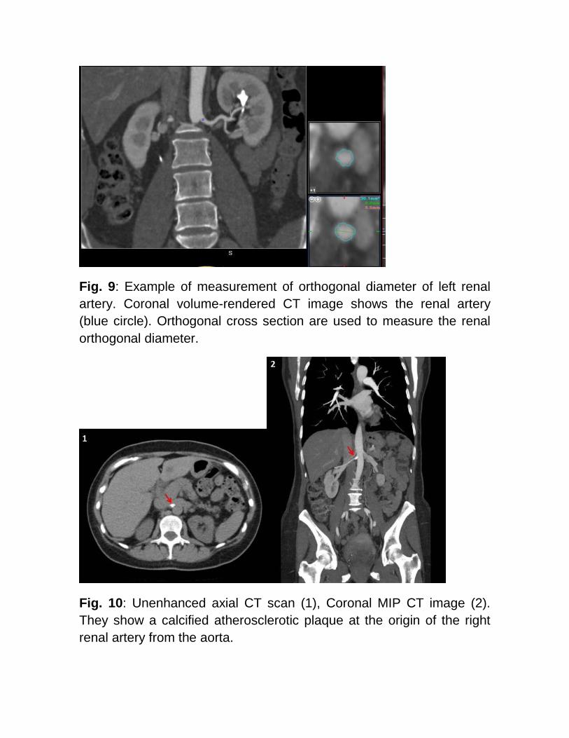
Fig. 9: Example of measurement of orthogonal diameter of left renal
artery. Coronal volume-rendered CT image shows the renal artery
(blue circle). Orthogonal cross section are used to measure the renal
orthogonal diameter.
Fig. 10: Unenhanced axial CT scan (1), Coronal MIP CT image (2).
They show a calcified atherosclerotic plaque at the origin of the right
renal artery from the aorta.

Fig. 11: Coronal MIP CT image Example of measurement of the
distance between the segmentary confluence of each renal vein and
the inferior vena cava.
Fig. 12: Coronal MIP CT image (1), 3D reconstruction (2). Late
segmentary confluence of the left renal vein

Fig. 13: Complete ureteral duplication. Coronal volume-rendered CT
image shows duplicate left renal pelvis and ureters.