ct of the temporal bone in achondroplasia - ajnr · achondroplasia is a genetic disorder of...
TRANSCRIPT

Steven R. Cobb 1
Mordechai Shohat2
C. Mark Mehringer1
Ralph Lachman 1
Received November 16. 1987; accepted after revision April 22. 1988.
1 Department of Radiology, Harbor-UCLA Medical Center, 1000 W. Carson St.. Torrance, CA 90509. Address reprint requests to S. R. Cobb.
2 Department of Pediatrics, Cedars-Sinai Medical Center, 8700 Beverly Blvd. , Los Angeles, CA 90048.
AJNR 9:1195-1199, November/December 1988 0195-6108/88/0906-1195 © American Society of Neuroradiology
CT of the Temporal Bone in Achondroplasia
1195
In an attempt to better define the changes affecting the temporal bone that might predispose achondroplastic dwarfs to otitis media, nine achondroplastic subjects who were evaluated for hearing loss underwent high-resolution CT scanning of the temporal bone. Comparisons were made with 10 nonachondroplastic subjects. A number of morphologic changes were seen, including (1) poor development of mastoid air cells, (2) foreshortening of the carotid canals, (3) narrowing of the skull base, (4) "towering" petrous ridges, and (5) relative "rotation" of the cochlea and other temporal bone structures. The most significant change was the rotational effect, which was more pronounced medially, resulting in an abnormal orientation of inner ear structures relative to middle ear structures and of middle ear structures relative to the external auditory canal. There was a notable lack of evidence for otitis media or its sequelae in any of the achondroplastic subjects. Audiograms were obtained in six of the nine achondroplastic subjects (two adults and four children). There was evidence of mixed hearing loss in the four children, but only of sensorineural hearing loss in the adults.
We believe that the persistent hearing loss in achondroplasia is not due to sequelae of otitis media as some authors have suggested. Intrinsic vestibulocochlear changes below the limits of resolution of high-resolution CT scanning may be responsible.
Achondroplasia is a genetic disorder of cartilage that leads to a characteristic pattern of dwarfism. Although achondroplasia is a prime example of mendel ian autosomal-dominant inheritance, mutation accounts for 85% of new cases. The reported frequency of achondroplasia has ranged from 15 per million live births to as high as 125 per million live births [1].
Long- and short-term complications have been reported . The spinal canal is characteristically small , particularly in the lumbar region, where short pedicles and narrowed interpedicular distances contribute to spinal stenosis; in many cases this is associated with progressive neurologic dysfunction involving the cauda equina and lumbosacral nerve roots . Herniation of intervertebral disks is common [2] . Respiratory complications have been described and include recurrent respiratory tract infections and obstructive sleep apnea [3] .
Gorlin et al. [4] and Cohen [5] have reported a high rate of otitis media in patients with achondroplasia. Glass et al. [6] noted a high frequency of hearing loss, particularly conductive hearing loss, and abnormal middle ear function in achondroplastic dwarfs, which they attributed to altered craniofacial morphology.
Our current study was undertaken in an effort to better characterize the changes in the temporal bone in achondroplasia and to determine if indeed there is a morphologic predisposition in these patients, definable by high-resolution CT, making them more susceptible to otitis media and its sequelae.
Subjects and Methods
Nine achondroplastic dwarfs 8-43 years old were evaluated by high-resolution CT for investigation of hearing loss. Clinically , hearing loss was believed to be bilateral. Audiograms

1196 COBB ET AL. AJNR:9, November/December 1988
were obtained in six of the nine achondroplastic subjects (two adults and four children). All were scanned with a Picker 1200SX scanner with a high-resolution bone imaging algorithm. Slice th ickness was 2 mm and images were obtained at 2-mm increments by using a 16-cm field size. Scans were routinely obtained in the axial plane at approximately 30" relative to the anthropologic baseline and in the coronal plane at approximately 105" [7J. Because of difficulty in maintaining the neck in the hyperextended position , coronal images were not obtained in three of the nine patients. The CT scans of 10 patients without achondroplasia chosen at random (but excluding patients over 45 years old to keep the two groups essentially agematched) were used for comparison.
Along with gross morphologic observations , cursor measurements were obtained of the diameter and length of the internal auditory canal (lAC). The diameter was measured at the midportion of the canal and the length was measured from the lateral extent of the canal to a point midway between the superior and inferior rim of the porus acusticus. Because of a marked cephalad angulation of the lACs in the achondroplastic patients, accurate measurements of the length of the canal could not be made in the axial plane.
The distance between the lACs (from the lateral extent of one canal to the lateral extent of the opposite canal) was also measured along with the angle formed by intersecting lines drawn along the length of the lACs on coronal images.
Results
Audiograms in six of the nine achondroplastic subjects revealed evidence for bilateral hearing loss in all six. In the four children tested there was mixed hearing loss (conductive and sensorineural). In the two adults tested there was only sensorineural hearing loss. Interestingly, none of those tested demonstrated purely conductive hearing loss.
Consistent changes were seen in the skull base and temporal bone of all nine subjects. These changes were more pronounced in the more medial petro us portion of the temporal bone as opposed to the mastoid and tympanic portions. Most of the changes were better demonstrated on coronal images. Table 1 summarizes some of the findings in the nine achondroplastic and 10 nonachondroplastic subjects.
The predominant effect on the temporal bone in achondroplaSia is upward tilting of the petrous portion with relative "rotation" of related bony structures from the horizontal plane. This upward tilt as seen on coronal images of the skull base gives the appearance of "towering" petrous ridges (Fig. 1). This appearance has been described by Pierre-Kahn et al. [8]. In a study of hydrocephalus and achondroplasia they
TABLE 1: Morphometric and Audiometric Results in Subjects with and Without Achondroplasia
lAC/lAC Measurements lAC Measurements
Group: Case No. Age Diameter Length Audiometric Findings (years)
Angle (0) Distance (mm) R L R L
With achondroplasia: 1 43 125 65.2 4.5 4.6 11.1 11.0 + (SNHL) 2 23 120 49.4 5.0 4.7 9.5 10.0 3 40 129 41 .2 5.0 5.1 8.6 9.0 + (SNHL) 4 8 - a 40.7 5.0 4.8 - a - a + (Mixed) 5 11 123 48.1 4.5 5.4 11.2 10.9 + (CHL, R; mixed, L) 6 8 - a 45.0 4.1 4.6 - a - a + (Mixed) 7 23 110 51.0 5.2 - b 8.7 - b
8 13 123 47.2 4.8 5.0 9.0 9.2 + (Mixed) 9 42 - a 44.0 5.5 4.8 - a - a
Mean 23 122 48.0 4.8 4.9 9.7 10.0 4.9c 9.9c
Without achondroplasia: 1 29 170 66.8 5.0 4.8 9.5 9.4 2 24 165 71.5 4.0 4.1 10.3 10.0 3 12 159 71.9 4.5 4.5 13.4 13.1 4 10 163 63.8 4.8 4.7 10.3 10.5 5 3 168 57.5 4.3 4.1 9.1 9.4 6 23 165 66.3 3.2 3.5 11.0 11.1 7 16 175 68.2 5.3 5.1 11.4 11.1 8 30 157 74.1 5.3 5.5 7.2 7.3 9 37 158 64.8 5.8 5.7 8.5 8.4 10 34 169 65.0 5.0 5.0 11 .3 11.0
Mean 22 165 67 .0 4.7 4.7 10.2 10.1 4.7c 10.2c
Note.-Audiometry was not performed in patients without achondroplasia or in three of the achondroplastic patients. lAC = internal aUditory canal; R = right; L = left; SNHL = sensorineural hearing loss; CHL = conductive hearing loss.
a Coronal images were not obtained in this patient; therefore, it was not possible to measure the lAC/lAC angle or to accurately measure the lAC length. b The left lAC was obscured by an artifact. C Mean of the right and left sides.
AJNR:9, November/December 1988 CT OF ACHONDROPLASTIC TEMPORAL BONE 1197
described upward tilting of the petrous ridges on conventional frontal tomograms of the skull base in achondroplasia. They also measured the angle formed by the upward tilting of the petrous ridges in 25 achondroplastic individuals. In their series this angle ranged from 107° to 130°. This appearance was not a manifestation of basilar invagination, since measurements of the Chamberlain line revealed only one case of basilar invagination among the 25 achondroplastic subjects.
Measurements of the angle of upward tilting of the petrous ridges (lAC/lAC angle) in our series paralleled those of PierreKahn et al. [8] , ranging from 110° to 129° (average, 122\ whereas in 10 nonachondroplastic subjects this angle ranged from 157° to 175° (average, 165} However, the lACs themselves appeared unaffected. The diameter of the lAC in achondroplasia was not significantly different from its diameter in nonachondroplastic subjects (4.9 vs 4.7 mm), and the
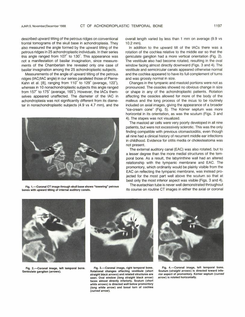
Fig. 1.-Coronal CT image through skull base shows "towering" petrous bones with upward tilting of internal auditory canals.
overall length varied by less than 1 mm on average (9.9 vs 10.2 mm).
In addition to the upward tilt of the lACs there was a rotation of the cochlea relative to the middle ear so that the geniculate ganglion had a more vertical orientation (Fig . 2). The vestibule also had become rotated, resulting in the oval window facing almost directly downward (Figs . 3 and 4). The vestibule and semicircular canals appeared otherwise normal and the cochlea appeared to have its full complement of turns and was grossly normal in size.
Changes in the tympanic and mastoid portions were not as pronounced . The ossicles showed no obvious change in size or shape in any of the achondroplastic patients. Rotation affecting the ossicles allowed for more of the body of the malleus and the long process of the incus to be routinely included on axial images, giving the appearance of a broader "ice-cream cone" (Fig . 5). The Korner septum was more horizontal in its orientation, as was the scutum (Figs . 3 and 4). The stapes was not visualized .
The mastoid air cells were very poorly developed in all nine patients, but were not excessively sclerotic. This was the only finding compatible with previous otomastoiditis, even though all nine had a clinical history of recurrent middle ear infections in childhood . Evidence for otitis media or cholesteatoma was not present.
The external auditory canal (EAC) was also rotated , but to a lesser degree than the more medial structures of the temporal bone. As a result, the labyrinthine wall had an altered relationship with the tympanic membrane and EAC. The promontory, which ordinarily would be plainly visible from the EAC on reflecting the tympanic membrane, was instead projected for the most part well above the scutum so that at best only the most inferior aspect was visible (Figs. 3 and 4).
The eustachian tube is never well demonstrated throughout its course on routine CT images in either the axial or coronal
Fig. 2.-Coronal image, left temporal bone. Fig. 3.-Coronal image, right temporal bone. Fig. 4.-Coronal image, left temporal bone. Scutum (straight arrows) is directed toward inferior aspect of promontory. Korner septum (curved arrow) is rotated horizontally.
Geniculate ganglion (arrows) . . Rotational changes affecting vestibule (short straight black arrows) and related structures are seen. Oval window (/ong straight black arrow) faces almost directly inferiorly. Scutum (short white arrows) is directed well below promontory (long white arrow) and basal turn of cochlea (curved arrow).

1198 COBB ET AL. AJNR:9. November/December 1988
Fig. 5.-Axial image, right temporal bone. Malleus (straight white arrow) and incus (curved arrow) appear together as broad "ice-cream cone" because of rotation into axial plane. Horizontal portion of carotid canal (black arrows) is foreshortened.
Fig. S.-Axial image, skull base. Foreshortened carotid canals (long arrows). Medial extents of both canals closely approximate midline. Tympanic orifice of eustachian tube is seen near lateral extent of carotid canals (shorf arrows).
Fig. 7.-Axial image, skull base. Carotid canals (straight arrows) are foreshortened and approximate midline at distal end. Mastoid processes (curved arrows) show no pneumatization.
plane, and its full course was not entirely delineated in our study.
More general changes were seen affecting the skull base as a whole. The carotid canal showed a tendency toward foreshortening in the achondroplastic patients, and, in addition, the distal petrous portions of the carotid canal more closely approximated the midline (Figs. 6 and 7). The initial ascending portion of the carotid canal was no longer vertical in orientation but was angled toward the midline. This same angulation affected the course of the jugular vein exiting the skull (Fig. 1). The skull base in general was narrower in achondroplasia. The distance between the distal extent of the lACs (lAC/lAC distance) in achondroplasia averaged 48.0 mm, whereas without achondroplasia this distance averaged 67.0 mm. Narrowing of the foramen magnum has been described previously and was seen to various degrees in our nine patients.
There were only slight differences between the adult and pediatric achondroplastic subjects (Table 1). The average diameter of the lAC was 4.9 mm in the adults compared with 4.8 mm in the pediatric group. The length of the canal in the adults averaged 9.7 mm. The pediatric length averaged 10.1 mm. The average lAC/lAC angle was 121
0
in the adults (vs 123
0
in children). The lAC/lAC distance showed the clearest difference (50.2 vs 45 .3 mm), but again the difference was slight, and given the limited number of measurements none of the differences can be considered statistically significant.
Discussion
Of the triad of parts that constitute the temporal bone, only the petrous part arises entirely in cartilage. The squamous and most of the mastoid portions develop in membrane. The ossification center for the mastoid portion appears from the ninth to the tenth fetal week. Up to the middle of the fifth month the cartilaginous petrous portion is represented by the otic capsule. It then ossifies so rapidly that by the end of the
sixth month it has been almost entirely converted into porous bone. Unlike a typical articular skeletal element arising in cartilage, which is not expected to reach adult dimensions until approximately 22 years, the otic capsule is as large as it will ever be in the 21-week fetus [9J.
It is generally accepted that the malleus and incus are derived from the first branchial arch (Meckel cartilage). By 4 months of fetal life they begin to change from cartilage to bone. These two ossicles derive their adult form by way of continuing deposition of endosteal bone during the course of fetal life similar to long bone growth in the appendicular skeleton. The stapes has a more complex development, having a dual origin from branchial cartilage (Reichert cartilage from the second branchial arch) and cartilage from the otic capsule (which contributes to the foot plate) [9, 1 OJ.
The tympanic cavity proper as well as the lining of the auditory (eustachian) tube arises from the expanding terminal end of the first (and possibly second) pharyngeal pouch. Late in the second fetal month, while the proximal portion of the pharyngeal pouch becomes constricted to form the auditory tube, the distal end expands into a flattened sac, the primordial tympanic cavity [9, 1 OJ.
The basic defect in achondroplasia is a disturbance of endochondral ossification caused by an inability to produce a sufficient quantity of columnar cartilage laid down as a precursor to trabecular bone. Rows of columnar cartilage lack parallel arrangement and are unequal in length. Lines of preparatory ossification are irregular. Trabeculae are short, thick, and lack a normal orderly arrangement. Periosteal ossification is affected little so that transverse bone growth is not markedly altered. The result is selective deficiency of bone growth leading to long bones that appear short and thick and a skull base and facial bones that are distorted.
Given this understanding of the disturbance of bone growth leading to achondroplasia, it should be expected that certain portions of the temporal bone would be directly affected, namely, those structures arising from endochondral bone

AJNR:9, November/December 1988 CT OF ACHONDROPLASTIC TEMPORAL BONE 1199
formation: the otic capsule and its derivatives, the petrous ridge, and the ossicles. The EAC and eustachian tube (osseous portion) are essentially membranous in origin, the EAC arising from mesodermal condensations around the epithelial tract of the descending otic capsules and the eustachian tube from similar condensations around the elongating first pharyngeal pouch. Thus, these structures should not be directly affected , and they do not appear to be. Interestingly enough, the ossicles, despite being formed in endochondral bone, show no real change in size or shape in achondroplastic patients. So, other than rotational changes previously described, there appears to be little evidence for a structural defect to account for hearing loss in the achondroplastic population. Moreover, contrary to the findings of Gorlin et al. [4] and Cohen [5], who reported a high frequency of otitis media in achondroplasia, we found an obvious lack of evidence for otitis media, cholesteatoma, or mastoiditis, either chronic or acute, in all our achondroplastic subjects.
Glass et al. [6] , who noted a high frequency of conductive hearing loss and abnormal middle ear function, implicated "altered craniofacial morphology." Parkin [11] also implicated altered craniofacial morphology in suggesting that palatal and pharyngeal maldevelopment contribute to poor eustachian tube function , giving rise to frequent episodes of otitis media in achondroplasia during early childhood. However, eustachian tube dysfunction does not appear to be a factor, since none of our current cases had evidence of poor middle ear drainage (i .e., otitis media or its sequelae). Moreover, the osseous portion of the eustachian tube, not being of endochondral origin , should not be expected to be implicated.
Caldarelli [12] and Sando et al. [13] ascribe the conductive hearing loss in achondroplasia to fusion of the ossicular chain to the surrounding bony structures of the tympanic cavity. They also described inner ear abnormalities, including a deformed cochlea, as associated anomalies. Bony fusion of the ossicular chain was not demonstrated in any of our nine patients, nor were cochlear malformations appreCiated. However, cochlear malformations sufficient to account for senso-
rineural hearing loss may simply be beneath the limits of resolution of even high-resolution CT scanning.
The notable lack of radiographic evidence for otit is media or its sequelae and the audiometric findings implicating the inner ear as the site of persistent hearing loss in achondroplasia suggest that the frequency of otomastoiditis and the evaluation of hearing loss in achondroplasia may need to be reviewed in light of high-resolution CT scanning of the temporal bone.
REFERENCES
1. Bailey JA. Disproportionate short stature. Diagnosis and management. Philadelphia: Saunders, 1973:81-82
2. Morgan OF, Young RF. Spinal neurological complications of achondroplasia. Results of surgical treatment. J Neurosurg 1980 ;52:463-472
3. Stokes DC, Phillips JA, Leonard CO, et al. Respiratory complications of achondroplasia. J Pediatr 1983;102:534-540
4. Gorlin RF, Pindborg JJ , Cohen MM. Syndromes of the head and neck. New York: McGraw-Hili , 1976 :15
5. Cohen MM. In: Dysmorphic syndromes with craniofacial manifestations. Stewart RE , Prescott G, eds. Oral facial genetics. St. Louis: Mosby, 1976: 523-526
6. Glass L, Shapiro I, Hodge SE , Bergstrom L, Rimoin DL. Audiological findings of patients with achondroplasia. Int J Pediatr Otorhinolaryngol 1981 ;3: 129-135
7. Chakeres OW, Spiegel PK. A systematic technique for compressive evaluation of oral bone by computed tomography. Radiology 1983;146 : 97-106
8. Pierre-Kahn A, Hirsh JF, Renier 0 , et al. Hydrocephalus and achondroplasia. A study of 25 observations. Childs Nerv Syst 1980;7:205-219
9. Anson BJ , Donaldson JA. The ear: developmental anatomy. In: Surgical anatomy of the temporal bone , 3rd ed. Philadelphia: Saunders, 1981: 23-58
10. Pansky B. Review of medical embryology. New York: MacMillan, 1982 ; 178 11. Parkin JL. Congenital malformations of the mouth and pharynx. In: Blue
stone CD, Stool SE, Arjona SK, eds. Pediatric otolaryngology, vol. 2. Philadelphia: Saunders, 1983;912-930
12. Caldarelli DO. Congenital middle ear anomalies associated with craniofacial and skeletal syndromes. In: Jaffe BF, ed. Hearing loss in children. A comprehensive text. Baltimore: University Park, 1977:310-340
13. Sando I, Suehiro S, Wood RP II. Congenital anomalies of the external and middle ear. In: Bluestone CD, Stool SE, Arjona SK, eds. Pediatric otolaryngology, vol. 1. Philadelphia: Saunders, 1983 :309-346