cte in peds crohn's 10.pdf
TRANSCRIPT
8/11/2019 CTE in peds Crohn's 10.pdf
http://slidepdf.com/reader/full/cte-in-peds-crohns-10pdf 1/9
REVIEW
CT enterography of pediatric Crohn disease
Jonathan R. Dillman & Jeremy Adler &
Ellen M. Zimmermann & Peter J. Strouse
Received: 9 August 2009 /Revised: 10 October 2009 /Accepted: 30 October 2009 /Published online: 20 November 2009# Springer-Verlag 2009
Abstract CT enterography is an important tool in the
noninvasive diagnosis and follow-up of pediatric Crohndisease. This imaging modality is particularly useful for
assessing extent of disease (including both intestinal and
extraintestinal manifestations), response to medical therapy,
and disease-related complications. The purpose of this
article is to provide a contemporary review of CT enter-
ography technique as well as the spectrum of intestinal and
extraintestinal findings in pediatric Crohn disease.
Keywords CT . Crohn disease . Children . Bowel imaging
Introduction
Crohn disease, a form of chronic inflammatory bowel
disease (IBD), is common in the pediatric population. The
current worldwide prevalence of Crohn disease is approx-
imately 130 per 100,000 individuals and its incidence is
rising [1]. The incidence of pediatric Crohn disease in the
United States is 4.56 per 100,000 individuals [1]. Approx-imately 15 – 25% of individuals with IBD will present
during childhood (<18 years of age) [1]. Crohn disease is
characterized by transmural bowel inflammation, skip
lesions, and episodic flares of disease activity interspersed
with periods of remission. Initial diagnosis, establishing
disease extent, monitoring response to medical manage-
ment, and identification of disease-related complications
typically relies on a combination of clinical assessment,
endoscopy (both optical and capsule), and radiologic
evaluation.
Radiologic evaluation of Crohn disease can include the
use of radiography, barium studies (upper gastrointestinal
series, small bowel follow-through [SBFT] examination,
contrast enema, and, less commonly, enteroclysis), com-
puted tomography enterography (CTE), magnetic resonance
enterography (MRE), and sonography. CTE and MRE
allow for complete radiologic evaluation of both the
intestinal and extraintestinal manifestations of abdomino-
pelvic Crohn disease. Benefits of CTE when compared to
MRE include better spatial resolution, fewer motion-related
artefacts, increased availability, greater radiologist familiar-
ity, lesser cost, and shorter examination time. Benefits of
MRE when compared to CTE include better contrast
resolution and a lack of ionizing radiation. As recent
studies have demonstrated similar sensitivities for CTE
and MRE for the detection of small bowel Crohn disease
[2, 3], MRE is likely to become the primary modality for
imaging of the small bowel in children. In certain children,
however, CTE will still be indicated, such as individuals
with pacemakers and other MRI-sensitive implanted devices.
A study by Jamieson et al. [4] in the pediatric population
concluded that CT shows sensitivity for the evaluation of
small bowel involvement in inflammatory bowel disease
J. R. Dillman (*) : P. J. Strouse
Department of Radiology, C.S. Mott Children’s Hospital,
University of Michigan Health System,
Section of Pediatric Radiology, 1500 E. Medical Center Drive,
Ann Arbor, MI 48109, USAe-mail: [email protected]
J. Adler
Department of Pediatrics and Communicable Diseases,
Division of Pediatric Gastroenterology,
University of Michigan Health System,
Ann Arbor, MI, USA
E. M. Zimmermann
Department of Internal Medicine, Division of Gastroenterology,
University of Michigan Health System,
Ann Arbor, MI, USA
Pediatr Radiol (2010) 40:97 – 105
DOI 10.1007/s00247-009-1465-5
8/11/2019 CTE in peds Crohn's 10.pdf
http://slidepdf.com/reader/full/cte-in-peds-crohns-10pdf 2/9
that is equal to or better than that of SBFT examination.
Their study also indicated that most children preferred CT
to barium studies [4]. A prospective study by Hara et al. [5]
evaluated adults with CTE, capsule endoscopy, ileoscopy,
and SBFT examination. They found that CTE, capsule
endoscopy, and ileoscopy have a higher yield in depicting
mild to moderate findings of Crohn disease than SBFT
examination, and that CTE is better for detecting transmuraland extraluminal abnormalities [5]. Additionally, they
concluded that CTE can depict nonobstructive Crohn
disease of the small bowel when conventional techniques,
such as ileoscopy and SBFT examination, produce negative
or inconclusive findings [5].
The purpose of this article is to present a contemporary
review of CT enterography technique as well as the
spectrum of intestinal and extraintestinal findings in
pediatric Crohn disease.
Pediatric CTE technique and radiation dose reduction
Pediatric CTE technique involves several modifications to
standard abdominopelvic CT imaging protocols. Prior to
imaging, a low-density oral contrast material (most com-
monly VoLumen, a low-density barium sulfate oral contrast
agent made by Bracco, Princeton, NJ) is typically admin-
istered to the child (using a weight-based algorithm at our
institution) to maximize bowel luminal distention [6 – 9].
Low-density contrast materials also allow for assessment of
bowel mucosal hyperenhancement, a finding that can be
obscured by higher-density oral contrast agents [6]. Orally
administered water can also be used as a low-density oral
contrast agent in CTE, although it may result in less optimal
small bowel distention. CT enteroclysis is a less often used
approach to opacify the small bowel in children. This
technique is more labor-intensive than CT enterography,
requires nasojejunal intubation of the child, and typically
uses higher-density contrast material. Brown et al. [10]
documented that CT enteroclysis can be safe, feasible, and
accurate in depicting small bowel pathology in children.
CTE almost always utilizes intravascular iodinated low-
osmolality (or iso-osmolality) contrast material unless
specifically contraindicated. We administer an intravenous
dose of 2 ml/kg (maximum dose, 125 ml) using a power
injector whenever possible. While we prefer to image
during the portal venous phase (approximately 70 s after
the initiation of contrast material injection), in part to allow
for adequate evaluation of the solid organs, others have
described enteric-phase imaging approximately 40 – 45 s
after the initiation of contrast material injection [7, 9].
Vandenbroucke et al. [9] concluded that Crohn disease is
adequately depicted on both the enteric and portal venous
imaging phases. While some have advocated the use of
anti-peristaltic agents, such as glucagon, prior to imaging to
minimize bowel-related motion artefacts, we believe that
this intervention is unnecessary given the rapid speed with
which images are acquired using modern multi-detector CT
scanners.
Images should be acquired using multi-detector CT with
submillimeter collimation to obtain an isotropic data set that
allows for the creation of a variety of two-dimensional (2-D) multiplanar reformatted and three-dimensional (3-D)
reconstructed images. From the source data, we reconstruct
2.5-mm axial images with 1.25-mm section overlap for
primary review. The submillimeter axial source images are
generally only reviewed as a problem-solving tool. Multi-
planar reformatted images, particularly in the coronal plane,
might assist in the detection of pathology [11]. We generate
both coronal and sagittal reformatted imaging series with a
2-mm section thickness. Finally, we create subvolume
maximum intensity projection (MIP) and front-cut
volume-rendered (VR) 3-D imaging series in the coronal
plane.A variety of methods can be employed to minimize
pediatric radiation exposure related to CTE. First, only a
single imaging phase should be acquired in order to adhere
to the ALARA (as low as reasonably achievable) principle.
Second, tube current (mA) should be minimized, as this
imaging parameter is directly proportional to radiation dose.
Tube current can be selected by using a weight-based
protocol or tube current modulation (a form of automatic
exposure control). Finally, the use of other nonionizing
imaging modalities, such as MRE or sonography, should be
strongly considered, particularly for follow-up imaging.
The effective radiation dose related to CTE in a 10-year-old
child is approximately 3.5 mSv, compared to approximately
1.8 – 2.2 mSv for SBFT (using, on average, 5.1 min of
fluoroscopy time and 3.3 abdominal radiographs) [12]. The
effective dose of SBFT examination can be extremely
variable, however, depending on the fluoroscope utilized,
fluoroscopy time and number of radiographs acquired can
be greater than CTE. The mean CTDIvol (a measure of
average absorbed radiation dose) for pediatric (ages 2 to
19 years) CTE at our institution is 4.9 mGy (when using a
standard adult 32-cm body reference phantom), with a
range from 1.29 to 12.95 mGy.
Bowel-related findings
Crohn disease can affect any portion of the gastrointestinal
(GI) tract from the mouth to the anus and most commonly
involves the small bowel, particularly the terminal ileum.
CTE specifically allows for noninvasive detailed evaluation
of the GI tract from the level of the distal esophagus
through the anus.
98 Pediatr Radiol (2010) 40:97 – 105
8/11/2019 CTE in peds Crohn's 10.pdf
http://slidepdf.com/reader/full/cte-in-peds-crohns-10pdf 3/9
A variety of small and large bowel (as well as gastric)
findings can be observed at CTE in pediatric Crohn disease.
The most common findings include abnormal bowel wall
thickening and hyperenhancement (Figs. 1, 2, 3, 4) [13,
14]. These findings generally become more pronounced
with increasing inflammation and disease activity. Multiple
patterns of bowel wall enhancement might be observed,
depending on the degree of mucosal, muscularis propria,and serosal hyperemia, inflammation, and fibrosis. Differ-
ential bowel wall enhancement might result in a striated
appearance, sometimes referred to as mural stratification or
the target sign (Figs. 1 and 2) [13]. More homogeneous
bowel wall enhancement suggests the presence of fibrosis
[13]. A recent study by Adler et al. [15], however, has
called this supposition into question, demonstrating that the
presence of fibrosis cannot be independently identified or
distinguished from inflammation at CTE in children and
adults.
A study by Booya et al. [7] concluded that the most
sensitive visual marker of Crohn disease at CTE is mural
hyperenhancement followed by mural thickening. Another
study, by Baker et al. [8], was able to distinguish normal
terminal ileum from terminal ileitis of active Crohn disease
using a combination of mural attenuation and mural
thickness. Alterations in bowel wall thickness and enhance-
ment at CTE over time might be useful to monitor changesin disease activity in response to therapy [16].
Bowel (and gastric) luminal diameter changes might be
noted in Crohn disease. Areas of abnormal luminal
narrowing with proximal dilatation, or strictures, can be
caused by either active inflammation or fibrosis. Strictures
can result in partial or complete bowel obstruction (Fig. 5)
or gastric outlet obstruction (Fig. 6). Findings that suggest
bowel obstruction include proximal luminal dilatation,
distal luminal decompression, and more proximal stasis of
intraluminal bowel contents (“small bowel feces” sign)
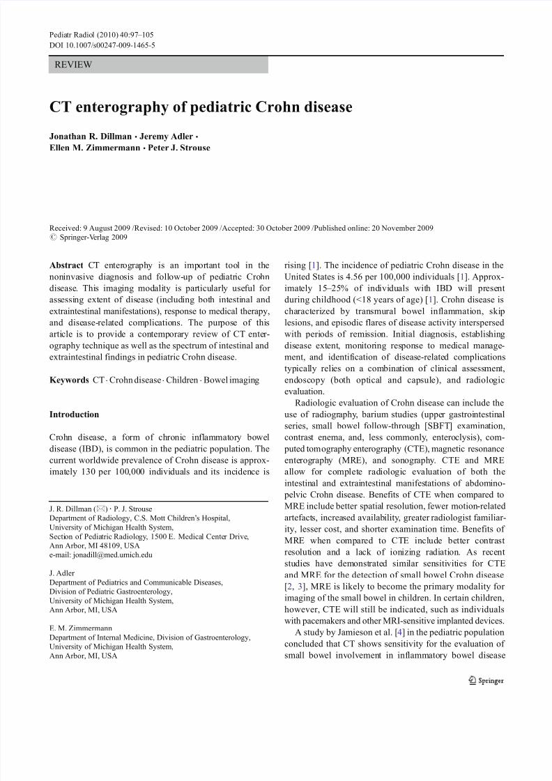
Fig. 1 Crohn disease in an 11-year-old girl. a Axial CTE image
reveals marked distal ileal mural thickening and differential mucosal
and mural hyperenhancement (mural stratification). There is adjacent
mesenteric fat inflammatory stranding (white arrowheads). b A
slightly more caudal image reveals mesenteric hypervascularity and
fibrofatty proliferation (black arrowheads). c Coronal-reformatted
subvolume maximum-intensity projection image confirms the above
findings. There is marked vasa recta prominence, the so-called comb
sign (arrows)
Pediatr Radiol (2010) 40:97 – 105 99
8/11/2019 CTE in peds Crohn's 10.pdf
http://slidepdf.com/reader/full/cte-in-peds-crohns-10pdf 4/9
(Fig. 7). There is no consensus on the value of CTE to
distinguish between bowel obstruction caused by active
inflammation versus fibrosis, and, therefore, the decision
for surgical management should rely mainly on the
response to medical management.
Mesentery-related findings
Several mesenteric abnormalities might be observed adja-
cent to bowel loops affected by Crohn disease. Mesenteric
vasculature, particularly the vasa recta, can appear notice-
ably prominent because of hyperemia-related engorgement,
sometimes referred to as the comb sign (Figs. 1, 3, and 4)
[13, 17]. A study by Lee et al. [17] found that perienteric
and pericolic mesenteric hypervascularity correlated with
the presence of active and more extensive Crohn disease.
They also found that increased mesenteric vascularity was
associated with more common occurrence of longitudinal
and perpendicular ulcerations at barium studies [17].
Mesenteric fat stranding, thought to be primarily caused
by inflammation, is commonly observed in Crohn disease
and can be variable in conspicuity (Figs. 1, 2, 3, 4, 7).
Abnormal mesenteric fibrofatty proliferation, or “creeping
fat,” can also be seen adjacent to affected bowel loops
(Figs. 1, 2, 3) [13, 14]. This finding might be particularly
conspicuous in children who typically have a paucity of
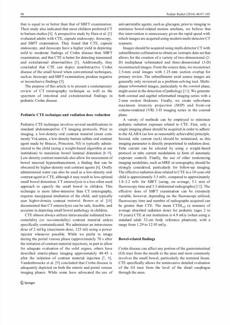
Fig. 2 Crohn disease in an 18-year-old with Down syndrome. a Axial
CTE image demonstrates marked descending colon mural thickening
(arrows) with adjacent fibrofatty proliferation, hypervascularity, andinflammatory fat stranding. Differential mucosal and serosal hyper-
enhancement result in mural stratification. b A more caudal image
reveals an enhancing sinus tract (arrow) as well as an adjacent loculated
fluid collection consistent with abscess (arrowheads)
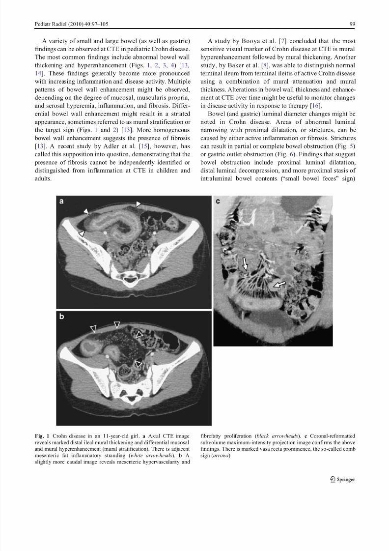
Fig. 3 Crohn disease in a 6-year-old girl. Axial CTE image reveals
bowel wall thickening and hyperenhancement involving multiple
loops of jejunum (arrows). Additionally, there is mesenteric lymph
node enlargement (asterisks), inflammatory fat stranding, hyper-
vascularity, and fibrofatty proliferation
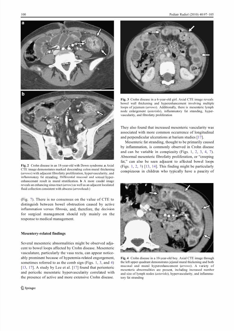
Fig. 4 Crohn disease in a 10-year-old boy. Axial CTE image through
the left upper quadrant demonstrates jejunal mural thickening and both
mucosal and mural hyperenhancement (arrows). A variety of
mesenteric abnormalities are present, including increased number
and size of lymph nodes (asterisks), hypervascularity, and inflamma-
tory fat stranding
100 Pediatr Radiol (2010) 40:97 – 105
8/11/2019 CTE in peds Crohn's 10.pdf
http://slidepdf.com/reader/full/cte-in-peds-crohns-10pdf 5/9
mesenteric fat. Finally, mesenteric lymph nodes can be
increased in number or size (Figs. 3, 4, and 8).
Penetrating disease
The bowel wall inflammatory changes of Crohn disease
vary greatly in depth, with lesions being either superficial,
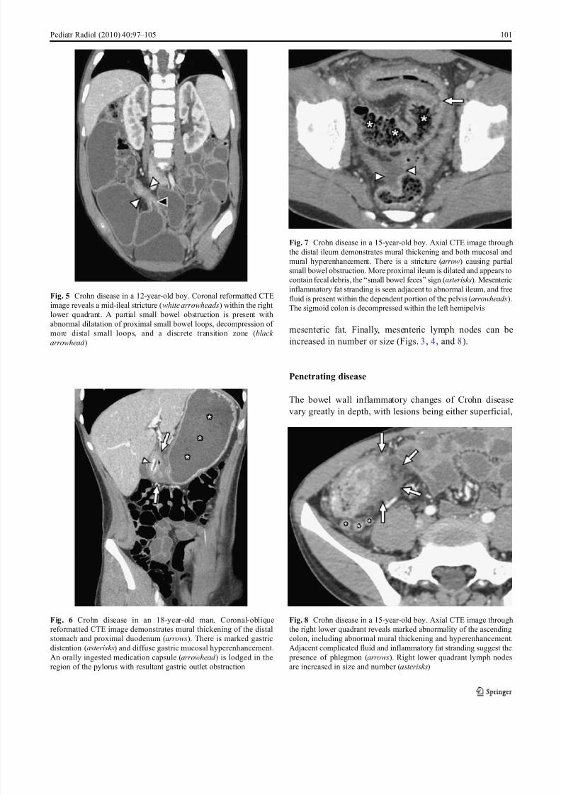
Fig. 5 Crohn disease in a 12-year-old boy. Coronal reformatted CTE
image reveals a mid-ileal stricture (white arrowheads) within the right
lower quadrant. A partial small bowel obstruction is present with
abnormal dilatation of proximal small bowel loops, decompression of
more distal small loops, and a discrete transition zone (black
arrowhead )
Fig. 6 Crohn disease in an 18-year-old man. Coronal-oblique
reformatted CTE image demonstrates mural thickening of the distal
stomach and proximal duodenum (arrows). There is marked gastric
distention (asterisks) and diffuse gastric mucosal hyperenhancement.
An orally ingested medication capsule (arrowhead ) is lodged in the
region of the pylorus with resultant gastric outlet obstruction
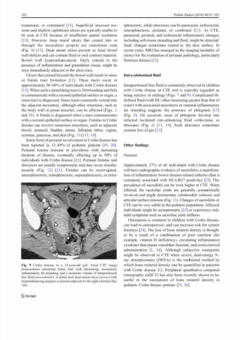
Fig. 7 Crohn disease in a 15-year-old boy. Axial CTE image through
the distal ileum demonstrates mural thickening and both mucosal and
mural hyperenhancement. There is a stricture (arrow) causing partial
small bowel obstruction. More proximal ileum is dilated and appears to
contain fecal debris, the “small bowel feces” sign (asterisks). Mesentericinflammatory fat stranding is seen adjacent to abnormal ileum, and free
fluid is present within the dependent portion of the pelvis (arrowheads).
The sigmoid colon is decompressed within the left hemipelvis
Fig. 8 Crohn disease in a 15-year-old boy. Axial CTE image through
the right lower quadrant reveals marked abnormality of the ascending
colon, including abnormal mural thickening and hyperenhancement.
Adjacent complicated fluid and inflammatory fat stranding suggest the
presence of phlegmon (arrows). Right lower quadrant lymph nodes
are increased in size and number (asterisks)
Pediatr Radiol (2010) 40:97 – 105 101
8/11/2019 CTE in peds Crohn's 10.pdf
http://slidepdf.com/reader/full/cte-in-peds-crohns-10pdf 6/9
transmural, or extramural [13]. Superficial mucosal ero-
sions and shallow (aphthous) ulcers are typically unable to
be seen at CTE because of insufficient spatial resolution
[13]. However, deep mural ulcers that extend into or
through the muscularis propria are sometimes seen
(Fig. 9) [13]. Deep mural ulcers present as focal bowel
wall defects and can contain fluid or oral contrast material.
Bowel wall hyperenhancement, likely related to the presence of inflammation and granulation tissue, might be
seen immediately adjacent to the ulcer tract.
Ulcers that extend beyond the bowel wall result in sinus
or fistula tract formation [13]. These tracts occur in
approximately 20 – 40% of individuals with Crohn disease
[13]. When such a penetrating tract is blind-ending and fails
to communicate with a second epithelial surface or organ, a
sinus tract is diagnosed. Sinus tracts commonly extend into
the adjacent mesentery, although other structures, such as
the body wall or psoas muscles, can be affected (Figs. 2
and 10). A fistula is diagnosed when a tract communicates
with a second epithelial surface or organ. Fistulas in Crohndisease can involve numerous structures, such as adjacent
bowel, stomach, bladder, uterus, fallopian tubes, vagina,
scrotum, pancreas, and skin (Fig. 11) [13, 18].
Some form of perianal involvement in Crohn disease has
been reported in 13 – 49% of pediatric patients [19, 20].
Perianal lesions increase in prevalence with increasing
duration of disease, eventually affecting up to 90% of
individuals with Crohn disease [21]. Perianal fistulae and
abscesses are usually symptomatic and may occur simulta-
neously (Fig. 12) [21]. Fistulae can be rectovaginal,
intersphincteric, transphincteric, suprasphincteric, or extra-
sphincteric, while abscesses can be pararectal, ischiorectal,
intersphincteric, perianal, or combined [21]. At CTE,
pararectal, perianal, and ischiorectal inflammatory changes,
including soft-tissue stranding and fluid, might be detected.
Such changes sometimes extend to the skin surface. In
recent years, MRI has emerged as the imaging modality of
choice for the evaluation of perianal pathology, particularly
fistulous disease [22].
Intra-abdominal fluid
Intraperitoneal free fluid is commonly observed in children
with Crohn disease at CTE and is typically regarded as
being reactive in etiology (Figs. 7 and 9). Complicated ill-
defined fluid (with HU often measuring greater than that of
water) with associated mesenteric or omental inflammatory
fat stranding suggests the presence of phlegmon [13]
(Fig. 8). On occasion, areas of phlegmon develop into
infected loculated rim-enhancing fluid collections, or abscesses (Fig. 2) [13, 14]. Such abscesses sometimes
contain foci of gas [13].
Other findings
Osseous
Approximately 27% of all individuals with Crohn disease
will have radiographic evidence of sacroiliitis, a manifesta-
tion of inflammatory bowel disease-related arthritis (that is
commonly associated with HLA-B27 positivity) [23]. The
prevalence of sacroiliitis can be even higher at CTE. When
affected, the sacroiliac joints are generally symmetrically
involved and might demonstrate subchondral sclerosis and
articular surface erosions (Fig. 13). Changes of sacroiliitis at
CTE can be very subtle in the pediatric population. Affected
individuals might be asymptomatic [23] or experience only
mild symptoms such as sacroiliac joint stiffness.
Osteopenia is common in children with Crohn disease,
can lead to osteoporosis, and can increase risk for certain
fractures [24]. The loss of bone mineral density is thought
to be a result of a combination of poor nutrition (for
example, vitamin D deficiency), circulating inflammatory
cytokines that impair osteoblast function, and corticosteroid
administration [1, 24]. Although subjective osteopenia
might be observed at CTE when severe, dual-energy X-
ray absorptiometry (DEXA) is the traditional method by
which bone mineral density can be quantified in patients
with Crohn disease [1]. Peripheral quantitative computed
tomography (pQCT) has also been recently shown to be
useful in the assessment of bone mineral density in
pediatric Crohn disease patients [25, 26].
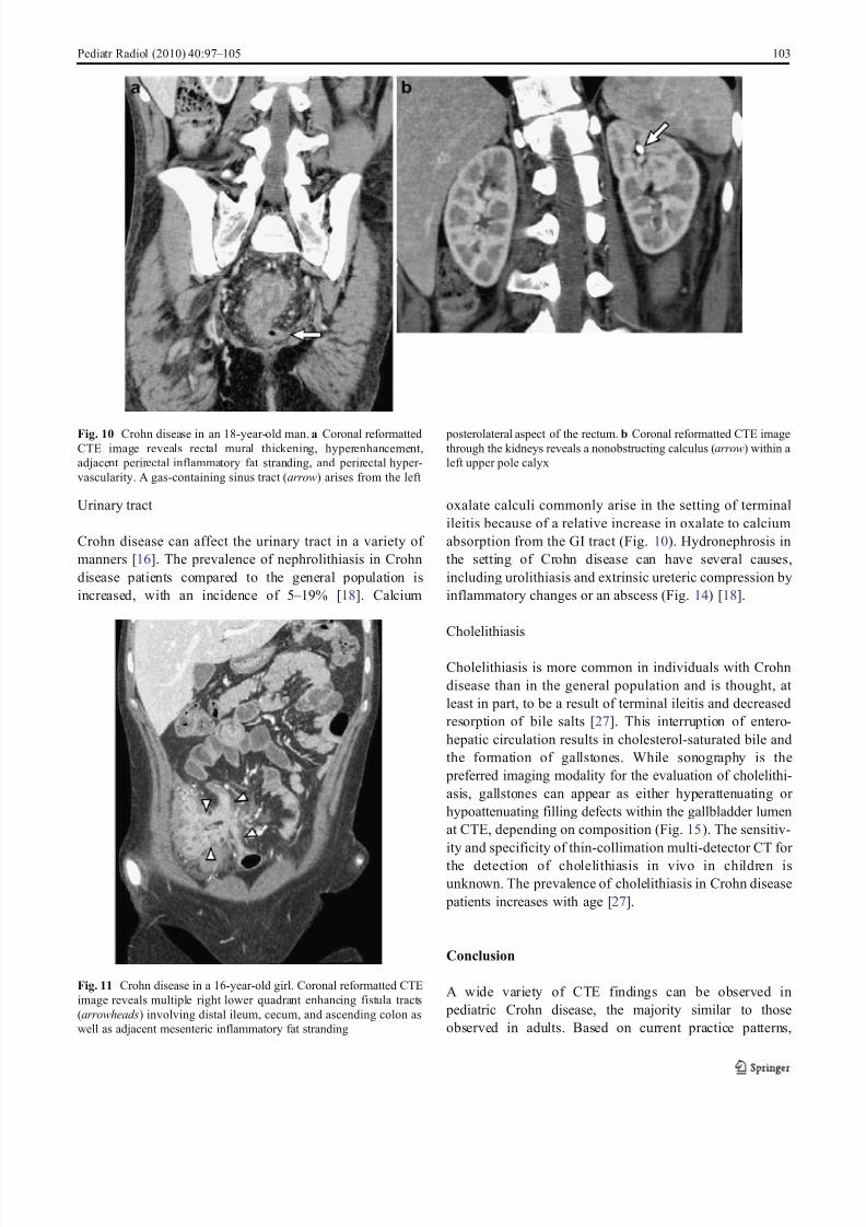
Fig. 9 Crohn disease in a 15-year-old girl. Axial CTE image
demonstrates abnormal distal ileal wall thickening, mesenteric
inflammatory fat stranding, and a moderate volume of intraperitoneal
free fluid (arrowheads). A distal ileal deep mural ulcer (arrow) with
hyperenhancing margins is present adjacent to the right external iliac
vein
102 Pediatr Radiol (2010) 40:97 – 105
8/11/2019 CTE in peds Crohn's 10.pdf
http://slidepdf.com/reader/full/cte-in-peds-crohns-10pdf 7/9
Urinary tract
Crohn disease can affect the urinary tract in a variety of
manners [16]. The prevalence of nephrolithiasis in Crohn
disease patients compared to the general population is
increased, with an incidence of 5 – 19% [18]. Calcium
oxalate calculi commonly arise in the setting of terminal
ileitis because of a relative increase in oxalate to calcium
absorption from the GI tract (Fig. 10). Hydronephrosis in
the setting of Crohn disease can have several causes,
including urolithiasis and extrinsic ureteric compression by
inflammatory changes or an abscess (Fig. 14) [18].
Cholelithiasis
Cholelithiasis is more common in individuals with Crohn
disease than in the general population and is thought, at
least in part, to be a result of terminal ileitis and decreased
resorption of bile salts [27]. This interruption of entero-
hepatic circulation results in cholesterol-saturated bile and
the formation of gallstones. While sonography is the
preferred imaging modality for the evaluation of cholelithi-
asis, gallstones can appear as either hyperattenuating or
hypoattenuating filling defects within the gallbladder lumen
at CTE, depending on composition (Fig. 15). The sensitiv-
ity and specificity of thin-collimation multi-detector CT for
the detection of cholelithiasis in vivo in children is
unknown. The prevalence of cholelithiasis in Crohn disease
patients increases with age [27].
Conclusion
A wide variety of CTE findings can be observed in
pediatric Crohn disease, the majority similar to those
observed in adults. Based on current practice patterns,
Fig. 11 Crohn disease in a 16-year-old girl. Coronal reformatted CTE
image reveals multiple right lower quadrant enhancing fistula tracts
(arrowheads) involving distal ileum, cecum, and ascending colon as
well as adjacent mesenteric inflammatory fat stranding
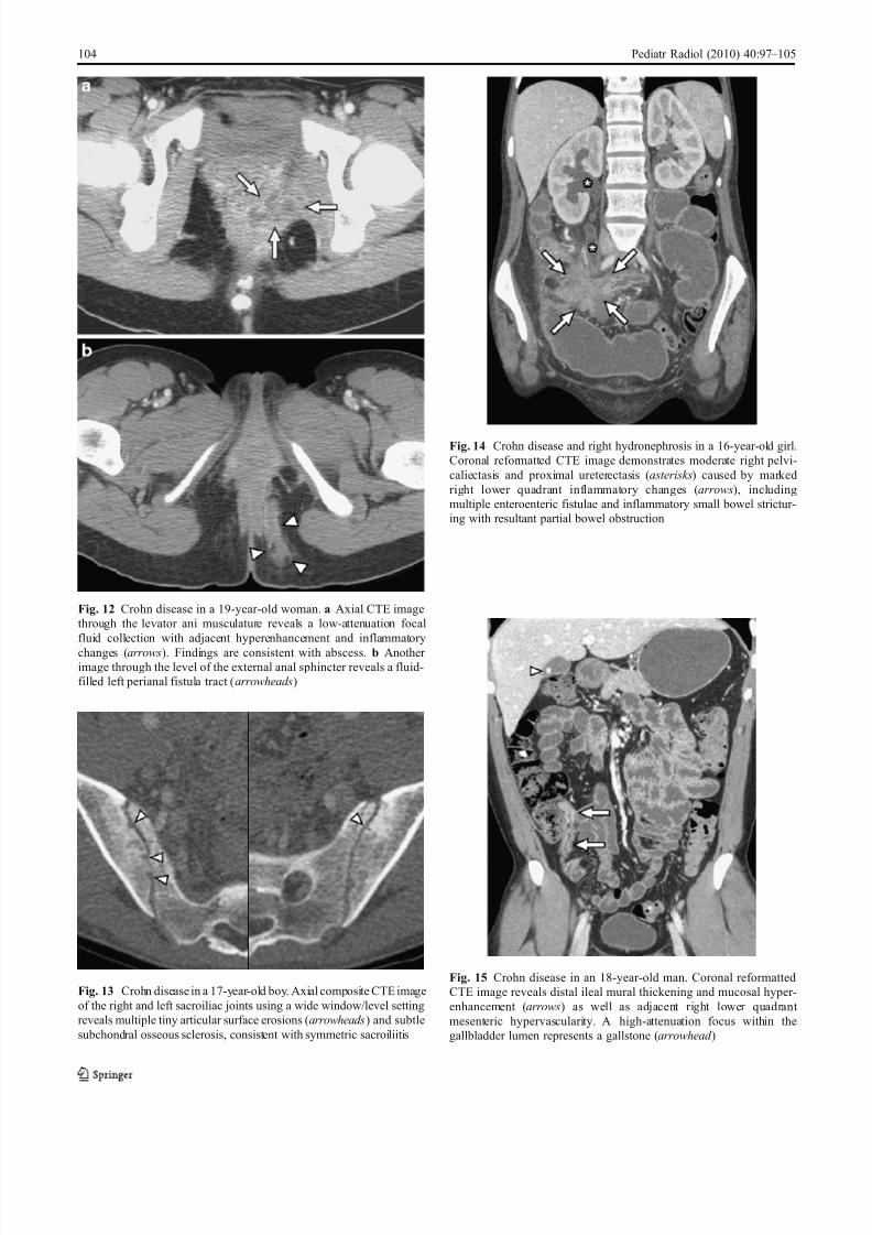
Fig. 10 Crohn disease in an 18-year-old man. a Coronal reformattedCTE image reveals rectal mural thickening, hyperenhancement,
adjacent perirectal inflammatory fat stranding, and perirectal hyper-
vascularity. A gas-containing sinus tract (arrow) arises from the left
posterolateral aspect of the rectum. b Coronal reformatted CTE imagethrough the kidneys reveals a nonobstructing calculus (arrow) within a
left upper pole calyx
Pediatr Radiol (2010) 40:97 – 105 103
8/11/2019 CTE in peds Crohn's 10.pdf
http://slidepdf.com/reader/full/cte-in-peds-crohns-10pdf 8/9
Fig. 15 Crohn disease in an 18-year-old man. Coronal reformatted
CTE image reveals distal ileal mural thickening and mucosal hyper-
enhancement (arrows) as well as adjacent right lower quadrant
mesenteric hypervascularity. A high-attenuation focus within the
gallbladder lumen represents a gallstone (arrowhead )
Fig. 14 Crohn disease and right hydronephrosis in a 16-year-old girl.
Coronal reformatted CTE image demonstrates moderate right pelvi-
caliectasis and proximal ureterectasis (asterisks) caused by marked
right lower quadrant inflammatory changes (arrows), including
multiple enteroenteric fistulae and inflammatory small bowel strictur-
ing with resultant partial bowel obstruction
Fig. 13 Crohn disease in a 17-year-old boy. Axial composite CTE image
of the right and left sacroiliac joints using a wide window/level setting
reveals multiple tiny articular surface erosions (arrowheads) and subtle
subchondral osseous sclerosis, consistent with symmetric sacroiliitis
Fig. 12 Crohn disease in a 19-year-old woman. a Axial CTE image
through the levator ani musculature reveals a low-attenuation focal
fluid collection with adjacent hyperenhancement and inflammatory
changes (arrows). Findings are consistent with abscess. b Another image through the level of the external anal sphincter reveals a fluid-
filled left perianal fistula tract (arrowheads)
104 Pediatr Radiol (2010) 40:97 – 105
8/11/2019 CTE in peds Crohn's 10.pdf
http://slidepdf.com/reader/full/cte-in-peds-crohns-10pdf 9/9
CTE plays an important role in the diagnosis of Crohn
disease in children as well as in establishing disease extent
and identifying disease-related complications. A variety of
techniques minimize pediatric radiation exposure related to
CTE. While CTE allows for complete assessment of
intestinal and extraintestinal pediatric Crohn disease, it is
our desire that MRE will soon become the primary imaging
modality by which inflammatory bowel disease in childrenis assessed.
References
1. Kim SC, Ferry GD (2004) Inflammatory bowel diseases in
pediat ric and adolescent patients: clinical, therape utic, and
psychosocial considerations. Gastroenterology 126:1550 – 1560
2. Siddiki HA, Fidler JL, Fletcher JG et al (2009) Prospective
comparison of state-of-the-art MR enterography and CT enter-
ography in small-bowel Crohn’s disease. AJR 193:113 – 121
3. Lee SS, Kim AY, Yang SK et al (2009) Crohn disease of the small
bowel: comparison of CT enterography, MR enterography, and
small-bowel follow-through as diagnostic techniques. Radiology
251:751 – 761
4. Jamieson DH, Shipman PJ, Israel DM et al (2003) Comparison of
multidetector CT and barium studies of the small bowel:
inflammatory bowel disease in children. AJR 180:1211 – 1216
5. Hara AK, Leighton JA, Heigh RI et al (2006) Crohn disease of the
small bowel: preliminary comparison among CT enterography,
capsule endoscopy, small-bowel follow-through, and ileoscopy.
Radiology 238:128 – 134
6. Applegate KE, Maglinte DD (2008) Imaging of the bowel in
children: new imaging techniques. Pediatr Radiol 38(Suppl 2):
S272 – S274
7. Booya F, Fletcher JG, Huprich JE et al (2006) Active Crohn
disease: CT findings and interobserver agreement for enteric
phase CT enterography. Radiology 241:787 – 795
8. Baker ME, Walter J, Obuchowski NA et al (2009) Mural
attenuation in normal small bowel and active inflammatory
Crohn’s disease on CT enterography: location, absolute attenua-
tion, relative attenuation, and the effect of wall thickness. AJR
192:417 – 423
9. Vandenbroucke F, Mortelé KJ, Tatli S et al (2007) Noninvasive
multidetector computed tomography enterography in patients with
small-bowel Crohn’s disease: is a 40-second delay better than 70
seconds? Acta Radiol 23:1 – 9 [Epub ahead of print]
10. Brown S, Applegate KE, Sandrasegaran K et al (2008) Fluoro-
scopic and CT enteroclysis in children: initial experience,
technical feasibility, and utility. Pediatr Radiol 38:497 – 510
11. Raptopoulos V, Schwartz RK, McNicholas MM et al (1997)
Multiplanar helical CT enterography in patients with Crohn’s
disease. AJR 169:1545 – 1550
12. Gaca AM, Jaffe TA, Delaney S et al (2008) Radiation doses from
small-bowel follow-through and abdomen/pelvis MDCT in pedi-
atric Crohn disease. Pediatr Radiol 38:285 – 291
13. Toma P, Granata C, Magnano G et al (2007) CT and MRI of paediatric Crohn disease. Pediatr Radiol 37:1083 – 1092
14. Jabra AA, Fishman EK, Taylor GA (1994) CT findings in
inflammatory bowel disease in children. AJR 162:975 – 979
15. Adler J, Punglia D, Dillman JR et al (2008) CT enterography
findings correlate with tissue inflammation but not fibrosis in
resected small bowel Crohn’s disease. Gastroenterology 134:A195
16. Hara AK, Alam S, Heigh RI et al (2008) Using CT enterography
to monitor Crohn’s disease activity: a preliminary study. AJR
190:1512 – 1516
17. Lee SS, Ha HK, Yang SK et al (2002) CT of prominent pericolic
or perienteric vasculature in patients with Crohn’s disease:
correlation with clinical disease activity and findings on barium
studies. AJR 179:1029 – 1036
18. Simoneaux SF, Patrick LE (1997) Genitourinary complications of
Crohn’s disease in pediatric patients. AJR 169:197 – 199
19. Markowitz J, Daum F, Aiges H et al (1984) Perianal disease in
children and adolescents with Crohn’s disease. Gastroenterology
86:829 – 833
20. Tolia V (1996) Perianal Crohn’s disease in children and
adolescents. Am J Gastroenterol 91:922 – 926
21. Solomon MJ (1996) Fistulae and abscesses in symptomatic
perianal Crohn’s disease. Int J Colorectal Dis 11:222 – 226
22. Halligan S, Stoker J (2006) Imaging of fistula in ano. Radiology
239:18 – 33
23. Peeters H, Vander Cruyssen B, Mielants H et al (2008) Clinical
and genetic factors associated with sacroiliitis in Crohn’s disease.
J Gastroenterol Hepatol 23:132 – 137
24. Leonard MB (2007) Glucocorticoid-induced osteoporosis in
children: impact of the underlying disease. Pediatrics 119(Suppl
2):S166 – S174
25. Dubner SE, Shults J, Baldassano RN et al (2009) Longitudinal
assessment of bone density and structure in an incident cohort of
children with Crohn’s disease. Gastroenterology 136:123 – 130
26. Gilsanz V, Perez FJ, Campbell PP et al (2009) Quantitative CT
reference values for vertebral trabecular bone density in children
and young adults. Radiology 250:222 – 227
27. Parente F, Pastore L, Bargiggia S et al (2007) Incidence and risk
factors for gallstones in patients with inflammatory bowel disease:
a large case-control study. Hepatology 45:1267 – 1274
Pediatr Radiol (2010) 40:97 – 105 105