current concepts in the diagnosis and treatment of ... · pdf fileschondral fractures of the...
TRANSCRIPT
1
*Address correspondence to John G. Kennedy, MD, FRCS (Orth), Hospital for Special Surgery, 535 East 70th St, New York, NY 10021 (e-mail: [email protected]).
No potential conflict of interest declared.
The American Journal of Sports Medicine, Vol. XX, No. XDOI: 10.1177/0363546509336336© 2009 American Orthopaedic Society for Sports Medicine
Current Concepts in the Diagnosis and Treatment of Osteochondral Lesions of the AnklePadhraig F. O’Loughlin, MD, Benton E. Heyworth,* MD, and John G. Kennedy, MD, FRCS (Orth)From the Hospital for Special Surgery, New York, New York
Osteochondral lesions of the ankle are a more common source of ankle pain than previously recognized. Although the exact pathophysiology of the condition has not been clearly established, it is likely that a variety of etiological factors play a role, with trauma, typically from ankle sprains, being the most common. Technological advancements in ankle arthroscopy and radiologic imaging, most importantly magnetic resonance imaging, have improved diagnostic capabilities for detecting osteochondral lesions of the ankle. Moreover, these technologies have allowed for the development of more sophisticated classification sys-tems that may, in due course, direct specific future treatment strategies. Nonoperative treatment yields best results when employed in select pediatric and adolescent patients with osteochondritis dissecans. However, operative treatment, which is dependent on the size and site of the lesion, as well as the presence or absence of cartilage damage, is frequently warranted in both children and adults with osteochondral lesions. Arthroscopic microdrilling, micropicking, and open procedures, such as osteochondral autograft transfer system and matrix-induced autologous chondrocyte implantation, are frequently employed. The purpose of this article is to review the history, etiology, and classification systems for osteochondral lesions of the ankle, as well as to describe current approaches to diagnosis and management.
Keywords: osteochondral defects; osteochondral lesions; osteochondritis dissecans; foot and ankle surgery; arthroscopy
lesion. In 1922, Kappis58 described a similar presentation within the talus, as did Phemister92 in 1924. Rendu98 was the first to describe articular fractures of the talus in 1932.
Many synonyms for OCLs of the ankle have been used, such as osteocartilaginous bodies, joint mice, intra-artic-ular fragmentary fractures, transchondral fractures, and osteochondral defects, with the acronym OCD being used interchangeably to refer to osteochondral defect or osteo-chondritis dissecans. We prefer to use the broader term osteochondral lesion to define a lesion of any origin that involves the articular surface and/or subchondral region of the talus or tibial plafond, thus affecting cartilage, bone, or both. The acronym OCD is reserved for osteochondritis dissecans lesions, which represent a specific subset of OCLs. Despite being first recognized several centuries ago, both the causes and preferred treatment strategy for OCLs remain the subject of frequent debate.
This review seeks to elucidate and compare theories regarding origins within a historical background, detail current diagnostic strategies, and provide a systematic approach to management of OCLs within the ankle joint.
ETIOLOGY
Proposed causes of ankle OCLs have included local avascu-lar necrosis,66 systemic vasculopathies, acute trauma,13,82 chronic microtrauma,37 endocrine or metabolic factors,93
DEFINITIONS AND HISTORY
Approximately 1 in 10 000 people per day suffers an ankle injury.59 In athletes, this number can be as high as 5.23 ankle injuries per 10 000 athlete-exposures, and higher still during active competition (9.35 per 10 000 athlete-exposures).85 In a recent systematic review of ankle injuries, Fong et al38 identified ankle injuries as being the most com-mon type of injury in 24 of 70 sports, with ankle sprain being the most frequent. Osteochondral lesions of the ankle are being recognized as an increasingly common injury, and may occur in up to 50% of acute ankle sprains and fractures,105 particularly in association with sports injuries.106,120
Recognition and understanding of osteochondral lesions (OCLs) of the ankle have developed in a gradual, stepwise fashion. In 1737, Monro79 described removal of a loose body from the ankle, which was believed to be of traumatic origin. Franz Konig66 first coined the term “osteochondritis disse-cans” in 1888, in reference to loose bodies found in the knee joint that he believed to be fragments from an avascular bone
Clinical Sports Medicine Update
AJSM PreView, published on June 26, 2009 as doi:10.1177/0363546509336336

2 O’Loughlin et al The American Journal of Sports Medicine
degenerative joint disease,117 joint malalignment,47 and genetic predisposition.80 Konig66 hypothesized that vascu-lar occlusion of subchondral bone leads to a process of local inflammation and development of a subchondral cyst. Although such a sequence may be specific to osteochondri-tis dissecans, the most widely accepted cause of OCLs of the ankle is trauma, usually in the form of ankle sprains. In a seminal study, Berndt and Harty13 reproduced the mechanism of injury for both lateral and medial talar dome OCLs. It was proposed that lateral injuries occur in inversion and dorsiflexion of the ankle, while posterome-dial injuries are the result of ankle plantar flexion and inversion. Flick and Gould37 studied more than 500 patients with OCLs and found that 98% of lateral dome lesions and 70% of medial dome lesions were associated with a history of trauma.
In several series, however, a significant percentage of patients with OCLs reported no trauma, lending support for the role of genetic, metabolic, or endocrine causes.93,99,100 Mubarak and Carroll80 described an autosomal dominant inheritance pattern in some osteochondritis dissecans patients characterized by endocrine dysfunction and col-lagen and epiphyseal abnormalities. Other authors have cited a high incidence of familial inheritance or popula-tions of patients with OCLs in multiple joints.18,44 Trias et al121 noted significant rates of hypothyroidism in 1 series of OCL patients. Although most patients with OCLs have a history of trauma, in some instances analysis of laboratory values related to endocrine and metabolic bone disorders, such as serum vitamin D, calcium, phosphorus, and parathyroid hormone, may be indicated, particularly in patients with bilateral or polyarticular disease or those with a family history of the condition.
CLASSIFICATION
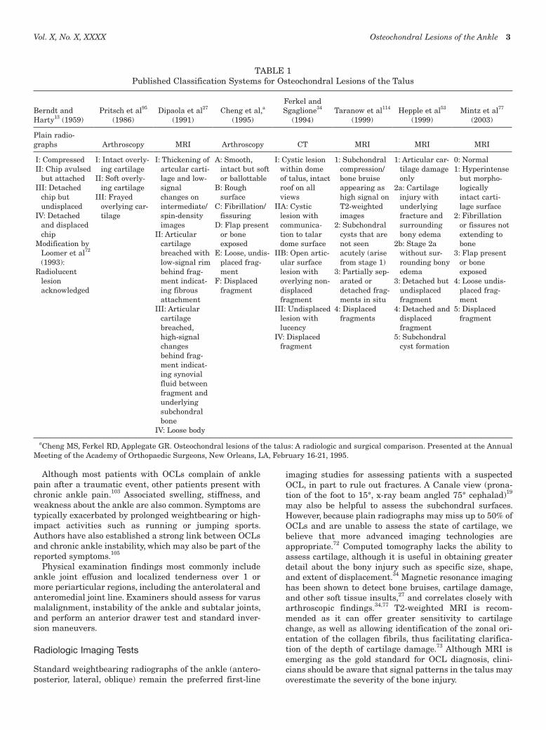
In 1959, Berndt and Hardy13 established a 4-stage classifica-tion system of ankle OCLs based on the severity of the lesion on plain radiographs (Figure 1). This was based on a thorough review of the literature pertaining to “tran-schondral fractures of the talus” from 1856 through 1956 combined with their own data from reproduction of transchondral fractures in 15 cadaveric specimens. Their classification was subsequently modified by Loomer et al72 in 1993 to include a fifth subtype of radiolucent, cystic lesions, as seen on CT scans. The principal advantage of the Berndt and Hardy system is its widespread use and sim-plicity. However, in 1 prospective study of 92 patients,72 50% of OCLs were not detected on plain radiographs. Moreover, the system is largely based on lesions with a traumatic origin, and does not differentiate or incorporate the spectrum of de novo lesions. As newer imaging tech-nologies have emerged, a variety of additional classification systems have been proposed (Table 1).5,27,53,72,114 Taranow et al114 used MRI to describe the condition of both the car-tilage and subchondral bone by employing the classic 4-stage grading to the bony component while describing the cartilage to be either viable and intact (grade A) or nonvi-able (grade B). The MRI-based system by Hepple et al53 is
based on the original Berndt and Harty classification, but uncouples traumatic, cystic, and idiopathic etiologies of OCLs. Mintz et al77 established a correlation between MRI and arthroscopic findings. Because of the large degree of signal change that can arise secondary to edema after even mild ankle injuries, some authors30 believe MRI may over-diagnose or overestimate the extent of OCLs, and they urge caution in the use of these classification systems.
Alternative classification systems using arthroscopic findings have emerged as well. Pritsch et al95 graded talar OCLs based on cartilage quality, as seen on arthroscopic visualization. Cheng et al (unpublished data, 1995) used arthroscopy to describe the condition of the talar cartilage, ranging from smooth, soft, and ballottable cartilage (stage A) to increasingly rough and fibrillated or fissured carti-lage to a more unstable lesion culminating in a loose, dis-placed fragment (stage F). The disadvantage of an arthroscopic classification system is that it focuses on the cartilage insult and is unable to consider the underlying bony component of the lesion.
DIAGNOSIS
Clinical Presentation
Osteochondral lesions of the ankle are being recognized as an increasingly common injury that may occur in up to 50% of acute ankle sprains and fractures.105 Advances in imaging techniques and an increasing number of ankle arthroscopies being performed each year, in conjunction with participation in sporting activities among all ages, are expected to contribute to a rise in frequency of this injury. The average age of patients with an OCL is 20 to 30 years, with a male preponderance of 70%, and bilaterality being reported in 10% of cases.22
Figure 1. Berndt and Harty classification system.
Vol. X, No. X, XXXX Osteochondral Lesions of the Ankle 3
Although most patients with OCLs complain of ankle pain after a traumatic event, other patients present with chronic ankle pain.103 Associated swelling, stiffness, and weakness about the ankle are also common. Symptoms are typically exacerbated by prolonged weightbearing or high-impact activities such as running or jumping sports. Authors have also established a strong link between OCLs and chronic ankle instability, which may also be part of the reported symptoms.105
Physical examination findings most commonly include ankle joint effusion and localized tenderness over 1 or more periarticular regions, including the anterolateral and anteromedial joint line. Examiners should assess for varus malalignment, instability of the ankle and subtalar joints, and perform an anterior drawer test and standard inver-sion maneuvers.
Radiologic Imaging Tests
Standard weightbearing radiographs of the ankle (antero-posterior, lateral, oblique) remain the preferred first-line
imaging studies for assessing patients with a suspected OCL, in part to rule out fractures. A Canale view (prona-tion of the foot to 15°, x-ray beam angled 75° cephalad)19 may also be helpful to assess the subchondral surfaces. However, because plain radiographs may miss up to 50% of OCLs and are unable to assess the state of cartilage, we believe that more advanced imaging technologies are appropriate.72 Computed tomography lacks the ability to assess cartilage, although it is useful in obtaining greater detail about the bony injury such as specific size, shape, and extent of displacement.34 Magnetic resonance imaging has been shown to detect bone bruises, cartilage damage, and other soft tissue insults,27 and correlates closely with arthroscopic findings.34,77 T2-weighted MRI is recom-mended as it can offer greater sensitivity to cartilage change, as well as allowing identification of the zonal ori-entation of the collagen fibrils, thus facilitating clarifica-tion of the depth of cartilage damage.73 Although MRI is emerging as the gold standard for OCL diagnosis, clini-cians should be aware that signal patterns in the talus may overestimate the severity of the bone injury.
TABLE 1Published Classification Systems for Osteochondral Lesions of the Talus
Berndt and Harty13 (1959)
Pritsch et al95
(1986)
Dipaola et al27
(1991)
Cheng et al,a
(1995)
Ferkel and Sgaglione34
(1994)
Taranow et al114
(1999)
Hepple et al53
(1999)
Mintz et al77
(2003)
Plain radio-graphs
Arthroscopy
MRI
Arthroscopy
CT
MRI
MRI
MRI
I: CompressedII: Chip avulsed
but attachedIII: Detached
chip but undisplaced
IV: Detached and displaced chip
Modification by Loomer et al72 (1993):
Radiolucent lesion acknowledged
I: Intact overly-ing cartilage
II: Soft overly-ing cartilage
III: Frayed overlying car-tilage
I: Thickening of artcular carti-lage and low-signal changes on intermediate/spin-density images
II: Articular cartilage breached with low-signal rim behind frag-ment indicat-ing fibrous attachment
III: Articular cartilage breached, high-signal changes behind frag-ment indicat-ing synovial fluid between fragment and underlying subchondral bone
IV: Loose body
A: Smooth, intact but soft or ballottable
B: Rough surface
C: Fibrillation/fissuring
D: Flap present or bone exposed
E: Loose, undis-placed frag-ment
F: Displaced fragment
I: Cystic lesion within dome of talus, intact roof on all views
IIA: Cystic lesion with communica-tion to talar dome surface
IIB: Open artic-ular surface lesion with overlying non-displaced fragment
III: Undisplaced lesion with lucency
IV: Displaced fragment
1: Subchondral compression/bone bruise appearing as high signal on T2-weighted images
2: Subchondral cysts that are not seen acutely (arise from stage 1)
3: Partially sep-arated or detached frag-ments in situ
4: Displaced fragments
1: Articular car-tilage damage only
2a: Cartilage injury with underlying fracture and surrounding bony edema
2b: Stage 2a without sur-rounding bony edema
3: Detached but undisplaced fragment
4: Detached and displaced fragment
5: Subchondral cyst formation
0: Normal1: Hyperintense
but morpho-logically intact carti-lage surface
2: Fibrillation or fissures not extending to bone
3: Flap present or bone exposed
4: Loose undis-placed frag-ment
5: Displaced fragment
aCheng MS, Ferkel RD, Applegate GR. Osteochondral lesions of the talus: A radiologic and surgical comparison. Presented at the Annual Meeting of the Academy of Orthopaedic Surgeons, New Orleans, LA, February 16-21, 1995.

4 O’Loughlin et al The American Journal of Sports Medicine
Anatomy and Location of Lesions
Talus. Articular cartilage in adults possesses neither a blood supply nor lymphatic drainage and is relatively inef-fective in responding to injury.16 Thus injuries confined to the cartilage alone stimulate only a slight reaction in the adjacent chondrocytes. The talus has 60% of its surface covered in cartilage, which may increase the risk of vascu-lar compromise.1 In contrast, involvement of the subchon-dral bone via penetration of the subchondral plate allows a typical inflammatory wound-healing response. Cells recruited from marrow elements attempt to fill the defect but the degree of success is predicated on age, as well as the size and location of the defect.
In 2007, Millington et al76 used a high-resolution ste-reophotography system to quantify the articular carti-lage topography and thickness within the ankle joint. These factors are important in the assessment of the extent of OCLs, to determine the best mode of treatment. The authors found that the thickest articular cartilage occurs over the talar shoulders. The mean thickness of both the talar (1.1 ± 018 mm) and tibial (1.16 ± 0.14 mm) cartilage was significantly thicker than the fibular carti-lage (0.85 ± 0.13 mm).
A recent study sought to evaluate the true frequency of OCLs on the talar dome by location and morphological characteristics.31 The authors developed a novel, 9-zone anatomical grid system. They identified 428 OCLs in 428 ankles and found that medial talar dome lesions were both more common and significantly larger than lateral lesions. With regard to specific zones on the talus, centromedial lesions were the most common (n = 227) with centrolateral the next most frequent site (n = 110). Posteromedial and anterolateral lesions were rarely found. A prior study by the same group studying MRI changes over time also reported that of 29 OCLs of the talus, 19 (66%) were located at the medial talar dome.30
Tibial Plafond. Osteochondral lesions of the tibial pla-fond (Figure 2) are rare, particularly in comparison to the incidence of OCLs of the talus. This may be due to differ-ences in the thickness and mechanical properties of the cartilage in these regions, as well as the rich arterial sup-ply to the distal tibia. Distal tibial cartilage has been shown to be stiffer than talar cartilage.6 Anterolateral and posteromedial cartilage at the distal tibia is also stiffer than the corresponding areas on the talar dome, with the softest cartilage found in the posterior half of the talus.
TREATMENT AND RESULTS
Nonoperative Treatment
Osteochondral lesions of the talus that are asymptomatic or are discovered as incidental findings can be treated nonoperatively. Low-grade OCLs, particularly osteochon-dritis dissecans lesions in the pediatric population, may resolve completely with variable need for immobilization or protected weightbearing. However, it is rarer to observe spontaneous healing in adult patients.28 One series
demonstrated good or excellent results in only 54% of patients with chronic, cystic talar lesions treated nonop-eratively.111 While rates of ankle osteoarthritis were low in this cohort, follow-up averaged only 38 months. In a com-prehensive review of treatment strategies for OCLs of the talus, Tol et al120 noted that of a total of 201 patients from 14 studies who had nonoperative treatment, 91 (45%) were reported to have had successful outcomes, with patients with chronic symptoms (>6 weeks) actually having better results (average success rate 56%) than the overall cohort. The authors divided the nonoperatively treated patients into 2 groups: group 1 pursued rest or restriction of sport or activities with or without the use of nonsteroidal anti-inflammatory drugs, while group 2 underwent cast immo-bilization for 3 weeks to 4 months. Group 1 had good or excellent results in 59% of patients, compared with 41% of patients in group 2. Typical indications for nonoperative treatment in these studies were minimal symptoms; Berndt and Harty stage I, II, and medial stage III lesions; or lesions with intact cartilage. Shearer et al111 have noted a poor correlation between changes in lesion size and clinical outcome. Therefore, a reduction in the size of the OCL that may be seen over time with conservative mea-sures may not necessarily correlate with symptomatic improvement, so conversion to operative treatment may be warranted, if symptoms persist.
Patients who are asymptomatic or minimally symptom-atic with lesions that involve cartilage alone may be treated nonoperatively with rest, ice, temporarily reduced weightbearing, and, in case of ankle malalignment, an orthosis.128 Clinicians should be aware, however, that non-operative management has shown relatively high rates of
Figure 2. Osteochondral lesion of the tibial plafond.

Vol. X, No. X, XXXX Osteochondral Lesions of the Ankle 5
failure in the literature.28,111 Further research into which patients may have successful outcomes with nonoperative care is warranted.
Surgical Management
The principal aim of surgical treatment is revasculariza-tion of the bony defect.7,40,43,67,104 Because articular hyaline cartilage is avascular and has poor regenerative capabili-ties, injuries that do not penetrate the subchondral plate have no stimulus for an inflammatory reaction and heal-ing. When the depth of a talar OCL injury extends to the subchondral bone, marrow cells are stimulated to produce new tissue in an attempt to fill the defect.4,87 However, this process involves the formation of fibrous cartilage, which lacks the favorable biomechanical properties of normal articular hyaline cartilage. In the case of smaller lesions, this fibrocartilage substitute may suffice, and is the basis behind techniques such as microfracture and micropicking. However, with larger lesions, fibrocartilage may not be adequate to support the longevity of the joint. Therefore, the majority of recently developed treatment approaches are aimed at providing a method of replacing damaged articular cartilage with tissue that more closely resembles hyaline cartilage. These have included primarily trans-plantation of osteochondral autograft plugs from distant donor sites, allograft transplantation, or harvesting and culturing chondrocytes that are later transplanted into the site of the osteochondral defect.
Cartilage Stabilization/Pinning
Traumatic osteochondral fragments that have not detached from the underlying bone may be suitable for fixation. Whenever possible, large unstable OCLs with a viable bony component are preferentially treated with stabiliza-tion rather than debridement alone, which may precipitate pain and degenerative changes within the joint.4,87 Although traditional OCL fixation has involved metal implants that require subsequent removal, more recent techniques have utilized compression or stabilization with bioabsorbable materials, such as polyglycolic acid (PGA) or polylactic acid (PLLA) bioabsorbable pins, which do not require removal. While the literature on use of these new materi-als in the ankle is limited, 1 small series, in which PGA/PLLA copolymer pins were used in conjunction with debri-dement of the bony bed, demonstrated healing in 6 of 7 cases, with no evidence of an inflammatory reaction in any cases.68
Retrograde Drilling
Retrograde drilling (RD) is indicated for subchondral bone lesions over which the overlying cartilage remains intact, with the clear advantage of protecting the integrity of the articular cartilage, compared with anterograde drilling (Figure 3).114 However, it is critical not only to decompress the lesion but also to address the structural integrity of the subchondral cyst or defect, to prevent subsequent articular collapse. Although previous authors have described the use of solid bone graft, given the difficulty in adequately filling
the contours of the lesion, other investigators have used surgical-grade calcium sulfate as an alternative bone-graft substitute that can be injected in liquid form into the defect after drilling.62 As an adjunct, a bone-marrow aspi-rate may be harvested from the iliac graft and its pluripo-tent cells isolated by centrifuge and mixed with the calcium graft to promote more rapid healing (Deland and Young, unpublished data, 2001). Retrograde drilling was first described by Lee and Mercurio70 for the knee in 1981 as an open procedure, but now is frequently performed arthroscop-ically in the ankle, along with fluoroscopic radiographic imaging. Because posteromedial and posterolateral lesions present a challenge when using a standard drill-targeting device arthroscopically, we have also employed the use of computer-assisted techniques to improve the accuracy of targeting lesions. These techniques have been employed successfully in other studies.24,60,101
Outcomes studies investigating RD have shown good results overall.8,67,70,114,118 Kono et al67 compared transmalle-olar drilling (TMD) with RD in 30 patients with unilateral OCLs without detachment of the cartilage, and re-look arthroscopy was performed at 1 year to assess the cartilage. In the TMD group, 11 lesions (58%) were unchanged (grade I) and 8 lesions (42%) had deteriorated from grade 0 to I, compared with the RD group, in which 3 lesions (27%) had improved from grade I to 0 and 8 (73%) were unchanged (2 grade 0 lesions, 6 grade I lesions). In another series of 16 patients with symptomatic OCLs of the medial talar dome treated arthroscopically with percutaneous RD through the sinus tarsi,114 mean American Orthopaedic Foot and Ankle Society (AOFAS) scores increased from 53.9 points to 82.6 points, with no complications reported.
Microfracture/Microdrilling
Microfracture and microdrilling (Figure 4) procedures have the same objective: to stimulate fibrocartilage devel-opment by breaching the subchondral plate with subse-quent introduction of serum factors and development of
Figure 3. Fluoroscopic image of talar retrograde drilling.

6 O’Loughlin et al The American Journal of Sports Medicine
scar tissue at the defect site. While the efficacy of micro-fracture in ankle OCLs is somewhat controversial,86 most series have demonstrated that it provides symptomatic relief.11,43,116 In the presence of a small (<6 mm), shear-type lesion characterized primarily by chondral damage, but minimal subchondral bone involvement, this technique may be optimal.87 Chuckpaiwong et al23 investigated 105 cases of talar OCLs treated with microfracture, reporting no failures of treatment with lesions smaller than 15 mm (n = 73) regardless of location, but only 1 successful out-come in lesions greater than 15 mm (n = 32). The authors also highlighted increasing age, higher body mass index, history of trauma, and presence of osteophytes as factors negatively affecting outcome.
Tissue Transplantation
For transplant of osteochondral constructs into the talus, perpendicular access is generally required to the injured
area.81 Most areas of the talar dome can be accessed per-pendicularly without the necessity for a malleolar osteotomy.26,81 Muir et al81 demonstrated that, on average, only 17% of the medial talar dome and 20% of the lateral talar dome could not be accessed without an osteotomy. After an anterolateral osteotomy, they reported an increase of 22% in sagittal exposure, while malleolar osteotomies provided access to the entire medial and lateral talar dome areas with a residual central 15% of the talar dome remaining inaccessible perpendicularly. Several well- accepted techniques for medial malleolar osteotomy have been described.88,119,129 Critical to all methods of osteotomy is a precise reduction and fixation to avoid fibrous non-union or malunion. Three-screw fixation (Figure 5) may be beneficial to reduce translation of bony fragments that can occur with 2-screw fixation (Figure 6).
Mosaicplasty. For treatment of larger talar lesions, Hangody et al51 described a method for autologous grafting using numerous cylindrical osteochondral plugs taken
Figure 4. Talar OCL micropicking.
Figure 5. Two parallel screw malleolar fixation technique, with evidence of displacement of osteotomy fragment.
Figure 6. Three-screw malleolar fixation technique, with evidence of satisfactory anatomic alignment.

Vol. X, No. X, XXXX Osteochondral Lesions of the Ankle 7
from the nonweightbearing segment of the medial or lat-eral femoral ridge of the knee and transferring them to a talar dome defect with a surface area of no more than 4 cm2 and approximately 10 mm in diameter. The authors recommend a mini-arthrotomy and identify OCLs in the medial or lateral aspect of the dome (rather than the cen-tral part of the talus) and otherwise normal tibial and talar articular surfaces as factors associated with better results.49 Good-to-excellent results have been reported in as high as 94% of patients in some series.9,51 In a study with 2- to 7-year follow-up, 36 talar OCL patients were reviewed, with excellent results in 26 patients, good in 6, and moderate in 2, based on the Hanover scoring system.50 A second-look arthroscopy procedure was performed in 8 patients and showed normal and congruent-appearing surfaces, with specific staining revealing type II–specific normal articular cartilage collagen and articular cartilage proteoglycans that were of similar quality to a control biopsy specimen. However, other authors have emphasized the technical challenge of reproducing a smooth articular surface, with protrusion of plugs in an “organ pipe” arrangement.75,89 Patient complaints such as a “catching” sensation and late-onset postoperative pain have also been described.84
Osteochondral Autologous Transfer System. Osteochon-dral autologous transfer system (OATS) has been advo-cated for the treatment of large cystic OCLs, such as type V lesions (Figure 7).53,72 Based on the course of a large cohort of patients with failures after simple drilling, curetting, debridement, or bone grafting, Scranton107 suggested that type V lesions (as described by Hepple et al53) greater than 6 mm in diameter with articular disruption should be indi-cated for OATS (Table 1). After arthroscopic identification or confirmation of a lesion greater than 6 mm in diameter with disrupted cartilage, conversion to open surgery is
pursued, with a posteromedial lesion generally requiring a medial malleolar osteotomy.107 Some authors advocate release of the anterior talofibular ligament, anterior subluxation, and forced plantar flexion to achieve ade-quate exposure for posterolateral lesions.108 The lesion is debrided at the edges and matched to a graft of equal size harvested from a nonweightbearing area of the ipsilateral knee. One series reported 90% patient satisfaction in a retrospective review of 50 OATS cases with a lesion diam-eter from 8 mm to 20 mm.108 In another series with aver-age follow-up of 16 months and an average lesion size of 12 mm × 10 mm, the mean postoperative AOFAS score was 88, the Lysholm knee score (assessing donor-site pain) was 97, and 89% of patients said they would have the procedure again.3 The authors believe OATS to be an effective salvage procedure for patients with failed previ-ous procedures and long-standing symptoms. Studies have suggested that viability of chondrocytes in the periphery of the graft may be affected by the acquisition method, with manual punches being associated with better survival of cells than use of a power trephine system.33,97
Osteochondral Allograft Transplantation. As an alterna-tive to OATS, osteochondral allograft transplantation may be more suitable for very large OCLs of the talus, and has the advantage of optimization of matching graft morpho-logic characteristics with the defect site, which is done with both radiologic and direct measurements intraoperatively.96,115 Raikin96 classified OCLs as “mas-sive” or not viable for standard repair options when the volume exceeds 3 cm3 and suggested that this grade may represent a sixth stage to the Berndt and Harty classifica-tion system. Some authors prefer fresh osteochondral allografts over fresh-frozen grafts, citing a decline in chon-drocyte viability in the latter.115 In such cases, transplan-tation should be performed within 7 days of the death of the donor. However, other authors report good results with frozen allografts that were frozen for less than 14 days before insertion.96 Typically, the defect is burred to create an even-edged rectangular defect with a flat base that can be packed with cancellous graft from distal tibia or donor talus to aid subsequent graft integration. The transplanted allograft is usually held in place by screw fixation.
Only 2 small series involving the use of an osteochondral allograft exist in the literature. Raikin96 reported on 6 cases, 5 of which involved the medial talar dome and 1, the lateral talar dome. Five of the 6 OCLs were of traumatic origin. Two patients had fresh allograft transplantation, and 4 had fresh-frozen talus allografts, with all approaches except for 1 involving a malleolar osteotomy. Mean AOFAS ankle scores improved from 42 preoperatively to 86 postop-eratively, which included 1 patient who went on to have an ankle arthrodesis for persistent pain. All patients stated they would have the procedure performed on the contralat-eral side if necessary. Gross et al46 reported on 9 cases of talar OCLs treated with fresh osteochondral allograft transplantation, 6 of which remained in situ at a mean follow-up of 11 years. The remaining 3 patients required ankle arthrodeses, secondary to resorption and fragmenta-tion of the graft.
Figure 7. An MRI scan of talus following an osteochondral autograft transfer system (OATS) procedure.
8 O’Loughlin et al The American Journal of Sports Medicine
Autologous Chondrocyte Implantation/Transplantation (ACI/ACT). Autologous chondrocyte transplantation is an alternative to osteochondral grafting techniques.74 The technique, as described by Giannini et al,41 involves har-vesting a small amount of cartilage arthroscopically from the knee ipsilateral to the ankle injury for chondrocyte cultures, which are grown in vitro for approximately 30 days. The OCL is debrided and filled with autologous can-cellous bone harvested from the ipsilateral distal tibial metaphysis. Periosteum is acquired from the ipsilateral proximal tibial metaphysis to cover the transplant area and is fixed with resorbable sutures. Before the flap is fully sutured down, the chondrocytes are transplanted in liquid media through the remaining unsutured area, which is then sutured and sealed with fibrin glue. Malleolar osteot-omy is fixed with 1 or more screws. At 1 year, the screws are removed, and at that time an ankle arthroscopy is per-formed to assess the graft site. The authors reported on 8 patients treated with ACT, with average preoperative to postoperative AOFAS scores improving from 32.1 points to 91 points at 2 years.41 Baums et al10 reported on 12 simi-larly treated patients, 11 of whom had good-to-excellent results after 63 months of follow-up, with an average pre-operative AOFAS score of 43.5 increasing to 88.4 postop-eratively. One recent study suggests that decreased postoperative pain may be an advantage of ACI, compared with other techniques.43 Gobbi et al43 compared surgical outcomes in 33 similarly sized talar OCLs treated with chondroplasty (11 cases), microfracture (10 cases), and OATS (12 cases). Although no significant difference was detected between the groups, with reference to AOFAS or single-assessment numeric evaluation scores, the numeric pain intensity was significantly greater at 24 hours post-operatively with OATS than with the other 2 techniques. Disadvantages of ACI include the cost of culturing hyaline cells, the need for 2 surgical procedures, and the durability of the graft.
Most recently, Giannini et al42 have reported on the results of ACI incorporating the use of a hyaluronan-based 3-dimensional scaffold (Hyalograft C, Fidia Advanced Biopolymers, Abana Terme, Italy) for symptomatic post-traumatic osteochondral talar dome lesions in 46 patients. It involved a 3-step process with initial cartilage harvest from the detached osteochondral fragment, chondrocyte culture on the Hyalograft C scaffold, and subsequent arthroscopic implantation of the 3-dimensional scaffold. They reported excellent clinical and histologic results, with an increase in AOFAS scores from 57.2 to 86.8. Hyaline-like cartilage regeneration was identified histologically in samples obtained at second-look arthroscopy in 3 patients at an average of 18 months after surgery.
Treatment of Tibial Plafond Lesions
Because of the rarity of tibial plafond OCLs, there are few reports in the literature related to treatment recommenda-tions. In the largest series involving distal tibial OCLs, Mologne and Ferkel78 retrospectively reviewed 880 con-secutive ankle arthroscopies, 23 (2.6%) of which involved treatment of tibial plafond OCLs. They concluded that
arthroscopic treatment methods used for talar OCLs (excision, curettage, and abrasion arthroplasty) were also effective for those of the distal tibia, based on average AOFAS ankle-hindfoot score improvement from 52 preop-eratively to 87 postoperatively and good or excellent results in 14 of 17 patients at medium-term follow-up.
Ueblacker et al123 reported on a new technique for retro-grade osteochondral autograft transplantation for treat-ment of OCLs of the proximal and distal tibia. Their series involved 5 patients, 2 of whom had painful chondral lesions of the distal anterocentral and posteromedial tibia. All patients were satisfied with the surgery. Follow-up arthros-copy showed the osteochondral cylinders well integrated and flush with the articular surface.
One case report described a female patient with bilateral distal tibial OCLs after several months of intensive mili-tary training.112 One month after cessation of active train-ing and nonoperative therapy, the severity of pain decreased considerably and the patient remained asymptomatic in her daily activities at 3 years. A second case report described osteochondral allografting of a distal tibial OCL, with 2-year follow-up radiographs demonstrating satisfac-tory incorporation of the graft without collapse and with preservation of joint space.21
Adjunctive Treatments/Future Directions
Viscosupplementation Therapy
Despite a dearth of convincing outcomes data to support their use, the popularity of intra-articular hyaluronic acid (HA) derivative injections, also known as viscosupplemen-tation therapy, continues to grow for arthritis and other conditions in a variety of joints. Pleimann et al94 were among the first authors to report on the use of HA injec-tions as an adjunct in the nonoperative treatment of ankle arthritis in 2002. Salk et al102 recently performed a controlled trial in which 22 patients were randomized to receive either 5 weekly intra-articular injections of Hyalgan (sodium hyaluronate) or saline placebo injections for ankle osteoarthritis, demonstrating significantly better improvement in the HA treatment group. Tytherleigh-Strong et al122 reported increased markers of articular cartilage survival and function in a sheep model, in which viscosupplementation therapy was used as an adjunct to osteochondral grafting of the knee. Other studies have sup-ported these findings as well. Most recently, Cohen et al25 conducted a double-blind randomized controlled study examining the safety and efficacy of intra-articular sodium hyaluronate in the treatment of pain associated with ankle osteoarthritis. Thirty consecutive patients were enrolled, and those treated demonstrated a significantly greater improvement from baseline on the Ankle Osteoarthritis Scale at 3 months than did the control group. The authors concluded that sodium hyaluronate may be a safe and effective option for pain associated with ankle osteoarthri-tis but advocated the need for larger studies.
Because of the presumed benefits of HA derivatives on synovial fluid and chondrocyte function, the senior author of this review (J.G.K.) routinely uses viscosupplementation

Vol. X, No. X, XXXX Osteochondral Lesions of the Ankle 9
as adjunctive treatment with all methods of surgical treat-ment of ankle OCLs. We are involved in a clinical trial that aims to establish how HA may help in maintaining the integrity of cartilage transplants in patients undergoing the OATS procedure. It is known that the peripheral rim of the graft suffers chondral cell death and that the graft itself may also have reduced chondral viability after trans-plant due to integrative problems as well as the impact forces involved in graft placement.91,127 The authors hypoth-esize that HA will act in these cases, as it does in degen-erative joint disease, to preserve existing cartilage and produce a more robust graft. However, it must be empha-sized that ongoing clinical trials are needed to confirm the efficacy of this treatment. Hyaluronic acid may be an adjunct to improve outcomes in compromised cartilage in the future, but at this time, it is not standard practice to employ it in this fashion.
Electrical/Electromagnetic Stimulation
Although the efficacy of electric and electromagnetic stimu-lation on bone repair and healing of cartilage defects is controversial, studies have suggested an upregulation of known molecular healing factors, such as transforming growth factor–beta (TGF-β) and various bone morphoge-netic proteins (BMPs, which are members of the TGF-β superfamily), as well as osteoclasts.2,14,20,71,126 One proposed mechanism is that pulsed electromagnetic fields stimulate chondrocyte proliferation by means of a nitrous oxide path-way.36 A recent study investigating bone formation and graft stabilization in a sheep model of osteochondral autograft treatment suggested that pulsed electromagnetic field treatment leads to improved results.12 Based on a growing body of evidence, the use of electric and electro-magnetic stimulation may play an increasing role in the future treatment of ankle OCLs.
Ultrasound Stimulation
Ultrasound is a propagating pressure wave that transfers mechanical energy into tissues.130 Low-intensity ultra-sound has been studied as a modality with properties that can enhance bone52,64 and cartilage healing.29,90 In a recently studied rabbit model with bilateral knee OCLs, the effects of low-intensity pulsed ultrasound in repairing osteochondral injuries was compared with the untreated contralateral side, revealing significantly higher scores in gross appearance grades, histologic grades, and proteogly-can quantity on the treated side.56 More evidence is needed to assess the role of low-intensity pulsed ultrasound in the acceleration of repair of osteochondral injuries.
Mesenchymal Stem Cells
Mesenchymal stem cells (MSCs) from bone marrow have been cultured in vitro and induced to form cartilage before implantation into the chondral defects in rabbits.124 Bone marrow has been aspirated from the iliac crests in a caprine model and chondrogenesis of the MSCs induced.17 Mesenchymal stem cells represent a valuable adjunct in
that they may be harvested with relative ease by means of a bone-marrow aspirate and a small number of pluripotent cells can be isolated, grown in vitro if necessary, and then introduced into osteochondral defects. They have the capa-bility to differentiate into articular cartilage and induce the formation of subchondral bone. Recently, MSCs have been used with success in hybrid scaffolds to repair osteochon-dral defects in animal models.48,55,65 Although still in the early stages of application, this unique approach may have great potential in treatment of human cartilage defects.
Platelet-Rich Plasma
At the site of any injury involving bone, a clot will form that consists of red blood cells, white blood cells, and plate-lets in a fibrin matrix. In bone healing, the alpha granules within the platelets are a valuable reservoir of exogenous factors.39 These factors include platelet-derived growth fac-tor, insulin-like growth factor, and TGF-β, which along with a number of other factors play a critical role in bone healing.57 Platelet-rich plasma has recently been studied in conjunction with autologous chondrocyte transfer, show-ing promise both as a scaffold in which to help hold ACI cells and as a reservoir of growth-stimulating factors.15,83
Computer-Aided Navigation and Robot-Assisted Surgery
As computer navigation techniques become more sophisti-cated and more user-friendly, their integration into ortho-paedic procedures increases (Figure 8). Computer navigation is particularly attractive for OCLs given the importance of precise localization and the potential for minimally inva-sive procedures.60,61 Robot-assisted surgery may also be ultimately favored over conventional orthopaedic tech-niques in the treatment of OCLs because of the optimiza-tion of accuracy and precision in the preparation of bone surfaces, and the potential for more reliable and reproduc-ible outcomes with regard to spatial accuracy. How rapidly these technologies advance in foot and ankle surgery, and orthopaedic surgery as a whole, remains to be seen.
Tissue Engineering
Tissue engineering can be defined as the application of biological, chemical, and engineering principles to the repair, restoration, or regeneration of living tissue by using biomaterials, cells, and factors alone or in combina-tion.69 There are 3 common tissue engineering approaches used to address osteochondral injuries113: extraction of the appropriate cells from the patient, in vitro culture, followed by transplantation back into the body defect that requires regeneration; placement of biologic factors, such as molecules or growth factors, into body defects; and use of 3-dimensional porous materials (eg, titanium, tanta-lum) to stimulate the ingrowth of new tissue. A combina-tion of these 3 approaches may also be used, such that there are osteoinductive, osteoconductive, and osteogenic elements, thus providing an optimal environment for bone growth.63

10 O’Loughlin et al The American Journal of Sports Medicine
The repair of OCLs requires a tissue engineering approach that aims to mimic the physiologic properties and structure of 2 different tissues (cartilage and bone) using a scaffold-cell construct.113 Hoque et al54 have devel-oped 2 such scaffolds, the first of which is composed of fibrin and polycaprolactone (PCL), and the second of which is composed of PCL and PCL–tricalcium phosphate. However, clinical efficacy has yet to be established.
Growth Factors. Growth factors are cytokines that are critical for optimal healing. Several growth factors have been shown to improve cartilage healing in vivo. Bone mor-phogenetic protein–2 appears to be intimately involved with the growth and differentiation of MSCs to chondro-blasts and osteoblasts.125 Sellers et al109,110 used recombi-nant human BMP-2 for the treatment of full-thickness defects of articular cartilage in rabbits, and found an accel-erated formation of new subchondral bone and an improved histologic appearance of the overlying articular surface. However, the direct application of growth factors is contro-versial, as the method of delivery and ability to maintain appropriate endogenous levels remains challenging.
Gene Therapy. Gene therapy involves the modification of cellular genetic information, and its application to the treatment of cartilage lesions involves transferring specific genes that code for certain growth factors into chondrocytes or progenitor cells. Vectors may be viral or nonviral, with viral vectors appearing to be a more efficient prospect.32 Delivery may be executed either in vivo or in vitro. In vitro delivery offers greater safety and control, although it repre-sents a greater technical challenge. Because of the theo-retical risk of insertional mutagenesis by the viral vectors integrating into the genome of the cells, safety remains a concerning obstacle to nonexperimental use at this stage.
By using tissue engineering to incorporate growth factors (osteoinductive), tissue scaffolds (osteoconductive), and gene therapy (osteogenic), Grande et al45 have successfully influ-enced the cartilage repair environment in the treatment of OCLs in a rabbit model. Periosteal stem cells with known osteoinductive potency were transduced with either BMP-7 or sonic hedgehog (Shh) gene. The cells were subsequently cultured and expanded and then seeded into bioresorbable
polymer scaffolds, with the resultant implant placed in full-thickness OCLs in 80 rabbits. Significant enhancement of the quality of the repair tissue with more hyaline-appearing cartilage and a smoother surface was observed in both BMP-7 and Shh gene–treated animals versus controls.
CONCLUSION
Osteochondral lesions of the ankle are being recognized with increasing frequency, in part because of heightened understanding and awareness, and in part because of improved MRI and arthroscopic technology. As a result, treatment strategies and techniques continue to be rapidly developed and improved upon. However, outcomes research remains sparse on this subject; despite a number of emerg-ing future directions that hold promise in the treatment of OCLs, more evidence is necessary before they can be treated with consistent efficacy and safety.
REFERENCES
1. Adelaar RS, Madrian JR. Avascular necrosis of the talus. Orthop Clin North Am. 2004;35(3):383-395, xi.
2. Akai M, Hayashi K. Effect of electrical stimulation on musculoskeletal systems; a meta-analysis of controlled clinical trials. Bioelectro-magnetics. 2002;23(2):132-143.
3. Al-Shaikh RA, Chou LB, Mann JA, Dreeben SM, Prieskorn D. Autologous osteochondral grafting for talar cartilage defects. Foot Ankle Int. 2002;23(5):381-389.
4. Anderson AF, Pagnani MJ. Osteochondritis dissecans of the femoral condyles: long-term results of excision of the fragment. Am J Sports Med. 1997;25(6):830-834.
5. Anderson IF, Crichton KJ, Grattan-Smith T, Cooper RA, Brazier D. Osteochondral fractures of the dome of the talus. J Bone Joint Surg Am. 1989;71(8):1143-1152.
6. Athanasiou KA, Niederauer GG, Schenck RC Jr. Biomechanical topography of human ankle cartilage. Ann Biomed Eng. 1995;23(5): 697-704.
7. Baker CL, Graham JM Jr. Current concepts in ankle arthroscopy. Orthopedics. 1993;16(9):1027-1035.
8. Bale RJ, Hoser C, Rosenberger R, Rieger M, Benedetto KP, Fink C. Osteochondral lesions of the talus: computer-assisted retrograde drilling—feasibility and accuracy in initial experiences. Radiology. 2001;218(1):278-282.
9. Bartha L, Vajda A, Duska Z, Rahmeh H, Hangody L. Autologous osteochondral mosaicplasty grafting. J Orthop Sports Phys Ther. 2006;36(10):739-750.
10. Baums MH, Heidrich G, Schultz W, Steckel H, Kahl E, Klinger HM. Autologous chondrocyte transplantation for treating cartilage defects of the talus. J Bone Joint Surg Am. 2006;88(2):303-308.
11. Becher C, Thermann H. Results of microfracture in the treatment of articular cartilage defects of the talus. Foot Ankle Int. 2005;26(8): 583-589.
12. Benazzo F, Cadossi M, Cavani F, et al. Cartilage repair with osteo-chondral autografts in sheep: effect of biophysical stimulation with pulsed electromagnetic fields. J Orthop Res. 2008;26(5):631-642.
13. Berndt AL, Harty M. Transchondral fractures (osteochondritis disse-cans) of the talus. J Bone Joint Surg Am. 1959;41:988-1020.
14. Bodamyali T, Bhatt B, Hughes FJ, et al. Pulsed electromagnetic fields simultaneously induce osteogenesis and upregulate transcription of bone morphogenetic proteins 2 and 4 in rat osteoblasts in vitro. Biochem Biophys Res Commun. 1998;250(2):458-461.
15. Brehm W, Aklin B, Yamashita T, et al. Repair of superficial osteochon-dral defects with an autologous scaffold-free cartilage construct in a caprine model: implantation method and short-term results. Osteoarthritis Cartilage. 2006;14(12):1214-1226.
Figure 8. Computer–aided orthopaedic surgery.

Vol. X, No. X, XXXX Osteochondral Lesions of the Ankle 11
16. Buckwalter JA, Mankin HJ. Articular cartilage: degeneration and osteoarthritis, repair, regeneration, and transplantation. Instr Course Lect. 1998;47:487-504.
17. Butnariu-Ephrat M, Robinson D, Mendes DG, Halperin N, Nevo Z. Resurfacing of goat articular cartilage by chondrocytes derived from bone marrow. Clin Orthop Relat Res. 1996;330:234-243.
18. Campbell CJ, Ranawat CS. Osteochondritis dissecans: the question of etiology. J Trauma. 1966;6(2):201-221.
19. Canale ST, Kelly FB Jr. Fractures of the neck of the talus: long-term evaluation of seventy-one cases. J Bone Joint Surg Am. 1978;60(2):143-156.
20. Chang K, Chang WH, Huang S, Huang S, Shih C. Pulsed electromag-netic fields stimulation affects osteoclast formation by modulation of osteoprotegerin, RANK ligand and macrophage colony-stimulating factor. J Orthop Res. 2005;23(6):1308-1314.
21. Chapman CB, Mann JA. Distal tibial osteochondral lesion treated with osteochondral allografting: a case report. Foot Ankle Int. 2005;26(11):997-1000.
22. Chew KT, Tay E, Wong YS. Osteochondral lesions of the talus. Ann Acad Med Singapore. 2008;37(1):63-66.
23. Chuckpaiwong B, Berkson EM, Theodore GH. Microfracture for osteochondral lesions of the ankle: outcome analysis and outcome predictors of 105 cases. Arthroscopy. 2008;24(1):106-112.
24. Citak M, Kendoff D, Kfuri M Jr, Pearle A, Krettek C, Hufner T. Accuracy analysis of Iso-C3D versus fluoroscopy-based navigated retrograde drilling of osteochondral lesions: a pilot study. J Bone Joint Surg Br. 2007;89(3):323-326.
25. Cohen MM, Altman RD, Hollstrom R, Hollstrom C, Sun C, Gipson B. Safety and efficacy of intra-articular sodium hyaluronate (Hyalgan) in a randomized, double-blind study for osteoarthritis of the ankle. Foot Ankle Int. 2008;29(7):657-663.
26. Dallari D, Fini M, Stagni C, et al. In vivo study on the healing of bone defects treated with bone marrow stromal cells, platelet-rich plasma, and freeze-dried bone allografts, alone and in combination. J Orthop Res. 2006;24(5):877-888.
27. Dipaola JD, Nelson DW, Colville MR. Characterizing osteochondral lesions by magnetic resonance imaging. Arthroscopy. 1991;7(1): 101-104.
28. Easley ME, Scranton PE Jr. Osteochondral autologous transfer sys-tem. Foot Ankle Clin. 2003;8(2):275-290.
29. Ebisawa K, Hata K, Okada K, et al. Ultrasound enhances transforming growth factor beta-mediated chondrocyte differentiation of human mesenchymal stem cells. Tissue Eng. 2004;10(5-6):921-929.
30. Elias I, Jung JW, Raikin SM, Schweitzer MW, Carrino JA, Morrison WB. Osteochondral lesions of the talus: change in MRI findings over time in talar lesions without operative intervention and implications for staging systems. Foot Ankle Int. 2006;27(3):157-166.
31. Elias I, Zoga AC, Morrison WB, Besser MP, Schweitzer ME, Raikin SM. Osteochondral lesions of the talus: localization and morphologic data from 424 patients using a novel anatomical grid scheme. Foot Ankle Int. 2007;28(2):154-161.
32. Evans CH, Ghivizzani SC, Herndon JH, Robbins PD. Gene therapy for the treatment of musculoskeletal diseases. J Am Acad Orthop Surg. 2005;13(4):230-242.
33. Evans PJ, Miniaci A, Hurtig MB. Manual punch versus power harvest-ing of osteochondral grafts. Arthroscopy. 2004;20(3):306-310.
34. Ferkel RD, Flannigan BD, Elkins BS. Magnetic resonance imaging of the foot and ankle: correlation of normal anatomy with pathologic conditions. Foot Ankle. 1991;11(5):289-305.
35. Ferkel RD, Sgaglione NA: Arthroscopic treatment of osteochondral lesions of the talus: long term results. Orthop Trans. 1994;17:1011.
36. Fitzsimmons RJ, Gordon SL, Kronberg J, Ganey T, Pilla AA. A pulsing electric field (PEF) increases human chondrocyte proliferation through a transduction pathway involving nitric oxide signaling. J Orthop Res. 2008;26:854-859.
37. Flick AB, Gould N. Osteochondritis dissecans of the talus (transchon-dral fractures of the talus): review of the literature and new surgical approach for medial dome lesions. Foot Ankle. 1985;5(4):165-185.
38. Fong DT, Hong Y, Chan LK, Yung PS, Chan KM. A systematic review on ankle injury and ankle sprain in sports. Sports Med. 2007;37(1):73-94.
39. Gandhi A, Bibbo C, Pinzur M, Lin SS. The role of platelet-rich plasma in foot and ankle surgery. Foot Ankle Clin. 2005;10(4):621-637, viii.
40. Giannini S, Buda R, Faldini C, et al. Surgical treatment of osteochon-dral lesions of the talus in young active patients. J Bone Joint Surg Am. 2005;87(suppl 2):28-41.
41. Giannini S, Buda R, Grigolo B, Vannini F. Autologous chondrocyte transplantation in osteochondral lesions of the ankle joint. Foot Ankle Int. 2001;22(6):513-517.
42. Giannini S, Buda R, Vannini F, Di Caprio F, Grigolo B. Arthroscopic autolo-gous chondrocyte implantation in osteochondral lesions of the talus: surgical technique and results. Am J Sports Med. 2008;36:873-880.
43. Gobbi A, Francisco RA, Lubowitz JH, Allegra F, Canata G. Osteochondral lesions of the talus: randomized controlled trial com-paring chondroplasty, microfracture, and osteochondral autograft transplantation. Arthroscopy. 2006;22(10):1085-1092.
44. Goldstone RA, Pisani AJ: Osteochondritis dissecans of the talus. N Y State J Med. 1965;65(19):2487-2494.
45. Grande DA, Mason J, Light E, Dines D. Stem cells as platforms for delivery of genes to enhance cartilage repair. J Bone Joint Surg Am. 2003;85(suppl 2):111-116.
46. Gross AE, Agnidis Z, Hutchison CR. Osteochondral defects of the talus treated with fresh osteochondral allograft transplantation. Foot Ankle Int. 2001;22(5):385-391.
47. Guettler JH, Demetropoulos CK, Yang KH, Jurist KA. Osteochondral defects in the human knee: influence of defect size on cartilage rim stress and load redistribution to surrounding cartilage. Am J Sports Med. 2004;32(6):1451-1458.
48. Han SH, Kim YH, Park MS, et al. Histological and biomechanical properties of regenerated articular cartilage using chondrogenic bone marrow stromal cells with a PLGA scaffold in vivo. J Biomed Mater Res A. 2008;87:850-861.
49. Hangody L: The mosaicplasty technique for osteochondral lesions of the talus. Foot Ankle Clin. 2003;8(2):259-273.
50. Hangody L, Kish G, Modis L, et al. Mosaicplasty for the treatment of osteochondritis dissecans of the talus: two to seven year results in 36 patients. Foot Ankle Int. 2001;22(7):552-558.
51. Hangody L, Rathonyi GK, Duska Z, Vasarhelyi G, Fules P, Modis L. Autologous osteochondral mosaicplasty: surgical technique. J Bone Joint Surg Am. 2004;86(suppl 1):65-72.
52. Heckman JD, Ryaby JP, McCabe J, Frey JJ, Kilcoyne RF. Acceleration of tibial fracture-healing by non-invasive, low-intensity pulsed ultra-sound. J Bone Joint Surg Am. 1994;76(1):26-34.
53. Hepple S, Winson IG, Glew D. Osteochondral lesions of the talus: a revised classification. Foot Ankle Int. 1999;20(12):789-793.
54. Hoque ME, Hutmacher DW, Feng W, et al. Fabrication using a rapid prototyping system and in vitro characterization of PEG-PCL-PLA scaffolds for tissue engineering. J Biomater Sci Polym Ed. 2005;16(12):1595-1610.
55. Jeong WK, Oh SH, Lee JH, Im GI. Repair of osteochondral defects with a construct of mesenchymal stem cells and a polydioxanone/poly(vinyl alcohol) scaffold. Biotechnol Appl Biochem. 2008;49 (Pt 2):155-164.
56. Jia XL, Chen WZ, Zhou K, Wang ZB. Effects of low-intensity pulsed ultrasound in repairing injured articular cartilage. Chin J Traumatol. 2005;8(3):175-178.
57. Joyce ME, Jingushi S, Scully SP, Bolander ME. Role of growth factors in fracture healing. Prog Clin Biol Res. 1991;365:391-416.
58. Kappis M. Weitere beitrage zur traumatisch-mechischen entstehung der spontanen knorpelablosungen (sogen. osteochondritis dis-secans). Dtsch Z Chir. 1922;171:13-29.
59. Katcherian D. Soft-tissue injuries of the ankle. In: Lutter LD, Mizel MS, Pfeffer GB, eds. Orthopaedic Knowledge Update: Foot and Ankle. Rosemont, IL: American Academy of Orthopaedic Surgeons; 1994:241-253.
60. Kendoff D, Geerling J, Mahlke L, et al. Navigated Iso-C(3D)-based drilling of a osteochondral lesion of the talus. Unfallchirurg. 2003;106(11):963-967.
61. Kendoff D, Hufner T, Citak M, et al. Navigated Iso-C3D-based percu-taneous osteoid osteoma resection: a preliminary clinical report. Comput Aided Surg. 2005;10(3):157-163.

12 O’Loughlin et al The American Journal of Sports Medicine
62. Kennedy JG, Suero EM, O’Loughlin PF, Brief A, Bohne WH. Clinical tips: retrograde drilling of talar osteochondral defects. Foot Ankle Int. 2008;29:616-619.
63. Khan SN, Cammisa FP Jr, Sandhu HS, Diwan AD, Girardi FP, Lane JM. The biology of bone grafting. J Am Acad Orthop Surg. 2005;13(1):77-86.
64. Khan Y, Laurencin CT. Fracture repair with ultrasound: clinical and cell-based evaluation. J Bone Joint Surg Am. 2008;90(suppl 1):138-144.
65. Kobayashi T, Ochi M, Yanada S, et al. A novel cell delivery system using magnetically labeled mesenchymal stem cells and an external magnetic device for clinical cartilage repair. Arthroscopy. 2008;24(1):69-76.
66. Konig F. Uber freie Korper in den Gelenken [On the presence of loose bodies in joints]. Deutsche Zeitschr f Chi. 1888;27:90-109.
67. Kono M, Takao M, Naito K, Uchio Y, Ochi M. Retrograde drilling for osteochondral lesions of the talar dome. Am J Sports Med. 2006;34(9):1450-1456.
68. Larsen MW, Pietrzak WS, DeLee JC. Fixation of osteochondritis dis-secans lesions using poly(l-lactic acid)/ poly(glycolic acid) copolymer bioabsorbable screws. Am J Sports Med. 2005;33(1):68-76.
69. Laurencin CT, Ambrosio AM, Borden MD, Cooper JA Jr. Tissue engi-neering: orthopedic applications. Annu Rev Biomed Eng. 1999;1:19-46.
70. Lee CK, Mercurio C. Operative treatment of osteochondritis disse-cans in situ by retrograde drilling and cancellous bone graft: a pre-liminary report. Clin Orthop Relat Res. 1981;158:129-136.
71. Lippiello L, Chakkalakal D, Connolly JF. Pulsing direct current-induced repair of articular cartilage in rabbit osteochondral defects. J Orthop Res. 1990;8(2):266-275.
72. Loomer R, Fisher C, Lloyd-Smith R, Sisler J, Cooner T. Osteochondral lesions of the talus. Am J Sports Med. 1993;21(1):13-19.
73. Lusse S, Claassen H, Gehrke T, et al. Evaluation of water content by spatially resolved transverse relaxation times of human articular car-tilage. Magn Reson Imaging. 2000;18(4):423-430.
74. Mandelbaum BR, Gerhardt MB, Peterson L. Autologous chondrocyte implantation of the talus. Arthroscopy. 2003;19(suppl 1):129-137.
75. Marcacci M, Kon E, Zaffagnini S, et al. Multiple osteochondral arthroscopic grafting (mosaicplasty) for cartilage defects of the knee: prospective study results at 2-year follow-up. Arthroscopy. 2005;21(4):462-470.
76. Millington SA, Grabner M, Wozelka R, Anderson DD, Hurwitz SR, Crandall JR. Quantification of ankle articular cartilage topography and thickness using a high resolution stereophotography system. Osteoarthritis Cartilage. 2007;15(2):205-211.
77. Mintz DN, Tashjian GS, Connell DA, Deland JT, O’Malley M, Potter HG. Osteochondral lesions of the talus: a new magnetic resonance grading system with arthroscopic correlation. Arthroscopy. 2003;19(4):353-359.
78. Mologne TS, Ferkel RD. Arthroscopic treatment of osteochondral lesions of the distal tibia. Foot Ankle Int. 2007;28(8):865-872.
79. Monro A. Part of the cartilage of the joint separated and ossified. In: Medical Essays and Observations. 2nd ed. Edinburgh: Ruddimans; 1737:305.
80. Mubarak SJ, Carroll NC. Familial osteochondritis dissecans of the knee. Clin Orthop Relat Res. 1979;140:131-136.
81. Muir D, Saltzman CL, Tochigi Y, Amendola N. Talar dome access for osteochondral lesions. Am J Sports Med. 2006;34(9):1457-1463.
82. Mullett H, Kennedy JG, Quinlan W. Subchondral talar cyst following open reduction and internal fixation of an ankle fracture. Foot Ankle Surgery. 1999;5(3):147-149.
83. Munirah S, Samsudin OC, Chen HC, Salmah SH, Aminuddin BS, Ruszymah BH. Articular cartilage restoration in load-bearing osteo-chondral defects by implantation of autologous chondrocyte-fibrin constructs: an experimental study in sheep. J Bone Joint Surg Br. 2007;89(8):1099-1109.
84. Nakagawa Y, Suzuki T, Kuroki H, Kobayashi M, Okamoto Y, Nakamura T. The effect of surface incongruity of grafted plugs in osteochondral grafting: a report of five cases. Knee Surg Sports Traumatol Arthrosc. 2007;15(5):591-596.
85. Nelson AJ, Collins CL, Yard EE, Fields SK, Comstock RD. Ankle injuries among United States high school sports athletes, 2005-2006. J Athl Train. 2007;42(3):381-387.
86. O’Driscoll S. Update on cartilage repair. Orthopedics. 2002;25(12):1342, 1388.
87. O’Driscoll SW. The healing and regeneration of articular cartilage. J Bone Joint Surg Am. 1998;80(12):1795-1812.
88. Oznur A. Medial malleolar window approach for osteochondral lesions of the talus. Foot Ankle Int. 2001;22(10):841-842.
89. Ozturk A, Ozdemir MR, Ozkan Y. Osteochondral autografting (mosaicplasty) in grade IV cartilage defects in the knee joint: 2- to 7-year results. Int Orthop. 2006;30(3):200-204.
90. Parvizi J, Parpura V, Greenleaf JF, Bolander ME. Calcium signaling is required for ultrasound-stimulated aggrecan synthesis by rat chondrocytes. J Orthop Res. 2002;20(1):51-57.
91. Patil S, Butcher W, D’Lima DD, Steklov N, Bugbee WD, Hoenecke HR. Effect of osteochondral graft insertion forces on chondrocyte viability. Am J Sports Med. 2008;36(9):1726-1732.
92. Phemister DB. The causes of and changes in loose bodies arising from the articular surface of the joint. J Bone Joint Surg. 1924;6: 278-315.
93. Pick MP. Familial osteochondritis dissecans. J Bone Joint Surg Br. 1955;37(1):142-145.
94. Pleimann JH, Davis WH, Cohen BE, Anderson RB. Viscosupplemen-tation for the arthritic ankle. Foot Ankle Clin. 2002;7(3):489-494.
95. Pritsch M, Horoshovski H, Farine I. Arthroscopic treatment of osteo-chondral lesions of the talus. J Bone Joint Surg Am. 1986;68(6): 862-865.
96. Raikin SM. Stage VI: massive osteochondral defects of the talus. Foot Ankle Clin. 2004;9(4):737-744, vi.
97. Redman SN, Dowthwaite GP, Thomson BM, Archer CW. The cellular responses of articular cartilage to sharp and blunt trauma. Osteoarthritis Cartilage. 2004;12:106-116.
98. Rendu A. Fracture intra-articulaire parcellaire de Ia poulie astra-glienne. Lyon Med. 1932;150:220-222.
99. Roberts N, Hughes R. Osteochondritis dissecans of the elbow joint; a clinical study. J Bone Joint Surg Br. 1950;32(3):348-360.
100. Roden S, Tillegard P, Unanderscharin L. Osteochondritis dissecans and similar lesions of the talus: report of fifty-five cases with special reference to etiology and treatment. Acta Orthop Scand. 1953;23(1):51-66.
101. Rosenberger RE, Fink C, Bale RJ, El Attal R, Muhlbacher R, Hoser C. Computer-assisted minimally invasive treatment of osteochon-drosis dissecans of the talus. Oper Orthop Traumatol. 2006;18(4): 300-316.
102. Salk RS, Chang TJ, D’Costa WF, Soomekh DJ, Grogan KA. Sodium hyaluronate in the treatment of osteoarthritis of the ankle: a con-trolled, randomized, double-blind pilot study. J Bone Joint Surg Am. 2006;88(2):295-302.
103. Santrock RD, Buchanan MM, Lee TH, Berlet GC. Osteochondral lesions of the talus. Foot Ankle Clin. 2003;8(1):73-90, viii.
104. Savva N, Jabur M, Davies M, Saxby T. Osteochondral lesions of the talus: results of repeat arthroscopic debridement. Foot Ankle Int. 2007;28(6):669-673.
105. Saxena A, Eakin C. Articular talar injuries in athletes: results of microfracture and autogenous bone graft. Am J Sports Med. 2007;35(10):1680-1687.
106. Schenck RC Jr, Goodnight JM. Osteochondritis dissecans. J Bone Joint Surg Am. 1996;78(3):439-456.
107. Scranton PE Jr. Osteochondral lesions of the talus: autograft and allograft replacement. Tech Foot Ankle Surg. 2004;3(1):25-39.
108. Scranton PE Jr, Frey CC, Feder KS. Outcome of osteochondral autograft transplantation for type-V cystic osteochondral lesions of the talus. J Bone Joint Surg Br. 2006;88(5):614-619.
109. Sellers RS, Peluso D, Morris EA. The effect of recombinant human bone morphogenetic protein-2 (rhBMP-2) on the healing of full-thickness defects of articular cartilage. J Bone Joint Surg Am. 1997;79(10):1452-1463.

Vol. X, No. X, XXXX Osteochondral Lesions of the Ankle 13
110. Sellers RS, Zhang R, Glasson SS, et al. Repair of articular cartilage defects one year after treatment with recombinant human bone morphogenetic protein-2 (rhBMP-2). J Bone Joint Surg Am. 2000;82(2):151-160.
111. Shearer C, Loomer R, Clement D. Nonoperatively managed stage 5 osteochondral talar lesions. Foot Ankle Int. 2002;23(7):651-654.
112. Sopov V, Liberson A, Groshar D: Bilateral distal tibial osteochondral lesion: a case report. Foot Ankle Int. 2001;22(11):901-904.
113. Swieszkowski W, Tuan BH, Kurzydlowski KJ, Hutmacher DW. Repair and regeneration of osteochondral defects in the articular joints. Biomol Eng. 2007;24(5):489-495.
114. Taranow WS, Bisignani GA, Towers JD, Conti SF. Retrograde drilling of osteochondral lesions of the medial talar dome. Foot Ankle Int. 1999;20(8):474-480.
115. Tasto JP, Ostrander R, Bugbee W, Brage M. The diagnosis and management of osteochondral lesions of the talus: osteochondral allograft update. Arthroscopy. 2003;19(suppl 1):138-141.
116. Thermann H, Becher C. Microfracture technique for treatment of osteochondral and degenerative chondral lesions of the talus. 2-year results of a prospective study. Unfallchirurg. 2004;107(1):27-32.
117. Thordarson DB. Talar body fractures. Orthop Clin North Am. 2001;32(1):65-77, viii.
118. Thordarson DB. Retrograde drilling of osteochondral lesions in the mediotalar dome. Foot Ankle Int. 2000;21(5):434-435.
119. Thordarson DB, Kaku SK. Results of step-cut medial malleolar osteotomy. Foot Ankle Int. 2006;27(12):1020-1023.
120. Tol JL, Struijs PA, Bossuyt PM, Verhagen RA, van Dijk CN. Treatment strategies in osteochondral defects of the talar dome: a systematic review. Foot Ankle Int. 2000;21(2):119-126.
121. Trias A, Mueller K, Ray RD. Epiphysial stapling. Surg Gynecol Obstet. 1961;113:315-323.
122. Tytherleigh-Strong G, Hurt ig M, Miniaci A. Intra-articular hyaluronan following autogenous osteochondral grafting of the knee. Arthroscopy. 2005;21(8):999-1005.
123. Ueblacker P, Burkart A, Imhoff AB. Retrograde cartilage transplanta-tion on the proximal and distal tibia. Arthroscopy. 2004; 20(1):73-78.
124. Wakitani S, Goto T, Pineda SJ, et al. Mesenchymal cell-based repair of large, full-thickness defects of articular cartilage. J Bone Joint Surg Am. 1994;76(4):579-592.
125. Wang EA, Rosen V, D’Alessandro JS, et al. Recombinant human bone morphogenetic protein induces bone formation. Proc Natl Acad Sci U S A. 1990;87(6):2220-2224.
126. Wang Z, Clark CC, Brighton CT. Up-regulation of bone morphoge-netic proteins in cultured murine bone cells with use of specific electric fields. J Bone Joint Surg Am. 2006;88(5):1053-1065.
127. Whiteside RA, Jakob RP, Wyss UP, Mainil-Varlet P. Impact loading of articular cartilage during transplantation of osteochondral autograft. J Bone Joint Surg Br. 2005;87(9):1285-1291.
128. Zengerink M, Szerb I, Hangody L, Dopirak RM, Ferkel RD, van Dijk CN. Current concepts: treatment of osteochondral ankle defects. Foot Ankle Clin. 2006;11(2):331-359, vi.
129. Ziran BH, Abidi NA, Scheel MJ. Medial malleolar osteotomy for exposure of complex talar body fractures. J Orthop Trauma. 2001;15(7):513-518.
130. Ziskin MC, Petitti DB: Epidemiology of human exposure to ultra-sound. Ultrasound Med Biol. 1988;14(5):429.