current neurovascular research, 2007 111-120 111 the
TRANSCRIPT
Current Neurovascular Research, 2007, 4, 111-120 111
1567-2026/07 $50.00+.00 ©2007 Bentham Science Publishers Ltd.
The Zebrafish Model: Use in Studying Cellular Mechanisms for a Spectrum of Clinical Disease Entities
Chi-Hsin Hsu, Zhi-Hong Wen, Chan-Shing Lin and Chiranjib Chakraborty*
Department of Marine Biotechnology and Resources, College of Marine Science, National Sun Yat-sen University, Kaohisung, Taiwan
Abstract: Although the zebrafish model provides an important platform for the study of developmental biology, recent work with the ze-brafish model has extended its application to a wide variety of experimental studies relevant to human disease. Currently, the zebrafish model is used for the study of human genetic disease, caveolin-associated muscle disease, homeostasis, kidney development and disease, cancer, cardiovascular disorders, oxidative stress, caloric restriction, insulin-like pathways, angiogenesis, neurological diseases, liver dis-ease, hemophilia, bacterial pathogenesis, apoptosis, osteoporosis, immunological studies, germ cell study, Bardet-Biedl syndrome gene (BBS11), Alzheimer's disease, virology studies and vaccine development. Here we describe the essential use of the zebrafish model that applies to several clinical diseases. With increased understanding of the cellular mechanisms responsible for disease, we can use knowledge gained from the zebrafish model for the development of therapeutics.
Key Words: Zebrafish, diseases model, disease mechanism, genetic diseases, muscle disease, hemostasis, kidney disease, cancer, cardiovas-cular disorders, oxidative stress, caloric restriction, insulin-like pathways, angiogenesis, neurological diseases, liver disease, hemophilia, bac-terial pathogenesis.
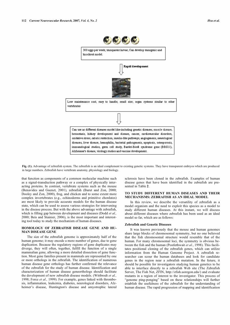
INTRODUCTION Presently, wide variety of model organisms are commonly used for studying the diseases and their mechanism, using Drosophila and Caenorhabditis elegans, and mammals, such as mice, rats and primates etc. But there is a wide research gap exists between the invertebrate and vertebrate model systems. In developmental biol-ogy, a similar gap has been filled by the zebrafish (Danio rerio). The zebrafish, a teleost native to tributaries of the Ganges river in India and Bangladesh, which is also very favorite in home aquari-ums in every country due to its hardy nature. Currently, it is one of the leading models for studying development and human diseases (Rubinstein, 2003). Zebrafish were established as a tool for aca-demic developmental biologists in the 1970s and 1980s due to their transparent embryos and rapid organogenesis. In the 1990s, zebraf-ish were used for the first vertebrate large scale mutagenesis screen, yielding thousands of mutations, some of which recapitulated hu-man diseases. The characteristics that make zebrafish a popular experimental animals (Fig. 1) to study the diseases mechanism include: (1) Zebrafish larvae are transparent. The eggs hatch rap-idly. The embryos are transparent and all cells remain visible up to the early larval stage. Organs, cells and tissues may be readily visu-alized in vivo and investigated in real-time (Eisen, 1996; Fishman, 1999). (2) Embryogenesis is rapid and large numbers of embryos are generated due to the high fecundity of zebrafish. Under ideal conditions, the females spawn up to 300 eggs per week. The eggs hatch rapidly. This enables the study of large numbers of meioses for positional cloning purposes. In addition, maintenance costs are significantly lower than those for mammals. (3) The zebrafish is amenable to molecular and genetic analysis through rapid determi-nation of temporal and spatial gene expression, examination of specific gene function by transgenic development, antisense gene knockdown and through large-scale mutagenesis (Driever et al., 1996; Knapik et al., 1998). (4) Zebrafish have cardiovascular, nerv-ous and digestive systems that are similar to those of mammals. As a vertebrate organism, zebrafish presents many organs and cell types similar to that of mammals. Organogenesis occurs rapidly, and major organs are present in larvae by 5 to 6 days post-
*Department of Marine Biotechnology and Resources, College of Marine Science, National Sun Yat-sen University, Kaohisung, Taiwan; E-mail: [email protected]� Received: August 16, 06, Revised: October 24, 06, Accepted: October 31, 06
fertilization. (5) A high degree of conservation exists between the human and zebrafish genomes (approximately 75% similarity). The Sanger Institute is in the process of sequencing the zebrafish ge-nome. Raw sequence totalling approximately 7.8 times the size of the genome is publicly available presently. Updated assemblies are generated twice a year. A complete assembled sequence is expected by the end of 2006-2007. The genetic map has been continually improving over the past 2 years, and currently >2000 microsat-ellite markers and up to 400 genes have been defined (Knapik et al., 1998; Postlethwait et al., 1998). Thus, the zebrafish has at-tracted number of scientists from various fields, such as develop-mental biology, neuroscience, hematopoiesis, nephrological or car-diovascular research. In fact, the zebrafish was recently described as 'the canonical vertebrate', due to the similarities between zebraf-ish and mammalian biology (Haffter et al., 1996; Fishman, 2001). Early events in zebrafish studies ensured that a variety of human disease conditions are primed to be studied utilizing the zebrafish model. An approach, the isolation of zebrafish orthologous of hu-man diseases causing genes has been undertaken. But, the use of the zebrafish in transgenesis and targeted mutagenesis for the model autosomal dominant and recessive disorders is not well advanced. Our aim of this review is to highlight what has been achieved to date with respect to human disease modeling in the zebrafish, more importantly, to study the diseases mechanisms and to understand molecular mechanism, molecular genetics with the creation of ze-brafish diseases model in a targeted manner.
MODEL ORGANISMS: ADVANTAGES AND LIMITA-TIONS OF THE VARIOUS SYSTEMS Presently, we consider the advantage and limitations of several model organisms with respect to the analysis of the mechanism of human diseases (Table 1). Several model systems can be used to analyze the function of a given human disease mechanism and hu-man genetic disorders. Organisms such as yeast (Saccharomyces) (Foury, 1997) and the colonial slime mold (Dictyostelium) (Chung et al., 2001; Firtel and Chung, 2000) are also using to analyze the phenomena that involve basic important eukaryotic cell functions, such as metabolism, regulation of the cell cycle, membrane target-ing and dynamics, protein folding, DNA damage and repair. Simple invertebrate systems such as Drosophila (Bernards and Hariharan, 2001; Reiter et al., 2001; Chien et al., 2005) or Caenorhabditis elegans (Aboobaker and Blaxter, 2000; Culetto and Sattelle, 2000) are excellent models for examining the coordinated actions of genes
112 Current Neurovascular Research, 2007, Vol. 4, No. 2 Hsu et al.
that function as components of a common molecular machine such as a signal-transduction pathway or a complex of physically inter-acting proteins. In contrast, vertebrate systems such as the mouse (Benavides and Guenet, 2001), zebrafish (Barut and Zon, 2000; Dooley and Zon, 2000), frog, and chicken and to some extent more complex invertebrates (e.g., echinoderms and primitive chordates) are most likely to provide accurate models for the human disease state, which can be used to assess various strategies for intervening in the disease process. But with the above advantage with zebrafish, which is filling gap between development and diseases (Dodd et al., 2000; Beis and Stainier, 2006), is the most important and interest-ing tool today to study the mechanism of human diseases.
HOMOLOGY OF ZEBRAFISH DISEASE GENE AND HU-MAN DISEASE GENE The size of the zebrafish genome is approximately half of the human genome; it may encode a more number of genes, due to gene duplication. Because the regulatory regions of gene duplicates may diverge, they will often, together, fulfill the function of a single mammalian gene, allowing a more detailed dissection of gene func-tion. Most gene families present in mammals are represented by one or more orthologs in the zebrafish. The identification of numerous human disease gene orthologs has further confirmed the relevance of the zebrafish for the study of human disease. Identification and characterization of human disease geneorthologs should facilitate the development of new zebrafish disease models. (Wittbrodt et al., 1998; Force et al., 1999). For example, genes linked with thrombo-sis, inflammation, leukemia, diabetes, neurological disorders, Alz-heimer’s disease, Huntington's disease and amyotrophic lateral
sclerosis have been cloned in the zebrafish. Examples of human disease genes that have been identified in the zebrafish are pre-sented in Table 2.
TO STUDY DIFFERENT HUMAN DISEASES AND THEIR MECHANISMS: ZEBRAFISH AS AN IDEAL MODEL In this review, we describe the versatility of zebrafish as a model organism and the need to exploit this species as a model to study different human diseases. At this instant, we will discuss about different diseases where zebrafish has been used as an ideal model so far, which are as follows:
Zebrafish and Genetic Diseases It was known previously that the mouse and human genomes share large blocks of chromosomal symmetry, but no one believed that the fish chromosomal structure would resemble that of the human. For many chromosomal loci, the symmetry is obvious be-tween the fish and the human (Postlethwait et al., 1998). This facili-tates positional cloning of the zebrafish genes, which can utilize information from the Human Genome Project. A zebrafish re-searcher can scour the human databases and look for candidate genes in the region near a zebrafish mutation. In the future, it should be possible for investigators studying human genetics to be able to interface directly to a zebrafish Web site (The Zebrafish Server, The Fish Net, ZFIN, http://zfish.uoregon.edu/) and evaluate mutants in a region of interest to the investigator. This process of “genome ping-ponging” based on these relationships will further establish the usefulness of the zebrafish for the understanding of human disease. The rapid progression of mapping and identification
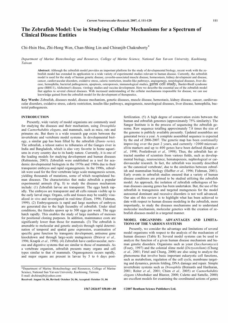
Fig. (1). Advantage of zebrafish system. The zebrafish is an ideal complement to existing genetic systems. They have transparent embryos which are produced in large numbers. Zebrafish have vertebrate anatomy, physiology and biology.

Zebrafish Model for Clinical Disease Entities Current Neurovascular Research, 2007, Vol. 4, No. 2 113
Table 1. Different Model Organisms- Their Advantage and Limitations
Species Advantages Limitations References
Yeast (Saccharomyces )
1.Excellent genetics 2.Very powerful second site screening 3. Powerful molecular techniques 4.Genes can be easily cloned 5.Genome sequence complete 6. Possess all basic eukaryotic cell organelles 7. Cell cycle control similar to animals
1.No distinct tissues
Foury, 1997
Nematode (Caenorhabditis
elegans)
1.Excellent genetics 2.Hermaphrodites, self-fertilization 3.Genes can be easily cloned 4.Transposon tagging 5.SNP mapping 6.Rapid cosmid rescue 7.Deletion collections span genome 8.RNAi effective 9.Genome sequence complete 10.Few cells: 959 cells, 302 neurons 11.Morphology fully characterized 12.Serial EM reconstruction 13.All cell lineages known 14.Time lapse microscopy of development 15.Laser ablation of single identified cells
1.Limited external morphology 2.Less similar to human than flies (61% of Drosophila genes have human counterparts vs. 43% of C. elegans genes) 3.Detailed direct analysis of gene expression patterns can be difficult 4.Some embryological manipulations difficult
Aboobaker and Blaxter, 2000;
Culetto and Sattelle, 2000
Fruit fly (Drosophila)
1.RNAi effective 2.Fast generation time 3.Second site suppressor/enhancer screens 4.Powerful molecular techniques 5.Genes can be easily cloned 6.Transposon tagging 7.SNP mapping 8.Transgenic animals easily generated 9.Mosaic analysis: determine where gene acts
1.Embryological manipulations difficult 2.Targeted gene disruption still difficult, although possible
Bernards and Hariharan 2001;
Reiter et al., 2001; Chien et al., 2002
Zebrafish
1.Simplest vertebrate with good genetics 2.Genome analysis well under way (good SNP and linkage maps) 3.Embryological manipulations possible 4.Organ systems similar to other vertebrates (e.g., eyes, heart, blood, gastrointestinal tract) 5.Rapid vertebrate development 6.Can develop transgenic and knockout animals 7. Low maintenance cost
1.very small fish; difficult to separate male and female Barut and Zon, 2000; Dooley and Zon 2000;
Dodd et al., 2000; Beis and
Stainier, 2006
Mouse
1.Construction of chimeric embryos possible 2.Availability of material at all stages. Source of primary cells for culture 3.Large mutant collection 4.Developmental overview same as for all mammals
1.Classic “forward” genetics difficult 2.Early-acting mutant phenotypes difficult to study (resorbed by mother) 3.Embryonic manipulations difficult (inside mother) 4.Development and life cycle relatively slow (months) 5.High cost, for both animals and facilities
Benavides and Guenet 2001; Taketo 2006; Notebaert and Meyer, 2006
Monkey
1.Very similar to humans 2.Developmental connections and physiology, postnatal 3.Anatomy of learning 4.Responses to injury
1.Fetal experiments difficult 2.No genetics 3.High cost for animals and their facilities
Wagner et al., 2006; Singh et al., 2006
Human
1.Some good family pedigrees 2.Genome sequence complete 3.Detailed behavior/ontogeny 4.Many diseases, self-reporting mutants (~5000 genetically based diseases)
1.Fetal material difficult 2.No experimental can be done on human
Couvineau et al., 2006; Newbold et al., 2006
SNP = single nucleotide polymorphism; RNAi = RNA interference.
114 Current Neurovascular Research, 2007, Vol. 4, No. 2 Hsu et al.
Table 2. Different Disease-related Genes that are Cloned in Zebraf-ish or Genes that are Introduce to Produce Transgenic Zebrafish
Diseases or Control a Process Genes Reference
Diabetes Insulin IA-2 autoantigen
Milewski et al., 1999; Cai et al, 2001
Leukemia runx1 cbfb
Blake et al., 2002
Thrombosis Factor VII COX-1 COX-2
Sheehan et al., 2001 Grosser et al., 2002
Cardiomyopathy cardiac troponin T titin
Sehnert and Stainier, 2001; Sehnert et al., 2002
Alzheimer's disease
presenilin-1 presenilin-2
acetylcholinesterase amyloid precursor protein
apoE
Leimer et al., 1999; Groth et al., 2002; Behra et al., 2002; Musa et al., 2001;
Monnot et al., 1999
Huntington's disease
Huntingtin Karlovich et al., 1998
Amyotrophic lateral sclerosis
sod-1 Dodd et al., 2000
Muscular dystrophy
dystroglycan dystrophin
Dp71
Parsons et al. 2002; Bolanos-Jimenez et al., 2001
Apoptosis Caspase-3 Chakraborty et al., 2006a; Chakraborty et al., 2006b
Cancer/tumor Myc myc-induced T cell leukemia
Langenau et al., 2003; Langenau et al., 2005
of gene function in zebrafish demonstrates the value of this organ-ism in the field of physiological genomics. With continued devel-opment of genomic tools for the zebrafish system, there is a greater likelihood of the identification of the function of newly discovered genes and determination of relevance to human disease. The initial characterization by Streisinger and co-workers (Streisinger et al., 1981; Streisinger et al., 1986) of the zebrafish provided a strong foundation for the ease of genetic studies. The combination of easy mutagenesis and powerful phenotypic screens of the earliest devel-opmental stages resulted in the undertaking of large-scale screens. Phenotypic analysis of embryonic development in animals obtained from two mutagenic screens of the zebrafish genome isolated muta-tions that affect virtually all major organ systems (Grunwald and Streisinger, 1992). The first large-scale mutagenesis screens of zebrafish to iden-tify developmental mutations were conducted by Nusslein-Volhard and colleagues in Germany in the early 1990s (Haffter et al., 1996), based on her Nobel prize-winning studies on saturation mutagenesis screens in Drosophila (Nusslein-Volhard and Wieschaus, 1980), and by Fishman and colleagues in Boston (Stainier et al., 1996). Their approach was to mutagenize males with a chemical to induce point mutations, and then use a breeding strategy to produce indi-viduals harboring homozygous mutations. A sufficient set of ge-nomics resources is a necessary requirement for the identification of chemically induced mutations. The development of zebrafish ge-nomics has been supported by a Trans-NIH Zebrafish Initiative (http://www.nih.gov/science/models/zebrafish/). Gene targeting by homologous recombination has not yet been accomplished in ze-brafish, although embryonic cell cultures have been used to gener-ate germ-line chimeras (Ma et al., 2001). However, ‘knock down’ techniques, in which antisense morpholino oligonucleotides are injected into embryos to partially block the translation of specific mRNAs, have become powerful tools for the dissection of gene function (Ekker, 2000; Nasevicius and Ekker, 2000).
Zebrafish as a Model for Caveolin-Associated Muscle Disease Caveolae are an abundant feature of many animal cells. Caveo-lin-3 is required for myofibril organization and muscle cell pattern-ing. However, the exact function of caveolae remains unclear. Nixon et al. (2005) used the zebrafish, as a system to understand caveolae function focusing on the muscle-specific caveolar protein, caveolin-3 (Cav3). They have identified caveolin-1, caveolin-2 and Cav3 in the zebrafish. Zebrafish Cav3 has 72% identity to human CAV3, and the amino acids altered in human muscle diseases are conserved in the zebrafish protein. During embryonic development, cav3 expression is apparent by early segmentation stages in the first differentiating muscle precursors, the adaxial cells and slightly later in the notochord. Expression of the zebrafish equivalent to a human muscular dystrophy mutant, CAV3P104L, causes severe disruption of muscle differentiation. In addition, knockdown of Cav3 resulted in a dramatic up-regulation of eng1 expression resulting in an in-crease in the number of muscle pioneer-like cells adjacent to the notochord. These studies provide new insights into the role of Cav3 in muscle development and demonstrate its requirement for correct intracellular organization and myoblast fusion. In future may pro-vide insights into both the protein interactions and cellular functions that underlie muscular dystrophy and other muscle diseases.
A Novel Model to Study Hemostasis A partial cDNA encoding zebrafish prothrombin has been cloned and used as a probe to study the temporal expression of prothrombin mRNA during early embryonic development by Ja-gadeeswaran and Liu (1997). The results revealed accumulation of prothrombin mRNA in diverse tissues such as the eyes and myo-tomes in early embryogenesis. They have also examined the enzy-matic activity of thrombin in converting fibrinogen to fibrin in indi-vidual embryos at different stages of development. They found that the fibrinforming activity does not temporally correlate with the first presence of thrombin mRNA in the early stages of embryo-genesis, but does correlate with the initiation of blood formation and also ability to observe the fibrin-forming activity in single indi-vidual embryo will facilitate studies on identifying recessive muta-tions affecting blood coagulation, such as the regulatory gene muta-tions controlling the clotting factor genes. Furthermore, this obser-vation of thrombin activity will also facilitate studies on the blood coagulation pathways in the early embryogenesis in this zebrafish model.
Model for Kidney Development and Disease in the Zebrafish Proper functioning of the kidney requires a structural integra-tion of glomerular podocytes and blood vessels. In zebrafish, evi-dence that podocytes act to organize vessel ingrowth can be seen in (Marshall, 1929) the expression patterns of genes that are known to play an important role in angiogenesis and (Silva et al., 1977) the recruitment of endothelial cells to clusters of podocytes in mutant embryos that lack the dorsal aorta, the normal blood supply for the pronephric glomerulus. Surprisingly, zebrafish mutants that lack blood flow as a result of defects in cardiac function (Rottbauer et al., 2001) fail to form a proper glomerular capillary tuft. This sug-gests that vascular shear force per se is required to drive capillary formation (Serluca et al. 2002). Although vascular cells seem nor-mal, they fail to express matrix metalloproteinase-2. Inhibition of matrix metalloproteinase activity by tissue inhibitor of metallopro-teinase-1 injections results in a similar failure to form the glomeru-lus (Serluca et al., 2002), indicating that degradation and remodel-ing of the glomerular basement membrane is a key step in capillary tuft formation. One of the most common human genetic diseases is polycystic kidney disease, which affects 1 in 1000 individuals (Calvet and Grantham, 2001). Kidney cysts are the result of grossly expanded kidney tubule lumens and, when present in sufficient size and number, lead to kidney fibrosis and end-stage renal failure. Drummond (2005) has identified a relatively large set of genetic loci associated with cystic pronephroi in zebrafish (Solnica-Krezel

Zebrafish Model for Clinical Disease Entities Current Neurovascular Research, 2007, Vol. 4, No. 2 115
et al., 1998). Recently, the results of a large-scale retroviral inser-tional mutagenesis screen have identified 10 zebrafish genes that when mutated cause pronephric cysts (Sun et al., 2004 ). The re-quirement for a relatively large number of genes underlying main-tenance of tubule structure is consistent with the idea that mainte-nance of lumen size and epithelial cell shape is a complex process that is controlled by many cellular proteins or signaling pathways.
Zebrafish as a Cancer Model System Cancer is a complex disease for millions of humans worldwide. The zebrafish is an ideal vertebrate system to model cancer. Despite more than 300 million years separating the last common ancestor of fish and humans, the biology of cancer is very much the same in these two organisms. Cancer is commonly seen in fish in the wild, and straightforward assays involving water-borne carcinogen expo-sure have demonstrated that teleosts develop a wide variety of be-nign and malignant tumors in virtually all organs, with a histology closely resembling that of human tumors (Hawkins et al., 1985; Spitsbergen et al., 2000). A comparison of the human genome se-quence and the soon to be completed zebrafish sequence demon-strates conservation of cell-cycle genes, tumor suppressors, and oncogenes. Beyond comparative genomics, there are many advan-tages to modeling cancer in the zebrafish system (Patton and Zon, 2001). A particularly exciting approach would be to combine the power of zebrafish genetics with chemical genetics by performing a chemical suppressor/enhancer screen on a zebrafish cancer model (Chan et al., 2002). The zebrafish cancer system can be viewed as a combination of vertebrate tumor biology, classical and chemical genetics, and genomics. Beginning with the pioneering work of Streisinger and colleagues, the zebrafish was envisioned as an ex-cellent model system for complex biology. The large-scale forward genetic screens in Tubingen and Boston in the 1990s furthered the system in order to understand early embryonic development (Driever et al., 1996; Haffter et al., 1996). A genetic screen in ze-brafish (Danio rerio) was performed to find mutations that cause genomic instability (gin), as scored by Streisinger's mosaic eye assay for the study of cancer which is developed by Moore et al. (2006). Many of the mutants obtained in these screens represent animal models of rare genetic diseases.
Zebrafish Mutants as a Model for Human Cardiovascular Dis-orders It is necessary to understand the molecular pathways underlying human dilated cardiomyopathies (DCM). Scientists have recently isolated in some of these lines novel genes essential for the devel-opment of proper vertebrate heart form and function (Rottbauer et al., 2001; Rottbauer et al., 2002). The same group (Rottbauer et al., 2005) have recently identified the genetic defect in zebrafish dead beat and were able to show for the first time that vascular endothe-lial growth factor (VEGF) signaling modulates cardiac contractility through its receptor FLT-1 and consecutively activation of phos-pholipase C �11. In an another study, Rottbauer et al. (2006) have isolated a mutation in zebrafish, tell tale heart (tel(m225)), which selectively perturbs contractility of the embryonic heart recently. By positional cloning, they have also identified tel to encode the zebrafish mlc-2 gene.
Model for Oxidative Stress and Apoptosis Oxidative stress has become a key paradigm in aging (Sohal and Weindruch, 1996). Little work has been done on oxidative stress during aging in any fish species. However, adaptive re-sponses to toxicological and oxidative stressors in fish have been found to be similar, though not identical, to mammals (Winston, 1991; Kelly et al., 1998). Fish possess the major antioxidant en-zymes, which generally occur at relatively higher levels than those of birds and mammals. Zebrafish appear to be an excellent organ-ism to investigate oxidative stress during aging. Studies on the ef-fects of UV radiation on antioxidant status and survival, and on the use of transgenic zebrafish to serve as sentinels for oxidative stress,
have been reported (Carvan et al., 2001; Black, 2002). Another group has initiated studies on oxidative stress by cloning several zebrafish antioxidant defense genes, including catalase (Gerhard et al., 2000). In response to many foreign compounds that generate oxidative stress, Carvan et al. (2001) induced the transcription of certain protective genes via specific DNA motifs called electrophile response elements (EPREs). They have shown that the treatment of zebrafish cell line ZEM2S with a variety of chemicals known to induce EPRE-dependent transcription in cultured mammalian cells, results in dose-dependent induction of the transiently-transfected EPRE-LUC reporter construct. Malek et al. (2004) reported the effects of a year-long 10 °C reduction in water temperature on gene expression in tail skeletal muscle from adult zebrafish. They deter-mined using an oligonucleotide microarray representing 15, 512 genes. Expression levels for approximately 600 genes were up-regulated by 1.7-fold or greater by the reduction in temperature, while a similar number of transcripts were down regulated by more than 1.7-fold. Using gene ontology (GO) classifications for molecu-lar function, two functional groups, “oxygen and reactive oxygen species metabolism” and “response to oxidative stress, ” were found to be overrepresented among up-regulated genes. However, temperature reduction did not suppress lipid peroxidation potential, protein carbonyl content, or 8-oxoguanine level. Several studies are doing apoptosis using zebra fish. Rybp (DEDAF) is a member of the Rybp/Yaf2 protein family and has been shown to encode pro-apoptotic functions and to be essential for mouse embryogenesis. The related Yaf2 protein has not been studied extensively at the cellular or organismal levels. Stanton et al. (2006) describe zebrafish yaf2 (zyaf2) and show that it is widely expressed during early embryogenesis, with subsequent enrichment of transcripts in the anterior head region. Finally, the observed acti-vation of Caspase 8 in the morphants is in accord with the ability of Yaf2 to inhibit Caspase 8-mediated apoptosis in cultured cells. Their findings implicate Yaf2 as a survival factor during early ze-brafish development and organogenesis.
Model for Caloric Restriction It is necessary for explaining how obesity has emerged as a public health epidemic and understanding the mechanism of energy flux. Another general advantage of a fish species as a model system is the ability to control caloric intake with reasonable accuracy. Scientists have performed short-term studies in zebrafish using a caloric restriction regimen akin to those used in rodent and primate studies (Pugh et al., 1999). They have designed specific diet formu-lations to ensure that calorically restricted and ad libitum fed fish consume equivalent amounts of essential nutrients yet decreased calories, primarily carbohydrates. Gene expression in tail muscle from restricted fish was profiled using a small-scale zebrafish cDNA array. Several mitochondrial genes were found to be down-regulated, similar to gene expression profiling results reported for skeletal muscle of rhesus monkeys subjected to caloric restriction (Kayo et al., 2001). Novak et al. (2005) tested the hypothesis that fasting induces a biphasic pattern of change in physical activity levels (PA) by measuring PA before and after long-term food dep-rivation in zebrafish. They have found that compared to control-fed fish, food-deprived fish showed a significant increase in PA levels during the first 2 days of food deprivation. Subsequently, fasted fish showed a significant chronic decrease in PA compared to fish fed at weight-maintenance levels. These data show a biphasic response of PA to caloric restriction which is comparable to mammals. In a separate group of fish, long-term food deprivation, associated with decreases in PA, induced a significant increase in brain prepro-orexin mRNA levels compared to feed controls and after 2 days of food deprivation, no change in orexin mRNA was noticed.
Model for Insulin-Like Pathways Studies conducted over the past several years on insulin-like signaling plays a key role in determining life span (Murakami et al.,
116 Current Neurovascular Research, 2007, Vol. 4, No. 2 Hsu et al.
2000). Some information on these pathways is already available in zebrafish. IGF receptors with characteristics of the mammalian type I IGF receptor have been described in zebrafish cells (Maures et al., 2002). The two major signal transduction pathways, MAPK and PI3 kinase, are activated by IGF-I (Pozios et al., 2001). Based on data from several fish species, including zebrafish, the major compo-nents of the IGF signaling system appear to be structurally and functionally similar to those in mammals, although several key differences are apparent. The insulin-like growth factor (IGF) sig-naling pathway has been highly conserved in animal evolution and, in mammals which plays a key role in embryonic growth and de-velopment for embryo, with the IGF-1 receptor (IGF-1R) being a crucial regulator of the signaling cascade. Eivers et al. (2004) re-ported the first functional role for the IGF pathway in zebrafish. Expression of mRNA coding for a dominant negative IGF-1R re-sulted in embryos that were small in size compared to controls and had disrupted head and CNS development. In other hand, up-regulation of IGF signaling following injection of IGF-1 mRNA, resulted in a greatly expanded development of anterior structures at the expense of trunk and tail. IGF-1R knockdown caused a signifi-cant decrease in the expression of Otx2, Rx3, FGF8, Pax6.2 and Ntl, while excess IGF signaling expanded Otx2 expression in pre-sumptive forebrain tissue and widened the Ntl expression domain in the developing notochord. The observation that IGF-1R knockdown reduced expression of two key organizer genes (chordin and goosecoid) suggested that IGF signaling plays a role in regulating zebrafish organizer activity. This data was supported by the expres-sion of IGF-1, IGF-2 and IGF-1R in shield-stage zebrafish embryos and the demonstration that IGF signaling influences expression of BMP2b, a gene that plays an important role in zebrafish pattern formation.
Model for Angiogenesis The process of building new blood vessels (angiogenesis) and controlling the propagation of blood vessels (anti-angiogenesis) are fundamental to human health, as they play key roles in wound heal-ing and tissue growth. A method has been develop to visualize blood vessels in the zebrafish include whole mount in situ hybridi-zation (Fouquet et al., 1997; Liao et al., 1997) detection of endoge-nous alkaline phosphatase activity, and microangiography (Wein-stein et al., 1995 ). Microangiography is also labor intensive and only useful for visualization of patent blood vessels in a complete circulatory system. Transgenic zebrafish with fluorescent blood vessels (Motoike et al., 2000; Lawson and Weinstein, 2002) repre-sent a less labor-intensive way of visualizing blood vessels in the zebrafish. Scientists have generated a transgenic line with fluores-cent blood vessels by driving expression of a green reef coral fluo-rescent protein (G-RCFP) (Matz et al., 2000) with a promoter for the vascular endothelial growth factor receptor 2 gene (VEGFR2, also referred to as Flk-1 or KDR). VEGFR2 is one of several recep-tors for VEGF family members in humans and is expressed specifi-cally in blood vessels (Gale and Yancopoulos, 1999). Several ze-brafish VEGFR2 cDNAs have been cloned and their expression pattern described (Chan et al., 2002; Fouquet et al., 1997; Liao et al., 1997; Thompson et al., 1998 ). These groups used a 6.5-KB genomic fragment 5’ to the VEGFR2 initiation codon to drive G-RCFP expression specifically in zebrafish blood vessels. Hereafter, the stable transgenic line generated is referred to as TG (VEGFR2: G-RCFP), using standard zebrafish nomenclature. Bayliss et al. (2006) reported the role of angiogenesis and the need for receptor signaling using chemical inhibition of the vascular endothelial growth factor receptor in the adult zebrafish tail fin. They were able to exert precise control over blood vessel regeneration Using a small-molecule inhibitor. An angiogenic limit to tissue regeneration was determined, as a vascular tissue containing skin, pigment, neu-ronal axons and bone precursors could regenerate up to about 1 mm. This indicates that tissues can regenerate without direct inter-action with endothelial cells and at a distance from blood supply.
Vascular endothelial growth factor (VEGF) is a major mediator of pathologic angiogenesis, a process necessary for the formation of new blood vessels to support tumor growth. Leung et al. (2006) provide a new opportunity for cotargeting G protein- and VEGF-dependent pathways to synergistically block pathologic angiogene-sis, which may lead to a safer and more efficacious therapeutic regimen to fight cancer. For tumor angiogenesis, Haldi et al. (2006) optimized parame-ters for xenotransplanting WM-266-4, a metastatic melanoma cell line, including zebrafish site and stage for transplantation, number of cells, injection method, and zebrafish incubation temperature. Since zebrafish are transparent until approximately 30 dpf, the in-teraction of labeled melanoma cells and zebrafish endothelial cells (EC) can be visualized by whole-mount immunochemical staining. After staining with Phy-V, a mouse anti-zebrafish monoclonal anti-body (mAb) that specifically labels activated EC and angioblasts, using immunohistology and 2-photon microscopy, they have ob-served activated zebrafish EC embedded in human melanoma cell masses. The zebrafish model offers a rapid efficient approach for assessing human cancer cells at various stages of tumorigenesis for angiogenesis.
Model for Neurological Disease Zebrafish are an ideal vertebrate model to study development of the visual system as they produce transparent embryos that develop rapidly, thereby facilitating morphological and behavioral testing. In a study, zebrafish connexin35 has been cloned from a P1 artifi-cial chromosome (PAC) library. Sequence analysis shows a high degree of similarity to the Cx35/36 orthologous group, which are expressed primarily in nervous tissue, including the retina. The gene encodes a 304-amino acid protein with a predicted molecular weight of approximately 35 kDa (McLachlan et al., 2003). Axonal guidance and vascular patterning share several guidance cues, in-cluding proteins in the netrin family. Wilson et al. (2006) demon-strate that netrins stimulate proliferation, migration, and tube for-mation of human endothelial cells in vitro and that this stimulation is independent of known netrin receptors. Suppression of netrin1a messenger RNA in zebrafish inhibits vascular sprouting, implying a proangiogenic role for netrins during vertebrate development. They have proposed that the attractive vascular and neural guidance func-tions of netrins offer a unique therapeutic potential. Parkinson's disease is characterized by a severe loss of dopaminergic neurons resulting in a range of motor deficits. The neurotoxin 1-methyl-4-phenyl-1, 2, 3, 6-tetrahydropyridine (MPTP) is known to cause a similar loss of dopaminergic neurons in the human midbrain with corresponding Parkinsonian symptoms. Several animal species have also shown sensitivity to MPTP, including primates, mice, goldfish, and, most recently, zebrafish. A study demonstrated by McKinley et al. (2005) that the effect of MPTP on dopaminergic neurons in zebrafish larvae is mediated by the same pathways that have been demonstrated in mammalian species. MPTP-induced neurodegen-eration was prevented by co-incubation with either the monoamine oxidase-B (MAO-B) inhibitor l-deprenyl or the dopamine trans-porter (DAT) inhibitor nomifensine. Furthermore, targeted inactiva-tion of the DAT gene by antisense morpholinos also protected neu-rons from MPTP damage. Thus, the mechanism for MPTP-induced dopaminergic neuron toxicity in mammals is conserved in zebrafish larvae. Effects on swimming behavior and touch response that re-sult from MPTP damage are partially ameliorated by both l-deprenyl and DAT knockdown.
Models for Liver Disease To identify zebrafish mutants to serve as models for hepatic pathologies, Sadler et al. (2005) screened for hepatomegaly at day 5 of embryogenesis in 297 zebrafish lines bearing mutations in genes that are essential for embryonic development. Seven mutants were identified, and three have phenotypes resembling different liver diseases. Mutation of the class C vacuolar protein sorting gene
Zebrafish Model for Clinical Disease Entities Current Neurovascular Research, 2007, Vol. 4, No. 2 117
vps18 results in hepatomegaly associated with large, vesicle-filled hepatocytes, which we attribute to the failure of endosomallysoso-mal trafficking. Additionally, these mutants develop defects in the bile canaliculi and have marked biliary paucity, suggesting that vps18 also functions to traffic vesicles to the hepatocyte apical membrane and may play a role in the development of the intrahe-patic biliary tree. Similar findings have been reported for individu-als with arthrogryposis-renal dysfunction-cholestasis (ARC) syn-drome, which is due to mutation of another class C vps gene. A second mutant, resulting from disruption of the tumor suppressor gene nf2, develops extrahepatic choledochal cysts in the common bile duct, suggesting that this gene regulates division of biliary cells during development. The third mutant is in the novel gene foie gras, which develops large, lipid-filled hepatocytes, resembling those in individuals with fatty liver disease. These mutants illustrate the utility of zebrafish as a model for studying liver development and disease, and provide valuable tools for investigating the molecular pathogenesis of congenital biliary disorders and fatty liver disease.
Hemophilia Model in Zebrafish Jagadeeswaran1 and Liu (1997) have developed artificial he-mophilia in zebrafish by treating them with copper and measured their clotting function by a newly developed sensitive clotting time assay. The clotting function can be detected rapidly and reliably in 30 hr larvae and in adult fish by measuring the blood clotting time. They have used this assay to screen wild type zebrafish and identi-fied fish with prolonged clotting time.
Zebrafish Model of Bacterial Pathogenesis With a natural and important pathogen of fish, Streptococcus iniae, Neely et al., (2000) have established a streptococcus zebraf-ish model of bacterial pathogenesis. Following injection into the dorsal muscle, zebrafish developed a lethal infection, with a 50% lethal dose of 103 CFU, and died within 2 to 3 days. The patho-genesis of infection resembled that of S. iniae in farmed fish popu-lations and that of several important human streptococcal diseases and was characterized by an initial focal necrotic lesion that rapidly progressed to invasion of the pathogen into all major organ systems, including the brain. Zebrafish were also susceptible to infection by the human pathogen Streptococcus pyogenes. This combination of a genetically amenable pathogen with a well-defined vertebrate host makes the streptococcus-zebrafish model of bacterial pathogenesis a powerful model for analysis of infectious disease. Refinement of these conditions may make it possible to conduct large-scale screens for zebrafish genes that are involved in host-pathogen inter-actions.
Model for Osteoporosis Glucocorticoid-induced osteoporosis (GIOP) is a major clinical problem given the widespread use of steroids and limited efficacy of biphosphonates. Existing animal models of GIOP are both slow and expensive. Hence, there is a need both for adjunctive modeling systems, as well as more efficacious therapies for the treatment of GIOP. Barrett et al. (2006) have addressed this issue through the creation of a zebrafish model of GIOP, which can be used for 96-well plate in vivo screening with an assay time of 5 days. The model demonstrates key similarities to human GIOP including a partial response to bisphosphonates.
Model for Immunological Studies Zebrafish has been advocated as an alternative animal model to study lymphocyte development, although the similarities in the genetic requirements of lymphopoiesis between fish and mammals have not yet been investigated. In a study, Schorpp et al. (2006) examine the role of the transcription factor Ikaros in zebrafish lym-phopoiesis. In fish larvae homozygous for an ikaros allele predicted to lack the C-terminal zinc fingers, T lymphopoiesis is absent; the presence of V(H)DmuJmu rearrangements in adolescent fish is
delayed in mutants. In adolescent mutant fish, T cells expressing tcrb and tcrd and B cells expressing igm are formed with low effi-ciency and display an oligoclonal Ag receptor repertoire.
Model for Germ Cell Study The migration of zebrafish primordial germ cell towards the region where the gonad develops is guided by the chemokine SDF-1a. Recent studies show that soon after their specification, the cells undergo a series of morphological alterations before they become motile and are able to respond to attractive cues. In all of these stages, zebrafish germ cells respond as individual cells to altera-tions in the shape of the sdf-1a expression domain, by directed mi-gration towards their target - the position where the gonad develops (Raz and Reichman-Fried, 2006).
Model for Virology Studies and Vaccine Development Zebrafish is a suitable animal model to study VHSV infection and immune (innate and adaptive) responses and, more importantly, we demonstrate for the first time the usefulness of the zebrafish as a vaccination model to viral diseases. The rhabdovirus viral hemor-rhagic septicemia virus (VHSV) is the etiological agent of one of the most important salmonid viral diseases. In a work by Novoa et al. (2006), the ability of VHSV to infect and replicate in zebrafish at low temperature (15 degrees C) was demonstrated. Zebrafish was also used to determine the effectiveness of the recombinant virus rIHNV-Gvhsv GFP as a live attenuated vaccine against the virulent VHSV strain.
Model for Bardet-Biedl Syndrome Gene (BBS11) In studies of rare diseases, the resolution of linkage mapping is limited by the number of available meioses and informative marker density. Functional analysis of the gene (BBS) in zebrafish and expression correlation analyses among other BBS genes in an ex-pression quantitative trait loci data set demonstrate that TRIM32 is a BBS gene. This study shows the value of high-density SNP geno-typing for homozygosity mapping and the use of expression corre-lation data for evaluation of candidate genes and identifies the pro-teasome degradation pathway as a pathway involved in BBS (Chiang et al. 2006). In an another study reported by Badano et al. (2006) where they have identified of a novel locus, MGC1203, that contributes epistatic alleles to Bardet-Biedl syndrome (BBS), a pleiotropic, oligogenic disorder. Finally, recapitulation of the hu-man genotypes in zebrafish shows that modest suppression of mgc1203 exerts an epistatic effect on the developmental phenotype of BBS morphants.
Model for Alzheimer's Disease Gamma-secretase cleavage, mediated by a complex of preseni-lin, presenilin enhancer (Pen-2), nicastrin, and Aph-1, is the final proteolytic step in generating amyloid beta protein found in brains of Alzheimer's disease patients and Notch intracellular domain critical for proper neuronal development. Campbell et al. (2006) have employed the zebrafish model to study the role of Pen-2 in neuronal survival. They have found that knockdown of Pen-2 using antisense morpholino led to a reduction of islet-1 positive neurons, and notch signaling was reduced in embryos lacking Pen-2 or other gamma-secretase components. Their results demonstrate that knockdown of Pen-2 directly induces a p53-dependent apoptotic pathway that contributes to neuronal loss and suggest that Pen-2 plays an important role in promoting neuronal cell survival and protecting from apoptosis in vivo. Presenilins play prominent roles in the molecular pathogenesis of Alzheimer's disease and during embryo development. Groth et al. (2002) have isolated a zebrafish presenilin orthologue (pre2), which shows a high degree of se-quence identity to the human PS2 protein. Zebrafish pre2 is mater-nally and ubiquitously expressed during early embryo development, whereas Pre2 protein expression is initiated between 6 and 12 hours post fertilization (hpf), suggesting strict regulation of pre2 transla-
118 Current Neurovascular Research, 2007, Vol. 4, No. 2 Hsu et al.
tion. An integrated microscope image analysis pipeline is developed for automatic analysis and quantification of phenotypes in zebrafish with altered expression of Alzheimer's disease (AD)-linked genes. Liu et al. (2006) results have showed that the computerized analysis is comparable to manual counting with equivalent accuracy and improved efficacy and consistency. Development of such an auto-mated data analysis pipeline represents a significant step forward to achieve accurate and reproducible quantification of neuronal pheno-types in large scale or high-throughput zebrafish imaging studies.
CONCLUSIONS A large number of evidence is accumulating that shows that zebrafish can accurately represents human biology and the drugs currently used by humans can have a predictable effect in zebrafish assays. Since mammalian disease models are expensive and gener-ally not conducive to highthroughput target validation and drug screening, the zebrafish system has an opportunity to provide a crucial link between high-throughput in vitro assays and in vivo mammalian disease models. However, development of disease-relevant assays and disease models in the zebrafish is still in its infancy. Zebrafish is a model system which will provide for both genetic and embryological manipulations. As would be the case for any new model organism, a substantial database of basic gerontological information for zebrafish is needed. Careful documentation during disease modeling easily and economically accomplished in zebraf-ish, would also be invaluable for the interpretation of age-related phenotypes. By virtue of a wave of biological resources accumulat-ing for the zebrafish, driven primarily by the ability to perform large-scale mutagenesis studies, zebrafish have become a major model system for biomedical research.
REFERENCES Aboobaker, AA, Blaxter, ML. (2000) Medical significance of Caenorhabdi-
tis elegans. Ann Med 32: 23-30. Badano, JL, Leitch, CC, Ansley, SJ, May-Simera, H, Lawson, S, Lewis,
RA, Beales, PL, Dietz, HC, Fishe, r S, Katsanis, N. (2006) Dissection of epistasis in oligogenic Bardet-Biedl syndrome. Nature 439: 326-30.
Barrett, R, Chappell, C, Quick, M, Fleming, A. (2006) A rapid, high con-tent, in vivo model of glucocorticoid-induced osteoporosis. Biotechnol J 1: 651-655.
Barut, BA, Zon, LI. (2000) Realizing the potential of zebrafish as a model for human disease. Physiol Genomics 2: 49-51.
Bayliss, PE, Bellavance, KL, Whitehead, GG, Abrams, JM, Aegerter, S, Robbins, HS, Cowan, DB, Keating, MT, O'Reilly, T, Wood, JM, Rob-erts, TM, Chan, J. (2006) Chemical modulation of receptor signaling inhibits regenerative angiogenesis in adult zebrafish. Nat Chem Biol 2: 228-229.
Behra, M, Cousin, X, Bertrand, C, Vonesch, JL, Biellmann, D, Chatonnet, A, Strahle, U. (2002) Acetylcholinesterase is required for neuronal and muscular development in the zebrafish embryo. Nat Neurosci 5: 111-118.
Beis, D, Stainier, DY. (2006) In vivo cell biology: following the zebrafish trend. Trends Cell Biol 16: 105-12.
Benavides, F, Guenet, JL. (2001) Murine models for human diseases. Medi-cina 61: 215-231.
Bernards, A, Hariharan, IK. (2001) Of flies and men—studying human disease in Drosophila. Curr Opin Genet Dev 11: 274-278.
Black, H. (2002) Fishing for answers to questions about the aging process. Bioscience 52: 15-18.
Blake, T, Adya, N, Kim, CH, Oates, AC, Zon, L, Chitnis A, Weinstein, BM, Liu, PP. (2000) Zebrafish homolog of the leukemia gene CBFB: Its ex-pression during embryogenesis and its relationship to scl and gata-1 in hematopoiesis. Blood 96: 4178-4184.
Bolanos-Jimenez, F, Bordais, A, Behra, M, Strahle, U, Mornet, D, Sahel, J, Rendon, A. (2001) Molecular cloning and characterization of dystro-phin and Dp71, two products of the Duchenne muscular dystrophy gene, in zebrafish. Gene 274: 217-226.
Cai, T, Krause, MW, Odenwald, WF, Toyama, R, Notkins, AL. (2001) The IA-2 gene family: Homologs in Caenorhabditis elegans, Drosophila and zebrafish. Diabetologia 44: 81-88.
Calvet, JP, Grantham, JJ. (2001)The genetics and physiology of polycystic kidney disease. Semin Nephrol 21: 107-123.
Campbell, WA, Yang, H, Zetterberg, H, Baulac, S, Sears, JA, Liu, T, Wong, ST, Zhong, TP, Xia, W. (2006) Zebrafish lacking Alzheimer presenilin enhancer 2 (Pen-2) demonstrate excessive p53-dependent apoptosis and neuronal loss. J Neurochem 96: 1423-40
Carvan, MJ, Sonntag, DM, Cmar, CB, Cook, RS, Curran, MA, Miller, GL. (2001) Oxidative stress in zebrafish cells: potential utility of transgenic zebrafish as a deployable sentinel for site hazard ranking. Sci Total En-viron 274: 183-96.
Chan, J, Bayliss, PE, Wood, JM, Roberts, TM. (2002) Dissection of angio-genic signaling in zebrafish using a chemical genetic approach. Cancer Cell 1: 257-267.
Chiang, AP, Beck, JS, Yen, HJ, Tayeh, MK, Scheetz, TE, Swiderski, RE, Nishimura, DY, Braun, TA, Kim, KY, Huang, J, Elbedour, K, Carmi, R, Slusarski, DC, Casavant, TL, Stone, EM, Sheffield, VC. (2006) Ho-mozygosity mapping with SNP arrays identifies TRIM32, an E3 ubiq-uitin ligase, as a Bardet-Biedl syndrome gene (BBS11). Proc Natl Acad Sci U S A 103: 6287-92.
Chien, S, Reiter, LT, Bier, E, Gribskov, M. (2002) Homophila: human dis-ease gene cognates in Drosophila. Nucleic Acids Res 30: 149-151.
Chung, CY, Funamoto, S, Firtel, RA. (2001) Signaling pathways controlling cell polarity and chemotaxis. Trends Biochem Sci 26: 557-566.
Couvineau, A, Tan, YV, Ceraudo, E, Lacapere, JJ, Murail, S, Neumann, JM, Laburthe, M. (2006) The human VPAC1 receptor: Identification of the N-terminal ectodomain as a major VIP-binding site by photoaffinity la-beling and 3D modeling. Ann N Y Acad Sci 1070: 205-9.
Culetto, E, Sattelle, DB. (2000) A role for Caenorhabditis elegans in under-standing the function and interactions of human disease genes. Hum Mol Genet 9: 869-877.
Dodd, A, Curtis, PM, Willams, LC, Love, DR. (2000) Zebrafish: bridging the gap between development and disease. Hum Mol Genet 9: 2443-2449.
Dodd, A, Curtis, PM, Williams, LC, Love, DR. (2000) Zebrafish: Bridging the gap between development and disease. Hum Mol Genet 9: 2443-2449.
Dooley, K, Zon, LI. (2000) Zebrafish: a model system for the study of hu-man disease. Curr Opin Genet Dev 10: 252-256.
Driever, W, Solnica-Krezel, L, Schier, AF, Neuhauss, SC, Malicki, J, Stem-ple, DL, Stainier, DY, Zwartkruis, F, Abdelilah, S, Rangini, Z, Belak, J, and Boggs, C. (1996) A genetic screen for mutations affecting embryo-genesis in zebrafish. Development 123: 37-46.
Driever, W, Solnica-Krezel, L, Schier, AF, Neuhauss, SC, Malicki, J, Stem-ple, DL, Stainier, DY, Zwartkruis, F, Abdelilah, S, Rangini, Z. (1996). A genetic screen for mutations affecting embryogenesis in zebrafish. Development 123: 37-46.
Drummond, IA. (2005) Kidney development and disease in the Zebrafish. J Am Soc Nephrol 16: 299-304.
Eisen, JS. (1996) Zebrafish make a big splash. Cell 87: 969-977. Eivers, E, McCarthy, K, Glynn, C, Nolan, CM, Byrne, L. (2004) Insulin-like
growth factor (IGF) signaling is required for early dorso-anterior devel-opment of the zebrafish embryo. Int J Dev Biol 48: 1131-1140.
Ekker, SC. (2000) Morphants: a new systematic vertebrate functional ge-nomics approach. Yeast 17: 302-306.
Firtel, RA, Chung, CY. (2000) The molecular genetics of chemotaxis: sens-ing and responding to chemoattractant gradients, Bioessays 22: 603-615.
Fishman, MC. (1999) Zebrafish genetics: The enigma of arrival. Proc Natl Acad Sci USA 96: 10554-10556.
Fishman, MC. (2001) Zebrafish: The canonical vertebrate. Science 294: 1290-1291.
Force, A, Lynch, M, Pickett, FB, Amores, A, Yan, YL, Postlethwait, J. (1999) Preservation of duplicate genes by complementary, degenerative mutations. Genetics 151: 1531-1545.
Fouquet, B, Weinstein, BM, Serluca, FC, Fishman, MC. (1997) Vessel patterning in the embryo of the zebrafish: guidance by notochord. Dev Biol 183: 37-48.
Foury, F. (1997) Human genetic diseases: a cross-talk between man and yeast. Gene 195: 1-10.
Zebrafish Model for Clinical Disease Entities Current Neurovascular Research, 2007, Vol. 4, No. 2 119
Gale, NW, Yancopoulos, GD. (1999) Growth factors acting via endothelial cell specific receptor tyrosine kinases: VEGFs, angiopoietins, and eph-rins in vascular development. Genes Dev 13: 1055-1066.
Gerhard, GS, Kauffman, EJ, Grundy, MA. (2000) Molecular cloning and sequence analysis of the Danio rerio catalase gene. Comp Biochem Physiol Biochem Mol Biol 127: 447-457.
Grosser, T, Yusuff, S, Cheskis, E, Pack, MA, FitzGerald, GA. (2002) De-velopmental expression of functional cyclooxygenases in zebrafish. Proc Natl Acad Sci USA 99: 8418-8423.
Groth, C, Nornes, S, McCarty, R, Tamme, R, Lardelli, M. (2002) Identifica-tion of a second presenilin gene in zebrafish with similarity to the hu-man Alzheimer's disease gene presenilin 2. Dev Genes Evol 212: 486-490.
Groth, C, Nornes, S, McCarty, R, Tamme, R, Lardelli, M. (2002) Identifica-tion of a second presenilin gene in zebrafish with similarity to the hu-man Alzheimer's disease gene presenilin2. Dev Genes Evol 212: 486-90.
Grunwald, D, Streisinger, G. (1992) Induction of recessive lethal and spe-cific locus mutations in zebrafish with ethylnitrosourea. Genet Res 59: 103-116.
Haffter, P, Granato, M, Brand, M, Mullins, MC, Hammerschmidt, M, Kane, DA, Odenthal, J, van Eeden, FJ, Jiang, YJ, Heisenberg, CP, Kelsh, RN, Furutani-Seiki, M, Vogelsang, E, Beuchle, D, Schach, U, Fabian, C, Nusslein-Volhard, C. (1996) The identification of genes with unique and essential functions in the development of the zebrafish Danio rerio. Development 123: 1-36.
Haldi, M, Ton, C, Seng, WL, McGrath, P. (2006) Human melanoma cells transplanted into zebrafish proliferate, migrate, produce melanin, form masses and stimulate angiogenesis in zebrafish. Angiogenesis 9: 139-151.
Hawkins, WE, Overstreet, RM, Fournie, JW, Walker, WW. (1985) Devel-opment of aquarium fish models for environmental carcinogenesis: tu-mor induction in seven species. J Appl Toxicol 5: 261-264.
Jagadeeswaran, P, Liu, YC. (1997) A Hemophilia Model in Zebrafish: Analysis of Hemostasis. Blood Cells Mol Dis 23: 52-57.
Jagadeeswaran, P, Liu, YC. (1997) Developmental expression of thrombin in zebrafish embryos: a novel model to study hemostasis. Blood Cells Mol Dis 23: 147-156.
Karlovich, CA, John, RM, Ramirez, L, Stainier, DYR, Myers, RM. (1998) Characterization of the Huntington's disease (HD) gene homolog in the zebrafish Danio rerio. Gene 217: 117-125.
Kayo, T, Allison, DB, Weindruch, R, Prolla, TA. (2001) Influences of aging and caloric restriction on the transcriptional profile of skeletal muscle from rhesus monkeys. Proc Natl Acad Sci USA 98: 5093-5098.
Kelly, KA, Havrilla, CM, Brady, TC, Abramo, KH, Levin, ED. (1998) Oxidative stress in toxicology: established mammalian and emerging piscine model systems. Environ Health Perspect 106: 375-384.
Knapik, EA, Goodman, M, Ekker, M, Chevrette, J, Delgado, S, Neuhauss, N, Shimoda, W, Driever, M, Fishman, Jacob. H. (1998) A microsatellite genetic linkage map for zebrafish (Danio rerio). Nat Genet 18: 338-343.
Langenau, DM, Feng, H, Berghmans, S, Kanki, JP, Kutok, JL, Look, AT. (2005) Cre/lox-regulated transgenic zebrafish model with conditional myc-induced T cell acute lymphoblastic leukemia. Proc Natl Acad Sci 102: 6068-73.
Langenau, DM, Traver, D, Ferrando, AA, Kutok, JL, Aster, JC, Kanki, JP, Lin, S, Prochownik, E, Trede, NS, Zon, LI, Look, AT. (2003) Myc-induced T cell leukemia in transgenic zebrafish. Science 299: 887-890.
Lawson, ND, Weinstein, BM. (2002) In vivo imaging of embryonic vascular development using transgenic zebrafish. Dev Biol 248: 307-318.
Leimer, U, Lun, K, Romig, H, Walter, J, Grunberg, J, Brand, M, Haass, C. (1999) Zebrafish (Danio rerio) presenilin promotes aberrant amyloid �- peptide production and requires a critical aspartate residue for its func-tion in amyloidogenesis. Biochemistry 38: 13602-13609.
Leonard, IZ. (1999) Zebrafish: A new model for human disease. Genome Res 9: 99-100.
Leung, T, Chen, H, Stauffer, AM, Giger, KE, Sinha, S, Horstick, EJ, Hum-bert, JE, Hansen, CA, Robishaw, JD. (2006) Zebrafish G protein gamma2 is required for VEGF signaling during angiogenesis. Blood 108: 160-116.
Liao, W, Bisgrove, BW, Sawyer, H, Hug, B, Bell, B, Peters, K, Grunwald, DJ, Stainier, DY. (1997) The zebrafish gene cloche acts upstream of a flk-1 homologue to regulate endothelial cell differentiation. Develop-ment 124: 381-389.
Liu, T, Lu, J, Wang, Y, Campbell, WA, Huang, L, Zhu, J, Xia, W, Wong, ST. (2006) Computerized image analysis for quantitative neuronal phe-notyping in zebrafish. J Neurosci Methods 153: 190-202.
Ma, C, Fan, L, Ganassin, R, Bols, N, Collodi, P. (2001) Production of ze-brafish germ-line chimeras from embryo cell cultures. Proc Natl Acad Sc USA 98: 2461-2466.
Malek, RL, Sajadi, H, Abraham, J, Grundy, MA, Gerhard, GS. (2004) The effects of temperature reduction on gene expression and oxidative stress in skeletal muscle from adult zebrafish. Comp Biochem Physiol C Toxi-col Pharmacol 138: 363-373.
Marshall, EK. (1929) The aglomerular kidney of the toadfish (Opsanus tau). Bull Johns Hopkins Hosp 45: 95-101.
Matz, MV, Fradkov, AF, Labas, YA, Savitsky, AP, Zaraisky, AG, Markelov, ML, Lukyanov, SA. (2000) Fluorescent proteins from non-bioluminescent Anthozoa species. Nat Biotech 17: 969-973.
McKinley, ET, Baranowski, TC, Blavo, DO, Cato, C, Doan, TN, Rubin-stein, AL. (2005) Neuroprotection of MPTP-induced toxicity in zebraf-ish dopaminergic neurons. Brain Res Mol Brain Res 141: 128-37.
McLachlan, E, White, TW, Ugonabo, C, Olson, C, Nagy, JI, Valdimarsson, G. (2003) Zebrafish Cx35: cloning and characterization of a gap junc-tion gene highly expressed in the retina. J Neurosci Res 73: 753-764.
Milewski, WM, Duguay, SJ, Chan, SJ, Steiner, DF. (1999) Conservation of PDX-1 structure, function, and expression in zebrafish. Endocrinology 139: 1440-1449.
Monnot, MJ, Babin, PJ, Poleo, G, Andre, M, Laforest, L, Ballagny, C, Aki-menko, MA. (1999) Epidermal expression of apolipoprotein E gene during fin and scale development and fin regeneration in zebrafish. Dev Dyn 214: 207-215.
Moore, JL, Rush, LM, Breneman, C, Mohideen, MA, Cheng, KC. (2006) Zebrafish genomic instability mutants and cancer susceptibility. Genet-ics 174: 585-600.
Motoike, T, Loughna, S, Perens, E, Roma, n BL, Liao, W, Chau, TC, Richardson, CD, Kawate, T, Kuno, J, Weinstein, BM, Stainier, DYR, Sato, TN. (2000) Universal GFP reporter for the study of vascular de-velopment. Genesis 28: 75-81.
Murakami, S, Tedesco, PM, Cypser, JR, Johnson, TE. (2000) Molecular genetic mechanisms of life span manipulation in Caenorhabditis ele-gans. Ann NY Acad Sci 908: 40-49.
Musa, A, Lehrach, H, Russo, VEA. (2001) Distinct expression patterns of two zebrafish homologues of the human APP gene during embryonic development. Dev Genes Evol 211: 563-567.
Nasevicius, A, Ekker, SC. (2000) Effective targeted gene ‘knockdown’ in zebrafish. Nat Gene 26: 216-220.
Neely, MN, Pfeifer, JD, Caparon, M. (2000) Streptococcus-zebrafish model of bacterial pathogenesis. Infect Immun 70: 3904-3914.
Newbold, K, Partridge M, Cook, G, Sohaib, SA, Charles-Edwards, E, Rhys-Evans, P, Harrington, K, Nutting, C. (2006) Advanced imaging applied to radiotherapy planning in head and neck cancer: a clinical review. Br J Radiol 79: 554-61.
Nixon, SJ, Wegner, J, Ferguson, C, Franc, P Me´ry, O, John, F, Hancock, Peter, D, Key, B, Westerfield, M, Parton RJ. (2005) Zebrafish as a model for caveolin-associated muscle disease; caveolin-3 is required for myofibril organization and muscle cell patterning. Hum Mol Genet 14: 1727-1743.
Notebaert, S, Meyer E. (2006) Mouse models to study the pathogenesis and control of bovine mastitis. a review. Vet Q 28: 2-13.
Novak, CM, Jiang, X, Wang, C, Teske, JA, Kotz, CM, Levine, JA. (2005) Caloric restriction and physical activity in zebrafish (Danio rerio). Neu-rosci Lett 383: 99-104.
Novoa, B, Romero, A, Mulero, V, Rodriguez, I, Fernandez, I, Figueras, A. (2006) Zebrafish (Danio rerio) as a model for the study of vaccination against viral haemorrhagic septicemia virus (VHSV). Vaccine 24: 5806-16.
Nusslein-Volhard, C, Wieschaus, E. (1980) Mutations affecting segment number and polarity in Drosophila. Nature 287: 795-801.
Parsons, MJ, Campos, I, Hirst, EM, Stemple, DL. (2002) Removal of dys-troglycan causes severe muscular dystrophy in zebrafish embryos. De-velopment 129: 3505-3512.
Patton, EE, Zon, LI. (2001) The art and design of genetic screens: zebrafish. Nat Rev Genet 2: 956-966.
Postlethwait, J, Yan, Y, Gates, M, Horne, S, Amores, A, Brownlie, A, Donovan, A, Egan, E, Force, A, Gong, Z. (1998) Vertebrate genome evolution and the zebrafish gene map. Nat Genet 18: 345-349.

120 Current Neurovascular Research, 2007, Vol. 4, No. 2 Hsu et al.
Pozios, KC, Ding, J, Degger, B, Upton, Z, Duan, C. (2001) IGFs stimulate zebrafish cell proliferation by activating MAP kinase and PI3-kinasesignaling pathways. Am J Physiol Regul Integr Comp Physiol 280: R1230-R1239.
Pugh, TD, Klopp, RG, Weindruch, R. (1999) Controlling caloric consump-tion: protocols for rodents and rhesus monkeys. Neurobiol Aging 20: 157-165.
Raz, E, Reichman-Fried, M. (2006) Attraction rules: germ cell migration in zebrafish. Curr Opin Genet Dev 16: 355-9.
Reiter, LT, Potocki, L, Chien, S, Gribskov, M, Bier, E. (2001) A systematic analysis of human disease-associated gene sequences in Drosophila melanogaster. Genome Res 11: 1114-1125.
Rottbauer, W, Baker, K, Wo, ZG, Mohideen, MA, Cantiello, HF, Fishman, MC. (2001) Growth and function of the embryonic heart depend upon the cardiac- specific L-type calcium channel alpha1 subunit. Dev Cell 1: 265-275.
Rottbauer, W, Just, S, Wessels, G, Trano, N, Most, P, Katus, HA, Fishman, MC. (2005) VEGF-PLC�� pathway controls cardiac contractility in the embryonic heart. Genes Dev 19: 1624-163.
Rottbauer, W, Saurin, AJ, Lickert, H, Shen, X, Burns, CG, Wo, ZG, Kem-ler, R, Kingston, R, Wu, C, Fishman, M. (2002) Reptin and pontin an-tagonistically regulate heart growth in zebrafish embryos. Cell 111: 661-672.
Rottbauer, W, Wessels G, Dahme T, Just S, Trano N, Hassel D, Burns CG, Katus HA, Fishman MC. (2006) Cardiac myosin light chain-2: a novel essential component of thick-myofilament assembly and contractility of the heart. Circ Res 99: 323-31.
Rubinstein, AL. (2003) Zebrafish: From disease modeling to drug discov-ery. Curr Opin Drug Discov Dev 6: 218-222
Sadler, KC, Amsterdam, A, Soroka, C, Boyer, J, Hopkins, N. (2005) A genetic screen in zebrafish identifies the mutants vps18, nf2 and foie gras as models of liver disease. Development 132: 3561 -3572.
Schorpp, M, Bialecki, M, Diekhoff, D, Walderich, B, Odentha, J, Maischein, HM, Zapata, AG, Boehm, T. (2006) Conserved functions of ikaros in vertebrate lymphocyte development: genetic evidence for dis-tinct larval and adult phases of T cell development and two lineages of B cells in zebrafish. J Immunol 177: 2463-76.
Sehnert, AJ, Huq, A, Weinstein, BM, Walker, C, Fishman, M, Stainier, DY. (2002) Cardiac troponin T is essential in sarcomere assembly and car-diac contractility. Nat Genet 31: 106-110.
Sehnert, AJ, Stainier, DY. (2002) A window to the heart: Can zebrafish mutants help us understand heart disease in humans? Trends Genet 18: 491-494.
Serluca, FC, Drummond, IA, Fishman, MC. (2002) Endothelial signaling in kidney morphogenesis: A role for hemodynamic forces. Curr Biol 12: 492-497.
Sheehan, J, Templer, M, Gregory, M, Hanumanthaiah, R, Troyer, D, Phan, T, Thankavel, B, Jagadeeswaran, P. (2001) Demonstration of the extrin-sic coagulation pathway in teleostei: Identification of zebrafish coagula-tion factor VII. Proc Natl Acad Sci USA 98: 8768-8773.
Silva, P, Stoff, J, Field, M, Fine, L, Forrest, JN, Epstein, FH. (1977) Mecha-nism of active chloride secretion by shark rectal gland: Role of Na-K-ATPase in chloride transport. Am J Physiol 233: F298-F306.
Singh, S, Miura, K, Zhou, H, Muratova, O, Keegan, B, Miles, A, Martin, LB, Saul, AJ, Miller, LH, Long, CA. (2006) Immunity to recombinant
plasmodium falciparum merozoite surface protein 1 (MSP1): protection in Aotus nancymai monkeys strongly correlates with anti-MSP1 anti-body titer and in vitro parasite-inhibitory activity. Infect Immun 74: 4573-4580.
Sohal, RS, Weindruch, R. (1996) Oxidative stress, caloric restriction, and aging. Science 273: 59-63.
Solnica-Kreze, L, Schier, AF, Neuhauss, SC, Stemple, DL, Zwartkruis, F, Rangini, Z, Driever, W, Fishman, MC. (1998) Early development of the zebrafish pronephros and analysis of mutations affecting pronephric function. Development 125: 4655-4667.
Spitsbergen, JM, Tsai, HW, Reddy, A, Miller, T, Arbogast, D, Hendr icks, JD, Bailey, GS. (2000) Neoplasia in zebrafish (Danio rerio) treated with 7, 12-dimethylbenz[a]anthracene by two exposure routes at differ-ent developmental stages. Toxicol Pathol 28: 705-715.
Stainier, DY, Fouquet, B, Chen, JN, Warren, KS, Weinstein, BM, Meiler, SE, Ohideen, MA, Neuhauss, SC, Solnica-Krezel, L, Schier, AF, Zwartkruis, F, Stemple, DL, alicki, J, Driever, W, Fishman, MC. (1996) Mutations affecting the formation and function of the cardiovascular system in the zebrafish embryo. Development 123: 285-292.
Stanton, SE, McReynolds, LJ, Evans, T, Schreiber-Agus, N. (2006) YAF2 inhibits caspase 8-mediated apoptosis and regulates cell survival during zebrafish embryogenesis. J Biol Chem 281: 28782-28793.
Streisinger, G, Singer, F, Walke, C, Knauber, D, Dower, N. (1986) Segrega-tion analyses and gene-centromere distances in zebrafish. Genetics 112: 311-319.
Streisinger, G, Walker, C, Dower, N, Knauber, D, Singer, F. (1981) Produc-tion of clones of omozygous diploid zebrafish (Brachydanio rerio). Na-ture 291: 293-296.
Sun, Z, Amsterdam, A, Pazour, GJ, Cole, DG, Miller, MS, Hopkins, N. (2004) A genetic screen in zebrafish identifies cilia genes as a principal cause of cystic kidney. Development 131: 4085-4093.
Taketo, MM. (2006) Mouse models of gastrointestinal tumors. Cancer Sci 97: 355-61.
Thompson, MA, Ransom, DG, Pratt, SJ, MacLennan, H, Kieran, MW, Detrich, HW, Vail, B, Huber, TL, Paw, B, Brownlie, AJ, Oates, AC, Fritz, A, Gates, MA, Amores, A, Bahary, N, Talbot, WS, Her, H, Beier, DR, Postlethwait, JH, Zon, LI. (1998) The cloche and spadetail genes differentially affect hematopoiesis and vasculogenesis. Dev Biol 197: 248-269.
Wagner, JE, Kavanagh, K, Ward, GM, Auerbach, BJ, Harwood, HJ Jr, Kaplan, JR. (2006) Old world nonhuman primate models of type 2 dia-betes mellitus. ILAR J 47: 259-71.
Weinstein, BM, Stemple, DL, Driever, W, Fishman, MC. (1995) Gridlock, a localized heritable vascular patterning defect in the zebrafish. Nat Med 1: 1143-1147.
Wilson, BD, Ii, M, Park, KW, Suli, A, Sorensen, LK, Larrieu-Lahargue, F, Urness, LD, Suh, W, Asai, J, Kock, GA, Thorne, T, Silver, M, Thomas, KR, Chien, CB, Losordo, DW, Li, DY. (2006) Netrins promote devel-opmental and therapeutic angiogenesis. Science 313: 640-644.
Winston, GW. (1991) Oxidants and antioxidants in aquatic animals. Comp Biochem Physiol 100: 173-176.
Wittbrodt, J, Meyer, A, Schart, M. (1998) More genes in fish? Bioessays 20: 511-515.
