current status of intestinal transplantation in...
TRANSCRIPT

~ .. 'r.t . ,
I ! :
\~'\~ Current Status of Intestinal Transplantation in Children
By Jorge Reyes. Javier Bueno. Samuel Kocoshis. Mike Green. Kareem Abu-Elmagd, Hiro Furukawa. Edward M. Barksdale. Sharon Strom. John J. Fung. Satoru Todo. William Irish. and Thomas E. Starzl
Pittsburgh, Pennsylvania
Purpose: A clinical trial of intestinal transplantation (ltx) under tacrolimus and prednisone immunosuppression was initiated in June 1990 in children with irreversible intestinal failure and who were dependent on total parenteral nutrition (TPN).
Methods: Fifty·five patients (28 girls. 27 boys) with a median age of 3.2 years (range. 0.5 to 18 years) received 58 intestinal transplants that included isolated small bowel (SS) (n = 17). liver SS (LSS) (n = 33). and multivisceral (MV) (n = 8) allo· grafts. Nine patients also received bone marrow infusion. and there were 20 colonic allografts. Azathioprine. cyclophosphamide. or mycophenolate mofetil were used in different phases of the series. Indications for It x included: gastroschisis (n = 14). volvulus (n = 13). necrotizing enterocolitis (n = 61. intestinal atresia (n = 8). chronic intestinal pseudoobstruction (n = 5). Hirschsprung's disease (n = 4). microvillus inclusion disease (n = 31. multiple polyposis (n = 11. and trauma (n = 1).
Results: Currently. 30 patients are alive (patient survival. 55%; graft survival. 52%). Twenty-nine children with functioning grafts are living at home and off TPN. with a mean follow-up of 962 (range. 75 to 2.424) days. Immunologic complications have included liver allograft rejection (n = 181. intestinal allograft rejection (n = 52), posttransplant Iymphoprolifera-
T HE EVOLUTION of experimental and clinical intestinal transplantation has been through several devel
opmental phases over the last three decades. The pioneering experimental models in dogs by Lillehei et all in 1959, and then subsequently the transplantation of the small intestine as part of a multi visceral composite graft by Starztl I year later established the technical basis for the transplantation of the intestine alone or with other organs. Many auempts at clinical intestinal transplantation were performed after 1964. initially under azathioprine/steroid immunosuppression, and then subsequently using cyclosporine/steroids.3 Unlike other organs. clinical success was almost unknown because of a high incidence of graft loss caused by technical complications and rejection. Also. the established unidirectional paradigm of transplantation and immunology found with bone marrow transplantation' and direct experimentationS predicted that graft-versus-host disease (GVHD) would be precipitated through the immunocytes in the lymphoid-rich. major histocompatibility complex disparate intestinal allograft.
These precepts were applied in a modified form..of this
Journal of Pediatric Surgery. Vol 33. No 2 (February). 1998: pp 243-254
tive disease (n = 16). cytomegalovirus (n = 16) and graftversus-host disease (n = 4). A combination of associated complications included intestinal perforation (n = 4). biliary leak (n = 3). bile duct stenosis (n = 11. intestinal leak (n = 6).
dehiscence with evisceration (n = 4). hepatic artery thrombosis (n = 3). bleeding (n = 9). portal vein stenosis (n = 1). intraabdominal abscess (n = 11). and chylous ascites (n = 4). Graft loss occurred as a result of rejection (n = 8). infection (n = 12). technical complications (n = 8). and complications of TPN after graft removal (n = 3). There were four retransplants (SS. n = 1; LSB n = 3).
Conclusions: Intestinal transplantation is a valid therapeutic option for patients with intestinal failure suffering complications of TPN. The complex clinical and immunologic course of these patients is reflected in a higher complication rate as well as patient and graft loss than seen after heart. liver. and kidney transplantation. although better than after lung transplantation. J Pediatr Surg 33:243-254. Copyright c> 1998 by W.B. Saunders Company.
INDEX WORDS: Intestinal failure. short gut. transplantation of: intestine. liver. multivisceral. liver small bowel. tacrolimus.
operation in 1987 with a 3-year-old reCIpient of a multivisceral abdominal graft that contained the stomach. duodenum. pancreas, small bowel. colon. and the liver.6 This was the first long-term survivor (6 months) of a functioning human intestinal graft. The dogmatic depletion of graft T lymphocytes by infusing the donor with OKT3 treatment before procurement and by ex vivo irradiation after their removal was believed to have precipitated the wide spread B cell lymphoma that caused
From the Thomas E. Stal7.l Transplamation Institute. University of Pirtsburglr Medical Center amf tire Intestinal Care Center at Chiidren:S Hospital of Pillsburgh. Pittsburgh. PA.
Presented at the 28th Annual Meeting of the American Pediatric Surgical Association. Naples. Florida. May 18-21.1997.
Aided by Research Grants from 'he Veterans Administration amf Project Grant No. DK 299621 from the National Institutes of Health. Bethesda. MD. Financed in pari by FlS 96/5147.
Address reprint requests to Jorge Reyes. MD. Associate Professor of Surge/); Director Pediatric Transplant Surgery. Thomas E. Starzl Transplantation Institule. Children s Hospital of Pillsburgh. 3705 Fifth Aile. Pillsburgh. PA J 52 /J.
Copyright c 1998 by W.B. Sautufers Company 0022·346819813302-OO18$03.00AJ
243
244
the death of this child. Subsequent limited success was achieved with cadaveric intestinal grafts transplanted alone, as liver/intestinal composite grafts. or as multi visceral allografts.7• lo A living-related donor intestinal segment was transplanted by Deitz in February 1988, which supported nutrition for 61 months. I I
The clinical application of intestinal transplantation was abruptly changed after 1989 by the emergence of FK506 (now tacrolimus). This superior immunosuppressant allowed rat and human intestinal grafts (alone or as part of a multivisceral complex) to be transplanted with routine success. 12.13 Also, the appreciation of the duality of the immune reaction after organ transplantation (hostversus-graft and graft-versus-host) has been postulated as the two-way paradigm of transplantation immunology.14 Here, two cell populations (of recipient and donor origin) reciprocally modulate immune responsiveness, including the induction of mutual nonreactivity with consequent organ allograft acceptance. 15 The expectation that intestinal transplantation would be feasible under these conditions has been realized and permitted the rapid growth of this field. We report here the first 55 pediatric recipients of intestinal allografts treated with this drug.
MATERIALS AND METHODS
Case Material
Between June. 1990 and January 1997. 55 children received 58 intestinal transplants. which included isolated small bowel (SB. n = 17). liver/small bowel (LSB. n = 33). and multivisceral (MV. n = 8) Jllografts. Two patients also received kidney allografts. There were 28 girls and 27 boys with age ranging between 0.5 to 18 years (median. 3.2 years). The average patient weight al the time of transplantation WJS 18 (range. 5.2 10 65.6) kg.
The diseases leJtiing 10 IOtestinal failure included anatomic ("surgi· cal") causes with a small length of bowel after resections. and functional ("nonsurgical") CJuses such as motility disorders and secretory/absorptive deficiencies (Table I). All patients were dependant on t01a1 pJrenteral nutrition (TPN) for a median time of 24 months (range. 6 to 180 months). and had experienced multiple episodes of TPN·related complications such as line sepsis. major vessel thrombosis. or hepalic dysfunction. Two patients were previous recipients of liver allografts. Evaluation of candidates were as previously described. 16 A total of 182 patients have been followed up. which included the 55 transplant recipients and 127 patients who did not undergo transplantation.
Table 1. Causes of Intestinal Failure in Children
Volvulus Gastroschisis Necrotizing enterocolitis Intestinal atresia Pseudoobstruction Microvillus inclusion disease Intestinal polyposis Hirschsprung's disease Trauma Total
• Associated ischemic. volvulus. or atretic disorders.
14
13' 6
8 5 3
4
1
55
REYES ET AL
These 55 children had undergone an average of tWO (range. zero to seven) previous laparotomies. Only four retained the large bowel including the ileocecal valve. The average length of the remaining proximal small bowel was 9.7 (range, 0 to 50) cm. and 16 patients had no remaining small bowel. The median bilirubin level at the time of transplant was 8.3 (range, 0.4 to 44.1) mg/dL. All of the patients were followed up until March 30,1997. The mean follow-up was 962 (range. 75 to 2424) days.
Donors
The intestinal grafts were obtained from ABO blood type-identical cadaveric donors. Acceptable weight ranges included 50'k less than to
20% greater than the recipient weight. This range was permissible because of the larger abdominal cavity in recipients suffering end· stage liver disease and the potential of shonening the bowel length in recipients of an isolated SB graft. The median donor age was 2 (range. 0.3 to 34) years. and the average weight was I g (range. 5.2 to 65.6) kg. Donor management and operative technique were as previously de· scribed. t7tS Conceptually. the organs were isolated preserving their vascular and parenchymal anatomy. and perfused with chilled University of Wisconsin solution. Cold ischemia time from aonic clamping until graft reperfusion averaged 7.83 (range. 2.8to 14.8) hours. None of the allografts had evidence of significant organ preservation damage. Lymphocytotoxic cross match was strongly positive in seven patients. Matching of human leukocyte antigen (HLA) was random. Graft pretreatment to deplete the lymphoid tissue in the intestinal allograft was not performed. Luminal flushing was performed only when a segment of allograft colon was used. Systemic antibiotics. and intestinal decontamination was used in all donors as previously described. 19
Transplallt Operation
The liver was included as pan of the allograft when there was TPN-induced end-stage liver disease. Inclusion of other portions of the gastrointestinal tract was dictated by the presence of extensive functional disease in the remaining gastrointestinal tract. The mean operative time was 13.2 (range, 5.6 to 24.6) hours. This was significantly shoner for the SB operation. which had a median time of 9.6 (range. 7.8 to 24.6) hours versus the LSB median procedure time of 13.5 (range. 5.6 to 22.5) hours, or the MY median time of 13.1 (range. 12.1 to 16.2) hours (P < .05).
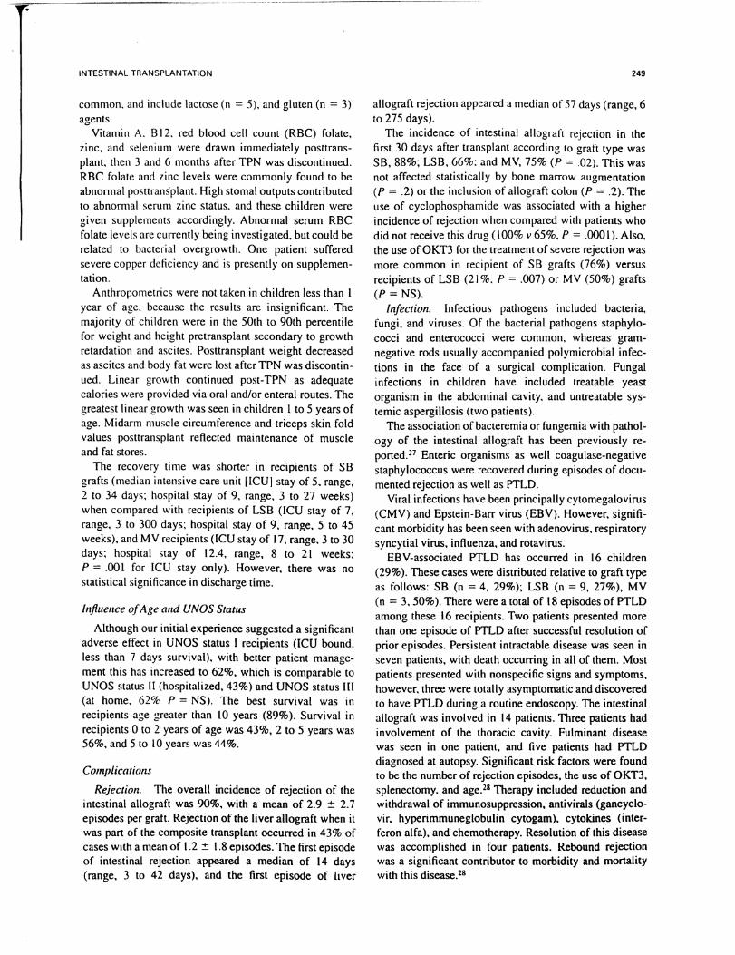
The patients given the isolated SB grafts had principally major vessel access complications (n = 6) or frequent episodes of infection (n = 8) in the face of minor liver function abnormalities (n = 6). In this group, results of liver biopsy performed at the time of transplant showed significant fibrosis in 13 (80%) patients. The 58 graft was based on a vascular pedicle of superior mesenteric anery and superior mesenteric vein. The venous return was directed into the recipients ponal vein (n = 8). superior mesenteric vein (n = 4). splenic vein (n = I), and the inferior vena cava (n = 4). Six patients had an interposition venous graft to the ponal vein (Fig I).
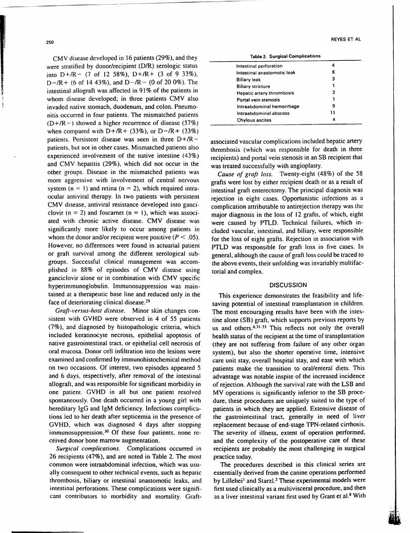
For the LSB and M V procedures. the graft was anastomosed "piggyback" to the skeletonized recipient vena cava. and anerialized from the infrarenal aorta via an aortic conduit homogrolft (Fig 2A and B).ln the LSB recipients. a permanent native portocaval shunt (n = 22) or a donor ponal vein to native ponal vein anastomosis (n = 10) was performed (Fig 2C). Two patients with a normal native liver received a modified MY procedure. which excluded the allograft liver as pan of the composite of organs. In these cases. the portal venous return was directed into the recipient portal vein (Fig 20). Reconstruction of the gastrointestinal and biliary ducts was with conventional techniques. In the first three cases. both ends of the intestlOal graft were exteriorized by the "chimney" method. Subsequemly. a tube jejunostomy replaced the chimney proximally.
When the recipient became independent of TPN. the enterostomies

INTESTINAL TRANSPLANTATION 245
1 B
Ive
Fig 1. (AI Isolated intestinal graft. (B) Arterialization and potential venous drainage options in the isolated intestinal graft. pV, portal vein; sv, splenic vein; SMV, superior mesenteric vein; IYC, inferior vena cava.
were taken down with an elltraperilOneal technique. at a median time of 113 (range. 21 to 516) days. Because of high postoperative stomal outputs and the need for continuous intravenous fluid therapy. 20 grafts contained a segment of colon that was distributed among all three recipient cohorts: SB (n = 4), LSB (n = 10), and MV (n = 6). Two patients (one with Hirschsprung's disease and another with familial polyposis) had endorectal pull-through procedures performed with allograft colon.2O [n seven recipients. the donor celiac ganglion was preserved in an attempt to facilitate reinnervation and consequently improve the motility of the allograft bowel.
Bone Marrow Transplantation
Augmentation of leukocyte chimerism was performed in nine patients. (eight primary and one retransplantation) by giving unaltered adjuvant donor bone marrow isolated from the thoracolumbar vertebrae of the donor. as previously described.ll ·n
Immunosuppression
Postoperative immunosuppression was ba.<;Cd on tacrolimus (FK506. Prograt) and steroids. Briefly. intravenous tacrolimus was started intraoperatively at a dose of 0.15 mglkg/d and converted to an oral dose when patients could tolerate oral intake. Plasma l2-hour trough levels of tacrolimus were measured daily initially by enzyme-linked immunoassay technique (ELISA) (target therapeutic level between I and 2 nglmL). then subsequently by whole blood levels. which were performed using micropanicle enzyme immunoassay (MEIA) technology (trough target levels of 15 to 30 nglmL). Hydrocortisone (25 mglkg) or methylprednisolone (\0 mglkg) was given immediately after graft reperfusion. This was followed by an intravenous methylprednisolone taper over 5 days from a starting dose of 5 mglkgld. Azathioprine (I to 2 mglkg/d) was added in selected patients to avert tacrolimus-induced renal tOllicity or enhance baseline immunosuppression in cases of recurrent rejection. The last 16 recipients received induction therapy using cyclophosphamide intravenously (2 mglkgld). and then were
subsequently switched to either mycophenolate mophetil (15-30 mg/kg twice a day) or azathioprine. Prostaglandin EI was started intraoperatively at O.03l1glkglmin and gradually increased to 0.09 lIg/kglmin until intravenous tacrolimus was discontinued. This was given both for its beneficial effects on renal perfusion as well as for its prevention of microvascular thrombosis. the damage-mediating event in acute cellular rejection and procurement injury. H
Rejection was treated initially with intravenous bolus methylprednisolone (10 mglkg) and optimization of tacrolimus levels (either intravenously or orally). Methylprednisolone taper therapy (similar to induction tapering doses) was used in cases of more severe rejection or when bolus therapy was inadequate. The use of OKT3 was the nellt line of therapy when steroid resistance occurred.
For the initial cohort of patients. weekly allograft mucosal biopsies were performed endoscopicaly. alternating with occasional cup forceps biopsies near the ileostomy. Subsequently. surveillance was performed only endoscopically with mucosal biopsies. which were performed twice weekly for the transplant admission. and then whenever clinically indicated thereafter. The histological diagnoses of rejection has been described elsewhere. !S.26
Surveillance of Graft- Versus-Host Disease
All suspicious skin lesions were biopsied and studied by routine histology testing. Detection of donor cells by immunohistologic staining for donor-specific HLA antigens and in situ hybridization technique using the Y chromosome-specific probe have been described elsewhere. ls
Prevention of Infection
All recipients received intestinal decontamination with polymyxin. tobramycin. and amphotericin B via the nasogastric tube for 2 weeks postoperatively. Weekly quantitative stool cultures were performed to assess overgrowth of resistant bacteria in greater than 108.
Broad-spectrum intravenous antibiotics were given for 5 days;

l
246
c
Fig 2. lAI Liver small bowel graft. (BI Complete multivisceral graft. (CI Systemic porta caval shunt, and recipient portal vein to donor portal vein anastomosis (insetl. (01 Modified multivisceral graft.
REYES ET AL

INTESTINAL TRANSPLANTATION
specific antimicrobial therapy was given for infections the patient may have had before transplant. The antiviral prophylactic strategy evolved during the study period. Twelve patients who underwent transplant between July 1990 and July 1992 received prophylaxis with oral acyclovir (gOO mg/m2) four times a day for 12 weeks after transplantation. Forty-three patients received intravenous gancyclovir (10 mg/ kg/d) divided in two doses for the first 2 weeks after transplantation. Twenty of the 43 subsequently received oral acyclovir for an additional 10 weeks. Seven of these 43 were given concomitant cytomegalovirus (CMV)-specific hyperimmuneglobulin (Cytogam. 100 mg/kg/d. Medimmune. Gaithersburg. MO). Antifungal therapy was used prophylactically only if there was evidence of prior infection. or at reexploration for any complication. Long-term protozoal prophylaxis was with trimethoprim-sulfamethoxazole.
NutritionaL Management
Standard TPN formulas were tapered gradually as oral or enteral feedings (via gastric or jejunal tube) were advanced. Tube feedings are initiated with an isotonic dipeptide formula containing medium-chain triglycerides and glutamine. This was later converted to a lactose-free and gluten-free diet that contained dietary fibers to promote normalization of intestinal motility and function. Most patients did not voluntarily eat adequate amounts early after the operation: therefore. enteral supplementation was required as soon as the intestinal tract became functional. This resistance to resumption of oral feeding has been particularly impressive in children.
StatisticaL Methods
Patient survival was calculated from the date of transplantation until death. Isolated SB recipients submitted to allograft enterectomy were censored at the time of discharge. because these patients may succumb to TPN-associated causes not related to the transplant procedure or immunosuppression. The primary graft survival was calculated from the date of transplantation until graft removal. retransplantation. or death. Survival curves were generated using the Kaplan-Meier method and compared by log rank (Mantel-Cox) test. The Pearson "ltest or Fisher's Exact test were used to compare proportions.
RESULTS
Patient and Graft SurvivaL
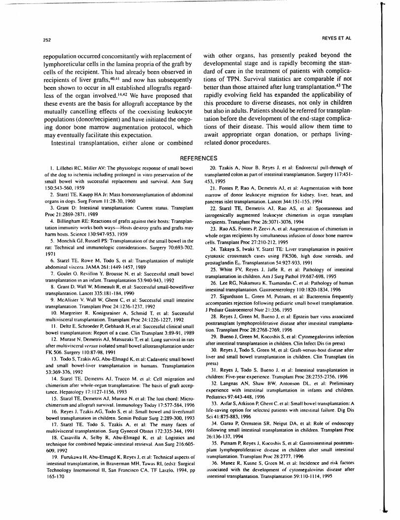
Thirty of 55 patients are still alive. All except six of the 30 surviving patients have a potential follow-up of at least I year (range. 0.2 to 6.7 years). The actuarial patient and primary graft survival rates at I. 3. and 5 years are 72%. 55%. and 55% for patient. and 66%. 48%. and 48% for graft. respectively (Fig 3). Of note. during this time. the I and 2 year survival of patients who have not undergone transplantation has been 30% and 22%. respectively.
IsoLated IllIestinal Grafts
This type of graft provided better patient survival and succeeded in restoring alimentary function at the highest rate at all follow-up times (range. 0.2 to 5.8 years). Actuarial survival rates at I. 3. and 5 years was 92% consistently for patient. and 73%. 61 %. and 61 % for the primary graft. respectively. All surviving patients who retained their graft have been liberated from TPN. Recently. one recipient was hospitalized with acute
100
80
~ 60
~ ii > 40 t " CIl
20
__ Patient survival
- - Graft survival
247
• . •. Patients not transplanted
, ------------
o+-__ --,-__ --,-__ --,-__ --,-__ --,-____ ~ 70 1080 1440 1800 2160 360
Time (Days)
Fig 3. Overall patient and graft survival rates. Of note is the survival rate of similar patients who did not reeeive a transplant.
pancreatitis and is on partial TPN. Five graft losses occurred at 27. 54. 119.332. and 774 days posttransplant. respectively. all as a result of untreatable rejection. two of them in the setting of posttransplant Iymphoproliferative disease (PTLD) (Fig 4). Of note. the three censored patients died of TPN-related causes at 94. 459. and 496 days postgraft removal.
Liver Small BoweL
Thirty-two recipients received 33 LSB grafts. of which 15 (47%) are alive 0.5 to 6.7 years after operation. The actuarial survival rates at I. 3. and 5 years was 65%. 44%. and 44% for patient. and 63%. 45%. and 45% for the primary graft. respectively. All survivors are at home with functioning grafts (Fig 4).
Multivisceral
There were seven primary and one retransplant MY graft recipients. of which three patients are alive and TPN free. The actuarial 1-. 3-. and 5-year survival rates were 57%. 43%. and 43% for patient and primary graft survival. respectively (Fig 4).
RetrallspLantatioll
Four patients underwent retransplantation. three LSB recipients on the same day as primary graft removal. and one SB recipient at 340 days after graft enterectomy. These patients lost their primary graft to acute intestinal rejection (n = 2). chronic intestinal rejection (n = I). PTLD (n = I). and hepatic artery thrombosis (n = I). Of the three LSB recipients. one patient received an MY retransplant. one patient received a liver-only retransplant after hepatic artery thrombosis. and another patient received another LSB retransplant after acute rejection and adenovirus hepatitis. The three LSB recipients died at 19 days. 47 days. and 57 days after the retransplantation of systemic bacterial infection (n = 1). rejection (n = I).
.......
Ii
I

248
100 I: ~ ____ ~ ________________________ _ I'
~- --: 80 L , , , ,
--:-1_-, L_ 1 ... ____ 1_ ........
1 __ ~---I,
'_ .. _ ...... ~~~_":'r.":':-:.":'r.":'.:-.. ":' - --
-S8en= 161 20 .. __ S8 It In = 3.2) P = 0.04 Ilol!.~rank If'sf)
____ MV en = 71
o+-----~------,_----~------~----~---
A 0
100
80 '
Oi ~ 60 ::
en
360 720 1080
Days arler Transplantation
• 1 ······t:.-.::.: __ _ 1
\
1-1-10 1800
.:: E 40
. - - ...... '":."": :"" .. ~."': :-. ':" .""': :-. ':" .. - - -'-' >. .. " E 20 --SB(n=16)
p = 0.58 (Iog-ronk tnt) 'i: c.. - - SB/l (n=32)
- - . - MV (n=7)
O+-~~~~~~~--.-~--r-~--r-~ B 0 360 7 0 1080 1440 1800
Days After Transplantation
Fig 4. (AI Patient survival rate by type of graft. *indicates 5B survival censored recipients whose follow-up was terminated after allograft enterectomy. (BI Graft survival by type of graft.
and PTLD (n = I). One S8 recipient received an S8 graft at retransplant 11 months after graft enterectomy. This patient, who is alive, was also given bone marrow from the second intestinal donor.
Influence of the Allograft Colon and Donor Bone MarroII' Augmentation
Although inclusion of allograft colon did not statistically affect survival, there was a trend toward an increase in patient and graft loss in recipients of this intestinal segment (Fig 5). The actuarial survival at 1,3, and 5 years was 56%, 39%, and 39% for primary graft survival and 64%, 45%. and 45% for patient survival when the allograft colon was part of the intestinal allograft versus a survival of 71 %, 51 %, and 51 % for graft and 70%, 59%, and 59% for patient, respectively when the allograft colon was not included (Fig 4). Although the potential functional advantage in weaning time of TPN or intravenous fluids when compared with patients who did not recei ve allograft colon was not seen, the allograft colon was successfully used as a reconstructive segment in two
REYES ET AL
patients who required a pull-through procedure because of disease involving the rectum.
The bone marrow augmentation trial included a cohort of 22 patients, of which nine received donor bone marrow. There was no significant difference in actuarial patient or graft survival between the two groups at I and 2 years.
Nutritional Status
Currently, 29 of 30 children (97%) have been weaned offTPN. The median time of weaning has been 41 (range, 14 to 210) days. Enteral feeding (Pediatric Vivonex or Tolerex) was introduced at a median of 9 days posttransplant (range, 3 to 37). Twenty-three (77%) of these children thrive on oral diet alone. Two children receive tube feedings only because of the refusal to eat, and are currently in feeding therapy. Four other children take oral feeds during the day and receive supplemental night time feeds until their oral intake is adequate to support growth. The hospitalized recipient of an S8 graft is transiently on TPN because of acute pancreatitis. Food allergies were
100
1- __ 1- ________ _
.... ;; E 20
--No Colon (n=37)
Colon (n= 18) t
~ ~ Oi >
"E " '" .:: .. ... '-'
0
A
100
80
60
40
20
p-O.28 (Iog-ronk tn.)
360 7 0 1080 1440 1800
Days After Transplantation
\ - --1 ______ ---
--No Colon (n=38)
Colon (n=20) p - 0.1121 (!oc-.. ak tnt)
O+-~--~~---'--~-.--~-r--~-.~ B 360 7.0 1080 1440 1800
Days After Transplantation
Fig 5. (AI Effect of allograft colon on patient survival rate. IBI Effect of allograft colon on graft survival rate.
., . T
INTESTINAL TRANSPLANTATION
common, and include lactose (n = 5), and gluten (n = 3) agents.
Vitamin A, B 12, red blood cell count (RBC) folate, zinc, and selenium were drawn immediately posttransplant, then 3 and 6 months after TPN was discontinued, RBC folate and zinc levels were commonly found to be abnormal posttrans·plant. High stomal outputs contributed to abnormal serum zinc status, and these children were given supplements accordingly. Abnormal serum RBC folate levels are currently being investigated, but could be related to bacterial overgrowth. One patient suffered severe copper deficiency and is presently on supplementation,
Anthropometrics were not taken in children less than I year of age, because the results are insignificant. The majority of children were in the 50th to 90th percentile for weight and height pretransplant secondary to growth retardation and ascites. Posttransplant weight decreased as ascites and body fat were lost after TPN was discontinued. Linear growth continued post-TPN as adequate calories were provided via oral and/or enteral routes. The greatest linear growth was seen in children I to 5 years of age. Midarm muscle circumference and triceps skin fold values posttransplant reflected maintenance of muscle and fat stores,
The recovery time was shorter in recipients of SB grafts (median intensive care unit [ICUI stay of 5, range, 2 to 34 days; hospital stay of 9, range, 3 to 27 weeks) when compared with recipients of LSB (lCU stay of 7, range, 3 to 300 days; hospital stay of 9, range, 5 to 45 weeks), and MV recipients (ICU stay of 17, range, 3 to 30 days; hospital stay of 12.4, range, 8 to 21 weeks; P = .001 for ICU stay only). However, there was no statistical significance in discharge time.
Influence of Age and UNOS Status
Although our initial experience suggested a significant adverse effect in UNOS status I recipients (lCU bound, less than 7 days survival), with better patient management this has increased to 62%, which is comparable to UNOS status II (hospitalized, 43%) and UNOS status III (at home, 62% P = NS). The best survival was in recipients age greater than 10 years (89%). Survival in recipients 0 to 2 years of age was 43%, 2 to 5 years was 56%, and 5 to 10 years was 44%.
Complications
Rejection. The overall incidence of rejection of the intestinal allograft was 90%, with a mean of 2.9 :t 2.7 episodes per graft. Rejection of the liver allograft when it was part of the composite transplant occurred in 43% of cases with a mean of 1.2 :t 1,8 episodes. The first episode of intestinal rejection appeared a median of 14 days (range. 3 to 42 days). and the first episode of liver
249
allograft rejection appeared a median of 57 days (range. 6 to 275 days).
The incidence of intestinal allograft rejection in the first 30 days after transplant according to graft type was SB, 88%; LSB, 66%; and MV, 75% (P = .02). This was not affected statistically by bone marrow augmentation (P = .2) or the inclusion of allograft colon (P = .2). The use of cyclophosphamide was associated with a higher incidence of rejection when compared with patients who did not receive this drug ( 100% v 65%, P = .000 I). Also, the use of OKT3 for the treatment of severe rejection was more common in recipient of SB grafts (76%) versus recipients of LSB (21%, P = .007) or MV (50%) grafts (P = NS).
Infection. Infectious pathogens included bacteria, fungi, and viruses. Of the bacterial pathogens staphylococci and enterococci were common, whereas gramnegative rods usually accompanied polymicrobial infections in the face of a surgical complication. Fungal infections in children have included treatable yeast organism in the abdominal cavity. and untreatable systemic aspergillosis (two patients).
The association of bacteremia or fungemia with pathology of the intestinal allograft has been previously reportedY Enteric organisms as well coagulase-negative staphylococcus were recovered during episodes of documented rejection as well as PTLD.
Viral infections have been principally cytomegalovirus (CMV) and Epstein-Barr virus (EBV). However. significant morbidity has been seen with adenovirus, respiratory syncytial virus. influenza. and rota virus.
EBV-associated PTLD has occurred in 16 children (29%). These cases were distributed relati ve to graft type as follows: SB (n = 4, 29%); LSB (n = 9, 27%), MV (n = 3,50%). There were a total of 18 episodes of PTLD among these 16 recipients. Two patients presented more than one episode of PTLD after successful resolution of prior episodes. Persistent intractable disease was seen in seven patients, with death occurring in all of them. Most patients presented with nonspecific signs and symptoms. however, three were totally asymptomatic and discovered to have PTLD during a routine endoscopy. The intestinal allograft was involved in 14 patients. Three patients had involvement of the thoracic cavity. Fulminant disease was seen in one patient, and five patients had PTLD diagnosed at autopsy. Significant risk factors were found to be the number of rejection episodes, the use of OKT3. splenectomy, and age,28 Therapy included reduction and withdrawal of immunosuppression. antivirals (gancyclovir, hyperimmuneglobulin cytogam), cytokines (interferon alfa), and chemotherapy. Resolution of this disease was accomplished in four patients. Rebound rejection was a significant contributor to morbidity and mortality with this disease.28
250
CM V disease developed in 16 patients (29%), and they were stratified by donor/recipient (DIR) serologic status into D+/R- (7 of 12 58%), D+IR+ (3 of 9 33%), D-IR+ (6 of 14 43%), and D-IR- (0 of 20 0%). The intestinal aJlograft was affected in 91 % of the patients in whom disease developed; in three patients CMV also invaded native stomach, duodenum, and colon. Pneumonitis occurred in four patients. The mismatched patients (D+IR -) showed a higher recurrence of disease (57%) when compared with D+IR+ (33%), or D-/R+ (33%) patients. Persistent disease was seen in three D+IRpatients, but not in other cases. Mismatched patients also experienced involvement of the native intestine (43%) and CMV hepatitis (29%), which did not occur in the other groups. Disease in the mismatched patients was more aggressive with involvement of central nervous system (n = I) and retina (n = 2), which required intraocular antiviral therapy. In two patients with persistent CMV disease, antiviral resistance developed into gancic10vir (n = 2) and foscamet (n = I), which was associated with chronic active disease. CMV disease was significantly more likely to occur among patients in whom the donor and/or recipient were positive (P < .05). However. no differences were found in actuarial patient or graft survival among the different serological subgroups. Successful clinical management was accomplished in 88% of episodes of CMV disease using ganciclovir alone or in combination with CMV specific hyperimmunoglobulin. Immunosuppression was maintained at a therapeutic base line and reduced only in the face of deteriorating clinical disease.29
Graft-versus-host disease. Minor skin changes consistent with GVHD were observed in 4 of 55 patients (7%), and diagnosed by histopathologic criteria, which included keratinocyte necrosis, epithelial apoptosis of native gastrointestinal tract, or epithelial cell necrosis of oral mucosa. Donor ceJl infiltration into the lesions were examined and confirmed by immunohistochemical method on two occasions. Of interest, two episodes appeared 5 and 6 days, respectively, after removal of the intestinal allograft, and was responsible for significant morbidity in one patient. GVHD in all but one patient resolved spontaneously. One death occurred in a young girl with hereditary IgG and IgM deficiency. Infectious complications led to her death after septicemia in the presence of GVHD, which was diagnosed 4 days after stopping immunosuppression. 3O Of these four patients. none received donor bone marrow augmentation.
Surgical complications. Complications occurred in 26 recipients (47%), and are noted in Table 2. The most common were intraabdominal infection. which was usually consequent to other technical events. such as hepatic thrombosis. biliary or intestinal anastomotic leaks. and intestinal perforations. These complications were significant contributors to morbidity and mortality. Graft-
Table 2. Surgical Complications
Intestinal perforation Intestinal anastomotic leak
Biliary leak
Biliary stricture Hepatic artery thrombosis
Portal vein stenosis Intraabdominal hemorrhage
Intraabdominal abscess Chylous ascites
REYES ET AL
4
6 3 1
3 1
9 11
4
associated vascular complications included hepatic artery thrombosis (which was responsible for death in three recipients) and portal vein stenosis in an SB recipient that was treated successfully with angioplasty.
Cause of graft loss. Twenty-eight (48%) of the 58 grafts were lost by either recipient death or as a result of intestinal graft enterectomy. The principal diagnosis was rejection in eight cases. Opportunistic infections as a complication attributable to antirejection therapy was the major diagnosis in the loss of 12 grafts. of which. eight were caused by PTLD. Technical failures. which included vascular. intestinal. and biliary. were responsible for the loss of eight grafts. Rejection in association with PTLD was responsible for graft loss in five cases. In general, although the cause of graft loss could be traced to the above events, their unfolding was invariably multifactorial and complex.
DISCUSSION
This experience demonstrates the feasibility and lifesaving potential of intestinal transplantation in children. The most encouraging results have been with the intestine alone (SB) graft. which supports previous reports by us and others.8.31-33 This reflects not only the overall health status of the recipient at the time of transplantation (they are not suffering from failure of any other organ system), but also the shorter operative time, intensive care unit stay. overall hospital stay. and ease with which patients make the transition to oral/enteral diets. This advantage was notable inspite of the increased incidence of rejection. Although the survival rate with the LSB and MV operations is significantly inferior to the SB procedure. these procedures are uniquely suited to the type of patients in which they are applied. Extensive disease of the gastrointestinal tract. generally in need of liver replacement because of end-stage TPN-related cirrhosis. The severity of illness. extent of operation performed. and the complexity of the postoperative care of these recipients are probably the most challenging in surgical practice today.
The procedures described in this clinical series are essentially derived from the canine operations performed by Lillehei I and Starzl.2 These experimental models were first used clinically as a multi visceral procedure. and then as a liver intestinal variant first used by Grant et al.8 With

,----------
INTESTINAL TRANSPLANTATION
the parallel advances in organ preservation and procurement. the surgical techniques necessary to provide the flexible application of these composite organs are ideally suited for the varied diseases encountered in children suffering from intestinal fai lure. '7 Because donor a vai labi 1-ity has been limited. this also allows for potential sharing and the use of isolated intestinal grafts from donors who would otherwise donate the liver graft only.
The postoperative care of these patients is extremely challenging and requires a multidisciplinary team with standardization of care specifically focused on intestinal transplant recipients. Fluid and electrolyte management as well as nutritional support can become challenging tasks when coupled with efforts to pursue rejection and sources of infection. The spectrum of potential surgical and clinical complications encountered in this series reflects not only the type of transplant operation. but also the severity of illness with which these patients present at the time of transplant. which inevitably effects survi val.
Successful retransplamation has met with consistent failure in the larger composite grafts. These recipients inevitably suffer multisystem organ dysfunction and a precarious clinical state at the time of retransplamation. However. in the isolated intestinal transplant patient with a failed graft. successful retransplantation was possible after graft enterectomy and an II-month "rest" period. This type of strategy may improve even further the results of patient and graft survival after isolated intestinal transplant. Because this type of rescue operation is not an option for failed complex grafts. we have not performed the composite procedures in patients who need only the intestine.
Intestinal allograft rejection remains the major stumbling block after intestinal transplantation. This complication may further precipitate opportunistic infections (consequent to increased immunotherapy) that become additive factors in patient and graft losses. [n general clinical manifestations of intestinal disease such as abdominal distention. pain. diarrhea. or fever. are highly unreliable markers for rejection. and should be investigated by appropriate endoscopic procedures before any therapeutic intervention is begun. By far the most effective means of diagnosing rejection is by serial surveillance endoscopies performed on a twice-a-week basis. 34 Early infiltrative changes can be diagnosed before endoscopic damage becomes evident. or. if endoscopy findings appear significant and diagnostic. therapeutic intervention can be initiated before interpretation of biopsy results. It is also possible to differentiate other sources of potential intestinal allograft disease that may clinically mimic rejection such as PTLD. CMV. or other bacteriaVviral enteritis. 35
The evidence here supports other observations that simultaneous liver grafting may reduce the risk of intestinal graft rejection.s The isolated SB graft not only had the highest incidence of rejection. but also required
251
more intense immunotherapy to control. as evidenced by the frequency ofOKT3 use.
Infectious complications generally plaque this patient population, and can be of bacterial, fungal, or viral origin. With rejection, the intestinal graft becomes permeable to bacteria from within the lumen. Paradoxically, such bacteremias can be prevented or treated only by heavier immunosuppression that may contribute to worsening the susceptability to infection. However, maintenance of an intact mucosal barrier is primordial and mandates appropriate levels of immunosuppression.
Our experience with CMV supports previous recommendations to avoid using seropositive grafts in a seronegative recipient.36 The frequency of CMV disease was similar in the other serological groups. Although CMV disease produced significant morbidity. successful therapy of CMV was accomplished in most patients. Also. the patient and graft survivals in those cases of CMV disease was not different when compared with patients who did not acquire CMV disease. Thus, we support the use of CMV-positive donors in patients who are awaiting composite grafts. because these patients generally may be too sick to await a seronegative donor.
The effect on patient and graft survival with PTLD has been pervasive. Associated losses from uncontrollable rejection have been precipitated with efforts to control the EB V infection.!8 Results after traditional therapeutic interventions. which have included decrease or withdrawal of immunosuppression. antiviral therapy, cytokine therapy, and chemotherapy. have been poor. The development of tools such as the use of quantitative EBV polymerase chain reaction in the peripheral blood has recently allowed for early diagnosis. as well as follow-up of patients with established PTLD. Therapeutic intervention using such a guideline has been helpful in preventing the end phases of this disease and may improve outcomeY This may eventually allow us to diagnose EBV infection before the development of PTLD. More experience with this form of preemptive therapy. and the avoidance of concomitant rejection, will likely impact on survival.
Clinical GVHD was significant only in one recipient who was a bearer of a preexisting immune deficiency and required reduction and then discontinuance of immunosuppression after an intestinal anastomotic leak. All other cases had minimal skin changes, which resolved spontaneously. The possibility of inherent immunodeficiencies in children will be more likely than with adults. and should be investigated when appropriate. It is interesting to note that the cases of GVHD were not found among the donor bone marrow-augmented patients. We have previously demonstrated the appearance of donor mononuclear cells in the peripheral blood of intestinal graft recipients. which. under the umbrella of effective immunosuppression. did not result in GVHD.39.4O This cell migration and
252
repopulation occurred concomitantly with replacement of lymphoreticular cells in the lamina propria of the graft by cells of the recipient. This had already been observed in recipients of liver grafts,40AI and now has subsequently been shown to occur in all established allografts regardless of the organ involved. 14,42 We have proposed that these events are the basis for allograft acceptance by the mutually cancelling effects of the coexisting leukocyte populations (donorlrecipient) and have initiated the ongoing donor bone marrow augmentation protocol, which may eventually facilitate this expectation.
Intestinal transplantation, either alone or combined
REYES ET Al
with other organs, has presently peaked beyond the developmental stage and is rapidly becoming the standard of care in the treatment of patients with complications of TPN. Survival statistics are comparable jf not better than those attained after lung transplantation.43 The rapidly evolving field has expanded the applicability of this procedure to diverse diseases, not only in children but also in adults. Patients should be referred for transplantation before the development of the end-stage complications of their disease. This would allow them time to await appropriate organ donation, or perhaps livingrelated donor procedures.
REFERENCES
t. Lillehei RC. Miller AV: The physiologic response of small bowel of the dog to ischemia including prolonged in vitro preservation of the
small bowel with successful replacement and survival. Ann Surg 150:543-560,1959
2. Starzl TE. Kaupp HA Jr: Mass homotransplantation of abdominal organs in dogs. Surg Forum II :28-30, 1960
3. Grant D: Intestinal transplantation: Current status. Transplant Proc 21 :2869-1871. 1989
4. Billingham RE: Reactions of grafts against their hosts: Transplan· tation immunity works both ways-Hosts destroy grafts and grafts may haml hosts. Science 130:947-953. 1959
5. Monchik GJ. Russell PS: Transplantation of the small bowel in the rat: Technical and immunologic considerations. Surgery 70:693-702. 1971
6. Starzl TE. Rowe M, Todo S, et al: Transplantation of multiple abdominal viscera. JAM A 261: 1449- 1457, 1989
7. Goulet O. Revillon y, Brousse N, et al: Successful small bowel transplantation in an infant. Transplantation 53:940-943, 1992
8. Grant D. Wall W. Mimeault R. et al: Successful small-boweilliver transplantation. Lancet 335: 18 I - I 84, 1990
9. McAlister V. Wall W, Ghent C. et al: Successful small intestine transplantation. Transplant Proc 14: I 236- I 237. 1992
10. Margreiter R. Konigsrainer A, Schmid T. et al: Successful multi visceral transplantation. Transplant Proc 24: I 226-1227, 1992
I I. Deitz E, Schroeder P, Gebhardt H, et al: Successful clinical small bowel transplantalion: Report of a case. C1in Transplant 3:89-91, 1989
12. Murase N, Demetris AJ, Matsuzaki T, et al: Long survival in rats after multi visceral ,'emu isolated small bowel allotransplantation under FK 506. Surgery 110:87-98, 1991
13. Todo S, Tzakis AG, Abu-Elmagd K, et al: Cadaveric small bowel and small bowel·liver transplantation in humans. Transplantation 53:369-376, 1992
14. Starzl TE. Demctns AJ, Trucco M. ct al: Cdl migration and chimerism after whole·organ transplantation: The basis of graft acceptance. Hepatology 17:1127-1156. 1993
15. Starzl TE. Demetns AJ, Murase N. CI al: The lost chord: Micro· chimerism and allograft survival. Immunology Today 17:577-584,1996
16. Reyes J. Tzakis AG, Todo S, et al: Small bowel and liver/small bowel transplamation in children. Semin Pediatr Surg 2:289-300, 1993
17. Starzl TE, TOOo S. Tzakis A. et al: The many faces of multivisceral transplantation. Surg Gynecol Obstet 172:335-344, 1991
18. Casavilla A, Selby R, Abu-Elmagd K, et al: Logistics and technique for combined hepatic-intestinal retrieval. Ann Surg 216:605-609,1992
19. Furukawa H. Abu-Elmagd K, Reyes J. et al: Technical aspects of intestinal transplantatIon, in Br.lverman MH, Tawas RL (eds): Surgical Technology International II, San Francisco CA. TF Laszlo, 1994, pp 165-170
20. Tzakis A, Nour B, Reyes J, et al: Endorectal pull-through of
transplanted colon as part of intestinal transplantation. Surgery 117:451-
453, 1995 21. Fontes P. Rao A, Demetris AJ, et al: Augmentation with bone
marrow of donor leukocyte migration for kidney. liver, heart, and
pancreas islet transplantation. Lancet 344: I 5 I-I 55, 1994
22. Starzl TE, Demetris AJ. Rao AS, et al: Spontaneous and iatrogenically augmented leukocyte chimerism in organ transplant
recipients. Transplant Proc 26:3071-3076,1994
13. Rao AS, Fontes P. Zeevi A, et al: Augmentation of chimerism in
whole organ recipients by simultaneous infusion of donor bone marrow
cells. Transplant Proc 27:210-212.1995
24. Takaya S, Iwaki Y. Starzl TE: Liver transplantation in positive
cytotoxic crossmatch cases using FK506. high dose steroids, and prostaglandin E,. Transplantation 54:927-933. 1991
25. White FY, Reyes J. Jalfe R, et al: Pathology of intestinal
transplantation in children. Am J Surg Pathol 19:687-698. 1995 26. Lee RG, Nakamura K. Tsamandas C. et al: Pathology of human
intestinal transplantation. Gastroenterology 110: 1820-1834. 1996
27. Sigurdsson L. Green M. Putnam. et al: Bacteremia frequently accompanies rejection following pediatric small bowel transplantation. J Pediatr Gastroenterol Nutr 21 :356, 1995
28. Reyes J, Green M. Bueno J, et al: Epstein barr virus associated posttransplant Iymphoproliferative disease after intestinal transplantation. Transplant Proc 28:2768·2769, 1996
29, Bueno J, Green M, Kocoshis S, et al: Cytomegalovirus infection after intestinal transplantation in children. Clin Infect Dis (in press)
30. Reyes J, TOOo S, Green M, et al: Graft-versus-host disease after liver and small bowel transplantation in children. Clin Transplant (in press)
31. Reyes J, Todo S, Bueno J, et al: Intestinal transplantation in children: Five-year experience. Transplant Proc 18:2755-2756. 1996
32. Langnas AN, Shaw BW, Antonson DL. ct al: Preliminary
experience with intestinal transplantation in infants and children. Pediatrics 97:443-448, 1996
33. Asfar S, Atkison p, Ghent C. et al: Small bowel transplantation: A
life-saving option for selected patients with intestinal failure. Dig Dis
Sci 41:875-883.1996 34. Garau P, Orenstein SR. Neigut DA, et al: Role of endoscopy
following small intestinal transplantation in children. Transplant Proc 26: 136- 137,1994
35. Putnam P, Reyes J, Kocoshis S, et al: Gastrointestinal posttransplant Iymphoproliferative disease in children after small intestinal
transplantation. Transplant Proc 28:2777. 1996 36. Manez R, Kusne S, Green M. et al: Incidence and risk factors
associated with the development of cytomegalovirus disease after intestinal transplantation. Transplantation 59: 110-1114, 1995

INTESTINAL TRANSPLANTATION
37. Green M. Reyes J. Jabbour N. et al: Use of quantitative PCR to predict onset of Epstein·Barr viral infection and post·transplantlymphoproliferative disease after intestinal transplantation in children. Trans· plant Proc 2S:2759-276O. 1996
3S. Iwaki Y. Starzl TE. Yagihashi A. et al: Replacement of donor lymphoid tissue in human small bowel transplants under FK506 immunosuppression. Lancet 337:SIS-SI9. 1991
39. Murase N. Demetris Al. Matsuzaki T. et al: Long survival in rats after multivisceral versus isolated small bowel allotransplantation under FK506. Surgery 110:S7-98. 1991
40. Kashiwagi N. Porter KA. Penn I. et al: Studies of homograft sex
253
and of gamma globulin phenotypes after orthotopic homotransplanta· tion of the human liver. Surg Forum 20:374-376. 1969
41. Porter KA: Pathology of the orthotopic homograft and heterograft. in Starzl TE (ed): Experience in Hepatic Transplantation. Philadelphia. PA. WB Saunders Company. 1969. pp 464-465
42. Randhawa PS. Starzl TE. Ramos H. et al: Allografts surviving for 26-29 years following living related kidney transplantation: Analysis by light microscopy. in situ hybridization for Y chromosome. and anti-HLA antibodies. Am 1 Kidney Dis 24:72-77. 1994
43. Grant D: Current results of intestinal transplantation. Intema· tional Transplant Registry. Lancet 347:1S01-1S03. 1996
Discussion
M. Langham (GaillesvilLe, FL): Dr Reyes presents a landmark paper, and [ congratulate you.
First. the patient survival of 57% supports the conclusion that this is rapidly becoming the standard of care. [n an institution that does several hundred liver transplants a year. an accumulated experience of 50 patients over 7 years points out that there are not a tremendous number of people in the country that need this therapy. The detailed reports of the results, with concentration of the experience in one or two institutions during the learning curve are worthwhile. and Dr Reyes' manuscript is to be recommended to the audience.
There are a couple of things [ would like to point out and ask about. Concerning patient survival with small bowel grafts only. you censored patients after graft enterectomy. so that the patient survival of 90% long tenn is misleading. In fact. several of those patients died after graft enterectomy and although they were not strictly transplant-related deaths. because there is no other therapy for these patients. the patients will eventually die if the graft fails.
Also. I would like you to comment on use of the colon. Although there is no significant difference in survival with inclusion of the colon. you had a decrease of 20% I-year graft survival when you used the colon. Although that did not reach statistical significance. it looked like a real difference to me. [ wondered if you are going to continue to use the colon, particularly in light of your adult experience.
1. Reyes (response): The success rate for small bowel transplanlation around the country and around the world has become reasonable. I included the data. specifically that patients who had allograft enterectomy were censored. and their survivals were tenninated at that point. I also included the fact that these deaths were TPN-related deaths and they died 91. 459. 496 days after graft removal. It is important to include that because these patients are not dying of their transplant operation. and this is an acceptable mode of analyzing therapeutic intervention that has been used for kidney and pancreas
transplantation. Once those allografts are removed. the follow-up of those patients is tenninated. If we are going to analyze survival rates with the isolated small bowel graft. we need to look at survival rate with the graft and survival rate without the graft. because 15% of patients in this country right now on long-term TPN will eventually require intestinal transplantation. Thus. we should compare survival rate with those 15% that fail. not the 85% that are making it on TPN.
We have evaluated more than 200 patients. some have undergone transplantation in other centers. We reject extremely complex candidates with other comorbid system diseases: affecting the lungs. heart. and brain. Sixty patients have died while on the waiting list. which reflects a high morbidity waiting to find an appropriate size of graft for these small children. Recently. we have developed a technique of reduced composite liver/small bowel graft. That patient is presently recovering from transplant and hopefully. in the future. [ will be able to present that type of technique.
T.e. Moore (Palos Verdes Estates, CA): We are very much indebted to Starzl's Pittsburg group for tackling this extremely difficult problem. Your experience with 43% mortality and a high incidence of both immunologic and nonimmunologic complications suggests that this procedure is barely viable. and should be perfonned only in Pittsburgh and a few other centers as Dr Langham has suggested. The long-term problems are also significant. Nicholas Tilney of Boston. evaluating cadaveric kidney transplants. found that only 20% are functioning at 10 years. The long-tenn outcome for small bowel grafts. because you are starting largely with small children. is doubtful. With the current advances in biomedical science and genetics we hope that in the future there will be much better results. I congratulate you for continuing this approach. Currently. one should concentrate on prevention of the short gut syndrome. From the causes that you presented here I think 75% to 85% are preventable by pretenn prelabor cesarean section for gastroschisis and the "patch. drain. and wait" surgical approach to necrotiz-

------- ----------------
ing entercolitis and midgut volvulus, which would essentially eliminate the short gut syndrome.
1. Reyes: I agree with your comments. 1. Vacanti (Boston, MA): Dr Reyes, I have a specific
question related to the high incidence of lymphoproliferative disease. Based on your experience, do you think it's related specifically to the intestinal graft, or is it related to the overall level of immunosuppression that is required?
1. Reyes (response): That's a very important question. It is related to the overall level of immunosuppression. Anybody who needs their FK506 levels maintained around 25 runs a high risk of being over immunosuppressed, but that's the level that we found is adequate for the maintenance of this graft without rejection. Other centers have had similar results. Until we find a protocol that would decrease the incidence of rejection and allow a lower level of FK506 administration, this is the type of morbidity we have to accept. With our present study of EBV with preemptive therapy we are able to make the diagnosis of EBV early, treat it more easily, and I think we'll decrease the morbidity of EBY. We also need a bener immunosuppressive protocol for this patient population.
As far as the graft is concerned, we haven't been able to explain how you can get a negative EBV PTLD. We believe that it is related to the type of graft and to antigenic stimuli from the lymphocytes or histiocytes that we are transplanting into these patients. Presently that theory is involved in one of the therapeutic applications that we are developing.
D. Croitoru (Norfolk, VA): What has changed over the last few years? The previous reports of combined liver/ small bowel transplantation were that you had better survival rates, and now you are showing that isolated small bowel immunosuppressant regimen or something else like that technique that has made a difference?
1. Reyes (response): The first thing that has changed is
REYES ET AL
that we are reporting our pediatric experience separate from the adult experience. The adult experience significantly changed the overall survival rate, both for the composite grafts and for the effect of the colon and no colon. With the adults the colon had a pervasive effect on survival from infections.
With respect to other things, the selection of recipient patients and donors has improved, and the overall experience with the number of people taking care of these patients also has improved. CMV was previously a significant morbidity in our initial experience with the adults. We learned from them how to manipulate the immunosuppression, not to withdraw it significantly, and to treat the CMV aggressively. That accounted for a significant improvement in survival.
E.S. Adkins (Wilmington, DE): In terms of strategy, when you have the very small patient who has obviously end-stage liver disease and is going to need both intestine and a liver, are you transplanting the liver first because you can't get an appropriate size intestine and liver? What is your approach to those little ones?
1. Reyes (response): Currently, we are using combined liverlbowel. We will not do an isolated liver for two reasons. First, we did try twice, stimulated by Steve Dunn at St Christopher's Hospital who had some initially favorable experience but then subsequently poor, which he presented at the ASTS this year. We failed, and we stopped doing that because it is a waste of a precious resource. Now we do a reduced composite liver/intestine, trying to increase the weight ranges and increase the availability of organs for these patients. It is a complex, in situ split technique in which we are able to maintain the left lateral segment with the whole composite of organs, and give the right side to an adult recipient. It's a big operation and we just did our first case. The patient is recovering now, but we are not ready to claim success.