curriculum / statutes & regulations
TRANSCRIPT
CURRICULUM / STATUTES & REGULATIONS FOR
5 YEARS DEGREE PROGRAMME IN
THORACIC SURGERY (MS Thoracic Surgery)
UNIVERSITY OF HEALTH SCIENCES, LAHORE
Curriculum/Statutes & Regulations -MS Thoracic Surgery
2
STATUTES
1. Nomenclature of the Proposed Course
The name of degree programme shall be MS Thoracic Surgery. This name is
well recognized and established for the last many decades worldwide.
2. Course Title:
MS Thoracic Surgery
3. Training Centers
Departments of Thoracic Surgery (accredited by UHS) in affiliated institutes of
University of Health Sciences Lahore.
4. Duration of Course
The duration of MS Thoracic Surgery course shall be five (5) years (first year
in Part I, first two years in Part II and next three years in Part III) with
structured training in a recognized department under the guidance of an
approved supervisor. The course is structured in three parts:
Part I is structured for the 1st calendar year. The candidate shall undertake
didactic training in Basic Medical Sciences, Behavioural Sciences, Biostatistics
& Research Methodology. At the end of first year, the examination shall be
held in Basic Sciences. The clinical training in fundamental concepts of
Surgery shall start from the 1st day of enrollment.
Part II is structured for the 1st and 2nd calendar year. The candidate shall
undertake clinical training in fundamental concepts of Surgery. At the end of
2nd year, the examination shall be held in fundamental concepts of Surgery.
The clinical training in Thoracic Surgery shall start from 3rd year onwards in
the recognized institutions.
Part III is structured for 3rd, 4th and 5th calendar years in MS Thoracic
Surgery. It has two components; Clinical and Research. The candidate shall
undergo clinical training to achieve educational objectives of MS Thoracic
Surgery (knowledge & skills) along with rotation in relevant fields. Over the
Curriculum/Statutes & Regulations -MS Thoracic Surgery
3
five years duration of the course, candidate will spend total time equivalent to
one calendar year for research during the training. Research can be done as
one block in 5th year of training or it can be done in the form of regular periodic
rotations over five years as long as total research time is equivalent to one
calendar year.
5. Admission Criteria
I. For admission in MS Thoracic Surgery course, the candidate shall be
required to have:
• MBBS degree
• Completed one year House Job
• One year experience in Thoracic Surgery/General surgery/Allied
surgical discipline in the given order of preference
• Registration with PMDC
• Passed Entry Test conducted by the University & aptitude interview
by the Institute concerned
• Having up to the mark credentials as per UHS rules (no. of
attempts in each professional, any gold medals or distinctions,
relevant work experience, Rural/ Army services, research
experience in a recognized institution, any research article
published in a National or International Journal) may also be
considered on case to case basis.
II. Exemptions: A candidate holding FCPS/MRCS/Diplomate/equivalent
qualification in General Surgery shall be exempted from Part-I & Part-II
Examinations and shall be directly admitted to Part-III Examinations,
subject to fulfillment of requirements for the examination.
6. Registration And Enrollment
• Total number of students enrolled for the course must not exceed 2 per
supervisor/year.
Curriculum/Statutes & Regulations -MS Thoracic Surgery
4
• The maximum number of trainees that can be attached with a supervisor at
a given point of time (inclusive of trainees in all years/phases of MS
training), must not exceed 6.
• Beds to trainee ratio at the approved teaching site shall be at least 5 beds
per trainee.
• The University will approve supervisors for MS courses.
• Candidates selected for the courses after their enrollment at the relevant
institutions shall be registered with UHS as per prescribed Registration
Regulation.
7. Accreditation Related Issues of the Institution
A. Faculty
Properly qualified teaching staff in accordance with the requirements of
Pakistan Medical and Dental Council (PMDC)
B. Adequate Space
Including class-rooms (with audiovisual aids), demonstration rooms, computer
lab and clinical pathology lab etc.
C. Library
Departmental library should have latest editions of recommended books,
reference books and latest journals (National and International).
Accreditation of Thoracic Surgery training program can be suspended on
temporary or permanent basis by the University, if the program does not
comply with requirements for residents training as laid out in this curriculum.
Program should be presented to the University along with a plan for
implementation of curriculum for training of residents.
Programs should have documentation of residents training activities and
evaluation on monthly basis.
To ensure a uniform and standardized quality of training and availability of
the training facilities, the University reserves the right to make surprise
visits of the training program for monitoring purposes and may take
appropriate action if deemed necessary.
Curriculum/Statutes & Regulations -MS Thoracic Surgery
5
AIMS AND OBJECTIVES OF THE COURSE AIM The aim of five years MS programme in Thoracic Surgery is to train residents
to acquire the competency of a specialist in the field so that they can
become good teachers, researchers and clinicians in their specialty after
completion of their training.
GENERAL OBJECTIVES
MS Thoracic Surgery training should enable a student to:
1. Access and apply relevant knowledge to clinical practice:
Maintain currency of knowledge
Apply scientific knowledge in practice
Appropriate to patient need and context
Critically evaluate new technology
2. Safely and effectively performs appropriate surgical procedures:
Consistently demonstrate sound surgical skills
Demonstrate procedural knowledge and technical skill at a level
appropriate to the level of training
Demonstrate manual dexterity required to carry out procedures
Adapt their skills in the context of each patient and procedure
Maintain and acquire new skills
Approach and carries out procedures with due attention to safety
of patient, self and others
Critically analyze their own clinical performance for continuous
improvement
3. Design and implement effective management plans:
Recognize the clinical features, accurately diagnose and manage
thoracic problems
Formulate a well-reasoned provisional diagnosis and management
plan based on a thorough history and examination
Formulate a differential diagnosis based on investigative findings
Curriculum/Statutes & Regulations -MS Thoracic Surgery
6
Manage patients in ways that demonstrate sensitivity to their
physical, social, cultural and psychological needs
Recognize disorders of the thoracic system and differentiate those
amenable to surgical treatment
Effectively manage the care of patients with thoracic trauma
including multiple system trauma
Effectively recognize and manage complications
Accurately identify the benefits, risks and mechanisms of action of
current and evolving treatment modalities
Indicate alternatives in the process of interpreting investigations
and in decision-making
Manage complexity and uncertainty
Consider all issues relevant to the patient
Identify risk
Assess and implement a risk management plan
Critically evaluate and integrate new technologies and techniques.
4. Organize diagnostic testing, imaging and consultation as needed:
Select medically appropriate investigative tools and monitoring
techniques in a cost-effective and useful manner
Appraise and interpret appropriate diagnostic imaging and
investigations according to patients' needs
Critically evaluates the advantages and disadvantages of different
investigative modalities
5. Communicate effectively:
Communicate appropriate information to patients (and their
family) about procedures, potentialities and risks associated with
surgery in ways that encourage their participation in informed
decision making
Communicate with the patient (and their family) the treatment
options including benefits and risks of each
Communicate with and co-ordinate health management teams to
achieve an optimal surgical environment
Initiate the resolution of misunderstandings or disputes
Curriculum/Statutes & Regulations -MS Thoracic Surgery
7
Modify communication to accommodate cultural and linguistic
sensitivities of the patient
6. Recognize the value of knowledge and research and its application to clinical
practice:
Assume responsibility for self-directed learning
Critically appraise new trends in Thoracic Surgery
Facilitate the learning of others.
7. Appreciate ethical issues associated with Thoracic Surgery:
Consistently apply ethical principles
Identify ethical expectations that impact on medico-legal issues
Recognize the current legal aspects of informed consent and
confidentiality
Be accountable for the management of their patients.
8. Professionalism by:
Employing a critically reflective approach to Thoracic Surgery
Adhering with current regulations concerning workplace harassment
Regularly carrying out self and peer reviewed audit
Acknowledging and have insight into their own limitations
Acknowledging and learning from mistakes
9. Work in collaboration with members of an interdisciplinary team where
appropriate:
Collaborate with other professionals in the selection and use of
various types of treatments assessing and weighing the indications
and contraindications associated with each type
Develop a care plan for a patient in collaboration with members of an
interdisciplinary team
Employ a consultative approach with colleagues and other
professionals
Recognize the need to refer patients to other professionals.
10. Management and Leadership
Effective use of resources to balance patient care and system resources
Identify and differentiate between system resources and patient needs
Curriculum/Statutes & Regulations -MS Thoracic Surgery
8
Prioritize needs and demands dealing with limited system resources.
Manage and lead clinical teams
Recognize the importance of different types of expertise which
contribute to the effective functioning of clinical team.
Maintain clinically relevant and accurate contemporaneous records
11. Health advocacy:
Promote health maintenance of patients
Advocate for appropriate health resource allocation
Promote health maintenance of colleagues and self scholar and
teacher
Curriculum/Statutes & Regulations -MS Thoracic Surgery
9
SPECIFIC LEARNING OUTCOMES
On completion of the training programme, Thoracic Surgery trainees including
those pursuing an academic pathway will be expected to have demonstrated
competence in all aspects of the published syllabus. The specific training
component would be targeted for establishing clearly defined standards of
knowledge and skills required to practice Thoracic Surgery at secondary and
tertiary care level with proficiency in the Basic and applied clinical sciences, Basic
Thoracic surgical care, Thoracic intensive care, Emergency medicine and
Complementary surgical disciplines.
A wide coverage of Basic Sciences like Anatomy, Physiology, Biochemistry,
Pathology, Microbiology, Pharmacology and Immunology pertaining to the
thorax and its contents.
A thorough knowledge, theoretical as well as practical, of the various
investigative procedures- invasive and non-invasive - including biochemical,
radiological and ultrasonographic investigations, radioisotope scanning and
dynamic function studies such as pulmonary function tests, cardiac
catheterization, oesophageal manometry, esophagoscopy, bronchoscopy: trans-
bronchial biopsy and bronchoalveolar lavage and thoracoscopy.
A detailed knowledge of and practical experience in clinical and operative
paediatric and adult thoracic surgery including surgery of the chest wall,
diaphragm, oesophagus, mediastinum, trachea, lungs and pleura.
A broad knowledge of relevant Cardiology, Respiratory Medicine and other
medical problems and related clinical skills specific for Thoracic Surgery;
Function of the gas exchange system and its evaluation
Diagnostic and therapeutic procedures such as pleural tapping, insertion
of intercostals drains.
Thoracoscopy: general principles.
Developmental abnormalities of the airways and lungs
Surgical treatment of the bullous emphysema.
Surgical treatment of tuberculosis
Curriculum/Statutes & Regulations -MS Thoracic Surgery
10
Molecular biology and immunology of lung cancer.
Benign tumors of the lower respiratory tract.
Lung carcinoma :diagnosis, staging, surgery for limited pulmonary
resection
Tracheobronchoplasty and sleeve resection for multimodality therapy of
carcinoma of the lung: irradiation, chemotherapy, and immunotherapy.
Indications for resection of pulmonary metastases.
Pulmonary embolism acute & chronic
Pulmonary fistulae
Lung transplantation.
Benign & malignant disorders of pleura
Pleural space problems & thoracoplasty
Thoracic infections caused by actinomycetes, fungi, opportunistic
organisms
Thoracic disorders in an immunocompromised host
Thoracic trauma and management
Management of thoracic outlet syndrome
Chest wall & sternal abnormalities & management of chest wall tumours
Diaphragm : developmental, traumatic, neoplastic disorders,
dysfunction & pacing
Mediastinal tumours, thymic tumors and managemnt of myasthenia
gravis
Trachea: tumors, strictures, tracheomalacia, tracheal resections &
reconstructions.
The esophagus: anatomy & functional evaluation.
Esophageal injuries
Medical & surgical treatment of hiatal hernia.
Barrette’s esophagus
Nissen fundoplication.
Hill procedure.
Belsey mark IV procedure.
Benign strictures of esophagus
Paraoesophageal hiatal hernia
Esophageal dysmotility
Curriculum/Statutes & Regulations -MS Thoracic Surgery
11
Treatment of achalasia & gastroesophageal reflux.
Thoracoscopic esophageal surgery.
Carcinoma of esophagus, surgical options resection, reconstruction,
palliative treatment of carcinoma of the esophagus.
Anesthesia for thoracic surgery
Thoracic incisions, complications of incision including sternal dehiscence
Prosthetic material ( biological / artificial) in thoracic surgery
Sutures: physical properties of sutures, needle types, recent advances
in suture materials.
Ventilatory assistance and support
Shock: types, diagnosis, management. Cardio pulmonary resuscitation.
Postoperative care and complications in thoracic surgery
Use of antibiotics in thoracic surgery
Research Experience:
All residents in the categorical program are required to complete an academic
outcomes-based research project during their training. This project can consist
of original bench top laboratory research, clinical research or a combination of
both. The research work shall be compiled in the form of a thesis, which is to
be submitted for evaluation by each resident before end of the training. The
designated Faculty will organize and mentor the residents through the process,
as well as journal clubs to teach critical appraisal of the literature.
Curriculum/Statutes & Regulations -MS Thoracic Surgery
12
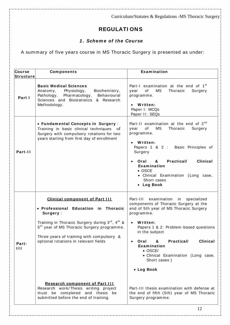
REGULATIONS
1. Scheme of the Course
A summary of five years course in MS Thoracic Surgery is presented as under:
Course Structure
Components Examination
Part I
Basic Medical Sciences Anatomy, Physiology, Biochemistry, Pathology, Pharmacology, Behavioural Sciences and Biostatistics & Research Methodology.
Part-I examination at the end of 1st year of MS Thoracic Surgery programme.
• Written: Paper I: MCQs
Paper II: SEQs
Part-II
• Fundamental Concepts in Surgery : Training in basic clinical techniques of Surgery with compulsory rotations for two years starting from first day of enrollment
Part-II examination at the end of 2nd
year of MS Thoracic Surgery programme.
• Written:
Papers 1 & 2 : Basic Principles of Surgery
• Oral & Practical/ Clinical
Examination • OSCE • Clinical Examination (Long case,
Short cases • Log Book
Part-III
Clinical component of Part III
• Professional Education in Thoracic
Surgery : Training in Thoracic Surgery during 3rd, 4th & 5th year of MS Thoracic Surgery programme. Three years of training with compulsory & optional rotations in relevant fields
Research component of Part III Research work/Thesis writing project must be completed and thesis be submitted before the end of training.
Part-III examination in specialized components of Thoracic Surgery at the end of 5th year of MS Thoracic Surgery programme.
• Written:
Papers 1 & 2: Problem-based questions in the subject
• Oral & Practical/ Clinical Examination
• OSCE/ • Clinical Examination (Long case,
Short cases )
• Log Book
Part-III thesis examination with defense at the end of fifth (5th) year of MS Thoracic Surgery programme.
Curriculum/Statutes & Regulations -MS Thoracic Surgery
13
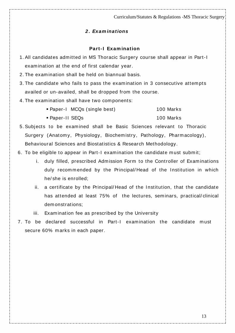
2. Examinations
Part-I Examination
1. All candidates admitted in MS Thoracic Surgery course shall appear in Part-I
examination at the end of first calendar year.
2. The examination shall be held on biannual basis.
3. The candidate who fails to pass the examination in 3 consecutive attempts
availed or un-availed, shall be dropped from the course.
4. The examination shall have two components:
Paper-I MCQs (single best) 100 Marks
Paper-II SEQs 100 Marks
5. Subjects to be examined shall be Basic Sciences relevant to Thoracic
Surgery (Anatomy, Physiology, Biochemistry, Pathology, Pharmacology),
Behavioural Sciences and Biostatistics & Research Methodology.
6. To be eligible to appear in Part-I examination the candidate must submit;
i. duly filled, prescribed Admission Form to the Controller of Examinations
duly recommended by the Principal/Head of the Institution in which
he/she is enrolled;
ii. a certificate by the Principal/Head of the Institution, that the candidate
has attended at least 75% of the lectures, seminars, practical/clinical
demonstrations;
iii. Examination fee as prescribed by the University
7. To be declared successful in Part-I examination the candidate must
secure 60% marks in each paper.
Curriculum/Statutes & Regulations -MS Thoracic Surgery
14
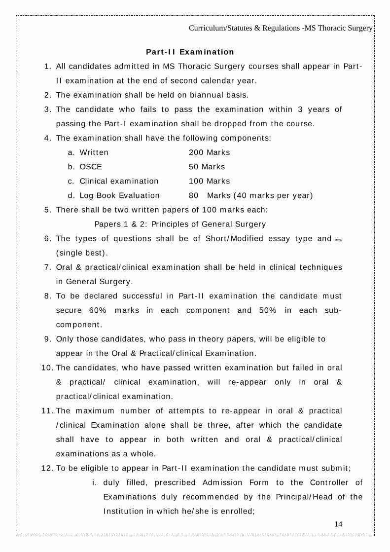
Part-II Examination
1. All candidates admitted in MS Thoracic Surgery courses shall appear in Part-
II examination at the end of second calendar year.
2. The examination shall be held on biannual basis.
3. The candidate who fails to pass the examination within 3 years of
passing the Part-I examination shall be dropped from the course.
4. The examination shall have the following components:
a. Written 200 Marks
b. OSCE 50 Marks
c. Clinical examination 100 Marks
d. Log Book Evaluation 80 Marks (40 marks per year)
5. There shall be two written papers of 100 marks each:
Papers 1 & 2: Principles of General Surgery
6. The types of questions shall be of Short/Modified essay type and MCQs
(single best).
7. Oral & practical/clinical examination shall be held in clinical techniques
in General Surgery.
8. To be declared successful in Part-II examination the candidate must
secure 60% marks in each component and 50% in each sub-
component.
9. Only those candidates, who pass in theory papers, will be eligible to
appear in the Oral & Practical/clinical Examination.
10. The candidates, who have passed written examination but failed in oral
& practical/ clinical examination, will re-appear only in oral &
practical/clinical examination.
11. The maximum number of attempts to re-appear in oral & practical
/clinical Examination alone shall be three, after which the candidate
shall have to appear in both written and oral & practical/clinical
examinations as a whole.
12. To be eligible to appear in Part-II examination the candidate must submit;
i. duly filled, prescribed Admission Form to the Controller of
Examinations duly recommended by the Principal/Head of the
Institution in which he/she is enrolled;
Curriculum/Statutes & Regulations -MS Thoracic Surgery
15
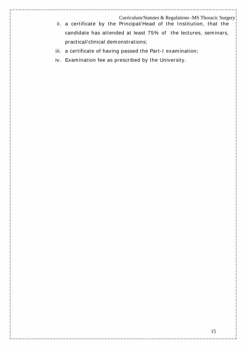
ii. a certificate by the Principal/Head of the Institution, that the
candidate has attended at least 75% of the lectures, seminars,
practical/clinical demonstrations;
iii. a certificate of having passed the Part-I examination;
iv. Examination fee as prescribed by the University.
Curriculum/Statutes & Regulations -MS Thoracic Surgery
16
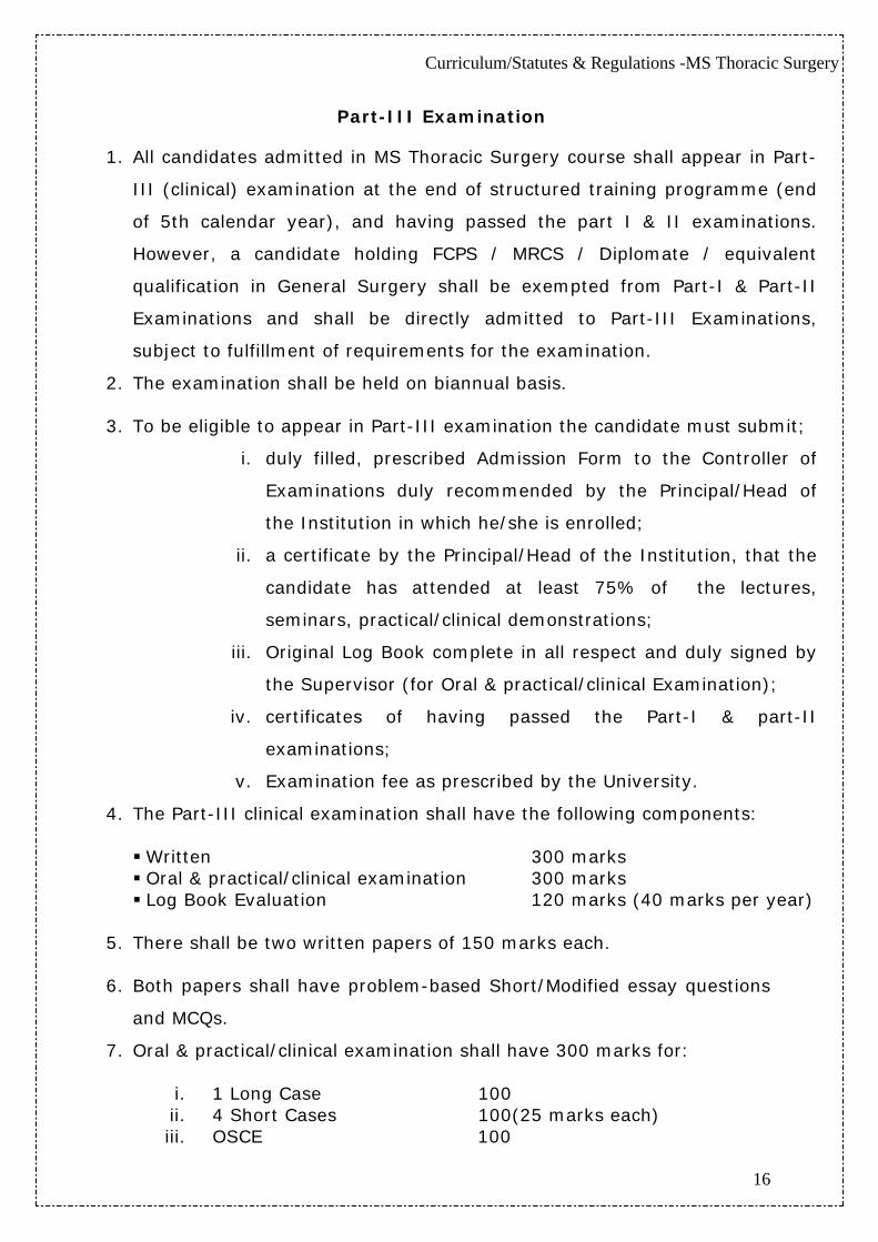
Part-III Examination
1. All candidates admitted in MS Thoracic Surgery course shall appear in Part-
III (clinical) examination at the end of structured training programme (end
of 5th calendar year), and having passed the part I & II examinations.
However, a candidate holding FCPS / MRCS / Diplomate / equivalent
qualification in General Surgery shall be exempted from Part-I & Part-II
Examinations and shall be directly admitted to Part-III Examinations,
subject to fulfillment of requirements for the examination.
2. The examination shall be held on biannual basis.
3. To be eligible to appear in Part-III examination the candidate must submit;
i. duly filled, prescribed Admission Form to the Controller of
Examinations duly recommended by the Principal/Head of
the Institution in which he/she is enrolled;
ii. a certificate by the Principal/Head of the Institution, that the
candidate has attended at least 75% of the lectures,
seminars, practical/clinical demonstrations;
iii. Original Log Book complete in all respect and duly signed by
the Supervisor (for Oral & practical/clinical Examination);
iv. certificates of having passed the Part-I & part-II
examinations;
v. Examination fee as prescribed by the University.
4. The Part-III clinical examination shall have the following components:
Written 300 marks Oral & practical/clinical examination 300 marks Log Book Evaluation 120 marks (40 marks per year)
5. There shall be two written papers of 150 marks each.
6. Both papers shall have problem-based Short/Modified essay questions
and MCQs.
7. Oral & practical/clinical examination shall have 300 marks for:
i. 1 Long Case 100 ii. 4 Short Cases 100(25 marks each) iii. OSCE 100
Curriculum/Statutes & Regulations -MS Thoracic Surgery
17
8. To be declared successful in Part-III examination the candidate must
secure 60% marks in each component and 50% in each sub-component.
9. Only those candidates, who pass in theory papers, will be eligible to
appear in the Oral & Practical/ Clinical Examination.
10. The candidates, who have passed written examination but failed in
Oral & Practical/ Clinical Examination, will re-appear only in Oral &
Practical / Clinical examination.
11. The maximum number of attempts to re-appear in oral & practical
/clinical Examination alone shall be three, after which the candidate
shall have to appear in both written and oral & practical/clinical
examinations as a whole.
12. The candidate with 80% or above marks shall be deemed to have
passed with distinction.
13. Log Book/Assignments: Through out the length of the course, the
performance of the candidate shall be recorded on the Log Book.
14. The Supervisor shall certify every year that the Log Book is being
maintained and signed regularly.
15. The Log Book will be developed & approved by the Advanced Studies &
Research Board.
16. The evaluation will be maintained by the Supervisor (in consultation
with the Co- Supervisor, if appointed).
17. The performance of the candidate shall be evaluated on annual basis,
e.g., 40 marks for each year in five years MS Thoracic Surgery course.
The total marks for Log Book shall be 200.The log book shall reflect the
performance of the candidate on following parameters:
• Year wise record of the competence of skills.
• Year wise record of the assignments.
• Year wise record of the evaluation regarding attitude & behaviour
• Year wise record of journal club / lectures / presentations / clinico-
pathologic conferences attended & / or made by the candidate.
Curriculum/Statutes & Regulations -MS Thoracic Surgery
18
3. Submission / Evaluation of Synopsis
1. The candidates shall prepare their synopsis as per guidelines provided
by the Advanced Studies & Research Board, available on UHS website.
2. The research topic in clinical subject should have 30% component
related to basic sciences and 70% component related to applied
clinical sciences. The research topic must consist of a reasonable
sample size and sufficient numbers of variables to give training to the
candidate to conduct research, to collect & analyze the data.
3. Synopsis of research project shall be submitted by the end of the 3rd
year of MS program. The synopsis after review by an Institutional
Review Committee, shall be submitted to the University for
consideration by the Advanced Studies & Research Board, through the
Principal / Dean /Head of the institution.
4. Submission of Thesis
1. Thesis shall be submitted by the candidate duly recommended by the
Supervisor.
2. The minimum duration between approval of synopsis and submission
of thesis shall be one year, but the thesis can not be submitted later
than 8 years of enrolment.
3. The research thesis must be compiled and bound in accordance with
the Thesis Format Guidelines approved by the University and available
on website.
4. The research thesis will be submitted along with the fee prescribed by
the University.
5. Thesis Examination
1. All candidates admitted in MS course shall appear in Part-III thesis
examination at the end of 5th year of their training course.
2. Only those candidates shall be eligible for thesis evaluation who
have passed Part I, II & III (clinical) Examinations.
3. The examination shall include thesis evaluation with defense.
Curriculum/Statutes & Regulations -MS Thoracic Surgery
19
4. The Vice Chancellor shall appoint three external examiners for thesis
evaluation, preferably from other universities and from abroad, out of
the panel of examiners approved by the Advanced Studies & Research
Board. The examiners shall be appointed from respective specialty.
Specialists from General Surgery and Allied surgical Disciplines may
also be appointed/co-opted, where deemed necessary.
5. The thesis shall be sent to the external examiners for evaluation,
well in time before the date of defense examination and should be
approved by all the examiners.
6. After the approval of thesis by the evaluators, the thesis defense
examination shall be held within the University on such date as
may be notified by the Controller of Examinations. The
Controller of Examinations shall make appropriate arrangements
for the conduct of thesis defense examination in consultation
with the supervisor, who will co-ordinate the defense examination.
7. The thesis defense examination shall be conducted by two External
Examiners who shall submit a report on the suitability of the
candidate for the award of degree. The supervisor shall act as
coordinator.
6. Award of MS Thoracic Surgery Degree
After successful completion of the structured courses of MS Thoracic Surgery and
qualifying Part-I, Part-II and Part-III examinations, the degree with title MS
Thoracic Surgery hall be awarded.
Curriculum/Statutes & Regulations -MS Thoracic Surgery
20
CONTENT OUTLINE
Part I MS Thoracic Surgery
Basic Sciences: Student is expected to acquire comprehensive knowledge of Anatomy, Physiology, Pathology (Microbiology), Biochemistry, Pharmacology relevant to surgical practice appropriate for Thoracic Surgery
1. Anatomy Cell Biology: Cytoplasm – Cytoplasmic matrix, cell membrane, cell organelles, cytoskeleton, cell inclusions, cilia and flagella.
Nucleus – nuclear envelope, nuclear matrix, DNA and other components of chromatin, protein synthesis, nucleolus, nuclear changes indicating cell death.
Cell cycle, mitosis, meiosis, cell renewal. Cellular differentiation and proliferation. Tissues of Body: Light and electron microscopic details and structural basis of function, regeneration and degeneration. Confocal microscopy.
The systems/organs of body – Cellular organization, light and electron microscopic features, structure function correlations, and cellular organization.
Structure of the Thoracic Wall Anterior chest wall Posterior chest wall Lines of orientation Sternum Costal cartilages Ribs Diaphragm Intercostal spaces Intercostal muscles Intercostal arteries and veins Intercostal nerves Suprapleural membrane Endothoracic fascia Major thoracic arteries and veins Muscles of the thoracic wall
The Thoracic Cavity Basic anatomy Mediastinum Contents of the anterior, posterior, middle, superior and inferior mediastinum
Relations of the contents of the mediastinum Pleurae
Curriculum/Statutes & Regulations -MS Thoracic Surgery
21
Blood, lymphatic and nerve supply of the pleura Normal anatomy of the diaphragm Origins and insertions of muscles Vascular and neural supply Foramina of the diaphragm Esophageal Vascular Morgagni and Bochdalek Contiguous structures
Esophagus
Blood supply Nerve supply Sphincters Muscular composition Mucosa
Heart External anatomy Valves Cusps Thoracic Muscle Conducting System Coronary Circulation Lymph Drainage and Nerve supply of the Heart The general structure of arteries, veins, and microcirculation related to
heart Upper Respiratory Tract Blood, lymphatic and nerve supply of the larynx, trachea and bronchi Muscles of the larynx and trachea
Lower Respiratory Tract Bronchopulmonary segments Lungs Bronchioles, alveoli Blood supply, lymph drainage and nerve supply of the lungs
Salient Features of the Embryology of the Thoracic cavity and its contents
2. Physiology
Normal physiology of the lung Chest wall mechanics Properties of thoracic muscle Thoracic muscle: electrical and mechanical properties. Large and small airway mechanics Alveolar mechanics and gas exchange
Curriculum/Statutes & Regulations -MS Thoracic Surgery
22
Pulmonary function tests Flow volume loops Non respiratory functions of lung Physiology of the esophagus Normal peristalsis Hormonal influences Neural influences Assessment of the esophagus Contrast studies Manometry pH studies Physiologic anatomy of the heart, the atria, ventricles, pericardium and myocardium
The cardiac cycle ; heart sounds Regulation of cardiac function. The normal electrocardiogram and characters of its various components. Physiology and abnormalities of apex beat. Functional classification of blood vessels Peripheral circulation: pressure and resistance Physiology of perfusion Physiology of hypothermia Acid-base balance
3. Biochemistry
Membrane biochemistry and signal transduction Gene expression and the synthesis of proteins Bioenergetics; fuel oxidation and the generation of ATP Enzymes and biologic catalysis Tissue metabolism
VITAMINS Classification, components, sources, absorption and functions
(physiological and biochemical role). Daily requirements, effects of deficiency and hypervitaminosis. Salient morphologic features of diseases related to deficiency or excess
of vitamins. MINERALS Sources of calcium, phosphorous, iron, iodine, fluorine, magnesium
and manganese. Trace elements and their clinical importance. Absorption and factors required for it. Functions and fate.
METABOLISM Metabolic rate and basal metabolic rate Factors influencing metabolic rate, principles of measurement.
Carbohydrates Classification and dietary sources. Digestion, absorption and utilization of dietary carbohydrates. Glucose
tolerance test.
Curriculum/Statutes & Regulations -MS Thoracic Surgery
23
Glycogenesis, glycolysis, gluconeogenesis, glycogenolysis, processes with the steps involved and effects of hormones.
Citric acid cycle, steps involved, its significance and the common final metabolic pathway.
Hexose monophosphate shunt: mechanism and significance. Lipids Classification of simple, derived and compound lipids. Dietary sources. Digestion, absorption, utilization and control. Fatty acid oxidation with steps involved. Ketogenesis and its significance. Lipotropic factors and their actions. Lipoproteins, types and
importance. Proteins and Amino Acids Classification and dietary sources of proteins. Digestion, absorption, utilization and control. Fate of amino acids. Urea formation with steps involved. Functions and effects of deficiency.
Nucleoproteins: Structure and metabolism. Pigment Metabolism Basic concept of endogenous and exogenous pigments. Causes of pigmentation and depigmentation.
Disorders of pigment metabolism, inherited disorders, acquired
disorders from deficiency or excess of vitamins, minerals, fats, carbohydrates, proteins etc.
BALANCED DIET Requisites of an adequate diet. Role of carbohydrates, fats, proteins, minerals, vitamins and water in
diet. Principles of nutrition as applied to medical problems
Biotechnology and concepts of molecular biology with special emphasis on use of recombinant DNA techniques in medicine and the molecular biology of cancer
4. Pathology
Pathological alterations at cellular and structural level along with brief introduction to Microbiology related to surgical procedures and Haematological pathology as related to Thoracic surgery: Cell Injury and adaptation
Reversible and Irreversible Injury Fatty change, Pathologic calcification Necrosis and Gangrene Cellular adaptation Atrophy, Hypertrophy,
Curriculum/Statutes & Regulations -MS Thoracic Surgery
24
Hyperplasia, Metaplasia, Aplasia Inflammation
Acute inflammation Cellular components and chemical mediators of acute inflammation Exudates and transudate Sequelae of acute inflammation Chronic inflammation Etiological factors and pathogenesis Distinction between acute and chronic (duration) inflammation Histologic hallmarks Types and causes of chronic inflammation, non-granulomatous & granulomatous
Haemodynamic disorders Etiology, pathogenesis, classification and morphological and clinical
manifestations of Edema, Haemorrhage, Thrombosis, Embolism, Infarction & Hyperaemia
Shock; classification etiology, and pathogenesis, manifestations. Compensatory mechanisms involved in shock Pathogenesis and possible consequences of thrombosis Difference between arterial and venous emboli
Neoplasia Dysplasia and Neoplasia Benign and malignant neoplasms Etiological factors for neoplasia Different modes of metastasis Tumor staging system and tumor grade
Immunity and Hypersensitivity Immunity Immune response Diagnostic procedures in a clinical Immunology laboratory Protective immunity to microbial diseases Tumour immunology Immunological tolerance, autoimmunity and autoimmune diseases. Transplantation immunology Hypersensitivity Immunodeficiency disorders Immunoprophylaxis & Immunotherapy
Haematology
Normal blood picture & variation in disease Related Microbiology
Role of microbes in various thoracic diseases Infection source Main organisms that cause chest organs diseases Surgically important micro-organisms in thoracic surgical procedures Nosocomial infections Pathogenic bacteria Vegetative organisms Spores
Curriculum/Statutes & Regulations -MS Thoracic Surgery
25
Important viruses Important parasites
Sterilization and disinfection Infection prevention
Immunization Personnel protection from communicable diseases Use of investigation and procedures in laboratory
Special Pathology
As related to cardiothoracic systems Lung: Allergy & lung Bronchial asthma Infectious conditions of lung Pneumonias Lung Abscess Pulmonary Tuberculosis Chronic obstructive pulmonary diseases Lung injury Adult respiratory distress syndrome (ARDS) Systemic diseases & lung Anomalies in lung Tumours Surgery & complications of lung Infections of lung
Pleura Pleural tumours Pleural infection Empyema
Oesophagus Congenital anomalies Oesophageal cancers Stricture oesophagus Achalasia cardia
Mediastinum Infections Tumours in mediastinum Thymus tumours
Pericardium Infections Pericardial tamponade
5. Pharmacology & Therapeutics
Introduction to Pharmacology Receptors Mechanisms of drug action Drug-receptor interactions Pharmacokinetic process
Absorption Distribution
Curriculum/Statutes & Regulations -MS Thoracic Surgery
26
Metabolism Elimination
Drug effect Beneficial responses Harmful responses Allergic responses Drug dependence, addiction Abuse and tolerance
Dosage forms and routes of administration Oral routes Parenteral routes Topical routes
The drug prescription Factors that influence drug effects Special considerations in elderly Special considerations in pediatric
6. Biostatistics & Research Methodology
Introduction to bio-statistics Introduction to bio- medical research Why research is important? What research to do?
Selecting a field for research Drivers for health research Participation in national and international research Participation in pharmaceutical company research Where do research ideas come from Criteria for a good research topic
Ethics in health research Writing a scientific paper Making a scientific presentation Searching the literature
7. Behavioural Sciences
Bio-psycho-social (bps) model of health care Use of non-medicinal interventions in clinical practice Communication skills Counseling Informational skills Crisis intervention/disaster management Conflict resolution Breaking bad news Medical ethics, professionalism and doctor-patient relationship Hippocratic oath Four pillars of medical ethics (autonomy, beneficence, non-malficence
and justice) Informed consent and confidentiality
Curriculum/Statutes & Regulations -MS Thoracic Surgery
27
Ethical dilemmas in a doctor’s life Delivery of culturally relevant care and cultural sensitivity Psychological aspects of health and disease Psychological aspect of health Psychological aspect of disease Stress and its management Psychological aspect of pain Psychological aspect of aging
Curriculum/Statutes & Regulations -MS Thoracic Surgery
28
Part II MS Thoracic Surgery
Fundamental Principles of Surgery
History of surgery Preparing a patient for surgery Principles of operative surgery: asepsis, sterilization and antiseptics Surgical infections and antibiotics Basic principles of anaesthesia and pain management Acute life support and critical care:
Pathophysiology and management of shock Fluids and electrolyte balance/ acid base metabolism Haemostasis, blood transfusion
Trauma: assessment of polytrauma, triage, basic and advanced trauma Accident and emergency surgery Wound healing and wound management Nutrition and metabolism Principles of burn management Principles of surgical oncology Principles of laparoscopy and endoscopy Organ transplantation Informed consent and medicolegal issues Molecular biology and genetics Operative procedures for common surgical manifestations e.g cysts, sinuses, fistula, abscess, nodules, basic plastic and reconstructive surgery
Principles of basic diagnostic and interventional radiography Principles and interpretation of conventional and advanced radiographic procedures
Common Surgical Skills
Incision of skin and subcutaneous tissue: o Langer’s lines o Healing mechanism o Choice of instrument o Safe practice Closure of skin and subcutaneous tissue: o Options for closure o Suture and needle choice o Safe practice Knot tying: o Choice of material o Single handed o Double handed o Superficial o Deep
Curriculum/Statutes & Regulations -MS Thoracic Surgery
29
Tissue retraction: o Choice of instruments o Placement of wound retractors o Tissue forceps
Use of drains: o Indications o Types o Insertion o Fixation o Management/removal Incision of skin and subcutaneous tissue: o Ability to use scalpel, diathermy and scissors Closure of skin and subcutaneous tissue: o Accurate and tension free apposition of wound edges Haemostasis: o Control of bleeding vessel (superficial) o Diathermy o Suture ligation o Tie ligation o Clip application o Plan investigations o Clinical decision making o Case work up and evaluation; risk management Pre-operative assessment and management: o Cardiorespiratory physiology o Diabetes mellitus o Renal failure o Pathophysiology of blood loss o Pathophysiology of sepsis o Risk factors for surgery o Principles of day surgery o Management of comorbidity Intraoperative care: o Safety in theatre o Sharps safety o Diathermy, laser use o Infection risks o Radiation use and risks o Tourniquets o Principles of local, regional and general anaesthesia Post-operative care: o Monitoring of postoperative patient o Postoperative analgesia o Fluid and electrolyte management o Detection of impending organ failure o Initial management of organ failure o Complications specific to particular operation o Critical care
Curriculum/Statutes & Regulations -MS Thoracic Surgery
30
Blood products: o Components of blood o Alternatives to use of blood products o Management of the complications of blood product transfusion
including children Antibiotics: o Common pathogens in surgical patients o Antibiotic sensitivities o Antibiotic side-effects o Principles of prophylaxis and treatment Safely assess the multiply injured patient: o History and examination o Investigation o Resuscitation and early management o Referral to appropriate surgical subspecialties Technical Skills o Central venous line insertion o Chest drain insertion o Diagnostic peritoneal lavage o Bleeding diathesis & corrective measures, e.g. warming, packing o Clotting mechanism; Effect of surgery and trauma on coagulation o Tests for thrombophilia and other disorders of coagulation o Methods of investigation for suspected thromboembolic disease o Anticoagulation, heparin and warfarin o Role of V/Q scanning, CT angiography and thrombolysis o Place of pulmonary embolectomy o Awareness of symptoms and signs associated with pulmonary embolism
and DVT o Role of duplex scanning, venography and d-dimer measurement o Initiate and monitor treatment
Diagnosis and Management of Common Paediatric Surgical Conditions:
• Child with abdominal pain • Vomiting child • Trauma • Chest wall pathologies
In terms of general experience it is expected that trainees would have gained exposure to the following procedures and to be able to perform those marked (*) under direct supervision.
Lymph node biopsy* Insertion of CV lines Excision of skin lesions*
• Emergency Procedures Incision and drainage of abscess* Insertion of pleural drain* Insertion of NG tube Thoracotomy*
Curriculum/Statutes & Regulations -MS Thoracic Surgery
31
Part III- MS Thoracic Surgery
Clinical Component 1. Didactic Training 2. Clinical Skills 3. Research and Thesis Writing
1. Chest Wall A. Anatomy, Physiology and Embryology Unit Objective: At the end of this unit the resident understands the anatomy, physiology, and embryology of the chest wall and interprets diagnostic tests. Learner Objectives: Upon completion of this unit the resident: 1. Understands the anatomy and physiology of the cutaneous, muscular, and bony components of the chest wall and their anatomic and physiologic relationships to adjacent structures. 2. Understands the anatomy of the vascular, neural, muscular, and bony components of the thoracic outlet. 3. Knows all operative approaches to the chest wall. 4. Knows the surgical anatomy, neural, vascular, and skeletal components of the chest wall, as well as the major musculocutaneous or pedicle flaps used in the chest. Contents: 1. Chest wall embryology
a. Ectodermal, mesodermal, endodermal 2. Chest wall anatomy
a. Skeletal b. Muscular c. Neural d. Vascular e. Relationships to adjacent structures
3. Diagnostic tests to define chest wall anatomy a. Chest x-ray b. CAT scans c. MRI scans d. Nuclear scans e. Pulmonary function tests
4. Major flaps of the chest wall and their vascular pedicles a. Latissimus dorsi b. Pectoralis major c. Serratus anterior d. Trapezius
Curriculum/Statutes & Regulations -MS Thoracic Surgery
32
e. Intercostal f. Pleural g. Pericardial fat pad h. Rectus abdominis i. Omental j. Vascularized rib graft
Clinical Skills: During the training program the resident: 1. Recognizes the normal and abnormal anatomy of the chest wall. 2. Reads and interprets tests to diagnose chest wall abnormalities. 3. Performs operations utilizing major chest wall flaps and the correct application of prosthetic materials. B. Acquired Abnormalities and Neoplasms Unit Objective At the end of this unit the resident understands acquired abnormalities and neoplasms of the chest wall and performs biopsy, incision, resection, reconstruction, and stabilization of the chest wall. Learner Objectives: Upon completion of this unit the resident: 1. Understands the diagnosis and management of various chest wall infections. 2. Evaluates and diagnoses primary and metastatic chest wall tumors, knows their histologic appearance, and understands the indications for incisional versus excisional biopsy. 3. Knows the radiologic characteristics of tumors. 4.Knows the indications for and methods of prosthetic chest wall reconstruction (e.g., methyl-methacrylate, Marlex®, Gortex®, Vicryl®, and Dacron® mesh). 5. Knows the types of chemotherapy and radiotherapy (induction neo-adjuvant and adjuvant therapy) of chest wall tumors and the indications for preoperative and postoperative therapy. 6. Knows the management of osteoradionecrosis of the chest wall. Contents: 1. Malignant neoplasms of the chest wall a. Chondrosarcoma b. Osteogenic sarcoma c. Malignant fibrous histiocytoma d. Rhabdomyosarcoma e. Lymphoma f. Myeloma g. Ewing's sarcoma h. Metastatic lesions i. Lung cancer invading the chest wall 2. Benign neoplasms of the chest wall
Curriculum/Statutes & Regulations -MS Thoracic Surgery
33
a. Fibrous dysplasia b. Chondroma c. Osteochondroma d. Eosinophilic granuloma Clinical Skills: During the training program the resident: 1. Performs a variety of surgical incisions to expose components of the chest wall and interior thoracic organs. 2. Performs surgical resections of primary and secondary chest wall tumors. 3. Identifies the need for major flaps of the chest wall. 4. Identifies the need for prosthetic replacement of the chest wall. 5. Performs surgical reconstruction of chest wall defects. C. Congenital Abnormalities and Thoracic Outlet Syndrome Unit Objective: At the end of this unit the resident understands congenital abnormalities, including those leading to thoracic outlet syndrome, and uses operative and non-operative therapy. Learner Objectives: Upon completion of this unit the resident: 1. Recognizes pectus excavatum and pectus carinatum, understands possible physiologic disturbances, and interprets diagnostic tests to identify such physiologic disturbances. 2. Understands the indications for the operative treatment of congenital chest wall abnormalities. 3. Knows the complications of reconstruction of congenital chest wall abnormalities and their management. 4. Understands the etiology, evaluation, differential diagnosis, and diagnostic criteria for thoracic outlet syndrome. 5. Knows the operative and non-operative management of thoracic outlet syndrome. Contents: 1. Pectus excavatum a. Components b. Evaluation and management (operative and non-operative) 1. Nuss procedure 2. Conventional repair (Ravitch) c. Plastic surgical alternatives 2. Pectus carinatum a. Components b. Evaluation and management (operative and non-operative) 3. Thoracic outlet anatomy a. Skeletal, muscular, vascular, neural 4. Diagnostic tests a. Clinical examination and physical exam b. Nerve conduction studies
Curriculum/Statutes & Regulations -MS Thoracic Surgery
34
c. Angiography d. CT scan e. MRI f. Non-invasive vascular studies 5. Forms of conservative management a. Physical therapy b. Weight reduction 6. Surgical management a. First rib resection (operative approaches) b. Cervical ribs c. Associated vascular abnormalities d. Management of intraoperative complications e. Re-operation Clinical Skills During the training program the resident: 1. Recognizes the varied presentations of thoracic outlet syndrome and interprets diagnostic tests. 2. Evaluates and treats patients with congenital chest wall malformations. 3. Reads and interprets diagnostic x-ray and performs physiologic examinations for congenital chest wall defects and thoracic outlet syndromes. 4. Performs the operative reconstruction of selected chest wall defects. 5. Performs first rib and cervical rib resection and repairs or releases vascular and neural abnormalities associated with thoracic outlet syndrome. 6. Manages intraoperative and postoperative complications associated with the repair of congenital chest wall abnormalities and thoracic outlet syndrome. 7. Performs re-operations for thoracic outlet syndrome.
2. Lungs & Pleura A. Anatomy, Physiology, Embryology and Testing Unit Objective: At the completion of this unit the resident understands the embryology and anatomy of the lungs and their relationship to adjacent structures, the physiology of airway mechanics, gas exchange, and blood flow, and applies the findings of invasive and non-invasive tests to patient management. Learner Objectives: Upon completion of this unit the resident: 1. Understands the segmental anatomy of the bronchial tree and bronchopulmonary segments. 2. Understands the arterial, venous and bronchial anatomy of the lungs and their inter-relationships. 3 Understands the lymphatic anatomy of the lungs, the major lymphatic nodal stations, and lymphatic drainage routes of the lung segments; including the anatomy of the thoracic duct. 4. Knows the indications for different thoracic incisions, the surgical anatomy encountered, and the physiological impact. 5. Knows the indications for plain radiography, CT scan, bone scan, magnetic resonance imaging, and PET scanning for staging of lung cancer.
Curriculum/Statutes & Regulations -MS Thoracic Surgery
35
6. Knows the indications, interpretation, and use of nuclear medicine ventilation/perfusion scanning (V/Q scan) to determine the operability of candidates for pulmonary resection; 7. Understands the methods of surgical staging (e.g., mediastinoscopy, Chamberlain procedure, scalene node biopsy, thoracoscopy). 8. Knows how to interpret pulmonary function tests. 9. Knows how to perform pulmonary function tests. Contents: 1. Normal anatomy and histology of the lung a. Segmental anatomy of the bronchial tree b. Bronchopulmonary segments (topography) c. Hilar anatomy d. Lymphatic anatomy and drainage of the lung e. Histologic anatomy and cell types of the lung f. Endoscopic anatomy of the larynx, trachea, and bronchi 2. Normal physiology of the lung a. Chest wall mechanics b. Large and small airway mechanics c. Alveolar mechanics and gas exchange 3. Imaging a. Chest x-ray b. CT scan of the chest and abdomen c. MRI of the chest d. Contrast angiography of major vessels within the chest e. Radioactive isotope scanning of organs within the chest 4. Surgical approaches a. Anterior thoracotomy b. Posterolateral thoracotomy c. Posterior thoracotomy d. Muscle sparing thoracotomy e. Mediastinotomy f. Transverse anterior sternotomy g. Incisions common to video assisted thoracic surgery h. Incisions common to cervical and anterior mediastinoscopy Clinical Skills: During the training program the resident: 1. Reads and interprets pulmonary function studies, ventilation/perfusion scans, pulmonary arteriograms and arterial blood gases, and correlates the results with operability. 2. Applies knowledge of thoracic anatomy to the physical examination of the chest, heart, and vascular tree. 3. Applies knowledge of thoracic anatomy to flexible and rigid endoscopy. 4. Uses knowledge of chest, pulmonary, and cardiac physiology to interpret tests involving the thoracic cavity and to understand and treat diseases of the chest and its contents. 5. Reads and interprets plain radiography, CT scans, magnetic resonance imaging, and PET scanning of the chest. 6. Participates in the performance of exercise tolerance tests and pulmonary
Curriculum/Statutes & Regulations -MS Thoracic Surgery
36
function tests. B. Non-Neoplastic Lung Diseases Unit Objective: At the end of this unit the resident understands infectious, inflammatory, and environmental conditions of the lung and performs operative and non-operative management. Learner Objectives: Upon completion of this unit, the resident: 1. Understands diagnostic procedures used to evaluate non-neoplastic lung disease. 2. Knows the common pathogens that produce lung infections, including their presentation and pathologic processes, and knows the treatment and indications for operative intervention. 3. Understands the natural history, presentation and treatment of chronic obstructive lung disease. 4. Knows the indications for bullectomy, lung reduction, and pulmonary transplantation. 5. Understands the pathologic results and alterations of pulmonary function due to bronchospasm. 6. Understands the principles of surgical resection for non-neoplastic lung disease. 7. Understands the mechanisms by which foreign bodies reach the airways, how they cause pulmonary pathology, and the management of patients with airway foreign bodies. 8. Understands the causes, physiology, evaluation and management of hemoptysis. 9. Knows the complications of lung resection and their management. Contents: 1. Common pulmonary pathogens a. Bacteria b. Fungi c. Mycobacterial (tuberculoisis and atypical [MOTT]) d. Viruses e. Protozoa f. Immunocompromised patients 2. Chronic obstructive pulmonary disease a. Natural history b. Presentation, evaluation c. Alteration of lung function d. Complications requiring operative treatment e. Treatment (operative and non-operative) 3. Bronchospasm a. Natural history b. Evaluation c. Complications requiring operative treatment d. Treatment (operative and non-operative) 4. Foreign bodies of the lung and airways
Curriculum/Statutes & Regulations -MS Thoracic Surgery
37
a. Common types b. Causes, pathology c. Evaluation d. Treatment (operative and non-operative) 5. Hemoptysis a. Causes b. Physiologic derangements c. Evaluation d. Treatment (operative and non-operative) 6. Pneumothorax a. Etiology b. Indications for treatment c. Types of treatment Clinical Skills: During the training program the resident: 1. Diagnoses and treats patients with bacterial, fungal, tuberculous, and viral lung infections. 2. Performs operative and non-operative management of lung abscess. 3. Performs resections of lung and bronchi in patients with non-neoplastic lung disease. 4. Manages patients with chronic obstructive lung disease, bronchospastic airway disease, foreign bodies of the airways, and hemoptysis. 5. Performs thoracentesis, mediastinoscopy, mediastinotomy, flexible and rigid bronchoscopy, thoracoscopy, and open lung biopsy. 6. Performs bronchoalveolar lavage and transbronchial lung biopsy. C. Neoplastic Lung Diseases Unit Objective: At the end of this unit the resident understands the etiology, natural history, pathology, evaluation, and management of lung neoplasms, and performs operative and nonoperative treatment. Learner Objectives: Upon completion of this unit the resident: 1. Understands TNM staging of lung cancer and its application to the diagnosis, therapeutic planning, and management of patients with lung cancer. 2. Evaluates and diagnoses neoplasia of the lung, using a knowledge of the histologic appearance of the major types. 3. Knows the signs of inoperability. 4. Understands the therapeutic options for patients with lung neoplasms. 5. Understands the principles of bronchoplastic surgery. 6. Understands the complications of pulmonary resection and their management. 7. Understands the role of induction and adjuvant therapy for lung neoplasms. 8. Understands the indications for resection of benign lung neoplasms. 9. Understands the indications for resection of pulmonary metastases.
Curriculum/Statutes & Regulations -MS Thoracic Surgery
38
Contents: 1. Benign tumors of the lung and airways a. Pathology, biologic behavior b. Evaluation, diagnosis, treatment (operative and non-operative) 2. Solitary lung nodule a. Differential diagnosis, evaluation, diagnostic techniques b. Treatment (operative and non-operative) 3. Malignant tumors of the lung and airways a. Pathology, biologic behavior b. Evaluation, diagnosis, treatment (operative and non-operative) 4. Metastatic tumors to the lungs a. Pathology and biologic behavior b. Evaluation, diagnosis, treatment (operative and non-operative) Clinical Skills: During the training program the resident: 1. Evaluates patients with lung neoplasia and recommends therapy based on their functional status, pulmonary function and tumor type. 2. Performs staging procedures (e.g., bronchoscopy, mediastinoscopy, mediastinotomy, and thoracoscopy). 3. Performs operations to extirpate neoplasms of the lung (e.g., local excision, wedge resection, segmental resection, lobectomy, pneumonectomy, sleeve lobectomy, carinal resection, chest wall resection). 4. Recognizes and manages complications of pulmonary resections (e.g., space problem, persistent air leak, bronchopleural fistula, bronchovascular fistula, empyema, cardiac arrhythmia). 5. Performs bedside bronchoscopies and placement of tracheostomies and/or minitracheostomies. 6. Recognizes and treats the early signs of non-cardiac pulmonary edema. D. Congenital Lung Diseases Unit Objective: At the end of this unit the resident understands the embryology, pathology and principles of management of congenital lung abnormalities and performs appropriate treatment. Learner Objectives: Upon completion of this unit the resident: 1. Recognizes various congenital lung abnormalities and understands their anatomy and indications for treatment. Contents: 1. Pulmonary sequestration a. Presentation (intralobar and extralobar) b. Evaluation and management c. Prognosis 2. Congenital lobar emphysema a. Presentation and physiology b. Evaluation and management
Curriculum/Statutes & Regulations -MS Thoracic Surgery
39
3. Cystic fibrosis a. Presentation and physiology b. Evaluation and management c. Complications and their management d. Role of pulmonary transplantation 4. Bronchogenic cysts a. Presentation b. Evaluation and indications for operation c. Operative options 5. Cystic adenomatoid malformation a. Presentation and physiology b. Evaluation and indications for operation c. Operative options Clinical Skills: During the training program the resident: 1. Evaluates patients with congenital lung abnormalities. 2. Performs operations for congenital lung abnormalities and their complications. E. Diseases of the Pleura Unit Objective: At the end of this unit the resident understands the benign and malignant abnormalities of the pleura, pleural effusions, and the evaluation and treatment of pleural diseases. Learner Objectives: Upon completion of this unit the resident: 1. Is familiar with the clinical presentation of benign and malignant diseases of the pleura. 2. Understands the types of pleural effusions, their evaluation and treatment. including chylothorax. 3.Understands the management of empyema with and without bronchopleural fistula. 4. Understands the indications, contraindications, and complications of video assisted thoracic surgery and has a working knowledge of the equipment. 5. Understands the treatment of benign and malignant diseases of the pleura. Contents: 1. Mesothelioma a. Pathology, biologic behavior, and natural history b. Treatment (operative and non-operative) 2. Pleural effusions a. Types b. Diagnosis c. Treatment (operative and non-operative) 3. Empyema a. Presentation with and without bronchopleural fistula b. Diagnosis c. Treatment (operative and non-operative) d. Surgical options (e.g., thoracentesis, tube thoracostomy, decortication,
Curriculum/Statutes & Regulations -MS Thoracic Surgery
40
rib resection, repair of bronchopleural fistula) Clinical Skills: During the training program the resident: 1. Evaluates pleural effusions and recommends appropriate therapy. 2. Performs invasive diagnostic studies (e.g., incisional and excisional biopsy, needle biopsy, fluid analysis). 3. Places tube thoracostomies and performs chemical or mechanical pleurodesis. 4. Performs initial drainage procedures and subsequent procedures for empyema (e.g., decortication, empyemectomy, rib resection, Eloesser flap, Clagett procedure, closure of bronchopleural fistula). 5. Performs video assisted thoracoscopic surgery as necessary for the diagnosis and treatment of pleural disease. 6. Places pleuroperitoneal shunts. 7. Performs pleurectomy for mesothelioma.
3. Trachea & Bronchi A. Anatomy, Physiology and Embryology Unit Objective: At the end of this unit the resident understands the anatomy, blood supply, physiology, and embryology of the trachea and bronchi and applies findings of radiography, pulmonary function tests, and endoscopy to patient care. Learner Objectives: Upon completion of this unit the resident: 1. Understands the anatomy and blood supply of the trachea and bronchi. 2. Understands the endoscopic anatomy of the nasopharynx, hypopharynx, larynx, trachea, and major bronchi. 3. Understands and interprets pulmonary function studies of the trachea and bronchi. 4. Understands the radiologic assessment of the trachea and bronchi. Contents: 1. Trachea a. Blood supply b. Histologic and gross anatomy c. Lymphatic anatomy and drainage d. Contiguous structures e. Radiographic anatomy and tests f. Endoscopic anatomy and tests 2. Bronchi a. Blood supply b. Histologic and gross anatomy c. Segmental anatomy d. Lymphatic relationships e. Radiographic anatomy and tests f. Endoscopic anatomy and tests 3. Physiologic evaluation
Curriculum/Statutes & Regulations -MS Thoracic Surgery
41
a. Pulmonary function tests b. Flow volume loops 4. Radiologic evaluation a. Plain radiographs b. Tomography c. CT scan d. Fluoroscopy e. MRI f. Barium swallow Clinical Skills: During the training program the resident: 1. Interprets plain radiographic analyses, CT scan, MRI, and pulmonary function studies involving the trachea and bronchi. 2. Performs endoscopy of the upper airway, trachea and major bronchi. B. Congenital and Acquired Abnormalities Unit Objective: At the end of this unit the resident understands congenital and acquired diseases of the trachea and adjacent structures, knows the physiology of tracheal abnormalities, and performs operative and non-operative management. Learner Objectives: Upon completion of this unit the resident: 1. Understands congenital abnormalities and idiopathic diseases of the trachea. 2. Understands the etiology, presentation and management of acquired tracheal and bronchial strictures and their prevention. 3. Understands the etiology, presentation and management of tracheoesophageal fistulas and tracheoinnominate artery fistulas. 4. Knows the operative approaches to the trachea and techniques of mobilization. 5. Knows the methods of airway management, anesthesia and ventilation for tracheal operations. 6. Knows the principles of tracheal surgery and release maneuvers. 7. Understands the complications of tracheal surgery and their management. 8. Understands the etiology, presentation, and principles of airway trauma management. 9. Understands the radiologic evaluation of tracheal abnormalities. Contents: 1. Radiologic assessment of the trachea and bronchi a. Plain x-rays b. CT scans c. MRI d. Barium swallow 2. Stricture of the trachea a. Post-intubation
Curriculum/Statutes & Regulations -MS Thoracic Surgery
42
b. Post-tracheostomy c. Post-traumatic 3. Strictures of the bronchi a. Transplant b. Stricture after sleeve resection c. Histoplasmosis 4. Anesthesia for tracheal operations a. Methods of airway control b. Extubation concerns 5. Operative approaches to the trachea a. Reconstruction of the upper trachea b. Reconstruction of the lower trachea c. Mediastinal tracheostomy 6. Tracheostomy and its complications a. Tracheal stenosis b. Tracheo-esophageal fistula c. Tracheo-innominate artery fistula d. Persistent tracheal stoma 7. Airway trauma a. Airway control b. Evaluation of associated injuries c. Principles of repair (primary and secondary) d. Protecting tracheostomies 8. Tracheomalacia, Bronchomalacia a. Diagnosis b. Strategies for management (operative and non-operative) Clinical Skills: During the training program the resident: 1. Evaluates diagnostic tests of the trachea and bronchi. 2. Performs laryngoscopy and bronchoscopy of the trachea and bronchi, including dilation of stenoses. 3. Performs tracheostomy 4. Evaluates patients for tracheal resection and plans the operation. 5. Performs tracheal resection and reconstruction for tracheal stenosis. 6. Performs placement of tracheal T-tubes. 7. Performs the operations for tracheo-esophageal fistula, tracheo-innominate fistula, subglottic stenosis, and traumatic airway injury. 8. Is knowledgeable regarding the indications and technique for placement of tracheobronchial stents. C. Neoplasms Unit Objective: At the end of this unit the resident has a working knowledge of neoplasms affecting the trachea and adjacent structures, and performs operative and non-operative management. Learner Objectives: Upon completion of this unit the resident:
Curriculum/Statutes & Regulations -MS Thoracic Surgery
43
1. Knows the types, histology, and clinical presentation of tracheal neoplasms; 2. Understands the radiologic evaluation and operative management of tracheal neoplasms; 3. Understands the methods of airway management; 4. Knows the indications for and the use of radiotherapy and chemotherapy. Contents: 1. Neoplasms of the trachea a. Benign b. Malignant c. Metastatic 2. Operative techniques a. Resection of tracheal tumors b. Methods of tracheal reconstruction c. Operative approaches 3. Prosthetics a. Silastic prosthetics b. Stents c. Types of tracheostomy tubes and tracheal T-tubes 4. Airway management a. Bronchoscopic “core out” b. Laser c. photodynamic therapy Clinical Skills: During the training program the resident: 1. Performs rigid and flexible bronchoscopy for diagnosis and “core-out”. 2. Performs resection of tracheal tumors. 3. Manages patients and their airways after tracheal resection. 4. Uses laser techniques in the management of endoluminal tumors. 5. Uses stents, tracheal T-tubes and tracheostomy tubes in the management of tracheal neoplasms. 6. Uses adjunctive therapy for the management of tracheal tumors.
4. Mediastinum & Pericardium A. Anatomy, Physiology and Embryology Unit Objective: At the end of this unit the resident understands the anatomy, physiology and embryology of the mediastinum and pericardium, the relationships of adjacent structures, and applies findings of invasive and non-invasive tests to patient management. Learner Objectives: Upon completion of this unit the resident: 1. Understands the anatomic boundaries of the mediastinum and the structures found within each region. 2. Understands the embryologic development of structures within the mediastinum and the variations and pathologic consequences of abnormally
Curriculum/Statutes & Regulations -MS Thoracic Surgery
44
located structures. 3. Understands the radiologic assessment of the mediastinum including CT scan, MRI, contrast studies, and angiography. 4. Understands pericardial pathology and it’s effect on cardiac physiology. Contents: 1. Anterior mediastinum a. Major structures b. Diagnostic studies 2. Middle mediastinum (visceral compartment) a. Major structures b. Diagnostic studies 3. Posterior mediastinum (paravertebral sulcus) a. Major structures b. Diagnostic studies Clinical Skills: During the training program the resident: 1. Reads and interprets mediastinal plain radiographs, CT scans, MRI, and contrast studies; 2. Applies knowledge of mediastinal anatomy and physiology to the diagnosis of mediastinal abnormalities; 3. Applies knowledge of pericardial physiology to the differential diagnosis of pericardial vs. myocardial abnormalities. B. Congenital Abnormalities of the Mediastinum Unit Objective: At the end of this unit the resident understands congenital mediastinal abnormalities and performs operative and non-operative management. Learner Objectives: Upon completion of this unit the resident: 1. Is able to diagnose mediastinal cysts. 2. Is familiar with the symptoms associated with mediastinal abnormalities. 3. Knows the indications for operations involving the mediastinum and the anatomic approaches. Contents: 1. Mediastinal cysts a. Anterior 1. Cystic hygroma b. Middle 1. Pericardial cysts 2. Bronchogenic cysts c. Posterior 1. Esophageal duplications 2. Neurogenic tumors 2. Symptoms of mediastinal abnormalities
Curriculum/Statutes & Regulations -MS Thoracic Surgery
45
3. Management (operative and non-operative) Clinical Skills: During the training program the resident: 1. Reads and interprets plain radiographs, CT scans, MRI's and contrast studies of congenital abnormalities of the mediastinum. 2. Diagnoses and manages patients with congenital abnormalities of the mediastinum. 3. Performs operations for congenital abnormalities of the mediastinum. C. Acquired Abnormalities of the Mediastinum Unit Objective: At the end of this unit the resident knows the differential diagnosis of mediastinal abnormalities and performs operative and non-operative treatment. Learner Objectives: Upon completion of this unit the resident: 1. Understands mediastinal infections and their management. 2. Understands the diagnostic tests available. 3. Understands evaluation and management of myasthenia gravis a. Relationship to thymoma b. Laboratory testing c. Indications for surgery d. Perioperative management e. Surgical techniques 4. Recognizes the histologic appearance of benign and malignant mediastinal neoplasms. 5.Understands the neoplastic and non-neoplastic mediastinal diseases. 6.Understands the operative management of benign and malignant mediastinal neoplasms. 7.Understands chemotherapy and radiotherapy in mediastinal neoplasm management. Contents: 1. Anterior mediastinal tumors a. Thymoma 1. Histologic appearance 2. Management b. Thyroid 1. Histologic appearance 2. Management c. Teratoma 1. Histologic appearance 2. Management d. Lymphoma 1. Histologic appearance 2. Management
Curriculum/Statutes & Regulations -MS Thoracic Surgery
46
e. Germ cell tumor 1. Histologic appearance 2. Management 2. Middle mediastinal tumors a. Lymphoma 1. Histologic appearance 2. Management b. Hamartoma 1. Histologic appearance 2. Management c. Cardiac tumors 1. Histologic appearance 2. Management 3. Posterior mediastinum (paravertebral sulcus) a. Neurilemoma 1. Histologic appearance 2. Management b. Neurofibroma 1. Histologic appearance 2. Management c. Pheochromocytoma 1. Histologic appearance 2. Management d. Ganglion neuroma 1. Histologic appearance 2. Management e. Dumbbell neurogenic tumor 1. Histologic appearance 2. Management 4. Mediastinal infection a. Postoperative b. Primary (Ludwig’s angina) c. Management (operative and non-operative) 5. Diagnostic tests a. Plain radiographs b. CT scans c. MRI d. Contrast studies e. Radionucleotide studies f. Ultrasound g. Fine needle aspiration h. Core biopsy i. Mediastinoscopy j. Serologic tests Clinical Skills: During the training program the resident 1. Performs diagnostic tests and operations on the mediastinum. 2. Diagnoses and manages mediastinal infection. 3. Recognizes the histologic appearance of mediastinal tumors.
Curriculum/Statutes & Regulations -MS Thoracic Surgery
47
4. Manages patients with mediastinal tumors. D. Congenital and Acquired Abnormalities of the Pericardium Unit Objective: At the end of this unit the resident understands pericardial diseases and performs operative and non-operative management. Learner Objectives: Upon completion of this unit the resident: 1. Understands the physiologic consequences of an enlarging increased pericardial effusion fluid and the techniques for diagnosis and management. 2. Understands the operative management of benign and malignant pericardial neoplasms. 3. Understands the physiologic consequences of pericardial constriction and the techniques for diagnosis and management. Contents: 1. Pericardial effusions a. Benign b. Malignant c. Diagnostic tests d. Management (operative and non-operative) 2. Constrictive pericarditis a. Infectious b. Postoperative c. Diagnostic tests to differentiate from restrictive disease d.Management (operative and non-operative) 3. Pericardial cysts and tumors a. Congenital cysts b. Benign tumors c. Malignant tumors d. Management (operative and non-operative) Clinical Skills: During the training program the resident: 1. Uses knowledge of abnormal pathophysiology to diagnose pericardial disease. 2. Evaluates and manages patients with pericardial cysts or tumors. 3. Performs diagnostic tests and therapeutic interventions for the treatment of pericardial tamponade, pericardial effusions, and constrictive pericardial disease.
5. Diaphragm A. Anatomy, Physiology and Embryology Unit Objective: At the end of this unit the resident understands the anatomy, physiology, and embryology of the diaphragm and its relationship to adjacent structures, and interprets radiographic studies.
Curriculum/Statutes & Regulations -MS Thoracic Surgery
48
Learner Objectives: Upon completion of this unit the resident: 1. Knows the embryologic origin of the diaphragm. 2. Understands the anatomy of the diaphragm and adjacent structures. 3. Understands the neural and vascular supply of the diaphragm and the pathologic consequences of injury. 4. Understands imaging studies for assessing the diaphragm; 5. Understands the consequences of incisions in the diaphragm; 6. Understands developmental anomalies of the diaphragm. Contents: 1. Normal anatomy of the diaphragm a. Origins and insertions b. Vascular and neural supply 2. Foramina of the diaphragm a. Esophageal b. Vascular c. Morgagni and Bochdalek 3. Contiguous structures a. Heart b. Lungs c. Vessels d. Chest wall Clinical Skills: During the training program the resident: 1. Uses knowledge of the normal anatomy and physiology of the diaphragm to treat primary or contiguous abnormalities. 2. Evaluates and interprets radiographic studies of the diaphragm, including fluoroscopy, CT scan, and MRI. B. Acquired Abnormalities, Neoplasms Unit Objective: At the end of this unit the resident understands acquired abnormalities of the diaphragm including traumatic injuries, inflammation, diaphragmatic paralysis and neoplasms, and performs the appropriate treatment. Learner Objectives: Upon completion of this unit the resident: 1. Understands the presentation of diaphragmatic rupture and associated injuries. 2. Knows evaluation methods for penetrating injuries of the diaphragm. 3. Knows management of infections immediately above and below the diaphragm. 4. Understands the etiology, presentation, diagnosis, and management of acquired diaphragmatic hernias. 5. Understands the etiology, diagnosis, and treatment of diaphragmatic paralysis.
Curriculum/Statutes & Regulations -MS Thoracic Surgery
49
6. Understands the primary and secondary tumors of the diaphragm and their management. 7. Understands reconstruction methods for the diaphragm. 8. Understands the indications for and techniques of diaphragmatic pacing. Contents: 1. Diaphragmatic rupture a. Clinical presentation b. Physiologic effects c. Operative management d. Management of associated injuries 2. Periphrenic abscess a. Clinical presentation b. Physiologic effects c. Operative management 3. Acquired diaphragmatic hernias a. Esophageal b. Eventration c. Treatment 4. Tumors of the diaphragm a. Mesenchymal origin (benign and malignant) b. Neurogenic (benign and malignant) c. Secondary (lung, esophageal, mesothelioma) d. Treatment 5. Paralysis of the diaphragm a. Causes b. Diagnosis c. Treatment Clinical Skills: During the training program the resident: 1. Interprets plain and contrast x-rays, fluoroscopy, CT scans, and MRI of the diaphragm. 2. Performs operative repair of acquired diaphragmatic abnormalities and provides preoperative and postoperative care. 3. Reconstructs defects of the diaphragm. 4. Performs diagnostic studies of the diaphragm (e.g., pneumoperitoneum, direct incisional and excisional biopsy, video assisted thoracoscopic surgery). 5. Performs diaphragmatic mobilization for exposure of the spine and aorta; 6. Performs operative removal of diaphragmatic tumors; 7. Inserts permanent diaphragmatic pacemakers. C. Congenital Abnormalities Unit Objective: At the end of this unit the resident understands congenital diaphragmatic abnormalities and their pathologic effects, and provides operative and non-operative management. Learner Objectives:
Curriculum/Statutes & Regulations -MS Thoracic Surgery
50
Upon completion of this unit the resident: 1. Understands the anatomy of congenital diaphragmatic hernias. 2. Understands the physiologic consequences of diaphragmatic hernias. 3. Knows the indications for operative repair of diaphragmatic hernias. 4. Diagnoses and manages infants and adults with diaphragmatic hernias. Contents: 1. Congenital diaphragmatic hernias a. Clinical presentations b. Pulmonary abnormalities c. Gastrointestinal abnormalities d. Cardiovascular abnormalities e. Treatment Clinical Skills: During the training program the resident: 1. Evaluates neonates with congenital diaphragmatic hernias. 2. Performs or participates in the operative treatment of infants with diaphragmatic hernias. 3. Participates in the preoperative and postoperative management of multi-system abnormalities of infants with congenital diaphragmatic hernias. 4. Performs operative treatment of adults with delayed presentation of diaphragmatic hernias. 5. Manages eventration of the diaphragm in children and adults.
6: Esophagus A. Anatomy, Physiology and Embryology Unit Objective: At the end of this unit the resident understands the anatomy, physiology, and embryology of the esophagus and the diagnostic tests of normal and abnormal function. Learner Objectives: Upon completion of this unit the resident: 1. Understands the anatomy, embryology, innervation, and vascular supply of the esophagus and adjacent structures. 2. Understands the physiologic function of the esophagus and pharynx. 3. Understands the radiographic evaluation of the esophagus. Contents: 1. Anatomy of the esophagus a. Histology b. Blood supply c. Nerve supply d. Sphincters e. Muscular composition f. Mucosa 2. Physiology of the esophagus
Curriculum/Statutes & Regulations -MS Thoracic Surgery
51
a. Normal peristalsis b. Hormonal influences c. Neural influences 3. Assessment of the esophagus a. Contrast studies b. Manometry c. pH studies d. Radionuclide scans e. Endoscopy f. Esophageal ultrasound Clinical Skills: During the training program the resident: 1. Interprets esophageal plain radiographs, contrast studies, CT scans, MRI, and intraluminal echo. 2. Orders and interprets manometric and pH studies of the esophagus. 3. Performs rigid and flexible endoscopy of the pharynx and esophagus. B. Congenital Abnormalities Unit Objective: At the end of this unit the resident understands congenital esophageal abnormalities, including esophageal atresia, tracheo-esophageal fistula and esophageal duplications, and performs operative and non-operative management. Learner Objectives: Upon completion of this unit the resident: 1. Understands the clinical presentations, types, diagnosis and treatment of esophageal atresia and congenital tracheo-esophageal fistula. 2. Understands the clinical presentation and diagnosis of esophageal duplication cysts. Contents: 1. Esophageal atresia/tracheo-esophageal fistula a. Types b. Clinical presentation c. Diagnosis d. Operative and non-operative management 2. Esophageal duplication a. Histology b. Clinical presentation c. Diagnosis d. Operative management Clinical Skills: During the training program the resident: 1.Evaluates patients with various types of esophageal atresia/tracheoesophageal fistula and recommends management. 2. Performs diagnostic tests of congenital esophageal diseases/
Curriculum/Statutes & Regulations -MS Thoracic Surgery
52
3.Performs or participates in the operative repair of tracheo-esophageal fistula. 4. Performs the operative management of esophageal duplication cysts. C. Acquired Abnormalities Unit Objective: At the end of this unit the resident understands the types and causes of acquired abnormalities of the esophagus and provides appropriate treatment. Learner Objectives: Upon completion of this unit the resident: 1. Understands the pathophysiology, histology, complications, and diagnosis of esophageal reflux. 2. Understands the indications for and principles of anti-reflux operations. 3. Understands the clinical presentation, diagnosis, and management of paraesophageal hernias. 4. Knows the clinical presentation, causes, diagnosis, and treatment of motility disorders of the esophagus. 5. Understands the clinical presentation, diagnosis, and management of esophageal perforation. 6. Understands the clinical presentation, diagnosis, and management of chemical injuries and trauma of the esophagus. 7. Understands the indications, methods, and operative approaches for esophageal replacement. 8. Understands the clinical presentation, diagnosis, and management of esophageal foreign bodies. 9. Understands the presentation and management of complications of esophageal operations. 10. Understands the etiology, presentation, and management of infections after esophageal injuries and operations. Contents: 1. Esophageal reflux a. Histology b. Clinical presentation c. Etiology d. Diagnosis e. Operative and non-operative management f. Management of complications (bleeding, ulceration, Barrett's mucosa, stricture) 2. Paraesophageal hernias a. Clinical presentation b. Diagnosis and indications for operation c. Operative management 3. Motility disorders a. Achalasia b. Scleroderma c. Spasm d. Diverticula
Curriculum/Statutes & Regulations -MS Thoracic Surgery
53
e. Clinical presentation f. Diagnosis g. Operative and non-operative management 4. Esophageal perforation a. Etiology b. Clinical presentation and diagnosis c. Operative and non-operative management 5. Trauma a. Chemical injuries b. Blunt and penetrating trauma c. Clinical presentation and diagnosis d. Operative and non-operative management 6. Esophageal replacement a. Stomach b. Jejunum c. Colon d. Free jejunal replacement 7. Foreign bodies a. Clinical presentation and diagnosis b. Methods of removal 8. Video assisted thoracic surgery for esophageal disorders a. Indications b. Techniques 9. Infections a. Moniliasis b. Diagnosis c. Treatment 10. Rings and webs a. Diagnosis b. Treatment Clinical Skills: During the training program the resident: 1. Interprets esophageal plain radiographs, contrast studies, CT scans, MRI, manometry, pH studies, and intraluminal echo. 2. Performs esophagoscopy, foreign body removal and biopsy. 3. Uses various operative approaches to different parts of the esophagus. 4. Performs anti-reflux operations including management of strictures. 5.Performs resection and reconstruction using various esophageal substitutes. 6.Evaluates and manages patients with esophageal motility disorders, performs myotomy and resection of diverticula. 7. Diagnoses, manages, and performs operations for esophageal perforation, chemical burns, and trauma. 8. Manages the complications of esophageal operations. 9. Uses video assisted thoracic surgery for esophageal diseases where appropriate.
Curriculum/Statutes & Regulations -MS Thoracic Surgery
54
D. Neoplasms Unit Objective: At the end of this unit the resident understands benign and malignant esophageal neoplasms and the various forms of treatment, and performs operative and nonoperative management. Learner Objectives: Upon completion of this unit the resident: 1. Understands the types of benign esophageal neoplasms, their clinical presentation, diagnosis, and treatment. 2. Understands the types of malignant esophageal neoplasms, their clinical presentation, diagnosis, histologic appearance, and treatment. 3. Understands the TNM staging of esophageal cancer. 4. Understands the role of chemotherapy and radiotherapy in esophageal cancer. 5. Understands the operative approaches, methods, and complications of esophageal resection and reconstruction. 6. Understands the indications for operative and non-operative treatment of esophageal cancer. 7. Understands the principles of patient management after esophageal resection. 8. Understands the nutritional management of patients with esophageal neoplasms. Contents: 1. Benign esophageal tumors a. Histology b. Fibrovascular polyps c. Leiomyoma d. Operative and non-operative management 2. Malignant esophageal tumors a. Histology b. Squamous cell carcinoma c. Adenocarcinoma d. Sarcoma e. Small cell carcinoma f. Melanoma g. Staging h. Adjuvant treatment i. Operative management j. Methods of palliation Clinical Skills: During the training program the resident: 1. Evaluates malignant and benign esophageal tumors and recommends overall management, including neo-adjuvant therapy. 2. Performs diagnostic tests for esophageal neoplasms and correlates the results with clinical staging.
Curriculum/Statutes & Regulations -MS Thoracic Surgery
55
3. Performs esophagectomy through various approaches. 4. Performs reconstruction with various esophageal substitutes. 5. Diagnoses and manages complications of esophageal surgery. 6. Manages nutritional needs after esophageal surgery. 7. Performs palliative operations for obstructing esophageal lesions. 8. Recommends appropriate postoperative or alternate therapy for advanced or recurrent disease.
7. Congenital Heart Disease A. Embryology, Anatomy and History Unit Objective: At the end of the unit the resident understands the embryology of the heart and great vessels as it relates to the normal anatomy of the heart, and the development of congenital cardiac anomalies. This knowledge should be applied to echocardiograms, angiocardiograms, and other imaging techniques. Learner Objectives: Upon completion of the unit the resident: 1. Knows the embryology and anatomy of the normal heart. 2. Knows the embryology and anatomy of major cardiac anomalies. 3. Interprets angiocardiograms, echocardiograms, and other images (video 3D reconstructed CT and MRI scans) and correlates these with normal and abnormal cardiac anatomy. 4. Knows the history of congenital cardiac surgery, and the intellectual development of operations used to manage each cardiac anomaly. Contents: 1. Anatomy and embryology of the normal heart. 2. Embryology and pathologic anatomy of each major congenital cardiac anomaly. 3. Interpretation of angiocardiograms, echocardiograms, and other images a. Normal heart b. Major congenital cardiac anomalies 4. History of cardiac surgery of congenital heart disease. Clinical Skills: During the training program the resident: 1. Applies knowledge of the normal and abnormal anatomy of the heart to the planning and performance of operations. 2. Interprets angiocardiograms, echocardiograms, and other images to diagnose congenital heart disease. 3. Uses knowledge to select the best procedure for individual patients. B. Physiology and Physiologic Evaluation Unit Objective: At the end of this unit the resident understands the physiology of the developing
Curriculum/Statutes & Regulations -MS Thoracic Surgery
56
fetal heart, the physiologic changes of advancing age and transition ex-utero, and the physiologic consequences of congenital heart disease. The resident understands the findings in and limitations of invasive and non-invasive tests to define physiologic abnormalities and uses them in operative and perioperative patient management. Learner Objectives: Upon completion of the unit the resident: 1. Understands normal fetal circulation. 2. Understands the transitional nature of circulation as the fetus becomes a neonate. 3. Understands the physiology of obstructions, of intra- and extracardiac shunts, of abnormal connections to the heart, and of combinations of these anomalies in the fetus, neonate, and child. Contents: 1. Fetal circulation a. Oxygen source b. Flow pattern of blood through the heart and circulation c. Cardiac output and its distribution d. Myocardial function e. Regulation of the circulation 2. Transitional and neonatal circulation a. General changes b. Pulmonary circulation changes (e.g., mechanical factors, oxygen effects, vasoactive substances, hormonal factors) c. Ductus arteriosus changes (factors effecting closure or maintaining patency) d. Foramen ovale changes (factors effecting closure or maintaining patency) e. Physiologic assessment of the neonate 3. Fundamental anatomic abnormalities and physiologic consequences a. Anatomic abnormalities: obstruction (e.g., aortic stenosis, pulmonary atresia); shunt lesions (e.g., atrial septal defect, ventricular septal defect); abnormal connections (e.g., transposition of the great vessels) b. Increased blood flow to a region c. Decreased blood flow to a region d. Combinations of increased or decreased blood flow to a region (e.g., tetralogy of Fallot, double outlet right ventricle, anomalous pulmonary veins) e. Application of these anatomic and physiologic principles to derive the common names for defects f. Hemodynamic manifestations of these anatomic and physiologic elements 4. Hemodynamic assessment a. Usefulness and limitations of echocardiographic doppler b. Usefulness and limitations of cardiac catheterization c. Calculations of regional flows and resistances d. Calculation of flow resistance and ratio
Curriculum/Statutes & Regulations -MS Thoracic Surgery
57
e. Pulmonary vascular resistance and pulmonary hypertension 5. Indications for operation a. Clinical symptoms and signs of obstructive lesions b. Clinical symptoms and signs of extra pathway lesions c. Clinical symptoms and signs of abnormal connections Clinical Skills: During the training program the resident: 1. Describes the physiologic changes of circulation during neonatal life. 2. Diagnoses clinically important congenital heart diseases in the neonate, infant, and child. 3. Applies a knowledge of anatomic abnormalities and their physiologic consequences to diagnose congenital heart defects. 4. Manages the physiologic aspects of the neonate, infant, and child with congenital heart disease preoperatively, intraoperatively, and postoperatively. 5. Stabilizes patients who are critically ill both preoperatively and postoperatively with congenital heart disease. 6. Performs calculations of blood flows and resistances from cardiac catheterization data. C. Cardiopulmonary Bypass for Operations on Congenital Cardiac Anomalies Unit Objective: At the end of this unit the resident has a working knowledge of the principles of cardiopulmonary bypass for congenital heart disease, the techniques of myocardial preservation, and the use of profound hypothermia and total circulatory arrest in the infant and child. Learner Objectives: Upon completion of the unit the resident: 1. Knows the indications for the various techniques of bypass (anatomy, pathophysiology, and technical requirements of the underlying cardiac defects). 2. Knows arterial and venous cannulation techniques for different intracardiac defects. 3. Understands the techniques of myocardial protection in the neonate and young infant. 4. Understands the use of varying levels of hemodilution and anticoagulation. 5. Understands perfusion flow and pressure control. 6. Knows the methods of body temperature manipulation, and the indications for and techniques of profound hypothermia with and without total circulatory arrest or regional perfusion techniques. Contents: 1. Monitoring for cardiopulmonary bypass a. Arterial pressure lines b. Central venous pressure, pulmonary artery pressure c. Temperature monitoring (nasopharyngeal, esophageal, rectal, bladder) d. O2 saturation, end-tidal CO2 e. Urine output
Curriculum/Statutes & Regulations -MS Thoracic Surgery
58
2. Cannulation a. Single venous (indications, technique) b. Double venous (indications, technique) c. Arterial (technique) d. Venting (indications, technique) e. Cardioplegia 3. Myocardial preservation techniques a. Crystalloid, blood b. Cold, warm c. Antegrade, retrograde d. Additives e. Fibrillation 4. Profound hypothermia and total circulatory arrest a. Indications b. Advantages, disadvantages c. Safe duration of total circulatory arrest d. Early cerebral complications e. Late intellectual, neurological, psychiatric outcome Clinical Skills: During the training program the resident: 1. Performs arterial and venous cannulation and initiates cardiopulmonary bypass. 2. Directs the perfusionist in the intraoperative management and conduct of cardiopulmonary bypass. 3. Performs or participates in the repair of congenital heart defects using cardiopulmonary bypass. 4. Performs intracardiac de-airing and manages separation from CPB. D. Left-To-Right Shunts Unit Objective: At the end of the unit the resident understands the diagnosis and treatment of left to- right shunts caused by congenital cardiac anomalies, and performs operative and non-operative treatment. Learner Objectives: Upon completion of the unit the resident: 1. Knows the anatomy, embryology, and physiology of the most common or important anomalies. 2. Knows the operative indications of the most common or important anomalies. 3. Knows the technical components of the operative repair of the most common or important anomalies. 4. Understands the role of medical management and interventional cardiology as treatment options. 5. Understands the postoperative care of each anomaly. Contents: 1. Atrial septal defect
Curriculum/Statutes & Regulations -MS Thoracic Surgery
59
a. Anatomy 1. Types of atrial septal defects 2. Key landmarks of the right atrium and location of the conduction system. 3. Associated anomalies (i.e., cleft mitral valve with primum defect, sinus venosus defect and partial anomalous pulmonary venous drainage) b. Clinical features 1. Natural history, indications for operation 2. Clinical signs and symptoms, physical exam 3. Chest x-ray and ECG 4. Echocardiogram and cardiac catheterization 5. Indications for surgery or catheter-based repair c. Operative repair and complications 1. Extracorporeal bypass and myocardial protection 2. Incisions in the heart 3. Techniques for defect closure 4. Treatment of associated anomalies (e.g., cleft mitral valve) 5. Complications of closure (e.g., air embolism, conduction abnormalities, residual defects) d. Outcome 1. Expected operative mortality 2. Long-term results 3. Complications 2. Ventricular septal defect a. Anatomy 1. Types 2. Location of the conduction system with the various types of VSD b. Clinical features 1. Clinical signs and symptoms, physical exam 2. Echocardiogram and cardiac catheterization 3. Chest x-ray and ECG 4. Natural history 5. Indications, contraindications, and timing of operation vs. catheter based repair (e.g., total repair vs. pulmonary artery banding) 6.Associated lesions e.g., coarctation, TGA c. Operative repair and complications 1. Extracorporeal bypass and myocardial protection 2. Incisions for different types of defects 3. Closure techniques (direct suture vs. patch) 4. Treatment of associated anomalies (e.g., atrial septal defect, right ventricular muscle bands) 5. Complications (rhythm disturbances, residual defects, and their management, air embolism) 6. Techniques of PA banding d. Outcomes 1. expected operative mortality 2. long-term results 3. complications 3. Patent ductus arteriosus a. Anatomy
Curriculum/Statutes & Regulations -MS Thoracic Surgery
60
b. Physiology 1. Neonate vs. older child 2. Effect of prostaglandin and prostaglandin inhibitors c. Diagnosis and clinical features 1. Symptoms and physical findings 2. Echocardiogram and cardiac catheterization 3. Chest x-ray and ECG 4. Natural history (neonate vs. older child, endocarditis) 5. Medical treatment vs. surgical, indications for closure (surgical vs. catheter based) 6. associated anomalies (e.g., ductus-dependent conditions) d. Operative repair and complications 1. Operative techniques for simple ductus 2. Management of the difficult ductus 3. Complications of operative repair e. Outcome 1. Expected operative mortality 2. Long-term results 3. Complications 4. Atrioventricular septal defect a. Anatomy 1. Types (complete, transitional, ostium primum ASD) 2. Atrioventricular valve pathologic anatomy 3. Location of conduction system b. Physiology 1. Shunts and resistance calculation 2. Complete vs. incomplete c. Diagnosis and clinical features 1. Symptoms and signs (infant vs. older patient, physical exam) 2. Echocardiogram, angiocardiogram, cardiac catheterization 3. Chest x-ray and ECG 4. Natural history (development of Eisenmenger's syndrome) 5. Indications for and timing of operation (size of shunt, endocarditis risk, total repair vs. pulmonary artery banding) d. Operative repair and complications 1. Cardiopulmonary bypass and myocardial protection 2. Incisions in the heart 3. Operative techniques 4. Complications and their management (residual defects, residual cleft “mitral valve” insufficiency, heart block) e. Outcome 1. Expected operative mortality 2. Long-term results 3. Complications 5. Double-outlet right ventricle a. Anatomy 1. Types (subaortic, subpulmonic, uncommitted) 2. Associated anomalies b. Clinical features 1. Natural history
Curriculum/Statutes & Regulations -MS Thoracic Surgery
61
2. Indications for and timing of operation 3. Signs and symptoms of each of the anatomic types 4. Chest x-ray, ECG 5. Echocardiogram and cardiac catheterization c. Operative repair and complications 1. Palliative operations vs. total repair (application of shunts, pulmonary artery band, total repair) 2. Cardiopulmonary bypass and myocardial protection 3. Approach to each anatomic subtype and placement of incisions in the heart 4. Specific operative techniques (e.g., suturing, placement of patches) 5. Complications and their management d. Outcome 1. Expected operative mortality 2. Long-term results 3. Complications 6. Aorto-pulmonary window a. Anatomy 1. Types b. Clinical features 1. Natural history (development of pulmonary vascular obstructive disease) 2. Symptoms and signs 3. Echocardiogram, angiocardiogram, cardiac catheterization 4. Chest x-ray, ECG 5. Indications for surgery c. Operative repair d. Outcome 1. Expected operative mortality 2. Long-term results 3. Complications Clinical Skills: During the training program the resident: 1. Participates in or performs the operative repair of atrial septal defects, ventricular septal defects, patent ductus arteriosus, and pulmonary artery banding. 2. Participates in or performs the repair of more complex cardiac anomalies. 3. Performs the preoperative evaluation of patients with each of these anomalies. 4. Manages postoperative care. E. Cyanotic Anomalies Unit Objective: At the end of this unit the resident knows the anatomy and physiology of anomalies that result in cyanosis, their diagnosis, their preoperative, operative, and postoperative management, and performs operative and non-operative treatment.
Curriculum/Statutes & Regulations -MS Thoracic Surgery
62
Learner Objectives: Upon completion of the unit the resident: 1. Knows the anatomy and physiology of each anomaly. 2. Knows the methods of diagnosis. 3. Understands the role of medical management and interventional cardiology as treatment options. 4. Knows the indications for and timing of operation. 5. Understands the technical components of operative repair. 6. Knows the postoperative care, expected outcome, long-term results, and complications. Contents: 1. Tetralogy of Fallot a. Anatomy and embryology 1. Embryology and anatomy of malaligned ventricular septal defect. 2. levels of right ventricular outflow tract obstruction b. Physiology 1. Genesis and medical management of “Tet spells” 2. Factors which affect degree of right-to-left shunt 3. Associated anomalies c. Clinical features 1. Symptoms and physical findings 2. Cardiac catheterization, echocardiogram, angiocardiogram 3. Chest x-ray, ECG 4. Natural history 5. Indications for and timing OF operation (pink vs. blue TOF) d. Operative repair and complications 1. Role of systemic-to-pulmonary artery shunt vs. total repair 2. Types of aortic-to-pulmonary artery shunts 3. Extracorporeal bypass and myocardial protection 4. Ventricular septal defect closure by transventricular or transatrial approach 5. Techniques for relief of right ventricular outflow tract obstruction and indications for transannular patching 6. Indications for conduit repair 7. Anatomic considerations (abnormal coronary anatomy, small PA’s,MAPCA’s) e. Outcome 1. Expected operative mortality 2. Long-term results 3. Complications 4. Late pulmonary artery/valve replacement/reoperation (percutaneous interventions) 2. Transposition of the great vessels (TGA) a. Anatomy 1. Simple TGA 2. Complex TGA (ventricular septal defect, pulmonary stenosis) b. Physiology 1. Concept of circulations in parallel and mixing c. Clinical features 1. Symptoms and physical findings 2. Echocardiogram, angiocardiogram, cardiac catheterization
Curriculum/Statutes & Regulations -MS Thoracic Surgery
63
- Conduits (composite, xenograft and homograft)
3. Chest x-ray, ECG 4. Natural history, role of balloon atrial septostomy 5. Indications for and timing of operations d. Operative repair and complications 1. Technique of open atrial septectomy 2. Cardiopulmonary bypass and myocardial protection 3. Operative techniques for total repair (Mustard and Senning, Arterial switch, Rastelli) 4. Palliative operations (PA band, systemic-to-pulmonary artery shunt) e. Outcome 1. Expected operative mortality 2. Long-term results 3. Complications 4. Arrhythmias after atrial repairs 5. Semilunar insufficiency, PA stenosis, coronary problems after arterial switch 6. Conduit obstruction after Rastelli 3. Truncus arteriosus a. Anatomy 1. Types of truncus arteriosus 2. Associated anomalies (VSD, left ventricular outflow tract obstruction, arch interruption, Di George syndrome) b. Clinical features 1. Symptoms and physical findings 2. Cardiac catheterization, echocardiogram, angiocardiogram 3. Chest x-ray, ECG 4. Natural history (development of pulmonary vascular obstructive disease) 5. Indications for and timing of operation c. Operative repair and complications 1. Extracorporeal bypass and myocardial protection 2. Operative techniques
- Modifications required for types II and III truncus 3. Techniques for repair of associated anomalies d. Outcome 1. Expected operative mortality 2. Long-term results 3. Complications 4. Conduit replacement long-term 4. Tricuspid atresia a. Anatomy 1. Types I and II, subtypes b. Physiology 1. Subtypes with right-to-left shunt 2. Subtypes with left-to-right shunt c. Clinical features 1. Symptoms and physical findings 2. Echocardiogram, angiocardiogram, cardiac catheterization 3. Chest x-ray, ECG 4. Natural history, role of balloon atrial septostomy 5. Indications for and timing of operation
Curriculum/Statutes & Regulations -MS Thoracic Surgery
64
6. Role of palliative operations (systemic-pulmonary artery shunts, PA banding, bidirectional Glenn, Fontan, other right heart bypass operations) d. Operative repair and complications 1. Palliative operations 2. Operations for right heart bypass (bidirectional Glenn, Fontan) e. Outcome 1. Expected operative mortality 2. Long-term results 3. Complications 5. Total anomalous pulmonary venous connection a. Anatomy 1. supracardiac, cardiac, infracardiac, mixed b. Physiology 1. obstructive vs. nonobstructive c. Clinical features 1. Symptoms and physical findings 2. Cardiac catheterization, echocardiogram, angiocardiogram 3. Chest x-ray, ECG 4. Natural history 5. Indications for and timing of operation d. Operative repair and complications 1. Extracorporeal bypass, myocardial protection 2. Operative techniques for different subtypes e. Outcome 1. Expected operative mortality 2. Long-term results 3. Complications 6. Ebstein's anomaly a. Anatomy b. Physiology 1. Concept of atrialized ventricle 2. Right ventricular outflow tract obstruction c. Clinical features 1. Symptoms and physical findings 2. Cardiac catheterization, echocardiogram, angiocardiogram 3. Chest x-ray, ECG 4. Natural history 5. Associated lesions (e.g., Wolf-Parkinson-White syndrome) 6. Indications for and timing of operation d. Operative repair and complications 1. Extracorporeal bypass and myocardial protection 2. Technique of tricuspid repair, obliteration of atrialized ventricle 3. Technique of tricuspid valve replacement e. Outcome 1. Expected operative mortality 2. Long-term results 3. Complications Clinical Skills: During the training program the resident:
Curriculum/Statutes & Regulations -MS Thoracic Surgery
65
1. Participates in or performs the major palliative operations for these congenital cardiac anomalies. 2. Participates in or performs operative repair of tetralogy, TGA, Truncus arteriosus, TAPVR, Ebstein's anomaly, and Fontan-type operations. 3. Performs preoperative evaluation and preparation. 4. Manages postoperative care. F. Obstructive Anomalies Unit Objective: At the end of this unit the resident understands the anatomy and physiology of obstructive anomalies of the left and right sides of the heart and aorta, their diagnosis, management, and postoperative care, and performs the operative and non-operative treatment. Learner Objectives: Upon completion of the unit the resident: 1. Knows the anatomy and physiology of each anomaly. 2. Knows the methods of diagnosis. 3. Understands the role of medical management and interventional cardiology. 4. Knows the indications for and timing of operation. 5. Knows the technical components of operative repair. 6. Understands the principles of postoperative care. 7. Knows the expected outcome, long-term results and complications. Contents: 1. Aortic stenosis a. Anatomy 1. Supravalvular, valvular, subvalvular (including subtypes) b. Physiology 1. Associated anomalies c. Clinical features 1. Symptoms and physical findings 2. Cardiac catheterization, echocardiogram, angiocardiogram 3. Chest x-ray, ECG 4. Natural history 5. Indications for and timing of operation d. Operative repair and complications 1. Extracorporeal bypass, myocardial protection 2. Operative techniques 3. Pros and cons of various techniques and patch configurations for supravalvular stenosis 4. Techniques of aortic valvotomy 5. Operations to enlarge the aortic annulus (e.g., Konno-Rastan procedure, Ross procedure) 6. Technique of apical aortic conduit 7. Myomectomy and myotomy for subaortic obstruction e. Outcome 1. Expected operative mortality 2. Long-term results
Curriculum/Statutes & Regulations -MS Thoracic Surgery
66
3. Complications 2. Pulmonary stenosis a. Anatomy 1. Valvular and supravalvular 2. Associated anomalies (e.g., atrial septal defect, ventricular septal defect, branch stenosis) b. Clinical features 1. Symptoms and physical findings 2. Echocardiogram, angiocardiogram, cardiac catheterization 3. Chest x-ray, ECG 4. Natural history; role of balloon valvuloplasty 5. Indications for and timing of operation c. Operative repair and complications 1. Extracorporeal bypass, myocardial protection 2. Incisions in the heart and great vessels 3. Operative considerations (technique of valvulotomy, indications for transannular patching, division of right ventricular muscle bands) 4. Complications (residual obstruction) d. Outcome 1. Expected operative mortality 2. Long-term results 3. Complications 3. Coarctation of the aorta a. Anatomy 1. Relationship to the ductus arteriosus 2. Associated anomalies (e.g., hypoplasia of transverse aorta, patent ductus arteriosus, LVOT obstruction) b. Physiology 1. Infant vs. older child 2. Ductal v. non-ductal dependent 3. Concept of collateral circulation c. Clinical features 1. Symptoms and physical findings (neonate with a closing ductus vs. older infant and child) 2. Echocardiogram, angiogram, cardiac catheterization 3. Chest x-ray, ECG 4. Natural history 5. Indications for and timing of operation vs. catheter based intervention 6. Role of prostaglandins in stabilizing neonates 7. Effect of associated anomalies (e.g., patent ductus arteriosus, aortic stenosis, ventricular septal defect) d. Operative repair and complications 1. Methods of repair (end-to-end vs. patch vs. subclavian angioplasty) 2. Methods of arch reconstruction 3. Complications (residual obstruction, paraplegia, chylothorax GI reperfusion syndromes) 4. Role of extracorporeal bypass or use e. Outcome 1. Expected operative mortality 2. Long-term results
Curriculum/Statutes & Regulations -MS Thoracic Surgery
67
3. Complications 4. Re-coarctation and the role of balloon angioplasty 4. Interrupted aortic arch a. Anatomy 1. Types A, B, and C 2. Associated anomalies (e.g., Di George syndrome, VSD) b. Physiology 1. Role of ductal patency, prostaglandin c. Clinical features 1. Symptoms and physical findings 2. Echocardiogram, angiocardiogram, cardiac catheterization 3. Chest x-ray, ECG 4. Natural history 5. Indications for and timing of operation 6. The role of prostaglandins in preoperative stabilization 7. Di George syndrome (hypocalcemia, need for irradiated blood) d. Operative repair and complications 1. Extracorporeal bypass, hypothermic arrest, regional cerebral perfusion 2. Median sternotomy vs. left thoracotomy 3. Techniques (e.g., end-to-end anastomosis, interposition grafting, absorbable vs. non-absorbable sutures) 4. Complications (e.g., residual obstruction, recurrent laryngeal nerve injury, chylothorax) 5. Repair of associated anomalies e. Outcome 1. Expected operative mortality 2. Long-term results 3. Complications 4. Reoperation 5. Management of Di George syndrome 5. Vascular ring a. Anatomy 1. Double aortic arch, right arch with left ligamentum arteriosus, anomalous subclavian artery, unusual rings, pulmonary artery sling b. Physiology 1. Compression of airway and esophagus c. Clinical features 1. Signs and symptoms 2. Barium esophagogram, CT scan, MRI d. Operative repair and complications 1. Techniques for exposure by left thoracotomy, indications for other approaches 2. Technique for correction of each type 3. Role of aortopexy 4. Complications (e.g., recurrent laryngeal nerve paralysis, chylothorax, residual tracheomalacia) e. Outcome 1. Expected operative mortality 2. Long-term results 3. Complications
Curriculum/Statutes & Regulations -MS Thoracic Surgery
68
4. Residual tracheomalacia Clinical Skills: During the training program the resident: 1. Performs corrections for patent ductus arteriosus and coarctation of the aorta. 2. Participates in or performs aortic valvotomy, repair of supravalvular and subvalvular aortic stenosis, pulmonary valvotomy, correction of subvalvular pulmonary stenosis, correction of vascular rings. 3. Participates in or performs operations for left ventricular outflow obstruction and interrupted aortic arch. 4. Performs preoperative evaluation and preparation. 5. Manages postoperative care. 6. Uses prostaglandins in the management of patients with neonatal coarctation, interrupted aortic arch, critical aortic stenosis. G. Miscellaneous Anomalies Unit Objective: At the end of this unit the resident is familiar with the anatomy, physiology, diagnosis, and operative treatment of unusual complex congenital anomalies and performs operative and non-operative treatment. Learner Objectives: Upon completion of the unit the resident: 1. Understands the natural history, evaluation, and treatment of coronary anomalies, congenital complete heart block, hypoplastic left heart syndrome, pulmonary atresia (with and without VSD), “corrected transposition”, single ventricle, cardiomyopathy, cortriatriatum, and cardiac tumors. 2. Understands the role of corrective and palliative operations for the above anomalies and of cardiac transplantation for appropriate cardiac pathology. 3. Understands the concepts and physiology of the various steps of the staged repair of single ventricle lesions. Contents: 1. Normal and abnormal anatomy 2. Physiology of each anomaly 3. Preoperative evaluation and diagnosis 4. Operative strategies and complications 5. Outcomes Clinical Skills: During the training program the resident: 1. Performs or assists in pacemaker insertion, systemic-to-pulmonary artery shunting for pulmonary atresia or stenosis (with or without VSD), and pulmonary artery banding for large left-to-right shunts. 2. Evaluates angiocardiograms, echocardiograms, and cardiac catheterizations of the above anomalies. 3. Develops treatment plans for the above anomalies. 4. Participates in or performs operative treatment for the above anomalies.
Curriculum/Statutes & Regulations -MS Thoracic Surgery
69
5. Manages postoperative care for the above anomalies. H. Principles of Postoperative Care Unit Objective: At the end of this unit the resident understands postoperative care of patients having palliation or correction of congenital cardiac anomalies and manages all aspects of their postoperative care. Learner Objectives: Upon completion of the unit the resident: 1. Knows the physiologic characteristics of neonates and small infants. 2. Understands the management of infants and children who have undergone operative correction of simple and complex congenital cardiac anomalies. 3. Understands the postoperative management of patients with systemictopulmonary artery shunts. 4. Understands the management of patients who have had a right heart bypass operation. 5. Understands the physiologic preoperative and postoperative management of patients with hypoplastic left heart syndrome. 6. Understands which infants and children are prone to have a pulmonary hypertensive crisis. 7. Knows the prevention, recognition, and treatment of pulmonary hypertensive crises. Contents: 1. Preoperative assessment and preparation a. Clinical and diagnostic data b. Physical examination 2. Expected postoperative course for each operation. 3. Ventilatory management a. Reactive pulmonary vasculature b. Left heart syndrome c. Right heart bypass operations 4. Pharmacologic management a. After right heart bypass operations b. With parallel circulation c. With reactive pulmonary vasculature Clinical Skills: During the training program the resident: 1. Manages ventilators for infants and children with and without obligatory
intracardiac shunts, 2. Assesses the cardiac output and pulmonary and systemic resistance in
infants and children. 3. Uses physiologic and pharmacologic manipulation of preload, myocardial
contractility, heart rate, pulmonary vascular resistance and afterload to optimize cardiac output in critically ill infants and children.
4. Evaluates the metabolic reserve of neonates and infants and provides prompt therapeutic intervention as indicated.
Curriculum/Statutes & Regulations -MS Thoracic Surgery
70
5. Anticipates problems and complications of postoperative pediatric patients and provides appropriate treatment.
7. Understands the indications and techniques of establishing ECMO.
8. Acquired Heart Disease
A. Coronary Artery Disease Unit Objective: At the end of this unit the resident understands the anatomy and physiology of the coronary circulation, the pathophysiologic changes of ischemic heart disease and the standard of care for short and long-term management. Learner Objectives: Upon completion of the unit the resident: 1. Understands coronary anatomy and the physiologic derangements caused by atherosclerosis including endothelial cell function and signaling. 2. Understands the development of atherosclerotic plaques and the current theories of plaque origination. 3. Knows the normal and variant anatomy of coronary circulation as well as the radiographic anatomy of the coronary arteries and the left and right ventricles. 4. Understands the indications for and techniques of coronary artery bypass operations including use of various conduits available. 5. Understands the risks and complications of coronary artery bypass operations, coronary angiography, and percutaneous coronary artery balloon angioplasty. 6. Understands the preoperative and postoperative care of patients undergoing coronary artery bypass grafting. 7. Is knowledgeable about outcomes of angioplasty and of operative and nonoperative treatment of coronary artery disease, using statistical methods. Contents: 1. Cardiac anatomy a. Left and right main coronary arteries b. Left anterior descending coronary artery c. Circumflex coronary artery d. Right coronary artery e. Coronary venous system f. Left and right ventricular anatomy 2. Radiographic cardiac and coronary anatomy a. Right anterior oblique views b. Left anterior oblique views c. Cranial view d. Ventriculography 3. Pathologic development of atherosclerotic plaque a. Endothelial injury b. Platelet factors
Curriculum/Statutes & Regulations -MS Thoracic Surgery
71
c. Cellular factors d. Serum factors 4. Coronary artery bypass grafting a. Indications and Rationale (knowledge of ACC/AHA clinical guidelines for bypass grafting) b. Conduits (arterial and venous – and familiar with different procurement techniques) c. Techniques (on and off-pump) d. Technical considerations e. Myocardial protection 5. Preoperative evaluation a. Symptoms of cardiac ischemia b. Non-invasive testing c. Invasive testing d. Decision making 6. Postoperative care a. Intensive care b. Acute care c. Long term management d. Late complications 7. Outcome a. Perioperative complications b. Expected operative mortality c. Long term results d. Mechanisms of graft stenosis development 8. Complications of ischemic heart disease a. Chronic mitral insufficiency b. Ruptured papillary muscle (non-operative and operative management) c. Ventricular septal defect (non-operative and operative management) d. Cardiac rupture (non-operative and operative management) e. Left ventricular aneurysm Clinical Skills: During the training program the resident: 1. Evaluates patients with angina pectoris, unstable angina pectoris, and
acute myocardial infarction. 2. Reads and interprets invasive and non-invasive tests of patients with
ischemic heart disease. 3. Performs operative and non-operative management of patients with
ischemic heart disease, including on and off-pump coronary artery bypass grafting using the internal mammary artery, radial artery, and saphenous vein.
4. Participates in or performs surgery for the complications of myocardial infarction.
5. Directs the critical care management of preoperative and postoperative patients with ischemic heart disease.
6. Can interpret exercise tolerance tests echocardiograms, MDCT, cardiac MRI, and cardiac catheterizations.
Curriculum/Statutes & Regulations -MS Thoracic Surgery
72
B. Myocarditis, Cardiomyopathy, Hypertrophic Obstructive Cardiomyopathy, Cardiac Tumors Unit Objective: At the end of this unit the resident understands the pathology and etiology of diseased myocardium, the natural history of the diseases and physiologic alterations, and performs operative and non-operative management. Learner Objectives: Upon completion of the unit the resident: 1. Understands the types of cardiac tumors (frequency, anatomic location, physiologic and pathologic derangements, diagnostic methods and surgical management). 2. Understands myocarditis (causes, physiologic changes, treatment, prognosis, and radiographic, EKG and echocardiographic changes). 3. Understands hypertrophic cardiomyopathy (genetic linkage, pathologic and anatomic changes, physiologic derangements, clinical features, diagnostic tests, natural history, medical and surgical treatment). 4. Knows the types of cardiomyopathies (causes, natural history, diagnostic methods, operative and nonoperative treatment). 5. Understands cardiac transplantation (immunology/rejection and treatment, physiology, indications, operative techniques, diagnostic techniques in follow-up). Contents: 1. Tumors a. Types, pathology, and difference in types and prevalence among children and adults b. Location c. Physiology d. Primary vs. metastatic e. Malignant pericardial effusion f. Diagnostic methods g. Treatment h. Prognosis 2. Myocarditis a. Pathologic changes b. Etiologies c. Clinical findings d. Radiographic changes e. Electrocardiography f. Echocardiography g. Treatment h. Outcome 3. Hypertrophic cardiomyopathy (HCM) (genetic issues) a. Pathologic changes b. Anatomic changes c. Pathophysiology d. Obstructive vs. non-obstructive e. Arrhythmias
Curriculum/Statutes & Regulations -MS Thoracic Surgery
73
f. Diagnosis g. History and physical examination 1. Echocardiography 2. Cardiac catheterization h. Mitral valve 1. Systolic anterior motion 2. Mitral regurgitation i. Treatment 1. Mitral valve replacement 2. Myectomy and myotomy 3. Pacing 4. Medical treatment 5. Catheter based septal artery ablation j. Outcome 1. Complications 2. Long-term results 4. Cardiomyopathy a. Dilated b. Restrictive c. Causes d. Pathology e. Pathophysiology f. Diagnosis 1. Echocardiography 2. Endomyocardial biopsy g. Clinical course h. Treatment i. Outcome 5. Cardiac transplantation a. Techniques b. Indications c. Immunology d. Immunosuppressive treatment e. Physiology f. Complications and infection g. Rejection 1. Diagnosis 2. Treatment h. Coronary artery disease development i. Organ harvesting, preservation j. Long term complications and outcome Clinical Skills: During the training program the resident 1. Evaluates and interprets chest x-rays, CT scans, MRI, echocardiograms, and cardiac catheterizations of patients with cardiac tumors, myocarditis, cardiomyopathy and hypertrophic cardiomyopathy (HCM). 2. Participates in or performs operative excision of cardiac tumors. 3. Participates in or performs operations for the treatment of HCM when indicated.
Curriculum/Statutes & Regulations -MS Thoracic Surgery
74
4. Participates in or performs heart transplants and provides preoperative and postoperative care. 5. Interprets echocardiography, cardiac catheterization, endomyocardial biopsy, and participates in (or understands technique of) donor heart harvesting. C. Abnormalities of the Aorta Unit Objective: At the end of this unit the resident understands the etiology and pathophysiology of diseases of the aorta and performs operative and non-operative treatment. Learner Objectives: Upon completion of the unit the resident: 1. Understands the etiology and the pathophysiology of aortic dissections and all other aneurysms involving the ascending, transverse, descending, and abdominal aorta. 2. Recognizes the potential morbidity and mortality associated with aortic aneurysms and develops appropriate treatment plans for their management. 3. Knows the operative and nonoperative management of patients with acute and chronic aortic dissections. 4. Understands the pathology of intramural hematoma, penetrating ulcer, and dissection and its management. Contents: 1. Aortic aneurysms (atherosclerotic, aortic dissections) a. Ascending b. Transverse c. Descending d. Abdominal 2. Operative and non-operative treatment (including pharmacologic and endoluminal therapy). a. Ascending b. Transverse c. Descending d. Abdominal 3. Intramural Hematoma, penetrating ulcer – diagnosis and therapy. Clinical Skills: During the training program the resident: 1. Evaluates and interprets plain radiography, echocardiography, CT scans, MRI, and contrast studies for diseases of the aorta. 2. Participates in or performs operative and non-operative management of thoracic aortic disease, including aneurysms, dissections, and occlusive disease. 3. Plans and directs the use of extracorporeal bypass, hypothermia, and circulatory arrest for aortic diseases. 4. Performs preoperative and postoperative care of patients with aneurysms,
Curriculum/Statutes & Regulations -MS Thoracic Surgery
75
dissections, and occlusive disease of the aorta. 5. Understands the use of endoluminal therapy of aortic disease. D. Cardiac Arrhythmias Unit Objective: At the end of this unit the resident understands the etiology and physiology of cardiac arrhythmias, and performs operative and non-operative treatment. Learner Objectives: Upon completion of the unit the resident: 1. Understands the etiology of cardiac arrhythmias and underlying physiologic disturbances. 2. Understands operative and non-operative management. 3. Knows the indications for and techniques of electrophysiologic studies and the application of this information to patient management. Contents: 1. Cardiac arrhythmias a. Atrial b. Ventricular 2. Non-operative management a. Anti-arrhythmic drugs b. Electrical cardioversion and pacing c. Catheter ablation 3. Operative management a. AICD b. Intraoperative mapping and ablation c. Permanent pacing systems d. Surgical treatment of atrial tachyarrhythmias Clinical Skills: During the training program the resident: 1. Performs the operative and non-operative management of patients with atrial arrhythmias. 2. Participates in or performs operative management of patients with ventricular arrhythmias, including placement of automatic implantable Cardioverter defibrillator. 3. Can interpret electrophysiologic studies. E. Valvular Heart Disease Unit Objective: At the end of this unit, the resident knows the normal and pathologic anatomy of the cardiac valves, understands their natural history, physiology and clinical assessment, and performs operative and non-operative treatment. Learner Objectives: Upon completion of the unit the resident: 1. Understands the normal and pathologic anatomy of the atrioventricular and semilunar valves.
Curriculum/Statutes & Regulations -MS Thoracic Surgery
76
2. Knows the natural history, pathophysiology, and clinical presentation of each major valvular lesion (mitral stenosis and incompetence, aortic stenosis and incompetence, tricuspid stenosis and incompetence). 3. Understands the surgical indications operative and non-operative therapeutic options for the treatment of each major valvular lesion. 4. Knows the techniques for repair and replacement of cardiac valves. 5. Knows the preoperative and postoperative management of patients with valvular heart disease. Contents: 1. Assessment of patients with valvular heart disease a. History and physical examination b. Echocardiogram c. Cardiac catheterization data 2. Choice of treatment a. Prosthetic valves b. Stented xenografts c. Non-stented human and xenograft valves (subcoronary and mini-root techniques) d. Autograft valves for aortic valve replacement e. Valve repair 3. Long term complications of replacement devices a. Thrombosis b. Embolus c. Prosthetic dysfunction d. infection e. hemolysis 4. Mitral valve a. Normal anatomy b. Normal function c. Mitral stenosis 1. Etiology and pathologic anatomy 2. Natural history and complications 3. Physiology 4. Non-operative treatment 5. Indications for intervention (risk stratification) 6. Merits of balloon valve dilation vs. operative repair or replacement 7. Techniques of valve repair and replacement 8. Intraoperative and postoperative complications and management 9. Early and late results of operative and balloon valvulotomy d. Mitral incompetence 1. Etiology and pathologic anatomy 2. Natural history and complications 3. Physiology (mechanisms of incompetence) 4. Non-operative treatment -for nonischemic etiology - for ischemic etiology 5. Indications for surgical intervention (risk stratification) - in isolation - with CAD
Curriculum/Statutes & Regulations -MS Thoracic Surgery
77
- in dilated cardiomyopathy 6. Techniques of valve repair - ring and suture annuloplasty - leaflet plication, excision - chordal/papillary muscle shortening - chordal transposition and artificial chordae 7. Perioperative care 8. Early and late results of repair and replacement 5. Aortic valve a. Normal anatomy b. Normal function c. Aortic stenosis 1. Etiology and pathologic anatomy 2. Natural history and complications 3. Physiology (ventricular hypertrophy, mitral incompetence, ischemia, arrhythmia) 4. Non-operative therapy 5. Indications for operative intervention (risk stratification) 6. Techniques of valve replacement and repair management of small aortic root -homograft and autograft valve replacement 7. Perioperative care considerations 8. Early and late results d. Aortic incompetence 1. Etiology - Indications for operative intervention in absence and pathologic anatomy 2. Natural history and complications 3. Physiology (LV dilatation and LV dysfunction) 4, Non-operative treatment 5. Of clinical symptoms -when complicated by endocarditis -when complicated by aortic root aneurysm 6. Techniques of valve repair and replacement -with endocarditis and aortic root abscess -with ascending and root aneurysm 7. Perioperative care consideration 8. Early and late results 6. Tricuspid valve a. Normal anatomy b. Normal function c. Tricuspid incompetence 1. Etiology and pathologic anatomy 2. Physiology 3. Indications for operation -functional incompetence -endocarditis 4. Techniques of repair, indications for replacement -ring and suture annuloplasty -endocarditis (valve excision vs. repair or replacement) 5. Perioperative care -management of RV dysfunction
Curriculum/Statutes & Regulations -MS Thoracic Surgery
78
-interventions to decrease pulmonary vascular resistance 6. Early and late results d. Tricuspid stenosis 1. Etiology and pathologic anatomy 2. Physiology 3. Differentiation from constrictive pericarditis 4. Indications for operative repair vs. replacement 5. Techniques of repair and replacement 6. Early and late results e. Multiple valve disease f. Pathophysiology 1. Connective tissue disorders 2. Rheumatic heart disease 3. Infectious causes 4. Choice of replacement valves 5. Conduct and sequence of operative repair Clinical Skills: During the training program the resident: 1. Evaluates, diagnoses and selects management strategies for patients with valvular heart disease, including participation in and interpretation of cardiac catheterizations and echocardiograms. 2. Makes use of the therapeutic options and relative risks of operative and nonoperative treatment for valvular heart disease in planning interventions. 3. Manages preoperative clinical preparation and early and intermediate postoperative care. 4. Performs valve repair and replacement for valvular disease, interprets intraoperative echo.
9. Thoracic Trauma A. Trauma of the Chest Wall Unit Objective: At the end of this unit the resident understands the pathophysiology of chest wall injury, and diagnoses, resuscitates and treats trauma patients. Learner Objectives: Upon completion of this unit the resident: 1. Evaluates patients with blunt or penetrating chest wall injury. 2. Understands the physiology and mechanics of operative drainage of the thoracic cavity. 3. Understands the operative and non-operative management of chest wall injuries. 4. Understands the pathophysiology of flail chest. Contents: 1. Thorax a. Rib fracture b. Flail chest/pulmonary contusion
Curriculum/Statutes & Regulations -MS Thoracic Surgery
79
c. Sucking chest wounds 2. Pneumothorax a. Simple b. Tension 3. Hemothorax a. Diagnosis b. Operative and non-operative management Clinical Skills: During the training program the resident: 1. Evaluates and treats chest wall injuries. 2. Performs emergency operations to repair chest wall injuries and provides postoperative management. B. Tracheobronchial and Pulmonary Trauma Unit Objective: At the end of this unit the resident understands the pathophysiology of tracheobronchial and pulmonary trauma, and diagnoses, resuscitates and treats patients with these injuries. Learner Objectives: Upon completion of this unit the resident: 1. Understands clinical presentation and radiologic findings of tracheobronchial injury. 2. Understands the principles of airway management. 3. Understands the bronchoscopic findings of tracheobronchial and pulmonary injury. 4. Understands the management of tracheobronchial and pulmonary injury. 5. Understands the injuries associated with tracheobronchial and pulmonary injury. Contents: 1. Tracheobronchial injury a. Signs and symptoms b. Radiologic findings c. Diagnosis and management 2. Airway control a. Intubation b. Bronchoscopy c. Emergency tracheostomy d. One-lung ventilation e. High-frequency ventilation 3. Pulmonary contusion a. Signs and symptoms b. Pathophysiology c. Radiologic findings d. Operative and non-operative management 4. Penetrating injury a. Signs and symptoms
Curriculum/Statutes & Regulations -MS Thoracic Surgery
80
b. Indications for operation c. Management of peripheral injuries d. Management of hilar injuries e. Air embolism Clinical Skills: During the training program the resident: 1. Evaluates and manages patients with tracheobronchial trauma. 2. Manages the airway of patients with tracheobronchial injuries. 3. Repairs tracheobronchial and associated injuries. 4. Performs non-operative management of pulmonary contusion. 5. Performs emergency operations to repair peripheral pulmonary and hilar injuries. 6. Uses precautions to avoid air embolism in patients with penetrating and blunt injuries. C. Esophageal Trauma Unit Objective: At the end of this unit the resident understands the pathophysiology of esophageal trauma, and diagnoses, resuscitates and treats patients with these injuries. Learner Objectives: Upon completion of this unit the resident: 1. Understands the etiology and presentation of esophageal trauma. 2. Understands the methods of assessment and diagnosis of esophageal trauma. 3. Understands the management of injuries that disrupt the esophagus. 4. Understands the management of complications of esophageal injury treatment. Contents: 1. Esophageal trauma a. Signs and symptoms b. Radiologic assessment (e.g., plain radiographs, CT scans, contrast studies) 2. Methods of repair a. Primary repair b. Resection and reconstruction c. Diversion 3. Complications a. Esophageal leak b. Esophageal obstruction c. Management Clinical Skills: During the training program the resident: 1. Evaluates and interprets diagnostic tests of patients with esophageal trauma. 2. Performs the operative treatment of patients with esophageal injuries. 3. Manages the complications of operations for esophageal injury.
Curriculum/Statutes & Regulations -MS Thoracic Surgery
81
D. Diaphragmatic Trauma Unit Objective: At the end of this unit the resident understands the pathophysiology of diaphragmatic trauma, and diagnoses, resuscitates, and treats patients with these injuries. Learner Objectives: Upon completion of this unit the resident: 1. Understands the presentation, evaluation, and treatment of blunt and penetrating diaphragmatic injuries. 2. Understands the evaluation and management of associated injuries. 3. Knows the presentation of delayed diaphragmatic injury, its diagnosis and management. Contents: 1. Blunt trauma a. Signs and symptoms b. Radiologic findings c. Indication for operation d. Operative approach e. Techniques of repair f. Delayed presentation g. Associated injuries 2. Penetrating trauma a. Signs and symptoms b. Radiologic findings c. Operative approaches and techniques of repair d. Management of associated injuries Clinical Skills: During the training program the resident: 1. Performs emergency evaluation and diagnosis of diaphragmatic and associated injuries. 2. Performs operative repair of acute and chronic diaphragmatic and associated injuries. 3. Knows the presentation of delayed diaphragmatic injury, its diagnosis and management. E. Cardiovascular Trauma Unit Objective: At the end of this unit the resident understands the pathophysiology of thoracic trauma resulting in injury to the heart and great vessels, and diagnoses, resuscitates and treats patients with these injuries. Learner Objectives: Upon completion of the unit the resident: 1. Evaluates patients who have sustained cardiovascular trauma. 2. Understands the physiology of deceleration injuries to the thoracic aorta.
Curriculum/Statutes & Regulations -MS Thoracic Surgery
82
3. Understands both invasive and noninvasive methods for the diagnosis of cardiovascular traumatic injuries. Contents: 1. Cardiac contusion a. Pathophysiology b. Noninvasive diagnostic techniques c. Management d. Follow-up and outcomes 2. Penetrating cardiovascular injuries a. Major vessel laceration b. Penetrating cardiac trauma c. Laceration of coronary arteries d. Pericardial tamponade e. Diagnostic methods f. Management 1. Operative approaches for specific injuries 2. Use of cardiopulmonary bypass or partial mechanical support 3. Management of concomitant injuries 3. Postoperative management a. Outcomes 4. Traumatic aortic transection a. Pathophysiology b. Anatomic locations and operative approaches c. Operative and non-operative management d. Role of endovascular therapy e. Management of associated injuries f. Outcomes Clinical Skills: During the training program the resident: 1. Evaluates and treats cardiac contusion. 2. Performs or participates in emergency operations to repair penetrating injuries of the heart and thoracic great vessels, and provides postoperative management. 3. Performs emergency operations to repair traumatic transections of the thoracic aorta and provide postoperative management.
10. Transplantation A. Cardiac Transplantation Unit Objective: At the end of this unit, the resident knows the principles of organ preservation, immunosuppressive therapy, signs and treatment of rejection, and the indications for and techniques of cardiac transplantation. Learner Objectives: Upon completion of the unit the resident: 1. Knows the indications and contraindications for cardiac transplantation. 2. Understands the management of immunosuppressive therapy in cardiac
Curriculum/Statutes & Regulations -MS Thoracic Surgery
83
transplantation. 3. Knows the techniques of cardiac transplantation. 4. Recognizes the signs and symptoms of cardiac rejection and knows the appropriate management. 5. Understands the evaluation and management of organ donors. 6. Knows the methods of organ harvest and preservation. 7. Is familiar with the techniques and complications of endomyocardial biopsy. 8. Knows the post-operative, intermediate-term and long-term outcomes. Contents: 1. Indications and contraindications for cardiac transplantation a. Patient evaluation b. Patient selection c. Informed consent 2 Immunosuppressive therapy in cardiac transplantation a. Evaluation of therapy b. Drugs c. Complications 3. Technique of cardiac transplantation a. Orthotopic b. Heterotopic 4. Donor preparation and organ harvest a. Brain death, legal and family-related issues b. Donor evaluation c. Methods of organ procurement and preservation 5. Cardiac rejection a. Signs and symptoms b. Endomyocardial biopsy c. Histologic evaluation d. Management e. Mechanical support and re-transplantation 6. Immunosuppressive therapy a. Immunosuppressive drugs and their side effects b. Polyclonal and monoclonal antibody therapy and side effects c. Complications 7. Outcomes Clinical Skills: During the training program the resident: 1. Manages organ donors. 2. Performs organ harvest and preservation. 3. Performs cardiac transplantation. 4. Manages the cardiac transplant recipient preoperatively and postoperatively. 5. Participates in the immunosuppressive therapy for cardiac transplantation. 6. Evaluates transplant recipients for signs of rejection or infection and initiates appropriate therapy. 7. Performs endomyocardial biopsy. B. Lung Transplantation Unit Objective:

Curriculum/Statutes & Regulations -MS Thoracic Surgery
84
At the end of this unit the resident understands the basic principles of lung preservation and immunosuppressive therapy, recognizes and treats rejection, and knows the indications for and performs lung transplantation. Learner Objectives: Upon completion of the unit the resident: 1. Understands the evaluation and management of organ donors. 2. Knows the indications and contraindications for lung transplantation. 3. Understands the management of immunosuppressive therapy in lung transplantation. 4. Knows the techniques of single and double lung transplantation. 5. Recognizes the signs and symptoms of lung rejection or infection and knows the appropriate management. 6. Knows the methods for harvesting and preserving donor lungs. 7. Is familiar with the techniques and complications of bronchoscopy of the transplanted lung. 8. Knows the post-operative, intermediate-term and long-term outcomes. Contents: 1. Indications and contraindications for lung transplantation a. Patient evaluation b. Patient selection c. Informed consent 2. Immunosuppressive therapy in lung transplantation a. Evaluation of therapy b. Drugs c. Complications 3. Technique of single and double lung transplantation a. Left lung b. Right lung c. Extracorporeal bypass techniques and indications for their use 4. Donor evaluation a. History b. Physiology c. Radiology 5. Donor preparation and organ harvest a. Brain death, legal and family-related issues b. Organ procurement and preservation c. Pharmacologic and technical aspects of donor lung harvest operations 6. Pulmonary rejection a. Signs and symptoms b. Endobronchial biopsy c. Histologic evaluation of rejection d. Management of rejection 7. Immunosuppressive therapy a. Immunosuppressive drugs and their side effects b. Antibody therapy and side effects c. Complications of immunosuppressive therapy 8. Outcomes
Curriculum/Statutes & Regulations -MS Thoracic Surgery
85
Clinical Skills: During the training program the resident: 1. Performs or participates in donor evaluation and management. 2. Performs or participates in donor lung harvest and preservation. 3. Performs or participates in lung transplantation. 4. Participates in the immunosuppressive therapy for lung transplantation. 5. Manages the lung transplant recipient preoperatively and postoperatively. 6. Evaluates transplant recipients for signs of rejection or infection, and initiates appropriate therapy. 7. Performs transbronchial biopsy. C. Heart-Lung Transplantation Unit Objective: At the end of this unit the resident understands the principles of heart-lung preservation and immunosuppressive therapy, recognizes and treats rejection, and knows the techniques of heart-lung transplantation. Learner Objectives: Upon completion of the unit the resident: 1. Knows the indications and contraindications for heart-lung transplantation. 2. Understands the management of immunosuppressive therapy of heart-lung transplantation. 3. Knows the operative techniques of heart-lung transplantation. 4. Recognizes the signs and symptoms of pulmonary rejection in cardiopulmonary transplantation. 5. Recognizes infection and rejection, and knows the appropriate management of each. 6. Understands the evaluation and management of heart-lung donors. 7. Knows the methods for harvesting and preserving heart-lung blocs. 8. Is familiar with the techniques and complications of radiologic and fiberoptic bronchoscopy of the transplanted lung in the heart-lung recipient. 9. Knows the post-operative, intermediate-term and long-term outcomes. Contents: 1. Immunosuppressive therapy in cardiopulmonary transplantation a. Evaluation of therapy b. Drugs c. Complications 2. Technique of heart-lung transplantation 3. Donor evaluation a. History b. Physiology c. Radiology 4. Donor preparation and harvest a. Brain death, legal and family-related issues b. Organ procurement and preservation c. Pharmacologic and technical aspects of donor heart-lung harvesting 5. Rejection in cardiopulmonary transplantation a. Signs and symptoms b. Frequency of cardiac rejection and indications for endomyocardial biopsy
Curriculum/Statutes & Regulations -MS Thoracic Surgery
86
c. Techniques for diagnosing lung rejection in the cardiopulmonary transplant patient d. Histologic evaluation of pulmonary rejection in the cardiopulmonary transplant patient e. Management of rejection in the cardiopulmonary transplant recipient 6. Immunosuppressive therapy a. Immunosuppressive drugs and their side effects b. Monoclonal and polyclonal antibody therapy and their side effects c. Complications Clinical Skills: During the training program the resident: 1. Participates in the evaluation and management of donors for cardiopulmonary transplantation. 2. Performs heart-lung bloc harvesting and preservation. 3. Performs heart-lung transplantation. 4. Participates in immunosuppressive therapy for transplantation. 5. Manages transplant recipients preoperatively and postoperatively. 6. Evaluates transplant recipients for signs of pulmonary rejection and infection and of cardiac dysfunction. 7. Performs endobronchial biopsy, thoracoscopic biopsy of the lung, and endocardial biopsy of cardiopulmonary transplantation patients, as indicated. 8. Outcomes.
11. Extracorporeal Bypass & Coagulation-Blood Products
A. Physiology of Extracorporeal Bypass Unit Objective: At the end of this unit the resident understands the physiology and pathologic derangements of pulsatile and non-pulsatile extracorporeal bypass, and has a working knowledge of oxygenators, perfusion systems, and ventricular support devices as they apply to adult patients. Learner Objectives: Upon completion of the unit the resident: 1. Understands the physiology and mechanics of membrane and bubble oxygenators. 2. Understands the mechanics and operation of roller and centrifugal pumps. 3. Understands the physiology of various extracorporeal bypass circuits and the alterations of blood elements caused by their use. 4. Knows the coagulation cascade and humoral changes which may occur with CPB support. 5. Understands the basic design and function of ventricular support devices. Contents: 1. Membrane oxygenators a. Physiology b. Design

Curriculum/Statutes & Regulations -MS Thoracic Surgery
87
c. Complications 2. Bubble oxygenators a. Physiology b. Design c. Complications 3. Roller head pumps a. Design b. Safety measures c. Complications 4. Centrifugal pumps a. Mechanism and design b. Safety measures c. Complications 5. Extracorporeal circuits a. Set-up (Full Cardiopulmonary Bypass vs. left heart assist) b. Types of tubing, filters, hemoconcentrators c. Safety measures d. Blood and artificial surface interaction e. Oxygenators (types, indications, benefits, disadvantages) f. Venous reservoir g. Cardiotomy reservoir h. Tubing (choice of adequate internal diameter and surface treatments) i. Osmotic pressure, oncotic pressure (use of mannitol, albumin) j. Blood gas control 6. Perfusion solutions a. Prime solutions b. Hemodilution c. Blood substitutes 7. Manipulation of: a. Flow b. Pressure c. Temperature Clinical Skills: During the training program the resident: 1. Uses knowledge of the effects of extracorporeal bypass to ensure its safe use. 2. Understands the set-up and operation of an extracorporeal circuit. 3. Plans and uses extracorporeal circuits in clinical practice. 4. Understands and treats physiologic derangements caused by blood-artificial surface interaction. 5. Plans and uses ventricular support devices in clinical practice. B. Techniques of Extracorporeal Bypass Unit Objective: At the end of this unit the resident understands the techniques of extracorporeal bypass and their application to solve specific clinical problems.
Curriculum/Statutes & Regulations -MS Thoracic Surgery
88
Learner Objectives: Upon completion of the unit the resident: 1. Understands the standard techniques for extracorporeal bypass. 2. Understands the techniques for left heart bypass and right heart bypass for the treatment of specific clinical problems. 3. Understands the techniques of cannulation for extracorporeal bypass. 4. Oversees the management of patients undergoing extracorporeal bypass. Contents: 1. Standard cardiopulmonary bypass a. Routes for cannulation (arterial and venous) b. Types of extracorporeal circuits c. Monitoring d. Complications 2. Anticoagulation for cardiopulmonary bypass a. Heparin and other agents b. Monitoring c. Reversal d. Complications 3. Special situations a. Left and/or right heart bypass b. Profound hypothermia and circulatory arrest Clinical Skills: During the training program the resident: 1. Performs cannulation for extracorporeal bypass using appropriate access routes. 2. Uses appropriate types of extracorporeal bypass to solve specific clinical problems. 3. Uses left and right heart bypass. C. Mechanical Support Unit Objective: At the end of this unit, the resident understands the indications for mechanical cardiac support and ECMO, patient selection, device selection, recognition and treatment of the complications of mechanical support, methods for weaning the patient from support, and “bridging” to transplantation. Learner Objectives: Upon completion of the unit the resident: 1. Understands the indications for cardiac support with mechanical devices or ECMO. 2. Understands alternatives to mechanical support (e.g., intra-aortic and intrapulmonary balloon pumping) 3. Knows the techniques for inserting these ventricular support devices. 4. Recognizes complications of the devices. 5. Understands the principles of weaning patients from these devices. 6. Understands the use of mechanical devices as a “bridge” to transplantation.
Curriculum/Statutes & Regulations -MS Thoracic Surgery
89
7. Knows the requirements for anticoagulation and monitoring of blood trauma 8. Understands Federal regulations that apply to the use of these devices. Contents: 1. Indications for mechanical support a. Deterioration of an established prospective transplant recipient b. Patient unable to be weaned from cardiopulmonary bypass but is a candidate for “postcardiotomy” usage or “bridging” to transplantation c. Acute myocardial infarction with balloon-dependent left heart failure 2. Respiratory failure a. Indications for ECMO b. Alternatives to ECMO 3. Alternatives to mechanical devices a. Balloon pumping (left and right) b. Centrifugal devices c. Impeller devices d. Pulsatile devices e. Total artificial heart 4. Techniques of insertion a. Cardiac b. ECMO 5. Complications a. Blood trauma b. Thrombosis c. Bleeding d. Infection 6. Weaning the patient from support devices and the use of mechanical devices to “bridge” to transplantation. a. Hemodynamic parameters used in weaning from cardiac support, criteria for weaning and rate of weaning. b. Concept of “rehabilitation” of the bridging patient and modification of transplantation criteria for the bridging patient. 7. Anticoagulation a. Requirements for various mechanical devices b. Detection of blood trauma c. Early detection of thrombotic problems Clinical Skills: During the training program the resident: 1. Evaluates and participates in the preoperative and postoperative management of patients requiring mechanical support. 2. Uses appropriate mechanical cardiac support and ECMO. 3. Manages the complications from the use of mechanical support and ECMO. 4. Weans patients from mechanical support and ECMO. 5. Manages patients bridging to transplantation. 6. Manages the anticoagulation of patients on mechanical support and ECMO.
Curriculum/Statutes & Regulations -MS Thoracic Surgery
90
D. Fundamentals of Coagulation Management and Blood Component Therapy Unit Objective: At the end of this unit the resident knows the physiology, methods, and techniques to manage the coagulation and fibrinolytic systems, and uses component therapy to treat specific clinical problems. Learner Objectives: At the end of the unit the resident: 1. Understands the major blood groups, the clotting cascade, and the pathophysiology of clotting (e.g., abnormal clotting, activation of compliment, Kallikrein, prostanoids). 2. Understands the specific hemorrhagic and thrombotic complications of cardiac surgery and their management. 3. Understands the methods used in blood component storage and the measures taken to ensure a safe blood supply. 4. Understands the use of specific blood components to treat abnormalities of red cell quantity and quality, platelet quantity and quality, and coagulation function. 5. Knows the preoperative risk factors for excessive blood loss and blood utilization. 6. Understands the operative and postoperative techniques to ensure blood conservation. Contents: 1. Blood characteristics a. Blood groups and specific antigens b. Cellular elements c. Clotting cascade d. Pathophysiology of clotting e. Drugs that affect clotting and platelet function 2. Hemorrhagic and thrombotic complications of cardiac surgery a. Diagnosis b. Preoperative, intraoperative, and postoperative management c. Heparin, Protamine d. Cardiac and vascular prostheses 3. Component therapy a. Packed red blood cells b. Fresh frozen plasma c. Platelets d. Cryoprecipitate e. Specific clotting factors 4. Blood conservation a. Indications for transfusion b. Autotransfusion c. Cell-plasma salvage d. Hemoconcentration e. Pharmacologic manipulation
Curriculum/Statutes & Regulations -MS Thoracic Surgery
91
Clinical Skills: During the course of the program, the resident: 1. Evaluates patients requiring component therapy and develops management strategies to correct abnormalities of the coagulation system. 2. Uses appropriate tests to ensure the safety of blood and blood components. 3. Uses appropriate blood conservation techniques.
12. Minor Procedures A. Bronchoscopy Unit Objective: At the end of this unit the resident understands the indications, patient preparation, and techniques of rigid and/or fibreoptic bronchoscopy, and performs these procedures under local, regional, or general anesthesia. Learner Objectives: Upon completion of this unit the resident: 1. Understands the indications, techniques, and complications of rigid and fibreoptic bronchoscopy of the larynx and tracheobronchial tree. Contents: 1. Rigid bronchoscopy a. Indications b. Patient selection c. Instrumentation d. Techniques under local and under general anesthesia e. Biopsy and brushing techniques f. Complications 2. Fibreoptic bronchoscopy a. Indications b. Patient selection c. Instrumentation d. Techniques under local and under general anesthesia e. Biopsy and brushing techniques f. Complications 3. Laser techniques a. Safety measures b. Types of application c. Delivery systems Clinical Skills: During the training program the resident: 1. Evaluates and manages patients requiring bronchoscopy. 2. Performs rigid and fiberoptic bronchoscopy using various anesthetic techniques. 3. Obtains diagnostic material using various biopsy techniques. 4. Uses laser techniques via bronchoscopy. 5. Uses stents via bronchoscopy.
Curriculum/Statutes & Regulations -MS Thoracic Surgery
92
B. Esophagoscopy Unit Objective: At the end of this unit the resident understands the indications, patient preparation, and techniques of rigid and fiberoptic esophagoscopy, and performs these procedures under local, regional, or general anesthesia. Learner Objectives: Upon completion of this unit the resident: 1. Understands the indications, techniques, and complications of rigid and fiberoptic esophagoscopy. Contents: 1. Rigid esophagoscopy a. Indications b. Patient selection c. Instrumentation d. Techniques under local and under general anesthesia e. Biopsy and brushing techniques f. Complications 2. Fiberoptic esophagoscopy a. Indications b. Patient selection c. Instrumentation d. Techniques under local and under general anesthesia e. Biopsy and brushing techniques f. Complications 3. Laser techniques a. Safety measures b. Types of application Clinical Skills: During the training program the resident: 1. Evaluates and manages patients requiring esophagoscopy. 2. Performs rigid and fiberoptic esophagoscopy using various anesthetic techniques. 3. Obtains diagnostic material using various biopsy techniques. 4. Uses laser techniques via esophagoscopy. 5. Uses stents via esophagoscopy. C. Permanent Pacemakers Unit Objective: At the end of this unit the resident understands the indications, implantation techniques, and complications and performs permanent pacemaker implantation. Learner Objectives: Upon completion of this unit the resident: 1. Understands the indications and contraindications for permanent cardiac
Curriculum/Statutes & Regulations -MS Thoracic Surgery
93
pacing. 2. Knows the techniques and complications of epicardial and transvenous cardiac pacemakers. 3. Understands phrenic nerve pacing. 4. Understands cardiomyoplasty pacing techniques. Contents: 1. Indications for pacemakers a. Sick sinus syndrome b. Heart block c. Hypertrophic obstructive cardiomyopathy d. Other 2. Techniques of pacemaker implantation a. Transvenous (single chamber and dual chamber) b. Epicardial (single chamber and dual chamber) c. Phrenic nerve pacing d. Cardiomyoplasty pacing 3. Types of pacemakers a. Single chamber b. Dual chamber c. Specialized applications d. Phrenic e. Cardiomyoplasty 4. Pacemaker complications a. Infections b. Pacing thresholds c. Exit block d. Pacemaker programming e. Lead fracture Clinical Skills: During the training program the resident: 1. Performs transvenous and epicardial pacemaker insertion using single and dual chamber pacemakers. 2. Manages complications of pacemakers (e.g., infections, programming problems, lead fractures). 3. Implants diaphragmatic pacemakers. D. Tube Thoracostomy Unit Objective: At the end of this unit the resident understands the indications, complications, and techniques and performs tube thoracostomy. Learner Objectives: Upon completion of this unit the resident: 1. Understands the indications and contraindications for tube thoracostomy. 2. Knows the techniques and complications of tube thoracostomy and their management.
Curriculum/Statutes & Regulations -MS Thoracic Surgery
94
Contents: 1 I indications for tube thoracostomy a. Pleural spaces b. Pleural effusions and empyema c. Lung parenchymal air leaks 2. Insertion techniques a. Instrument placement b. Trocar placement c. Direct vision (e.g., thoracotomy) d. Local, regional or general anesthesia 3. Complications a. Lung parenchymal injury b. Neurovascular intercostal injury c. Infection d. Post chest tube pneumothorax Clinical Skills: During the training program the resident: 1. Evaluates patients for tube thoracostomy. 2. Performs tube thoracostomy under local, regional and general anesthesia. 3. Treats the complications of tube thoracostomy. E. Central Venous Lines and Arterial Lines Unit Objective: At the end of this unit the resident understands the indications, contraindications, management, and complications of central venous lines and arterial lines, and performs placement. Learner Objectives: Upon completion of this unit the resident: 1. Understands the indications, contraindications, management and complications of central venous lines and arterial lines. Contents: 1. Central venous lines a. Indications b. Contraindications c. Complications d. Catheter types, including pulmonary artery catheters 2. Arterial lines a. Indications b. Contraindications c. Complications d. Catheter types Clinical Skills: During the training program the resident: 1. Performs central venous line insertions by appropriate techniques (e.g., internal jugular vein, subclavian vein, femoral vein).
Curriculum/Statutes & Regulations -MS Thoracic Surgery
95
2. Performs arterial line insertions by appropriate techniques (e.g., radial, brachial, femoral and pedal arteries). 3. Manages complications of central venous and arterial lines.
13. Geriatrics & the Thoracic Surgery Patient
A. Normal Physiologic Changes with Age Unit Objective: At the end of this unit the resident understands how normal aging affects the physiology of organ systems. The resident understands how to care and manage patients with age-related physiologic end-points. Learner Objectives: Upon completion of the unit the resident understands the effect of normal aging on measured physiologic variables (laboratory values, etc). Contents: 1. Change in cardiovascular physiology with age a. Cardiac output b. Blood pressure control c. Heart rate d. Dysrhythmias e. Aerobic capacity 2. Change in pulmonary physiology with age a. Pulmonary mechanics b. Arterial blood gases 3. Change in renal physiology with age a. Renal mass b. Serum creatinine and creatinine clearance c. Volume regulation 4. Change in endocrine physiology with age a. Glucose control b. Thyroid function c. Adrenal function 5. Change in gastrointestinal physiology with age a. Esophageal motility b. Colonic motility and function c. Hepatic function Clinical Skills: During the training program the resident should manage patients with appropriate age-related end-points: B. Pharmacology Unit Objectives:
Curriculum/Statutes & Regulations -MS Thoracic Surgery
96
At the end of this unit the resident understands how pharmacology is affected by normal aging. Learner Objectives: Upon completion of the unit the resident understands the effect of aging on: 1. Pharmacokinetics 2. Drug metabolism 3. Drug actions 4. Drug interactions Content: 1. Pharmacokinetics a. Volume of distribution b. Protein binding 2. Metabolism a. Hepatic metabolism b. Renal metabolism c. Drug levels and monitoring d. Drug half-lives 3. Drug actions a. Drug potency/therapeutic window b. Changes in target receptor levels with age 4. Drug interactions a. Interactions of drugs commonly used in the elderly. Clinical Skills: During the training program the resident: 1. Prescribes drugs and dosages (oral and intravenous) appropriately adjusted for age. 2. Recognizes drug interactions and side effects and appropriately treats them. 3. Appropriately monitors organ function with drug administration. C. Nutrition: Unit Objectives: At the end of the unit the resident understands age-specific factors which may impair nutritional status. The resident understands the impact of malnutrition on geriatric care. The resident understands and may utilize the different modes of nutritional support. Learner Objectives: Upon completion of the unit the resident understands the effect of aging on: 1. Nutritional assessment. 2. Metabolic rate, total body oxygen consumption and caloric requirements. 3. Protein requirements. 4. Fluid and salt homeostasis. Content: 1. Nutritional assessment
Curriculum/Statutes & Regulations -MS Thoracic Surgery
97
a. Weight loss b. Body mass index c. Dietary intake 2. Metabolic requirements a. Total body oxygen consumption b. Caloric c. Protein d. Volume 3. Nutritional support a. Enteric feedings (gastrostomy, jejunostomy) b. Parenteral c. Appropriate laboratory values Clinical Skills: During the training program the resident: 1. Conducts nutritional assessment including appropriate social history. 2. Manages nutritional support using enteric and parenteral routes. D. Pre-Admission Living Situation: Unit Objectives: At the end of the unit the resident understands the importance of a patient’s prehospital living situation on hospital care and expected outcomes. Learner Objectives: Upon completion of the unit the resident is able to assess a patient’s functional status and relate this to anticipated perioperative and post-discharge course. Content: 1. Living situation a. Home b. Assisted living c. Stairs 2. Functional capacity a. Duke aerobic activity score b. Activities of daily living c. Gait and mobility 3. Social support a. Marital status b. Friends/family 4. Transportation Clinical Skills: During the training program the resident: 1. Conducts the preoperative evaluation, including an assessment of the patient’s living situation. 2. Uses this information to project its impact on operative risk, perioperative course and anticipated post-discharge disposition. 3. Participates in discharge planning.
Curriculum/Statutes & Regulations -MS Thoracic Surgery
98
E. Hearing and Vision: Unit Objectives: At the end of the unit the resident understands the prevalence of the changes in hearing and vision that result from normal aging. The resident understands how these changes effect clinical care. Learner Objectives: Upon completion of the unit the resident understands the impact on patient care of the affects of normal aging on: 1. Hearing changes 2. Visual change Content: 1. Hearing a. Changes in hearing with normal aging b. Types of hearing loss 1. Evaluation of hearing 2. Sensorineural c. Treatment of hearing loss d. Communication skills with hearing impaired patients 2. Vision a. Changes in vision with normal aging b. Types of visual impairment and specific therapies 1. Diabetic retinopathy 2. Macular degeneration 3. Cataracts c. Caring for visually impaired patients Clinical Skills: During the training program the resident: 1. Manages patients pre- and postoperatively with hearing and/or visual impairment. F. Cardiovascular System Unit Objectives: At the end of the unit the resident understands the effect of age on the incidence, prevalence, and management of cardiovascular diseases. Learner Objectives: Upon completion of the unit the resident 1. Understands how the incidence, prevalence, and presentation of coronary artery disease changes with patients age. 2. Understands how the incidence, prevalence, and management of hypertension change with patient age. 3. Understands how patient’s age effects the management of myocardial infarction. 4. Understands how age affects surgical outcomes for coronary artery bypass surgery, valve operation and aortic surgery.
Curriculum/Statutes & Regulations -MS Thoracic Surgery
99
5. Understands how the incidence and prevalence of heart failure changes with patient age. 6. Understands how the incidence and prevalence of aortic and mitral valve disease changes with patient age. 7. Understands how the incidence, prevalence and management of dysrhythmias change with patient age. 8. Understands how the incidence, prevalence and management of thoracic aortic aneurysms. Content: 1. Coronary artery disease a. Incidence and prevalence with age b. Clinical presentation c. Medical management d. Risks/outcomes with surgical revascularization 2. Heart failure a. Incidence and prevalence with age b. Medical management c. Surgical options 3. Valvular heart disease a. Aortic valve disease 1. Age distribution and causes of aortic stenosis and regurgitation 2. Risk/outcomes with aortic valve replacement b. Mitral valve disease 1. Age distribution and causes of mitral stenosis and regurgitation 2. Pathology of mitral regurgitation as a function of age 3. Risk/outcomes with mitral valve repair and replacement 4. Hypertension a. Incidence and prevalence with age b. Management 5. Dysrhythmias a. Incidence and prevalence with age of conduction adnormalities b. Incidences for pacemaker implantation c. Incidence, prevalence and management with age of atrial fibrillation Clinical Skills: During the training program the resident: 1. Counsel’s patients and families with appropriate data pertaining to age-specific surgical risks and outcomes. 2. Performs cardiovascular operations on geriatric patients 3. Provides age-specific management of cardiovascular conditions
Curriculum/Statutes & Regulations -MS Thoracic Surgery
100
Part-III Thesis Component (Fifth year of MS Thoracic Surgery Programme)
Thoracic Surgery & Research
Note: The knowledge and skills described in this unit may be developed before or during clinical training in thoracic surgery. The unit does not presuppose an interruption of clinical training. These skills may be strengthened and augmented by laboratory training, courses, and formal education in clinical or laboratory research. Unit Objective: At the end of this unit the resident understands the application of the scientific method to thoracic surgery, is able to describe problems in research terms and to design a scientific approach to the solution of an unsolved problem in thoracic surgery. The resident becomes facile in the interpretation and critical evaluation of the thoracic surgery literature. Learner Objectives: Upon completion of this unit the resident: 1. Understands the scientific method as it applies to basic and clinical research; 2. Knows how to access the literature including computerized and conventional library searches. 3. Is able to interpret published material critically. 4. Understands the role of statistics in validating scientific inferences, including the appropriate application of statistical tests commonly used in the thoracic literature, their limitations and deficiencies. 5. Understands the role of power, significance, and sample size in interpreting data. 6. Knows how to develop and design a research proposal and complete the process of solving a problem scientifically. Contents: 1. Review of the literature a. Technique of performing a literature review b. Critical analysis of published material 2. Scientific basis of practice a. The basis of current knowledge and beliefs b. How new knowledge is generated c. Valid comparisons d. Inferences from data 3. Scientific method a. Description of a researchable problem b. Formulation of a testable hypothesis c. Data collection and analysis d. Validation of a hypothesis using statistical methods
Curriculum/Statutes & Regulations -MS Thoracic Surgery
101
Clinical Skills: During the training program the resident: 1. Reads published material and listens to presentations critically. 2. Demonstrates understanding of the essential steps of the research process by preparing and submitting a manuscript for publication in a peer-reviewed journal or gives a presentation at Grand Rounds which meets the satisfaction of his/her teachers. Either an oral and a written presentation are appropriate. 3. Demonstrates competence by: a. Defining an analyzable problem or scientific question b. Assembling an appropriate literature review c. Synthesizing and analyzing available data d. Formulating an informed and insightful discussion e. Composing a properly constructed, critically reviewed bibliography or list of literature citations 4. Shows an understanding of the appropriate application of statistical tests to the problem. 5. Demonstrates an understanding of the appropriate application of other commonly used statistical tests such as univariate analysis, multivariate analysis, analysis of variance, and the use of T-tests for paired data and multiple comparisons. (Residents should know the limitations, deficiencies and proper applications of these commonly used statistical tests). 6. Shows evidence of ability to critically analyze major clinical research papers in the thoracic literature which guide practice.
7. Applies knowledge of the scientific method to design and execute at least one formal analysis to solve a problem related to thoracic surgery. RESEARCH/ THESIS WRITING Total of one year will be allocated for work on a research project with thesis writing. Project must be completed and thesis be submitted before the end of training. Research can be done as one block in 5th year of training or it can be stretched over five years of training in the form of regular periodic rotations during the course as long as total research time is equivalent to one calendar year. Research Experience
The active research component program must ensure meaningful, supervised research experience with appropriate protected time for each resident while maintaining the essential clinical experience. Recent productivity by the program faculty and by the residents will be required, including publications in peer-reviewed journals. Residents must learn the design and interpretation of research studies, responsible use of informed consent, and research methodology and interpretation of data. The program must provide instruction in the critical assessment of new therapies and of the surgical literature. Residents should be advised and supervised by qualified staff members in the conduct of research. Clinical Research Each resident will participate in at least one clinical research study to become familiar with:
1. Research design
Curriculum/Statutes & Regulations -MS Thoracic Surgery
102
2. Research involving human subjects including informed consent and operations of the Institutional Review Board and ethics of human experimentation
3. Data collection and data analysis 4. Research ethics and honesty 5. Peer review process
This usually is done during the consultation and outpatient clinic rotations.
Case Studies or Literature Reviews Each resident will write, and submit for publication in a peer-reviewed journal, a case study or literature review on a topic of his/her choice. Laboratory Research Bench Research Participation in laboratory research is at the option of the resident and may be arranged through any faculty member of the Division. When appropriate, the research may be done at other institutions. Research involving animals Each resident participating in research involving animals is required to: 1. Become familiar with the pertinent Rules and Regulations of the
University of Health Sciences Lahore i.e. those relating to "Health and Medical Surveillance Program for Laboratory Animal Care Personnel" and "Care and Use of Vertebrate Animals as Subjects in Research and Teaching"
2. Read the "Guide for the Care and Use of Laboratory Animals" 3. View the videotape of the symposium on Humane Animal Care Research involving Radioactivity Each resident participating in research involving radioactive materials is required to 1. Attend a Radiation Review session 2. Work with an Authorized User and receive appropriate instruction from
him/her.
Curriculum/Statutes & Regulations -MS Thoracic Surgery
103
METHODS OF INSTRUCTION/COURSE CONDUCTION
As a policy, active participation of students at all levels will be encouraged. Following teaching modalities will be employed:
1. Lectures 2. Seminar Presentation and Journal Club Presentations 3. Group Discussions 4. Grand Rounds 5. Clinico-pathological Conferences 6. SEQ as assignments on the content areas 7. Skill teaching in ICU, Operation theatres, emergency and ward
settings 8. Attend genetic clinics and rounds for at least one month. 9. Self study, assignments and use of internet 10. Bedside teaching rounds in ward 11. OPD & Follow up clinics 12. Long and short case presentations
In addition to the conventional teaching methodologies interactive strategies like conferences will also be introduced to improve both communication and clinical skills in the upcoming consultants. Conferences must be conducted regularly as scheduled and attended by all available faculty and residents. Residents must actively request autopsies and participate in formal review of gross and microscopic pathological material from patients who have been under their care. It is essential that residents participate in planning and in conducting conferences. 1. Clinical Case Conference Each resident will be responsible for at least one clinical case conference each month. The cases discussed may be those seen on either the consultation or clinic service or during rotations in specialty areas. The resident, with the advice of the Attending Surgeon on the Consultation Service, will prepare and present the case(s) and review the relevant literature. 2. Monthly Student Meetings Each affiliated medical college approved to conduct training for MS Thoracic Surgery will provide a room for student meetings/discussions such as: a. Journal Club Meeting b. Core Curriculum Meetings c. Skill Development
Curriculum/Statutes & Regulations -MS Thoracic Surgery
104
a. Journal Club Meeting
A resident will be assigned to present, in depth, a research article or topic of his/her choice of actual or potential broad interest and/or application. Two hours per month should be allocated to discussion of any current articles or topics introduced by any participant. Faculty or outside researchers will be invited to present outlines or results of current research activities. The article should be critically evaluated and its applicable results should be highlighted, which can be incorporated in clinical practice. Record of all such articles should be maintained in the relevant department.
b. Core Curriculum Meetings All the core topics of Thoracic Surgery should be thoroughly discussed during these sessions. The duration of each session should be at least two hours once a month. It should be chaired by the chief resident (elected by the residents of the relevant discipline). Each resident should be given an opportunity to brainstorm all topics included in the course and to generate new ideas regarding the improvement of the course structure
c. Skill Development
Two hours twice a month should be assigned for learning and practicing clinical skills. List of skills to be learnt during these sessions is as follows:
1. Residents must develop a comprehensive understanding of the indications, contraindications, limitations, complications, techniques, and interpretation of results of those technical procedures integral to the discipline
2. Residents must acquire knowledge of and skill in educating patients about the technique, rationale and ramifications of procedures and in obtaining procedure-specific informed consent. Faculty supervision of residents in their performance is required, and each resident's experience in such procedures must be documented by the program director.
3. Residents must have instruction in the evaluation of medical literature, clinical epidemiology, clinical study design, relative and absolute risks of disease, medical statistics and medical decision-making.
4. Training must include cultural, social, family, behavioral and economic issues, such as confidentiality of information, indications for life support systems, and allocation of limited resources.
5. Residents must be taught the social and economic impact of their decisions on patients, the primary care physician and society. This can be achieved by attending the bioethics lectures
Curriculum/Statutes & Regulations -MS Thoracic Surgery
105
6. Residents should have instruction and experience with patient
counseling skills and community education. 7. This training should emphasize effective communication techniques
for diverse populations, as well as organizational resources useful for patient and community education.
8. Residents should have experience in the performance of Thoracic Surgery related clinical laboratory and radionuclide studies and basic laboratory techniques, including quality control, quality assurance and proficiency standards
9. Each resident will manage at least the following essential Thoracic surgical cases and observe and participate in each of the following procedures, preferably done on patients under supervision initially and then independently.
3. Annual Grand Meeting Once a year all residents enrolled for MS Thoracic Surgery should be invited to the annual meeting at UHS Lahore. One full day will be allocated to this event. All the chief residents from affiliated institutes will present their annual reports. Issues and concerns related to their relevant courses will be discussed. Feedback should be collected and suggestions should be sought in order to involve residents in decision making. The research work done by residents and their literary work may be displayed. In the evening an informal gathering and dinner can be arranged. This will help in creating a sense of belonging and ownership among students and the faculty.
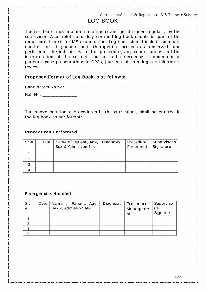
Curriculum/Statutes & Regulations -MS Thoracic Surgery
106
LOG BOOK The residents must maintain a log book and get it signed regularly by the supervisor. A complete and duly certified log book should be part of the requirement to sit for MS examination. Log book should include adequate number of diagnostic and therapeutic procedures observed and performed, the indications for the procedure, any complications and the interpretation of the results, routine and emergency management of patients, case presentations in CPCs, journal club meetings and literature review. Proposed Format of Log Book is as follows: Candidate’s Name: _________________________________
Roll No. _____________
The above mentioned procedures in the curriculum, shall be entered in the log book as per format: Procedures Performed
Sr.# Date Name of Patient, Age,
Sex & Admission No. Diagnosis Procedure
Performed Supervisor’s Signature
1
2
3
4 Emergencies Handled Sr.#
Date Name of Patient, Age, Sex & Admission No.
Diagnosis Procedure/Management
Supervisor’s Signature
1 2 3 4
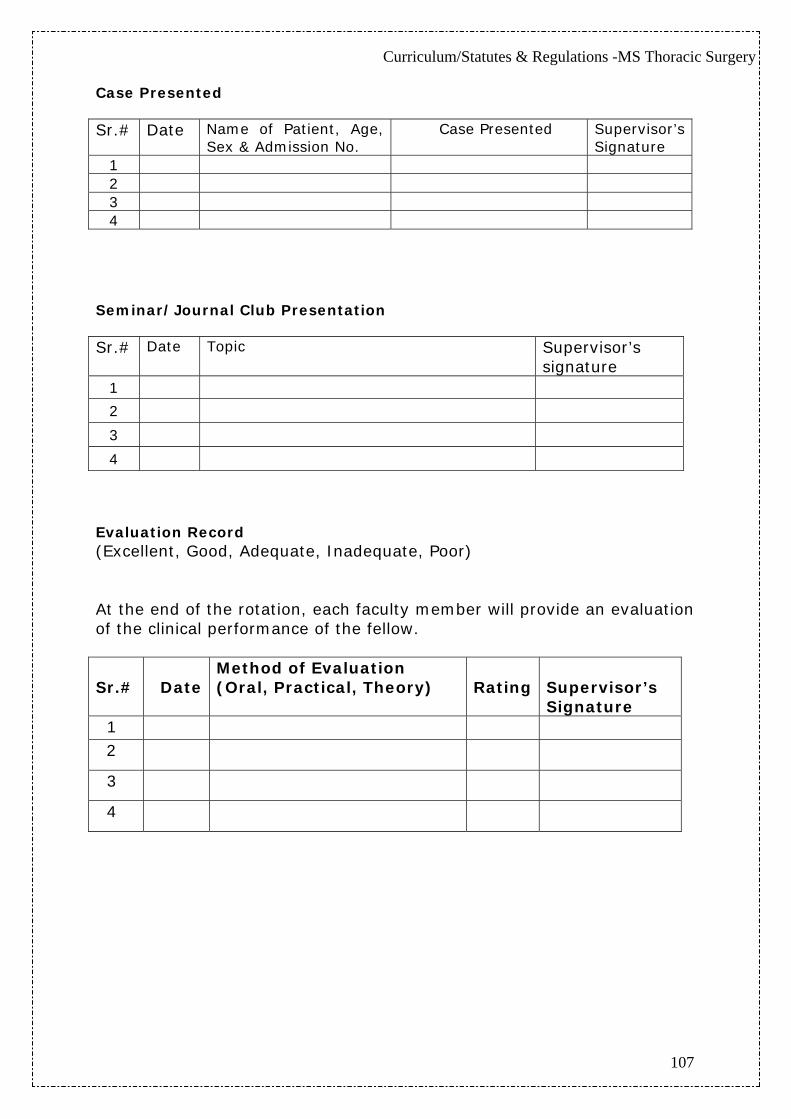
Curriculum/Statutes & Regulations -MS Thoracic Surgery
107
Case Presented Sr.# Date Name of Patient, Age,
Sex & Admission No. Case Presented Supervisor’s
Signature 1 2 3 4
Seminar/Journal Club Presentation Sr.# Date Topic Supervisor’s
signature 1
2
3
4
Evaluation Record (Excellent, Good, Adequate, Inadequate, Poor) At the end of the rotation, each faculty member will provide an evaluation of the clinical performance of the fellow. Sr.#
Date
Method of Evaluation (Oral, Practical, Theory)
Rating
Supervisor’s Signature
1 2
3
4
Curriculum/Statutes & Regulations -MS Thoracic Surgery
108
EVALUATION & ASSESSMENT STRATEGIES Assessment It will consist of action and professional growth oriented student-centered integrated assessment with an additional component of informal internal assessment, formative assessment and measurement-based summative assessment. Student-Centered Integrated Assessment It views students as decision-makers in need of information about their own performance. Integrated Assessment is meant to give students responsibility for deciding what to evaluate, as well as how to evaluate it, encourages students to ‘own’ the evaluation and to use it as a basis for self-improvement. Therefore, it tends to be growth-oriented, student-controlled, collaborative, dynamic, contextualized, informal, flexible and action-oriented. In the proposed curriculum, it will be based on:
• Self Assessment by the student • Peer Assessment • Informal Internal Assessment by the Faculty
Self Assessment by the Student Each student will be provided with a pre-designed self-assessment form to evaluate his/her level of comfort and competency in dealing with different relevant clinical situations. It will be the responsibility of the student to correctly identify his/her areas of weakness and to take appropriate measures to address those weaknesses. Peer Assessment The students will also be expected to evaluate their peers after the monthly small group meeting. These should be followed by a constructive feedback according to the prescribed guidelines and should be non-judgmental in nature. This will enable students to become good mentors in future. Informal Internal Assessment by the Faculty There will be no formal allocation of marks for the component of Internal Assessment so that students are willing to confront their weaknesses rather than hiding them from their instructors.
Curriculum/Statutes & Regulations -MS Thoracic Surgery
109
It will include:
a. Punctuality b. Ward work c. Monthly assessment (written tests to indicate particular areas
of weaknesses) d. Participation in interactive sessions
Formative Assessment Will help to improve the existing instructional methods and the curriculum in use Feedback to the faculty by the students: After every three months students will be providing a written feedback regarding their course components and teaching methods. This will help to identify strengths and weaknesses of the relevant course, faculty members and to ascertain areas for further improvement. Summative Assessment It will be carried out at the end of the programme to empirically evaluate cognitive, psychomotor and affective domains in order to award diplomas for successful completion of courses.
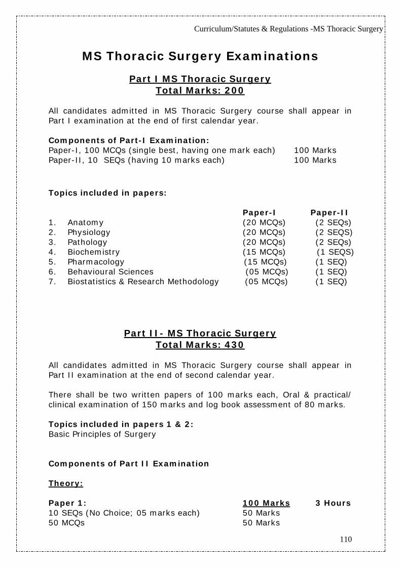
Curriculum/Statutes & Regulations -MS Thoracic Surgery
110
MS Thoracic Surgery Examinations
Part I MS Thoracic Surgery
Total Marks: 200 All candidates admitted in MS Thoracic Surgery course shall appear in Part I examination at the end of first calendar year. Components of Part-I Examination: Paper-I, 100 MCQs (single best, having one mark each) 100 Marks Paper-II, 10 SEQs (having 10 marks each) 100 Marks
Topics included in papers:
Paper-I Paper-II 1. Anatomy (20 MCQs) (2 SEQs) 2. Physiology (20 MCQs) (2 SEQS) 3. Pathology (20 MCQs) (2 SEQs) 4. Biochemistry (15 MCQs) (1 SEQS) 5. Pharmacology (15 MCQs) (1 SEQ) 6. Behavioural Sciences (05 MCQs) (1 SEQ) 7. Biostatistics & Research Methodology (05 MCQs) (1 SEQ)
Part II- MS Thoracic Surgery Total Marks: 430
All candidates admitted in MS Thoracic Surgery course shall appear in Part II examination at the end of second calendar year. There shall be two written papers of 100 marks each, Oral & practical/ clinical examination of 150 marks and log book assessment of 80 marks. Topics included in papers 1 & 2: Basic Principles of Surgery Components of Part II Examination Theory: Paper 1: 100 Marks 3 Hours 10 SEQs (No Choice; 05 marks each) 50 Marks 50 MCQs 50 Marks
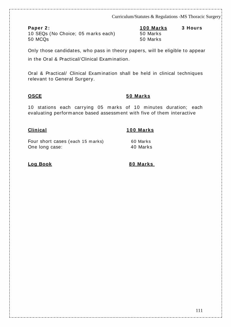
Curriculum/Statutes & Regulations -MS Thoracic Surgery
111
Paper 2: 100 Marks 3 Hours 10 SEQs (No Choice; 05 marks each) 50 Marks 50 MCQs 50 Marks Only those candidates, who pass in theory papers, will be eligible to appear
in the Oral & Practical/Clinical Examination.
Oral & Practical/ Clinical Examination shall be held in clinical techniques relevant to General Surgery. OSCE 50 Marks 10 stations each carrying 05 marks of 10 minutes duration; each evaluating performance based assessment with five of them interactive Clinical 100 Marks Four short cases (each 15 marks) 60 Marks One long case: 40 Marks Log Book 80 Marks
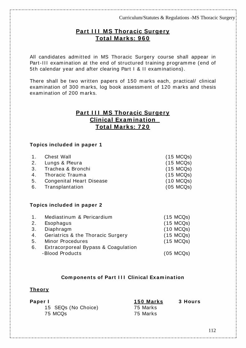
Curriculum/Statutes & Regulations -MS Thoracic Surgery
112
Part III MS Thoracic Surgery
Total Marks: 960 All candidates admitted in MS Thoracic Surgery course shall appear in Part-III examination at the end of structured training programme (end of 5th calendar year and after clearing Part I & II examinations). There shall be two written papers of 150 marks each, practical/ clinical examination of 300 marks, log book assessment of 120 marks and thesis examination of 200 marks.
Part III MS Thoracic Surgery Clinical Examination
Total Marks: 720 Topics included in paper 1 1. Chest Wall (15 MCQs) 2. Lungs & Pleura (15 MCQs) 3. Trachea & Bronchi (15 MCQs) 4. Thoracic Trauma (15 MCQs) 5. Congenital Heart Disease (10 MCQs) 6. Transplantation (05 MCQs)
Topics included in paper 2 1. Mediastinum & Pericardium (15 MCQs) 2. Esophagus (15 MCQs) 3. Diaphragm (10 MCQs) 4. Geriatrics & the Thoracic Surgery (15 MCQs) 5. Minor Procedures (15 MCQs) 6. Extracorporeal Bypass & Coagulation -Blood Products (05 MCQs)
Components of Part III Clinical Examination Theory Paper I 150 Marks 3 Hours 15 SEQs (No Choice) 75 Marks 75 MCQs 75 Marks
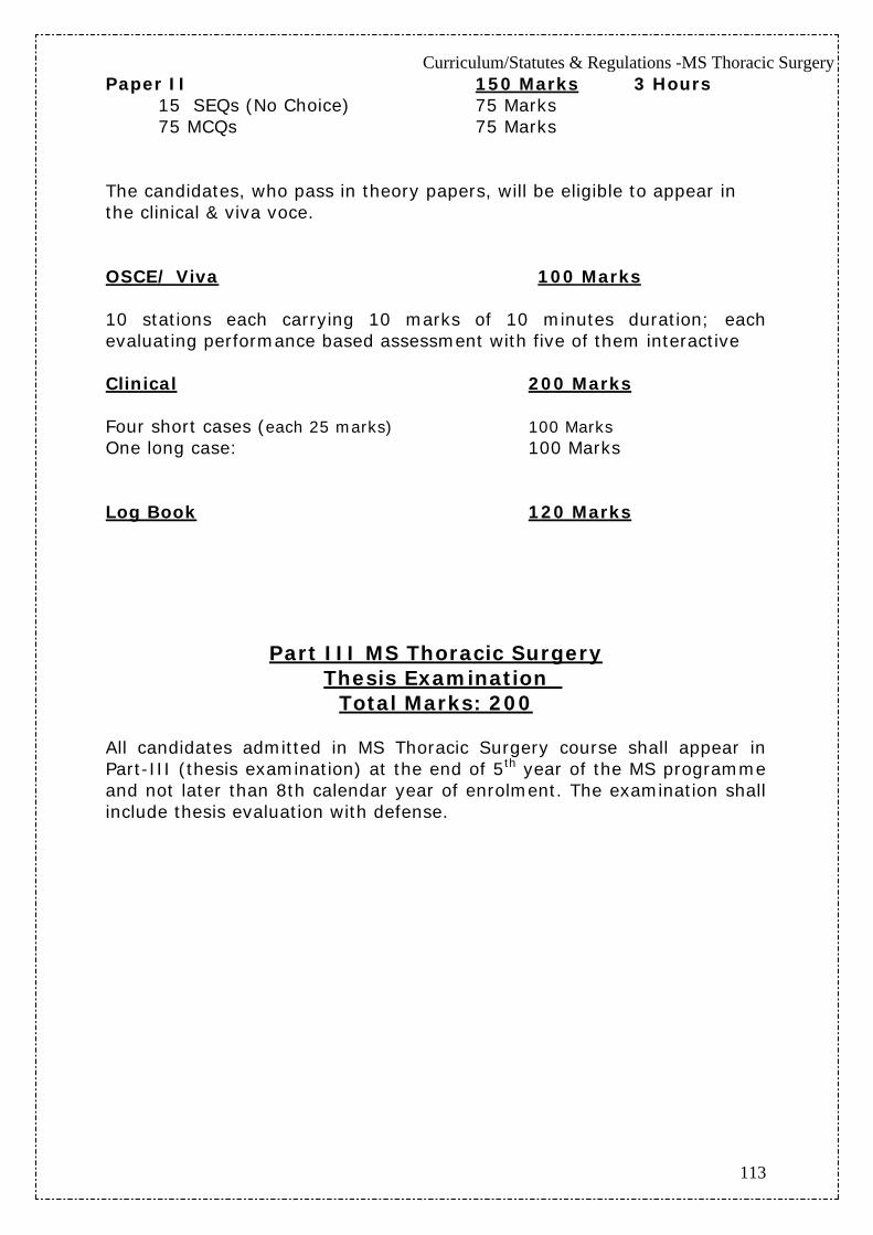
Curriculum/Statutes & Regulations -MS Thoracic Surgery
113
Paper II 150 Marks 3 Hours 15 SEQs (No Choice) 75 Marks 75 MCQs 75 Marks The candidates, who pass in theory papers, will be eligible to appear in the clinical & viva voce. OSCE/ Viva 100 Marks 10 stations each carrying 10 marks of 10 minutes duration; each evaluating performance based assessment with five of them interactive Clinical 200 Marks Four short cases (each 25 marks) 100 Marks One long case: 100 Marks Log Book 120 Marks
Part III MS Thoracic Surgery
Thesis Examination Total Marks: 200
All candidates admitted in MS Thoracic Surgery course shall appear in Part-III (thesis examination) at the end of 5th year of the MS programme and not later than 8th calendar year of enrolment. The examination shall include thesis evaluation with defense.