cutaneous presentation of neuroblastoma in 8 …...singh m, khan l, sachan s, mishra v, sachan a....
TRANSCRIPT
SSR Inst. Int. J. Life Sci. ISSN (O): 2581-8740 | ISSN (P): 2581-8732
Singh et al., 2019
DOI:10.21276/SSR-IIJLS.2019.5.4.6
Copyright © 2015 - 2019| SSR-IIJLS by Society for Scientific Research under a CC BY-NC 4.0 International License Volume 05 | Issue 04 | Page 2365
Cutaneous Presentation of Neuroblastoma in 8 Months Old Boy: A
Rare Case Report
Mahendra Singh1, Lubna Khan2*, Swetlana Sachan3, Vandana Mishra4, Anveksha Sachan3
1Professor & Head, Department of Pathology, GSVM Medical College, Kanpur, India 2Professor, Department of Pathology, GSVM Medical College, Kanpur, India
3Junior Resident III, Department of Pathology, GSVM Medical College, Kanpur, India 4Assistant Professor, Department of Pathology, GSVM Medical College, Kanpur, India
*Address for Correspondence: Dr. Lubna Khan, Professor, Department of Pathology, GSVM Medical College, Kanpur, India E-mail: [email protected]
Received: 30 Apr 2019/ Revised: 16 June 2019/ Accepted: 02 July 2019
ABSTRACT
Background: Neuroblastoma is the neurogenic, extracranial solid tumor of infancy and children emerging anywhere along with the peripheral sympathetic nervous system. Methods: An eight months old boy presented with chief complaints of sub-cutaneous non-tender nodule on the flexor aspect of forearm, pain and fullness of abdomen, emesis, mild bone pain. Complete blood profile and other biochemical parameter were within normal limits. Results: Fine needle aspiration cytology revealed tumour cells having high nucleo-cytoplasmic ratio bare nuclei, which were round to oval in shape. Homer wright rosette arrangement was formed by tumor cells that were radially arranged in a circle. The neuropil, which was stained as eosinophilic material was seen in the center of rosette arrangement. Conclusions: Hence on the basis of age and rare cutaneous presentation along with cytological findings; clinical features and radiological findings we can provide a diagnosis of metastasis neuroblastoma.
Key-words: Extracranial, Eosinophilic fibrillary material, Homer wright rosette, Neuropil, Neuroblastoma
INTRODUCTION
Neuroblastoma is the third most common neurogenic, an
extracranial solid tumor of infancy and children emerging
anywhere along the peripheral sympathetic nervous
system [1]. It accounts for 8-10% of all cancers in children [2]. Nearly 50% of neuroblastomas were diagnosed in
children younger than 5 years of age [3]. Its diagnosis
becomes poor and poor as the age advances. It was
sporadic or non-familial in origin [4].
They arise from primitive cells and are seen in the
adrenal medulla and sympathetic ganglia of the
sympathetic nervous system. These aggressive cells
begin to grow uncontrollably.
How to cite this article
Singh M, Khan L, Sachan S, Mishra V, Sachan A. Cutaneous Presentation of Neuroblastoma in 8 Months Old Boy: A Rare Case Report. SSR Inst. Int. J. Life Sci., 2019; 5(4): 2365-2368.
Access this article online
https://iijls.com/
Neuroblastomas may lead to signs such as swelling in the
face, neck, arms and upper chest, headaches, dizziness,
changes to consciousness, drooping eyelids and small
pupils. They may also lead to signs of paraneoplastic
syndromes that include constant diarrhea, fever, high
blood pressure (causing irritability), rapid heartbeat,
flushing of the skin, and sweating. Because
neuroblastomas consist of embryonic cells, they were
especially common among small children: up to 90% of
the patients are younger than six years old [5].
CASE REPORT
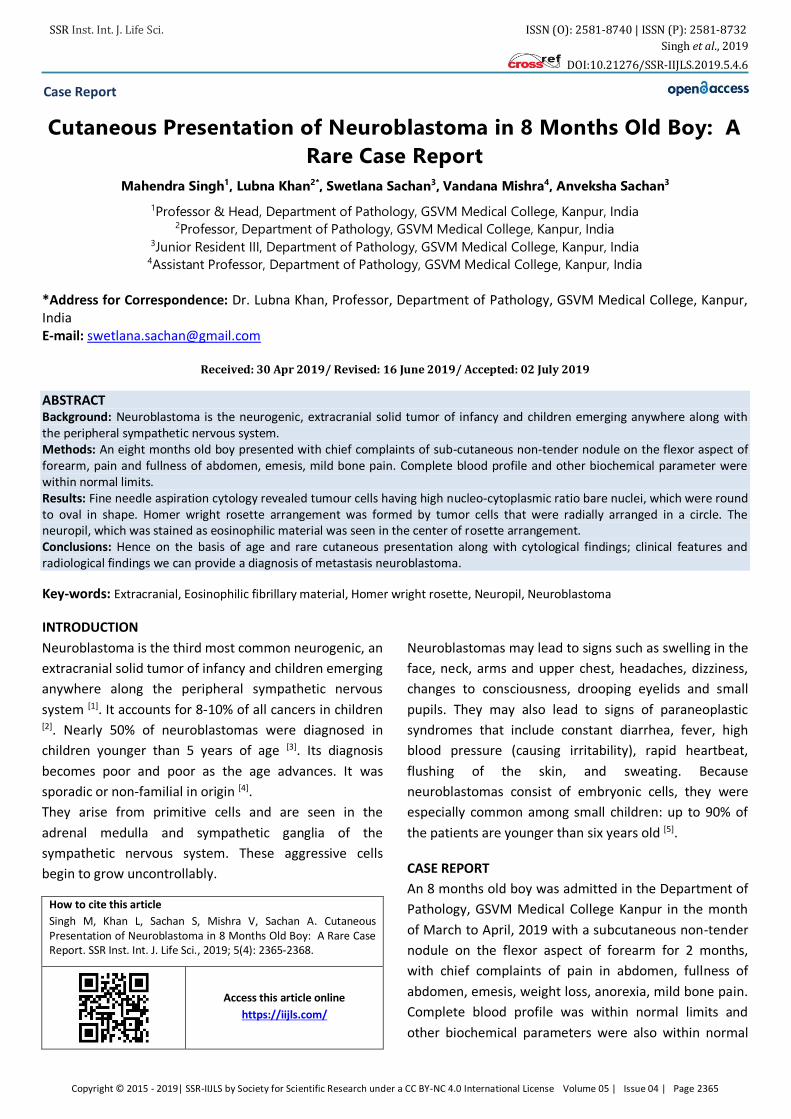
An 8 months old boy was admitted in the Department of
Pathology, GSVM Medical College Kanpur in the month
of March to April, 2019 with a subcutaneous non-tender
nodule on the flexor aspect of forearm for 2 months,
with chief complaints of pain in abdomen, fullness of
abdomen, emesis, weight loss, anorexia, mild bone pain.
Complete blood profile was within normal limits and
other biochemical parameters were also within normal
Case Report
SSR Inst. Int. J. Life Sci. ISSN (O): 2581-8740 | ISSN (P): 2581-8732
Singh et al., 2019
DOI:10.21276/SSR-IIJLS.2019.5.4.6
Copyright © 2015 - 2019| SSR-IIJLS by Society for Scientific Research under a CC BY-NC 4.0 International License Volume 05 | Issue 04 | Page 2366
limits. Multi slice spiral C.T. of the abdomen showed well
defined, lobulated heterogeneously enhancing mass
lesion in relation to the superomedial aspect of the right
kidney with few calcification and encasement of
abdominal vessels with skeletal metastasis suggesting
neuroblastoma. Ultrasound of the patient revealed a
solid, isoechoic to hypoechoic mass 3x2 cm in diameter
was located near the right adrenal gland.
Fig. 1: Red cutaneous nodular swelling in the flexor
aspect of forearm
Fig. 2 (B,C): Ultrasonography reveals well defined
isoechoic to hypoechoic rounded mass lesion noted in
right suprarenal region with sharply defined margin with
upper pole of kidney and posterior segment of right lobe
of liver. No evidence of any internal echogenic foci or
cystic degeneration
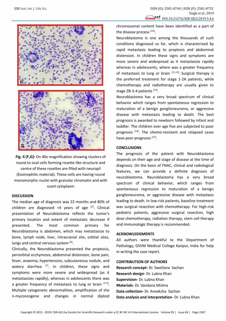
Fine needle aspiration cytology
On Aspiration- Blood mixed material was obtained.
Microscopic examination- Good cellularity smear
revealed tumor cells having bare nuclei which were
round to oval in shape. Homer wright rosettes
arrangements were formed by tumor cells that are
radially arranged in a circle. The neuropil (eosinophilic
fibrillary material) was seen in the center of rosette
arrangement. Red blood cells were seen around tumor
cells. These tumor cells showed increased nucleo-
cytoplasmic ratio and their nuclei were round to oval in
shape with anisokaryosis, nuclear chromatin was
granular.
Fig. 3 (D,E): On 10x magnification showing scattered as well as clusters of small round neuroblastic cells, few of them forming rosette like structure. Background shows
eosinophilic fibrillary material
SSR Inst. Int. J. Life Sci. ISSN (O): 2581-8740 | ISSN (P): 2581-8732
Singh et al., 2019
DOI:10.21276/SSR-IIJLS.2019.5.4.6
Copyright © 2015 - 2019| SSR-IIJLS by Society for Scientific Research under a CC BY-NC 4.0 International License Volume 05 | Issue 04 | Page 2367
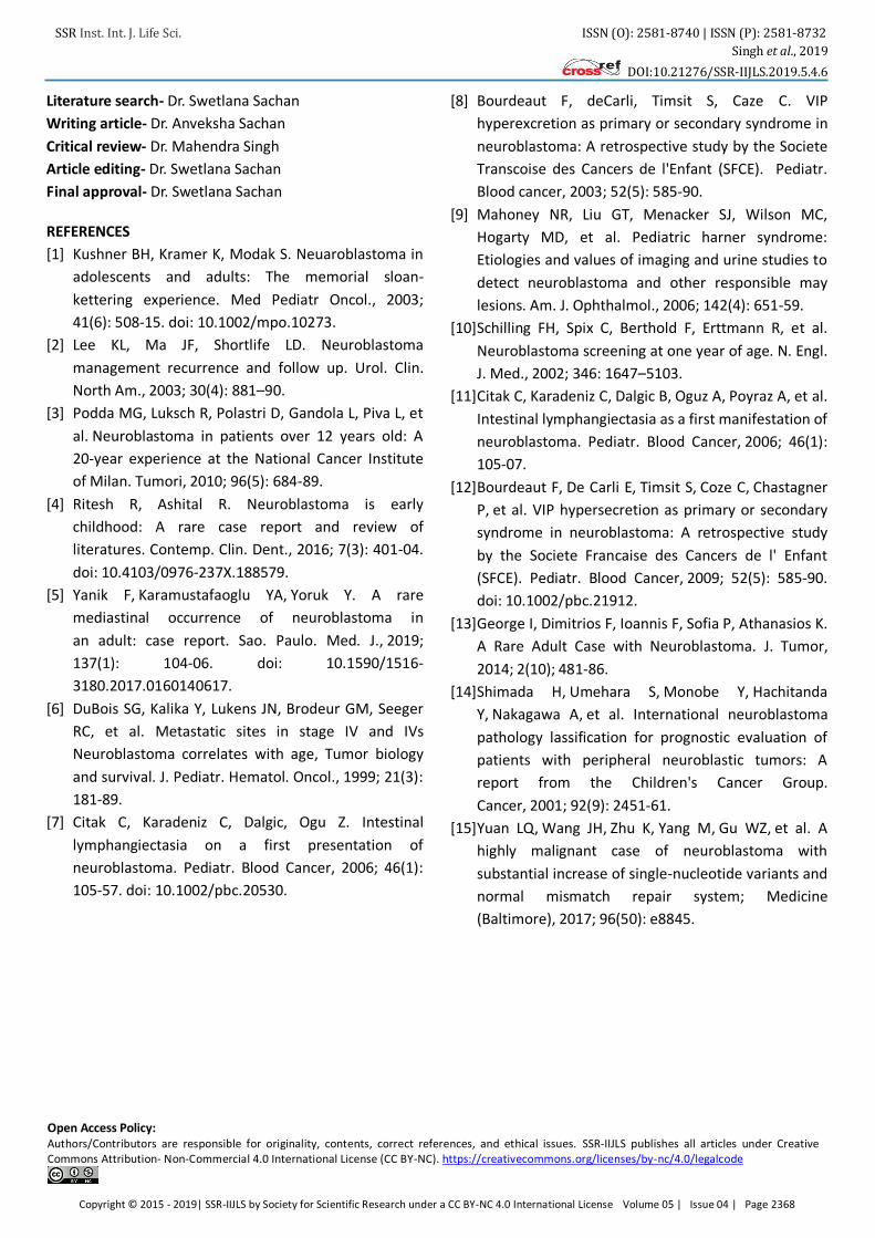
Fig. 4 (F,G): On 40x magnification showing clusters of
round to oval cells forming rosette like structure and
centre of these rosettes are filled with neuropil
(Eosinophilic material). These cells are having round
monomorphic nuclei with granular chromatin and with
scant cytoplasm
DISCUSSION
The median age of diagnosis was 22 months and 80% of
children are diagnosed <4 years of age [2]. Clinical
presentation of Neuroblastoma reflects the tumor's
primary location and extent of metastatic decrease if
presented. The most common primary for
Neuroblastoma is abdomen, which may metastasize to
bone, lymph node, liver, intracranial site, orbital sites,
lungs and central nervous system [6].
Clinically, the Neuroblastoma presented the proptosis,
periorbital ecchymosis, abdominal distension, bone pain,
fever, anaemia, hypertension, subcutaneous nodule, and
watery diarrhea [7]. In children, these signs and
symptoms were more severe and widespread (as it
metastasizes rapidly), whereas in adolescents there was
a greater frequency of metastasis to lung or brain [7-9].
Multiple cytogenetic abnormalities, amplification of the
n-myconcogene and changes in normal diploid
chromosomal content have been identified as a part of
the disease process [10].
Neuroblastoma is one among the thousands of such
conditions diagnosed so far, which is characterized by
rapid metastasis leading to proptosis and abdominal
distension. In children these signs and symptoms are
more severe and widespread as it metastasize rapidly
whereas in adolescents, where was a greater frequency
of metastasis to lung or brain [11,12]. Surgical therapy is
the preferred treatment for stage 1-2Α patients, while
chemotherapy and radiotherapy are usually given to
stage 2B-3-4 patients [13].
Neuroblastoma has a very broad spectrum of clinical
behavior which ranges from spontaneous regression to
maturation of a benign ganglioneuroma, or aggressive
disease with metastasis leading to death. The best
prognosis is awarded to newborn followed by infant and
toddler. The children over age five are subjected to poor
prognosis [14]. The chemo-resistant and relapsed cases
have poor prognosis [15].
CONCLUSIONS
The prognosis of the patient with Neuroblastoma
depends on their age and stage of disease at the time of
diagnosis. On the basis of FNAC, clinical and radiological
features, we can provide a definite diagnosis of
neuroblastoma. Neuroblastoma has a very broad
spectrum of clinical behavior, which ranges from
spontaneous regression to maturation of a benign
ganglioneuroma, or aggressive disease with metastasis
leading to death. In low-risk patients, baseline treatment
was surgical resection with chemotherapy. For high-risk
pediatric patients, aggressive surgical resection, high
dose chemotherapy, radiation therapy, stem cell therapy
and immunologic therapy is recommended.
ACKNOWLEDGMENTS
All authors were thankful to the Department of
Pathology, GSVM Medical College Kanpur, India for help
in writing the case report.
CONTRIBUTION OF AUTHORS
Research concept- Dr. Swetlana Sachan
Research design- Dr. Lubna Khan
Supervision- Dr. Lubna Khan
Materials- Dr. Vandana Mishra
Data collection- Dr. Anveksha Sachan
Data analysis and interpretation- Dr. Lubna Khan
SSR Inst. Int. J. Life Sci. ISSN (O): 2581-8740 | ISSN (P): 2581-8732
Singh et al., 2019
DOI:10.21276/SSR-IIJLS.2019.5.4.6
Copyright © 2015 - 2019| SSR-IIJLS by Society for Scientific Research under a CC BY-NC 4.0 International License Volume 05 | Issue 04 | Page 2368
Literature search- Dr. Swetlana Sachan
Writing article- Dr. Anveksha Sachan
Critical review- Dr. Mahendra Singh
Article editing- Dr. Swetlana Sachan
Final approval- Dr. Swetlana Sachan
REFERENCES
[1] Kushner BH, Kramer K, Modak S. Neuaroblastoma in
adolescents and adults: The memorial sloan-
kettering experience. Med Pediatr Oncol., 2003;
41(6): 508-15. doi: 10.1002/mpo.10273.
[2] Lee KL, Ma JF, Shortlife LD. Neuroblastoma
management recurrence and follow up. Urol. Clin.
North Am., 2003; 30(4): 881–90.
[3] Podda MG, Luksch R, Polastri D, Gandola L, Piva L, et
al. Neuroblastoma in patients over 12 years old: A
20-year experience at the National Cancer Institute
of Milan. Tumori, 2010; 96(5): 684-89.
[4] Ritesh R, Ashital R. Neuroblastoma is early
childhood: A rare case report and review of
literatures. Contemp. Clin. Dent., 2016; 7(3): 401-04.
doi: 10.4103/0976-237X.188579.
[5] Yanik F, Karamustafaoglu YA, Yoruk Y. A rare
mediastinal occurrence of neuroblastoma in
an adult: case report. Sao. Paulo. Med. J., 2019;
137(1): 104-06. doi: 10.1590/1516-
3180.2017.0160140617.
[6] DuBois SG, Kalika Y, Lukens JN, Brodeur GM, Seeger
RC, et al. Metastatic sites in stage IV and IVs
Neuroblastoma correlates with age, Tumor biology
and survival. J. Pediatr. Hematol. Oncol., 1999; 21(3):
181-89.
[7] Citak C, Karadeniz C, Dalgic, Ogu Z. Intestinal
lymphangiectasia on a first presentation of
neuroblastoma. Pediatr. Blood Cancer, 2006; 46(1):
105-57. doi: 10.1002/pbc.20530.
[8] Bourdeaut F, deCarli, Timsit S, Caze C. VIP
hyperexcretion as primary or secondary syndrome in
neuroblastoma: A retrospective study by the Societe
Transcoise des Cancers de l'Enfant (SFCE). Pediatr.
Blood cancer, 2003; 52(5): 585-90.
[9] Mahoney NR, Liu GT, Menacker SJ, Wilson MC,
Hogarty MD, et al. Pediatric harner syndrome:
Etiologies and values of imaging and urine studies to
detect neuroblastoma and other responsible may
lesions. Am. J. Ophthalmol., 2006; 142(4): 651-59.
[10]Schilling FH, Spix C, Berthold F, Erttmann R, et al.
Neuroblastoma screening at one year of age. N. Engl.
J. Med., 2002; 346: 1647–5103.
[11]Citak C, Karadeniz C, Dalgic B, Oguz A, Poyraz A, et al.
Intestinal lymphangiectasia as a first manifestation of
neuroblastoma. Pediatr. Blood Cancer, 2006; 46(1):
105-07.
[12]Bourdeaut F, De Carli E, Timsit S, Coze C, Chastagner
P, et al. VIP hypersecretion as primary or secondary
syndrome in neuroblastoma: A retrospective study
by the Societe Francaise des Cancers de l' Enfant
(SFCE). Pediatr. Blood Cancer, 2009; 52(5): 585-90.
doi: 10.1002/pbc.21912.
[13]George I, Dimitrios F, Ioannis F, Sofia P, Athanasios K.
A Rare Adult Case with Neuroblastoma. J. Tumor,
2014; 2(10); 481-86.
[14]Shimada H, Umehara S, Monobe Y, Hachitanda
Y, Nakagawa A, et al. International neuroblastoma
pathology lassification for prognostic evaluation of
patients with peripheral neuroblastic tumors: A
report from the Children's Cancer Group.
Cancer, 2001; 92(9): 2451-61.
[15]Yuan LQ, Wang JH, Zhu K, Yang M, Gu WZ, et al. A
highly malignant case of neuroblastoma with
substantial increase of single-nucleotide variants and
normal mismatch repair system; Medicine
(Baltimore), 2017; 96(50): e8845.
Open Access Policy: Authors/Contributors are responsible for originality, contents, correct references, and ethical issues. SSR-IIJLS publishes all articles under Creative Commons Attribution- Non-Commercial 4.0 International License (CC BY-NC). https://creativecommons.org/licenses/by-nc/4.0/legalcode