cvs6 cvs
TRANSCRIPT

Lecture №6. The acute and chronic coronary syndrome.
Lecture №7. The syndrome acute and chronic cardiac failure.
Acute and chronic coronary syndrome.
Acute and chronic cardiac failure.
1

Unstable Angina and Non-ST-Elevation Myocardial Infarction.
• Patients with ischemic heart disease fall into two large groups:
• patients with chronic coronary artery disease (CAD) who most commonly present with stable angina
• and patients with acute coronary syndromes (ACSs).
• The latter group, in turn, is composed of patients with acute myocardial infarction (MI) with ST-segment elevation on their presenting electrocardiogram
• and those with unstable angina and non-ST-segment elevation MI (UA/NSTEMI).
2

Definition• The diagnosis of UA is based largely on the clinical
presentation.
• Stable angina pectoris is characterized by chest or arm discomfort that may not be described as pain but is reproducibly associated with physical exertion or stress and is relieved within 5–10 min by rest and/or sublingual nitroglycerin .
3

Definition• UA is defined as angina pectoris or equivalent ischemic
discomfort with at least one of three features:
(1) it occurs at rest (or with minimal exertion), usually lasting >10 min;
(2) it is severe and of new onset (i.e., within the prior 4–6 weeks);
and/or (3) it occurs with a crescendo pattern (i.e., distinctly more severe, prolonged, or frequent than previously).
The diagnosis of NSTEMI is established if a patient with the clinical features of UA develops evidence of myocardial necrosis, as reflected in elevated cardiac biomarkers.
4
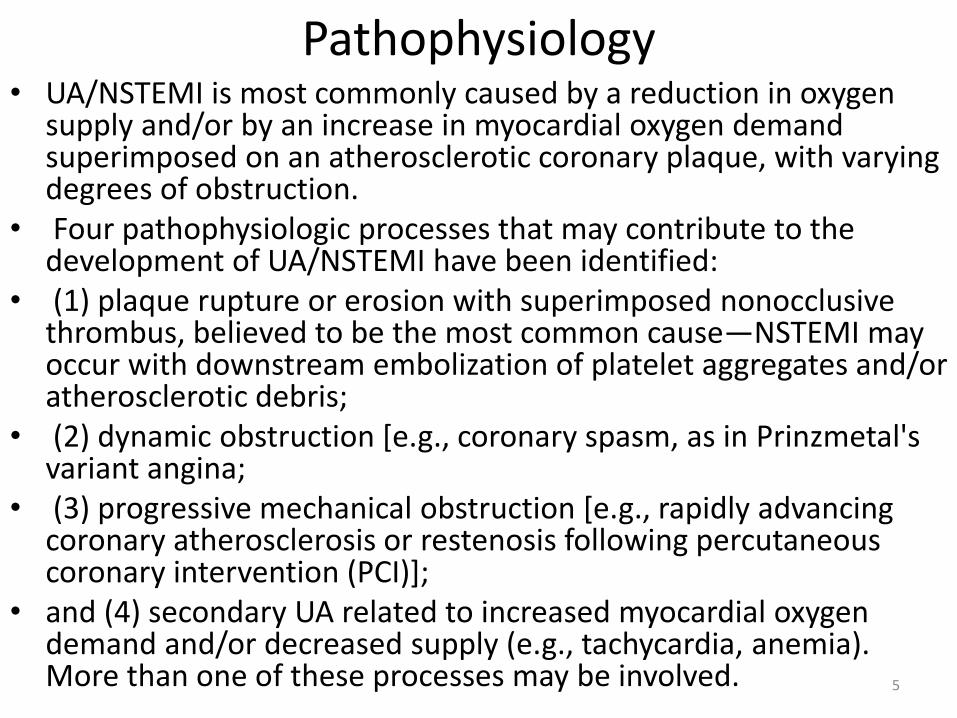
Pathophysiology• UA/NSTEMI is most commonly caused by a reduction in oxygen
supply and/or by an increase in myocardial oxygen demand superimposed on an atherosclerotic coronary plaque, with varying degrees of obstruction.
• Four pathophysiologic processes that may contribute to the development of UA/NSTEMI have been identified:
• (1) plaque rupture or erosion with superimposed nonocclusivethrombus, believed to be the most common cause—NSTEMI may occur with downstream embolization of platelet aggregates and/or atherosclerotic debris;
• (2) dynamic obstruction [e.g., coronary spasm, as in Prinzmetal'svariant angina;
• (3) progressive mechanical obstruction [e.g., rapidly advancing coronary atherosclerosis or restenosis following percutaneouscoronary intervention (PCI)];
• and (4) secondary UA related to increased myocardial oxygen demand and/or decreased supply (e.g., tachycardia, anemia). More than one of these processes may be involved. 5

Pathophysiology• Among patients with UA/NSTEMI studied at
angiography, approximately 5% have left main stenosis, 15% have three-vessel CAD, 30% have two-vessel disease, 40% have single-vessel disease, and 10% have no critical coronary stenosis;
• some of the latter have Prinzmetal's variant angina
• The "culprit lesion" on angiography may show an eccentric stenosis with scalloped or overhanging edges and a narrow neck.
• Angioscopy may reveal "white" (platelet-rich) thrombi, as opposed to "red" thrombi, more often seen in patients with acute STEMI. Patients with UA/NSTEMI often have multiple plaques vulnerable to disruption.
6

Clinical PresentationHistory and Physical Examination
• The clinical hallmark of UA/NSTEMI is chest pain, typically located in the substernal region or sometimes in the epigastrium, that radiates to the neck, left shoulder, and left arm.
• This discomfort is usually severe enough to be considered painful.
• Anginal "equivalents" such as dyspnea and epigastric discomfort may also occur, and these appear to occur more often in women.
7
Clinical PresentationHistory and Physical Examination
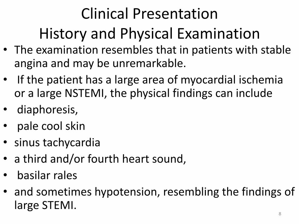
• The examination resembles that in patients with stable angina and may be unremarkable.
• If the patient has a large area of myocardial ischemia or a large NSTEMI, the physical findings can include
• diaphoresis,
• pale cool skin
• sinus tachycardia
• a third and/or fourth heart sound,
• basilar rales
• and sometimes hypotension, resembling the findings of large STEMI.
8

ElectrocardiogramIn UA,
• ST-segment depression,
• transient ST-segment elevation,
• and/or T-wave inversion occur in 30–50% of patients, depending on the severity of the clinical presentation.
• In patients with the clinical features of UA, the presence of new ST-segment deviation, even of only 0.05 mV, is an important predictor of adverse outcome.
• T-wave changes are sensitive for ischemia but less specific, unless they are new, deep T-wave inversions (0.3 mV).
9

Cardiac Biomarkers• Patients with UA who have elevated biomarkers of
necrosis, such as CK-MB and troponin (a much more specific and sensitive marker of myocardial necrosis), are at increased risk for death or recurrent MI.
• Elevated levels of these markers distinguish patients with NSTEMI from those with UA.
• There is a direct relationship between the degree of troponin elevation and mortality.
• However, in patients without a clear clinical history of myocardial ischemia, minor troponin elevations have been reported and can be caused by congestive heart failure, myocarditis, or pulmonary embolism, or they may be false-positive readings.
• Thus, in patients with an unclear history, small troponinelevations may not be diagnostic of an ACS. 10
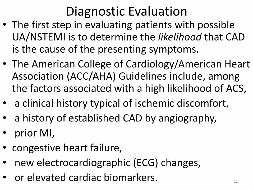
Diagnostic Evaluation • The first step in evaluating patients with possible
UA/NSTEMI is to determine the likelihood that CAD is the cause of the presenting symptoms.
• The American College of Cardiology/American Heart Association (ACC/AHA) Guidelines include, among the factors associated with a high likelihood of ACS,
• a clinical history typical of ischemic discomfort,
• a history of established CAD by angiography,
• prior MI,
• congestive heart failure,
• new electrocardiographic (ECG) changes,
• or elevated cardiac biomarkers. 11

Diagnostic Evaluation• Factors associated with an intermediate likelihood
of ACS in patients with the clinical features of this condition but without the above high-risk factors are:
• age >70 years,
• male gender,
• diabetes mellitus,
• known peripheral arterial or cerebrovascular disease,
• and old ECG abnormalities.
12

Diagnostic Pathways
• Four major diagnostic tools are used in the diagnosis of UA/NSTEMI in the ED:
• the clinical history,
• the ECG,
• cardiac markers,
• and stress testing.
13

The goals are
• to (1) recognize or exclude MI (using cardiac markers),
• (2) evaluate for rest ischemia (chest pain at rest, serial or continuous ECGs),
• and (3) evaluate for significant CAD (using provocative stress testing).
• Typical pathways begin with assessment of the likelihood that the presenting symptoms are due to ischemia.
14

Diagnostic Pathways• Patients with a low likelihood of ischemia are usually
managed with an ED-based critical pathway (which in some institutions is carried out in a "chest pain unit”.
• Evaluation of such patients includes clinical monitoring for recurrent ischemic discomfort, serial ECGs, and cardiac markers, typically performed at baseline and at 4–6 h and 12 h after presentation.
• If new elevations in cardiac markers (CK-MB and/or troponin) or ECG changes are noted, the patient is admitted to the hospital.
• If the patient remains pain-free and the markers are negative, the patient may go on to stress testing. This may be performed as early as 6 h after presentation in the ED or chest pain center, or on an outpatient basis within 72 h. 15

Diagnostic Pathways• For most patients, standard treadmill ECG stress testing
is used, but for patients with fixed abnormalities on the ECG (e.g., left bundle branch block), perfusion or echocardiographic imaging is used.
• For patients who cannot walk, pharmacologic stress is used.
• By demonstrating normal myocardial perfusion, sestamibi or thallium imaging can reduce unnecessary hospitalizations by excluding acute ischemia.
• CT angiography is used with increasing frequency to exclude obstructive CAD.
16
17

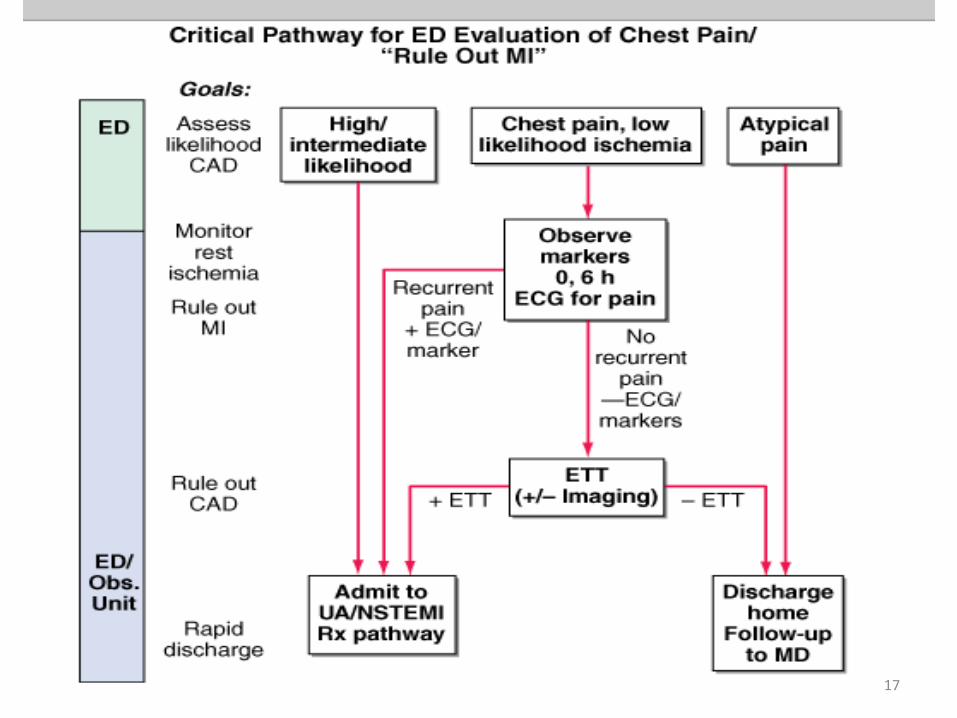
Diagnostic evaluation of patients presenting with suspected UA/NSTEMI.
• The first step is to assess the likelihood of coronary artery disease (CAD). Patients at high or intermediate likelihood are admitted to the hospital. Those with clearly atypical chest pain are sent home.
• Patients with a low likelihood of ischemia enter the pathway and are observed in a monitored bed in the emergency department (ED) or observation unit over a period of 6 h, and 12-lead electrocardiograms are performed if the patient has recurrent chest discomfort.
18

Diagnostic evaluation of patients presenting with suspected UA/NSTEMI
• A panel of cardiac markers (e.g., troponin and CK-MB) is drawn at baseline and 6 h later.
• If the patient develops recurrent pain, has ST-segment or T-wave changes, or has positive cardiac markers, he/she is admitted to the hospital and treated for UA/NSTEMI.
• If the patient has negative markers and no recurrence of pain, he/she is sent for exercise treadmill testing, with imaging reserved for patients with abnormal baseline electrocardiograms (e.g., left bundle branch block or left ventricular hypertrophy).
• If positive, the patient is admitted; if negative, the patient is discharged, with follow-up to his/her primary physician. ETT, exercise tolerance test; MI, myocardial infarction.
19

Risk Stratification and Prognosis• Patients with documented UA/NSTEMI exhibit a wide
spectrum of early (30 days) risk of death, ranging from 1 to 10%, and of new or recurrent infarction of 3–10%.
• Assessment of "global risk" can be accomplished by clinical risk scoring systems such as that developed from in the Thrombolysis in Myocardial Infarction (TIMI) Trials, which includes seven independent risk factors: age 65 years, three or more risk factors for CAD, documented CAD at catheterization, development of UA/NSTEMI while on aspirin, more than two episodes of angina within the preceding 24 h, ST deviation 0.5 mm, and an elevated cardiac marker
• Other risk factors include diabetes mellitus, left ventricular dysfunction, and elevated levels of creatinine, atrialnatriuretic peptides, and C-reactive protein.
20

Early risk assessment• Early risk assessment (especially using troponin, ST-
segment changes, and/or a global risk scoring system) is useful both in predicting the risk of recurrent cardiac events and in identifying those patients who would derive the greatest benefit from antithrombotic therapies more potent than unfractionated heparin, such as low-molecular-weight heparin (LMWH) and glycoprotein (GP)IIb/IIIa inhibitors, and from an early invasive strategy.
• For example, in the TACTICS-TIMI 18 Trial, an early invasive strategy conferred a 40% reduction in recurrent cardiac events in patients with a positive troponin level, whereas no benefit was observed in those with a negative troponin level. 21
Early risk assessment• C-reactive protein, a marker of vascular
inflammation, and B-type natriuretic peptide, a marker of increased myocardial wall tension, correlate independently with increased mortality (and, in some studies, recurrent cardiac events) in patients presenting with UA/NSTEMI.
• Multimarker strategies are now gaining favor both to define the pathophysiologic mechanisms underlying a given patient's presentation more fully and to stratify the patient's risk further.
22

Plate 28 T
23

Plate 28 B
24

Plate 24Myocardial
25

Plate 24Myocardial
26
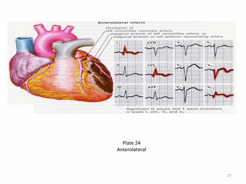
Plate 24Anterolateral
27

Plate 25Myocardial
28

Plate 25Posterior
29

The syndrome of coronary failure• The syndrome coronary failure being due to
myocardial ischemia ( narrowing of coronary vessels).
• Causes: atherosclerosis, sometimes without organic changes in vessels in result vasospasm.
• Different acute and chronic coronary failure.• Acute coronary syndrome its acute condition of
cardiac diseases, which include Q-wave dependent myocardial infarction, non-Q-wave myocardial infarction, unstable angina pectoris.
• Chronic coronary failure manifestation development atherosclerotic cardiosclerosis( arrhytmias, signs of cardiac failure).
30

• Acute coronary syndrome manifestation chest pain with irradiation to the left hand, shoulder, neck, superior part of the stomach. The chest pain beginning after physical activity. The patient is frightened of death, pallor of skin, cold sweat. Border of heart expansion, sound of heart weakened, may be arrhythmias, pericardial rub at myocardial infarction ( first-second days).
• ECG: depression of s.ST and inversion T wave or elevation of s.ST (in angina pectoris). Presence pathological large Q wave and elevation of s.ST with negative T wave (myocardial infarction).
• Common blood count: leucocytosis, increase ESR, increase of enzymes.
31

Ischemic heart diseaseMyocardial ischemia develops when there is an imbalance between supply of oxygen and the
myocardial demand.ETIOLOGY :1. Decreased coronary blood flow due to mechanical obstruction such as:• Atheroma - occluding one or more major coronary arteries.• Spasm of coronary artery• Thrombosis• Embolus• Coronary arteritis (e.g. in SLE)• Congenital abnormalities of coronary artery.
2 Increased myocardial oxygen requirement• Increased cardiac output: in thyrotoxicosis• Myocardial hypertrophy: usually from aortic stenosis or hypertension.3. Decreased flow of oxygenated blood to myocardium• Anemia• Hypotension - causing decreased coronary perfusion pressure.4. Cardiac syndrome X• Angina occurring in patient with normal coronary arteries, resulting from disease of coronary
microcirculation is called syndrome X. This is also called microvascular angina.
32

Atherosclerosis• The most common cause of myocardial ischemia
and angina is the formation of atheroma. Atheroma is a fibrofatty plaque in the intima of large and medium size arteries producinnarrowind of the lumen of the vessels. Exact causc of atheroma formation is not known. A 50%r reduction in reduction in luminal diameter produces a reduction in luminal cross- sectional area of approxymately 70%. Tis is considered significant obstruction and patient becomes symptomatic on exertion whenincreased blood flow is required that can not be supplied according to the demand.
33

Risk factors for atherosclerosisFixed risk factorsAge:• Risk increases with age, rare in childhood except in familial
hyperlipidemia.Male sex:• Men have higher incidence of ischemic heart disease than
premenopausal women. After menopause the incidence of atherosclerosis in women reaches that in men. This protection in premenopausal women is probably due to estrogen..
Family history:• A positive family history means ischemic heart disease in
first-degree relatives before the age of 45 years in male and before 50 years in female.
•
34

Modifiable risk factors• (Changeable with treatment)Strong association:• Hyperlipidemia:High scrum cholesterol especially increased low –density lioprotein
(LDL) and decreased high-density lioproteins (HDL) is strongly associated with coronary atheroma.
• HypertensionBoth systolic and diastolic hypertension are associated with increased
risk of coronary artery disease. The risk is same for men and women.
• Cigarette smokingRisk of coronary artery is directly related to number of cigarette
smoked. The risk from smoking declines to almost normal after ten years of quitting.
• Diabetes mejlitusDiabetes or even just an abnormal glucose tolerance test is strongly
associated with vascular disease.
35

ANGINA PECTORIS
Angina pectoris is a clinical syndrome characterized by paroxysmal chest pain due to transient myocardial ischemia. It may occur whenever there is imbalance between myocardial oxygen supply and demand. The most common cause is atherosclerosis; however angina may also develop in aortic stenosis (AS) and hypertrophic cardiomyopathy (HOCM) even there is no coronary atheroma.
36

PATHOPHYSIOLOGY OF STABLE ANGINA, UNSTABLE ANGINA AND MYOCARDIAL INFARCTION
• Stable angina:Stable angina is the angina that occurs when coronary perfusion is
impaired by fixed or stable atheroma of coronary arteries i-c patient has fixed capacity of exertion after that he starts feeling chest pain.
• Unstable angina:Unstable angina is the angina that is characterized by rapidly
worsening chest pain, pain on minimal exertion or pain at rest. The culprit lesion is usually a complex ulcerated or fissured atheromawith adherent platelet - rich thrombus and local coronary spasm.
Unstable angina = ulcerated atheroma + thrombus formation abrupt reduction of blood flow caused by thrombus formation angina at rest.
• Myocardial infarctionPathophysiology of unstable angina and myocardial infarction is same
(i-e thrombus formation on atherosclerotic plaque) however in unstable angina obstruction of artery is incomplete while in Ml there is total obstruction.
37
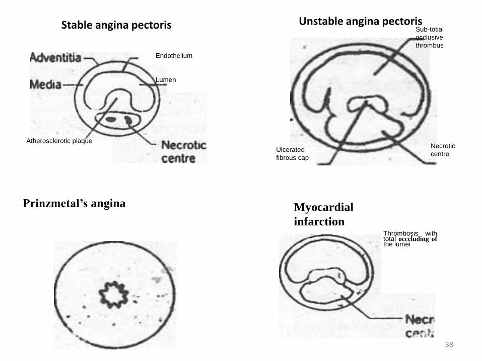
Prinzmetal’s angina
Atherosclerotic plaque
Myocardial
infarctionThrombosis withtotal occcluding ofthe lumei
Ulcerated
fibrous cap
Necrotic
centre
Sub-totial
occlusive
thrombus
Stable angina pectoris Unstable angina pectoris
Endothelium
Lumen
38

TYPES OF ANGINA1. Classical or exertional angina
It occurs due to increased myocardial oxygen demand during exertion or emotion in a patien narrow coronary arteries. It is relieved promptly by rest and by nitroglycerine.
2. Variant or Prinzmetal's angina
It Occurs at rest and is not a result of increasec myocardial demand. It is produced by the episodic reduction of myocardial blood supply due to coronary artery spasm. Underlying atherosclerotic disease may or may not be present. This type of angina occurs more frequently in women (under age 50 years) especially early in the morning awakening patients from sleep and the pain is usually more severe and more prolonged than i classical angina. It tends to involve right coronary artery. It is characteristically associated with ST elevation rather than depression (as seen classical angina).
3. Unstable angina
Unstable angina refers to angina of recent onset (less than one month), worsening angina characterized by increased frequency and duration of episode, or angina at rest not responding readily to therapy. 39
• 4. Decubitus anginaThis is angina that occurs when the patient lies down. . it
usually occurs in association with impaired left ventricular function. Patient with this symptoms usually has severe coronary artery disease.
• 5. Nocturnal angina.This is the angina that awakes the patient from sleep. It
may be provoked by vivid dreams. It may occur due to critical coronary artery obstruction or coronary spasm.
• 6. Cardiac syndrome XPatient presents with angina, positive exercise test (ETT)
and angiographically normal coronary arteries. It may be because of functional abnormalities of coronary microcirculation (no dilatation of arterioles at the timeof stress).
40

SYMPTOMS
• Chest pain Site, radiation, character, duration, precipitation factors)
• dyspnea
• Associated symptoms
SIGNS
• During attack patient looks anxious. Dyspnea, pale face, and cold sweats may also be present. In between attacks, physical examination is frequently negative except findings of risk factors.
41

Investigations
• ECG
• Exercise tolerance test
• Isotope scanning
• EchoCG
• Coronary angiograthy
42
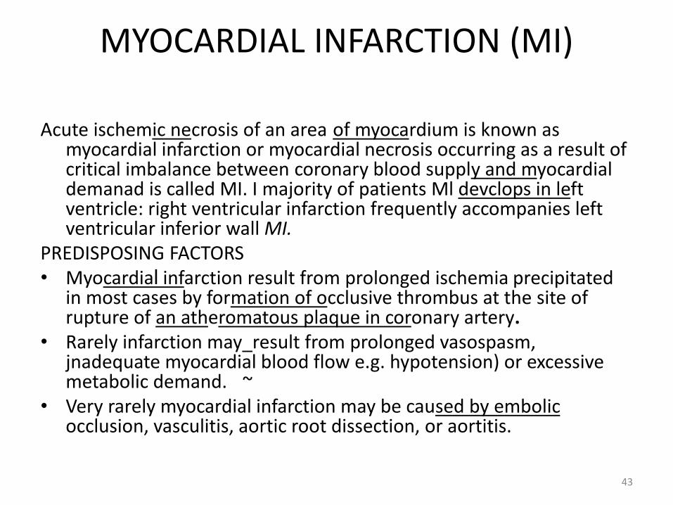
MYOCARDIAL INFARCTION (MI)
Acute ischemic necrosis of an area of myocardium is known as myocardial infarction or myocardial necrosis occurring as a result of critical imbalance between coronary blood supply and myocardial demanad is called MI. I majority of patients Ml devclops in left ventricle: right ventricular infarction frequently accompanies left ventricular inferior wall MI.
PREDISPOSING FACTORS• Myocardial infarction result from prolonged ischemia precipitated
in most cases by formation of occlusive thrombus at the site of rupture of an atheromatous plaque in coronary artery.
• Rarely infarction may_result from prolonged vasospasm, jnadequate myocardial blood flow e.g. hypotension) or excessive metabolic demand. ~
• Very rarely myocardial infarction may be caused by embolicocclusion, vasculitis, aortic root dissection, or aortitis.
43

Precipitating factors
• Reduced myocardial perfusion due to hypotension as a result hemorrhage or septic shock.
• Increased myocardial oxygen demand secondary to aortic stenosis, fever and tachycardia.
• Respiratory tract infections, hypoxemia of any cause, hypoglycemia, sympathomimetics drugs.
44

CLINICAL FEATURES Symptoms• Chest painThe pain is retrosternal similar to angina in location
and radiation but it occurs at rest or with less activity and is more severe and lasts longer (> 30 min); Pain may begin with exercise or psychological stress but may occur at rest without оbvious precipitating factors. It is not relieved by nitroglycerine. Some patients note only dull ache or numbness of the wrists jn association with severe retrosternal discomfort, pain may also begin in_epigastrium and simulates abdominal disorders.
45
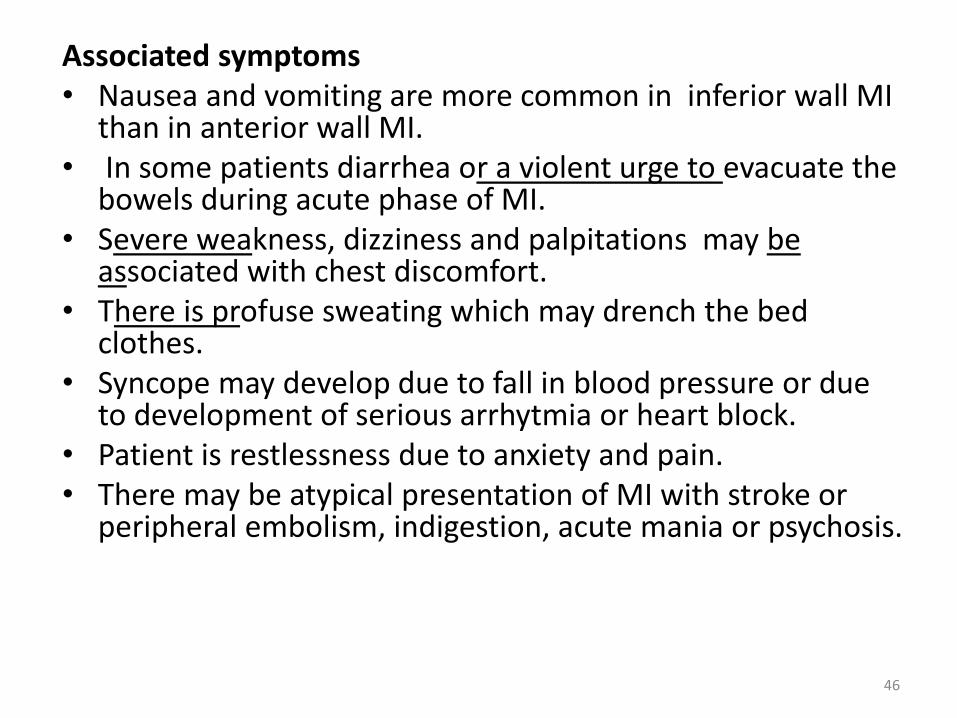
Associated symptoms• Nausea and vomiting are more common in inferior wall MI
than in anterior wall MI.• In some patients diarrhea or a violent urge to evacuate the
bowels during acute phase of MI.• Severe weakness, dizziness and palpitations may be
associated with chest discomfort.• There is profuse sweating which may drench the bed
clothes.• Syncope may develop due to fall in blood pressure or due
to development of serious arrhytmia or heart block. • Patient is restlessness due to anxiety and pain.• There may be atypical presentation of MI with stroke or
peripheral embolism, indigestion, acute mania or psychosis.
46

On examination:
– General
– Pulse
– Blood pressure
– Temperature
– Respiratory rate
– Examination of precordium
– Lungs
47
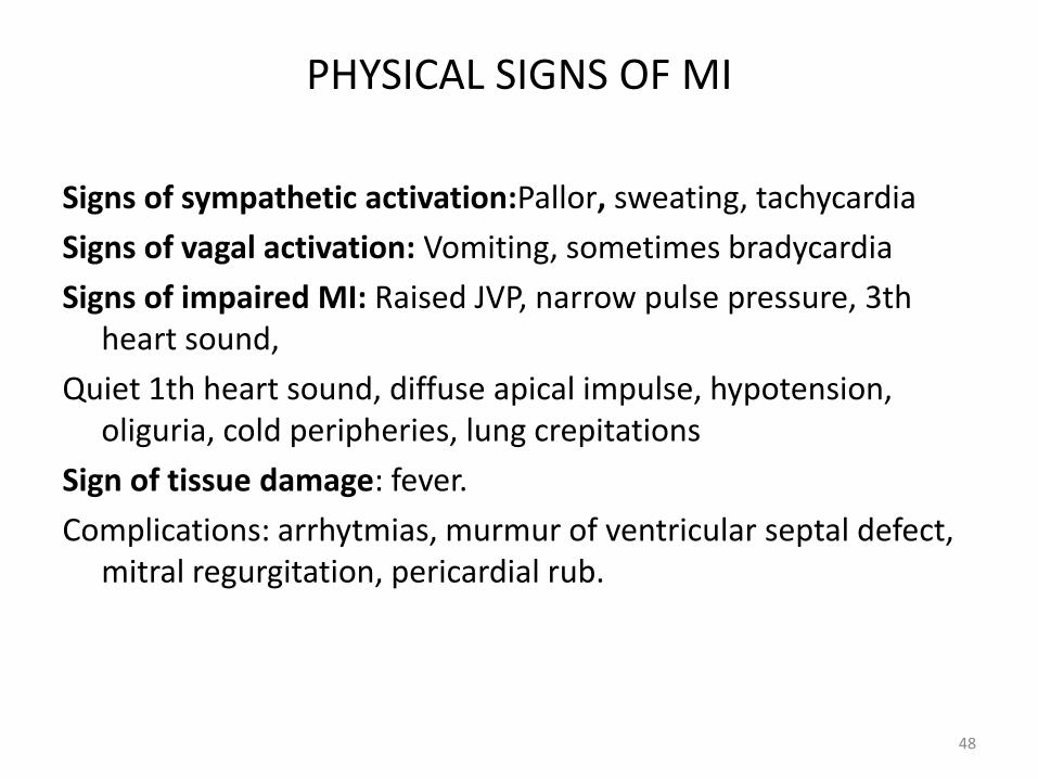
PHYSICAL SIGNS OF MI
Signs of sympathetic activation:Pallor, sweating, tachycardia
Signs of vagal activation: Vomiting, sometimes bradycardia
Signs of impaired MI: Raised JVP, narrow pulse pressure, 3th heart sound,
Quiet 1th heart sound, diffuse apical impulse, hypotension, oliguria, cold peripheries, lung crepitations
Sign of tissue damage: fever.
Complications: arrhytmias, murmur of ventricular septal defect, mitral regurgitation, pericardial rub.
48

Investigations of MI
• A history of ischemic type chest pain.
• ECG changes of myocardial infarction.
• Initially a rise and then fall of cardiac enzymes.
• ECG:
• Peaked (hyperacute) T waves -> ST segment elevation -> Formation of Q waves -> T wave inversion. These changes may occur over a few hours to several days.
49

ECG localization of MI
Infarct location Leads showing changes Likely coronary artery involved
Inferior wall MI II, III, aVF RCA
Septal MI V1-V2 LAD
Anterior wall MI V3-V$ LAD
Anterioseptal V1-V4 LAD
Extensive anterior wall MI I, aVL, V1-V8 LAD
Lateral wall MI I, aVL, V5-V6 Circumflex
High lateral wall MI I, aVL Circumflex
Posterior wall MI Prominent R in V1 RCA or Circ.
Right ventricular MI ST elevation in V1 and right- sided V4 with anterior wall MI
RCA
50

Types of MIQ-wave infarction (transmural infarction)• In this type of myocardial infarction pathological Q-waves
develop on ECG. These infarctions result from complete thrombotic occlusion of coronary artery and manifest on ECG by symmetrically peaked T waves replaced after several minutes by ST-segment elevation.
Ngn-Q wave infarction• It is also called Non- ST elevation infarction (NSTEMI) and
Subendocardial infarctictn.• Non- Q wave or non- ST elevation MI develops from high-
grade but non-occlusive thrombi (obstruction of coronary artery is not complete). This infarction is associated with_ST segment depression an/or T wave inversion without evolution of pathologic Q waves._There_js__alsosome loss R waves in leads facing the infarct.
51

CARDIAC ENZYMESMI leads to detectable rise in the plasma concentration of enzymes
normally confined within cardiac cells. The enzymes most widely used in the detection of MI are the folloving:
• Cardiac specific troponinsTroponin T and tropon I: These enzymes are highlу specific to cardiac
injury_and can detect small infarctions that are below the detection limit for CK-MB. Troponin T and I rise early (within 2 -4 hours) and remains elevated (troponin I, 7- 10 days and troponinT, 10-14 days).
Therefore these enzymes are especially 2-3 days after infarction becauseCK-MB reruns to normal till that time while theseenzymes_remain elevated. Although LDH is also elevated for 10 day but it is non-specific and is also elevated in other conditions, this is why troponin T or I is preferred. Troponin T kits are available in the market and this lest can be performed at bedside. It is now most widely used test for cardiac marker.
52

• Creatine Kinase (CK)It rises within_4 -8 hours, peaks at 24 hours and generally returns to normal
by 2-3 days.Creatine kinase has three isoenzymes-CK - MB - Present in heart. -CK - MM - Present in skeletal muscles -CK - BB - Present in brainTherefore CK may be falsely positive in muscle disease, alcohol intoxication,
diabetes, skeletal muscle trauma, exercise, convulsions, intramuscular injections and pulmonary embolism.
CK-MB is more specific and performed to detect myocardial necrosis.
• Serum myoglobinMyoglobin released from injured myocardium comes into circulation quite
early and is very sensitrve for detection of infarction (1-4 hours), however it is not very specific because minor skeletal muscle trauma also releases myoglobin. In patients presenting less than 6 hours of symptoms onset and with ST elevation in whom the diagnosis of MI is in doubt, an_elevated myoglobin level is associated with an increased risk of mortality.
.
53

OTHER SERUM MARKERSBefore availability of troponins several other markers were used for
confirmation of MI, they were non-specific and usually not performed now except in government hospitals laboratories and in small centers. You must be aware of these markers' because in viva exam you may be asked to interpret the report of cardiac enzymes.
1. Asparate aminotransferase (AST) also called serum glutamicoxaloacetic transaminase (SGOT). It peaks at 24-48 hours and may fall to normal by 72 hours. It is nonspecific to heart and may also be released by damaged RBCs, kidney, liver and lung.
2. Lactate dehydrogenase (LDH)It peaks at 3-4 days and remains elevated for 10-14 days (important for the diagnosis of MI in patients presenting after few days of MI). It is also nonspecific and is also released from damaged liver, skeletal muscles, and red blood cells.
54
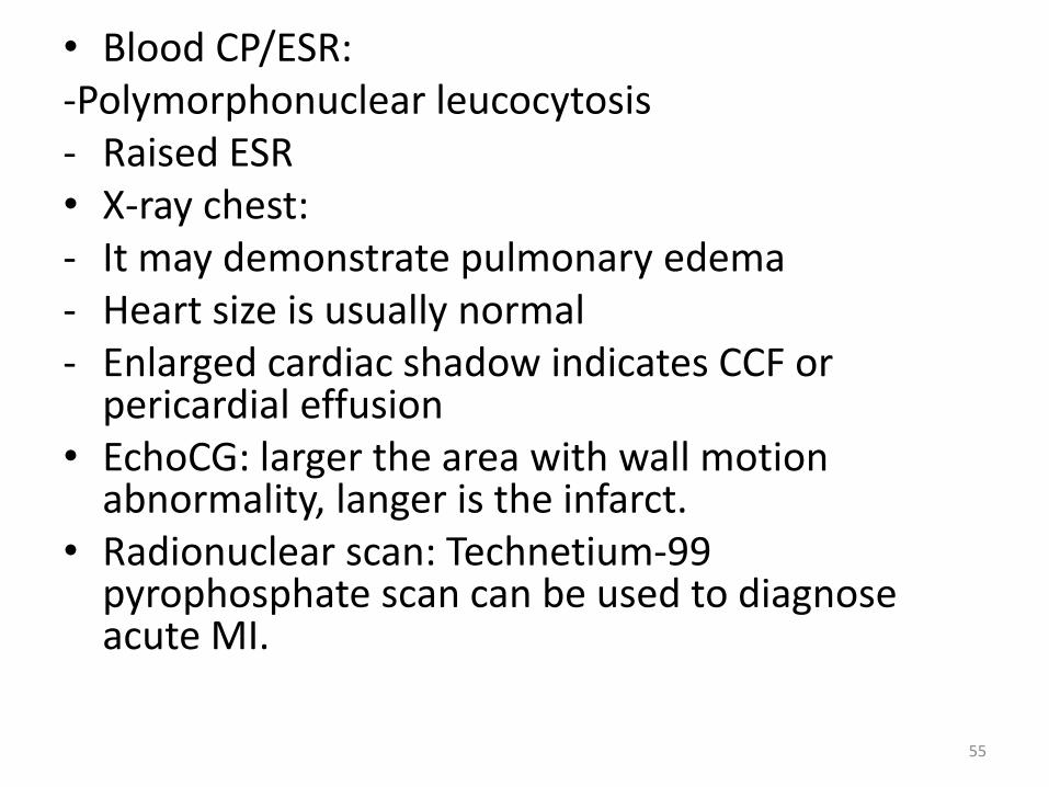
• Blood CP/ESR:-Polymorphonuclear leucocytosis- Raised ESR• X-ray chest:- It may demonstrate pulmonary edema- Heart size is usually normal- Enlarged cardiac shadow indicates CCF or
pericardial effusion• EchoCG: larger the area with wall motion
abnormality, langer is the infarct.• Radionuclear scan: Technetium-99
pyrophosphate scan can be used to diagnose acute MI.
55
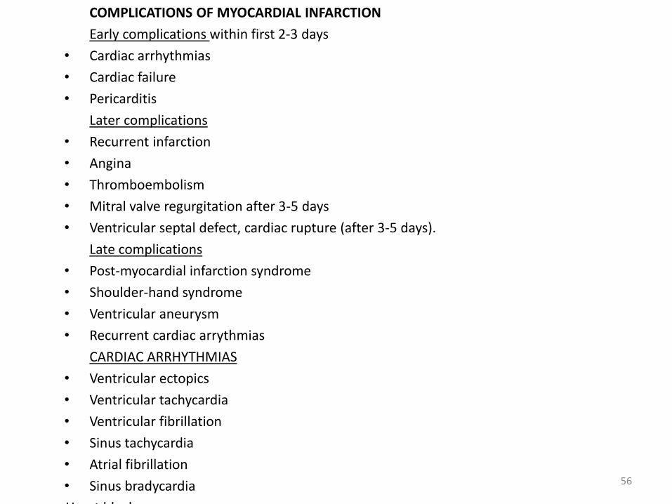
COMPLICATIONS OF MYOCARDIAL INFARCTION
Early complications within first 2-3 days
• Cardiac arrhythmias
• Cardiac failure
• Pericarditis
Later complications
• Recurrent infarction
• Angina
• Thromboembolism
• Mitral valve regurgitation after 3-5 days
• Ventricular septal defect, cardiac rupture (after 3-5 days).
Late complications
• Post-myocardial infarction syndrome
• Shoulder-hand syndrome
• Ventricular aneurysm
• Recurrent cardiac arrythmias
CARDIAC ARRHYTHMIAS
• Ventricular ectopics
• Ventricular tachycardia
• Ventricular fibrillation
• Sinus tachycardia
• Atrial fibrillation
• Sinus bradycardia
• Heart block
56

THE INFORMATION BLOCK
• The syndrome of cardiac failure - the pathological syndromedeveloping owing to an inconsistency of contraction functions of amyocardium on maintenance by oxygen of bodies and tissues of anorganism in rest and at loadings.
• To the factors, influencing on contraction ability of a myocardiumand defining productivity of left ventricule and requirement of amyocardium in 02, the concern preloading and postloading,condition contraction of myocardium, HR and a degree of damageof tissues.
• Preloading, or stretching degree myocardial tissues in rest, isdefined by end-diastolic volume and thickness of a wall of heart. Achronic stretching and HLV change the end-diastolic pressure. Inclinic a functional condition of ventricule ( contraction condition)judge on size of cardiac output ( stroke volume of leftventricules/end-diastolic volume)
57

• Postloading, or force of resistance to shortening oftissues of a myocardium after their stimulation for anexit from a relaxation condition, is defined by pressurein a cavity, volume of a cavity and thickness of a wall ofventricule at the moment of opening of aortic valve.
• Productivity of a myocardium influence availability ofsubstrata and presence of damages: decrease indelivery 02 and supply by the basic substrats (fat acidsand glucose) can conduct to damage of a myocardiumwith reduction of force of reduction of heart and itsproductivity.
• Damage of tissues, acute (caused by a myocardialinfarction) or chronic (with fibrosis, developed as aresult of tissues damage), not only locally breaksmechanics of a myocardium, but also imposesadditional loading on the tissues which has keptviability.
58

The causes: the cardiac failure can be caused defeat of the valvular device (a volume overload, pressure), coronary vessels or a myocardium.
• The valvular defeats of heart accompanied by stenoses or regurgitation, increase working loading by a myocardium, and eventually lead to its deterioration his contruction conditions.
• Damage of coronary arteries, reducing cardiac muscle blood supply, causes an ischemia or infarction with loss as a result of a part of functionally active myocardium.
• The myocardium deseases consist of the various conditions, which damages it contraction ability (myocarditis, myocardium dystrophies)
59

Due to decrease of contraction abilities of myocardium the left ventricule it is emptied not complete, that is shown by reduction of cardiac output. If this indicator decreases on 10 % or more end-diastolic pressure in the left ventricule, or preloading increases; in spite of the fact that it the compensate mechanism directed to increase of efficiency of systolic contraction, as a result leads to increase of venous and capillary pressure in lungs and haenostasiadevelops , thus decrease of cardiac output. The systemtemicarterial pressure is supported by compensatory growth of resistance of peripheral vessels, but it leads to the further increase in postloading and complicates mechanical works of ventricules.
Сoronary failure develops acute and chronically. In typical cases acute coronary failure develops at the patients who have transferred an extensive myocardial infarction or after rupture of the valve of heart. Chronic coronary failure is observed more often at persons with slowly progressing cardiomyopathy or heart valvular damages.
60

• At acute coronary failure - sudden reduction cardiac output is often accompanied by a hypotension without peripheral hypostases.
• At chronic coronary failure - the blood pressure, on the contrary, is long supported at normal level, but in tissues there is a fluid accumulation.
The majority of clinical displays of coronary failure develops as a result of fluid accumulation in system of one or both circles of blood circulation. This fluid usually accumulation in the vascular channel, located above the damaged chamber of heart. So, in case of mechanical overload of left ventricule (aortic stenosis) or its weaknesses (postinfarction myocardium changes), in result of haemostasia in lungs develop dyspnea and orthopnea. This condition called left ventricular coronary failure. If suffer right ventricule, as in case of a pulmonary hypertensia (for example, at thomboemboly of pulmonary artery) hypostases, stagnant hepatomegaly, expansion of veins are expressed. If coronary failure remains for a long time at patients with damage of valvular apparats of an aorta or systemic hypertensia at late stages of disease, develops stagnant hepatomegaly and enlarged of veins.
61

• Chronic coronary failure. Classification (Strajesco N.D., VasilenkoV. H, Muharljamov N.M., 1978).
• I stage (initial or hidden): fatigability, dyspnea and tachycardia at physical activity. In the early (preclinical) period: inadequacy of functions of heart comes to light only against extreme loadings or during carrying out of functional tests (three-stage test of the Master). In the presence of dyspnea, sweating, tremor of fingers, cyonosis of a nose and lips after 10 minutes and more after loading the initial condition of the patient is slowly restored.
• II stage (expressed):A - developments of hypostasis in one of blood circulation circles.It is clinically shown by a dyspnea, palpitation, fast fatigue at moderate
physical activity, it is accompanied with acrocyonosis, a tachycardia, increase in the sizes of heart, insignificant increase in a liver, presence of damp rattles in the inferior departments of lungs; X-ray- strengthening of pulmonary drawing and roots of lungs of hypostasis character;
62

B- proof deep infringements of blood circulation in both circles of blood circulation. Clinically: the dyrpnea in rest, оrthopnea (the compelled sitting position), cough in horizontal position, the big dense liver with the roundish or slightly pointed edge, painful at palpation, acts under costal edges on 6-8 sm, massive hypostases of shins and feet, acrocyonosis, is possible etrial fibrillation, heart borders are expanded, tones of heart are weakened, a tachycardia «a gallop rhythm», damp rattles in the inferior departments of lungs, hydrothorax, a hydropericardium, аcytis.
X-ray: consolidation of roots of lungs, pleural and interlobal fluid.ECHOKG: increase the end-diastolic size of left ventricule, the end-
systolic size of left ventricule, the anterio-posterior size left, - right atriums, right ventricule , decrease of cardiac output.
• III - stage (final, dystrophic) - an exhaustion of patients (cardiac kachesia ), orthopnea in rest, a skin flabby, acrocyonosis, massive hypostases of the inferior finitenesses, аnasarka, the sizes of heart are increased, tones of heart are weakened, a tachycardia, is possible atrial fibrillation, considerably increased liver, can be a jaundice.
63

• Classification of coronary failure by New-York Association of heart (NYHA).
• I-FC - patients with disease of heart without restriction of physical activity, symptomless left ventricular dysfunction
• II-FC - patients with disease of heart with restriction of easy physical activity
• II-FC - patients with disease of heart with considerable restriction of physical activity, moderate cardiac failure
• IV-FC - patients are incapable to carry out the easiest loading without sensation of discomfort.
64
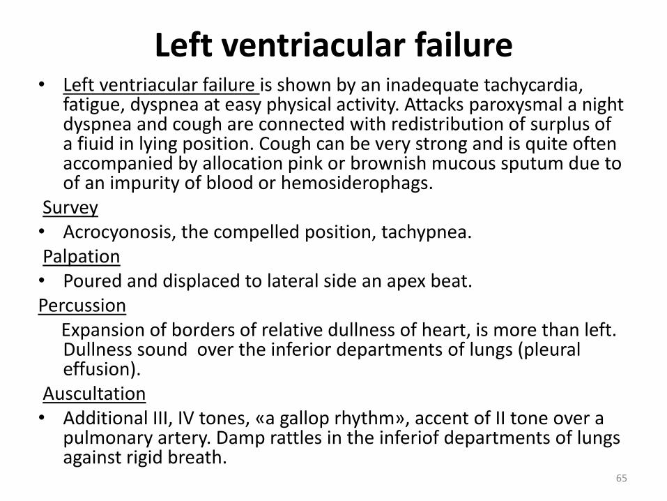
Left ventriacular failure• Left ventriacular failure is shown by an inadequate tachycardia,
fatigue, dyspnea at easy physical activity. Attacks paroxysmal a night dyspnea and cough are connected with redistribution of surplus of a fiuid in lying position. Cough can be very strong and is quite often accompanied by allocation pink or brownish mucous sputum due to of an impurity of blood or hemosiderophags.
Survey• Аcrocyonosis, the compelled position, tachypnea.Palpation• Poured and displaced to lateral side an apex beat.Percussion
Expansion of borders of relative dullness of heart, is more than left. Dullness sound over the inferior departments of lungs (pleural effusion).
Auscultation• Additional III, IV tones, «a gallop rhythm», accent of II tone over a
pulmonary artery. Damp rattles in the inferiof departments of lungs against rigid breath.
65

Acute left ventricular failire(a hypostasis of lungs)Represents display menacing to a life the acute left
ventricular insufficiency, caused suddenly occurrence of a pulmonary venous hypertensia. Prompt growth of pressure of filling of left ventricule leads to a fast exit of a fluid part of plasma through walls of pulmonary capillaries to in intersticial space and alveoles. At the patient develop the dificult dyspnea, cyanosis, tachypnea, the anxiety and fear, appears sensation of an asthma. Are usual pallor and plentiful sweating. Pulse can be thready, the blood pressure is sometimes difficult for defining. Breath is complicated, more of damp and dry rattles in front and behind in both lungs. At some patients the strong bronchospasm or stridor (a cardiac asthma) develops. Heavy hipoxemia and sharp cuanosis.
66

Right ventricular failureRight ventricular failure (with a pulmonary venous hypertensia or
without it). Leading symptoms - fatigue, feeling of burst in a neck, sensation of completeness in a stomach, morbidity over a liver, a swelling of anklebones, hypostases of shins, асytes.
• ECG: there are no specific signs of cardiac failure. Changes on an ECG can specify in causes cardiac failure (hypertrophy of left ventricular, MI , LBBB of RBBB).
• X-ray of bodies of a thorax: hypostasis in pulmonary veins and increase of pulmonary drawing. Kerly’s lines reflect long increase of pressure in left atrium and represent a chronic thickening the interlobar septs, caused by a hypostasis.
• EchoCG: the sizes of left ventricule and other chambers of heart, size of cardiac output and infringement of movement of walls. To define degree of dilatation of left ventricule and hypertrophies of left ventricule, pathological changes of valves of heart, fluid in pericardium cavities, intracardiac thrombus and tumours.
67