cwhc-rcsf - centre for coastal health...pathogenic avian influenza, mycobacterium avium, newcastle...
TRANSCRIPT
A REVIEW OF POTENTIAL HEALTH HAZARDS TO HUMANS AND LIVESTOCK FROM CANADA GEESE (Branta canadensis)
AND CACKLING GEESE (Branta hutchinsii)
Prepared for:
The Canadian Wildlife Service
Prepared by: Erin Fraser, BSc DVM MSc
Stu Fraser, BFA BEd
CENTRE FOR COASTAL HEALTH On behalf of
Canadian Cooperative Wildlife Health Centre
February 15, 2010
[1]
TABLE OF CONTENTS 1. Executive summary ............................................................................................................................................................ 2
2. Reason for request ............................................................................................................................................................. 5
3. Methodology ......................................................................................................................................................................... 7
4. Uncertainty ............................................................................................................................................................................ 9
5. Hazard Identification ...................................................................................................................................................... 12
6. Adapted Risk Assessment ............................................................................................................................................. 15
6.A. Release assessment ................................................................................................................................................. 15
Risk setting ..................................................................................................................................................................... 15
Mechanisms for exposure to humans from goose pathogens ................................................................... 18
Mechanisms for exposure to livestock from goose pathogens ................................................................. 18
6.B. Exposure assessment ............................................................................................................................................. 19
6.C. Consequence assessment ...................................................................................................................................... 22
7. Synthesis ............................................................................................................................................................................... 23
8. Recommendations ........................................................................................................................................................... 27
9. Reference List ..................................................................................................................................................................... 30
Appendix A: Overview of pathogen‐specific criteria related to possible transmission from geese to humans in North America. ................................................................................................................................................. 56
Appendix B: Overview of pathogen‐specific criteria related to possible transmission from geese to livestock in North America. ............................................................................................................................................... 67
Appendix C: Characteristics of identified hazards that could impact humans (magnitude of potential impact) ....................................................................................................................................................................................... 81
Appendix D: Characteristics of identified hazards that could impact livestock (magnitude of potential impact) ................................................................................................................................................................... 88

[2]
1. EXECUTIVE SUMMARY Canada and Cackling geese are protected under Canada’s Migratory Birds Convention Act1. This act prohibits capturing or killing of geese, or damaging, destroying, removing, or disturbing their nests, except as provided for under the Migratory Birds Regulations2. Canada geese, along with all other migratory birds, are protected and managed by the Canadian Wildlife Service (CWS) of Environment Canada. The Migratory Birds Regulations provide for management actions under permit to “remove or eliminate migratory birds or nests where it is necessary to do so to avoid injury …” CWS requested the Canadian Cooperative Wildlife Health Centre (CCWHC) to perform a risk assessment in order to support their decision‐making for permit issuance for removing protected geese because of concerns for human or livestock health. The most significant conclusion of this review was that there is an insufficient basis in available evidence to conduct a reliable and meaningful risk assessment of infectious hazards. Most of the data requirements to make a valid assessment of risks simply could not be met with the available data on pathogens potentially transmitted by geese. There were very large gaps in some of the following key determinants of risk: prevalence of pathogens and parasites in geese, epidemiological information to link the pathogen in geese to cases in people or livestock, goose fecal distribution patterns, and human or livestock exposure patterns (i.e. the nature and extent of contact between geese and humans and geese and livestock). As a result, a reliable, evidence‐based risk assessment of health risks to people or livestock from free‐ranging geese could not legitimately be performed with the existing availability and quality of data. Hazard identification There was evidence that many pathogens of importance to humans and livestock can infect Canada and Cackling geese and be shed into the environment by geese, and vice versa. However, there was a very large gap in our understanding of the ecological and epidemiological factors that may contribute to the transmission from geese to humans or livestock. As discussed above, effective risk analyses (identification, assessment, management and communication) require data inputs from quality surveillance information (Table 1). Although there was a large list of pathogens associated with geese and other members of the Anatidae family, there was scarce serological, microbiological or epidemiological data as evidence of transmission between geese and humans and geese and livestock. Release assessment We can conclude that there are several plausible routes of exposure to infectious material from geese, but estimates of the probability of these exposures are not attainable with existing data. 1 http://www.cws‐scf.ec.gc.ca/legislations/laws1_e.cfm 2 http://laws.justice.gc.ca/en/M‐7.01/C.R.C.‐c.1035/

[3]
The most plausible source of infection for both people and livestock appears to be via contaminated aquatic environments. Another plausible route is through direct contact with infected fecal material (this includes infected airborne particles). These routes provide the opportunity for the widest possible exposure for both people and livestock. In this report we highlight some of the pathogens that we feel should be of greatest concern to CWS and as a result be considered in their risk management and communication strategies. However, it must be noted that none of the pathogens examined had sufficient evidence (i.e. temporality, specificity, consistency, etc) to demonstrate a causal relationship between exposure to a pathogen of goose origin and a resulting infection in either people or livestock. Due to the fact that Canada and Cackling geese are aquatic birds that aggregate in large numbers and produce a prolific amount of feces, concerns have been raised about their role in water contamination. As a result, there has been an emphasis on research of waterfowl‐related pathogens and in particular on E. coli, Cryptosporidium, and Giardia. This research bias could affect the assessment of risks from waterborne transmission from geese in that more important, but unresearched pathogens are not considered as much as these three pathogens. In addition, the perceptions of waterborne pathogen risks are shifting as a consequence of more recent molecular and epidemiological information that is coming on stream and showing more host specificity than previously thought. Hence, the assessment of pathogens in this report might be biased by the older literature that could not make distinctions between strains that may be highly host‐specific and therefore present a diminished risk for interspecies transmission (Hansen et al, 2009; Graczyk et al, 2008). Consequence assessment In this report, the pathogens that were categorized as ‘high’ potential impact to human health had more than one of the following features: 1) are capable of causing severe illness that can result in hospitalization; 2) had the ability to spread epidemically from person‐to‐person; 3) had high fatality rates; 4) did not have effective treatment or preventive methods; or 5) were nationally reportable. The pathogens that filled more than one of these criteria included: highly pathogenic avian influenza (HPAI), enterotoxigenic E. coli (ETEC), and West Nile virus. Pathogens of medium and low impact are shown in Appendix C. In livestock, the pathogens with a ‘high’ ranking had more than one of the following features: 1) are capable of causing severe illness; 2) had the ability to spread epidemically within or between herds or flocks; 3) had high fatality rates; 4) did not have effective treatment or preventive methods; or 5) were nationally reportable to the Canadian Food Inspection Agency. The pathogens that filled more than one of these criteria included: highly pathogenic avian influenza, Mycobacterium avium, Newcastle disease, and West Nile virus. We found no direct evidence that linked human or livestock health outcomes to geese. Hence, this consequence assessment is the consequence of the pathogens in general and not the pathogen from geese specifically. Almost all of the hazards listed have multiple sources including people, other species and environmental sources.
[4]
Synthesis Major deficits exist in our understanding of the frequency of disease‐causing agents in free‐ranging geese. Perhaps more importantly, there was virtually no information on the frequency or probability with which pathogens from wild geese are transmitted from geese to people or livestock. We found a fairly extensive list of potential infectious and parasitic pathogens from geese that could infect people, poultry or other livestock and thus the potential for shared diseases exists. We concluded that the nature of the risks are not generic across Canada and are very context specific. The risks are affected by the potential exposure pathway (with waterborne being the route of most concern); people or livestock being exposed (immune compromised people and domestic poultry being of highest risk) and the specific pathogens of concern (enterohemorhagic E. coli, avian influenza, waterborne protozoan pathogens Cryptosporidium and Giardia, West Nile virus and Newcastle disease virus in poultry are the most reasonable to be concerned about). Recommendations
• Invest in monitoring and research that will provide the CWS with the necessary components to develop an evidence‐based risk assessment. Some of the key areas of investigation should be improving our understanding of exposure probabilities (i.e.. nature and extent of goose interactions with people and livestock) but also elucidating the prevalence of specific pathogens for which we have no information for geese, as well as further genetic characterization of strains of pathogens and how they are related to similar human or livestock pathogens. There is a large number of possible hazards widespread over a variety of risk settings in Canada. Investment in such a program of research would need to be substantial and long‐term to make significant shifts in our understanding of these determinants of risk.
• Model risks for some of the priority pathogens identified here as a means of accounting for the high degree of uncertainty. Uncertainty can either be modeled using quantitative or qualitative methods. Increasingly in areas in which there are large data gaps or uncertainty, a mixed approach, such as multi‐criteria decision making can be used. However, models would need to account for the issue of context which modifies risk (exposure setting, nature of the at risk group, etc).
• Develop and improve risk reduction and communication strategies that focus on specific higher risk settings (i.e. aquatic resources, hospital settings, high livestock density areas).
• Form a working group to develop national standards for the management of peri‐urban goose populations. Expertise from public health, regulatory agencies, resource managers, engineers, epidemiologists, and biologists along with input from the public are needed to develop a consensus on goose management practices. A consensus approach has been used to good effect in other situations where there is a high level of uncertainty.
[5]
2. REASON FOR REQUEST The Canadian Cooperative Wildlife Health Centre (CCWHC) was asked by the Canadian Wildlife Service (CWS), Environment Canada to carry out a risk assessment of Cackling (Branta hutchinsii) and Canada (Branta canadensis) geese on human and livestock health. The CCWHC requested the Centre for Coastal Health (CCH) to complete a risk assessment. The CCH is the Pacific regional node of the CCWHC. The CCH has a strong background and expertise in performing risk assessments on a variety of issues ranging from assessing risks in the translocation of endangered species, to chronic wasting disease, to assessing disease risks in aquaculture settings. Canada and Cackling geese are protected under Canada’s Migratory Birds Convention Act3. This act prohibits capturing or killing of geese, or damaging, destroying, removing, or disturbing their nests, except as provided for under the Migratory Birds Regulations4. Canada Geese, along with all other migratory birds, are protected and managed by the Canadian Wildlife Service (CWS), Environment Canada. The Migratory Birds Regulations provide for management actions to “remove or eliminate migratory birds or nests where it is necessary to do so to avoid injury …” CWS requested the Canadian Cooperative Wildlife Health Centre (CCWHC) to perform a risk assessment in order to support their decision‐making for permit issuance for removing protected geese because of concerns for human or livestock health. They often receive requests for permits to “destroy eggs, relocate or kill birds (primarily Canada Geese), particularly from urban environments. Among other concerns, the justification accompanying the request is sometimes about concerns for human health and in other situations for livestock health.” They have found that they have insufficient information on the actual health risks to humans or livestock on which to base their decisions. Some of the cases presented to them are straightforward in the sense that there is clearly property damage occurring as a result of the presence of geese. It is the cases where the issue of ‘risks to humans or livestock health’ is the stated reason for the request that the CWS requires a more detailed analysis of the risks to determine whether the requirements of the Migratory Birds Regulations would be met and thus support their decision about whether or not to issue a permit (pers. comm., Kathryn Dickson, CWS). Most goose populations have increased significantly throughout North America over the last several decades5. The public has become increasingly concerned with the both the aesthetic of high numbers of geese and the associated contamination of green spaces, beaches, and ponds. In addition, perhaps as a result of recent dissemination of, and associated media attention towards pathogens such as highly pathogenic Avian influenza (HPAI) and West Nile virus (WNV), there is increasing public concern that pathogens carried by Canada geese pose risks to human and livestock health. As a result of these concerns, there has been an increase in the number of
3 http://www.cws‐scf.ec.gc.ca/legislations/laws1_e.cfm 4 http://laws.justice.gc.ca/en/M‐7.01/C.R.C.‐c.1035/ 5 http://www.cws‐scf.ec.gc.ca/mgbc/trends/index.cfm?lang=e&go=info.bird&speciesid=1720

[6]
complaints received by Parks Departments and other agencies, such as the CWS, which are responsible for managing Canada and Cackling geese in Canada. There are a variety of risks that geese do or might present including risks to public health, agriculture, conservation, economics, animal welfare, or the environment. The types of potential risks to public health from geese could be grouped into the following categories: physical (i.e. trauma), psychological, or infectious. The categories of risk to livestock include: physical harm (i.e. trauma) and infectious disease. The bulk of public discourse (i.e. media) related to Canada or Cackling geese focus on these species as nuisance animals in public spaces and potential sources of pathogens infectious for humans or livestock. The risk of goose to human trauma or psychological harm is most pronounced in the aviation industry where there have been a number of incidents in which Canada geese have collided with airplanes (Eschenfelder, 2001). Canada geese are known to be highly protective during nesting season and could frighten people and other animals in their efforts to defend their territory against intruders. However, these types of incidents remain unquantified (Allan et al, 1995). There was also some reference to the risk to humans from slipping in the feces of Canada geese that are abundantly deposited in areas of high goose and human usage (i.e.. parks, golf courses, etc) (Allan et al, 1995; Conover and Chasco, 1985); however, we were unable to locate any data to support this claim or document the extent of this problem. Potential risks to economic activities (i.e.. parks agencies, golf courses, private property value; not including livestock‐related economics and trade), agriculture (crops), conservation (threats to migrant subspecies from overabundant resident goose populations), as well as to the environment (i.e.. overgrazing, eutrophication and algal growth of ponds/lakes) posed by Canada geese have been documented elsewhere and are beyond the scope of this risk assessment (Manny et al, 1994; Conover, 1991; Flegler et al, 1987; Conover and Chasco, 1985). A variety of reports and assessments have been produced in regards to the management of goose populations throughout North America (USDA, 2004; Clark, 2003; US Fish and Wildlife, 2002; Gabig, 2000; Canada Goose Committee Atlantic Flyway Technical Section, 1999). A common theme in these documents is the reference to the health risks that geese pose to people and livestock. However, it is interesting to note that even though the public discourse around the Canada goose as a nuisance places a strong emphasis on human health risks, the majority of complaints to the USDA‐APHIS Wildlife Service in West Virginia were found primarily to be related to damage to property (82%) or agriculture (15%) rather than human health and safety (3%) (USDA, 2004). As per the request by CWS, the focus of this report is on the role of Canada and Cackling geese in the transport, dissemination, and possible transmission of pathogens of importance to human and livestock health.
[7]
3. METHODOLOGY
LITERATURE REVIEW A literature search for peer‐reviewed articles was undertaken using PubMed, Google Scholar, Science Direct and Agricola database systems/search engines. The following terms were used: ‘geese’, ‘Canada goose’, ‘Anatidae’, ‘waterfowl, ‘migratory birds’, ‘infection’, ‘disease’, ‘management’ and specific pathogens known to be associated with birds or waterfowl such as ‘Avian Influenza’, ‘Salmonella’, ‘West Nile Virus’, etc. In addition, we searched for literature, using the same search engines, on the pathogens identified in our search for bird/goose‐associated literature, which was associated with human or livestock disease or infection. A stronger emphasis was placed on scientific literature that was published after 1980 due to both accessibility reasons and to the improvement in pathogen detection and elucidation methodologies (i.e. molecular tools) that have the potential to provide more information on the likelihood of transmission from geese to either humans or livestock.
Non peer‐reviewed literature, or ‘gray literature’, was searched using Google using the following terms: ‘Canada geese/goose’, ‘Cackling geese/goose’, ‘management’, ‘population’, ‘migration’, biology’, ‘disease’, and ‘interaction’. We also carried out specific searches of the following agency websites:
• Canadian Food Inspection Agency (www.inspection.gc.ca) • Canadian Wildlife Service, Environment Canada (www.cws‐scf.ec.gc.ca) • Canadian municipal parks departments (Kelowna, Kitchener, Montreal, Niagara,
Toronto, Vancouver, Victoria) • Canadian Cooperative Wildlife Health Centre (www.ccwhc.ca) • United States Department of Agriculture, Animal and Plant Health Inspection Service,
Wildlife (USDA/APHIS), National Wildlife Research Centre (www.aphis.usda.gov/wildlife_damage/nwrc)
• United States Fish and Wildlife Service, Migratory Bird Program (www.fws.gov) Canadian regulations and policies around the management of Canada and Cackling geese populations were reviewed:
• Canada Environmental Protection Act (http://laws.justice.gc.ca/en/C‐15.31/) • Migratory Birds Convention Act (http://laws.justice.gc.ca/eng/M‐7.01/index.html) • Migratory Birds Regulations (http://laws.justice.gc.ca/eng/C.R.C.‐c.1035/index.html)
We also obtained additional clinical and epidemiologic information on specific pathogens and disease manifestations through the following online sources: Centre for Disease Control (CDC), World Organization for Animal Health (OIE), and the Merck Veterinary Manual. For the purposes of this risk assessment, risks posed by Canada and Cackling geese were considered together. None of the literature we encountered on the pathogens associated with geese indicated that there were any differences between Canada or Cackling geese. In fact, Cackling geese, because of its relatively recent status as a separate species (2004), were not mentioned in the pathogen‐focused body of literature that we reviewed.
[8]
HAZARD IDENTIFICATION, REVIEW, AND PRIORITIZATION Hazards to humans or livestock from geese were identified using the following criteria:
Step 1: Can the potential hazard infect or be transmitted by Canada or Cackling geese or another member of the Anatidae family that includes geese, ducks, and swans? Step 2: Can the potential hazard produce negative impacts on humans or livestock? Step 3: Is the potential hazard present in North America?
If the answer to either the first or second steps was yes or uncertain AND it is present in North America, then the disease or pathogen was identified as a hazard. For each hazard identified, we reviewed the literature for relevant criteria that could contribute to an estimation of the probability of transmission from geese to humans and geese to livestock (see Appendices A and B). The variables that were included were:
• prevalence of pathogen in wild geese in North American • prevalence of pathogen in humans or livestock in North America • detection and persistence of the agent in the environment • evidence for cross‐species transmission • evidence for goose to human or goose to livestock transmission
The impact on human or livestock health, should a pathogen be transmitted from Canada or Cackling geese, were individually evaluated and rated (Appendices C and D). There was insufficient information to rate the impact of the pathogens quantitatively, so all ratings were carried out qualitatively using the criteria described below. The variables that were considered to assess the impact in either people or livestock for each of the hazards included:
• nature and severity of disease • morbidity, mortality, hospitalization rates • availability of suitable treatment and prevention measures • national disease reporting requirement6,7
The categories of impact were determined based on the following criteria: Negligible No, rare or sporadic disease resulting from infections. Very effective
treatments or control measures available. No impacts on trade. Not reportable.
Low Disease can occur uncommonly from infection and/or symptoms are generally mild or self limiting. No impacts on livestock herd/flock
6 List of human reportable diseases in Canada: http://dsol‐smed.phac‐aspc.gc.ca/dsol‐smed/ndis/list_e.html 7 Animal disease reporting to the CFIA: http://www.inspection.gc.ca/english/anima/disemala/guidee.shtml Reportable diseases: “[diseases that are] usually of significant importance to human or animal health or to the Canadian economy” Immediately notifiable diseases (for laboratories only): “diseases [that] are exotic to Canada for which there are not control or eradication programs” Annually notifiable diseases (for laboratories only): “diseases for which Canada must submit an annual report to the World Organization for Animal Health (OIE) indicating their presence in Canada, but are not classified as reportable or immediately notifiable”

[9]
productivity. Effective treatment or control is available. Hospitalization or death is unexpected. No impacts on trade. Not reportable.
Medium Resulting disease can cause illness requiring medical attention and sometimes hospitalization. Deaths can occur in individuals and/or some herd/flock impacts are possible. Possible, but low, impacts on trade. The disease is reportable and treatment options are either less effective or limited in availability.
High Resulting disease can cause severe illness and can spread epidemically, often requiring hospitalization in people. Deaths occur in individuals and/or herd/flock impacts occur. Livestock trade is affected. The disease is reportable. No treatment or prevention methods are available.
For many pathogens there is a range in the nature and severity of disease. There are often extreme manifestations and sequelae of infection that come about for a variety of reasons. Most often these more severe outcomes are due to risk factors in the susceptible species such as: immune status, age, gender, or predisposing conditions. Our final impact assessment is based on the average manifestation of disease but highlights where immune status or other risk factors might escalate severity of disease.
4. UNCERTAINTY The framework that is commonly used and promoted by the World Organization for Animal Health (OIE) to assess risks in animal populations involves three components: release, exposure and consequence assessments (Zepeda et al, 2001). The release assessment determines whether the disease is present (or potentially present) in either the country of origin or species of interest, which, in this case, are the Canada and Cackling goose. The exposure assessment describes the pathways of exposure and associated probabilities that could cause infection in other populations, which, in this case, are people or livestock (note, for the purposes of this report, we include domestic birds and mammals raised for agricultural purposes as livestock). The consequence assessment describes the biologic (i.e. mortality and morbidity rates) and economic (i.e. trade restrictions, days off work) impacts should the disease occur in the species of concern (people and livestock in this case). Risk is the combination of the likelihood of occurrence of an adverse event and the magnitude of the consequences (Zepeda et al, 2001).
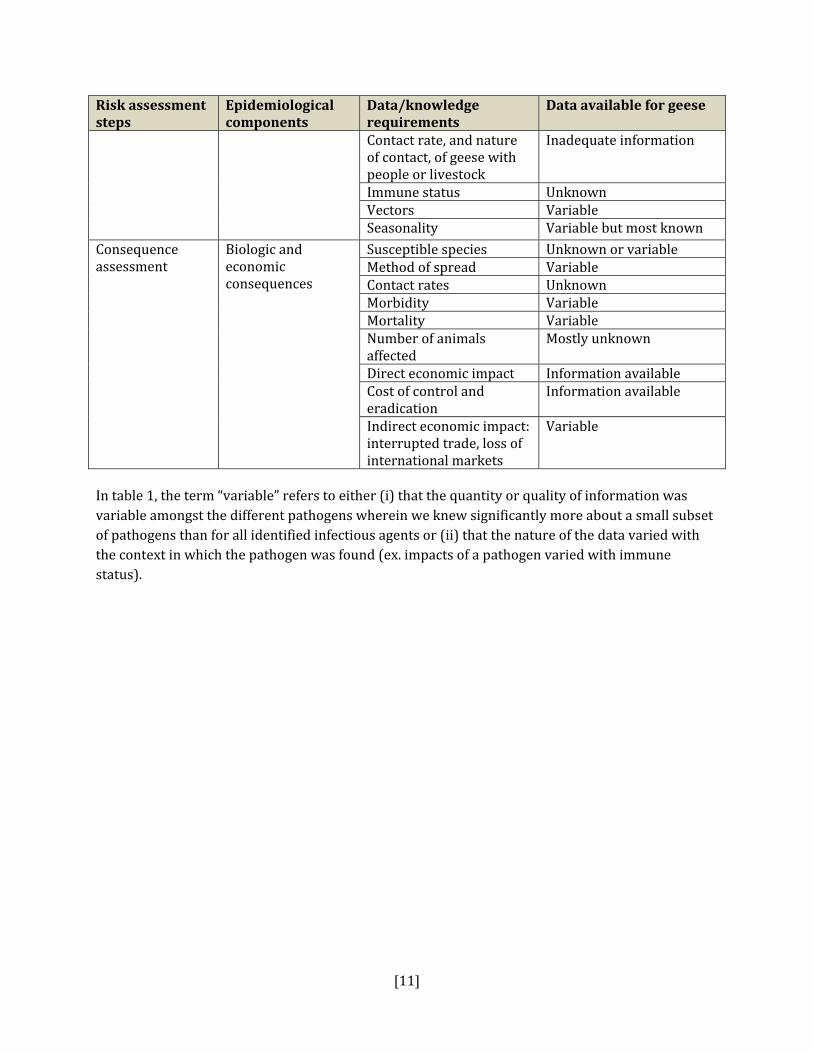
Table 1 outlines the data requirements for risk assessment and uncertainty analyses, adapted from Zepeda (2001). For this risk assessment concerning pathogens in wild geese, most of the data requirements simply cannot be met with the available data on pathogens potentially transmitted by geese. There are very large gaps in some of the following key determinants of risk: prevalence of pathogens and parasites in geese, epidemiological information to link the pathogen in geese to cases in people or livestock, fecal distribution patterns, and human or livestock exposure patterns (i.e.. the nature and extent of contact between geese and humans and geese and livestock). As a

[10]
result, a reliable, evidence‐based risk assessment of health risks to people or livestock from free‐ranging geese cannot legitimately be performed with the existing availability and quality of data. Unfortunately, this is not very helpful to the CWS as it must make decisions in the face of uncertainty and the absence of critical information on the true nature of risks that geese pose to human and livestock health. Therefore, we have adapted the risk assessment framework described above into a format that provides CWS with current information on any supporting evidence for pathogen transmission from geese and also provides some tools to help prioritize pathogens into areas of potential risk. Table 1: Information requirements for risk assessment and uncertainty analyses
Risk assessment steps
Epidemiological components
Data/knowledge requirements
Data available for geese
Hazard identification
List of pathogenic agents that could be associated with geese
Pathogens exotic to Canada
Inadequate surveillance
Emerging pathogens Inadequate surveillanceEpidemiology of each endemic, epidemic and emerging pathogens in relation to geese
Limited to isolated surveys for the most part focused on a subset of pathogens
Knowledge on the presence or absence of pathogen in Canada
Methods to demonstrate absence of pathogen
Iinsufficient testing
Release assessment
Prevalence of pathogen in Canada
Survey and surveillance results
Inadequate information
Survey methodology Variable Confidence level, precision, expected prevalence
Unknown
True prevalence Unknown Epidemiological characteristics of the disease and the pathogen
Incubation Variable Carriers Variable but largely
unknown Morbidity Inadequate informationMortality Inadequate informationMethod of spread Inadequate informationPathogenesis Variable Target organs Variable Susceptible species Variable
Diagnostic tests Test sensitivity and specificity
Inadequate information
Exposure assessment
Characteristics of the susceptible populations and environmental factors
Pathways of exposure Variable and unquantifiedFlock densities Some information
available. Flock distributions Some information
available.
[11]
Risk assessment steps
Epidemiological components
Data/knowledge requirements
Data available for geese
Contact rate, and nature of contact, of geese with people or livestock
Inadequate information
Immune status Unknown Vectors Variable Seasonality Variable but most known
Consequence assessment
Biologic and economic consequences
Susceptible species Unknown or variableMethod of spread Variable Contact rates Unknown Morbidity Variable Mortality Variable Number of animals affected
Mostly unknown
Direct economic impact Information availableCost of control and eradication
Information available
Indirect economic impact: interrupted trade, loss of international markets
Variable
In table 1, the term “variable” refers to either (i) that the quantity or quality of information was variable amongst the different pathogens wherein we knew significantly more about a small subset of pathogens than for all identified infectious agents or (ii) that the nature of the data varied with the context in which the pathogen was found (ex. impacts of a pathogen varied with immune status).

[12]
5. HAZARD IDENTIFICATION This section provides a list of the pathogens that could be transmitted between geese and people or geese and livestock (Table 2). This is not a complete list of all pathogens of consequence to geese but narrowed to those that could potentially infect people or livestock. They were selected based on literature review (see methodology section). Due to the scope of the project and the significant number of agents that needed to be considered, only a cursory review of nematodes and helminths of geese was carried out. Table 2: List of potential hazards that are found in North America in members of the Anatidae family (geese, ducks, swans) and in either people or livestock
Pathogens Demonstrated infection in people
Demonstrated infection in livestock
References
Bacterial Actinobacillis suis No Yes, swine Gottschalk, 2000; Maddux et al, 1987Arcobacter spp. Yes Yes, all species Collado et al, 2009; Houf et al, 2009; Atabay
et al, 2008; Ho et al, 2006: Vandenberg et al, 2004
Borrelia anserine (avian borreliosis)
No Yes, poultry Lisboa et al, 2009; Ataliba et al, 2007
Borrelia burgdorferi Yes Yes, poultry Ogden et al, 2009; Reed et al, 2003; Piesman et al, 1996; Burgess, 1989
Campylobacter spp. Yes Yes, all species Hughes et al, 2009; Colles et al, 2008; French et al, 2009; Converse, 1999
Chlamydophila psittaci Yes Yes, poultry Olsen, 2009; Laroucau, 2008; Fallacara, 2004; Longbottom and Coulter, 2003; Converse, 1999
Clostridium botulinum Type C (avian botulism)
No Yes, poultry(note: this is an intoxication from ingestion of spores rather than an infection from C. botulinum
Lu et al, 2009; Rocke, 2006; Jean et al, 1995; Wobeser et al, 1987
C. perfringens (‘necrotic enteritis’)
Yes Yes, all species Olsen, 2009; Holtby, 2008; Leal et al, 2008; Turcsan, 2001; Wobeser, 1987
Erysipelothrix rhusiopathiae (Erysipelas)
Yes Yes, turkeys and pigs
Olsen, 2009
Escherichia coli Yes Yes, all species Edge and Hill, 2007; Kullas et al, 2002; Feare et al, 1999
Helicobacter spp. Yes Yes, all species Tsiodras et al, 2008; Fox et al, 2006Legionella pneumophilia Yes No Clark, 2003; Fields et al, 2002; Yu et al, 2002;
Fabbi et al, 1998 Listeria monocytogenes Yes Yes, all species Lianou and Sofos, 2007;
Swaminathan et al, 2007; Converse et al, 1999

[13]
Pathogens Demonstrated infection in people
Demonstrated infection in livestock
References
Mycobacterium avium Yes Yes, all species Olsen, 2009; Inderlied et al, 1993; Mycoplasma spp. Yes Yes, poultry Dobos‐Kovacs et al, 2009; Olsen, 2009;
Waites et al, 2004; Rosengarten et al, 2001; Bradbury et al, 1988; Baseman and Tully, 1997; Stipkovits et al, 1993
Pasteurella multocida (Fowl cholera)
Yes Yes, poultry Olsen, 2009; Pedersen et al, 2003; Samuel et al, 1997; Botzler, 1991
Salmonella enteric Yes Yes, all species Olsen, 2009; Converse et al, 1999: Feare et al, 1999
Streptococcus spp (Group D) (S. bovis, S. gallolyticus subsp. Pasteurianus)
Yes Yes, all species Herrera et al, 2009; Hogg and Pearson, 2009; Onoyama, 2009; Barnett et al, 2008; Devriese et al, 1998
Vibrio spp. (non‐cholera) Yes Yes, all species Bush et al, 2006 ; Hinz et al, 1999; Buck, 1990; Schlater et al, 1981
Yersinia pseudotuberculosis
Yes Yes, all species Tauxe, 2004: Niskanen et al, 2003
Fungal Aspergilla spp. Yes Yes, poultry Beytut et al, 2004; Akan et al, 2002; Stroud et
al, 1982; Adrian et al, 1978 Candida albicans Yes Yes, poultry Buck, 1990; Beemer et al, 1973
Cryptococcus spp. Yes Yes, all species Duncan et al, 2006; Filion et al, 2006; Soogarun et al, 2006; Baro et al, 1998
Parasitic Coccidia spp. No Yes, poultry Converse et al, 1999; Gomis et al, 1996;
Chave et al, 1993 Cryptosporidia spp. Yes Yes, all species Schuster et al, 2005; Zhou et al, 2004;
Graczyk, 1998; Giardia spp. Yes Yes, all species Thompson et al, 2008; Graczyk and Lucy,
2007; Schuster et al, 2005; Graczyk, 1998; Leucocytozoon spp. No Yes, poultry Shutler et al, 2009; Hellgren et al, 2008;
Nakamura et al, 2008; Bennett et al, 1982; Herman et al, 1975
Nematodes: Amidostomum ssp. , Epomidiostomum spp., Trichostrongylus spp.
No Yes, domestic waterfowl
Nowicki et al, 1995
Sarcocystis spp. Yes Yes, all species Dubey et al, 2006; Fayer, 2004; Kutkiene et al, 2008 and 2004
Toxoplasma gondii Yes Yes, all species Dorny et al, 2009; Dubey et al, 2002, 2006 and 2007; Tenter et al, 2000
Trematodes: Schistosome cercariae (swimmer’s itch)
Yes No Brant and Loker, 2009; Skírnisson et al, 2009

[14]
Viral Arboviruses (Eastern and Western Equine Encephalitis, St. Louis Encephalitis, West Nile Virus)
Yes Yes, all species Reimann et al, 2008; Thomas et al, 2007; Wojnarowicz et al, 2007; Austin et al, 2004; Banet‐Noach et al, 2003; McLean et al, 2002; Swayne et al, 2001
Avian adenovirus No Yes, poultry Chen et al, 2009Avian herpesvirus (duck viral enteritis)
No Yes, domesticated waterfowl
Campagnolo et al, 2001; Converse and Kidd, 2001; Gough and Hansen, 2000
Avian influenza Yes Yes, poultry Brown et al, 2008; Ward et al, 2008; Pasick et al, 2007; Van Reeth et al, 2007; Clark and Hall, 2006; Gill et al, 2006; Olsen et al, 2006
Avian pneumovirus No Yes, poultry Cook, 2000; Shin et al, 2000 Avian pox virus No Yes, poultry Buller and Palumbo, 1991; Cox, 1980Coronavirus Yes Yes, all species Jonassen et al, 2005; Weiss and Navas‐
Martin, 2005 Duck Hepatitis Virus No Yes, domestic
waterfowl Marion et al, 2005
Goose parvovirus No Yes, domestic geese
Yang et al, 2009; Irvine et al, 2008
Newcastle disease virus No Yes, poultry Iowa State, 2008; Alexander et al , 1998 and 1999; Palmer and Trainer, 1970
New‐type gosling viral enteritis
No Yes, domestic geese
Chen et al, 2009
Goose Hemorrhagic Polyomavirus
No Yes, domestic geese
Pingret, 2008; Lacroux et al, 2004
Reticuloendotheliosis virus
No Yes, poultry Lin et al, 2009

[15]
6. ADAPTED RISK ASSESSMENT
6.A. RELEASE ASSESSMENT RISK SETTING There is a vast body of literature describing the biology of Canada and Cackling geese (Mowbray et al, 2002; Allan et al, 1995; CWS8). We include here only a brief overview of some of the aspects of their biology that help us understand the risk setting in which Canada or Cackling geese may contribute to the transport, dissemination, or transmission of pathogens of importance to humans or livestock in Canada. Canada and Cackling geese range and migrate widely throughout North America9. They are largely migratory species that breed in Canada and in the Northern US and overwinter in southern Canada, the US and in northern Mexico. Until recently, all “white‐cheeked geese” in North America were considered to comprise several races of a single species, Branta canadensis, commonly known as Canada geese. In 2004, the American Ornithologists Union declared that the genetic differences between two groups of races were sufficient that they represent two species, including the species Branta hutchinsii, or Cackling geese, in addition to those that comprise the Canada goose species. In general, geese found nesting or moulting in urban areas of southern Canada during spring and summer in the central and eastern regions of Canada are considered to be a mix of races, perhaps predominantly giant Canada geese (Branta canadensis maxima), whereas those found in western regions of Canada are called western Canada geese (predominantly Branta canadensis moffitti) (Smith et al, 1999). Collectively these are referred to as “temperate‐breeding” Canada geese. On the other hand, Cackling geese are arctic‐nesting birds and are present in southern Canada only during the migration period. Thus the majority of opportunities for conflicts with people occur with temperate breeding populations of Canada geese. However, in some situations migrating Cackling geese may also cause conflicts.
Surveys of temperate‐breeding nesting pairs in Ontario showed an increase from 2,400 pairs in 1970 to over 90,000 pairs in 2006, with an average annual population growth rate of about 13% (Hughes, 2009). These trends are also evident in each of the four North American flyways: Atlantic, Mississippi, Central, and Pacific Flyways (Gabig, 2000; Canada Goose committee, Atlantic Flyway Technical section, 1999). Current estimates of temperate‐breeding goose populations in North America are as follows: 1.1 million in the Atlantic Flyway, 1.3 million in the Mississippi Flyway; 1.1 million in the Central Flyway; and 0.1 million in the Pacific Flyway (Coluccy, Ducks Unlimited). Some estimate that temperate‐breeding populations now make up over 65% of the overall Canada and Cackling goose population in North America. The majority of the temperate breeding birds
8 http://wildspace.ec.gc.ca/life.cfm?ID=CAGO&Page=More&Lang=e 9 http://wildspace.ec.gc.ca/life.cfm?ID=CAGO&Page=RangeMap&Lang=e

[16]
geographically nest below 47°N latitude, with a smaller proportion below 49°N latitude (Hughes, 2009).
Temperate‐breeding populations have successfully taken hold in North America for a variety of reasons including: the success of captive breeding and re‐introduction programs in the 1960s and 1970s; their highly adaptable nature; landscape change and urbanization that has created ideal grazing zones (i.e. golf courses, parks, etc); paucity of predators; and possibly climate change (Conover and Chasko, 1985). The impact of this dramatic and sustained growth in temperate‐breeding goose populations has potentially provided increased opportunities for geese to interact with humans and livestock.
Canada and Cackling geese are generally long‐lived and have high survival rates. Temperate‐breeding geese tend to have higher survival and reproductive rates and are exposed to more favourable weather conditions for breeding than their migrant relatives. Temperate‐breeding geese are grazers and generally prefer new‐growth sedges and grasses. As a result they are particularly attracted to urban landscapes where recreational areas (parks, golf courses) and private properties are abundant and have wide expanses of manicured grass in close proximity to ponds and lakes. Canada and Cackling geese have very adaptable feeding habits and also graze on agricultural crops when they are available (Mowbray et al, 2002; Conover, 1991). Canada geese produce copious amounts of feces which are a potential source of exposure to humans or livestock of pathogens carried and disseminated by geese. Estimates of fecal output range from 0.39 to 0.90 kilograms daily (Filion et al, 2006; USDA, 2004). Feare et al (1999) found that the majority of fecal deposits by Canada geese were made within 100 metres of the water’s edge, which increases the opportunity for contamination of waterways. Microbial source tracking, combining antimicrobial resistance analysis and DNA fingerprinting methods, has been used to demonstrate the extent of the contribution of Canada geese to elevated fecal counts at several beaches in Ontario (Edge and Hill, 2007). In urban settings, there are a variety of ways in which humans and geese come into direct or indirect contact with each other. The most plausible urban site for contact is in recreational areas such as parks, beaches, playground, sport fields, and golf courses. Human risk from pathogens from geese is influenced by the way in which people interact directly with geese (i.e.. feeding them, controlling them, surveying them, etc) as well as how people make use of these public spaces (sports, sunbathing, landscape maintenance, picnicking, swimming, etc). For example, those walking in parks will have a different level of risk, than those sunbathing or playing games where the ball might roll through goose feces. The potential risk of contact with goose feces is also influenced by personal mitigation strategies (avoiding direct contact with geese, washing hands, cleaning off shoes, etc). In rural settings, the impact of geese in agriculture has been noted as a potential cause for concern (Clark, 2003). Historically, the most frequently described impact of geese in agricultural settings was around the issue of crop damage (Fleger et al, 1987). However, more recently the question of the role of wild birds, including geese on the transmission of avian influenza to domestic poultry
[17]
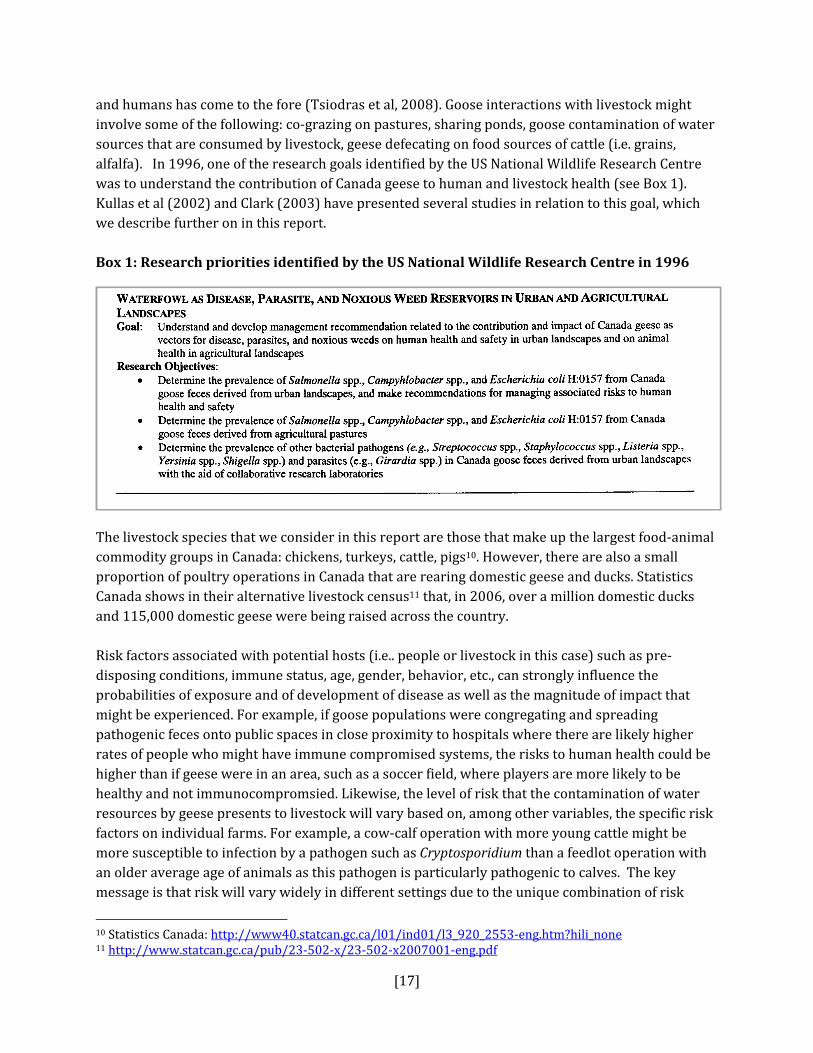
and humans has come to the fore (Tsiodras et al, 2008). Goose interactions with livestock might involve some of the following: co‐grazing on pastures, sharing ponds, goose contamination of water sources that are consumed by livestock, geese defecating on food sources of cattle (i.e. grains, alfalfa). In 1996, one of the research goals identified by the US National Wildlife Research Centre was to understand the contribution of Canada geese to human and livestock health (see Box 1). Kullas et al (2002) and Clark (2003) have presented several studies in relation to this goal, which we describe further on in this report. Box 1: Research priorities identified by the US National Wildlife Research Centre in 1996
The livestock species that we consider in this report are those that make up the largest food‐animal commodity groups in Canada: chickens, turkeys, cattle, pigs10. However, there are also a small proportion of poultry operations in Canada that are rearing domestic geese and ducks. Statistics Canada shows in their alternative livestock census11 that, in 2006, over a million domestic ducks and 115,000 domestic geese were being raised across the country. Risk factors associated with potential hosts (i.e.. people or livestock in this case) such as pre‐disposing conditions, immune status, age, gender, behavior, etc., can strongly influence the probabilities of exposure and of development of disease as well as the magnitude of impact that might be experienced. For example, if goose populations were congregating and spreading pathogenic feces onto public spaces in close proximity to hospitals where there are likely higher rates of people who might have immune compromised systems, the risks to human health could be higher than if geese were in an area, such as a soccer field, where players are more likely to be healthy and not immunocompromsied. Likewise, the level of risk that the contamination of water resources by geese presents to livestock will vary based on, among other variables, the specific risk factors on individual farms. For example, a cow‐calf operation with more young cattle might be more susceptible to infection by a pathogen such as Cryptosporidium than a feedlot operation with an older average age of animals as this pathogen is particularly pathogenic to calves. The key message is that risk will vary widely in different settings due to the unique combination of risk
10 Statistics Canada: http://www40.statcan.gc.ca/l01/ind01/l3_920_2553‐eng.htm?hili_none 11 http://www.statcan.gc.ca/pub/23‐502‐x/23‐502‐x2007001‐eng.pdf

[18]
factors that each presents, and a generic risk assessment for all pathogens and settings is not feasible.
MECHANISMS FOR EXPOSURE TO HUMANS FROM GOOSE PATHOGENS Pathogens from Canada and Cackling geese could be transmitted to humans by several routes including: direct transmission through airborne transmission, or contact with infected feathers, skin, droppings or external lesions, waterborne or food‐borne transmission, occupational exposure to infected materials, transmission via arthropod vectors, and indirect transmission through the same routes if geese transmit the agent to livestock and livestock then are a source of infection for people (see Figure 1) (Tsiodras et al, 2008). Each route of exposure is discussed in more detail in the next section (exposure assessment). In order for a person to develop a disease from a pathogenic organism in the feces of wild geese, the following steps must take place:
1) an organism that is pathogenic to humans must be present in the feces of geese; 2) the organism in the feces must survive in the environment after their deposition on the
ground or in water; 3) geese must deposit feces in an amount and manner where people might come into contact
with it, 4) a person must contact the contaminated feces, ingest a sufficient amount (infectious dose),
become infected, and produce symptomatic illness (Feare et al, 1999).
MECHANISMS FOR EXPOSURE TO LIVESTOCK FROM GOOSE PATHOGENS Figure 1 demonstrates the pathways of exposure for livestock to pathogens disseminated by geese. Direct transmission could occur through aerosol transmission or through direct contact with infected feathers, skin, droppings or external lesions. Infected birds could contaminate water with feces and respiratory secretions that could result in a waterborne transmission through ingestion. Indirect transmission to livestock could also occur through the same routes if geese transmit the agent to humans and humans then transmit the pathogen to livestock. Disease transmission via arthropod vectors is another possible route for livestock infection from geese. In order for livestock to develop a disease from a pathogenic organism in the feces of wild geese, the following steps must take place:
1) an organism that is pathogenic to livestock must be present in the feces of wild geese; 2) the organism in the feces must survive in the environment after their deposition on the
ground or in water; 3) geese must deposit feces in an amount and manner where livestock might come into contact
with it, 4) livestock must contact the contaminated feces, ingest a sufficient amount (infectious dose),
become infected, and produce symptomatic illness (Feare et al, 1999).

[19]
Figure 1: Potential exposure pathway of pathogens between geese, livestock and people (adapted from Clark, 2003)
6.B. EXPOSURE ASSESSMENT In Appendix A there is an overview of the factors that might affect the probability of transmission between infected geese and humans including: presence in North America, prevalence in wild geese, prevalence in humans in North America, persistence of organism in the environment, evidence for cross‐species transmission (of any sort not just humans), evidence for goose to human transmission, and some relevant references. Appendix B presents an overview of the factors that might affect the probability of transmission between infected geese and livestock including: presence in North America, prevalence in wild geese, prevalence in livestock in North America, persistence of organism in the environment, evidence for cross‐species transmission, evidence for goose to livestock transmission, and some relevant references. In this section we discuss the plausibility of human and livestock infection from geese taking place by the various routes of transmission described above. We can conclude that there are several plausible routes of exposure to infectious material from geese, but estimates of the probability of these exposures are not attainable with existing data.
Plausibility of transmission by route of infection Airborne transmission: Airborne transmission generally could occur when there is very close contact between humans or livestock and infected dust, or water droplets, feces or respiratory secretions from infected geese and there is inhalation deep enough to allow for infection. For example, if members of the public were feeding the birds, picking up young birds, or otherwise within very close proximity they could potentially be exposed in this way. This mode of exposure is also plausible for those responsible for handling geese (i.e. occupational exposure of bird control
Geese
Pasture
Silage
Feed
Ponds
Worker
Other wildlife
Waste
Livestock
[20]
specialists, wildlife rehabilitation specialists, biologists, veterinarians, etc) and also for landscape maintenance personnel who could be exposed to infected airborne particles through some of the following activities: gardening, cutting grass, moving soil, raking leaves or beaches, etc. In livestock, airborne transmission might occur if geese were in close proximity to the animals (i.e. co‐grazing, livestock drinking from pond inhabited by geese. It is also plausible that wind may play a role in distributing pathogen‐carrying particles. Airborne particles, particularly those of smaller size, can be carried over long distances. Some of the factors that affect the occurrence of infections acquired by long‐distance airborne transmission include: 1) dilution, 2) the infectious dose, 3) the number of infectious particles, 4) the duration of shedding of the infectious agent, and 5) the persistence of the agent in the environment (Tellier, 2006). The majority of this information is missing for goose‐related pathogens. Direct contact with infected feathers, skin, feces or external lesions: This route, particularly contact with feces, seems most plausible for people in areas of high recreational use of habitats frequented by geese and people, such as urban parks. Livestock must be in close proximity to geese (i.e. co‐grazing) in order for transmission via this route to take place. Canada geese have been shown to produce nearly 1 kg of feces per day (range 0.39 – 0.9 kg) (Ilion et al, 2006; USDA, 2004). The range of risk groups that present a reasonable presumption of exposure potential for pathogen transmission might include: children, bathers, picnickers, sunbathers, and landscapers. Of this group young children are probably at greatest risk due to their methods of play and propensity to consume dirt. Stanek and Calabrese (1995) demonstrated that children on average consume 138 mg of dirt daily. Also, bacteria have been shown to persist longer in sand than in water as they adhere to sediment particles, so people, and particularly children, playing in sand might be at greater risk of exposure (Benskin et al, 2009). Free‐ranging livestock or livestock reared in outdoor settings are more at risk for exposure than those kept in closed systems with proper biosecurity, which should prevent transmission of pathogens by direct contact with geese feathers, droppings, skin, or external lesions. The majority (up to 90% in the US) of chickens and turkeys, and about three‐quarters (72% in the US) of swine herds are raised in intensive, closed settings (Graham et al, 2008). However, there is a growing consumer demand for free‐range animal products that is driving an increase in open‐farming systems to meet the demand. It is reasonable to assume that these more open systems where chickens or turkeys spend a portion of their day in outdoor settings would create more opportunity for direct exposure to geese and their pathogens than those reared in entirely enclosed settings. Waterborne transmission: Infected birds can contaminate water with feces and respiratory secretions that could potentially result in a waterborne transmission through ingestion, or potentially, as a result of swimming (Benskin et al, 2009). A wide range of waterfowl species have been shown to contribute substantially to fecal coliform counts in human drinking water sources and are often suspects in transmission of other waterborne bacterial, parasitic and viral pathogens such as Campylobacter, Cryptosporidium, Giardia, Avian Influenza virus, Helicobacter, etc (Graczyk et al, 2008; Tsiodras et al, 2008; Fox et al, 2006; Alderisio and DeLuca, 1999). Waterborne

[21]
transmission is potentially one of the most important routes of transmission given the possibility for wide dissemination through municipal or household water sources and the level of access of waterfowl to relevant aquatic environments. It is not unreasonable to assume that livestock might be at greater risk than people from waterborne pathogen transmission as many farms rely on drinking water sources that are often untreated such as rivers, streams and ponds that might be contaminated by pathogens of goose origin. Occupational exposure: Exposure to infected tissues, blood and feces is another source of direct transmission. Veterinarians, hunters, biologists, wildlife control officers, wildlife rehabilitators are some of the risk groups for occupational exposure. These groups, perhaps more than any other, may be more inclined to take some preventive measures to mitigate against pathogen exposure such as those listed in the Public Health Agency of Canada’s “Fact Sheet: Guidance on Precautions for the Handling of Wild Birds12”. This could be an indirect transmission route for livestock if their handlers or veterinarians were in contact with infected goose tissues and pass it on to them. Foodborne transmission: Food‐borne infections in humans could develop as a result of the consumption of contaminated geese, particularly through the ingestion of raw or undercooked meat, blood, organs, etc (Tsiodras et al, 2008). Indirect transmission to humans could also occur through the same routes if geese transmit the agent to livestock (see Figure 1). Livestock could acquire pathogens from geese through their food‐stuffs that were contaminated with pathogens from geese during the production, harvesting, processing or distribution process. Arthropodborne transmission: Disease transmission via arthropod vectors is another possible route for infection from geese. Humans or livestock may be affected by arthropods either directly by bites, stings, or infestation of tissues, or indirectly through pathogen transmission. Ticks and mosquitoes are the most important genera for pathogen transmission. The most significant mode of vector‐borne disease transmission is by biological transmission by blood‐feeding arthropods. The pathogen multiplies within the arthropod vector, and the pathogen is transmitted when the arthropod takes a blood meal from the person or animal. Arthropods are also capable of infecting people or livestock indirectly through mechanical transmission of the agent when they physically carry pathogens from one place or host to another, usually on body parts. Key determinants of arthropod‐borne diseases include the:
• abundance of vectors and intermediate and reservoir hosts; • prevalence of pathogens suitably adapted to the vectors and the host; • local environmental conditions such as temperature and humidity; and • behaviour and immune status of the human population.
In summary, there are a number of biologically plausible routes of exposure to the hazards listed in Table 2. However, there are insufficient data to quantify the probability or average exposure rates for any of the pathogens. The exposure route of greatest concern, based on the extent of coverage in the peer‐reviewed literature as well as in the lay media, is direct contact of people (contact in parks 12 http://www.phac‐aspc.gc.ca/influenza/fs‐hwb‐fr‐mos‐eng.php#4

[22]
or from water sources) and livestock (on‐farm contact or from water sources) to infected goose feces. Airborne transmission of pathogens from geese to poultry (i.e. Avian influenza, Newcastle disease) is also widely discussed in the literature and media, but less so than for direct transmission from contact with feces. If feces are the major concern, high concentrations of geese with high rates of fecal deposition would increase the likelihood of exposure to hazards. This likelihood increases if fecal deposition is occurring in areas of high human use (density) or livestock density. This might suggest that exposure risk mitigation strategies to contain goose fecal matter is an appropriate area of focus (i.e. (1) dispersing birds to decrease the density of fecal contamination in areas of high human or livestock density; (2) habitat modification to make areas less attractive to Canada Geese; (3) waste management via removal of feces from affected areas; and (4) aquatic resource management) (Smith et al, 1999; Allan et al, 1995).
6.C. CONSEQUENCE ASSESSMENT This section covers the types of consequences, or impact, that could arise as a result of human or livestock infection from geese. Appendix C presents an overview of the factors that affect the impact of each pathogen should it be transmitted to humans from geese including: the route of transmission, clinical manifestation (what does the agent do?), severity of the illness (is it treatable? how often are people hospitalized? how often do they die from the disease?), whether or not the disease is nationally reportable, and some relevant references. Appendix D presents an overview of the factors that affect the impact of each pathogen should it be transmitted to livestock from geese including: the route of transmission, clinical manifestation (what does the agent do?), severity of the illness (is it treatable?, how severe are the signs? how often do livestock die from the disease?), national disease reporting requirements, and some relevant references. In each of these tables the hazards are each given an impact category (negligible, low, medium or high) based on the criteria described in the methods section. In humans, the pathogens with a ‘high’ ranking had more than one of the following features: 1) caused severe illness, often resulting in hospitalization; 2) had the ability to spread epidemically from person‐to‐person; 3) had high fatality rates; 4) did not have effective treatment or preventive methods; and 5) and were nationally reportable. The pathogens that filled more than one of these criteria included: highly pathogenic avian influenza (HPAI) and enterotoxigenic E. coli (ETEC). Pathogens of medium and low impact are shown in Appendix C. In livestock, the pathogens with a ‘high’ ranking had more than one of the following features: 1) caused severe illness; 2) had the ability to spread epidemically within or between herds or flocks; 3) had high fatality rates; 4) did not have effective treatment or preventive methods; and 5) and were nationally reportable to the Canadian Food Inspection Agency. The pathogens that filled more than one of these criteria included: highly pathogenic avian influenza (poultry), Mycobacterium avium, and Newcastle disease (poultry). Once again, we found no direct evidence that linked human or livestock health outcomes to geese. Hence, this consequence assessment is the

[23]
consequence of the pathogens in general and not the pathogen from geese. Almost all of the hazards listed have multiple sources including people, other species and environmental sources.
7. SYNTHESIS There is evidence that many pathogens of importance to humans and livestock can infect Canada and Cackling geese and be shed into the environment by geese, and vice versa. However, there is a very large gap in our understanding of the ecological and epidemiological factors that may contribute to the transmission from geese to humans or livestock. As discussed above, effective risk analyses (identification, assessment, management and communication) require data inputs from quality surveillance information (Table 1). Although there was a large list of pathogens associated with geese and other members of the Anatidae family, there was scarce serological, microbiological or epidemiological data as evidence of transmission between geese and humans and geese and livestock. In a traditional risk assessment, this section would involve making overall risk estimations for the transmission of each identified hazard by combining the results of the release/exposure (probability estimates) and consequence (magnitude of impact) assessments. But in this case, we were not able to estimate probability so we are left with identifying some of the priority pathogens and routes of exposures that CWS should consider in its risk management strategies. These pathogens were identified based on the level of potential impact to human health and livestock health and trade and the plausibility and related evidence for transmission. What type of evidence is needed to conclude that pathogens of goose origin are a cause of adverse health outcomes in people or livestock? The Bradford‐ Hill criteria that are used as guidelines to evaluate evidence of disease causation are relevant in this scenario (See Box 2). The evidence of transmission from geese to either humans or livestock must at a minimum be: 1) plausible, 2) specific (i.e.. same species, strain, serotype), ideally to the molecular level, and 3) associated by time and geographic location.

[24]
BOX 2: BradfordHill criteria for causation (Wikipedia, adapted from Hill, 1965)
Strength: A small association does not mean that there is not a causal effect. Consistency: Consistent findings observed by different persons in different places with different samples strengthen the likelihood of an effect. Specificity: Causation is likely if a very specific population at a specific site and disease with no other likely explanation. The more specific an association between a factor and an effect is, the bigger the probability of a causal relationship. Temporality: The effect has to occur after the cause (and if there is an expected delay between the cause and expected effect, then the effect must occur after that delay).
Biological gradient: Greater exposure should generally lead to greater incidence of the effect. However, in some cases, the mere presence of the factor can trigger the effect. In other cases, an inverse proportion is observed: greater exposure leads to lower incidence. Plausibility: A plausible mechanism between cause and effect is helpful. Coherence: Coherence between epidemiological and laboratory findings increases the likelihood of an effect. However, Hill noted that "... lack of such [laboratory] evidence cannot nullify the epidemiological effect on associations" Experiment: "Occasionally it is possible to appeal to experimental evidence". Analogy: The effect of similar factors may be considered
The most plausible source of infection for both people and livestock appears to be via contaminated aquatic environments. Another plausible route is through direct contact with infected fecal material (this includes infected airborne particles). These routes provide the opportunity for the widest possible exposure for both people and livestock. Below we discuss some of the pathogens that we feel should be of greatest concern to CWS and as a result be considered in their risk management and communication strategies. However, it must be noted that the only Bradford‐Hill criteria that any of the geese‐related pathogens meet are plausibility and some meet the ‘consistency’ criteria due to geo‐spatial associations. Therefore, all hazards fail to meet Bradford‐Hill’s criteria so no conclusions about causal linkages can be made. Even though we highlight several pathogens here, some of them have high species specificity and low virulence capacity so potentially present a low to negligible risk of transmission. Due to the fact that Canada and Cackling geese are aquatic birds that aggregate in large numbers and produce a prolific amount of feces, concerns have been raised about their role in water contamination. As a result, there has been an emphasis on research of waterfowl‐related pathogens and in particular for E. coli, Cryptosporidium, and Giardia. This research bias could affect the assessment of risks from waterborne transmission from geese in that more important, but un‐ or under‐researched pathogens are not considered as much as these three pathogens. In addition, the perceptions of waterborne pathogen risks are shifting as a consequence of more recent molecular

[25]
and epidemiological information that is coming on stream and showing more host specificity than previously thought. Hence, the assessment of pathogens in this report might be biased by the older literature that could not make distinctions between strains that may be highly host‐specific and therefore present a diminished risk for interspecies transmission (Hansen et al, 2009; Graczyk et al, 2008). Of all the potential hazards presented in this report, E. coli is the pathogen for which there exists the most amount of information and research. It is most commonly looked for in fecal surveys and enumerated in water quality tests; it is also often top of mind by the public as a potential risk from goose feces. Alderisio and DeLuca (1999) found that Canada geese contributed on average 1.28 x 10⁵ fecal coliforms per fecal deposit to surface water. Although E. coli has over 200 specific serological types, the majority of which are harmless to humans or livestock, there are several virulent forms that can cause severe illness (Edge and Hill, 2007; Clark, 2003; Kullas et al, 2002; Roscoe, 2001; Feare et al, 1999). Virulent strains, such as Enterohaemorrhagic E. coli (EHEC), only infrequently cause disease in animals; ruminants, and cattle in particular, are the primary reservoir. Poultry and pigs are not considered to be a source of EHEC (Caprioli et al, 2005). It is not unreasonable to assume that Canada or Cackling geese could potentially acquire EHEC, or other serotypes, from on‐farm sources such as infected pastures, manure piles or slurry and then mechanically transfer to other locals and contaminate aquatic environments used by people or livestock (Kudva et al, 1998). However, this possible route of exposure and transfer, to the best of our knowledge, has not been demonstrated. This again speaks to the importance of the unique risk settings that must be considered when assessing health risks such as goose feeding ecology, migration patterns, farming practices (i.e. fertilization of fields with manure), and the distribution and types of farms and their relationship with goose ecology. Improved water‐quality indicators are needed, and are currently being developed, that move beyond simple coliform counts as indicators of risk to methodologies that track specific host sources of pathogens (Hansen et al, 2009; Hamilton et al, 2006). These tools will contribute essential information for the assessment of risks from goose fecal contamination in aquatic environments that currently cannot be ascertained. Significant data gaps remain on the determinants of risk such as the prevalence of pathogens that are shed by geese and the exposure to these pathogens through aquatic environments (i.e. drinking water sources for both people and livestock and exposure patterns during recreational activities such as swimming). Cryptosporidium and Giardia are considered to be the most common causes of human waterborne outbreaks and gastroenteritis world‐wide and are the most important parasitic pathogen in livestock (non‐poultry) (Plutzer et al, 2009; Thompson et al, 2008). Oocysts (the transmissive stage) of Cryptosporidium and Giardia are very robust and can survive in water for prolonged periods in adverse conditions; they are considered to be ubiquitous in aquatic environments (Gracyzk et al, 2008). Canada geese can distribute Cryptosporidium strains that are pathogenic to people and ruminants but the majority of the strains they shed have been shown to be goose‐specific and not pathogenic to other species (Gracyzk et al, 1998). However, temperate‐breeding Canada geese have been shown to acquire C. hominis (human‐adapted species) oocysts from

[26]
garbage and other contaminated sites (Zhou et al, 2004) so they have the potential to mechanically transfer this agent. Cattle are considered to be the primary reservoir of C. parvum, so increased opportunities for geese to contact contaminated feces from cattle (i.e. co‐grazing, field fertilization with manure) has the potential to increase goose exposure and then possible transmission from geese back to aquatic environments of high human or livestock use. Zhou et al (2004) found that only 10% of recovered Cryptosporidium sp. from goose feces were C. parvum and C. hominis, the species that are pathogenic to humans, and concluded that Canada geese “might only serve as accidental carriers of cryptosporidia infectious to humans and probably play a minor role in the animal‐to‐human transmission cycle of the pathogen”. HPAI can cause devastating illness in poultry flocks, as seen in recent outbreaks in British Columbia and Saskatchewan (Berhane et al, 2009; Pasick et al, 2007). Avian influenza virus infection is endemic in a wide range of wild bird species, particularly species associated with water such as Anseriformes and Charadriiformes (Ward et al, 2009). There are conflicting theories and evidence in the literature about the role of wild birds in the transmission cycle of highly pathogenic avian influenza (HPAI) (i.e. H5N1) of importance to humans (Berhane et al, 2009; Ward et al, 2009; Tsiodras et al, 2008; Clark and Hall, 2006; Olsen et al, 2006; FAO media centre13). However on balance, wild birds do not appear to have a dominant role in the transmission cycle of HPAI as it has rarely been isolated from wild birds and is thought to result from local transmission from poultry rather than from de novo generation in wild birds (Tsiodras et al, 2008). Wild waterfowl are often infected with low pathogenic avian influenza (LPAI) viruses, and the theory is that wild birds could transmit LPAI to poultry flocks, which then act as mixing vessels for new, and potentially highly pathogenic, strains of influenza that could then infect people. Waterfowl, particularly ducks, can excrete LPAI in relatively high concentrations in their feces (Parmley et al, 2007; Pasick et al, 2007; Olsen et al, 2006). Influenza virus can remain infectious in freshwater for up to 4 days at 22⁰C and more than 30 days at 0⁰C. The relatively high virus prevalence in waterfowl might be due, in part, to efficient transmission through the waterborne fecal‐oral route of transmission (Olsen et al, 2006). However, factors such as species, sex, age, and season have been shown to influence prevalence rates in ducks (Pasick et al, 2007; Halvorson et al, 1985). In response to the HPAI outbreak in domestic poultry in British Columbia in 2004, a Canadian surveillance program for influenza A viruses in wild aquatic birds was initiated the following year to address information gaps on the role of wild birds in the pathogen transmission. The Canadian Cooperative Wildlife Health Centre was tasked with coordinating the Inter‐agency Wild Bird Influenza Survey14 to meet the following objectives:
1) “create an inventory of influenza A viruses that occur in wild birds in different areas across Canada;
13 FAO media centre. March 23,2010. On the trail of avian influenza: International task force concerned over declining support for H5N1 monitoring, despite disease persistence and spread. http://www.fao.org/news/story/en/item/40827/icode/
14 http://www.cws‐scf.ec.gc.ca/nwrc‐cnrf/default.asp?lang=en&n=00DF74F2‐1

[27]
2) to characterize these viruses to a sufficient degree that it may be possible to determine whether they may be the source, in whole or in part, of any future outbreaks in domestic animals or humans;
3) to monitor for the presence of particular influenza A viruses or their genetic components in the Canadian wild bird population;
4) to establish an archive of influenza A virus strains that would permit rapid retrospective analysis in response to disease outbreaks; and
5) to build and maintain an integrated, multiagency field, laboratory, regulatory, and communications capacity within Canada to carry out influenza A virus sampling, identification, and molecular characterization of large volumes of samples.” (Pasick et al, 2007)
Results from this survey have show that “on average, 30% of all live ducks sampled, 5% of other species of live birds and 3% of birds found dead have tested positive for avian influenza, all of North American lineage and of low pathogenicity” (Parmley et al, 2009). Geese were sampled in this survey in only 2006 and 2007. Of all geese sampled, including Snow geese, Ross’ geese, Greater White‐fronted geese, Canada and Cackling geese, 5.5% (207/3738) were positive for Avian Influenza during this period; Canada or Cackling geese accounted for 32% (66/207) of the positive geese samples. Between 2005 and 2009, there were nine dead goose submissions (4 Snow geese, 5 Canada geese) that were positive for Avian Influenza. In 2007, the year of the H7N3 BC outbreak, an H7N3 LPAI strain was detected in a single Canada goose that was submitted to the CCWHC as part of the Inter‐agency Wild Bird Influenza Survey. The relationship between this finding and the poultry outbreak in the Fraser Valley are unknown but could potentially be due to spill‐over from infected poultry to the goose. Berhane et al (2009) report on an HPAI outbreak in the Saskatchewan poultry industry where they indicate that wild birds played a role in seeding the outbreak with a low pathogenic avian influenza strain that converted into a more highly virulent strain and caused significant losses to the industry. In this case, phylogenetic analysis demonstrated a close relationship of Saskatchewan/2007 H7N3 with recent viruses of free‐flying waterfowl in North America. In summary, the exposure setting is of paramount importance in determining risk; the proximity of geese to poultry operations and the nature of their contact with domestic flocks will be a key determinant of risk for development and transmission of HPAI.
8. RECOMMENDATIONS For this risk assessment concerning pathogens in wild geese, most of the data requirements simply cannot be met with the available data on pathogens potentially transmitted by geese. There are very large gaps in some of the following key determinants of risk: prevalence of pathogens and parasites in geese, epidemiological information to link the pathogen in geese to cases in people or livestock, fecal distribution patterns, and human or livestock exposure patterns (i.e. the nature and extent of contact between geese and humans and geese and livestock). As a result, a reliable, evidence‐based risk assessment of health risks to people or livestock from free‐ranging geese cannot legitimately be performed with the existing availability and quality of data.

[28]
The Canadian Wildlife Service is in a challenging position to manage this issue in which there are rising populations of Canada and Cackling geese, especially of the temperate breeding variety, that are increasingly coming into conflict with human and agricultural activities. In addition, the CWS is faced with very large information gaps on the risk of geese for human and livestock to help it with its decision‐making processes. Some strategies for the CWS to consider include:
• Investing in monitoring and research that will provide the CWS with the necessary components to develop an evidence‐based risk assessment. Some of the key areas of investigation should be improving our understanding of exposure probabilities (i.e.. nature and extent of goose interactions with people and livestock) but also elucidating the prevalence of specific pathogens for which we have no information for geese, as well as further genetic characterization of strains of pathogens and how they are related to similar human or livestock pathogens. In the last several years, there has been an increasing development and application of advanced diagnostic and survey tools that are helping to elucidate the relationship between goose, human and livestock pathogens. Some of the methods that are increasingly being used include: molecular methods such as sequence analysis (i.e. Mycobacterium spp.), phylogenetic analyses, pulsed‐field gel electrophoresis, polymerase chain reaction (i.e. E. coli), serologic methods (i.e. influenza, psittacosis), and epidemiological methods (i.e. Salmonella, West Nile virus). These pathogen detection methodologies coming on stream give us hope that these gaps can be narrowed over the coming years with molecular‐level data that can demonstrate strain overlaps between geese and other species and help track the source of human and livestock infections and outbreaks, particularly those with waterborne origin and for which waterfowl are suggested as a possible source. However, there is a large number of possible hazards widespread over a variety of risk settings in Canada. Investment in such a program of research would need to be large and long‐term to make significant shifts in our understanding of these determinants of risk.
• Modeling of risks for some of the priority pathogens identified here as a means of accounting for the high degree of uncertainty. Uncertainty can either be modeled using quantitative or qualitative methods. Increasingly in areas in which there are large data gaps or uncertainty, a mixed approach, such as multi‐criteria decision making can be used. However, models would need to account for the issue of context which modifies risk (exposure setting, nature of the at risk group etc). Models are also only as good as the data available hence any modeling attempts would be opinion based or would need to use analytical methods to account for data uncertainty.
• Developing and improving risk reduction and communication strategies that focus on specific higher risk settings (i.e. aquatic resources, hospital settings, and high livestock density areas).
• Forming a working group to develop national standards for the management of peri‐urban goose populations. Expertise from public health, regulatory agencies, resource managers, engineers, epidemiologists, and biologists along with input from the public are needed to

[29]
develop a consensus on goose management practices. A consensus approach has been used to good effect in other situations where there is a high level of uncertainty. For example, the Northeast Wildlife Damage Management Research and Outreach Cooperative15 was formed in the US “to enhance coordination and collaboration among wildlife agencies, universities, and other cooperators to promote consistent, multi‐state approaches for resolving wildlife‐related concerns”. This group has examined management issues associated with Canada geese, white‐tailed deer, black bears, and beavers. Community‐based approaches have also been shown to be effective in developing location‐specific methods to address nuisance wildlife issues (Curtis et al, 2005).
15 http://wildlifecontrol.info/newdm/Pages/default.aspx

[30]
9. REFERENCE LIST Abulreesh HH, Paget TA and Goulder R. Waterfowl and the bacteriological quality of amenity ponds. J Water Health. 2004. 2(3):183‐189.
Abdelzaher A, Wright M, Ortega C, Solo‐Gabriele H, Miller G, Elmir S, Newman X, Shih P, Bonilla JA, Bonilla TD, Palmer CJ, Scott T, Lukasik J, Harwood VJ, McQuaig S, Sinigalliano C, Gidley M, Plano L, Zhu X, Wang JD, Fleming L. Presence of pathogens and indicator microbes at a nonpoint source subtropical recreational marine beach. Appl Environ Microbiol. 2010. 76(3):724‐32. Adams BL, Bates TC, and Oliver JD. Survival of Helicobacter pylori in a natural freshwater environment. Appl Environ Microbiol. 2003. 69(12): 7462–7466. Adrian WJ, Spraker TR and Davies RB. Epornitics of aspergillosis in mallards (Anas platyrhynchos) in north central Colorado. J Wildl Dis. 1978. 14(2):212‐7. Akan M, Haziroğlu R, Ilhan Z, Sareyyüpoğlu B and Tunca R. A case of aspergillosis in a broiler breeder flock. Avian Dis. 2002.46(2):497‐501. Alderisio KA and DeLuca N. Seasonal enumeration of fecal coliform bacteria from the feces of RingBilled gulls (Larus delawarensis) and Canada geese (Branta Canadensis). Appl and Environ Microbiol. 1999.65(12): 5628‐5630.
Alexander DJ, Banks J, Collins MS, Manvell RJ, Frost KM, Speidel EC and Aldous EW. Antigenic and genetic characterisation of Newcastle disease viruses isolated from outbreaks in domestic fowl and turkeys in Great Britain during 1997. Vet Rec. 1999. 145: 417‐421.
Alexander DJ, Morris HT, Pollitt WJ, Sharpe CE, Eckford RL, Sainsbury RMQ, Mansley LM, Gough RE and Parsons G. Newcastle disease outbreaks in domestic fowl and turkeys in Great Britain during 1997. Vet Rec. 1998. 143: 209‐212.
Allan JR, Kirby JS and Feare CJ. The biology of Canada geese Branta Canadensis in relation to the management of feral populations. Wildlife Biol. 1995. 1: 129‐143.
Anderson JF. Mammalian and avian reservoirs for Borrelia burgdorferi. Annals New York Academy of Sciences. 1988: 180‐191. 539: 180‐191.
Anderson JF, Johnson RC, Magnarelli LA and Hyde FW. Involvement of birds in the epidemiology of the Lyme Disease agent Borrelia burdorferi. Inf and Imm. 1986.51(2):394‐396.
Arsenault J, Letellier A, Quessy S, Normand V and Boulianne M. Prevalence and risk factors for Salmonella spp. and Campylobacter spp. caecal colonization in broiler chicken and turkey flocks slaughtered in Quebec, Canada. Prev Vet Med. 2007. 81(4):250‐64. Artois M, Bicout D, Doctrinal D, Fouchier R, Gavier‐Widen D, Globig A, Hagemeijer W, Mundkur T, Munster V, Olsen B. Outbreaks of highly pathogenic avian influenza in Europe: the risks associated with wild birds. Rev Sci Tech. 2009. 28(1):69‐92.
Atabay HI, Corry JEL, On SLW. Identification of unusual Campylobacterlike isolates frompoultry products as Helicobacter pullorum. J Applied Microbiol. 1998. 84: 1017–1024

[31]
Atabay HI, Unver A, Sahin M, Otlu S, Elmali M and Yaman H. Isolation of various Arcobacter species from domestic geese (Anser anser). Vet Micro Bio. 2008. 128: 400‐405.
Ataliba AC, Resende JS, Yoshinari N and Labruna MB. Isolation and molecular characterization of a Brazilian strain of Borrelia anserine, the agent of fowl spirochaetosis. Re Vet Sc. 2007. 83: 145‐149.
Austin RJ, Whiting TL, Anderson RA and Drebot MA. An outbreak of West Nile virusassociated disease in domestic geese ( Anser anser domesticus) upon an initial introduction into a geographic region, with evidence of bird to bird transmission. Can Vet J. 2004. 45: 117‐123.
Ball NW, Smyth JA, Weston JH, Borghmans BJ, Palya V, Glávits R, Ivanics E, Dán Á and Todd D. Diagnosis of goose circovirus infection in Hungarian geese samples using polymerase chain reaction and dot blot hybridization tests. Avian Path. 2004. 33(1):51‐58.
Banet‐Noach C, Simanov L and Malkinson M. Direct (nonvector) transmission of West Nile virus in geese. Avian Path. 2003. 32(5):489494.
Barker IK and Lindsay LR. Lyme borreliosis in Ontario: determining the risks. CMAJ. 2000. 162 (11): 1573‐1574.
Barnett J, Ainsworth H, Boon JD and Twomey DF. Streptococcus galloyticus subsp. Pasteurianus septicaemia in goslings. Vet J. 2008. 176: 251‐253.
Baro T, Torres‐Rodri´guez JM, de Mendoza MH, Morera Y and Alía C. First Identification of Autochthonous Cryptococcus neoformans var. gattii Isolated from Goats with Predominantly Severe Pulmonary Disease in Spain. J Clin Microbiol. 1998. 36(2):458‐461. Baseman JB and Tully JG. Mycoplasmas: sophisticated, reemerging, and burdened by their notoriety. Emerg Infect Dis. 1997.3:21‐32. Bedard JAGG. Assessment of faecal output in geese. J App Ecol. 1986. 23:77‐90. Beemer AM, Kuttin ES and Katz Z. Epidemic venereal disease due to Candida albicans in geese in Israel. Avian Dis. 1973. 17(3):639‐649. Bennett GF, Nieman DJ, Turner B, Kuyt E, Whiteway M and Greiner EC. Blood parasites of prairie anatids and their implication in waterfowl management in Alberta and Saskatchewan. J Wildl Dis. 1982.18(3):287‐96. Benskin CMH, Wilson K, Jones K and Hartley IR. Bacterial pathogens in wild birds: a review of the frequency and effects of infection. Biological Reviews. 2009. 84(3):349373. Bercic RL, Slavec B, Lavric M, Narat M, Zorman‐Rojs O, Dovc P and Bencina D. A survey of avian Mycoplasma species for neuraminidase enzymatic activity. Vet Microbiol. 2008. 130(3‐4):391‐7. Berhane Y, Hisanaga T, Kehler H, Neufeld J, Manning L, Argue C, Handel K, Hooper‐McGrevy K, Jonas M, Robinson J, Webster RG and Pasick J. Highly pathogenic avian influenza virus A (H7N3) in domestic poultry, Saskatchewan, Canada. 20072009. 2009. Emerg Infect Dis. 15(9):1492‐5.

[32]
Beytut E, Özcan K and Erginsoy S. Immunohistochemical detection of fungal elements in the tissues of goslings with pulmonary and systemic aspergillosis. Acta Veterinaria Hungarica. 2004. 52 (1): 71–84.
Biet F, Boschiroli ML, Thorel MF and Guilloteau LA. Zoonotic aspects of Mycobacterium bovis and Mycobacterium aviumintracellulare complex (MAC). Vet Res. 2005. 36(3):411‐36. Blanchong JA, Samuel MD, Goldberg DR, Shadduck DJ and Lehr MA. Persistence of pasteurella multocida in wetlands following avian cholera outbreaks. J of Wildlife Dis.2006. 42(1):33‐39. Boettger CM and Dohms JE. Separating Mycoplasma gallisepticum field strains from nonpathogenic avian mycoplasmas. Avian Dis. 2006. 50(4):605‐7. Boldur I, Cohen A, Tamarin‐Landau R, and Sompolinsky D. Isolation of Legionella pneumophila from calves and the prevalence of antibodies in cattle, sheep, horses, antelopes, buffaloes and rabbits. Vet Microbiol. 1987.13(4):313‐20. Bŏnner BM, Lutz W, Jäger L, Redmann T, Reinhardt B, Reichel U, Krajewski V, Weiss R, Wissing J, Knichmeier W, Gerlich WH, Wend UC, Kaleta EF. Do Canada geese (Branta Canadensis Linnaeus, 1758) carry infectious agents for birds and man? Eur J Wildl Re. 2004. 50:78‐84. Booth CM, Matukas LM and Tomlinson GA, et al. Clinical features and shortterm outcomes of 144 patients with SARS in the greater Toronto area. JAMA. 2003. 289(21):2801‐2809.
Boreham RE, McCowan MJ, Ryan AE, Allworth AM and Robson JM. Human trichostrongyliasis in Queensland. Pathology. 1995. 27(2):182‐185. Botzler RG. Epizootiology of Avian cholera in wildfowl. J Wildlife Dis. 1991. 27(3):367‐395.
Bowes, V. A. An outbreak of aspergillosis in wild waterfowl. Can. Vet. J. 1990. 31: 303–304.
Bradbury JM, Jordan FTW, Shimizu T, Stipkovits L and Vargas ZS. Mycoplasma anseris sp. nov. found in geese. J Systematic Bacteriol. 1988. 38: 74‐76.
Brand CJ and Docherty DE. Postepizootic surveys of waterfowl for duck plague(duck virus enteritis). Avian Dis. 1988. 32(4):722‐30.
Brant SV and Loker ES. Schistosomes in the southwest United States and their potential for causing cercarial dermatitis or ‘swimmer’s itch’. J of Helminthol. 2009. 83. 191‐198.
Brooke C and Riley TV. Erysipelothrix rhusiopathiae: bacteriology, epidemiology and clinical manifestations of an occupational pathogen. J Med Microbiol. 1999.48(9):789‐799.
Brown DWG. Foot and mouth disease in human beings. Lancet. 2001.12;357(9267):1463. Brown JD, Stallknecht DE and Swayne DE. Experimental infection of swans and geese with highly pathogenic Avian influenza virus (H5N1) of Asian lineage. Emerg Infect Dis. 2008. 14(1):136‐144. Buck JD. Isolation of Candida albicans and halophilic Vibrio spp. from aquatic birds in Connecticut and Florida. Appl Environ Microbiol. 1990. 56(3): 826‐828.

[33]
Buckland R and Guy G. Goose production. FAO animal production and health paper #154. Food and Agricultural Organization of the United Nations Rome. 2002. 151. Bueno VF, Banerjee P, Banada PP, José de Mesquita A, Lemes‐Marques EG, and Bhunia AK. Characterization of Listeria monocytogenes isolates of food and human origins from Brazil using molecular typing procedures and in vitro cell culture assays. Int J Environ Health Res. 2010.20(1):43‐59. Buller RM and Palumbo GJ. Poxvirus pathogenesis. Microbiol Mol Biol Rev. 1991. 55(1): 80‐122. Bundesamt für Veterinärwesen. Risk assessment concerning the introduction of avian influenza into captive bird stocks in Switzerland. . Bulletin des médecins. 2006. 87 (35):1496‐1497. Buntz, B, Bradbury JM, Vuillaume A, and Rousselot‐Paillet D. lsolation of Mycoplasma gallisepticum from geese. Avian Pathol. 1986. 15:615‐617. Burgess EC. Experimental inoculation of mallard ducks (Anas platyrynchos platyrynchos) with Borrelia burgdorferi.J Wild Dis. 1989. 25(1):99‐102.
Bush, JM, Hyatt DR, Bolte D and Pandher K. Isolation of Vibrio cholera from the brain of a feedlot heifer with meningoencephalitis. J. Vet Diagn Invest. 2006. 18: 594‐596.
Cafarchia C, Romito D, Coccioli C, Camarda A and Otranto D. Phospholipase activity of yeasts from wild birds and possible implications for human disease. Med Mycol. 2008. 46(5):429‐434. Caldwell JM and Levine JF. Domestic wastewater influent profiling using mitochondrial realtime PCR for source tracking animal contamination. J Microbiol Methods. 2009. 77: 17‐22. Campagnolo ER, Banerjee M, Panigrahy B and Jones RL. An outbreak of duck viralenteritis (duck plague) in domestic Muscovy ducks (Cairina moschata domesticus)in Illinois. Avian Dis. 2001. 45(2):522‐528. Canada Goose Committee, Atlantic Flyway Technical Section. Atlantic Flyway Resident Canada Goose Management Plan. 1999. Capriolia A, Morabitoa S, Brugére H and Oswald E. Enterohaemorrhagic Escherichia coli: emerging issues on virulence and modes of transmission. Vet. Res. 2005. 36 :289–311. Cardoen S, Van Huffel X, Berkvens D, Quoilin S, Ducoffre G, Saegerman C, Spreybroeck N, Imberechts H, Herman L, Ducatelle R and Dierick K. Evidencebased semiquantitative methodology for prioritization of foodborne zoonoses. Foodborne Path Dis. 2009. 6(9): 1083‐1097. Ceelen LM, Decostere A, Chiers K, Ducatelle R, Maes D, Haesebrouck F. Pathogenesis of Helicobacter pullorum infections in broilers. Int J Food Microbiol. 2007. 116(2): 207‐213. Chave CM, Reynaud MC and Gounel JM. Isospora sp from ducks. Infectivity for the goose, four anatids and the domestic fowl. Vet Res. 1993. 24(5):430‐433.

[34]
Chen S, Cheng A, Wang M, Zhu D, Luo Q, Liu F and Chen X. Detection and localization of a goose adenovirus in experimentally infected goslings, using indirect immunofluorescence with paraffinembedded tissue sections. Avian Path. 2009. 38(2):167‐174. Cheng WH, Huang YP and Wang CH. Isolation and identification of reticuloendotheliosis viruses from chickens. J. Vet. Med. Sci. 2006. 68:1315–1320.
Ciampa N, Finelu R, Fleury MD, Flint J, Nesbitt A, Murray R and Pauzé P. Canadian integrated surveillance report: salmonella, camplylobacter, verotoxigenic E. coli and shigella, from 200 to 2004. Centre for Food‐borne, Environmental and Zoootic Infectious Diseases, Infectious Disease and Emergency Preparedness Branch Public Health Agency of Canada. 2009. 3553.
Clark L. A review of pathogens of agricultural and human health interest found in Canada geese. Wildlife Damage Management, Internet Center for USDA National Wildlife Research Center‐Staff Publications. 2003:326‐334.
Clark L and Hall J. Status as reservoirs, and risks to humans and agriculture. Avian Influenza in Wild Birds Ornithlog Monogr. 2006. 60:3‐29.
Clinchy M, and Barker IK. Dynamics of parasitic infections at four sites within lesser snow geese (Chen caerulescens caerulescens) from the breeding colony at La Pérouse Bay, Manitoba, Canada. J Parasitol. 1994. 80(4):663‐6.
Cole D, Drum DJV, Stallknecht DE, White DG, Lee MD, Ayers S, Sobsey M and Maurer JJ. Freeliving Canada geese and antimicrobial resistence. Em Infec Dis. 2005. 11(6): 935‐938.
Collado L, Guarro J and Figueras MJ. Prevalence of Arcobacter in meat and shellfish. J Food Prot. 2009. 72(5):1102‐6.
Colles FM, Dingle KE, Cody AJ, and Maiden MCJ. Comparison of Campylobacter populations in wild geese with those in starlings and free range poultry on the same farm. App Environ Microbio. 2008. 74 (11):3583‐3590.
Colles FM, Jones TA, McCarthy ND, Sheppard SK, Cody AJ, Dingle KE, Dawkins MS and Maiden MCJ. Campylobacter infection of broiler chickens in a freerange environment. Environ Microbio. 2008. 10(8):20422050.
Coluccy J. Understanding waterfowl resident Canadas. A new breed of goose? Ducks Unlimited. understanding waterfowl series. Accessed on Jan 15, 2010 from http://www.ducks.org/Conservation/WaterfowlBiology/2113/UnderstandingWaterfowlResidentCanadas.html
Conover MR. Herbivory by Canada geese: Diet selection and its effect on lawns. Ecological Applications. 1991. 1:231‐236.
Conover MR and Chasko GG. Nuance Canada geese problems in the Eastern United States. Wildlife Society Bull. 1985. 13(3): 228‐233.
Conover MR and Kania GS. Characteristics of feeding sites used by urbansuburban flocks of Canada geese in Connecticut. Wild. Soc. Bull. 1991:36‐38.
Converse KA and Kidd GA. Duck plague epizootics in the United States, 19671995. J Wildlife Dis. 2001. 37(2): 347‐357.

[35]
Converse KA, Wolcott M, Docherty D and Cole R. Screening for potential human pathogens in fecal material deposited by resident Canada geese on areas of public utility. National Wildlife Health Center. 1999.
Cook N. Avian pneumovirus infections of turkeys and chickens. Vet J. 2000. 160(2):118‐125. Cook N, Bridger J, Kendall K, Gomara MI, El‐Atar L and Gray J. The zoonotic potential of rotavirus. Journal of Infection. 2004. 48(4): 289‐302. Cox WR. Avian pox infection in a Canada goose (Branta canadensis). J Wildl Dis. 1980. 16(4):623‐626. Crouch CF and Acres SD. Prevalence of rotavirus and coronavirus antigens in the feces of normal cows. 1984. Can J Comp Med. 48(3): 340–342.
Curtis PD, Julian GJS and Mattfeld GF. A model of collaborative programming to address wildlife issues: The northeast wildlife damage management research and outreach cooperative. Urban Ecosystems. 2005.8(2):237243.
Dai Y, Liu M and Li W. Protective efficacy of commercial Newcastle disease vaccines against challenge of goose origin virulent Newcastle disease virus in geese. Avian Dis. 2008.52:467‐471.
Dechet A, Yu PA, Koram N and Painter J. Nonfoodborne vibrio infections: an important cause of morbidity and mortality in the United States, 19972006. Clin Infect Dis 2008. 46: 970‐976.
Decker DJ and Chase LC. Human dimensions of living with wildlife: a management challenge for the 21st century. Wildlife Society Bulletin. 1997. 25(4): 788‐795. Degernes LA. Waterfowl toxicology: a review. Vet Clin Exot Anim. 2008. 11: 283‐300. Desser SS and Ryckman AK. The development and pathogenesis of Leucocytozoon simondi in Canada and domestic geese in Algonquin Park, Ontario. Can J Zool. 1976. 54(5):634‐643. Devane ML, Robson B, Nourozi F, Scholes P and Gilpin BJ. A PCR marker for detection in surface waters of faecal pollution derived from ducks. Water Res. 2007. 41: 3553‐3560. Devriese LA, Vandamme P, Pot B, Vanrobaeys M, Kersters K and Haesebrouck F. Differentiation between Streptococcus gallolyticus strains of human clinical and veterinary origins Streptococcus bovis strains from the intestinal tracts of ruminants. J Clin Microbiol. 1998. 36: 3520‐3523.
Dick RE and Hendee JC. Human responses to encounters with wildlife in urban parks. Leisure Sciences. 1986. 8(1):6377.
Dittrich S, Koopmans M and de Roda Husman N. Health risk assessment of exposure to geese droppings in recreational waters in the Netherlands. Poster. National Institute for Public Health and the Environment. 2009.
Dobos‐Kovács M, Varga Z, Czifra G and Stipkovits L. Salpingitis in geese associated with Mycoplasma sp. Strain 1220. Av Path. 2009. 38(3): 239‐243.
[36]
Dorny P, Praet N, Deckers N and Gabriel S. Emerging foodborne parasites. Vet Parasitol. 2009. 163(3):196‐206. Dubey JP. A review of toxoplasmosis in wild birds. Veterinary Parasitology 2002.106: 121–153. Dubey JP. Toxoplasma gondii infections in chickens (Gallus domesticus): prevalence, clinical disease, diagnosis and public health significance. Zoonoses Public Health. 2009. 57 :60‐73. Dubey JP and Lindsay DS. Neosporosis, toxoplasmosis, and sarcocystosis in ruminants. Vet Clin North Am Food Anim Pract. 2006. 22(3):645‐71. Dubey JP, Webb DM , Sundar N , Velmurugan GV , Bandini LA , Kwok OCH, and Su C. Endemic avian toxoplasmosis on a farm in Illinois: clinical disease, diagnosis, biologic and genetic characteristics of Toxoplasma gondii isolates from chickens (Gallus domesticus), and a goose (Anser anser).Vet Parasit. 2007. 148:207–212. Dubska L, Literak I, Kocianova E, Taragelova V and Sychra O. Differential role of passerine birds in distribution of Borrelia spirochetes, based on data from ticks collected from birds during the postbreeding migration period in central Europe. App Environ Microbiol. 2009. 75(3):596‐602. Duncan C, Schwantje H, Stephen C, Campbell J and Bartlett K. Cryptococcus gattii in wildlife of Vancouver Island, British Columbia, Canada. J Wildl Dis. 2006. 42(1):175‐8. Duneau D, Boulinier T, Gómez‐Diáz E, Petersen A, Tveraa T, Barrett RT and McCoy KD. Prevalence and diversity of Lyme borreliosis bacteria in marine birds. Infect Genetics Evol. 2008. 8: 352‐359. Edge TA and Hill S. Multiple lines of evidence to identify the sources of fecal pollution at a freshwater beach in Hamilton Harbour, Lake Ontario. Water Res. 2007. doi:10.1016/j.watres.2007.05.012
Edge TA, Hill S, Stinson G, Seto P and Marsalek J. Experience with the antibiotic resistance analysis and DNA fingerprinting in tracking faecal pollution at two lake beaches. 2007. Water Sci Techn. 56(11): 51‐58.
Eisen RJ and Eisen L. Spatial modeling of human risk of exposure to vectorborne pathogens based on epidemiological versus arthropod data. J Med Entomol. 2008. 45(2):181‐192.
Eschenfelder P. Wildlife hazards to aviation. ICAO/ACI airports conference. Miami, Florida, April 24, 2001. CSL, United Kingdom. Fabbi M, Pastoris MC, Scanziani E, Magnino S, and Matteo LD. Epidemiological and environmental investigations of Legionella pneumophila infection in cattle and case report of fatal pneumonia in a calf. J Clin Microbiol. 1998. 36(7): 1942‐1947. Faggi E, Gargani G, Pizzirani C, Pizzirani S and Saponetto N. Cryptococcosis in domestic mammals. Mycoses. 1993. 36(5‐6):165‐70. Fallacara DM, Monohan CM, Morishita TY, Bremer CA and Wack RF. Survey of parasites and bacterial pathogens from freeliving waterfowl in zoological setting. Avian Dis. 2004. 48: 759‐767. Fayer R. Sarcocystis spp. in human infections. Clin Microbiol Rev. 2004.17(4):894‐902.

[37]
Fayer R, Morgan U and Upton SJ. Epidemiology of Cryptosporidium: transmission, detection and identification. Int J Parasitol 2000.30:1305–1322.
Feare CJ, Sanders MF, Blasco R and Bishop JD. Canada goose (Branta canadensis) droppings as a potential source of pathogenic bacteria. Perspect. Pub. Health. 1999. 119: 146.
Fidalgo SG and Riley TV. Detection of Erysipelothrix rhusiopathiae in clinical and environmental samples. Methods Mol Biol. 2004.268:199‐205. Fields BS, Benson RF and Besser RE. Legionella and legionnaires' disease: 25 years of investigation. Clin Microbiol Rev. 2002. 15(3):506‐26. Filion T, Kidd S and Aguirre K. Isolation of Cryptococcus laurentii from Canada goose guano in rural upstate New York. Mycopathologia. 2006. 162: 363‐368.
Flegler EJ, Prince HH and Johnson WC. Grazing by Canada geese on winter wheat yield. Wildlife Soc Bull. 1987. 15(3): 402‐405.
Fox JG, Taylor NS, Howe S, Tidd M, Xu S, Paster BJ and Dewwhirst FE. Helicobacter anseris sp. nov. and Helicobacter brantae sp. non. ,isolated from feces of resident Canada geese in the greater Boston area. Applied Environ Microbiolo. 2006. 72(7):4633‐4637.
French NP, Midwinter A, Holland B, Collins‐Emerson J, Pattison R, Colles F and Carter P. Molecular epidemiology of Campylobacter jejuni isolates from wild bird fecal material in children’s playgrounds. App Environ Microbiol. 2009. 75(3):779‐783. Friend M and Franson JC. Technical Editors: Renal coccidiosis field manual of wildlife disease — general field procedures and diseases of birds. Biological Resources Division. Chapter 27. Information and Technology Report 1999–001. USGS. 1999. Fukushima H and Gomyoda M. Intestinal carriage of Yersinia pseudotuberculosis by wild birds and mammals in Japan. Appl Environ Microbiol. 1991. 57: 1152‐1155. Gabig PJ. Large Canada geese in the central flyway: management of depredation, nuisance and human health and safety issues. Prepared for The Central Flyway Council. 2000. Gaudie CM, Wragg PN, Barber AM. Outbreak of disease due to Candida krusei in a small dairy herd in the UK. Vet Rec. 2009. 165(18):535‐7. Gaukler SM, Linz GM, Sherwood JS, Dyer NW, Bleier WJ, Wannemuehler YM, Nolan LK and Logue CM. Escherichia coli, Salmonella, and Mycobacterium avium subsp. paratuberculosis in wild European starlings at a Kansas cattle feedlot. Avian Dis. 2009. 53(4):544‐551. Gilchrist, P. Involvement of freeflying wild birds in the spread of the viruses of avian influenza, Newcastle disease and infectious bursal disease from poultry products to commercial poultry. World's Poultry Science Journal. 2005.61(2):198214. Gill JS, Webby R, Gilchrist MJ and Gray GC. Avian influenza among waterfowl hunters and wildlife professionals. Emerg Infect Dis 2006.12(8):1284‐1286.

[38]
Goldberg DR, Samuel MD, Thomas CB, Sharp P, Krapu GL, Robb JR, Kenow KP, Korschgen CE, Chipley WH and Conroy MJ, et al. The occurrence of mycoplasmas in selected wild North American waterfowl. J Wildl Dis. 1995.31(3):364‐71. Gomis S, Didiuk AB, Neufeld J and Wobeser G. Renal coccidiosis and other parasitologic conditions in lesser snow goose goslings at Thaanne River, west coast Hudson Bay. J Wildl Dis. 1996. 32(3):498‐504. Gough RE and Hansen WR. Characterization of a herpesvirus isolated from domestic geese in Australia. Avian Pathol. 2000. 29(5):417‐22. Gough RE, Alexander DJ, Collins MS, Lister SA and Cox WJ. Routine virus isolation or detection in the diagnosis of disease in birds. Avian Path. 1988. 17(4):893‐907. Gortázar C, Ferroglio E, Höfle U, Frölich K and Vicente J. Diseases shared between wildlife and livestock: a European perspective. Eur J Wildl Res. 2007. 53:241–256.
Gottschalk M. Actinobacillus species in animal disease: A topical subject. Vet J. 2000. 159(1):5‐7.
Graczyk TK, Fayer R, Trout JM, Lewis EJ, Farley CA, Sulaiman I and Lal AA. Giardia sp. cysts and infectious Cryptosporidium parvum oocysts in the feces of migratory Canada geese (Branta canadensis). App Environ Micobiol. 1998. 64(7):2736‐2738.
Graczyk TK, Majewska AC and Schwab KJ. The role of birds in dissemination of human waterborne enteropathogens. Trends Parasitol. 2008. 24(2):55‐59.
Graczyk TK and Lucy FE. Quality of reclaimed waters: a public health need for source tracking of wastewaterderived protozoan enteropathogens in engineered wetlands. Trans R Soc Trop Med Hyg. 2007. 101(6):532‐3. Graham JP, Leibler JH, Price LB, Otte JM, Pfeiffer DU, Tiensin T and Silbergeld EK. The animalhuman interface and infectious disease in industrial food animal production: rethinking biosecurityand biocontainment. Public Health Reports. 2008. Vol. 123: 282‐299. Greig JD and Ravel A. Analysis of foodborne outbreak data reported internationally for source attribution. Int J Food Microbiol. 2009.130(2):77‐87. Gronesova P, Ficova M, Mizakova A, Kabat P, Trnka A, and Betakova T. Prevalence of avian influenza viruses, Borrelia garinii, Mycobacterium avium and Mycobacterium avium subsp. Paratuberculosis in waterfowl and terrestrial birds in Slovakia, 2006. Av Path. 2008. 37(5): 537‐543. Grunder AA, Benkel B, Sabour P and Gavora JS. Research note: avian leukosis retroviral genes are not detected in geese. Poult Sci. 1993. 72(2):363‐367. Guerin JL, Gelfi J, Dubois L, Vuillaume A, Boucraut‐Baralon C and Pingret IL. A novel polyomavirus (goose hemorrhagic polyomavirus) is the agent of hemorrhagic nephritis enteritis of geese. J Virol. 2000. 74: 4523‐4529. Gunning RF and Morton BJ. Outbreak of erysipelas in farmed geese. Vet Rec. 1988.122(8):191.

[39]
Haesebrouck F, Pasmans F, Flahou B, Chiers K, Baele M, Meyns T, Decostere A and Ducatelle R. Gastric helicobacters in domestic animals and nonhuman primates and their significance for human health. Clin Microbiol Rev. 2009. 22(2):202‐23. Halvorson DA, Kelleher CJ and Senne DA. Epizootiology of avian influenza: effect of season on incidence in sentinel ducks and domestic turkey’s in Minnesota. App Environ Microbiol. 1985. 49(4):914‐919. Hamilton MJ, Yan T and Sadowasky MJ. Development of goose and duck specific DNA markers to determine sources of Escherichia coli in waterways. App Environ Microbiol. 2006. 72(6): 4012‐4019. Han XY, Tarrand JJ, Infante R, Jacobson KL and Truong M. Clinical significance and epidemiologic analyses of Mycobacterium avium and Mycobacterium intracellulare among patients without AIDS. J Clin Microbiol. 2005.43(9):4407‐4412.
Hannu T, Mattila L, Nuorti JP, Ruutu P, Mikkola J, Siitonen A and Leirisalo‐Repo M. Reactive arthritis after an outbreak of Yersinia pseudotuberculosis serotype O:3 infection. Ann Rheum Dis. 2003. 62(9):866‐9.
Hansen DL, Ishii S, Sadowsky MJ, and Hicks RE. Escherichia coli populations in Great Lakes waterfowl exhibit spatial stability and temporal shifting. Appl Environ Microbiol. Mar 2009. 75 (6):1546‐1551.
Harkinezhad T, Verminnen K, De Buyzere M, Rietzschel E, Bekaert S and Vanrompay D. Prevalence of Chlamydophila psittaci infections in a human population in contact with domestic and companion birds. Journal of Medical Microbiol. 2009. 58(9):12071212. Hellgren O, Bensch S and Malmqvist B. Bird hosts, blood parasites and their vectorsassociations uncovered by molecular analyses of blackfly blood meals. Molecular Ecol. 2008. 17(6):1605‐13. Herman CM, Barrow JH Jr, and Tarshis IB. Leucocytozoonosis in Canada geese at the Seney National Wildlife Refuge. Journal of Wildlife Diseases. 1975. 11(3): 404‐411. Hernández‐Garduño E, Rodrigues M and Elwood RK. The incidence of pulmonary nontuberculous mycobacteria in British Columbia, Canada. Int J Tuberc Lung Dis. 2009. 13(9):1086‐1093. Herrera P, Kwon YM and Ricke SC. Ecology and pathogenicity of gastrointestinal Streptococcus bovis. anaerobe. 2009. 15(1‐2):44‐54. Hershberger E, Oprea SF, Donabedian SM, Perri M, Bozigar P, Bartlett P and Zervos MJ. Epidemiology of antimicrobial resistance in enterococci of animal origin. JAntimicrob Chemother. 2005. 55(1):127‐30. Hertel J, Hamburger J, Haberl B and Haas W. Detection of bird schistosomes in lakes by PCR and filterhybridization. Exp Parasitol. 2002. 101(1):57‐63. Hess M. Detection and differentiation of avian adenoviruses: a review. Avian Pathol. 2000. 29(3):195‐206.
Hill AB. The environment and disease: association or causation? Proceedings of the Royal Society of Medicine. 1965. 58: 295‐300.

[40]
Hinz KH, Pfützner H and Behr KP. Isolation of mycoplasmas from clinically healthy adult breeding geese in Germany. Zentralbl Veterinarmed B. 1994. 41(2):145‐147. Hinz KH, Ryll M and Glünder G. Isolation and identification of Vibrio metschnicovii from domestic ducks and geese. Zentralbl Veterinarmed B. 1999. 46(5):331‐339. Hlinak A, Müller T, Kramer M, Mühle RU, Liebherr H and Ziedler K. Serological survey of viral pathogens in bean and whitefronted geese from Germany. J Wildl Dis. 1998. 34(3):479‐486. Hoang LM, Maguire JA, Doyle P, Fyfe M and Roscoe DL. Cryptococcus neoformans infections at Vancouver hospital and health sciences centre (19972002) Epidemiology,microbiology and histopathology. J Med Microbiol. 2004. 53: 935‐940. Hogg R and Pearson A. Streptococcus gallolyticus subspecies gallolyticus infection in ducklings. Vet Rec. 2009. 5;165(10):297‐8. Ho HT, Lipman LJ and Gaastra W. Arcobacter, what is known and unknown about a potential foodborne zoonotic agent. Vet Microbiol. 2006. 115(1‐3):1‐13.
Holtby I, Tebbutt GM, Grant KA, McLauchlin J, Kett J and Pinkney S. A Clostridium perfringens food poisoning outbreak associated with consumption of chicken curry supplied by a home caterer. Public Health. 2008. 122. 1311e1314
Horrocks SM, Anderson RC, Nisbet DJ and Ricke SC. Incidence and ecology of Campylobacter jejuni and coli in animals. Anaerobe. 2009. 15(1‐2):18‐25. Houf K, On SL, Coenye T, Debruyne L, De Smet S and Vandamme P. Arcobacter thereius sp. nov., isolated from pigs and ducks. Int J Syst Evol Microbiol. 2009. 59(Pt 10):2599‐604. Hubálek Z. An annotated checklist of Pathogenci microorganisms associated with migratory birds. J Wildlife Dis. 2004. 40(4):639‐659. Hughes, L A, Bennett M, Coffey P, Elliott J, Jones TR, Jones R C, Lahuerta‐Marin A, Mcniffe K, Norman D,Williams N J and Chantrey J. Risk factors for the occurrence of Escherichia coli virulence genes eae, stx1 and stx2 in wild bird populations. Epi & Infect. 2009. 137(11):1574‐1582. Hughes LA, Bennett M, Coffey P, Elliott J, Jones TR, Jones RC, Lahuerta‐Marin A, Leatherbarrow H, McNiffe K, Norman D, Williams NJ and Chantrey J. Molecular epidemiology and characterization of Campylobacter spp. isolated from wild bird populations in northern England. Appl Environ Microbiol. 2009. 75(10):3007‐3015. Hughes RJ. BIRD TRENDS: The rise of temperatebreeding Canada geese in Ontario IN: A report on results of national ornithological surveys in Canada. Canadian Wildlife Service. 2009 Number 10, Winter. Hunter PR, Thompson RCA. The zoonotic transmission of Giardia and Cryptosporidium. Int J Parasitol 2005. 35:1181‐1190.

[41]
Hussong D, Damaré JM, Limpert RJ, Sladen WJL, Weiner RM and Colwell RR. Microbial impact of Canada geese (Branta canadensis) and whistling swans (Cygnus columbianus columbianus) on aquatic ecosystems. App Environ Microbiol. 1979. 37(1):14‐20.
Inderlied CB, Kemper CA and Bermudez LEM. The Mycobacterium avium complex. Clin Microbio Rev. 1993. 6(3): 266‐310.
Iowa State University. Newcastle disease. Avian Paramyxovirus‐1 infection. Goose Paramyxovirus infection. The Center for Food Security and Public Health. 2008. 1‐7.
Irvine R, Ceeraz V, Cox B, Twomey F, Young S, Bradshaw J, Featherstone C, Holmes JP, Ainsworth H and Jones R. Goose parvovirus in Great Britain. Vet Rec. 2008. Letter: 461.
Ivanics E, Palya V, Glavits R, Dan A, Palfi V, Revesz T, and Benko M. The role of egg drop syndrome virus in acute respiratory disease of goslings. Avian Pathol. 2001. 30(3):201‐208. Jansson DS, Feinstein R, Kardi V, Tama´s Mato´ C and Palya V. Epidemiologic investigation of an outbreak of goose Parvovirus infection in Sweden. Avian Dis. 2007. 51:609–613. Jargiwsky LW. Canada geese manure health nuisance research: Giardia lamblia cysts. The Monmouth County Board of Health. 1999.
Jean D, Fecteau G, Scott D, Higgins R, and Quessy S. Clostridium botulinum type C intoxication in feedlot steers being fed ensiled poultry litter. Can Vet J. 1995. 36: 626‐628.
Jeannotte ME, Slavić D, Frey J, Kuhnert P, and MacInnes JI. Analysis of nonporcine isolates of Actinobacillus suis. Vet Microbiol. 2002.85(1):83‐93. Jellison KL, Distel DL, Hemond HF and Schauer DB. Phylogenetic analysis of the hypervariable region of the 18S rRNA gene of Cryptosporidium oocysts in feces of Canada geese (Branta canadensis): evidence for five novel genotypes. App Environ Microbiol. 2004. 70(1):452‐458.
Jellison KL, Lynch AE and Ziemann JM. Source tracking identifies deer and geese as vectors of humaninfectious Cryptosporidium genotypes in an urban/suburban watershed. Environ Sci. Technol. 2009. 43(12): 4267‐4272.
Jeon WJ, Lee WK, Joh SJ, Kwon JH, Yang CB, Yoon YS and Choi KS. Very virulent infectious bursal disease virus isolated from wild birds in Korea: epidemiological implications. Virus Research. 2008. 137: 153‐156.
Ji B, and Collins MT. Seroepidemiologic survey of Borrelia burgdorferi exposure of dairy cattle in Wisconsin. Am J Vet Res. 1994.55(9):1228‐31. Jöbstl M, Heuberger S, Indra A, Nepf R, Köfer J and Wagner M. Clostridium difficile in raw products of animal origin. Int J Food Microbiol. 2010 Jan 4. [Epub ahead of print] PubMed PMID: 20079946 Jonassen CM, Kofstad T, Larsen IL, Løvland A, Handeland K, Follestad A and Lillehaug C. A molecular identification and characterization of novel coronaviruses infecting graylag geese (Anser anser), feral pigeons (Columbia livia) and mallards (Anas platyrhynchos) J Gen Virol. 2005. 86: 1597‐1607. Kaleta EF. Herpesviruses of birdsa review. Avian Pathol. 1990. 19(2):193‐211.

[42]
Karanis P, Kourenti C, and Smith H. Waterborne transmission of protozoan parasites: a worldwide review of outbreaks and lessons learnt. J Water Health. 2007. 5:1–38.
Karmali MA , Gannon V and Sargeant JM. Verocytotoxinproducing Escherichia coli (VTEC). Vet Microbiol. 2010. 140: 360–370. Kassa, H, Harrington BJ, and Bisesi MS. Cryptosporidiosis: a brief literature review and update regarding Cryptosporidium in feces of Canada geese (Branta canadensis). J. Environ. Health. 2004. 66:34–39. Kauppinen J, Hintikka E, Iivanainen E, and Katila M. PCRbased typing of Mycobacterium avium isolates in an epidemic among farmed lesser whitefronted geese (Anser erythropus).Vet Microbiol. 2001.81(1):41‐50. Kawaoka Y, Krauss S and Webster RG. Aviantohuman transmission of the PB1 gene of influenza A viruses in the 1957 and 1968 pandemics. J of Virol. 1989. 63(11):4603‐4608. Kawaoka Y, Otsuki K, Mitani T, Kubota T, and Tsubokura M. Migratory waterfowl as flying reservoirs of Yersinia species. Res Vet Sci. 1984. 37(3):266‐8. Keirans JE, Hutcheson HJ, Durden LA, and Klompen JS. Ixodes (Ixodes) scapularis (Acari:Ixodidae): redescription of all active stages, distribution, hosts, geographical variation, and medical and veterinary importance. J Med Entomol. 1996.33(3):297‐318. Kijlstra, A and Jongert, E. Toxoplasmasafe meat: close to reality? Trends Parasitol. 2009. 25:18–22. KimM‐C, Kwon Y‐K, Joh S‐J, Kwon J‐H, Kim J‐H, and Kim S‐J. Development of onestep reverse transcriptase–polymerase chain reaction to detect duck Hepatitis virus type 1.Av Dis. 2007. 51:540–545. Kisary J and Zsak L. Comparative studies on duck viral enteritis (DVE) virus strains in geese. Av Pathol. 1983. 12(4):395‐408. Kleineinz GT, McDermott CM, and Chomeau V. Evaluation of avian waste and bird counts as predictors of Escherichia coli contamination at Door County, Wisconsin Beaches. J Great Lakes Res. 2006. 32: 117‐123.
Kolářová L. Schistosomes causing cercarial dermatitis: a minireview of current trends in systematics and of host specificity and pathogenicity. Folia Parasitologica. 2007. 54: 81–87.
Kudva IT, Blanch K, Hovde CJ. Analysis of Escherichia coli O157:H7 survival in ovine or bovine manure and manure slurry. Appl Environ Microbiol. 1998. 64(9):3166‐3174.
Kullas H, Coles M, Rhyan J, and Clark L. Prevalence of Escherichia coli serogroups and human virulence factors in faeces of urban Canada geese (Branta canadensis). Int J Environ Heal Res. 2002. 12: 153‐162.

[43]
Kupferwasser I, Darius H and Muller AM, et al. Clinical and morphological characteristics in Streptococcus bovis endocarditis: a comparison with other causative microorganisms in 177 cases. Heart. 1998. 80(3):276‐80.
Kutkiené L, Prakas P, Sruoga A, and Butkauskas D. Sarcocystis in the birds family Corvidae with description of Sarcocystis cornixi sp. nov. from the hooded crow (Corvus cornix). Parasitol Res. 2009. 104: 329‐336. Kutkienė L and Sruoga A. Sarcocystis spp. in birds of the order Anseriformes. Parasitol Res. 2004. 92:171–172. Kutkiené L, Sruoga A, and Butkauskas D. Sarcocystis sp. from the goldeneye (Bucephala clangula) and the mallard (Anas platyrhynchos): cyst morphology and ribosomal DNA analysis. Parasitol Res. 2008. 102(4):691‐696. Kutkiené L, Sruoga A, and Butkauskas D. Sarcocystis sp. from whitefronted goose (Anser albifrons): cyst morphology and life cycle studies. Parasitol Res. 2006. 99:562‐565. Lacroux C, Andreoletti O, Payre B, Pingret JL, Dissais A and Guerin JL. Pathology of spontaneous and experimental by Goose haemorrahagic polyomavirus. Av Path. 2004. 33(3): 351‐358. Lanciotti RS, Roehrig JT, Deubel V, Smith J, Parker M, and Steele K, et al. Origin of the west nile virus responsible for an outbreak of encephalitis in the north eastern United States. Science. 1999. 286:2333‐2337. Lane RS, Kucera TF, Barrett RH, Mun J, Wu C and Smith VS. Wild turkey (Meleagris gallopavo) as a host of ixodid ticks, lice and lyme disease spirochetes (Borrelia burgdorferi sensu lato) in California state parks. J Wild Dis. 2006. 42(4): 759‐771. Laroucau K, de Barbeyrac B, Vorimore F, Clerc M, Bertin C, Harkinezhad T, Verminnen K, Obeniche F, Capek I, Bebear C, Durand B, Zanella G, Vanrompay D, Garin‐Bastuji and Sachse K. Chlamydial infections in duck farms associated with human cases of psittacosis in France. Vet Mic. 2008. 135(1):82‐89.
Latge JP. Aspergillus fumigates and aspergillosis. Clin Microbiol Rev. 1999. 12: 310‐350.
Leal J, Gregson DB, Ross T, Church DL and Laupland KB. Epidemiology of Clostridium species bacteria in Calgary, Canada 20002006. J of Infect Epi. 2008. 57(3):198‐203.
Lee YJ, Choi YK, Kim YJ, Song MS, Jeong OM, Lee EK, Jeon WJ, Jeong W, Joh SJ, Choi KS, Her M, Kim MC, Kim A, Kim MJ, Lee EH, Oh TG, Moon, HJ, Yoo DW, Kim JH, Sung MH, Poo H, Kwon JH and Kim CJ. Highly pathogenic avian influenza virus (H5N1) in domestic poultry and relationship with migratory birds, South Korea. Emerg Infect Dis. 2008. 14(3):487‐491.
Leighton BJ, Zervos S and Webster JM. Ecological factors in schistosome transmission, and an environmentally benign method for controlling snails in a recreational lake with a record of schistosome dermatitis. Parasitol Int. 2000. 49(1):9‐17. Leroy O, Gangneux JP, Montravers P, Mira JP, Gouin F, Sollet JP, Carlet J, Reynes J, Rosenheim M, Regnier B and Lortholary O. Epidemiology, management, and risk factors for death of invasive

[44]
Candida infections in critical care: a multicenter, prospective, observational study in France (20052006). Crit Care Med. 2009. 37(5):1612‐1618. Lévesque B, Giovenazzo P, Guerrier P, Laverdière D, Prud'Homme H. Investigation of an outbreak of cercarial dermatitis. Epidemiol Infect. 2002. 129(2):379‐386.
Levisohn S and Kleven SH. Avian mycoplasmosis (Mycoplasma gallisepticum). Revue Scientifique et Technique. Office International des Epizooties. 2000. 19. 425442. Lewis RE. Overview of the changing epidemiology of candidemia. Curr Med Res Opin. 2009.25(7):1732‐1740. Lianou A and Sofos JN. A review of the incidence and transmission of Listeria monocytogenes in readytoeat products in retail and food service environments. JFood Prot. 2007. 70(9):2172‐98. Lin CY, Chen CL, Wang CC and Wang CH. Isolation, identification, and complete genome sequence of an avian reticuloendotheliosis virus isolated from geese. Vet Microbiol. 2009. 136: 246‐249.
Lisbôa RS, Teixeira RC, Rangel CP, Santos HA, Massard CL and Fonseca AH. Avians spirochetosis in chickens following experiemental transmission of Borrelia anserine by Argas (pericargas) miniatus. Avian Dis. 2009. 53: 166‐168.
Loken BR, Spencer CN and Granath WO Jr. Prevalence and transmission of cercariae causing schistosome dermatitis in Flathead Lake, Montana. J Parasitol. 1995. 81(4):646‐649. Longbottom D and Coulter LJ. Animal chlamydioses and zoonotic implications. J Comp Path. 2003. 128: 217‐244.
Lu J, Santo Domingo JW, Hill S and Edge TA. Microbial diversity and hostspecific sequences of Canada goose feces. Applied Environ Microbiol. 2009. 75(18): 5919‐5926.
Luangtongkum T, Jeon B, Han J, Plummer P, Logue CM and Zhang Q. Antibiotic resistance in Campylobacter: emergence, transmission and persistence. Future Microbiol. 2009. 4(2):189‐200.
Lund EE, Chute AM and Vernon ME. Experimental infections with Histomonas meleagridis and Heterakis gallinarum in ducks and geese. J Parasitol. 1974. 60(4):683‐686.
Lyautey E, Hartmann A, Pagotto F, Tyler K, Lapen DR, Wilkes G, Piveteau P, Rieu A, Robertson WJ, Medeiros DT, Edge TA, Gannon V, and Topp E. Characteristics and frequency of detection of fecal Listeria monocytogenes shed by livestock, wildlife, and humans. Can J Microbiol. 2007.53(10):1158‐1167. Ma W, Lager KM, Vincent AL, Janke BH, Gramer MR, and Richt JA. The role of swine in the generation of novel influenza viruses. Zoonoses Public Health. 2009 May 20. [Epub ahead of print]
MacInnes JI, Gottschalk M, Lone AG, Metcalf DS, Ojha S, Rosendal T, Watson SB and Friendship RM. Prevalence of Actinobacillus pleuropneumoniae, Actinobacillus suis, Haemophilus parasuis, Pasteurella multocida, and Streptococcus suis in representative Ontario swine herds. Can J Vet Res. 2008. 72(3):242‐248.
[45]
MacNeill AC and Barnard T. Necropsy results in freeflying and captive Anatidae in British Columbia. CVJ. 1978. 19: 17‐21. Maddux RL, Chengappa MM and McLaughlin BG. Isolation of Actinobacillus suis from a Canada goose (Branta canadensis). J Wildlife Dis. 1987. 23(3): 483‐484.
Majowicz SE, et al. Descriptive analysis of endemic cryptosporidiosis cases reported in Ontario, 19961997. Can J Public Health. 2001. 92: 62‐66. Manny, BA, Johnson WC, and Wetzel RG. Nutrient additives by waterfowl to lakes and reservoirs: predicting their effects on productivity and water quality. Hydrobiologia. 1994. 279:121‐32. Marion PL, Cullen JM , Azcárraga RR, Van Davelaar MJ, and Robinson WS. Experimental transmission of duck Hepatitis B virus to pekin ducks and to domestic geese. Hepatol. 2005. 7(4):724‐731. McDougal HC and Vaught RW. An epizootic of aspergillosis in Canada geese. J Wild Management. 1968. 32: 415‐417.
McEvoy J M and Giddings CW. Cryptosporidium in commercially produced turkeys onfarm and postslaughter. Letters in Applied Microbiology. 2009. 48(3):302306. McLean RG. West Nile virus in North Amercian birds. Ornithological Monographs. 2006. 60: 44–64. McLean RG, Ubico SR, Bourne D, Komar N. West Nile virus in livestock and wildlife. Curr Top Microbiol Immunol. 2002. 267:271‐308. Mead PS, Slutsker L, Dietz V, McCaig LF, Bresee JS, Shapiro C, Griffin PM, and Tauxe RV. Foodrelated illness and death in the United States. Emerging Infect Dis. 1999. 5(5): 607‐625. Meinhardt PL, Casemore DP and Miller KB. Epidemiologic aspects of human cryptosporidiosis and the role of waterborne transmission. Epidemiol Rev. 1996. 18(2):118‐136. Merck & Co. Merck Veterinary Manual. 2008. Merck & Co., Inc.: Whitehouse Station NJ, USA. Meyerholtz DK, Vanloubbeeck YE, Hostetter SJ, Jordan DM and Fales‐Williams AJ. Surveillance of amyloidosis and other diseases at necropsy in captive trumpeter swans (Cygnus buccinator). J Vet Diagn Invest. 2005. 17(3):295‐298. Miguez‐Burbano MJ, Flores M, Ashkin D, Rodriguez A, Granada AM, Quintero N, and Pitchenik A. Nontuberculous mycobacteria disease as a cause of hospitalization in HIVinfected subjects. Int J Infect Dis. 2006. 10(1):47‐55. Milne JE and Charlton MN. Escherichia coli in water and ground water at beaches in Lake Huron, Lake Ontario, and Hamilton Harbour. Environ Can Nat Water Res Inst Aquatic Ecosystem Management Research Branch.

[46]
Morshed MG, Scott JD, Fernando K, Geddes G, McNabb A, Mak S, and Durden LA. Distribution and characterization of Borrelia burgdorferi isolates from Ixodes scapularis and presence in mammalian hosts in Ontario, Canada. J Med Entomol. 2006.43(4):762‐73. Mowbray, TB, Craig RE, Sedinger JS and Trost RE. Canada goose (Branta canadensis). The birds of North America online (A. Poole, Ed.). Ithaca: Cornell Lab of Ornithology; 2002. Retrieved from the Birds of North America Online: http://bna.birds.cornell.edu/bna/species/682 Murphy C, Carroll C, and Jordan KN. Environmental survival mechanisms of the foodborne pathogen Campylobacter jejuni. Journal of Applied Microbiology. 2006. 100: 623–632. Murphy J, Devane ML, Robson B and Gilpin BJ. Genotypic characterization of bacteria cultured from duck faeces. J of App Microbiol. 2005.99(2):301309.
Mutalib A, Keirs R, Maslin W, Topper M and Dubey JP. Sarcocystisassociated encephalitis in chickens. Avian Dis. 1995. 39(2):436‐440.
Nagano T, Kiyohara T, Suzuki K, Tsubokura M and Otsuki K. Identification of pathogenic strains within serogroups of Yersinia pseudotuberculosis and the presence of nonpathogenic strains isolated from animals and the environment. J Vet Med Sci. 1997. 59(3):153‐158.
Nakamura K, Ogiso M, Shibahara T, Kasuga H and Isobe T. Pathogenicity of Leucocytozoon caulleryi for specific pathogenfree laying hens.J Parasitol. 2001.87(5):1202‐1204. Ref p13 quotes 2008. Nakamura K, Kuwano A, Itahana H and Kato M. Effective period for the duration of different administration of sulfamonomethoxine for chickens with Leucocytozoon caulleryi. Journal of Animal Protozooses (Japan). 2007. 22(1):31‐37. Ref p13 quotes 2008. Neff JA. Outbreak of Aspergillosis in mallards. J of Wildlife Management. 1955. Neufeld JL, Embury‐Hyatt C, Berhane Y, Manning L, Gankske S and Pasick J. Pathology of highly pathogenic Avian influenza virus (H5N1) infection in Canada geese (Branta canadensis): preliminary studies. Vet Path. 2009. 46: 966‐970.
Niskanen T, Waldenström J, Fredriksson‐Ahomaa M, Olsen B, Korkeala H. virFpositive Yersinia pseudotuberculosis and Yersinia enterocolitica found in migratory birds in Sweden. Appl Environ Microbiol. 2003. 69(8):4670‐4675. Nowgesic E, Fyfe M and Hockin J, et at. Outbreak of Yersinian pseudotuberculosis in British ColumbiaNovember 1988. Can Commun Dis Rep. 1999. 25: 97‐100. Nowicki A, Roby DD, and Woolf A. Gizzard nematodes of Canada geese wintering in Southern Illinois. J Wildlife Dis. 1995. 31(3):307‐313. Nuorti JP, Niskanen T, Hallanvuo S, Mikkola J, Kela E, Hatakka M, Fredriksson‐Ahomma M, Lyytikäi ene O, Siitonen A, Korkeala H and Ruutu P. A widespread outbreak of Yersinia pseudotuberculosis 0:3 infection from iceburg lettuce. J Infect Dis. 2004. 189:766‐774.

[47]
Ogden ID, Dallas JF, MacRae M, Rotariu O, Reay KW, Leitch M, Thomson AP, Sheppard SK, Maiden M, Forbes KJ, and Strachan NJ. Campylobacter excreted into the environment by animal sources: prevalence, concentration shed, and host association. Foodborne Pathog Dis. 2009. 6(10):1161‐1170. Ogden NH. Lindsay LR, Hanincová K, Barker IK, Bigras‐Poulin M, Charron DF, Heagy A, Francis CM, O’Callaghan CJ, Schwartz I and Thompson RA. Role of migratory birds in the introduction and range expansion of Ixodes scapularis ticks and of Borrelia burgdorferi and Anaplasma phagocytophilum in Canada. Applied Environ Microbiol. 2008. 74(6):1780‐1790. Ogden NH, Lindsay LR, Morshed M, Sockett PN and Artsob H. The emergence of Lyme disease in Canada. CMAJ. 2009. 180(12): doi:10.1503/cmaj.080148. OIE. Technical disease card. Foot and Mouth Disease. Accessed from: http://www.oie.int/eng/maladies/en_technical_diseasecards.htm (accessed Jan 28, 2010) OIE. Technical disease card. Newcastle Disease. Accessed from: http://www.oie.int/eng/maladies/en_technical_diseasecards.htm (accessed Jan 28, 2010)
Olsen GH. Bacterial and parasitc diseases of Anseriformes. Vet Clin Exot Anim. 2009. 12. 475‐490
Olsen B and et al. Global Patterns of Influenza A Virus in Wild Birds. Science. 2006. 312: 384‐388.
Olson ME, O’Handley RM, Ralston BJ, McAllister TA, Thompson RCA. Update on Cryptosporidium and Giardia infections in cattle. Trends Parasitol. 2004. 20: 185–191. Onoyama S, Ogata R, Wada A, Saito M, Okada K and Harada T. Neonatal bacterial meningitis caused by Streptococcus gallolyticus subsp. Pasteurianus. J Med Microbiol. 2009. 58: 1252‐1254. Osterholm MT, Chin TDY, Osbourne DO, Dull HB, Dean AG, Fraser DW, Hayes PS, and Hall WN. A 1957 outbreak of Legionnaire’s disease associated with a meat packing plant. Am J Epide. 1983. 117(1):60‐68. O’Toole D, Williams ES, Woods LW, Mills K, Boerger‐Fields A, Montgomery DL, Jaeger P, Edwards WH, Christensen D and Marlatt W. Tularemia in range sheep: an overlooked syndrome? J Vet Diagno Invest. 2008. 20: 508‐513. Otter A, Livesey C, Hogg R, Sharpe R and Gray D. Risk of botulism in cattle and sheep arising from contact with broiler litter. Vet Rec. 2006.159(6):186‐187. Palmer SF and Trainer DO. Serologic evidence of NewCastle disease virus in Canada geese. Avian Dis. 1970. 14(3):494‐502.
Pannwitz G, Wolf C and Harder T. Active surveillance for Avia influenza virus infection in wild birds by analysis of avian fecal samples from the environment. J Wildlife Dis. 2009. 45. (2): 512‐518.
Parmley EJ, Bastien N, Booth TF, Bowes V, Buck PA, Breault A, Caswell D, Daoust P‐Y, Davies JC, Elahi SM, Fortin M,Kibenge F, King R, Li Y, North N, Ojkic D, Pasick J, Pryor SP, Robinson J, Rodrigue J, Whitney H, Zimmer P, Leighton FA. Wild bird influenza survey, Canada, 2005. Emerg Infect Dis. 2008. 14(1): 84‐87.

[48]
Parmley J, Lair S, Leighton FA. Canada’s interagency wild bird influenza survey. Integrative Zoology. 2009. 4:257.
Pasick J, Berhane Y, Embury‐Hyatt C, Copps J, Kehler H, Handel K, Babiuk Y, Hooper‐McGrevy K, Li Y, Le QM and Phuongs SL. Susceptibility of Canada geese (Branta canadensis) to highly pathogenic Avian influenza virus (H5N1). Emerg Infect Dis. 2007. 13 (12):1821‐1827.
Pasick J, Berhane Y and Hooper‐McGrevy K. Avian influenza: the Canadian experience. Rev Sci Tech. 2009.28(1):349‐58. Pasick J, Berhane Y, Kehler H et al. Survey of influenza A viruses circulating in wild birds in Canada 2005 to2007. Avian Diseases. 2009. (in press). Patiris PJ, Oceguera LF 3rd, Peck GW, Chiles RE, Reisen WK, and Hanson CV. Serologic diagnosis of West Nile and St. Louis encephalitis virus infections in domestic chickens. Am J Trop Med Hyg. 2008. 78(3):434‐441. Pedersen K, Dietz HH, Jorgensen JC, Christensen TK, Bregnballe T and Andersen TH. Pasteurella multocida from outbreaks of Avian Cholera in wild and captive birds in Denmark. J Wildlife Dis. 2003. 39(4):808‐816.
Pennycott TW. Scaly leg, papillomas and pox in wild birds. Vet Rec. 2003. 152(14):444.
Pennycott TW, Park A, and Mather HA. Isolation of different serovars of Salmonella enterica from wild birds in Great Britain between 1995 and 2003. Vet Rec. 2006. 158: 817‐820.
Petersen LR and Hayes EB. West Nile virus in the Americas. Med Clin North Am. 2008. 92(6):1307‐1322. Petersen LR and Marfin AA. West Nile virus: a primer for the clinician. Ann Intern Med. Aug 6 2002. 137(3):173‐179. Piesman J, Dolan MC, Schriefer ME and Burkot TR. Ability of experimentally infected chickens to infect ticks with the Lyme disease spirochete, Borrelia burdorferi. The Amer Soc Trop Med and Hyg. 1996. 54(3): 294‐298.
Pingret JL, Boucraut‐Baralon C and Guérin JL. Goose haemorrhagic polymavirus infection in ducks. The Vet Rec. Letter. 2008. 162(5):164.
Pintar KD, Pollari F, Waltner‐Toews D, Charron DF, McEwen SA, Fazil A, and Nesbitt A. A modified casecontrol study of cryptosporidiosis (using nonCryptosporidiuminfected enteric cases as controls) in a community setting. Epidemiol Infect. 2009. 137(12):1789‐1799. Plutzer J and Tomor B. The role of aquatic birds in the environmental dissemination of human pathogenic Giardia duodenalis cysts and Crytosporidium oocysts in Hungary. Parasit Internation. 2009. 58: 227‐231. Press N, Fyfe M, Bowie W, Kelly M. Clinical and microbiological followup of an outbreak of Yersinia pseudotuberculosis serotype Ib. Scand J Infect Dis. 2001. 33(7):523‐526.
[49]
Purvis JR, Gawlik DE, Dronen NO and Silvy NJ. Helminths of wintering geese in Texas. J of Wildlife Dis. 1997. 33(3):660‐663. Radomski N, Thibault VC, Karoui C, de Cruz K, Cochard T, Gutiérrez C, Supply P, Biet F, and Boschiroli ML. Genotypic diversity of Mycobacterium avium subspecies from human and animal origins, studied by MIRUVNTR and IS1311 RFLP typing methods. J Clin Microbiol. 2010 Jan 27. [Epub ahead of print] Radostits OM, Blood DC, Gay CC. Veterinary Medicine: A textbook of the diseases of cattle, sheep, pigs, goats and horses. 8th Edition. Ballière Tindall: Toronto, Ontario. Raj GD, Sivakumar S, Manohar BM, Nachimuthu K, and Nainar AM. An in vitro and in vivo evaluation of the virulence of egg drop syndrome virus for the chicken reproductive tract. Avian Pathol. 2001. 30(1):13‐20. Ravel A, Greig J, Tinga C, Todd E, Campbell G, Cassidy M, Marshall B, Pollari F. Exploring historical Canadian foodborne outbreak data sets for human illness attribution. J Food Prot. 2009. 72(9):1963‐1976. Reed KD, Meece JK, Henkel JS and Shukla SK. Birds, migration and emerging zoonoses: West Nile virus, Lyme disease, Influenza A and Enteropathogens. Clin Med Res. 2003. 1(1): 5‐12.
Reed C, von Reyn CF, Chamblee S, Ellerbrock TV, Johnson JW, Marsh BJ, Johnson LS, Trenschel RJ and Horsburgh CR Jr. Environmental risk factors for infection with Mycobacterium avium complex. Am J Epidemiol. 2006. 164(1):32‐40. Reimann CA, Hayes EB, DiGuiseppi C, Hoffman R, Lehman JA, Lindsey NP, Campbell GL and Fischer M. Epidemiology of neuroinvasive arboviral disease in the United States, 19992007. Am J Trop Med Hyg. 2008. 79(6):974‐979. Riddell C. Viral hepatitis in domestic geese in Saskatchewan. Avian Dis. 1984. 28(3):774‐782. Riddell C, den Hurk JV, Copeland S and Wobeser G. Viral tracheitis in goslings in Saskatchewan. Avian Dis. 1992. 36(1):158‐163.
Rocke TE, The global importance of avian botulism. Waterbirds around the world. The Stationery Office, Edinburgh, UK. 2006: 422‐426.
Roscoe DE. A survey to estimate the prevalence of Salmonella sp., Shigella sp., Yersinia sp. Bactreia and Cryptosporidia sp., Giardia sp. protozoa in resident Canada geese (Branta canandensis) in New Jersey, 2001.
Rosengarten R, Citti C, Much P, Spergser J, Droesse M and Hewicker‐Trautwein M. The changing image of mycoplasmas: from innocent bystanders to emerging and reemerging pathogens in human and animal diseases. Contrib Microbiol. 2001. 8:166‐185.
Ruecker NJ, Braithwaite SL, Topp E, Edge T, Lapen DR, Wilkers G, Robertson W, Medeiros D, Sensen CW and Neumann NF. Tracking host sources of Cryptosporidium spp. in raw water for improved health risk assessment. Applied Environ Microbiol. 2007. 73(12):3945‐3957.
Saif YM. Editor. Diseases of poultry. 11th edition. 2003. Iowa State University Press: Ames, Iowa.

[50]
Samina I, Khinich Y, Simanov M and Malkinson M. An inactivated West Nile virus vaccine for domestic geeseefficacy study and a summary of 4 years of field application. J of Vaccine. 2005. 23:4955‐4958.
Samuel MD, Goldberg DR, Shadduck DJ, Price JI and Cooch EG. Pasteurella multocida serotype 1 isolated from a lesser snow goose: evidence of a carrier state. J Wildlife Dis. 1997. 33(2):332‐335.
Samuel MD, Shadduck DJ and Goldberg DR. Avian cholera exposure and carriers in greater whitefronted geese breeding in Alaska, USA. J Wildl Dis. 2005. 41(3):498‐502. Scallan E, Jones TF, Cronquist A, et al. Factors associated with seeking medical care and submitting a stool sample in estimating the burden of foodborne illness. Foodborne Pathog Dis. 2006. 3:432‐438. Schlater LK, Blackburn BO Jr. Harrington R, Draper DJ, Van Wagner J, and Davis BR. A nonO1 Vibrio cholerae isolated from a goose. Avian Dis.1981. 25:199–201. Schumann T, Hotzel H, Otto P and Johne R. Evidence of interspecies transmission and reassortment among avian group A rotaviruses. Virology. 2009. 386(2):334‐343. Schuster CJ, Ellis AG, Robertson WJ, Charron DF, Aramini JJ, Marshall BJ, and Medeiros DT. Infectious disease outbreaks related to drinking water in Canada, 19742001. Can J Public Health. 2005. 96(4):254‐258. Schwarzová K, Betáková T, Neméth J and Mizáková A. Detection of Borrelia burgdorferi sensu lato and Chlamydophila psittaci in throat and cloacal wwabs from birds migrating through Slovakia. Folia Microbiol. 2006. 51(6): 653‐658. Sekizaki T, Nishiya H, Nakajima S, Nishizono M, Kawano M, Okura M, Takamatsu D, Nishino H, Ishiji T, and Osawa R. Endocarditis in chickens caused by subclinical infection of Streptococcus gallolyticus subsp. gallolyticus. Avian Dis. 2008. 52(1):183‐186. Senne DA, Pedersen JC, Hutto DL, Taylor WD, Schmitt BJ and Panigrahy B. Pathogenicity of West Nile virus in chickens. Avian Diseases. 2000. 44(3): 642‐649.
Shin HJ, M. Njenga K, McComb B, Halvorson DA, and Nagaraja KV. Avian pneumovirus (APV) RNA from wild and sentinel birds in the United States has genetic homology with RNA from APV isolates from domestic turkeys. Journal of Clinical Microbiology. 2000. 38(11): 4282‐4284. Shutler D, Lowe AG and Robinson SR. Relationships between circulating leucocytes and Leucocytozoon simondi in mallard, Anas platyrhynchos, ducklings. Comparative Biochemistry and Physiology Part A: Molecular & Integrative Physiology.2010. 156(1):46‐49.
Silkie SS, Nelson KL. Concentrations of hostspecific and generic fecal markers measured by quantitative PCR in raw sewage and fresh animal feces. Water Research. 2009. 43: 4860‐4871.
Sinclair JR, Newton A, Hinshaw K, Fraser G, Ross P, Chernak E, Johnson C and Warren N. Tularemia in a park, Philadelphia, Pennsylvania. 2008. Emerg Infect Dis. 14(9): 1482‐1483.
Skírnisson K, Aldhoun JA and Kolařová L. A review on swimmer’s itch and the occurrence of bird schistosomes in Iceland. J of Helminthol. 2009. 83: 165‐171.

[51]
Smart JL, Jones TO, Clegg FG, and McMurty MJ. Poultry waste associated type C botulism in cattle. Epi and Infect. 1987. 98 (1): 73‐79.
Smith A. Infection prevalence and vectorborne transmission: are vectors always to blame? Trends in Parasitology. 2008. 24(11): 492‐496. Smith AE, Craven SR and Curtis PD. Managing Canada geese in urban environments: a technical guide. Jack Berryman Institute. 1999. Publication 16. Cornell University Cooperative Extension, Ithaca, N.Y. Soler D, Brieva C and Ribón W. Mycobacteriosis in wild birds: the potential risk of disseminating a littleknown infectious disease. Rev Salud Publica (Bogota). 2009. 11(1):134‐144.
Songer JG, Trinh HT, Killgore GE, Thompson AD, McDonald LC and Limbago BM. Clostridium difficile in retail meat products, USA, 2007. Emerg Infect Dis. 2009. 15(5):819‐823.
Soogarun S, Wiwanitkit V, Palasuwan A, Pradniwat P, Suwansaksri J, Lertlum T, Maungkote T. Detection of Cryptococcus neoformans in bird excreta. Southeast Asian J Trop Med Public Health. 2006. 37(4):768‐70. Stallknecht DE. Ecology and epidemiology of Avian Influenza viruses in wild bird populations: waterfowl, shorebirds, pelicans, cormorants, etc. 2003. Avian Diseases. 1997. Special Issue. Fourth International Symposium on Avian Influenza, Proceedings. 47: 61‐69.
Stanek EJ and Calabrese EJ. Daily estimates of soil ingestion in children. Environmental Health Perspectives. 1995. Available at: http://works.bepress.com/edward_calabrese/3
Steinert M, Hentschel U and Hacker J. Legionella pneumophila: an aquatic microbe goes astray. FEMS Microbiol Rev. 2002. 26(2):149‐62.
Stipkovits L, Glavits R, Ivanics E and Szabo E. Additional data on Mycoplasma disease of goslings. Av Path. 1993. 22(1): 171‐176.
Stirling J, Griffith M, Dooley JS, Goldsmith CE, Loughrey A, Lowery CJ, McClurg R, McCorry K, McDowell D, McMahon A, Millar BC, Rao J, Rooney PJ, Snelling WJ, Matsuda M and Moore JE. Zoonoses associated with petting farms and open zoos. Vector Borne Zoonotic Dis. 2008. 8(1):85‐92. Stone WB and Okoniewski JC. Necropsy findings and environmental contaminants in common loons from New York. J Wildl Dis. 2001. 37(1):178‐84. Stroud RK and Duncan RM. Occlusion of the syrinx as a manifestation of aspergillosis in Canada geese. J Am Vet Med Assoc. 1982. 181(11):1389‐1390. Swaminathan B and Gerner‐Smidt P. The epidemiology of human listeriosis. Microbes Infect. 2007. 9(10):1236‐43. Subcommittee on Pacific Population of Western Canada Geese. Pacific flyway management plan for the pacific population of western Canada geese. Pacific Flyway Study Committee. 2000. Portland, Oregon. Unpubl. rept.

[52]
Sundsfjiord A, Simonsen GS and Courvalin P. Human infections caused by glycopeptides –resistant Enterococcus spp: are they a zoonosis? Clin Microbiol Infect. 2001. 7. Supp 4: 16‐33.
Swayne DE, Beck JR, Smith CS, Shieh WJ and Zaki SR. Fatal encephalitis and myocarditis in young domestic geese (Anser anser domesticus) caused by West Nile virus. Emerg Infect Dis. 2001. 7(4): 751‐753.
Swayne DE and Slemons RD. Using mean infectious dose of highand lowpathogenicity Avian Influenza viruses originating from wild duck and poultry as one measure of infectivity and adaptation to poultry. Av Dis. 2008. 52: 455‐460.
Talbot HK, Crowe JE Jr, Edwards KM, Griffin MR, Zhu Y, Weinberg GA, Szilagyi PG, Hall CB, Podsiad AB, Iwane M, and Williams JV. New vaccine surveillance network. Coronavirus infection and hospitalizations for acute respiratory illness in young children . J Med Virol. 2009.81(5):853‐856. Tauxe RV. Salad and pseudoappendicitis: Yersinia pseudotuberculosis as a foodborne pathogen. J Infect Dis. 2004. 189(5):761‐763. Taylore DN, Brown M and McDermott KT. Waterborn transmission of Campylobacter enteritis. Microb Ecol. 1982. 8: 347‐354.
Tellier R. Review of aerosol transmission of Influenza A virus. Emerging Infectious Diseases. 2006. 12(11):1657‐1662. Tenter AM, Heckeroth AR and Weiss LM. Toxoplasma gondii: from animals to humans. Int J Parasitology. 2000. 30:1217‐1258. Thoen CO, Himes Em and Campbell JH. Isolation of Mycobacterium avium serotype 3 from a white headed tree duck (Dendrocygna viduata). Av Dis. 1976. 20(3): 587‐592.
Thomas NJ, Hunter B and Atkinson CT. Infectious diseases of wild birds. Journal of Exotic Pet Medicine. 2008. 17 (3): 234‐235. Thompson RCA, Palmer CS and O’Handley R. The public health and clinical significance of Giardia and Cryptosporidium in domestic animals. Vet J. 2008. 177: 18-25.
Todd D, Weston JH, Soike D and Smyth JA. Genome sequence determinations and analysis of novel Circoviruses from goose and pigeon. Viral. 2001. 286: 354‐362.
Tsiodras S, Kelesidis T, Kelesidis L, Bauchinger U, and Falagas ME. Human infections associated with wild birds. Journal of Infection. 2008. 56(2): 8398. Tsuji LJS, Martin ID, Martin ES, LeBlanc A and Dumas P. Springharvested game birds in the Western James Bay region of Northern Ontario, Canada: the amount of organochlorines in matched samples of breast muscle, skin and abdominal fat. Environ Monit Assess. 2008. 146(91): 91‐104. Transport Canada. TP 13549 Sharing the skies: an aviation industry guide to the management of wildlife hazards. Accessed Jan 15, 2010.
[53]
from<http://www.tc.gc.ca/civilaviation/aerodromeairnav/standards/wildlifecontrol/tp13549/menu.htm> Traub RJ. The veterinary public health significance of Giardia and Cryptosporidium: getting things in perspective. Vet J. 2008. 177:309‐310. Turcsan J, Varga L, Turcsán Z, Szigeti J and Farkas L. Occurrence of Anaerobic Bacterial, Clostridial, and Clostridium perfringens spores in raw goose livers from a poultry processing plant in Hungary. Journal of Food Protection. 200164. ( 8): 1252‐1254(3).
USDA APHIS Wildlife Services. Environmental assessment Canada goose damage management in West Virginia. 2004:1‐82. US Fish and Wildlife Service. Migratory Birds. Draft environmental impact statement: resident Canada goose management. 2002. Accessed on Feb 15, 2010 from http://www.fws.gov/migratorybirds/currentbirdissues/management/cangeese/deis.html Van Driessche E, Houf K, Van Hoof J, De Zutter L, and Vandamme P. Isolation of Arcobacter species from animal feces. FEMS Microbiology Letters 2003.229:243‐248. Vandenberg O, Dediste A, Houf K, Ibekwem S, Souayah H, Cadranel S, Douat N, Zissis G, Butzler JP and Vandamme P. Arcobacter species in humans. Emerg Infect Dis. 2004. 10(10):1863‐1867. Van Reeth K. Avian and swine influenza viruses: our current understanding of the zoonotic risk. Vet Res. 2007. 38: 243‐260. van't Wout JW and Bijlmer HA. Bacteremia due to Streptococcus gallolyticus, or the perils of revised nomenclature in bacteriology. Clin Infect Dis. 2005. 40(7):1070‐1071. Verbrugge LM, Rainey JJ, Reimink RL, and Blankespoor HD. Prospective study of swimmers itch and severity. J of Parasit. 2004. 90(4):697‐704. Visser IJ, Vellema P, van Dokkum H and Shimada T. Isolation of Vibrio cholerae from diseased farm animals and surface water in The Netherlands. Vet Rec. 1999. 144: 451‐452. Visser IJ, Vellema P, Weitenberg AM and Bik EM. Severe watery diarrhea caused by Vibrio cholera in lambs. Tijdschr Diergeneeskd. 1997. 122: 600‐603.
Voetsch AC, Van Gilder TJ, Angulo FJ, Farly MM, Shallow S, Marcus R, Cieslak PR, Deneen VC and Tauxe RV. Foodnet estimate of the burden of illness caused by nontyphoidal Salmonella infections in the United States. Clinical Infec Dis. 2004. 38(Sup3): S127‐134.
Waites KB, and Talkington DF. Mycoplasma pneumoniae and Its Role as a Human Pathogen. Clin Microbiolog Rev. 2004. 17(4): 697–728. Waldenström J, On SLW, Ottvall R, Hasselquist D, Harrington CS, and Olsen B. Avian reservoirs and zoonotic potential of the emerging human pathogen Helicobacter canadensis. Appl Environ Microbiol. 2003. 69(12): 7523–7526.

[54]
Wallensten A, Salter M, Bennett S, Brown I, Hoschler K, Oliver I. No evidence of transmission of H5N1 highly pathogenic avian influenza to humans after unprotected contact with infected wild swans. Epidemiol Infect. 2010. 138(2):210‐213. Wang Q, Chang BJ, and Riley TV. Erysipelothrix rhusiopathiae.Vet Microbiol. 2009 Aug 8. [Epub ahead of print]
Wan H, Chen L, Wu L and Liu X. Newcastle disease in geese: natural occurrence and experimental infection. Avian Pathology. 2004. 33(2): 216 – 221.
Ward MP, Maftei DN, Apostu CL and Suru AR. Association between outbreaks of highly pathogenic Avian Influenza subtype H5N1 and migratory waterfowl (family Anatidae) populations. Zoonoses Pub Health. 2008. 56: 1‐9. Webster RG, Guan Y, Peiris M, Walker D, Krauss S, and Zhou NN, et al. Characterization of H5N1 influenza viruses that continue to circulate in geese in southeastern China. J Virol. 2002. 76:118‐126. Webster LM, Johnson PC, Adam A, Mable BK and Keller LF. Absence of three known benzimidazole resistance mutations in Trichostrongylus tenuis, a nematode parasite of avian hosts. Vet Parasitol. 2008. 158(4):302‐310. Weiss SR, and Navas‐Martin S. Coronavirus pathogenesis and the emerging pathogen severe acute respiratory syndrome Coronavirus. Microbiology and Molecular Biology Reviews. 2005. 69(4): 635‐664. Welsh RD, Ely RW and Holland RJ. Epizootic of Yersinia pseudotuberculosis in a wildlife park. J Am Vet Med Assoc. 1992. 201(1):142‐144. Wilske B. Epidemiology and diagnosis of Lyme borreliosis. Ann Med. 2005. 37(8):568‐579. Windingstad RM, Duncan RM and Thornburg D. Outbreak of avian cholera on the wintering grounds of the Mississippi Valley Canada goose flock. J Wildl Dis. 1983.19(2):95‐7. Winkler WG, Trainer DO and Easterday BC. Influenza in Canada geese. Bull Org. Mond Sante Hull Wld Hlth Org. 1972. 47: 507‐513. Wobeser G and Brand CJ. Chlamydiosis in 2 biologists investigating disease occurrences in wild waterfowl. Wildlife Society Bulletin. 1982. 10(2): 170-172. Wobeser G, Leighton FA and Cawthorn RJ. Occurrence of Sarcocystis lankester, 1882, in wild geese in Saskatchewan. Can J Zool. 1981. 59:1621–1624. Wobeser G, Marsden S and MacFarlane RJ. Occurrence of toxigenic Clostridium botulinum type C in the soil of the wetlands in Saskatchewan. J of Wildlife Dis. 1987. 23(1): 67‐76.
Wobeser G, Ngeleka M, Appleyard G, Bryden L and Mulvey MR. Tularemia in deer mice (Peromyscus maniculatus) during a population interruption in Saskatchewan, Canada. J Wildlife Dis. 2007. 43(1):23‐31.

[55]
Wobeser G and Rainnie DJ. Epizootic Necrotic enteritis in wild geese. J Wild Dis. 1987. 23(3): 376‐385.
Wojnarowicz C, Olkowski A and Schwean‐Lardner K. First Canadian outbreak of West Nile virus disease in farmed domestic ducks in Saskatchewan. Can Vet J. 2007. 48(12):1270‐1271. World Health Organization. WHO/SDE/WSH/06.1 Water, Sanitation and Health Public Health and Environment Review of latest available evidence on potential transmission of avian influenza (H5N1) through water and sewage and ways to reduce the risks to human health. Geneva 2006. Last updated 10/10/2007.
Yan C, Yue CL, Yuan ZG, He Y, Yin CC, Lin RQ, Dubey JP and Zhu XQ. Toxoplasma gondii infection in domestic ducks, freerange and caged chickens in southern China. Vet Parasitol. 2009. 165(3‐4):337‐340.
Yang JL, Cheng AC, Wang MS, Pan KC, Li M, Guo YF, Li CF, Zhu DK and Chen XY. Development of a fluorescent quantitative realtime polymerase chain reaction assay for the detection of Goose parvovirus in vivo. Virology J. 2009. 6:142‐148.
Yoder JS and Beach MJ. Giardiasis surveillanceUnited States, 20032005. MMWR Surveill Summ. 2007. 56(7):11‐18. Yu VL, Plouffe JF, Pastoris MC, Stout JE, Schousboe M, Widmer A, Summersgill J, File T, Heath CM, Paterson DL and Chereshsky A. Distribution of Legionella species and serogroups isolated by culture in patients with sporadic community acquired Legionellosis: an international collaborative survey. The J of Infect Dis. 2002.186:127–128. Zhang F, Li S, Yang J, Pang W, Yang L, and He C. Isolation and characterization of Chlamydophila psittaci isolated from laying hens with cystic oviducts. Av Dis. 2008. 52:74–78. Zepeda C, Salman M, and Ruppanner R. International trade, animal health and veterinary epidemiology: challenges and opportunities. Prev Vet Med. 2001. 48: 261‐271. Zhou L, Kassa H, Tischler ML, and Xiao L. Hostadapted Crytosporidium spp. in Canada geese (Branta canadensis). App Environ Microbiol. 2004. 70. (7): 4211‐4215. Zsak L and Kisary J. Characterisation of adenoviruses isolated from geese. Avian Pathol. 1984. 13(2):253‐264.
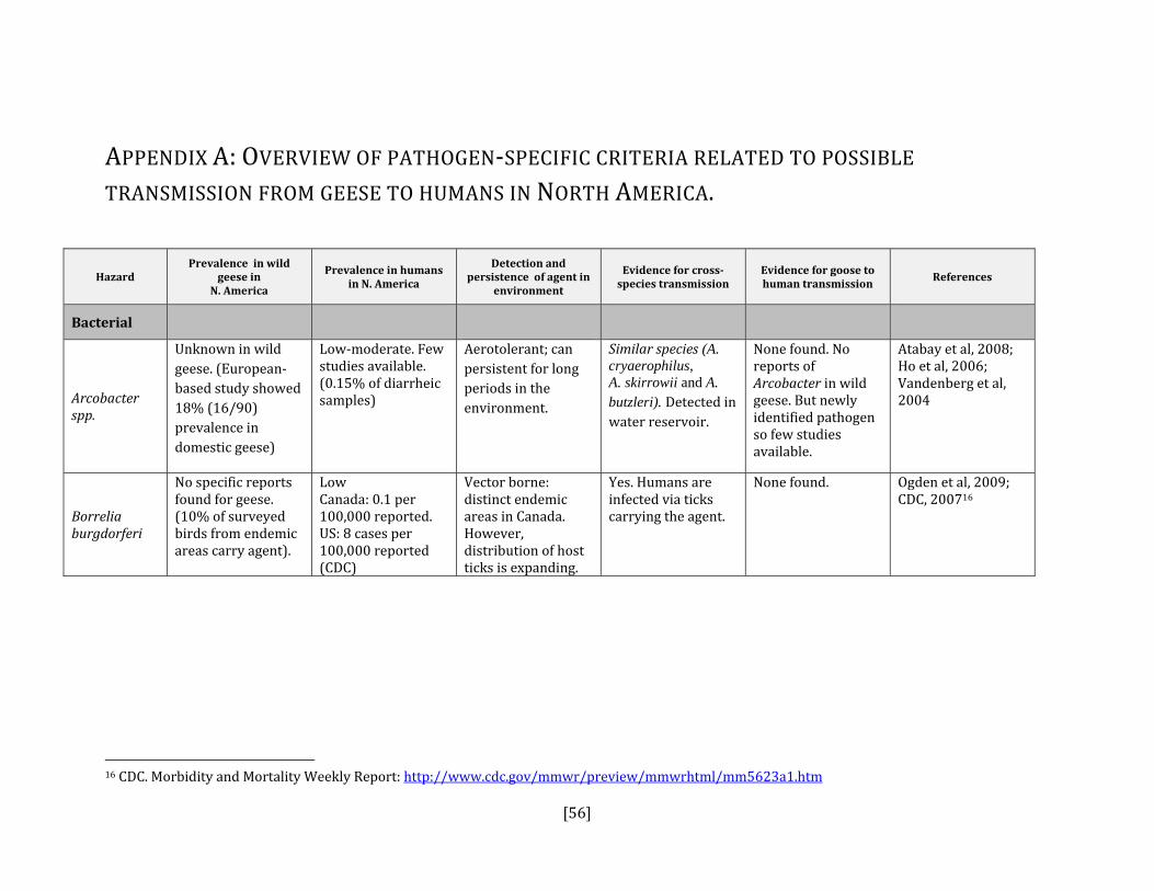
[56]
APPENDIX A: OVERVIEW OF PATHOGEN‐SPECIFIC CRITERIA RELATED TO POSSIBLE TRANSMISSION FROM GEESE TO HUMANS IN NORTH AMERICA.
Hazard Prevalence in wild
geese in N. America
Prevalence in humans in N. America
Detection and persistence of agent in
environment
Evidence for crossspecies transmission
Evidence for goose to human transmission
References
Bacterial
Arcobacter spp.
Unknown in wild geese. (European‐ based study showed 18% (16/90) prevalence in domestic geese)
Low‐moderate. Few studies available. (0.15% of diarrheic samples)
Aerotolerant; can persistent for long periods in the environment.
Similar species (A. cryaerophilus, A. skirrowii and A. butzleri). Detected in water reservoir.
None found. No reports of Arcobacter in wild geese. But newly identified pathogen so few studies available.
Atabay et al, 2008; Ho et al, 2006; Vandenberg et al, 2004
Borrelia burgdorferi
No specific reports found for geese. (10% of surveyed birds from endemic areas carry agent).
Low Canada: 0.1 per 100,000 reported. US: 8 cases per 100,000 reported (CDC)
Vector borne: distinct endemic areas in Canada. However, distribution of host ticks is expanding.
Yes. Humans are infected via ticks carrying the agent.
None found. Ogden et al, 2009; CDC, 200716
16 CDC. Morbidity and Mortality Weekly Report: http://www.cdc.gov/mmwr/preview/mmwrhtml/mm5623a1.htm

[57]
Hazard Prevalence in wild
geese in N. America
Prevalence in humans in N. America
Detection and persistence of agent in
environment
Evidence for crossspecies transmission
Evidence for goose to human transmission
References
Campylobacter spp.
Variable prevalence in fecal samples: 0‐100%.
High. Most common reportable enteric disease. Annually, 30‐40 cases per 100,000 in Canada.17
Generally poor survival in open air, can survive in cool water. Microaerophilic, sensitive to heat and UV. Higher rates in cooler temps with lower light.
Well‐known zoonosis, commonly acquired from poultry meat. Little evidence of cross‐contamination genotypes among geese, chickens, starlings (Colles).
High degree of host specificity in goose isolates (i.e. high number of unique clonal complexes noted in geese). Several shared clonal complexes (at low prevalence <1%) between geese and humans (ST‐21, 45 and 1034) (not causally related). Probable link between waterborne campylobacter outbreak in humans and pink‐footed geese in Norway.
CCDR, 200918; Ogden et al, 2009; Colles et al, 2008; French et al, 2008; Murphy et al, 2006; Clark, 2003; Converse et al, 1999
17 Canadian Integrated Surveillance Report: http://www.phac‐aspc.gc.ca/publicat/ccdr‐rmtc/09vol35/35s3/ 18 CCDR,2009. http://www.phac‐aspc.gc.ca/publicat/ccdr‐rmtc/09pdf/35s3‐eng.pdf

[58]
Hazard Prevalence in wild
geese in N. America
Prevalence in humans in N. America
Detection and persistence of agent in
environment
Evidence for crossspecies transmission
Evidence for goose to human transmission
References
Chlamydophila psittaci
No reports found for geese. 10‐30% of surveyed avian populations have been found positive.
Rare19 (<1 case per 100,000).
Resistant to drying. Can persist in feces or respiratory discharge for up to several months.
Many examples of transmission. Human outbreak linked to domestic duck farm (both samples) (both isolates exhibited the same PCR‐RFLP restriction pattern).
Several cases of C. psittaci reported from occupational exposure during wild bird necropsies, including, among other species, Canada geese and lesser snow geese.
Longbottom and Coulter, 2003; Wobeser and Brand, 1982
C. perfringens
Outbreaks in geese reported in Canada.
Low (1.8 per 100,000/year of all Clostridium spp. 40% of these are C. perfringens) food‐borne related cases.
Widely distributed in the environment.
Food‐borne infections from uncooked poultry have been documented (no causal link established with infected chickens).
No evidence found. Holtby et al, 2008; Leal et al, 2008; Wobeser et al, 1987
Erysipelothrix rhusiopathiae (Erysipelas)
Unknown but one report of outbreak on goose farm noted. Worldwide distribution.
Rare. Can survive for up to a month in soil.
Transmission from pigs most common cause of human infections.
No evidence found. Brooke and Riley, 1999; Gunning and Morton, 1998
19 http://www.ccohs.ca/oshanswers/diseases/psittacosis.html

[59]
Hazard Prevalence in wild
geese in N. America
Prevalence in humans in N. America
Detection and persistence of agent in
environment
Evidence for crossspecies transmission
Evidence for goose to human transmission
References
Escherichia coli20
High in fecal samples (2‐100% depending on season). ETEC: 13% EHEC: 6% (no O157:H7 detected in geese). EIEC: 4.6% EAEC: 1.3% Goose feces with human virulence factors: 2%.
~ 5 cases per 100,000 reported.
Prevalence increases with temperature and humidity (higher rates in spring/summer). Recovery rates do not seem to relate to fecal density. Persists for up to one month in water and for much longer on soil or other substrate.
Low level of virulence factors detected in goose feces (2%) (STa and K1 factors).
No direct evidence found. E.coli from geese detected in areas of high human use (beaches, parks). 19‐38% of water samples from Ontario beaches shown to be contaminated by goose feces (using PCR signal tests).
Edge and Hill, 2007; Kullas et al, 2002; Feare et al, 1999; Mead et al, 1999
Helicobacter spp.
40% of surveyed Canada geese had helicobacter spp in their feces. (PCR). 28% prevalence by fecal culture. Goose ‐specific species detected (H. anseris and H. brantae). Also H. canadensis.
Unknown for species shared by geese and people. But one survey found a prevalence of H. pullorum of 4%.
Yes. Survives well in fresh water.
H. pullorum shown to infect both chickens and humans (disease‐causing for both but not causally linked).
No direct evidence.H. canadensis detected in diarrheic people and in geese in Europe (not causally linked).
Tsiodras et al, 2008; Fox et al, 2006; Adams et al, 2003; Waldenstrom et al, 2003
20 ETEC = enterotoxogenic E. coli; EHEC = enterohemorrhagic E.coli ; EIEC = enteroinvasive E. coli; EAEC = enteroagglomerative E. coli
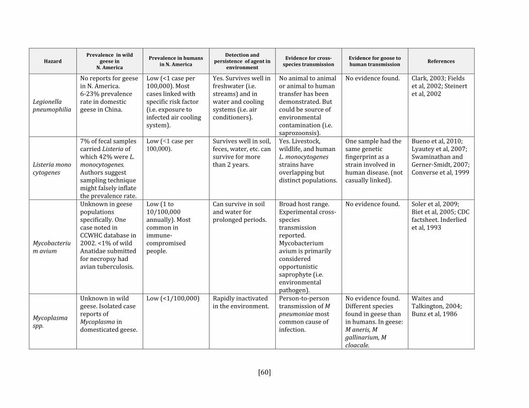
[60]
Hazard Prevalence in wild
geese in N. America
Prevalence in humans in N. America
Detection and persistence of agent in
environment
Evidence for crossspecies transmission
Evidence for goose to human transmission
References
Legionella pneumophilia
No reports for geese in N. America. 6‐23% prevalence rate in domestic geese in China.
Low (<1 case per 100,000). Most cases linked with specific risk factor (i.e. exposure to infected air cooling system).
Yes. Survives well in freshwater (i.e. streams) and in water and cooling systems (i.e. air conditioners).
No animal to animal or animal to human transfer has been demonstrated. But could be source of environmental contamination (i.e. saprozoonsis).
No evidence found. Clark, 2003; Fields et al, 2002; Steinert et al, 2002
Listeria monocytogenes
7% of fecal samples carried Listeria of which 42% were L. monocytogenes. Authors suggest sampling technique might falsely inflate the prevalence rate.
Low (<1 case per 100,000).
Survives well in soil, feces, water, etc. can survive for more than 2 years.
Yes. Livestock, wildlife, and human L. monocytogenes strains have overlapping but distinct populations.
One sample had the same genetic fingerprint as a strain involved in human disease. (not casually linked).
Bueno et al, 2010; Lyautey et al, 2007; Swaminathan and Gerner‐Smidt, 2007; Converse et al, 1999
Mycobacterium avium
Unknown in geese populations specifically. One case noted in CCWHC database in 2002. <1% of wild Anatidae submitted for necropsy had avian tuberculosis.
Low (1 to 10/100,000 annually). Most common in immune‐compromised people.
Can survive in soil and water for prolonged periods.
Broad host range. Experimental cross‐species transmission reported. Mycobacterium avium is primarily considered opportunistic saprophyte (i.e. environmental pathogen).
No evidence found. Soler et al, 2009; Biet et al, 2005; CDC factsheet. Inderlied et al, 1993
Mycoplasma spp.
Unknown in wild geese. Isolated case reports of Mycoplasma in domesticated geese.
Low (<1/100,000) Rapidly inactivated in the environment.
Person‐to‐person transmission of M pneumoniae most common cause of infection.
No evidence found. Different species found in geese than in humans. In geese: M aneris, M gallinarium, M cloacale.
Waites and Talkington, 2004; Bunz et al, 1986

[61]
Hazard Prevalence in wild
geese in N. America
Prevalence in humans in N. America
Detection and persistence of agent in
environment
Evidence for crossspecies transmission
Evidence for goose to human transmission
References
Salmonella enterica
Low. 0 to 2.5% prevalence in Canada geese.
2nd most common reportable enteric disease. 10‐20 cases per 100,000.
Can survive in the environment for at least nine months.
Commonly documented transmission from animals and their product (i.e. eggs) to humans.
No evidence found. CCDR, 200921; Voetsch et al, 2004; Clark, 2003; Roscoe, 2001; Feare et al, 1999; Mead et al, 1999
Streptococcus spp (Group D) (S. bovis, S. gallolyticus subsp. Pasteurianus)
Unknown. One report of S. galloyticus in farmed geese.
Uncommon in people.
Short‐lived survival in environment (hours to several days).
Similar strains of S. gallolyticus shown in humans and pigeons (not casually linked).
No evidence found. Herrera et al, 2009; Barnett et al, 2008; Devriese et al, 1998;
Vibrio spp.(noncholera)
Low. Only one study found 6% prevalence in Canada geese (low sample size (16)). Non‐01 vibrio demonstrated in farmed geese.
Low. (<1 / 100,000). Rates increased by 80% between 1996 and 2001 in the US.
Primarily survives in marine environment.
Different species detected in geese vs humans.
No evidence found. FDA22; Dechet et al, 2008; Buck et al, 1990; Schlater et al, 1981
Yersinia pseudotuberculosis
Low. Two studies did not recover Yersinia spp. from ‘resident’ goose fecal samples.
Considered to be rare in people.
Survives well in the environment and in water.
Unknown but animal source(s) suspected.
No evidence found. Tauxe, 2004; Roscoe, 2001Feare et al, 1999;
21 CCDR,2009. http://www.phac‐aspc.gc.ca/publicat/ccdr‐rmtc/09pdf/35s3‐eng.pdf 22 FDA, Bad Bug Book: Foodborne Pathogenic Microorganisms and Natural Toxins Handbook Vibrio cholerae Serogroup non‐01 http://www.fda.gov/Food/FoodSafety/FoodborneIllness/FoodborneIllnessFoodbornePathogensNaturalToxins/BadBugBook/ucm070419.htm

[62]
Fungal
Aspergilla spp.
Reports of die‐offs in wild waterfowl, including geese, from feeding on contaminated crops. Commonly found in captive geese. 15 cases noted in CCWHC database (passive surveillance) from 1990 to 20009.
Low. Most common in immuno‐compromised patients.
Commonly found in the environment.
Common mold in soil and fungus so environment exposure most important route.
No evidence found. Latge, 1999; Bowes, 1990; Adrian et al, 1978; McDougal and Vaught, 1968
Candida albicans
Unknown. One report found of disease in farmed geese.
Fourth most commonly recovered blood culture isolate in the US.
Normal flora of humans and animals.
This is a commensal organism; part of the normal GI flora of humans and animals. No direct transfer.
No evidence found. Leroy et al, 2009; Buck et al, 1990; Beemer et al, 1973
Cryptococcus spp.
C laurentii was found in 5 samples from Canada geese in NY state.
Uncommon. Recent outbreak in people and animals of C. gattii in Pacific Northwest. C. laurentii recently shown to cause several cases of human disease in immune‐compromised patients.
Survives in the specialized niches. Ie. pigeon guano (C. neoformans), in the tropics and sub‐tropics, and now Vancouver Island (C. gattii).
Pigeon guano is known to be a source of spores for C. neoformans. Few cases of bird to human transfer have been reported.
No evidence found.
Filion et al, 2006; Hoang et al, 2004;

[63]
Parasitic
Cryptosporidium spp.
Low to moderate. 10 and 23% prevalence in cloacal and fecal samples, respectively. In the study (Zhou, 2004) with 23% prevalence, 92% of specimens were goose‐adapted genotypes that so far have not been shown to be infectious to people. Infectious cysts of C. hominis and of the zoonotic genotype of C. parvum have been detected in geese but at low levels (2.4%).
1.85 per 100,000 in Canada (2004 stats). Adult population has a moderate level of seropositivity (~30%). Cryptosporidium spp. responsible for half of waterborne disease outbreaks worldwide.
Survives in 15‐20⁰C water: 3‐12 months.
Geese can act as mechanical carriers of infectious oocysts of C. parvum and distribute into the environment and into water. Geese shown to carry cryptosporidia of goose, ruminant, duck, and human origins. More recent molecular evidence indicates that cryptosporidia are more strongly host‐adapted that previously thought.
Source tracking of C. parvum in a drinking water supply linked to Canada geese, but oocysts in this case were non‐goose host‐adapted species shown to be infectious to people. Another study detected goose‐adapted strain not known to be infectious in people. No human cases have been directly linked to geese.
Jellison et al, 2009; Pintar et al, 2009; Graczyk et al, 2008; Ruecker et al, 2007; Hunter and Thompson, 2005; Zhou et al, 2004; Roscoe, 2001; Fayer et al, 2000; Graczyk et al, 1998
Giardia spp.
15% prevalence in cloacal samples (Roscoe). Detected in pooled fecal samples from each of 9 nine sites sampled (Graczyk).
13.08 cases per 100,000 in Canada (2004 stats). Giardia spp. responsible for ~40% of waterborne disease outbreaks worldwide.
Survives in 15‐20⁰C water: ~2 months.
Avian isolates of Giardia have caused significant infections in mice. More recent work demonstrates a more significant role of intra‐human transmission and places a lower significance on zoonotic transmission.
Direct transmission from geese to people has not been demonstrated.
PHAC, 200923; Graczyk et al, 2008; Yoder and Beach, 2007; Karanis et al, 2007; Hunter and Thompson, 2005; Roscoe, 2001; Graczyk et al, 1998
23 Public Health Agency of Canada: Notifiable disease surveillance. http://dsol‐smed.phac‐aspc.gc.ca/dsol‐smed/ndis/index‐eng.php

[64]
Sarcocystis spp.
Unknown. S. rileyi 65% prevalence found in the white‐fronted goose (Anser albifrons) in Europe.
Low. Case reports only. Did not find any population level studies.
Various stages of the lifecycle take place in the environment.
Different species detected. S. hominis or S. suihominis pathogenic for humans. S. rileyi infect but generally not pathogenic for birds.
No evidence found. Kutkiene et al, 2008; Fayer, 2004
Schistosome cercariae (swimmer’s itch)
Low in Canada geese (< 3% prevalence). Three cases noted in CCWHC database (passive surveillance). Primarily carried by ducks.
6.8 episodes per 100 water‐exposure days.
Cycles between waterfowl (primarily ducks) and snails (intermediate stage).
Literature indicates that ‘waterfowl’ (and intermediate hosts) source of infection for people. Literature focuses on snails.
Only found one study that detected low rate of infection in Canada geese. Another study in BC found 0% (0/122) fecal samples with S. cercariae. No direct evidence of transmission from geese.
Verbrugge et al, 2004; Leighton et al, 2000; Loken et al, 1995;
Toxoplasma gondii
First report in 2004 of detection of T. gondii in a single Canada goose (type III) from Mississippi. Seroprevalence of waterfowl <16% Several reports of clinical toxoplasmosis in small number of geese.
0‐100% seroprevalence in varied human populations.
Sporulated oocysts are very persistent in the environment (up to 18 months). Tissue cysts can survive in refrigerated (1‐4⁰C) meat for up to 3 weeks and can survive freezing (up to ‐8⁰C) for longer than a week.
Low genetic variability (Types I, II, and III) among T. gondi isolates. Types I and II are thought to be more pathogenic for humans than Type III. Chickens thought to be an important source of infection for humans (Dubey in press).
No direct evidence found.
Yan et al, 2009; Dubey et al, 2007 and 2002;Tenter et al, 2000
Viral

[65]
Arboviruses (Eastern and Western Equine Encephalitis, St. Louis Encephalitis, West Nile Virus)
Reports of outbreaks in domestic geese in Manitoba (25% mortality) and domestic ducks in Saskatchewan (39% mortality). One case of WNV in a Canada goose in CCWHC database. No other reports found.
Low. Average of 700 cases in Canada. Very low rates reported of human infection by SLE, EEE, WEE.
WNV able to survive in overwintering Culex mosquitoes in temperate climates.
Yes. Similar WNV strains between humans and birds. Highest mortality rates shown in corvids.
The WNV circulating in North America has shown similarities to strains from infected geese in Israel. Experimentally, geese had viremia titre sufficient to infect mosquitoes and act as a reservoir host. Transmission between geese by direct contact has been demonstrated.
Wojnarowicz et al, 2007; MacLean, 2006; Austin et al, 2004
Avian influenza
Waterfowl, primarily dabbling ducks, are a natural reservoir for AIV. Detected in 0.8% of Canada geese. Dominant strains in waterfowl are Low Pathogenic and include H3N8, H4N6, H4N8, H6N2, H6N8, and H9N2. One case of H7N3 (low pathogenic strain) detected in a dead Canada goose from British Columbia in 2007.
Very low. Two cases of human infection (conjunctivitis) linked to poultry outbreak of type H5N2 in British Columbia in 2005.
AIV can survive for prolonged periods in water.
Yes. Transmission from domestic poultry to humans has been demonstrated. The only evidence of direct transmission from wild birds to people was between H5N1‐ infected swans and people plucking their feathers (Azerbaijan). Contradicted by a British example of humans exposed to H5N1‐ infected swans did not develop disease or seroconvert.
Serologic evidence (H11/N9) of transmission between waterfowl and a duck hunter and two wildlife professionals with many years of exposure to waterfowl has been shown.
Wallenstein et al, 2010; Berhane et al, 2009; Tsiodras et al, 2008; Parmley et al, 2008 ; Pasick et al, 2007; Clark and Hall, 2006; Olsen et al, 2006; Gill et al, 2006

[66]
Coronavirus (SARS and nonSARS variants)
Unknown in Canada goose. Detected by cloacal swab in Graylag geese in Norway.
SARS: negligible (previous outbreak contained). Non‐SARS: low (<4% prevalence).
Can survive for several days on a variety of surfaces but converts to a non‐infectious form after about 90 minutes.
Some molecular evidence is available but not conclusive (civet cats in Asia to humans).
No evidence found. Jonassen et al, 2005; Weiss and Navas‐Martin, 2005
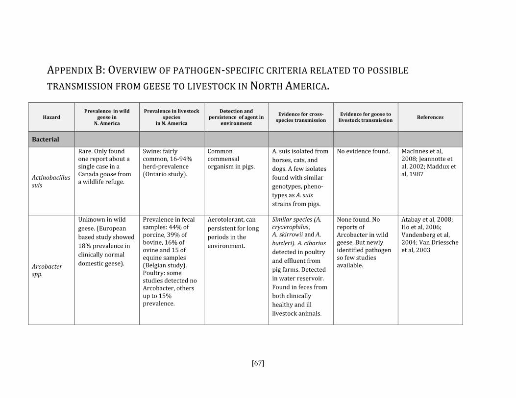
[67]
APPENDIX B: OVERVIEW OF PATHOGEN‐SPECIFIC CRITERIA RELATED TO POSSIBLE TRANSMISSION FROM GEESE TO LIVESTOCK IN NORTH AMERICA.
Hazard Prevalence in wild
geese in N. America
Prevalence in livestock species
in N. America
Detection and persistence of agent in
environment
Evidence for crossspecies transmission
Evidence for goose to livestock transmission
References
Bacterial
Actinobacillus suis
Rare. Only found one report about a single case in a Canada goose from a wildlife refuge.
Swine: fairly common, 16‐94% herd‐prevalence (Ontario study).
Common commensal organism in pigs.
A. suis isolated from horses, cats, and dogs. A few isolates found with similar genotypes, pheno‐types as A. suis strains from pigs.
No evidence found. MacInnes et al, 2008; Jeannotte et al, 2002; Maddux et al, 1987
Arcobacter spp.
Unknown in wild geese. (European based study showed 18% prevalence in clinically normal domestic geese).
Prevalence in fecal samples: 44% of porcine, 39% of bovine, 16% of ovine and 15 of equine samples (Belgian study). Poultry: some studies detected no Arcobacter, others up to 15% prevalence.
Aerotolerant, can persistent for long periods in the environment.
Similar species (A. cryaerophilus, A. skirrowii and A. butzleri). A. cibarius detected in poultry and effluent from pig farms. Detected in water reservoir. Found in feces from both clinically healthy and ill livestock animals.
None found. No reports of Arcobacter in wild geese. But newly identified pathogen so few studies available.
Atabay et al, 2008; Ho et al, 2006; Vandenberg et al, 2004; Van Driessche et al, 2003
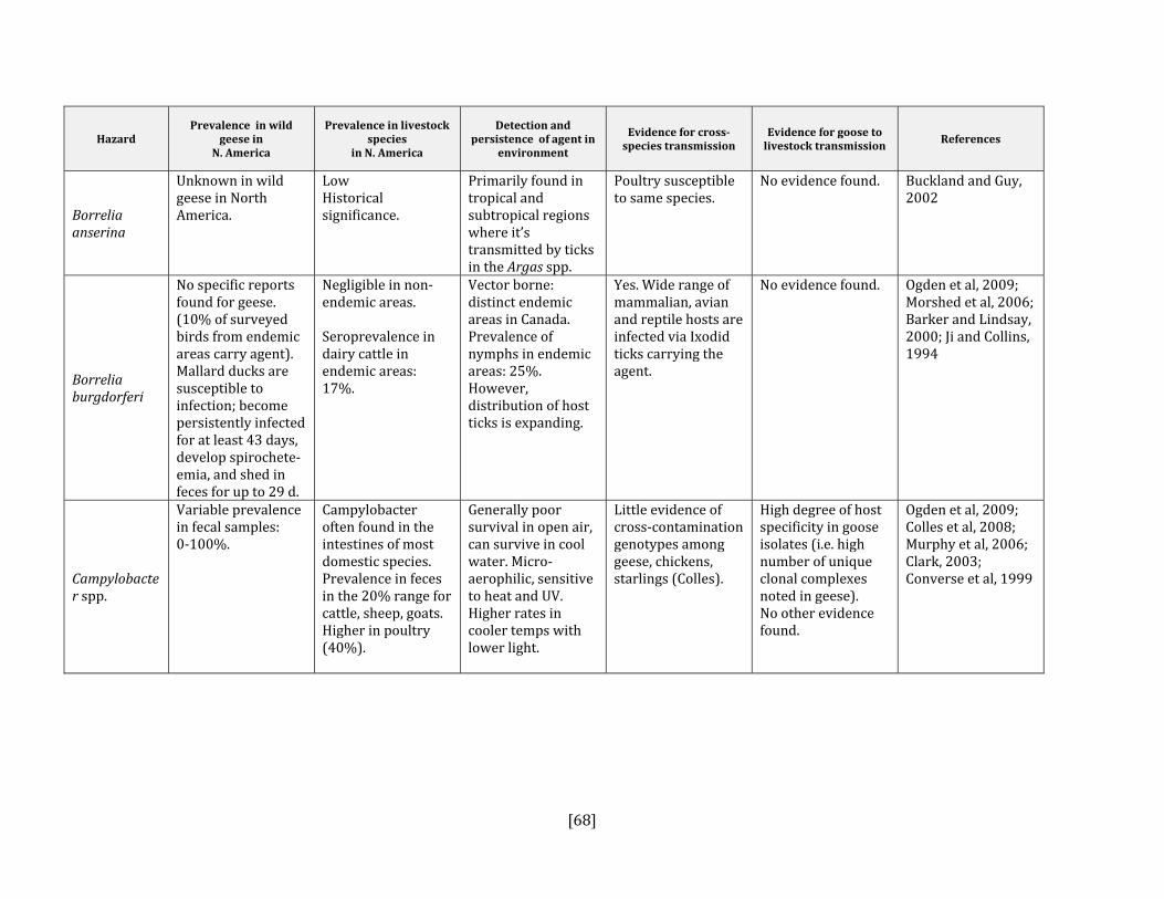
[68]
Hazard Prevalence in wild
geese in N. America
Prevalence in livestock species
in N. America
Detection and persistence of agent in
environment
Evidence for crossspecies transmission
Evidence for goose to livestock transmission
References
Borrelia anserina
Unknown in wild geese in North America.
Low Historical significance.
Primarily found in tropical and subtropical regions where it’s transmitted by ticks in the Argas spp.
Poultry susceptible to same species.
No evidence found. Buckland and Guy, 2002
Borrelia burgdorferi
No specific reports found for geese. (10% of surveyed birds from endemic areas carry agent). Mallard ducks are susceptible to infection; become persistently infected for at least 43 days, develop spirochete‐emia, and shed in feces for up to 29 d.
Negligible in non‐endemic areas. Seroprevalence in dairy cattle in endemic areas: 17%.
Vector borne: distinct endemic areas in Canada. Prevalence of nymphs in endemic areas: 25%. However, distribution of host ticks is expanding.
Yes. Wide range of mammalian, avian and reptile hosts are infected via Ixodid ticks carrying the agent.
No evidence found. Ogden et al, 2009; Morshed et al, 2006; Barker and Lindsay, 2000; Ji and Collins, 1994
Campylobacter spp.
Variable prevalence in fecal samples: 0‐100%.
Campylobacter often found in the intestines of most domestic species. Prevalence in feces in the 20% range for cattle, sheep, goats. Higher in poultry (40%).
Generally poor survival in open air, can survive in cool water. Micro‐aerophilic, sensitive to heat and UV. Higher rates in cooler temps with lower light.
Little evidence of cross‐contamination genotypes among geese, chickens, starlings (Colles).
High degree of host specificity in goose isolates (i.e. high number of unique clonal complexes noted in geese). No other evidence found.
Ogden et al, 2009; Colles et al, 2008; Murphy et al, 2006; Clark, 2003; Converse et al, 1999

[69]
Hazard Prevalence in wild
geese in N. America
Prevalence in livestock species
in N. America
Detection and persistence of agent in
environment
Evidence for crossspecies transmission
Evidence for goose to livestock transmission
References
Chlamydophila psittaci
No reports found for geese. 10‐30% of surveyed avian populations.
Outbreaks reported in domestic turkeys and ducks. Chickens have been shown to be quite resistant to C. psittaci.
Resistant to drying. Can persist in feces or respiratory discharge for up to several months.
Yes. Demonstrated transmission to humans.
No evidence found. Zang et al, 2008; Longbottom and Coulter, 2003
Clostridium botulinum
Large scale outbreaks, in waterfowl throughout North America, with very high mortality rates.
Rare. Cluster outbreaks have occurred in cattle and sheep that have ingested contaminated poultry litter.
C. botulinum is a strict anaerobe that forms dormant spores in adverse conditions. Spores are very resistant to environmental decay.
Most exposure is due to presence of spores in environ‐ment. However, spores also carried in tissues of birds and distributed to new environments in their feces.
No evidence found. Otter et al, 2006; Rocke, 2006; Wobeser and Rannie, 1987
Clostridium perfringens
Outbreaks in geese reported in Canada. Usually from ‘grain overload’ after grazing on crops following periods of low nutritional intake (i.e. migration).
Uncommon. Widely distributed in the environment.
Human food‐borne infections from uncooked poultry to have been documented (no causal link established with infected chickens).
No evidence found. Leal et al, 2008; Wobeser et al, 1987
Erysipelothrix rhusiopathiae (Erysipelas)
Unknown but one report of outbreak on goose farm noted. Worldwide distribution.
Present on most pig farms. Commonly vaccinated against in swine.
Can survive for up to a month in soil.
Transmission from pigs most common cause of human infections.
No evidence found. Gunning and Morton, 1998

[70]
Hazard Prevalence in wild
geese in N. America
Prevalence in livestock species
in N. America
Detection and persistence of agent in
environment
Evidence for crossspecies transmission
Evidence for goose to livestock transmission
References
Escherichia coli24
High in fecal samples (2‐100% depending on season). ETEC: 13% EHEC: 6% (no O157:H7 detected in geese). EIEC: 4.6% EAEC: 1.3% Goose feces with human virulence factors: 2%.
Common cause of neonatal disease in calves and piglets. 3‐40% of diarrheic calves are positive for enterotoxigenic E. coli.
Prevalence increases with temp. and humidity (higher rates in spring/summer). Recovery rates relate poorly to fecal density. Can persist for 1 month in water and much longer on soil or other substrate.
Low level of human virulence factors detected in goose feces (2%) (STa and K1 factors).
No direct evidence found. However, serotypes of importance to humans and human virulence factors were detected at low levels in geese.
Edge and Hill, 2007; Kullas et al, 2002; Feare et al, 1999; Mead et al, 1999; Radostits et al, 1994
Helicobacter spp.
40% of surveyed Canada geese had Helicobacter spp in their feces. (PCR). 28% prevalence by fecal culture. Goose ‐specific species detected (H. anseris and H. brantae). Also H. canadensis.
Unknown for species indentified in geese. H. pullorum relatively common in poultry. Species specificity (pigs carry H. suis, cattle carry H. bovis).
Yes. Survives well in fresh water.
The pathogenic potential of H. anseris and H. brantae are unknown. H. pullorum shown to infect both chickens and humans (disease‐causing for both but not‐ causally linked).
No evidence found. Couldn’t find evidence of same species in geese and livestock species.
Haesebroucket al, 2009; Tsiodras et al, 2008; Fox et al, 2006; Adams et al, 2003; Waldenstrom et al, 2003; Atabay et al, 1998
24 ETEC = enterotoxogenic E. coli; EHEC = enterohemorrhagic E.coli ; EIEC = enteroinvasive E. coli; EAEC = enteroagglomerative E. coli
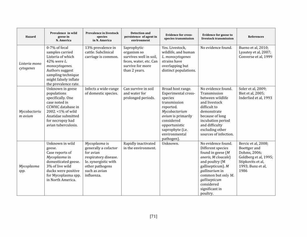
[71]
Hazard Prevalence in wild
geese in N. America
Prevalence in livestock species
in N. America
Detection and persistence of agent in
environment
Evidence for crossspecies transmission
Evidence for goose to livestock transmission
References
Listeria monocytogenes
0‐7% of fecal samples carried Listeria of which 42% were L. monocytogenes. Authors suggest sampling technique might falsely inflate the prevalence rate.
13% prevalence in cattle. Subclinical carriage is common.
Saprophytic organism so survives well in soil, feces, water, etc. Can survive for more than 2 years.
Yes. Livestock, wildlife, and human L. monocytogenes strains have overlapping but distinct populations.
No evidence found. Bueno et al, 2010; Lyautey et al, 2007; Converse et al, 1999
Mycobacterium avium
Unknown in geese populations specifically. One case noted in CCWHC database in 2002. <1% of wild Anatidae submitted for necropsy had avian tuberculosis.
Infects a wide‐range of domestic species.
Can survive in soil and water for prolonged periods.
Broad host range. Experimental cross‐species transmission reported. Mycobacterium avium is primarily considered opportunistic saprophyte (i.e. environmental pathogen).
No evidence found. Transmission between wildlife and livestock difficult to demonstrate because of long incubation period and difficulty excluding other sources of infection.
Soler et al, 2009; Biet et al, 2005; Inderlied et al, 1993
Mycoplasma spp.
Unknown in wild geese. Case reports of Mycoplasma in domesticated geese. 3% of live wild ducks were positive for Mycoplasma spp. in North America.
Mycoplasma is generally a cofactor for avian respiratory disease. Ie. synergistic with other pathogens such as avian influenza.
Rapidly inactivated in the environment.
Unknown. No evidence found.Different species found in geese (M aneris, M cloacale) and poultry (M gallisepticum). M gallinarium in common but only M. gallisepticum considered significant in poultry.
Bercic et al, 2008; Boettger and Dohms, 2006; Goldberg et al, 1995; Stipkovits et al, 1993; Bunz et al, 1986

[72]
Hazard Prevalence in wild
geese in N. America
Prevalence in livestock species
in N. America
Detection and persistence of agent in
environment
Evidence for crossspecies transmission
Evidence for goose to livestock transmission
References
Pasteurella multocida
Unknown. < 5% seroprevalence in Greater White‐fronted geese (Anser albifrons frontalis) in Alaska. Epizootics, with significant mortalities, more common in wintering grounds in US than in Canada. “Outbreaks have been happening regularly on breeding grounds in the eastern arctic (annually since 2004) and Saint Lawrence Estuary (not annually but commonly, for decades) in common eider colonies, but a few geese including Canada geese (in eastern arctic) have also died during these outbreaks. Not sweeping numbers for geese compared to the eiders.” (pers. comm., Catherine Soos)
Commonly occurs in respiratory tracks of domestic species (sheep, cattle, poultry).
Reasonably short‐lived in the environment. Shown to survive less than seven weeks in wetlands following avian cholera outbreaks.
Reported in a wide variety of birds and mammals. Identical strains (using PFGE) noted between poultry flocks and wild birds (Eiders). Experimental transmission demonstrated between Lesser Snow geese and Pekin ducklings.
No direct evidence found. Only one report found of outbreak in Canada geese (Windingstad et al, 1983).
Blanchong et al, 2006; Samuel et al, 2005; Pedersen et al, 2003; Samuel et al, 1997; Botzler, 1991; Windingstad et al, 1983
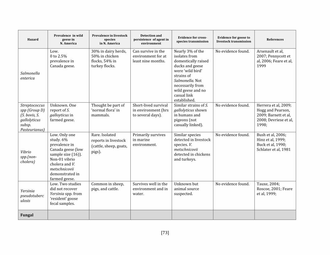
[73]
Hazard Prevalence in wild
geese in N. America
Prevalence in livestock species
in N. America
Detection and persistence of agent in
environment
Evidence for crossspecies transmission
Evidence for goose to livestock transmission
References
Salmonella enterica
Low. 0 to 2.5% prevalence in Canada geese.
30% in dairy herds, 50% in chicken flocks, 54% in turkey flocks.
Can survive in the environment for at least nine months.
Nearly 3% of the isolates from domestically raised ducks and geese were ‘wild bird’ strains of Salmonella. Not necessarily from wild geese and no casual link established.
No evidence found.
Arsenault et al, 2007; Pennycott et al, 2006; Feare et al, 1999
Streptococcus spp (Group D) (S. bovis, S. gallolyticus subsp. Pasteurianus)
Unknown. One report of S. galloyticus in farmed geese.
Thought be part of ‘normal flora’ in mammals.
Short‐lived survival in environment (hrs to several days).
Similar strains of S. gallolyticus shown in humans and pigeons (not casually linked).
No evidence found. Herrera et al, 2009; Hogg and Pearson, 2009; Barnett et al, 2008; Devriese et al, 1998;
Vibrio spp.(noncholera)
Low. Only one study. 6% prevalence in Canada geese (low sample size (16)). Non‐01 vibrio cholera and V. metschnicovii demonstrated in farmed geese.
Rare. Isolated reports in livestock (cattle, sheep, goats, pigs).
Primarily survives in marine environment.
Similar species detected in livestock species. V. metschnicovii detected in chickens and turkeys.
No evidence found. Bush et al, 2006; Hinz et al, 1999; Buck et al, 1990; Schlater et al, 1981
Yersinia pseudotuberculosis
Low. Two studies did not recover Yersinia spp. from ‘resident’ goose fecal samples.
Common in sheep, pigs, and cattle.
Survives well in the environment and in water.
Unknown but animal source suspected.
No evidence found.
Tauxe, 2004; Roscoe, 2001; Feare et al, 1999;
Fungal
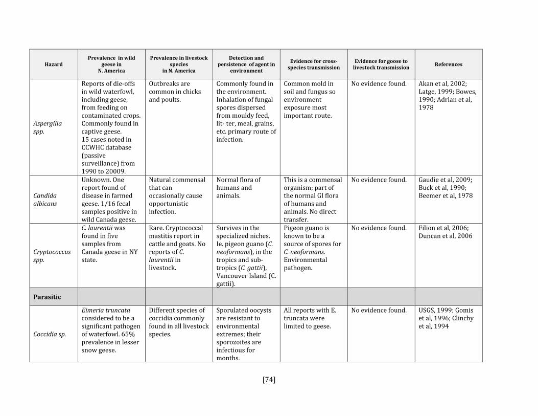
[74]
Hazard Prevalence in wild
geese in N. America
Prevalence in livestock species
in N. America
Detection and persistence of agent in
environment
Evidence for crossspecies transmission
Evidence for goose to livestock transmission
References
Aspergilla spp.
Reports of die‐offs in wild waterfowl, including geese, from feeding on contaminated crops. Commonly found in captive geese. 15 cases noted in CCWHC database (passive surveillance) from 1990 to 20009.
Outbreaks are common in chicks and poults.
Commonly found in the environment. Inhalation of fungal spores dispersed from mouldy feed, lit‐ ter, meal, grains, etc. primary route of infection.
Common mold in soil and fungus so environment exposure most important route.
No evidence found. Akan et al, 2002; Latge, 1999; Bowes, 1990; Adrian et al, 1978
Candida albicans
Unknown. One report found of disease in farmed geese. 1/16 fecal samples positive in wild Canada geese.
Natural commensal that can occasionally cause opportunistic infection.
Normal flora of humans and animals.
This is a commensal organism; part of the normal GI flora of humans and animals. No direct transfer.
No evidence found. Gaudie et al, 2009; Buck et al, 1990; Beemer et al, 1978
Cryptococcus spp.
C. laurentii was found in five samples from Canada geese in NY state.
Rare. Cryptococcal mastitis report in cattle and goats. No reports of C. laurentii in livestock.
Survives in the specialized niches. Ie. pigeon guano (C. neoformans), in the tropics and sub‐tropics (C. gattii), Vancouver Island (C. gattii).
Pigeon guano is known to be a source of spores for C. neoformans. Environmental pathogen.
No evidence found. Filion et al, 2006; Duncan et al, 2006
Parasitic
Coccidia sp.
Eimeria truncata considered to be a significant pathogen of waterfowl. 65% prevalence in lesser snow geese.
Different species of coccidia commonly found in all livestock species.
Sporulated oocysts are resistant to environmental extremes; their sporozoites are infectious for months.
All reports with E. truncata were limited to geese.
No evidence found. USGS, 1999; Gomis et al, 1996; Clinchy et al, 1994

[75]
Hazard Prevalence in wild
geese in N. America
Prevalence in livestock species
in N. America
Detection and persistence of agent in
environment
Evidence for crossspecies transmission
Evidence for goose to livestock transmission
References
Cryptosporidium spp.
Low to moderate. 10 and 23% prevalence in cloacal and fecal samples, respectively. Zhou et al (2004) found 23% prevalence, 92% of specimens were goose‐adapted genotypes. Infectious cysts of C. hominis and of the zoonotic genotype of C. parvum have also been detected in geese but at low levels (2.4%).
Common in cattle, particularly in calves. C. parvum and C. andersoni are the most common species.
Survives in 15‐20⁰C water: 3‐12 months.
Geese can act as mechanical carriers of infectious oocysts of C. parvum and distribute into the environment and into water. More recent molecular evidence indicates that cryptosporidia are more strongly host‐adapted that previously thought.
Source tracking of C. parvum in a drinking water supply linked to Canada geese. Geese shown to carry Cryptosporidia spp. of goose, ruminant, duck, and human origins.
Jellison et al, 2009; Pintar et al, 2009; Graczyk et al, 2008; Ruecker et al, 2007; Hunter and Thompson, 2005; Zhou et al, 2004; Roscoe, 2001; Fayer et al, 2000; Graczyk et al, 1998
Giardia spp.
15% prevalence in cloacal samples (Roscoe). Detected in pooled fecal samples from each of 9 nine sites sampled (Graczyk).
Common in livestock.
Survives in 15‐20⁰C water: ~2 months.
Avian isolates of Giardia have caused significant infections in mice. More recent work demonstrates a more significant role of intra‐human transmission and places a lower significance on zoonotic transmission.
High concentrations of cysts in Canada geese suggest infection rather than mechanical carriage of Giardia (i.e. increased transmission rate).
Graczyk et al, 2008; Olsen et al, 2004; Yoder and Beach, 2007; Karanis et al, 2007; Hunter and Thompson, 2005; Roscoe, 2001; Graczyk et al, 1998
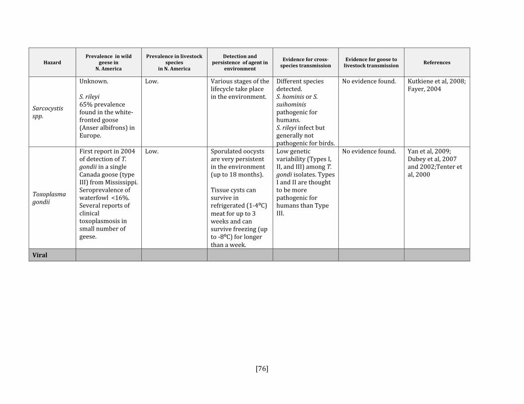
[76]
Hazard Prevalence in wild
geese in N. America
Prevalence in livestock species
in N. America
Detection and persistence of agent in
environment
Evidence for crossspecies transmission
Evidence for goose to livestock transmission
References
Sarcocystis spp.
Unknown. S. rileyi 65% prevalence found in the white‐fronted goose (Anser albifrons) in Europe.
Low. Various stages of the lifecycle take place in the environment.
Different species detected. S. hominis or S. suihominis pathogenic for humans. S. rileyi infect but generally not pathogenic for birds.
No evidence found. Kutkiene et al, 2008; Fayer, 2004
Toxoplasma gondii
First report in 2004 of detection of T. gondii in a single Canada goose (type III) from Mississippi. Seroprevalence of waterfowl <16%. Several reports of clinical toxoplasmosis in small number of geese.
Low. Sporulated oocysts are very persistent in the environment (up to 18 months). Tissue cysts can survive in refrigerated (1‐4⁰C) meat for up to 3 weeks and can survive freezing (up to ‐8⁰C) for longer than a week.
Low genetic variability (Types I, II, and III) among T. gondi isolates. Types I and II are thought to be more pathogenic for humans than Type III.
No evidence found. Yan et al, 2009; Dubey et al, 2007 and 2002;Tenter et al, 2000
Viral

[77]
Hazard Prevalence in wild
geese in N. America
Prevalence in livestock species
in N. America
Detection and persistence of agent in
environment
Evidence for crossspecies transmission
Evidence for goose to livestock transmission
References
Arboviruses (Eastern and Western Equine Encephalitis, St. Louis Encephalitis, West Nile Virus)
WNV: Reports of outbreaks in domestic geese in Manitoba (25% mortality) and domestic ducks in Saskatchewan (39% mortality). No reports of WNV in Canada geese.
WNV: low in chickens and turkeys. Chickens and turkeys develop very low virus titres and more limited clinical signs than geese.
WNV able to survive in overwintering Culex mosquitoes in temperate climates.
Yes. Similar WNV strains between humans and birds. Highest mortality rates shown in corvids.
The WNV circulating in North America has shown similarities to strains from infected geese in Israel. Experimentally, geese had virus titre sufficient to infect mosquitoes and act as a reservoir host. Transmission between geese by direct contact has been demonstrated.
Wojnarowicz et al, 2007; MacLean, 2006; Austin et al, 2004; Banet‐Noach et al, 2003; Swayne et al, 2001; Senne et al, 2000
Avian adenovirus
Antibodies in geese and ducks are widespread. Outbreak in domestic geese in Canada reported. In this case, no contact between domestic geese and wild geese had been noted.
Distributed widely throughout the world. High seroprevalence.
In general, short‐lived in the environment. Inactivated after 30 minutes in aqueous solutions.
Only experimental evidence of transmission whereby goose strains grow in vitro on poultry cells.
No evidence found. Chen et al, 2009; Saif, 2003; Ivanics et al, 2001; Riddell, 1984
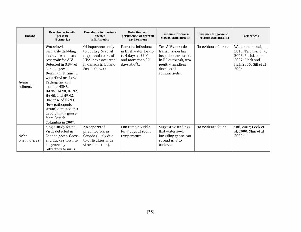
[78]
Hazard Prevalence in wild
geese in N. America
Prevalence in livestock species
in N. America
Detection and persistence of agent in
environment
Evidence for crossspecies transmission
Evidence for goose to livestock transmission
References
Avian influenza
Waterfowl, primarily dabbling ducks, are a natural reservoir for AIV. Detected in 0.8% of Canada geese. Dominant strains in waterfowl are Low Pathogenic and include H3N8, H4N6, H4N8, H6N2, H6N8, and H9N2. One case of H7N3 (low pathogenic strain) detected in a dead Canada goose from British Columbia in 2007.
Of importance only to poultry. Several major outbreaks of HPAI have occurred in Canada in BC and Saskatchewan.
Remains infectious in freshwater for up to 4 days at 22⁰C and more than 30 days at 0⁰C.
Yes. AIV zoonotic transmission has been demonstrated. In BC outbreak, two poultry handlers developed conjunctivitis.
No evidence found. Wallenstein et al, 2010; Tsiodras et al, 2008; Pasick et al, 2007; Clark and Hall, 2006; Gill et al, 2006
Avian pneumovirus
Single study found. Virus detected in Canada geese. Geese and ducks shown to be generally refractory to virus.
No reports of pneumovirus in Canada (likely due to difficulties with virus detection).
Can remain viable for 7 days at room temperature.
Suggestive findings that waterfowl, including geese, can spread APV to turkeys.
No evidence found. Safi, 2003; Cook et al, 2000; Shin et al, 2000;
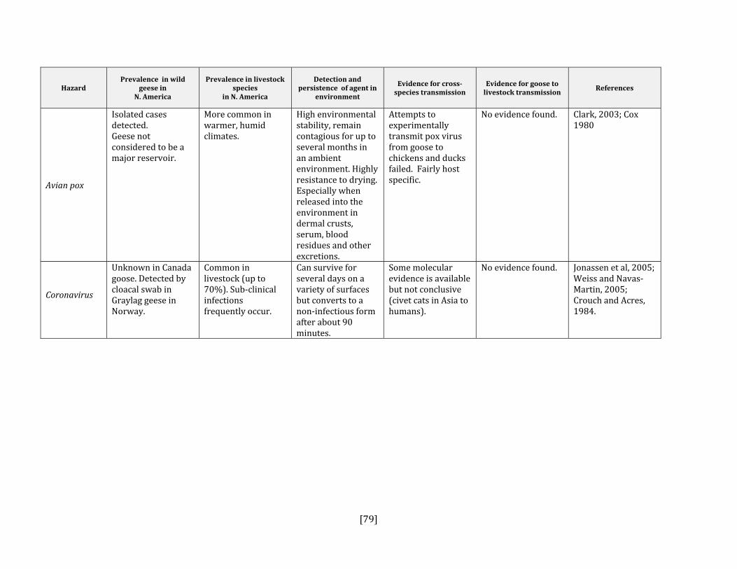
[79]
Hazard Prevalence in wild
geese in N. America
Prevalence in livestock species
in N. America
Detection and persistence of agent in
environment
Evidence for crossspecies transmission
Evidence for goose to livestock transmission
References
Avian pox
Isolated cases detected. Geese not considered to be a major reservoir.
More common in warmer, humid climates.
High environmental stability, remain contagious for up to several months in an ambient environment. Highly resistance to drying. Especially when released into the environment in dermal crusts, serum, blood residues and other excretions.
Attempts to experimentally transmit pox virus from goose to chickens and ducks failed. Fairly host specific.
No evidence found. Clark, 2003; Cox 1980
Coronavirus
Unknown in Canada goose. Detected by cloacal swab in Graylag geese in Norway.
Common in livestock (up to 70%). Sub‐clinical infections frequently occur.
Can survive for several days on a variety of surfaces but converts to a non‐infectious form after about 90 minutes.
Some molecular evidence is available but not conclusive (civet cats in Asia to humans).
No evidence found. Jonassen et al, 2005; Weiss and Navas‐Martin, 2005; Crouch and Acres, 1984.

[80]
Hazard Prevalence in wild
geese in N. America
Prevalence in livestock species
in N. America
Detection and persistence of agent in
environment
Evidence for crossspecies transmission
Evidence for goose to livestock transmission
References
Newcastle disease virus (NDV)
High seroprevalence (44% in adults) and geographically widespread in Canada. Waterfowl considered natural reservoir, particularly of mild form of virus.
Highly virulent form has been eradicated from developed countries.
Prolonged survival on eggshells and in feces.
Wild birds suspected in transmission to poultry in Europe, but not proven. Virulent strains isolated from migratory waterfowl have been experimentally transmitted to domestic poultry that showed evidence of pathogenicity acquired during passage in the infected chicken population.
No evidence found. Dai et al, 2008; Clark, 2003; Alexander et al, 1999; Palmer and Trainer, 1970

[81]
APPENDIX C: CHARACTERISTICS OF IDENTIFIED HAZARDS THAT COULD IMPACT HUMANS (MAGNITUDE OF POTENTIAL IMPACT)
Hazard Route of exposure
What does it do?
Severity of illness
Nationally Reportable
(humans)
25
References
Magnitude ranking
Treatable
Severity of
symptoms
Frequency of
hospitalization26
Frequency of
fatality
Bacterial
Arcobacter spp. Ingestion Enteritis, bacteremia
yes Mild to moderate
Low Low No Ho et al, 2006; Vandenberg et al, 2004 Lo
w
Borrelia burgdorferi
Vector
Multisystemic: general malaise, skin, cardiac,
musculoskeletal, and neurological
symptoms
yes
Mild (when detected early) Moderate‐ severe if left undiagnosed Morbidity: low Canada: 0.1 per
100,000 reported. In US: 8 cases per 100,000
Rare
Rare No Ogden et al, 2009; CDC, 200727 M
edium
25 List of human reportable diseases in Canada: http://dsol‐smed.phac‐aspc.gc.ca/dsol‐smed/ndis/list_e.html 26 Note: hospitalization rates documented here likely overestimate the true hospitalization rate due to inherent biases in the data and reporting methods. Ie. under‐reporting, hospital‐based studies, etc. 27 CDC. Morbidity and Mortality Weekly Report: http://www.cdc.gov/mmwr/preview/mmwrhtml/mm5623a1.htm

[82]
Hazard Route of exposure
What does it do?
Severity of illness
Nationally Reportable
(humans)
25
References
Magnitude ranking
Treatable
Severity of
symptoms
Frequency of
hospitalization26
Frequency of
fatality
Campylobacter spp. Ingestion Enteritis,
arthritis, sepsis Yes Mild‐moderate
Low
25‐100 cases per year2
Low
0.07 per 1000 cases2
Yes
CCDR, 200928 Morbidity: Most common reportable
enteric disease. Annually, 30‐40 cases per 100,000 in Canada29
Medium
Chlamydophila psittaci Aerosol General malaise,
pneumonia Yes Mild Rare
Rare
No Longbottom and Coulter,
2003 Negligible
C. perfringens Ingestion Enteritis, gas
gangrene, sepsis
Yes, but resistance an issue
Moderate to severe
depending on underlying
conditions (e.g. age, cancer,
kidney disease)
Often for hospital‐acquired cases and patients at
risk
High, dependent on study design (i.e. hospital‐based vs communit
y ) (25‐50%)
No Leal et al, 2008
Medium
28 CCDR,2009. http://www.phac‐aspc.gc.ca/publicat/ccdr‐rmtc/09pdf/35s3‐eng.pdf 29 Canadian Integrated Surveillance Report: http://www.phac‐aspc.gc.ca/publicat/ccdr‐rmtc/09vol35/35s3/

[83]
Hazard Route of exposure
What does it do?
Severity of illness
Nationally Reportable
(humans)
25
References
Magnitude ranking
Treatable
Severity of
symptoms
Frequency of
hospitalization26
Frequency of
fatality
Erysipelothrix rhusiopathiae (Erysipelas)
Direct contact
Cutaneous infection,
endocarditis Yes
Mild (cutaneous
form) to severe (systemic form)
low
Low (cutaneous form to high
(systemic form)
No Brooke and Riley, 1999 Lo
w
Escherichia coli Ingestion
Enteritis, hemolytic uremic
syndrome (with enterotoxigenic
strain)
Yes Mild to severe depending on
strain
Low to high
depending on strain
Low to moderate depending on strain
Yes (ETEC)
Karmali et al, 2010; OIE, 2008
High (ETEC,
EHEC).
Otherwise
low
Helicobacter spp. Ingestion
Chronic gastritis, peptic ulcer
disease, recently linked to gastric adenocarcinoma and lymphoma
Not always
Mild to severe Low Rare No
Tsiodras et al, 2008; Fox et al,
2006 Low
Legionella pneumophilia
Aerosol Pneumonia Yes Moderate Moderate
Moderate (7‐24%), highest in immune compromised and elderly
Yes Fields et al,
2002; Steinert et al, 2002 M
edium
Listeria monocytogenes Ingestion
Enteritis, meningitis, sepsis, fetal infection
Yes Mild to severe Moderate Moderate (20‐30%) Yes
Swaminathan and Gerner‐Smidt, 2007 M
edium

[84]
Hazard Route of exposure
What does it do?
Severity of illness
Nationally Reportable
(humans)
25
References
Magnitude ranking
Treatable
Severity of
symptoms
Frequency of
hospitalization26
Frequency of
fatality
Mycobacterium avium
Ingestion (water borne) and
aerosol
Pneumonia Yes Mild to moderate
Low (primarily in HIV patients)
Low (~10% in
HIV patients and much lower in non‐HIV)
Yes Inderlied et al, 1993 Lo
w
Mycoplasma spp. Aerosol
Tracheobronchitis, pneumonia, CNS and skin manifestations
Yes Mild to moderate Low Low No
Waites and Talkington,
2004 Low
Salmonella enterica
Ingestion Enteritis Yes Mild to moderate
Low (<20%)
Low (<1%) Yes
CCDR, 200930; Voetsch et al,
2004 M
edium
Streptococcus spp (Group D) (S.
bovis, S. gallolyticus subsp.
Pasteurianus)
Ingestion
Endocarditis, enteritis,
implicated in colorectal neoplasia (S.
bovis)
Yes Moderate to severe High
High for S. bovis
endocarditis (45%) Moderate for non S. bovis
endocarditis (25%)
No Herrera et al, 2009;
Medium
30 CCDR,2009. http://www.phac‐aspc.gc.ca/publicat/ccdr‐rmtc/09pdf/35s3‐eng.pdf

[85]
Hazard Route of exposure
What does it do?
Severity of illness
Nationally Reportable
(humans)
25
References
Magnitude ranking
Treatable
Severity of
symptoms
Frequency of
hospitalization26
Frequency of
fatality
Vibrio spp.(noncholera)
Water‐borne agent:
Ingestion or direct contact
Enteritis, septicemia,
wound infection Yes Mild to
moderate
Moderate to high
depending on Vibrio sp. (20‐80%)
Low (10‐20%) No FDA31; Dechet
et al, 2008
Medium
Yersinia pseudotuberculo
sis
Ingestion Enteritis Yes Mild Moderate
(30%) Low No Nuorti et al,
2004; Hannu et al, 2003 Lo
w
Fungal
Aspergilla spp. Aerosol Ingestion
Pneumonia Not always
Moderate to severe
High Moderate No Latge, 1999
Medi
um
Candida albicans Commensal org
Oropharyngeal, genital, and systemic manifestations
Yes Negligible to moderate
Low –moderate depending on underlying conditions
Low‐moderate depending on underlying conditions
No Leroy et al, 2009
Low
31 FDA, Bad Bug Book: Foodborne Pathogenic Microorganisms and Natural Toxins Handbook Vibrio cholerae Serogroup non‐01 http://www.fda.gov/Food/FoodSafety/FoodborneIllness/FoodborneIllnessFoodbornePathogensNaturalToxins/BadBugBook/ucm070419.htm

[86]
Hazard Route of exposure
What does it do?
Severity of illness
Nationally Reportable
(humans)
25
References
Magnitude ranking
Treatable
Severity of
symptoms
Frequency of
hospitalization26
Frequency of
fatality
Cryptococcus spp.
Aerosol Cutaneous, respiratory and neurologic manifestations
Yes Negligible to severe
Low Low No
Hoang et al, 2004
Low
Parasitic
Cryptosporidium spp.
Ingestion Enteritis Yes Low to moderate (in immune‐compromised patients)
Low Low Yes Pintar et al, 2009; Fayer et al, 2000
Medium
Giardia spp. Ingestion Enteritis
Yes Mild Low Low Yes Yoder and Beach, 2007
Medium
Sarcocystis spp. Ingestion Enteritis No. Only supportive therapy
Negligible to mild
Rare Rare No Fayer, 2004
Low
Schistosome cercariae (swimmer’s itch)
Direct contact (water)
Dermatitis Supportive
Low Rare Rare No Leighton et al, 2000
Low

[87]
Hazard Route of exposure
What does it do?
Severity of illness
Nationally Reportable
(humans)
25
References
Magnitude ranking
Treatable
Severity of
symptoms
Frequency of
hospitalization26
Frequency of
fatality
Toxoplasma gondii
Ingestion Lymphadenitis, encephalitis, myocarditis, retinochoroiditis, neonatal death
Yes Negligible to severe
Low Low to moderate (in immunocompromised)
No Tenter et al, 2000
Medium
Viral
Arboviruses (Eastern and Western Equine Encephalitis, St. Louis Encephalitis, West Nile Virus)
Vector Encephalitis, meningitis, general malaise
No. Only supportive therapy
Negligible –Severe
Low Low Yes Petersen and Marfin, 2002; Petersen and Hayes, 2008
High (W
est
Nile)
Avian influenza Aerosol, waterborne
General malaise, pneumonia [antivirals are only effective if therapy started within 1st 48hrs after clinical onset]
Yes. (see column on left)
Low to severe Low‐high depending on variant
Low‐high depending on variant
Yes Clark and Hall, 2006
High (HPAI)
Coronavirus (SARS and nonSARS variants)
Aerosol General malaise, pneumonia, respiratory failure (SARS variant)
No. Supportive therapy.
Low to severe depending on variant
Low to high depending on variant
Low to high depending on variant
No Talbot et al, 2009; Weiss et al, 2005
Low (non‐
SARS)
High (SARS)

[88]
APPENDIX D: CHARACTERISTICS OF IDENTIFIED HAZARDS THAT COULD IMPACT LIVESTOCK (MAGNITUDE OF POTENTIAL IMPACT)
32 Animal disease reporting to the CFIA: http://www.inspection.gc.ca/english/anima/disemala/guidee.shtml Reportable diseases: “[diseases that are] usually of significant importance to human or animal health or to the Canadian economy” Immediately notifiable diseases (for laboratories only): “diseases [that] are exotic to Canada for which there are not control or eradication programs” Annually notifiable diseases (for laboratories only): “diseases for which Canada must submit an annual report to the World Organization for Animal Health (OIE) indicating their presence in Canada, but are not classified as reportable or immediately notifiable”
Hazard Livestock species
Route of exposure
What does it do?
Treatable
Severity of
signs
Fatality rate
Vaccine
preventable
Reporting
requirem
ents
32
References
Magnitude
ranking
Bacterial Actinobacillis suis
Swine Direct, normal flora in oral cavity
Arthritis, pneumonia, pericarditis, nephritis
Yes Negligible to severe
High None Radostits et al, 1994
medium
Arcobacter spp. Cattle, pigs, sheep
Ingestion (possibly direct contact)
Reproductive disorders, enteritis
Yes Negligible to moderate
Low No
None Houf et al, 2009; Ho et al, 2006
Low
Borrelia anseria (avian borreliosis)
Poultry Vector Septicemia
Yes Mild to severe
High Yes Annually notifiable
Lisboa et al, 2009
High
Borrelia burgdorferi
Cattle and sheep
Vector Arthritis, decreased milk production
Yesnegligible to mild
Low None Radostits et al, 1994
Low

[89]
Campylobacter spp.
Cattle, swine, poultry
Ingestion Enteritis in young animals, abortions in cattle, swine, sheep
Yes Negligible to moderate
Low No None Radostits et al, 1994
Low
Chlamydophila psittaci
Poultry and domestic waterfowl
Aerosol, ingestion, vector
Pneumonia, septicemia
Yes Negligible to moderate
low Immediately notifiable
Coulter and Longbottom, 2003
Medium
Clostridium botulinum Type C (avian botulism)
Poultry and domestic waterfowl
Ingestion Flaccid paralysis
No Moderate to severe
Moderate to high
Yes Annually notifiable
Merck, 2008 Medium
C. perfringens (‘necrotic enteritis’)
Cattle, sheep, pigs, goats
Ingestion Enteritis Often not treatable
Moderate to severe
Moderate to high
Yes
Annually notifiable
Radostits et al, 1994
Medium
Erysipelothrix rhusiopathiae (Erysipelas)
Swine, sheep
Ingestion Cutaneous infection, arthritis, septicemia
Yes in acute cases
Moderate to severe
Moderate to high
Yes Annually notifiable
Radostits et al, 1994
Medium
Escherichia coli All Ingestion Enteritis Yes Mild Mild Yes Annually notifiable (STEC)
Karmali et al, 2010
Medium
Helicobacter spp. Poultry, swine
Ingestion EnteritisHepatitis
Yes Negligible to moderate
Low No None Tsiodras et al, 2008; Ceelen et al, 2007; Fox et al, 2006 ; Atabay et al, 1998
Medium
Listeria monocytogenes
All Ingestion Septicemia, encephalitis, reproducti
Yes Mild to severe
Moderate
Annually notifiable
Merck, 2008; Radostits et al, 1994
Medium

[90]
ve disorders
Mycobacterium avium
Cattle, pigs, Poultry
Aerosol or ingestion
Chronic weight loss, multi‐systemic disease
No Severe High Annually notifiable
Radostits et al, 1994
High
Mycoplasma sp All Aerosol or direct transmission
Pneumonia, arthritis, reproductive disorders, peritonitis, air‐sacculitis, salpingitis
Varies Mild to severe
Low to high
Annually notifiable
Merck, 2008; Stipkovits et al, 1993
Medium
Pasteurella multocida (Fowl cholera)
All Ingestion Septicemia
Yes Severe High Yes Immediately notifiable
Pedersen et al, 2003
Medium
Salmonella enterica
All Ingestion Enteritis, septicemia
Yes if started early
Low to moderate depending on serovar
Low to moderate depending on serovar
Annuallynotifiable
Radostits et al, 1994
Medium
Streptococcus spp (Group D) (S. bovis, S. gallolyticus subsp. Pasteurianus)
Cattle, poultry
Ingestion Cattle: bloat, rumen acidosis, mastitis Poultry: septicemia, endocarditis
Yes Moderate
Low to moderate
None Herrera et al, 2009; Sekizaki et al, 2008
Low
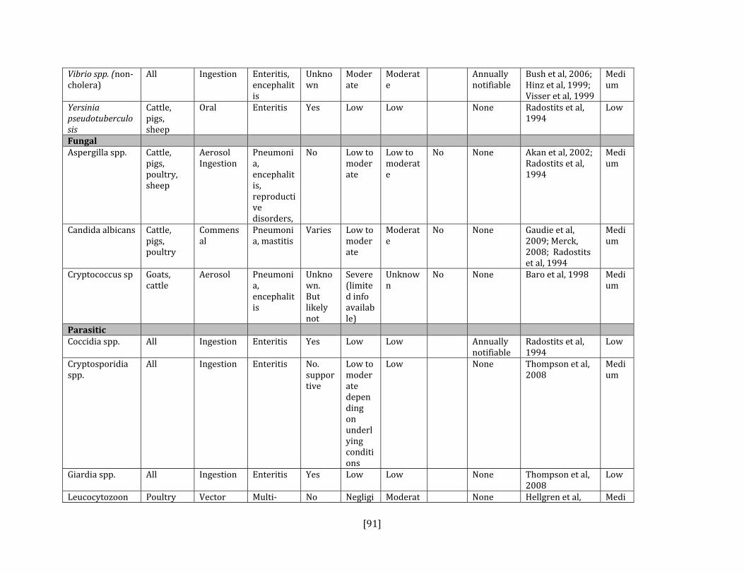
[91]
Vibrio spp. (non‐cholera)
All Ingestion Enteritis, encephalitis
Unknown
Moderate
Moderate
Annually notifiable
Bush et al, 2006; Hinz et al, 1999; Visser et al, 1999
Medium
Yersinia pseudotuberculosis
Cattle, pigs, sheep
Oral Enteritis Yes Low Low None Radostits et al, 1994
Low
Fungal Aspergilla spp. Cattle,
pigs, poultry, sheep
Aerosol Ingestion
Pneumonia, encephalitis, reproductive disorders,
No Low to moderate
Low to moderate
No None Akan et al, 2002; Radostits et al, 1994
Medium
Candida albicans Cattle, pigs, poultry
Commensal
Pneumonia, mastitis
Varies Low to moderate
Moderate
No None Gaudie et al, 2009; Merck, 2008; Radostits et al, 1994
Medium
Cryptococcus sp Goats, cattle
Aerosol Pneumonia, encephalitis
Unknown. But likely not
Severe (limited info available)
Unknown
No None Baro et al, 1998 Medium
Parasitic Coccidia spp. All Ingestion Enteritis Yes Low Low Annually
notifiable Radostits et al, 1994
Low
Cryptosporidia spp.
All Ingestion Enteritis No. supportive
Low to moderate depending on underlying conditions
Low None Thompson et al, 2008
Medium
Giardia spp. All Ingestion Enteritis Yes Low Low None Thompson et al, 2008
Low
Leucocytozoon Poultry Vector Multi‐ No Negligi Moderat None Hellgren et al, Medi

[92]
spp.
systemic ble to moderate
e 2008; Merck, 2008; Bennett et al, 1982
um
Nematodes: Amidostomum ssp. , Epomidiostomum spp., Trichostrongylus spp.
Domestic waterfowl
Ingestion Lethargy Yes Low Low None Nowicki et al, 1995
Low
Sarcocystis spp. Cattle, goats, sheep, poultry
Ingestion Weight loss and unthriftiness, abortions, encephalitis
No Low to moderate
Low None Dubey et al, 2006; Mutalib et al, 1995
Medium
Toxoplasma gondii
Pigs, goats, sheep, poultry (rare)
Ingestion Abortions, pneumonia, encephalitis, endocarditis
Only supportive therapy
Moderate to high
Moderate
Annually notifiable
Dubey et al, 2006 and 2007
Medium
Viral Arboviruses (Eastern and Western Equine Encephalitis, St. Louis Encephalitis, West Nile Virus)
Poultry, domestic waterfowl
Vector Encephalitis
No Negligible to high
Negligible to high
Immediately notifiable
Austin et al, 2004; Senne et al, 2000
High
Avian adenovirus
Poultry, domestic waterfowl
Ingestion Decreased egg production, poor eggshell quality, enteritis,
No Moderate
Low Immediately notifiable
Merck, 2008; Ivanics et al, 2001; Raj et al, 2001; Hess, 2000
Medium

[93]
pneumonia
Avian herpesvirus (duck viral enteritis)
Domestic waterfowl
Direct, Ingestion (water)
Multi‐systemic
No Moderate
Low to high
Annually notifiable
Merck, 2008; Campagnolo et al, 2001; Kaleta et al, 1990
Medium
Avian influenza Pigs, poultry
Ingestion, Aerosol
Pneumonia, decreased egg production, multisystemic disease
No Variable depending on strain
Variable depending on strain
Reportable (Highly pathogenic)
Lager et al, 2009; Pannwitz et al, 2009; Olsen et al, 2006
High
Avian pneumovirus
Poultry, turkeys in particular
Direct Respiratory disease
No Moderate to high
Low to moderate
Yes Immediately notifiable
Cook, 2000; Shin et al, 2000; Merck, 2008
Medium
Avian pox virus Poultry Direct, inhalation, mechanical
Cutaneous lesions, systemic disease
No Moderate
Low (cutaneous form) High for (diptheric/systemic form)
None Merck, 2008; Buller et al, 1991
Low
Coronavirus Group I and II: pigs and cattle Group III: poultry
Aerosol, Ingestion
Respirator disease, enteritis, systemic
No Moderate to high
Moderate to high
None Merck, 2008; Jonassen et al, 2005
Medium
Duck hepatitis virus
Domestic waterfowl
Oral Hepatitis, Sudden death
No High in young
Low in adults, high in young
Yes Immediately notifiable
Merck, 2008: Kim et al, 2007
Medium
Goose parvovirus (Derzsy’s
Domestic geese
Ingestion Multisystemic
No High High Yes Immediately notifiable
Yang et al, 2009; Jansson et al, 2007
Medium

[94]
disease) Newcastle disease virus
Poultry Aerosol, Ingestion
Respiratory disease, neurologic signs, enteritis
No variable depending on strain
variable depending on strain
Reportable
OIE Technical Disease Card
High
New‐type gosling viral enteritis
Domestic geese
Ingestion Enteritis No High High None Chen et al, 2009 Medium
Goose Hemorrhagic Polyomavirus
Domestic geese
Ingestion Nephritis No High High Yes None Lacroux et al, 2004; Guerrin et al, 2000
Medium
Reticuloendotheliosis virus
Poultry Direct Neoplasia No High in young
High in young
Yes None Lin et al, 2009; Cheng et al, 2006
Medium