c:windowstemp otes2cbb501999 manual · conference of operative dentistry educators (code) regional...
TRANSCRIPT
Conference of Operative Dentistry Educators
(CODE)
REGIONAL REPORTS
FALL 1999
http://www.uchsc.edu/sd/rstdenti/OpDSect/code.html
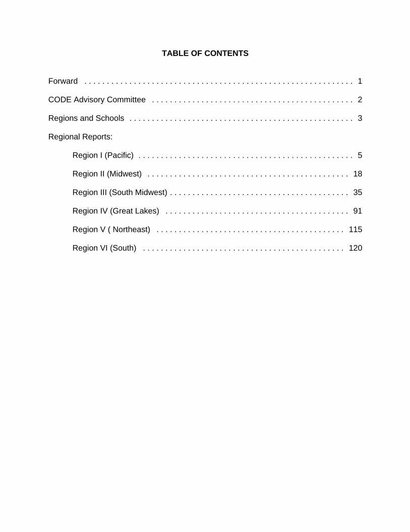
TABLE OF CONTENTS
Forward . . . . . . . . . . . . . . . . . . . . . . . . . . . . . . . . . . . . . . . . . . . . . . . . . . . . . . . . . . . . 1
CODE Advisory Committee . . . . . . . . . . . . . . . . . . . . . . . . . . . . . . . . . . . . . . . . . . . . . 2
Regions and Schools . . . . . . . . . . . . . . . . . . . . . . . . . . . . . . . . . . . . . . . . . . . . . . . . . . 3
Regional Reports:
Region I (Pacific) . . . . . . . . . . . . . . . . . . . . . . . . . . . . . . . . . . . . . . . . . . . . . . . . 5
Region II (Midwest) . . . . . . . . . . . . . . . . . . . . . . . . . . . . . . . . . . . . . . . . . . . . . 18
Region III (South Midwest) . . . . . . . . . . . . . . . . . . . . . . . . . . . . . . . . . . . . . . . . 35
Region IV (Great Lakes) . . . . . . . . . . . . . . . . . . . . . . . . . . . . . . . . . . . . . . . . . 91
Region V ( Northeast) . . . . . . . . . . . . . . . . . . . . . . . . . . . . . . . . . . . . . . . . . . 115
Region VI (South) . . . . . . . . . . . . . . . . . . . . . . . . . . . . . . . . . . . . . . . . . . . . . 120

1 1999 manual
Conference of Operative Dentistry Educators (CODE)Forward - Larry D. Haisch, D.D.S.
CODE continues to be an invaluable resource for the sharing of information discussing concerns and solutionsby Operative Dentistry educators at the Regional meetings. The collegial interaction and networking whichtakes place can only continue to improve the teaching of Operative Dentistry in North America.
As charged by the Executive Council of the Operative Section of American Association of Dental Schools, theNational Director attended the Fall meetings of Regions II, III, and IV. The other regional meetings will beattended in the next two years. The experiences and observation of those meetings reconfirmed the commentsmade in the previous paragraph.
Utilizing electronic media for communication (e-mail) and information (Web site), has been accomplished butat a basic level. The Fall 1999 CODE regional reports are to be placed on the Web Site for the first time. Inthis manner more of our colleagues should be able to access the information. Each person needs to advisetheir administration of CODE and what it accomplishes - direct them to the information. Remember we are allpart of the profession of dentistry. Communication is necessary not only within our organization, but with otherdental organizations. The Academy of Operative Dentistry, the dental licensure boards, the Academy ofGeneral Dentistry, the American Dental Association, to name a few.
CODE is to be or will be based on the desires of the membership. The CODE Advisory Committee needs tohear from you as to future direction.
Thank you to the 1999 CODE Fall Regional Meeting host/coordinators for all their efforts on behalf of CODE.Drs. James Simon (Region I), Pat Kelsey (Region II), William Tate (Region III), Robert Rashid (Region IV),Richard Lichtenthal (Region V), and Jim Knight (Region VI).
Thank you to the Regional Directors for the work accomplished in their respective regions, and theassistance/advise, guidance provided the National Director. Finally, thank you to Dr. Craig Passon for assistingCODE in improved communication via the web site.
http://www.uchsc.edu/sd/rstdenti/OpDSect/code.htmlThe web site will be transferred to the AADS server in the future.
2 1999 manual
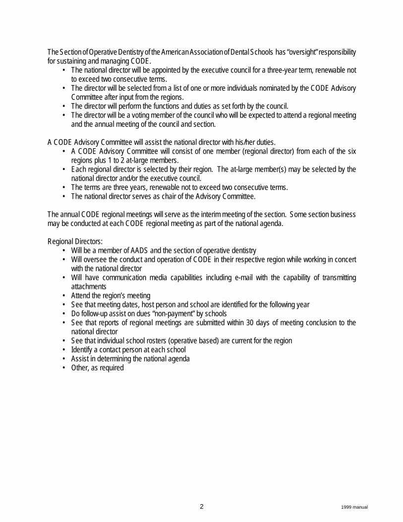
The Section of Operative Dentistry of the American Association of Dental Schools has “oversight” responsibilityfor sustaining and managing CODE.
• The national director will be appointed by the executive council for a three-year term, renewable notto exceed two consecutive terms.
• The director will be selected from a list of one or more individuals nominated by the CODE AdvisoryCommittee after input from the regions.
• The director will perform the functions and duties as set forth by the council.• The director will be a voting member of the council who will be expected to attend a regional meeting
and the annual meeting of the council and section.
A CODE Advisory Committee will assist the national director with his/her duties.• A CODE Advisory Committee will consist of one member (regional director) from each of the six
regions plus 1 to 2 at-large members.• Each regional director is selected by their region. The at-large member(s) may be selected by the
national director and/or the executive council.• The terms are three years, renewable not to exceed two consecutive terms.• The national director serves as chair of the Advisory Committee.
The annual CODE regional meetings will serve as the interim meeting of the section. Some section businessmay be conducted at each CODE regional meeting as part of the national agenda.
Regional Directors:• Will be a member of AADS and the section of operative dentistry• Will oversee the conduct and operation of CODE in their respective region while working in concert
with the national director• Will have communication media capabilities including e-mail with the capability of transmitting
attachments• Attend the region’s meeting• See that meeting dates, host person and school are identified for the following year• Do follow-up assist on dues “non-payment” by schools• See that reports of regional meetings are submitted within 30 days of meeting conclusion to the
national director• See that individual school rosters (operative based) are current for the region• Identify a contact person at each school• Assist in determining the national agenda• Other, as required

3 1999 manual
CODE ADVISORY COMMITTEE
Region Regional DirectorTerm
(subsequent terms - 3 years)
I Pacific Dr. Jim SimonUniversity of the PacificSan Francisco, California
2000-2002
II Midwest Dr. John KillipUniversity of MissouriKansas City, Missouri
2000-2002
III South Midwest Dr. Terry FriutsUniversity of OklahomaOklahoma City, Oklahoma
1998-2000
IV Great Lakes Dr. Bob RashidOhio State UniversityColumbus, Ohio
1998-2000
V Northeast Dr. Warren SchererNew York UniversityNew York, New York
1998-2001
VI South Dr. Kevin FrazierMedical College of GeorgiaAugusta, Georgia
1998-2001
II At-Large Poonam JainSouthern Illinois UniversityAlton, IL
1998-2001
II National Director Dr. Larry D. HaischNational DirectorUniversity of NebraskaLincoln, Nebraska
1998-2001
4 1999 manual
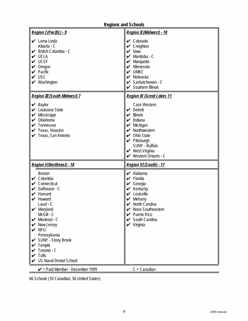
Regions and SchoolsRegion I (Pacific) - 9 Region II (Midwest) - 10
U
UUUUUUU
Loma LindaAlberta - CBritish Columbia - CUCLAUCSFOregonPacificUSCWashington
UUUUUUUUUU
ColoradoCreightonIowaManitoba - CMarquetteMinnesotaUMKCNebraskaSaskatchewan - CSouthern Illinois
Region III (South Midwest) 7 Region IV (Great Lakes 11
UUUUUUU
BaylorLouisiana StateMississippiOklahomaTennesseeTexas, HoustonTexas, San Antonio
UUUUUUU
UU
Case WesternDetroitIllinoisIndianaMichiganNorthwesternOhio StatePittsburghSUNY - BuffaloWest VirginiaWestern Ontario - C
Region V(Northeast) - 18 Region VI (South) - 11
UUUUU
U
UUU
UUUUU
BostonColumbiaConnecticutDalhousie - CHarvardHowardLaval - CMarylandMcGill - CMontreal - CNew JerseyNYUPennsylvaniaSUNY - Stony BrookTempleToronto - CTuftsUS Naval Dental School
UUUUUUUUUUU
AlabamaFloridaGeorgiaKentuckyLouisvilleMeharryNorth CarolinaNova SoutheasternPuerto RicoSouth CarolinaVirginia
U = Paid Member - December 1999 C = Canadian
66 Schools (10 Canadian, 56 United States)
5 1999 manual
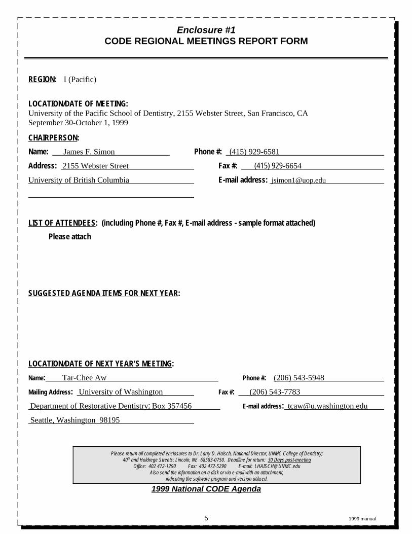
Enclosure #1CODE REGIONAL MEETINGS REPORT FORM
REGION: I (Pacific)
LOCATION/DATE OF MEETING:University of the Pacific School of Dentistry, 2155 Webster Street, San Francisco, CASeptember 30-October 1, 1999
CHAIRPERSON:Name: James F. Simon Phone #: (415) 929-6581
Address: 2155 Webster Street Fax #: (415) 929-6654
University of British Columbia E-mail address: [email protected]
LIST OF ATTENDEES: (including Phone #, Fax #, E-mail address - sample format attached)Please attach
SUGGESTED AGENDA ITEMS FOR NEXT YEAR:
LOCATION/DATE OF NEXT YEAR’S MEETING:Name: Tar-Chee Aw Phone #: (206) 543-5948
Mailing Address: University of Washington Fax #: (206) 543-7783
Department of Restorative Dentistry; Box 357456 E-mail address: [email protected]
Seattle, Washington 98195
Please return all completed enclosures to Dr. Larry D. Haisch, National Director, UNMC College of Dentistry;40th and Holdrege Streets; Lincoln, NE 68583-0750. Deadline for return: 30 Days post-meeting
Office: 402 472-1290 Fax: 402 472-5290 E-mail: [email protected] send the information on a disk or via e-mail with an attachment,
indicating the software program and version utilized.
1999 National CODE Agenda
6 1999 manual
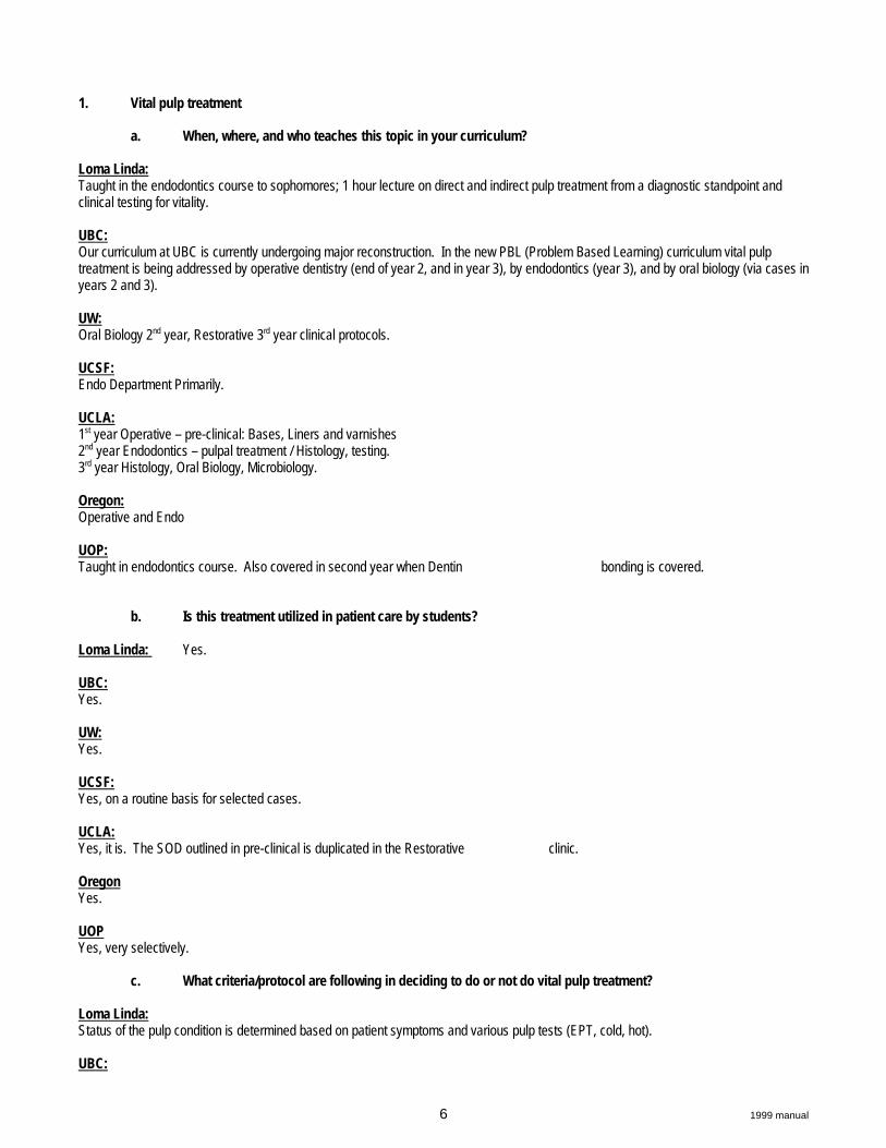
1. Vital pulp treatment
a. When, where, and who teaches this topic in your curriculum?
Loma Linda:Taught in the endodontics course to sophomores; 1 hour lecture on direct and indirect pulp treatment from a diagnostic standpoint andclinical testing for vitality.
UBC:Our curriculum at UBC is currently undergoing major reconstruction. In the new PBL (Problem Based Learning) curriculum vital pulptreatment is being addressed by operative dentistry (end of year 2, and in year 3), by endodontics (year 3), and by oral biology (via cases inyears 2 and 3).
UW:Oral Biology 2nd year, Restorative 3rd year clinical protocols.
UCSF:Endo Department Primarily.
UCLA:1st year Operative – pre-clinical: Bases, Liners and varnishes2nd year Endodontics – pulpal treatment / Histology, testing.3rd year Histology, Oral Biology, Microbiology.
Oregon:Operative and Endo
UOP:Taught in endodontics course. Also covered in second year when Dentin bonding is covered.
b. Is this treatment utilized in patient care by students?
Loma Linda: Yes.
UBC:Yes.
UW:Yes.
UCSF:Yes, on a routine basis for selected cases.
UCLA:Yes, it is. The SOD outlined in pre-clinical is duplicated in the Restorative clinic.
OregonYes.
UOPYes, very selectively.
c. What criteria/protocol are following in deciding to do or not do vital pulp treatment?
Loma Linda:Status of the pulp condition is determined based on patient symptoms and various pulp tests (EPT, cold, hot).
UBC:
7 1999 manual
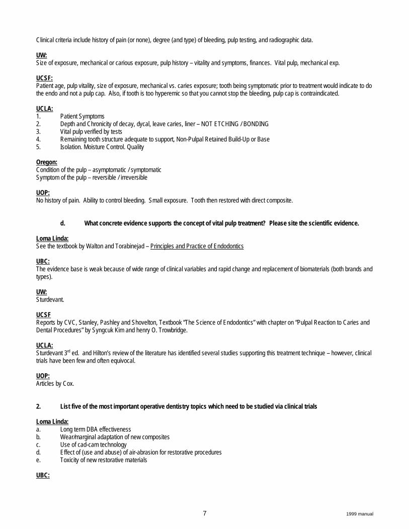
Clinical criteria include history of pain (or none), degree (and type) of bleeding, pulp testing, and radiographic data.
UW:Size of exposure, mechanical or carious exposure, pulp history – vitality and symptoms, finances. Vital pulp, mechanical exp.
UCSF:Patient age, pulp vitality, size of exposure, mechanical vs. caries exposure; tooth being symptomatic prior to treatment would indicate to dothe endo and not a pulp cap. Also, if tooth is too hyperemic so that you cannot stop the bleeding, pulp cap is contraindicated.
UCLA:1. Patient Symptoms2. Depth and Chronicity of decay, dycal, leave caries, liner – NOT ETCHING / BONDING3. Vital pulp verified by tests4. Remaining tooth structure adequate to support, Non-Pulpal Retained Build-Up or Base5. Isolation. Moisture Control. Quality
Oregon:Condition of the pulp – asymptomatic / symptomaticSymptom of the pulp – reversible / irreversible
UOP:No history of pain. Ability to control bleeding. Small exposure. Tooth then restored with direct composite.
d. What concrete evidence supports the concept of vital pulp treatment? Please site the scientific evidence.
Loma Linda:See the textbook by Walton and Torabinejad – Principles and Practice of Endodontics
UBC:The evidence base is weak because of wide range of clinical variables and rapid change and replacement of biomaterials (both brands andtypes).
UW:Sturdevant.
UCSFReports by CVC, Stanley, Pashley and Shovelton, Textbook “The Science of Endodontics” with chapter on “Pulpal Reaction to Caries andDental Procedures” by Syngcuk Kim and henry O. Trowbridge.
UCLA:Sturdevant 3rd ed. and Hilton’s review of the literature has identified several studies supporting this treatment technique – however, clinicaltrials have been few and often equivocal.
UOP:Articles by Cox.
2. List five of the most important operative dentistry topics which need to be studied via clinical trials
Loma Linda:a. Long term DBA effectivenessb. Wear/marginal adaptation of new compositesc. Use of cad-cam technologyd. Effect of (use and abuse) of air-abrasion for restorative procedurese. Toxicity of new restorative materials
UBC:

8 1999 manual
Vital pulp treatment, long-term and dentin and enamel bonding/sealing, resin modified glass ionomers, interfaced vs. non-interfacedbonded amalgams, electronic caries detection, assessment of amalgam and opposite restorations for replacement vs. repair, andevaluation of appropriateness of nonintervention. (We count different in Canada)
UW:a. Durability of posterior composites (direct & indirect)b. Durability of all-ceramic restorationsc. Significance of fluoride-releasing materialsd. Significance of resin/resin-ionomer cementse. Does usage of new tech – air abrasion, lasers, plasma-arch lights, result in better outcomesf. Restoration of vital pulp exposures in all agesg. Strengthening of teeth by bonded compositesh. Posts, cores – efficacy of various systems
UCSF:a. Dentin Bonding and longterm bondstrengthb. Wear Resistance of Posterior Compositec. Repairability of composite vs. complete replacementd. Future need or uses for amalgam (Build-ups only?, restorations?, etc.)e. Future of Lasers in tooth preparation for restoration
UCLA:a. Liners and varnishes vs. DBA’s with amalgamsb. Etiology and treatment of cervical lesions – abfractions, etc…c. Protocols for minimally invasive operative = NO TX vs. SEAL vs. FLOWABLE COMPOSITEd. Longevity of ceramic inlays, posterior compositese. RESIN MODIFIED CEMENTS: Longevity / retention / micro-leakage. v.s. “traditional” cements.
Oregon:Composite / bonding / sealant. Do away with amalgam?Air abrasion Painless dentistryTreatment planning
UOP:a. Vital Pulp Treatmentb. Minimal Caries Treatment – electronic caries Detectionc. Air abrasiond. Durability of posterior esthetic materialse. Esthetic Post and cores
3. Calibration of faculty
a. What is the protocol for calibrating and standardizing your operative faculty? Address current faculty and newfaculty. Also for pre-clinical and clinical.
Loma Linda:Loma Linda uses a grading from that is criteria based. A separate sheet with specific criteria is provided and discussed with current andnew faculty. Students are also provided with the same criteria sheet.
This same form is used from the pre-clinical courses on through to the clinical competencies so that the student is familiar with the gradingcriteria. With this form, the instructor is only concerned with identifying the criteria-based errors and is not burdened with a gradedetermination. The grade is figured separately based on the achievement of the criteria.
UBC:A, B. Meetings prior to each session to calibrate; dedicated “supervising” faculty to assist and oversee calibration; post-session evaluations;course post-mortems; quarterly gatherings of faculty (part-time and full-time together) with special topic (expect) presentations. Ourstandardization and interrater calibration has been rendered all the more challenging with the advent of PBL and ICC (Integrated Clinical
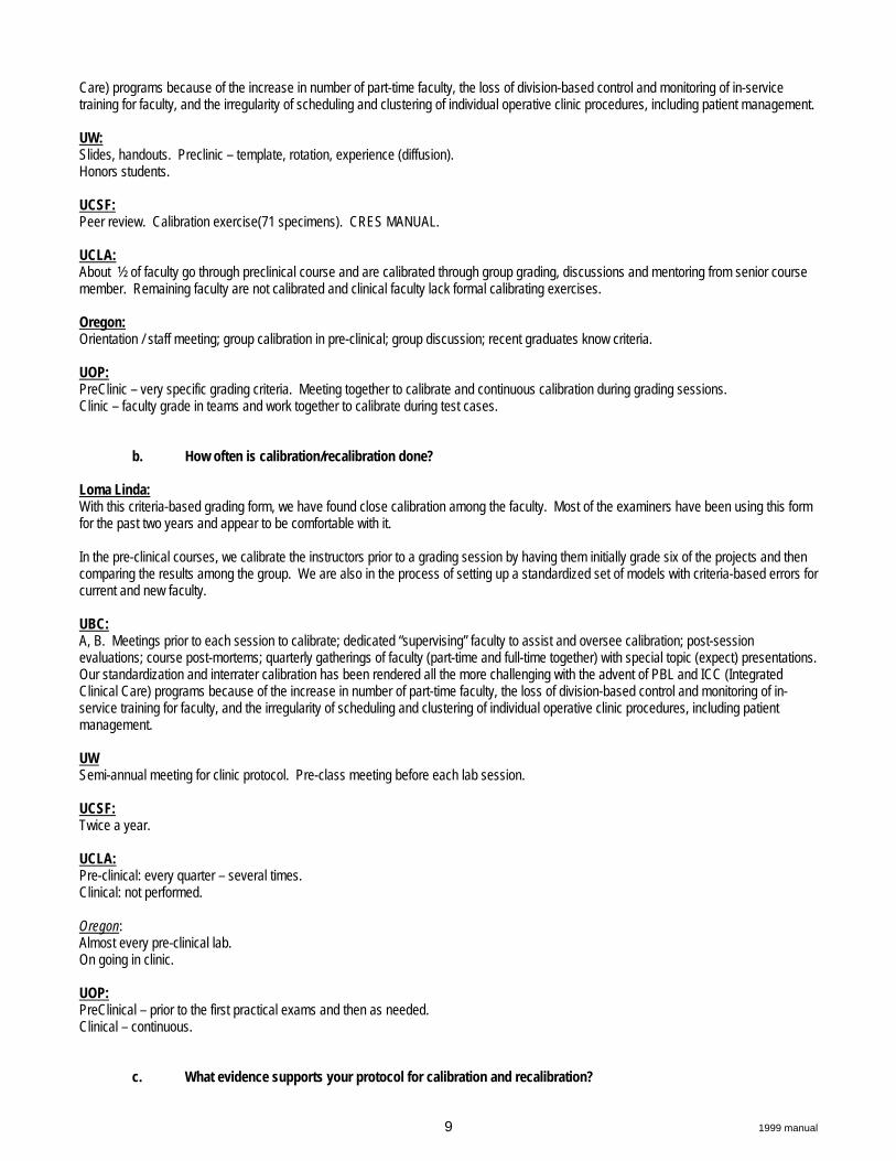
9 1999 manual
Care) programs because of the increase in number of part-time faculty, the loss of division-based control and monitoring of in-servicetraining for faculty, and the irregularity of scheduling and clustering of individual operative clinic procedures, including patient management.
UW:Slides, handouts. Preclinic – template, rotation, experience (diffusion). Honors students.
UCSF:Peer review. Calibration exercise(71 specimens). CRES MANUAL.
UCLA:About ½ of faculty go through preclinical course and are calibrated through group grading, discussions and mentoring from senior coursemember. Remaining faculty are not calibrated and clinical faculty lack formal calibrating exercises.
Oregon:Orientation / staff meeting; group calibration in pre-clinical; group discussion; recent graduates know criteria.
UOP:PreClinic – very specific grading criteria. Meeting together to calibrate and continuous calibration during grading sessions.Clinic – faculty grade in teams and work together to calibrate during test cases.
b. How often is calibration/recalibration done?
Loma Linda:With this criteria-based grading form, we have found close calibration among the faculty. Most of the examiners have been using this formfor the past two years and appear to be comfortable with it.
In the pre-clinical courses, we calibrate the instructors prior to a grading session by having them initially grade six of the projects and thencomparing the results among the group. We are also in the process of setting up a standardized set of models with criteria-based errors forcurrent and new faculty.
UBC:A, B. Meetings prior to each session to calibrate; dedicated “supervising” faculty to assist and oversee calibration; post-sessionevaluations; course post-mortems; quarterly gatherings of faculty (part-time and full-time together) with special topic (expect) presentations. Our standardization and interrater calibration has been rendered all the more challenging with the advent of PBL and ICC (IntegratedClinical Care) programs because of the increase in number of part-time faculty, the loss of division-based control and monitoring of in-service training for faculty, and the irregularity of scheduling and clustering of individual operative clinic procedures, including patientmanagement.
UWSemi-annual meeting for clinic protocol. Pre-class meeting before each lab session.
UCSF:Twice a year.
UCLA:Pre-clinical: every quarter – several times.Clinical: not performed.
Oregon:Almost every pre-clinical lab.On going in clinic.
UOP:PreClinical – prior to the first practical exams and then as needed.Clinical – continuous.
c. What evidence supports your protocol for calibration and recalibration?

10 1999 manual
Loma Linda: Individual scores given by each instructor are recorded and then compared with the group. Each instructor then receives the overallcomposite scores and is able to self-evaluate where he/she compares within the group.
UBC:Regular retrieval of student feedback about faculty members and course offerings (both in simulation and clinically), and comparison ofevaluations (both qualitatively and quantitatively) of students.
UW:Historical/empirical/anecdotal.
UCSF:Competency exam graders need to be calibrated so as to have consistency in grading the students preparations and restorations. The CEis a requirement for graduation (Perio, Op. Dent, C & B, Endo, Behavioral Sciences) and standardization is essential to fairness. Newfaculty have to be instructed on the competency criteria so as not to interject personal biases.
UCLA:None. KNIGHT’s work out of Detroit Mercy is quite good and may be implemented at UCLA (some day).
Oregon:Consistency of what we do in clinic.
UOP:Individual instructor scores are recorded and compared to the group. Students are asked to evaluate clinical and preclinical faculty at theend of each course.
4. Competency evaluation
a. How is competency based operative evaluation determined?
Loma Linda:Students are required to do the following competencies:Junior year: Class II Ag; Cl III, IV, or V composite; cusp replacement Ag; and full gold crownSenior year: Bridge, ceramo-metal, partial coverage crown and two mock boards.
UBC:Competency based tests (process and product) and daily formative feedback.
UW:Competency exam – weekly criteria-based evaluation. Clinic – daily grade sheets. Written diagnosis and treatment planning test at end of2nd year.
UCSF:CRES MANUAL CRITERIAL APPLIED TO COMPETENCY CLINICAL EXAMINATIONS.1. SKILLS ATTAINMENT2. COMPETENCY EXAM
UCLA:Class II Amalgam Exam at evaluation of Pre-clinical and passing grades in all other quarters.Competency exams, 5 amalgam, 2 composite + 1-2 mockboards.
Oregon:Group evaluation
UOP:Third year – 5 Amalgam Test Cases
2 Composite Test Cases

11 1999 manual
Second Year – 2 Amalgam Test Cases1 Composite Test Cases
b. How is the weak student identified?
Loma Linda:Through the grading and evaluating process.
UBC:The weak student is identified through formative feedback and specific instructor identification, and by poorer/failing performance oncompetency tests.
UW:Evaluation procedures – grade sheets, incident reports, tracking of student progress database.
UCSF:Daily notations on progress and performance in the Blue Coaches Group notebooks by daily attending faculty. These are reviewedregularly. Students are interviewed by Group Coach once each quarter formally and as often as needed informally to discuss progress,areas needing improvement, professionalism, etc. Weaker students are discussed at periodic meetings of the Group Coaches and coursedirectors.
UCLA:By bench instructors via daily work/exams and very very rarely by clinical faculty during patient care.
Oregon:Quickly.
UOP:PreClinic – instructor evaluationsClinic – instructor evaluation
c. What measures are taken to upgrade competency of the weak student?
Loma Linda:Remediation and retaking the competency or mock board.
UBC:Personal interview, review, and practice.
UW:Individual remediation. Basic science tutor, dental school tutors, faculty (FT instructor or Clinical affairs). Big brother/big sister matchups. Faculty advisor programs.
UCSF:A more concerted effort is given to bringing the weaker student up to par. The more advanced students in each coaching group areencouraged to help the weaker students as time permits. Weaker students will often have to take the CEs several times to reach a passinggrade reflecting at least minimal competency.
UCLA:Remediation exercises as determined by the course chair and exam make-ups. Monday evening tutorial 2.5 hours with one instructor.
Oregon:Tutoring 1 on 1.
UOP:PreClinic – Saturday morning sessions with one instructor and two Second Year students.
12 1999 manual
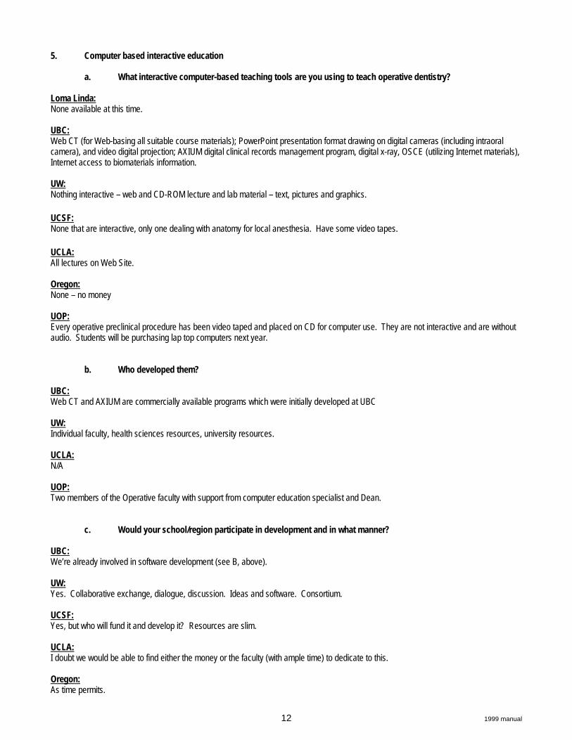
5. Computer based interactive education
a. What interactive computer-based teaching tools are you using to teach operative dentistry?
Loma Linda:None available at this time.
UBC:Web CT (for Web-basing all suitable course materials); PowerPoint presentation format drawing on digital cameras (including intraoralcamera), and video digital projection; AXIUM digital clinical records management program, digital x-ray, OSCE (utilizing Internet materials),Internet access to biomaterials information.
UW:Nothing interactive – web and CD-ROM lecture and lab material – text, pictures and graphics.
UCSF:None that are interactive, only one dealing with anatomy for local anesthesia. Have some video tapes.
UCLA:All lectures on Web Site.
Oregon:None – no money
UOP:Every operative preclinical procedure has been video taped and placed on CD for computer use. They are not interactive and are withoutaudio. Students will be purchasing lap top computers next year.
b. Who developed them?
UBC:Web CT and AXIUM are commercially available programs which were initially developed at UBC
UW:Individual faculty, health sciences resources, university resources.
UCLA:N/A
UOP:Two members of the Operative faculty with support from computer education specialist and Dean.
c. Would your school/region participate in development and in what manner?
UBC:We’re already involved in software development (see B, above).
UW:Yes. Collaborative exchange, dialogue, discussion. Ideas and software. Consortium.
UCSF:Yes, but who will fund it and develop it? Resources are slim.
UCLA:I doubt we would be able to find either the money or the faculty (with ample time) to dedicate to this.
Oregon:As time permits.

13 1999 manual
UOP:We have already given copies to many of the West Coast Schools on various preparations.
6. Posterior esthetic restorations - report on direct and indirect
a. Are they included in the curriculum? What discipline?
Loma Linda:Direct restorations: Part of Operative I (Freshman) and Operative II (Junior) coursesIndirect restorations: Part of Operative II (Junior) course
UBC:Yes.
UW:Yes. Operative and Fixed.
UCSF:Both direct and indirect composite restorations are included in the Operative Dentistry curriculum as well as ceramic inlays and onlays. Indirect composite inlays are sent to a commercial lab for fabrication as well as porcelain restorations. Taught in Preclinical course. Laceycovers indirect in lecture. No lab in clinic.
UCLA:Yes in operative for inlays, fixed for onlays, operative for composites / direct procedures.
Oregon:Yes.
UOP:Yes. Operative.
b. Where (pre-clinic and clinic)
Loma Linda:Pre-clinical: Freshman and Junior yearsClinical: Senior study club and with certain covering instructors. Not widely available as a general clinical procedure at this time.
UBC:Both preclinical (simulation) and clinical.
UW:2nd year operative, fixed 2nd year ceramic (didactic). 4th year clinical class – for indirect all-ceramic restorations. Instructor discretion forclinical post comp.
UCSF:Both.
UCLABoth.
UOPTaught in preclinical and are allowed to place in Senior Esthetic clinic with close supervision.
c. Required experiences?
Loma LindaNone at this time as part of graduation requirements
14 1999 manual
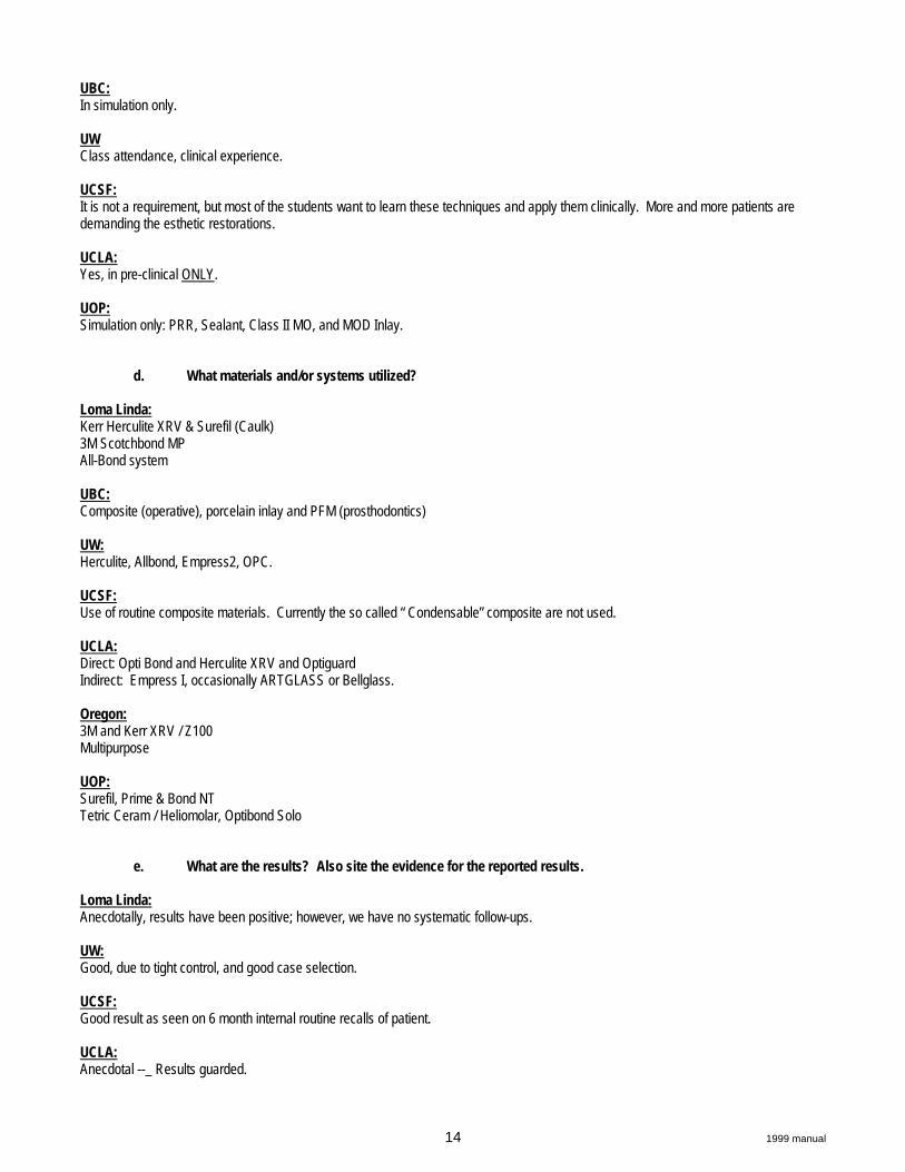
UBC:In simulation only.
UWClass attendance, clinical experience.
UCSF:It is not a requirement, but most of the students want to learn these techniques and apply them clinically. More and more patients aredemanding the esthetic restorations.
UCLA:Yes, in pre-clinical ONLY.
UOP:Simulation only: PRR, Sealant, Class II MO, and MOD Inlay.
d. What materials and/or systems utilized?
Loma Linda:Kerr Herculite XRV & Surefil (Caulk)3M Scotchbond MPAll-Bond system
UBC:Composite (operative), porcelain inlay and PFM (prosthodontics)
UW:Herculite, Allbond, Empress2, OPC.
UCSF:Use of routine composite materials. Currently the so called “ Condensable” composite are not used.
UCLA:Direct: Opti Bond and Herculite XRV and OptiguardIndirect: Empress I, occasionally ARTGLASS or Bellglass.
Oregon:3M and Kerr XRV / Z100Multipurpose
UOP:Surefil, Prime & Bond NTTetric Ceram / Heliomolar, Optibond Solo
e. What are the results? Also site the evidence for the reported results.
Loma Linda:Anecdotally, results have been positive; however, we have no systematic follow-ups.
UW:Good, due to tight control, and good case selection.
UCSF:Good result as seen on 6 month internal routine recalls of patient.
UCLA:Anecdotal --_ Results guarded.

15 1999 manual
UOP:Presently are at one year recall on 30 Surefil Molar restoration as part of research project sponsored by Caulk/Dentsply.

16 1999 manual
CODE Questions
1. What is CODE doing well?
UW:Participation, discussion, exchange of ideas.
UCSF:A good means for faculty of different schools to get together and compare notes. The yearly report from all the regional is way information.
UCLABringing minds and ideas together – very well done!
UOPInviting State Board members to attend.
2. Where do you desire improvement?
UW:Standardizing teaching, closer association with licensing boards, ADA, NIDCR, IADR, AADS etc., setting standard of care/testing/teaching.
UCSF:To develop an E-mail network to further communication. Semi-annual newsletter (besides the report).
3. In $24 US/$30 Canadian school dues adequate? Comments are expected.
UWSeems minimal – does it cover the activities of CODE?
UOP:Does it cover cost?
4. How can participation by faculty in CODE regional meetings be encouraged/improved?
UW:Credible, worthwhile, accomplishment – connection to Operative Academy activities. Impact on testing standards. Researchcollaboration? Action/follow up/after talking.
UCSF:Funding by the Dental School. Hold in conjunction with a regional Dental Meeting, such as CDA, or at the National AADS Meeting.
UOP:Send reports and announcements to attendees not Department Chairs.

17 1999 manual
Please indicate the office / position with complete mailing address at your school to which regional reports, dues statements,roster requests, etc., are to be mailed. NOTE: Regional Director – this information from each school is to be transmitted to theNational Director.
UCSF W. Stephan Eakle, DDSBox 0758Restorative DentistryUniversity of California707 Parnassus Ave.San Francisco, CA 94143
UCLA Dr. Jay Watson, ChairRestorative DentistryUCLA School of Dentistry10833 LeConte Ave.Los Angeles, CA 90095-1668
UOP James F. Simon, DDSChairperson, Operative DentistryUOP School of Dentistry2155 Webster St.San Francisco, CA 94115
UCLA Dan Tan, DDSDepartment of Restorative DentistrySchool of DentistryLoma Linda UniversityLoma Linda, CA 92350
UCLA Edmond R. Hewlett, DDSAssociate ProfessorDivision of Restorative DentistryUCLA School of DentistryBox 951668Los Angeles, CA 90095-1668
Alberta Rick Easton 4032 BDentistry-Pharmacy CentreUniversity of AlbertaEdmonton AlbertaCanada T6G 2N8
UW Tar-Chee Aw, DDS, MSAssistant ProfessorUniversity of WashingtonDepartment of Restorative DentistryBox 357456Seattle, WA 98195
BCU Lance M. Rucker, DDSAssociate Professor and ChairmanDivision of Operative DentistryThe University of British Columbia2199 Wesbrook MallVancouver, B.C.Canada V6T 1Z3
CODE Region I Attendees
NAME UNIVERSITY PHONE # FAX # E-MAIL ADDRESS
Stanley M. Plies UCSF Dental School (415) 476-860 (415) 476-0858
Lance M. Rucker UBC (British Columbia (604) 822-4158 (604) 822-3562 [email protected]
Richard Stevenson UCLA School of Dentistry (310) 794-7314 (310) 206-5539 [email protected]
Michael Hardin OHSU School of Dentistry (503) 494-8993 [email protected]
Daniel Tan Loma Linda (909) 558-4640 [email protected]
Daryl W. Miller WREB (509) 663-0541 (509) 663-1875
Tar C. Aw U. of Washington (206) 543-5948 (206) 543-7783 [email protected]
James Simon UOP (415) 929-6537 [email protected]

18 1999 manual
Enclosure #1CODE REGIONAL MEETINGS REPORT FORM
REGION: II (Midwest)
LOCATION/DATE OF MEETING:Creighton University School of Dentistry, September 20, 21, 1999
CHAIRPERSON:
Name: Pat Kelsay Phone #: (402) 280-5093
Address: 2500 California Plaza Fax #: (402) 280-5094
Omaha, NE 68178-0240 E-mail address: [email protected]
LIST OF ATTENDEES: (including Phone #, Fax #, E-mail address - sample format attached)
Please attach
SUGGESTED AGENDA ITEMS FOR NEXT YEAR:
LOCATION/DATE OF NEXT YEAR’S MEETING:
Name: John Killip Phone #: (816) 235-2100
Mailing Address: UMKC School of Dentistry Fax #: (816) 235-2157
650 E. 25th Street E-mail address: [email protected]
Kansas City, MO 64108-2748 Date: September 18, 19, 2000
Please return all completed enclosures to Dr. Larry D. Haisch, National Director, UNMC College of Dentistry;40th and Holdrege Streets; Lincoln, NE 68583-0750. Deadline for return: 30 Days post-meeting
Office: 402 472-1290 Fax: 402 472-5290 E-mail: [email protected] send the information on a disk or via e-mail with an attachment,
indicating the software program and version utilized.
19 1999 manual
1999 National CODE Agenda
Based on review of the 1998 Fall Regional CODE reports by the CODE Advisory Committee. Refer to Conference of Operative DentistryEducators Regional Reports, Fall 1998 – previously sent to each CODE dues paying member school, the CODE Advisory Committee andthe Executive Council of the Operative Section, American Association of Dental Schools.
1. Vital pulp treatment
a.a. When, where, and who teaches this topic in your curriculum?
Creighton University: Sophomore year preclinical curriculum taught in the Operative and Endodontic laboratories by faculty.
University of Missouri/KC:Operative section: Lecture presentation in the second semester of this first year.Lecture presentations second year and third year.We have recently changed Endo faculty and I am not familiar with their materials.
University of Iowa: Freshman Operative LectureSophomore Operative LectureSophomore Pediatric LectureJunior Pediatric Lecture
University of Colorado:This topic is taught through out the curriculum in the basic and clinical sciences. Several divisions talk about vital pulp treatment; all with adifferent focus or purpose however. The clinical curriculum is where most of the learning occurs through applied practice. No single entitywithin this School is responsible for the sole teaching of this subject. However, it is generally assumed that Operative Dentistry is ultimatelyresponsible.
Southern Illinois Univ.:Sophomore Operative – lectures and pre-clinic (extracted teeth)Junior and senior student presentationsEndodontics, pedo
University of Minnesota:3rd yearOperative DentistryDr. Zidan
UNMC College of Dentistry:Division of Operative Dentistry, 2nd year preclinical course (didactic); 3rd and 4th year operative dentistry clinics (practical).
b. Is this treatment utilized in patient care by students?
Creighton University:Yes
University of Missouri/KC:Yes occasionally in the clinic along with CaOH, in two of our teams the faculty are using bond material under amalgam restorations while inthe other two teams we use the CaOH and Copalite.
University of Iowa:Yes
University of Colorado:Yes, students utilized the treatment taught for vital pulp treatment in the clinics.
Southern Illinois University:Yes
20 1999 manual
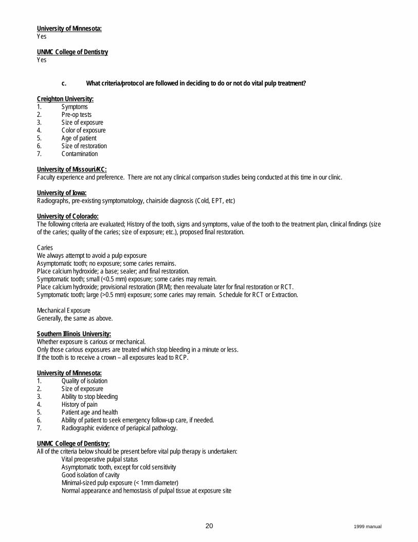
University of Minnesota:Yes
UNMC College of DentistryYes
c. What criteria/protocol are followed in deciding to do or not do vital pulp treatment?
Creighton University:1. Symptoms2. Pre-op tests3. Size of exposure4. Color of exposure5. Age of patient6. Size of restoration7. Contamination
University of Missouri/KC:Faculty experience and preference. There are not any clinical comparison studies being conducted at this time in our clinic.
University of Iowa:Radiographs, pre-existing symptomatology, chairside diagnosis (Cold, EPT, etc)
University of Colorado:The following criteria are evaluated; History of the tooth, signs and symptoms, value of the tooth to the treatment plan, clinical findings (sizeof the caries; quality of the caries; size of exposure; etc.), proposed final restoration.
CariesWe always attempt to avoid a pulp exposureAsymptomatic tooth; no exposure; some caries remains. Place calcium hydroxide; a base; sealer; and final restoration.Symptomatic tooth; small (<0.5 mm) exposure; some caries may remain. Place calcium hydroxide; provisional restoration (IRM); then reevaluate later for final restoration or RCT.Symptomatic tooth; large (>0.5 mm) exposure; some caries may remain. Schedule for RCT or Extraction.
Mechanical ExposureGenerally, the same as above.
Southern Illinois University:Whether exposure is carious or mechanical.Only those carious exposures are treated which stop bleeding in a minute or less.If the tooth is to receive a crown – all exposures lead to RCP.
University of Minnesota:1. Quality of isolation2. Size of exposure3. Ability to stop bleeding4. History of pain5. Patient age and health6. Ability of patient to seek emergency follow-up care, if needed.7. Radiographic evidence of periapical pathology.
UNMC College of Dentistry:All of the criteria below should be present before vital pulp therapy is undertaken:
Vital preoperative pulpal statusAsymptomatic tooth, except for cold sensitivityGood isolation of cavityMinimal-sized pulp exposure (< 1mm diameter)Normal appearance and hemostasis of pulpal tissue at exposure site
21 1999 manual
d. What concrete evidence supports the concept of vital pulp treatment? Please site the scientific evidence. Additional comments welcome.
Creighton University:Pameiser 6 Scientific presentation – June 1999
University of Missouri/KC:Refer to attached articles
University of Iowa:“Treatment Options for the Vital Exposed Pulp,” Swift, E, et al PPAD, Vol. 11, Aug. 99, pp. 735-739
University of Colorado:We rely on the research of Harold Stanley and Charlie Cox for vital pulp treatment.
Southern Illinois University:Although many studies have been published, they have been done on healthy teeth on primates. Healthy but contaminated pulps are notsimilar to pulps responding to decay.
University of Minnesota:1. Patterson and Watts, Further studies on the exposed germ free dental pulp. Int’l Endo J (1987) 20; 112-121.2. Cox et al., Biocompatibility of Surface-sealed dental materials against exposed pulps. J. Prosthet Dent (1987) 57: 1-8.3. Fitzgerald and Heys, A clinical and histological evaluation of conservative pulpal therapy in human teeth. Oper Dent (1991) 16:
101-112.
UNMC College of Dentistry:Concerning calcium hydroxide vital pulp treatment, decades of use and validation of its present form (suspended in a resin vehicle) byStanley (Stanley, HR. Dycal therapy for pulp exposures, Oral Surg, Oral Med 24:818-827, 1972) and many subsequent investigators. Weacknowledge that the stimulation of reparative dentin is of less importance than previously thought, and recognize the fragility and solubilityof calcium hydroxide products, attempting to overlay them with resin-modified glass ionomer whenever space permits.
Concerning vital pulp treatment by etching exposure sites and surrounding dentin, and application of a hydrophilic resin adhesive, we havenot incorporated this into our curriculum because other investigations have not confirmed the findings of Cox et al (Cox, CF et al. Biocompatibility of surface-sealed dental materials against exposed dental pulps. J Prosthet Dent 57:1-8, 1987), and some sharplycontradict these findings (Pameijer, CH, Stanley, HR. The disastrous effects of the “Total Etch” technique in vital pulp therapy in primates). The available evidence, all primate studies, does not justify discarding an effective technique.
2. List five of the most important operative dentistry topics which need to be studied via clinical trials
Creighton University:a. Condensable compositesb. Pulp capping proceduresc. Flowable compositesd. Management of excessive occlusal weare. Crown margin design – placement
University of Missouri/KC:a. Critical need for cavity design in light of the newly developed materials.b. This one is hard to word. Comparison of long term results of when current materials are used in accordance with the
manufacturer’s protocol.c. Longevity of posterior composite (condensable/flowable).d. Comparison of bonding agents versus Copalite under amalgam restorations.e. Pulp cappingf. Root caries
University of Iowa:a. Restoration longevity/survival rates.b. Caries inhibition by fluoride releasing materials.c. Complex amalgams vs. cast coverage.d. Restoration of endodontically restored teeth.e. Early detection methods of caries.

22 1999 manual
University of Colorado:a. Caries inhibition of fluoride releasing restorative materials.b. Clinical performance of indirect esthetic restorations.c. Clinical performance of amalgam bonding in lieu of other forms of retention.d. Outcomes of minimally invasive operative dentistry versus traditional operative dentistry.e. Clinical evidence to support the use of compomers for restorations.
Southern Illinois University:a. Treatment of stained grooves on occlusal surfaces of posterior teeth.b. Pulp cappingc. Decay removal – carious vs. sclerotic dentin under amalgam restorations.d. Long term clinical trials on bonded amalgams and composite restorations.e. How long does the dentin composite resin bond last in the oral cavity?
University of Minnesota:a. Criteria for selection and treatment of dried pulp exposures.b. Predicting and preventing cusp fractures.c. Accuracy of caries risk assessment and outcomes of caries control measures.d. Etiology and treatment (or no treatment) of abfraction lesions.
UNMC College of Dentistry:a. Performance of ceramic restorations of various materials and preparation design.b. Performance of large posterior resin restorations.c. Performance of various materials in restoration of root caries.d. Performance of various materials in restoration of large abfraction lesions.e. Performance of hybrid ionomer luting agents with ceramic restorations.
3. Calibration of faculty
a. What is the protocol for calibrating and standardizing your operative faculty? Address current faculty and newfaculty. Also for pre-clinical and clinical.
Creighton University:Calibration and standardization is done on a weekly basis in the preclinical laboratories. Grades are given by multiple faculty anddiscussion ensues if variances are observed. The clinical faculty are standardized less frequently. The grades given by each clinicalinstructor are analyzed and reported. Behavior modification is expected.
University of Missouri/KC:Two summers ago we had the opportunity of spending ½ day per week with our operative and generalist faculty. During that time we hadmembers of each department educate and calibrate the faculty members. Since that time the operative calibration has consisted ofsessions with the preclinical faculty prior to each semester and during exam periods in the laboratory. We have not had the opportunity towork with the clinical faculty or generalist faculty. I have not even had ½ a day with each of the new generalist faculty members that havebeen added since 1997. Nor have these generalist faculty members been rotated through the preclinical labs in all of the disciplines.
University of Iowa:ClinicalNothing at present other than written criteria in evaluation and competency forms.Currently working of new model using slides with simulated preparations.PreclinicalSet of models for calibration showing degrees of variation from ideal.
University of Colorado:We have no FORMAL calibration program. The faculty of restorative dentistry meet often to discuss issues of importance to changes inoperative dentistry treatment; we communicate freely with one and other about changes that should be made; most faculty work in the pre-clinical courses and the clinic. Most of the faculty rotated through the pre-clinical courses to learn the new topics being taught.
Southern Illinois University:Independent grading of student preps followed by discussions.Each practical exam – grading session.
University of Minnesota:

23 1999 manual
Clinical faculty: no calibration of current or new faculty. 3 full-time faculty grade competency exams.Preclinical faculty: new faculty attend lectures. Current faculty attend short meeting with course director prior to each lab to review what isexpected of students. Course director reviews criteria prior to each competency exam then does a post-exam review of all scores toidentify misapplication of grading criteria. One examiner grades only one cavity preparation or restoration feature for all students. Student’s grade is a sum of all examiners grades which tends to balance “easy” grades and “hard” grades.
UNMC College of Dentistry:All full-time faculty attend all operative dentistry lectures, and preclinical practical examinations and clinical competency examinations areexclusively graded by the full-time faculty. This collaborative grading and associated discussion, amongst a small group (5) of faculty hasserved as an effective means of calibration, for both new and long-time faculty members.
b. How often is calibration/recalibration done?
Creighton University:Weekly in freshman and sophomore years.Quarterly in the Junior clinic.
University of Missouri/KC:Not often enough.
University of Iowa:Clinical
PreclinicalEach year with new faculty.
University of Colorado:Not done on a regular basis. This is a continuous process with no regular schedule.
Southern Illinois University:At the beginning of the fall semester.At every practical exam.
University of Minnesota:Answered in “a”
UNMC College of Dentistry:Approximately 4 times/semester preclinically, and yearly for clinical competency examinations. Calibration for the latter is simplified by useof pass/non-pass grading.
c. What evidence supports your protocol for calibration and recalibration? (Please site)
Creighton University:Student performance on competency examinations.
University of Missouri/KC:None at the present time.
University of Iowa:
University of Colorado:No evidence to offer at this time.
Southern Illinois University:
University of Minnesota:None
24 1999 manual
UNMC College of Dentistry: None
4. Competency education
a. How is competency based operative evaluation determined?
Creighton University:Competency examinations
University of Missouri/KC:Clinical proficiency exams conducted on typodonts in the fall and winter semester of the senior year. Patient mock boards in the wintersemester of the senior year.
University of Iowa:Freshman – Practical examinationsSophomores – Competency discontinued – only daily feedback and evaluation formsJuniors – Set criteria for given number of competency restorations
University of Colorado:Students work under a comprehensive care clinic format. There are no specific operative dentistry “requirements” (numbers) nor are thereany specific procedures which must be preformed (for example numbers of class II amalgams). The students treat their patients accordingto the patients needs and in a timely fashion. Operative dentistry competency evaluation is determined through several measures. Theseinclude; numbers of operative dentistry related activities the student experiences (there is no set number); kinds of operative dentistryactivities experienced; quality of clinical performance during that treatment session; subjective assessment by the students ComprehensiveCare Group Leader; and performance on operative dentistry clinical competency examinations (there are 9 to be taken at interval over oneand a half years). The sum of these measures indicates a students progress towards competency.
Southern Illinois University:Through clinical competenciesDidactic gradesOverall through rigorous overseeing of grades by SPAC (Student Progress and Awards Committee)
University of Minnesota:Competency is composite, amalgam, cast gold mock boards3 full-time faculty evaluate
UNMC College of Dentistry:Clinical competencies, amalgam and resin restorations, in the 3rd and 4th years.
b. How is the weak student identified?
Creighton University:Pass/Fail of the competency exam
University of Missouri/KC:Good question. Not a good answer. Poor quality care provided to patients. In one of our clinical teams the faculty meet weekly anddiscuss student progress, we have a good handle on who our weak students are. In our other two clinical teams this process is justbeginning to occur.
University of Iowa:Failures on competenciesClinical procedure log for daily performanceE-mail to course director concerning problems with daily performance (documentation)
University of Colorado:They are identified by their Group leader and brought to the attention of the Chairs. We have an intermediate level of evaluation at whichthe students are expected to achieve a certain level of performance. Based on the outcomes of the measures at that time weak studentscan be identified.
25 1999 manual
Southern Illinois University:Didactic gradesFailure to complete all clinical competenciesDaily clinical work
University of Minnesota:Preclinic: bench instructor’s evaluations and practical exams
UNMC College of Dentistry:Poor performance in daily work is confidentially recorded in a faculty notebook in clinic, to hopefully identify weak students before theyidentify themselves through failure of a competency exam
c. What measures are taken to upgrade competency of the weak student?
Creighton University:Remediation and retesting
University of Missouri/KC:Additional typodont work and very close faculty supervision. Both very time consuming processes for the student and especially faculty.
University of Iowa:Remedial workMay revert to dentoform if necessaryTutors (pre-clinical)
University of Colorado:Weak students are identified and then are required to work more closely with their Group leader for a set period of time or until a level ofachievement is reached. Special enhancement sessions can be established in addition to the one-on-one assistance. Very weak studentsare set back and may have to repeat a semester or a year.
Southern Illinois University:Remediation of preclinical courses.Tutors for help in lab.Some students may be sent back to lab if incompetent in clinic.
University of Minnesota:Preclinic: course director meets with student to identify appropriate remediation. Usually a 3rd or 4th year student is assigned to tutor theweak student under the direction of the course director.
UNMC College of Dentistry:Measures range from coaching, to reading assignments, to removal of a student’s clinical privileges until remediation is undertaken on adentoform patient simulator, depending on the severity of the deficiencies.
5. Computer based interactive educationRegion VI (South) is developing two comprehensive restorative treatment planning cases to be used as problem basedlearning exercises. It is hoped these cases can be used to develop authoring software that could be used foreducational purposes. A joint venture with Dental Interactive Simulation Corporation (DISC) warrants consideration.
a. What interactive computer-based teaching tools are you using to teach operative dentistry?
Creighton University:None
University of Missouri/KC:NoneWe have just taken delivery on one Dent Sim unit and have not engaged students in its use.
University of Iowa:Computer based lectures – non-interactive

26 1999 manual
University of Colorado:We have no formal interactive computer-based teaching tools at this time. However, most of the operative dentistry courses will be on theWeb. We are moving in the direction of online courses with minimal “lecturing”. With the entire campus moving to a new site within thenext five to ten years there is a moratorium on extensive construction on the present campus. The new campus will make extensive use ofthis sort of technology.
A simulation lab is planned but will not be build on this campus. The School does own a DenX Dental Simulator. However, it is justbeginning to be used. There has been no formal development on this instrument. We also have the Visible Human Haptic Roboticslaboratory available to us.
Southern Illinois University:NoneElmo in the preclinicAnatomy and dental morphology have some interactive software
University of Minnesota:None
UNMC College of Dentistry:None
b. Who developed them?
Creighton University:
University of Missouri/KC:
University of Iowa:
University of Colorado:Dr. Craig Passon is the person doing most of the development in this area.
Southern Illinois University:
University of Minnesota:
UNMC College of Dentistry:
c. Would your school/region participate in development and in what manner?
Creighton:Yes – Testing
University of Missouri/KC:I would like to work with other schools – I do not know the amount of time or financial support that would be identified for this project.
University of Iowa:Yes
University of Colorado:Perhaps. The tools are what need to be developed; the content should be supplied by each school for their school.
Southern Illinois University:
27 1999 manual
University of Minnesota:Possibly but would prefer collaboration with other schools to develop outcome assessments to support treatment planning decisions.
UNMC College of Dentistry:No, unless evidence becomes available that such tools improve the clinical performance of dental students.
6. Posterior esthetic restorations – report on direct and indirect
a. Are they included in the curriculum? What discipline?
Creighton University:Yes – Operative
University of Missouri/KC:Direct Operative second semester two lectures three lab periods.Direct Crown & Bridge lectures related to build up materials.Indirect????
University of Iowa:Yes, preclinical and clinical.
University of Colorado:Yes, they are included. Operative Dentistry
Southern Illinois University:Yes, Operative only
University of Minnesota:Yes, Operative and Pros (PFM)
UNMC College of Dentistry:Direct posterior resin restorations are part of the operative dentistry curriculum, and used routinely in operative dentistry clinics. Teachingof indirect posterior esthetic restorations is shared between fixed prosthodontics and operative dentistry.
b. Where (pre-clinic and clinic)
Creighton University:Clinic and preclinic
University of Missouri/KC:See above – clinic used for small two surface as well as single surface restorations.
University of Iowa:Both
University of Colorado:Direct are taught in the pre-clinic and clinic curriculumsIndirects are taught in the pre-clinic curriculum only.
Southern Illinois University:Both
University of Minnesota:Pre-clinic and clinic
UNMC College of Dentistry:Both
c. Required experiences?

28 1999 manual
University of Missouri/KC:Preclinical – typodont and natural teeth exercises.Clinical – no specific requirements comprehensive patient care.
University of Iowa:Sophomore – not required, but all do posterior esthetic restorations.Junior – Class I and II requirements
University of Colorado:No, Operative Dentistry has no requirements for any restoration. Students can perform direct esthetic restorations at any time they areappropriate. They do not place indirect esthetic restorations. Students are not penalized for not placing posterior esthetic restorations.
Southern Illinois University:No
University of Minnesota:No
UNMC College of Dentistry:No
d. What materials and / or systems utilized?
Creighton University:TPH 6 Heat / Pressure 6 / Prodigy / Optibond
University of Missouri/KC:Caulk TPH
University of Iowa:Herculite, Prodigy, P-60, Z100, Z250, Heliomolar
University of Colorado:We use Kerr Prodigy with Kerr Optibond Fl or Coltene Synergy Compact with Kerr Optibond. We have no flowable composite. Our sealantis Ultradent UltraSeal XT Plus.No indirect restorations are placed.
Southern Illinois University:ConceptBelleglass HPTargis Vectris
University of Minnesota:Direct composite – 2100 and SBMPIndirect porcelain – Empress (in-house)
UNMC College of Dentistry:Direct - Z100/ScotchBond MultipurposeIndirect - Empress
e. What are the results? Also site the evidence for the reported results.
Creighton University:Indirect results – marginal – time consuming – question the advantage over direct composites.Direct results – betterReplacement frequency and secondary caries
University of Missouri/KC:No in house clinical trials.
29 1999 manual
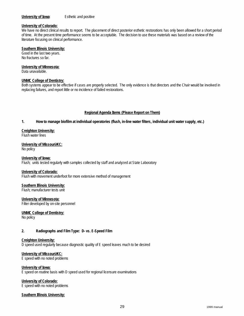
University of Iowa: Esthetic and positive
University of Colorado:We have no direct clinical results to report. The placement of direct posterior esthetic restorations has only been allowed for a short periodof time. At the present time performance seems to be acceptable. The decision to use these materials was based on a review of theliterature focusing on clinical performance.
Southern Illinois University:Good in the last two years.No fractures so far.
University of Minnesota:Data unavailable.
UNMC College of Dentistry:Both systems appear to be effective if cases are properly selected. The only evidence is that directors and the Chair would be involved inreplacing failures, and report little or no incidence of failed restorations.
Regional Agenda Items (Please Report on Them)
1. How to manage biofilm at individual operatories (flush, in-line water filters, individual unit water supply, etc.)
Creighton University:Flush water lines
University of Missouri/KC:No policy
University of Iowa:Flush; units tested regularly with samples collected by staff and analyzed at State Laboratory
University of Colorado:Flush with movement underfoot for more extensive method of management
Southern Illinois University:Flush; manufacturer tests unit
University of Minnesota:Filter developed by on-site personnel
UNMC College of Dentistry:No policy
2. Radiographs and Film Type: D- vs. E-Speed Film
Creighton University:D speed used regularly because diagnostic quality of E speed leaves much to be desired
University of Missouri/KC:E speed with no noted problems
University of Iowa:E speed on routine basis with D speed used for regional licensure examinations
University of Colorado:E speed with no noted problems
Southern Illinois University:

30 1999 manual
E speed routinely but D speed for licensure examinations
University of Minnesota:E speed with frustrations over diagnostic quality
UNMC College of Dentistry:E speed with no noted concerns
CODE Questions
1. What is CODE doing well?
Creighton University:Acting as liaison between Operative faculty and regional examination.
University of Missouri/KC:CODE is alive and very well in our region.
University of Iowa:Inter school communication.Social interaction with school.
University of Colorado:The regional meetings are the most important function of CODE at this time. CODE has made good progress to reinvigorate itself this pastyear and half.
Southern Illinois University:Interaction among faculty, camaraderie.Exchange of ideas, discussion of problems.
University of Minnesota:
UNMC College of Dentistry:
2. Where do you desire improvement?
Creighton University:
University of Missouri/KC:
University of Iowa:Treatment outcomesFaculty calibration instruments
University of Colorado:The CODE regions need to join forces to develop national projects which benefit operative dentistry at all schools.CODE should also be a leader in developing positions about the future of operative dentistry.
Southern Illinois University:Clinical research, multi-center trials
University of Minnesota:
UNMC College of Dentistry:

31 1999 manual
3. Is $25 US/$30 Canadian school dues adequate? Comments are expected.
Creighton University:Dues are minimal.
University of Missouri/KC:I am not familiar with the financial needs of the organization – you tell us if they are meeting your needs.
University of Iowa:Raise if necessary financially.
University of Colorado:This all depends on the mission of CODE. Do we need more money? What do we want to do and what will it take. This sounds like a jobfor the CODE Advisory committee.
Southern Illinois University:Yes, unless regional director feels otherwise.
University of Minnesota:
UNMC College of Dentistry:
4. How can participation by faculty in CODE regional meetings be encouraged/improved?
Creighton University:
University of Missouri/KC:At UMKC we see the value and have many of our generalist faculty members asking to attend the meeting annually. We are bringing onegeneralist faculty member to the meetings each year. CODE is important to UMKC.
University of Iowa:Local agenda must be good.Good social activities.Encourage social and educational interaction.
University of Colorado:Give them something to come for. What benefit is derived from attending their regional meeting that could not have been gained withoutcoming?
Southern Illinois University:Participation is, in general, quite good.
University of Minnesota:
UNMC College of Dentistry:
Suggested National Agenda Items
1. Esthetic Onlay - - Materials and Tooth Preparation

32 1999 manual
2. Postoperative Sensitivity Associated With Class II Direct Composite Resins
3. Technique Sensitivity and Success of Single Liquid Adhesives versus Multiple Component Systems
4. How to Teach a Lesion Specific Preparation in a Preclinical Environment (as opposed to a material specific approach)
5. Status of Operative Dentistry Within Schools: Is It Its Own Entity or Part of a Larger Department?
Suggested Regional Agenda / CODE Items
1. Report data on Class II direct composite failure studies
2. CRDTS and WREB Examination updates

33 1999 manual
MIDWESTERN CODE MEETING ATTENDEES
Name School Phone E-mail Address
Paul Tamisiea Creighton 402 280-5076 [email protected]
Floyd Tanner WREB Representative
John Killip UMKC 816 235-2100 [email protected]
Susan McMillan UMKC 816 235-2100 [email protected]
Brian Williams UMKC
Satish Khera Iowa 319 335-7207 [email protected]
Carol Stanislav Creighton 402 280-5076 [email protected]
Greg Johnson UMKC 816 235-2100
Craig Passon Colorado 303 315-6370 [email protected]
Don Jones Nebraska
Bill Brackett Nebraska
Dave Covey Nebraska
Pat Kelsey Creighton 402 280-5093 [email protected]
Larry Haisch Nebraska 402 472-1290 [email protected]
Gerald Denehy Iowa 319-335-7207 [email protected]
Deb Cobb Iowa 319 335-7207 [email protected]
Poonam Jain Southern Illinois 618 474-7073 [email protected]
Craig Phair Minnesota 612 625-7945 [email protected]
Henry St. Germain Nebraska
Scott Shaddy Creighton 402 280-5076

34 1999 manual
Enclosure #1CODE REGIONAL MEETINGS REPORT FORM
REGION: III
LOCATION/DATE OF MEETING:University of Texas HSC – HoustonDental BranchDepartment of Restorative Dentistry & Biomaterials6516 John Freeman AvenueHouston, Texas 77225-0068November 17-19, 1999
CHAIRPERSON:
Name: William H. Tate Phone #: (713) 500-4264
Address: U of Texa HSC - Houston Fax #: (713) 500-4100
Dental Branch; Dept. of Restorative Dent. & Biomaterials E-mail address: [email protected] John Freeman Ave; Houston, TX 77225-0068
LIST OF ATTENDEES: (including Phone #, Fax #, E-mail address - sample format attached)Please attach
SUGGESTED AGENDA ITEMS FOR NEXT YEAR:• Discuss the perceived value of air abrasion units for cavity preparations.• Discuss limitations of direct posterior resin composite restorations. What are the criteria for treatment planning these restorations in each
institution¹s clinics.• Discuss materials used to finish and polish posterior resin composite restorations. Which are most effective and which are most cost
effective.• Is tooth whitening taught in the operative department. Discuss fees, requirements and tracking of these procedures.• Discuss the possibility of developing a bank of test questions from old board exams related to operative dentistry.• Dentin desensitization, cavity desensitization.• Use of condensable posterior composite.• Amalgam liners, what types?• Treatment of root caries.
LOCATION/DATE OF NEXT YEAR’S MEETING:Name: Terry Fruits Phone #: (405) 271-5735Mailing Address: Dept. of Operative Dentistry Fax #: (405) 271-3006 U. of OK, College of Dent.; P.O. Box 26901 E-mail address: [email protected] Oklahoma City, OK 73190 Date: TBA
Please return all completed enclosures to Dr. Larry D. Haisch, National Director, UNMC College of Dentistry;40th and Holdrege Streets; Lincoln, NE 68583-0750. Deadline for return: 30 Days post-meeting
Office: 402 472-1290 Fax: 402 472-5290 E-mail: [email protected] send the information on a disk or via e-mail with an attachment,
indicating the software program and version utilized.
35 1999 manual
Region III CODE MeetingNovember 17-19, 1999
University of Texas HSC – HoustonDental Branch, Houston, Texas
As usual, the National agenda is a challenge. The agenda is based on the suggestions made by each Region plus the separate responsesfrom the Regional Directors. One question is related to the Special Projects activity conjoint with the Academy of Operative Dentistry andthe Section of Operative Dentistry of AADS. The approach to their mission is “Recommendations for Clinical Practice in OperativeDentistry.”
Each Region is to attempt to respond in an “evidence based” manner vs. a consensus based or anecdotal information. Is the responsebased on research and clinical experience? A suggested example of concern shared by Dorothy McComb at the last meeting of theExecutive Council of the Operative Section ….. Caries – detecting dyes. “Most consensus seems to indicate that these are necessary toremove all decay as they stain bacteria. However, the evidence shows that the dyes stain dentin below a certain level of mineralization,including normal dentin at the amelodentinal junction and circumpulpal dentin. There will be over removal of sound dentin if the dye is usedas a marker of decay.”
Give thought and discussion at the Regional meeting to evidence based responses.
Each Region is encouraged to have a Regional agenda, which is also reported on with the National Agenda. The Regional meeting reportsare to be submitted in publishable form within 30 days of the conclusion of the meeting to the National Director.
1999 National CODE Agenda
Based on review of the 1998 Fall Regional CODE reports by the CODE Advisory Committee. Refer to Conference of Operative DentistryEducators Regional Reports, Fall 1998 – previously sent to each CODE dues paying member school, the CODE Advisory committee andthe Executive Council of the Operative Section, American Association of Dental Schools.
LSU School of Dentistry
36 1999 manual
1999 National CODE Agenda
1. Vital pulp treatmenta. When, where, and who teaches this topic in your curriculum?
i. Operative Dentistry and Biomaterials, Freshman year, Introduction to Operative Dentistry – 1 hour lecture:Sealers, liners and bases, preclinic laboratory project: apply calcium hydroxide liner and RMGI liner/base anddentin bonding agent
ii. Operative Dentistry and Biomaterials, Sophomore year, Dental Materials Science – 1 hour lecture: Sealers,liners, and bases.
iii. Pediatric Dentistry, Sophomore year, 2 hour lecture: Treatment of Traumatic Injuries iv. Endodontics, Sophomore year, no lecture, assigned chapter : Vital pulp therapyv. Operative Dentistry and Biomaterials, Junior year, Advanced Operative Dentistry – 1 hour lecture: Vital pulp
therapyvi. When indicated vital pulp therapy is performed in the second year in Introduction to Clinical Operative
Dentistry, in the third year in Advanced Operative Dentistry, and in the fourth year in General Dentistry.
b. Is this treatment utilized in patient care by students? YES
c. What criteria are followed in deciding to do or not to do vital pulp treatment?i. There is no history of spontaneous pain.ii. Pulpal response to thermal or electrical testing is within normal limits.iii. Pain elicited during pulp testing with hot or cold stimulus does not linger after the tooth returns to mouth
temperature. Not longer than 20-30 seconds.iv. No radiographic evidence of a periradicular lesion.v. The tooth is not strategic for future fixed or removable prosthodontics
d. What concrete evidence supports the concept of vital pulp treatment? Please site scientific evidence.Additional comments welcome.i. Vital pulp therapy effectiveness of treatment.
Sawusch, E.H.: Direct and indirect pulp capping with two new products. J. Amer Dent Assoc 104:459-462,1982. Indirect pulp capping with Dycal treated with Improved Dycal Thirty-six indirect pulp caps- permanentteeth 67 teeth indirect pulp- 100% successHaskell JADA 97:607-12, 1978 Calcium hydroxide sealed with???130 human permanent 87% success. mean 11.7 yrs. (5-22 yrs)Nyborg, H. 1955 Capping of the pulp 296-364 CaOH paste with Ringers 81 teeth permanent teethevaluated Histologically & clinically. 124 teeth clinical evaluation only 62% success at recall > 2 yr. Formost teeth. 86% success rate Law DB and Lewis TM. The effect of calcium hydroxide on deep carious lesions. Oral Surg 14:1130-1137,1961. Ca(OH)2 and sterile water20 permanent teeth 80% success, two years clinical and radiographicevaluation. 36 permanent teeth for 13.4 exp Dycal vs regular Dycal 21/23 successful with exp. Dycal notedassoc. With failed cap and failed permanent restoration. Horsted Endod Dent Traumatol 1:29-34, 1985. Calcium hydroxide ZOE or ZnPO4 cement and restoration. 510 human permanent (210 direct exam) Exposures-70% cavity preparations, 15% carious, 15% unknown82% 5 yr survival rate.No difference between carious and non carious exposures. Matsuo T; Nakanishi T; Shimizu H; Ebisu S. A clinical study of direct pulp capping applied to carious-exposed pulps. J Endod 1996;22(10):551-6 carious-exposed 44 teeth. 81.8% success; degree of bleeding ofpulpal exposure was related to the success rate YEARS F?U???Kashiwada Bull Toko Dent Med Univ 38:45-52. 1991 10% hypochlorite- 4 min. hemostasis and disinfection.Clearfil Photo Bond applied and cured. Teethmate cured Human permanent Exposed teeth restored withporcelain inlay or metal inlay or onlays.60/64(94%) exposures successful after 18 mosHeitmann Quint Int 26:765-770, 1995. CaOH placed, etched CaOH removed primed Heliobond, 20 s VLCX 2 Lined with Tetric then Restored with Tetric. Permanent human molars and premolars100% success, 2-6months recall

LSU School of Dentistry
37 1999 manual
Kotoh J Dent Res 76:160(Abstr. 1192) 1997. 5 liner bond 3 LinerBond II, 3 Superbond C&B, 1 D-Liner andSuper Bond, 2 Ca(OH)2 & Superbond mixed All permanent teeth.100% success, 17 mos recallCiavarelli L. De Fazio P, Scarano A. Piattelli A. Histological analysis of direct capping with enamel dentin system in vivo. J Dent Res 77:(Abs#912), pg 21 capped with Prime & bond 2.1 restored with z-100. Humanpermanent bicuspids. Ext 15, 30, 45, 90, 180, 360 days after capping.Gp 1 earliest showed mild to moderateinflammation absence of inflammation shows reversable effects and there is new bridge formationKatoh Y. Wound healing process of pulp directly capped with adhesive resins. J Dent Res 77:(Abs#913), pg220, 1999. 10% NaOCl for 1-5 min. Capped with caOH restored witg composite resin, Capped with 3 typesof adhesive resin. 21 teeth in 9 patients exposed vital teeth 1.7mm diameter Extracted 37-194 days aftermean 79 days Healing began with disappearance of inflammation, collagen fiber matrix formation,calcification, calcification of the matrix, and dentin bridge formation. Progess began at the perphery &progressed to centerde Blanco LP Treatment of crown fractures with pulp exposure. Oral Surg Oral Med Oral Pathol Oral RadiolEndod 1996;82:564-8.Thirty permanent incisors with vital pulps and crown fractures were treated by a partialpulpotomy amputation of 1 to 2 mm of the exposed pulp, calcium hydroxide powder, & temporary restorationClinical and radiographic assessment of the hard tissue barrier was done after 3 months and again after 1 to8 years. 100% success.Cvek M.A clinical report on partial pulpotomy and capping with calcium hydroxide in permanent incisors withcomplicated crown fracture. J of Endo 4:232-37, 1978. Partial pulpotomy and calcium hydroxide dressing. 60perm incisors.treated due to trauma Mean f/u was 31 mos. 98% success no clinical signs or radiographicsignsFuks AB, Chosack A, Klein H Eidelman E. Partial pulpotomy as a treatment alternative for exposed pulpsin crown-fractured permanent incisors. Endod Dent Traumatol 1987;3:100-2. Partial pulpotomy and calciumhydroxide dressing 63 permanent incisors treated due to trauma 94% success rate clinical and x-rayevaluation
ii. Pulp Capping success rates in primates
Pitt Ford OOO 71:338-42, 1991 Dycal, VLC Dycal, Prisma-Bond immediately or 24 Hrs. Am and ZOE seal64 teeth Pulpal inflammation in 1/30 After two mos. Complete dentin bridging in 28/30 teeth capped withDycal or VLC Dycal. Prisma bond showed complete dentin bridging in 4/2 teeth and pulpal inflammation in2/22.Pameijjer CH Stanley HR. The disastrous effects of the Total Etch technique in vital pulp capping inprimates. Am J Dent 1998;11:S45-S54. Total etch & All-Bond 2 Total etch & ProBond Total Etch andPermagen Total etch & Ultrablend CaOH Total etch and Dycal No etch & Dycal 7/ group% non vital @ 75days 57% 29% 83% 57% 14% 0%Otsuki M, Sonoda H. Kitasako, M. Arakawa. Pulpal response to two adhesive systems using self-etchingprimers. J Dent Res 77:(Abs#1243), pg 261, 1999. Clearfil Liner Bond 2V restored with VLC or autocurecomposite resin. Other group. Treated with Mac-bond II. 90 teeth Animals sacrificed at 3,30 90 days. Nodifference in pulpal response. Only slight pulpal inflammation with the self etching primers in non humanprimate teeth.Dogon IL, Erickson ER, Dobeck JM. Pulp capping with two adhesives with and without Ca(OH)2 in M.Fasicularis. J Dent Res 77:(Abs#797), pg 205, 1999. Compare Single Bond, Scotchbond MP to Dycal.Measured at 3, 6, and 9 mos. 43 of 47 exposures had bridging with Dycal. Only 4/23 with MP and 3 of 23had bridging with Single Bond.Pameijer CH, Stanley HR. Pulp capping with total etch and other experimental methods. J Dent Res77:(Abs#911), pg 219, 1999. Evaluated at days 5, 25, & 70. Exposed pulps applied Consepsis for 60 s. &capped with 2% NaOCL & total etch.105 18-21 teeth/ group 92 % bridge formation attempted 85% formedcomplete. Gp 2 mod Ca PCA 100% bridge formation at 25& 75 days. Gp 3 Mg(OH)2 42% bridge formation. Gp 4 Benzalkonium chloride 10% no attempt at bridge formation. Gp 5 Fuji glass ionomer cement. Had 50%bridging. Conclusion the belief that any material placed on a pulp will allow bridge formation as long as thecavity is disinfected is a fallacy. Kitasako Y; Inokoshi S; Fujitani M; Otsuki M; Tagami J. Short-term reaction of exposed monkey pulpbeneath adhesive resins. Oper Dent 1998(6):308-17. Monkey pulpal responses to four adhesive resin

LSU School of Dentistry
38 1999 manual
systems used as direct pulp cap histopathologically evaluated at 3, 7, 14, 30, and 60 days after operation. No serious inflammatory reaction of the pulp, such as necrosis or abscess formation, was observed.Slightinflammatory cell infiltration was the main inflammatory reaction of the exposed pulp, and the exposed areabecame occluded with dentin bridging as the observation period increased. Healing of exposed dental pulpbeneath adhesive resin capping slightly differed depending on the materials used.
2. List five of the most important Operative Dentistry topics which need to be studied via clinical trials
a. Efficacy of fluoride-releasing materialsb. Marginal integrity of indirect restorations over timec. Finishing of composite resins – surface textured. Systemic effects of resinse. Finishing of porcelain onlays
3. Calibration of faculty
a. What is the protocol for calibrating and standardizing your Operative faculty? Address current faculty andnew faculty. Also for pre-clinical and clinical.
Time is set aside for the department to work on projects similar to what the students are required to complete. The projects are graded by each instructor without knowing whose work is being evaluated. Using a desk top video camerathe project is displayed to allow for a group discussion.
b. How often is calibration/recalibration done?
Once a year. For a clinical evaluation we use two instructors to grade competency exams. Each instructorindependently evaluates the step, then comes to an agreement on the final grade.
c. What evidence supports your protocol for calibration and recalibration? (Please cite)
Don’t have any.
4. Competency education
a. How is competency based Operative education determined?
The school administration is requiring competency-based education in all areas. For operative dentistry this isdetermined by decision of the course directors and department head. The decision for specific restorations is basedpartly on state board examinations and the direction our department believes operative dentistry will proceed into thefuture. This direction is derived from several sources – dentists in the community, literature reflecting trends in dentistryand presentations in the dental community..
b. How is the weak student identified?
Weaker students are identified by their performance qualitatively and quantitatively. Instructors can observe directly thedifficulty a students has both clinically and pre-clinically. Scores from daily work and performance on competencyexams. At certain intervals a required number of restorations are expected to be completed. Inadequacies wouldrequire closer observation of that student.
c. What measures are taken to upgrade competency of the weak student?Poor performance in both clinic and pre-clinical area require remediation. Students will be give extra attention andadditional work to complete before continuing. All competencies must completed to a satisfactory level to finish thecourse.
5. Computer based interactive education

LSU School of Dentistry
39 1999 manual
Region VI (South) is developing two comprehensive restorative treatment planning cases to be used as problem based learningexercises. It is hoped these cases can be used to develop authoring software that could be used for educational purposes. Ajoint venture with Dental Interactive Simulation Corporation (DISC) warrants consideration.
a. What interactive computer-based teaching tools are you using to teach Operative Dentistry?
None. Only additional help is that for some lectures that are presented electronically, a CD-Rom is available in thelibrary to review the presentation.
b. Who developed them?
Within our department. Usually the person who presents the lecture.
c. Would your school/region participate in development and in what manner?
Yes. Because of shortage of manpower we could possibly only beta-test a program.
6. Posterior esthetic restorations – report on direct and indirect
a. Are they included in the curriculum? What discipline?
Yes. Operative Dentistry
b. Where (pre-clinic and clinic)
Both
c. Required experiences?
In the freshman course only direct composite restorations. In our sophomore course the last four weeks are devoted toindirect composite inlay restorations in a preclinical setting. In our third year direct posterior composites are required aswell as successfully completing a Class II resin competency. They are not required to do an indirect resin but cansubstitute a gold casting with a resin inlay. We expect to have porcelain onlays available within the next couple ofyears.
d. What materials and/or systems utilized?
Our students are fabricating their own composite inlays from either Z100 or Herculite XRV. We also have BelleGlassavailable.
e. What are the results? Also cite the evidence for the reported results.
Last year was the first year to offer the sophomores a course in indirect resin inlays. In this preclinical course allrestorations were successfully completed. The students produced their own restorations using natural teeth. Allrestorations were turned in. I sectioned the teeth to measure and compare the margins and film thickness of two dualcured resin cements we were considering to use. All restorations fit were acceptable. This included a system requiringthe adhesive to be light-cured before the restoration was seated. In clinic we have very few problems. I know this isshort term but that’s all we’ve got.
Regional Agenda Items
1. Do you teach and/or are you using air abrasion or laser technology?
We do not teach these techniques. We are using them for some research projects measuring cutting efficacy and output ofmaterial.
2. What is your diagnostic criteria and teaching for fissure caries. What is your criteria for evaluating conservative pit andfissure preparations and restorations?
LSU School of Dentistry
40 1999 manual
Explorer catching and visual inspection of fissure. Absence of space in fissure and not sticking with explorer. Smooth margins ofrestorations.
3. How do you prepare your students for the National Board Examination?
Use of National Board Examination question in our exams. 1993 is the most current exam we have.
CODE Questions
1. What is CODE doing well?
Provides opportunity for peer support group of operative instructors
2. Where do you desire improvement?
3. Is $25 US/$30 Canadian school dues adequate? Comments are expected.
I’m not aware of expenses, so I assume this fee is adequate.
4. How can participation by faculty in CODE regional meetings be encouraged/improved?
Time is our most precious resource. Possibly scheduling meeting on Weekends or times when school is out.
5. Please indicate the office/position with complete mailing address at your school to which regional reports, duesstatements, roster requests, etc., are to be mailed. NOTE: Regional Director – this information from each school is tobe transmitted to the National Director.
Alan H. Ripps, D.M.D.LSU School of Dentistry, Box 137Department of Operative Dentistry & Biomaterials1100 Florida AvenueNew Orleans, Louisiana 70119
University of Oklahoma College of Dentistry
41 1999 manual
1999 National CODE Agenda
1. Vital pulp treatment
a. When, where and who teaches this topic in your curriculum?
Department of Operative Dentistry in the preclinical courses and in clinic.Department of Pedodontic Dentistry in preclinical courses and in clinic.Department of Endodontics in preclinic and in clinic.
b. Is this treatment utilized in patient care by students?
Direct and Indirect pulp caps are performed by students in our Operative clinics.Pulpotomies are performed in out pedodontic clinics.
c. What criteria/protocol are followed in deciding to do or not do vital pulp treatment?
Basic pulp cap criteria are:
i. Area of exposed pulp is very small "pinpoint."ii. Hemorrhage appears normal and is easily controllable.iii. No evidence of periapical lesions (infections) on radiographs.iv. Tooth must be vital (Responsive to pulp tests)v. Have no history of spontaneous pain (Pain that begins without stimuli such as cold, hot, sugar, or biting
pressure)vi. External stimuli (i.e., cold, hot, etc.) should elicit a fairly normal response from the tooth. Any pain from these
stimuli should not remain for an extended period of time following the removal of the stimuli.vii. Tooth will not need to be relied upon in the future as an abutment for a fixed or removable partial denture.viii. Patient is not medically compromised. Also must consider the age of the patient in regard to the ability of the
pulp tissue to survive this insult.
d. What concrete evidence supports the concept of vital pulp treatment? Please site the scientific evidence. Additional comments welcome.
Animal studies show evidence that dycal, resin modified glass ionomer, and adhesive resins can effectively providevital pulp therapy in the form of a direct or indirect pulp cap procedure.
Kitasako Y, Inokoshi S, Tagami J. Effects of direct resin pulp capping techniques on short term response ofmechanically exposed pulps. Journal of Dentistry, 1999; 27:257-63.
Kitasako Y, et al. Short-term reaction of exposed monkey pulp beneath adhesive resins. Operative Dentistry, 1998;23:308-17.
Tarim B, Hafez AA, Cox CF. Pulpal response to a resinSmodified glass-ionomer material on nonexposed and exposedmonkey pulps. Quintessence International, 1998; 29:535-42.
Cox CF, et al. Biocompatibility of primer, adhesive and resin composite systems on non-exposed and exposed pulps ofnon-human primate teeth. Amercan Journal of Dentistry, 1998; 11Spec No:S55-63.
Akimoto N, et al. Biocompatibility of Clearfil Liner Bond 2 and Clearfil AP-X system on nonexposed and exposedprimate teeth. Quintessence International, 1998; 29:177-88.
Cox CF, et al. Biocompatibility of various dental materials: pulp healing with a surface seal. International Journal ofPeriodontics & Restorative Dentistry, 1996; 16:240-51.
Tsuneda Y et al. A histopathological study of direct pulp capping with adhesive resins. Operative Dentistry, 1995;20:223-9.
University of Oklahoma College of Dentistry
42 1999 manual
(Paterson RC. Bacterial contamination and the exposed pulp. Br Dent J, 1976; 140:231-236.)
A fairly good success rate for direct pulp cap procedures was found in the following study on human patients:
MatsuoT, Nakanishi T, Shimizu H, Ebisu S. A clinical study of direct pulp capping applied to carious-exposed pulps.Journal of Endodontics, 1996; 22:551-6.
2. List five of the most important Operative Dentistry topics which need to be studied via clinical trials.
a. Long term effectiveness of posterior resin composite restorations. We need to know the limitations of this material. Arewe allowing dental manufacturers, patients, and the monetary goals of the dental practice dictate which treatments andmaterials we utilize in the treatment of our patients? (For example the extensive use of posterior resin compositerestorations)
b. Either eliminate polymerization shrinkage in resin composite, or definitively establish the best methods for controlling itclinically.
c. What quantity of fluoride needs to be released from a restorative material to be truly clinically effective in preventingrecurrent caries? Are any of today¹s products really meeting that standard for a meaningful duration?
d. Determine the long term effectiveness of the dentinal seal developed with the total etch/dentin bonding agents forbonded amalgam restorations. Will it eventually hydrolyze and leak, if so when?
e. Determine the long-term effectiveness of direct posterior resin composite restorations versus indirect posterior resincomposite restorations.
3. Calibration of faculty
a. What is the protocol for calibrating and standardizing your Operative faculty? Address current faculty andnew faculty. Also for pre-clinical and clinical.
Our grading system for all preparations and insertions are based on a list of objective criteria. There are specific criteriafor all the various types of "ideal" preparations and insertions. These criteria are supplied to the students and alldepartment members so that both are aware of the grading criteria. The students have several preclinical lab projectswhich involve evaluating their own and their classmates¹ preparations and insertions utilizing these criteria. New facultymembers are initially calibrated by allowing them to evaluate several preparations and insertions on typodont teeth thatwere completed by students in past years and have been previously evaluated by the faculty. The new facultymembers can then compare their evaluation to those done previously by more experienced departmental faculty toestablish an initial concept of the department¹s grading system.
During each semester, all members of the department individually evaluate each student¹s practical examinations.After the results of the evaluations are collated, the entire department meets to discuss any grades that fall beyond acertain acceptable level of variation (More than one grade variation is not acceptable). This allows the faculty to discussthe deficiencies observed in the preparation and calibrate how much it should affect the assigned grade.
b. How often is calibration/recalibration done?
About four times per semester. After each practical examination.
c. What evidence supports your protocol for calibration and recalibration?
None to our knowledge, other than the fact that over the past 25 years the results of this technique has yieldedevaluations that are reliable between our faculty members. Both the grades assigned and the reasons for assigning thegrades are very reliable from one instructor to another. No statistical analysis has been performed.
4. Competency education

University of Oklahoma College of Dentistry
43 1999 manual
a. How is competency based Operative education determined?
Preclinical Competency Assessment:
Practical & Didactic Exams S Students are evaluated on performance of preparations and insertions of Class I & IIamalgam restorations and Class III, IV, and V composite restorations. Daily Projects - Students are evaluated on allaspects concerning the fundamentals of operative dentistry during daily projects in the preclinical lab sessions.
Daily Clinical Judgement Evaluations S Students are evaluated during each session of the preclincal laboratory on thefollowing criteria.
•Organization/NeatnessMisplacement or loss of grade formsFailure to secure signatures at necessary check stepsDisorganized work areasIncomplete armamentariaFailure to bring course syllabus to classDull hand instruments
•Knowledge of ProcedureGross violation of procedural steps and principlesShows lack of knowledge concerning daily procedure
•Time ManagementLeaving laboratory early before projects are completedAbsent from laboratory sessionNon-productive use of time
•Professional MannerFailure to secure Permission to ProceedBehavior that reflects a lack of concern for classmatesInability to accept constructive criticismCynical or unprofessional attitudeImproper attireNo lab coat or safety glassesNot wearing gloves or mask
•General ProgressMultiple projects past due dateStruggling with basic concepts or skills
Clinical Competency Assessment:
Clinical Procedure Exams (Practical Exams) - Students are evaluated on performance of preparations and insertions of Class I & IIamalgam restorations and Class III, IV, and V composite restorations.Daily Clinical Procedures - Students are evaluated on all aspects concerning the fundamentals of operative dentistry during dailyprocedures in the clinical sessions.
Daily Clinical Judgement Evaluations S Students are evaluated during each session of the preclincal laboratory on the following criteria:
Professionalism (Appearance and attitude)Patient Management (Empathy, Education)Clinical Judgment (Application of Knowledge)Organization of Work Area (Efficiency, Neatness, & Instruments)Operative SkillTime ManagementRecordsAttendance in Clinic
b. How is the weak student identified?
University of Oklahoma College of Dentistry
44 1999 manual
Quarterly assessments are made of the progress of all students in both the preclinical and clinical courses. Any studentthat is significantly behind in clinical activity or in completion of preclinical projects is contacted and counseled.Students that have been identified as deficient in the quality of their work are also notified and counseled. Someremediation type of work may be necessary is the deficiencies are too significant.
c. What measures are taken to upgrade competency of the weak student?
The student may be assigned a faculty mentor or a tutor. Often the deficient student may be required to complete someform of remediation on any aspect for which they have been identified as deficient. Students in extreme difficulties maybe asked to repeat the year if the deficiencies are not correctable.
5. Computer based interactive education
Region VI (South) is developing two comprehensive restorative treatment planning cases to be used as problem based learningexercises. It is hoped these cases can be used to develop authoring software that could be used for education purposes. A jointventure with Dental Interactive Simulation Corporation (DISC) warrants consideration.
a. What interactive computer-based teaching tools are you using to teach Operative Dentistry?
No interactive computer software is being used to teach Operative Dentistry at this time.
b. Who developed them?
NA
c. Would your school/region participate in development and in what manner?
We would participate to the degree that our computer expertise and budget would allow.
6. Posterior esthetic restorations S report on direct and indirect
a. Are they included in the curriculum? What discipline?
Direct posterior esthetic restorations are included in the operative dentistry courses. Compomers or Glass Ionomersare included in the pedodontic dentistry courses.
b. Where (pre-clinic and clinic)
The students are introduced to posterior resin composite restorations in the preclinic course the first semester of theirsophomore year. They are currently allowed to treatment plan and restore small occlusal lesions on molars orpremolars, and small two surface class II lesions on premolars in esthetically sensitive areas.
c. Required experiences?
None as of yet.
d. What materials and or systems utilized?We use total etch techniques utilizing Allbond 2 and TPH or Herculite Composite.
e. What are the results? Also cite the evidence for the reported results.
We do not have long term results in our clinics yet. We have only recently begun to allow an increased number of theseprocedures to be performed in the clinic. The students seem to have a great deal of difficulty effectively finishing theresin composite to the cavosurface margins. They tend to have difficulty recognizing areas where the resin compositematerial is high.
We have no evidence to cite for any of these reported results.

University of Oklahoma College of Dentistry
45 1999 manual
Regional Agenda Items
1. Do you teach and/or are you using air abrasion or laser technology?
No. Our AEGD program purchased a unit, and it is my understanding that they have found very little practical use for it.
2. What is your diagnostic criteria and teaching for fissure caries? What is your criteria for evaluating conservative pitand fissure preparations and restorations?
As mentioned in an earlier response, we are attempting to introduce more conservative treatments involving the use of resincomposites and sealants into our clinics. Our criteria is based on the following:
Diagnostic criteria for pit and fissure caries includes a combination of the visual observation of the tooth surface and theutilization of a sharp explorer to feel for softness in the enamel. Radiographs are also utilized but they are usually of very little aidin detecting this type of caries in the incipient state.
Treatment planning for the restoration of pit and fissure caries relies upon a combination of factors: 1) the extent of the cariouslesion; 2) the caries history of the patient; 3) the presence of any abnormal occlusal functions (bruxing, clenching, etc).
Criteria for evaluating the student¹s performance on conservative pit and fissure preparations and restorations includes thefollowing:
a. Decision-making exhibited by the student in determining the proper extent of tooth removal and appropriate restorativematerial to be used.
b. Complete yet conservative removal of the caries leaving sound tooth structure.c. Complete and effective seal of all questionable pits and fissures.d. Adherence to basic cavity principles (as stated in Black¹s seven steps of cavity preparation) as applicable to the
specific conservative type of preparation and restorative material.
3. How do you prepare your students for the National Board Examination?
We have arranged for our students to take a mock National Board Examination offered by the University of Illinois. This may givethem some idea of their weak areas so they may better prepare for the actual board exam.
Many of our students have been encourages to enroll in the Kaplan Program which is a preparation course for the National BoardExam.
We attempt to make sure that we are covering topics related to the types of questions asked most frequently.
CODE Questions
1. What is CODE doing well?
I think that in the last year or two there has been a good effort at improving the overall organization of CODE.
2. Where do you desire improvement?
I would love to see the encouragement of more collegial sharing via a web site. This could be an ongoing interchange of ideas.Perhaps a topic of the month with the opportunity for the schools across the country to offer their thoughts and ideas on a bulletinboard type of set up. I don¹t know if this if possible or practical but it is an idea. This might disseminate more information oncurrent topics without the headaches of paperwork.
3. Is $25 US/$30 Canadian school dues adequate? Comments are expected.

University of Oklahoma College of Dentistry
46 1999 manual
These dues are adequate if they are meeting the financial needs of running the national CODE organization. We are not privy tothe financial requirements of CODE, so it is difficult to answer this. We are pleased with the functioning of CODE, but if there is adeficiency that we are not aware of we would be willing to support a small increase in the dues.
4. How can participation by faculty in CODE regional meetings be encouraged/improved?
Some of the difficulty in allowing more faculty to attend from any one school is the fact that someone has to stay home to "mindthe store". Other than having the meeting at a time when students are not in class, there may not be a way to avoid this problem.
5. Please indicate the office/position with complete mailing address at your school to which regional reports, duesstatement, roster requests, etc., are to be mailed. NOTE: Regional Director S this information from each school is to betransmitted to the National Director.
Dr. Terry FruitsDepartment of Operative DentistryUniversity of Oklahoma Health Sciences Center1001 Stanton L. Young Blvd.Oklahoma City, OK 73190

University of Tennessee College of Dentistry
47 1999 manual
1999 National CODE Agenda
1. Vital pulp treatment
a. When, where, and who teaches this topic in your curriculum?
Endodontics provides the majority of discussion of this topic. The endodontic department spends 3 hours in D-2 yearlecture followed by small group seminar discussions. This subject material is reinforced daily within the endodontic,emergency and restorative clinics. The D-4 year also contains an advanced endodontics course which contains 4hours of lecture on vital pulpal treatment as well as 4 more hours on trauma to the vital pulp. The operative lectures inthe D-1 and D-2 years contain a 1 hour lecture in each session on pulpal protection.
b. Is this treatment utilized in patient care by students?
Daily and as a matter of routine
c. What criteria/protocol are followed in deciding to do or not do vital pulp treatment?
Criteria as outlined in Chapter 21 of Walton’s “Preventive Endodontics: Protecting the Pulp”. The criteria includes:
Depth of cavity preparationAge of patientHistory of the tooth (caries, multiple restorations, etc)Frictional heatDrying of the dentinExposure of dentinQuality of the existing restorationRole of bacteriaPins
d. What concrete evidence supports the concept of vital pulp treatment? Please site the scientific evidence. Additional comments welcome.
Walton’s text contains 93 references at the end of Chapter 21. Also, Seltzer and Benders text “The Dental Pulp” andHarold Stanley’s book and slides on pulp are other cited references.
2. List five of the most important Operative Dentistry topics which need to be studied via clinical trials
a. Direct posterior composites (all types of systems)
b. Long term clinical analysis of various curing systems (including lasers)
c. Air abrasion: long term clinical performance of materials inserted in air abraded preparations.
3. Calibration of faculty
a. What is the protocol for calibrating and standardizing your Operative faculty? Address current faculty andnew faculty. Also for pre-clinical and clinical.
New faculty members are paired with of two or more full time faculty members both clinically and preclinically. Additionally, new faculty members attend the preclincical lecture and laboratory sessions in operative.
b. How often is calibration/recalibration done?
Annually or as new faculty members are added to the staff. Recalibration generally performed prior to the laboratorycourses beginning.
University of Tennessee College of Dentistry
48 1999 manual
c. What evidence supports your protocol for calibration and recalibration? (Please cite)
4. Competency education
a. How is competency based Operative education determined?
UT is currently working on competency statements and methods of measuring competencies. Currently an operativefeedback sheet is utilized within the clinic which provides some indication of the students overall and specificperformance within the clinical environment.
b. How is the weak student identified?
Pre-clinical: through practical exams Clinical: analysis of feedback sheets indicating specific areas of weakness
c. What measures are taken to upgrade competency of the weak student?
Preclinical students are provided individual instruction and remedial work Clinical students may be required to performcertain procedures on a typodont or mannequin prior to being allowed to perform specific treatment on patients. Theseare determined on a case by case basis.
5. Computer based interactive education
Region VI (South) is developing two comprehensive restorative treatment planning cases to be used as problem based learningexercises. It is hoped these cases can be used to develop authoring software that could be used for educational purposes. Ajoint venture with Dental Interactive Simulation Corporation (DISC) warrants consideration.
a. What interactive computer-based teaching tools are you using to teach Operative Dentistry?
None at this point in time
b. Who developed them?
c. Would your school/region participate in development and in what manner?
6. Posterior esthetic restorations – report on direct and indirect
a. Are they included in the curriculum? What discipline?
Not currently
b. Where (pre-clinic and clinic)
N/A
c. Required experiences?
N/A
d. What materials and/or systems utilized?
N/A
e. What are the results? Also cite the evidence for the reported results.
N/A

University of Tennessee College of Dentistry
49 1999 manual
Regional Agenda Items
1. Do you teach and/or are you using air abrasion or laser technology?
No
2. What is your diagnostic criteria and teaching for fissure caries. What is your criteria for evaluating conservative pit andfissure preparations and restorations?
Clinical and radiographic evaluation of individual lesions. Conservative pit and fissure preparations and restorations may rangefrom sealants, preventive resin restorations, or amalgams depending on each individual case.
3. How do you prepare your students for the National Board Examination?
Some course directors provide written study guides and/or offer elective lecture material covering commonly asked boardmaterial. Students also generally purchase the dental decks for their individual use.
CODE Questions
1. What is CODE doing well?
2. Where do you desire improvement?
3. Is $25 US/$30 Canadian school dues adequate? Comments are expected.
4. How can participation by faculty in CODE regional meetings be encouraged/improved?
5. Please indicate the office/position with complete mailing address at your school to which regional reports, duesstatements, roster requests, etc., are to be mailed. NOTE: Regional Director – this information from each school is tobe transmitted to the National Director.
University of Texas Health Science Center San Antonio, Dental School1
1999 National CODE Agenda
1. Vital pulp treatment
a. When, where, and who teaches this topic in your curriculum?
Vital pulp therapy for permanent teeth is taught by Operative Dentistry, using a protocol that has been developed bymultiple departments including the Department of Endodontics. It is introduced in the sophomore preclinical OperativeDentistry course and is expanded via a lecture during the junior course. It is further taught, one-on-one during clinicalcourses, as patient needs indicate.
b. Is this treatment utilized in patient care by students?
Yes
c. What criteria/protocol are followed in deciding to do or not do vital pulp treatment?
Protocol for decision to perform an indirect pulp capping (IPC) procedure calls for pre-operative diagnosis andtreatment planning. Indications for IPC include deep caries lesion(s) encroaching on the pulp, no history ofspontaneous pain, and a pre-operative determination of tooth vitality.
A direct pulp cap is never planned, but, for the rare occurrence of a mechanical pulp exposure during tooth preparation,it may be indicated. For a direct pulp capping procedure, the tooth should be a permanent tooth and the exposureshould be small, trauma to the pulp minimal, and the history and preoperative evaluation indicate the pulp is healthy.
A copy of the current protocol for direct and indirect pulp capping procedures is attached (Attachment 1).
d. What concrete evidence supports the concept of vital pulp treatment? Please cite the scientific evidence.Additional comments welcome.
Aponte, A.J., Hartsook, J.T. & Crowly, M.C.: Indirect pulp capping success verified. J. Dentistry for Children 33(3):164-166, 1966.
Quote: P 166, "1. In this study, residual carious dentin following calcium hydroxide indirect pulp capping was free ofmicroorganisms six to 46 months (average 28 months) after treatment, in 93 per cent of the 30 primary molarsinvestigated. 2. The dentin of these 30 teeth upon re-entry, was hard to the touch and had a shiny appearance. 3.Evidence of reparative dentin was present repeatedly in the postoperative bitewing radiographs."
Berk, H. & Krakow, A.A.: A comparison of the management of pulpal pathosis in deciduous and permanent teeth. OralSurgery, Oral Medicine, Oral Pathology 34(6):944-955, 1972.
Quote: P 953, "...the rationale for indirect pulp capping is that few bacteria remain in the deeper dentin layers and that,even if they find their way into the pulp, they would be inactivated. Yet, when one is confronted with the problemclinically, there is still no definitive way of determining whether one is dealing with an infected carious lesion or hasreached a bacteria-free demineralized (or incompletely mineralized) zone. Disclosing dyes which are being developedhave not as yet proved effective for making this distinction (Kronchke, A, Int. Dent J. 20:338-343, 1970). The decision,therefore, must still be made by the clinician, who would be guided by the quality of the dentin, the depth of the lesion,and the clinical symptoms and radiographic appearance. When dentin is soft and mushy so as to offer only minimalresistance to a sharp excavator, it should always be removed. When caries extends into that portion of the toothoriginally occupied by the pulp chamber and can be excavated to uncover harder dentin which offers reasonableresistance to a sharp excavator, indirect pulp capping can be employed. This indicates that the pulp has respondedphysiologically to the carious process by forming reparative dentin... When time is not a factor, it may be desirable toplace the final restoration at the same visit."

University of Texas HSC San Antonio, Dental School
51 1999 manual
Berk, H. & Krakow, A.A.: Efficient vital pulp therapy. Dental Clinics of North America, Jul 1965, pp 373-385.
Quote: P 375, "After the cavity preparation has been completed, all the necrotic and obviously infected dentin isremoved from the side wall, and as much is removed from the pulpal floor as can be excavated with a sharp spoonexcavator without exposing the pulp, provided vital dentin or semi-hard dry flaky dentin has been encountered.Whether this is incompletely calcified secondary dentin or decalcified primary dentin is of no consequence, since thislayer is often free of bacteria and, if properly treated , will calcify. The cavity preparation is cleansed, and a layer ofcalcium hydroxide (Pulpdent cavity liner) is placed on the floor of the cavity to coat all the exposed dentin.... and a finalrestoration is placed at the same visit."
Charbeneau, G.T.: Principles and Practice of Operative Dentistry, 3rd Ed, Lea & Febiger, Philadelphia, 1988.
Quote: P 170, "Much controversy has existed concerning the specific technique invoked in indirect pulp capping. Earlyresearch (Howes et al, J. Dent. Res. 43:808, 1964) indicated that indirect capping with calcium hydroxide left apersistence of cultivable organisms for prolonged periods. Such work indicated the necessity to reenter these teethafter the pulp had produced reparative dentin, remove the residual caries and finally place a permanent restorativematerial. More recent research (Aponte et al, J. Dent. Child., 33:164, 1966; Fairbourn et al, J. Am. Dent Assoc.100:547, 1980; Knight & Marchelya, Master's Thesis, Univ of Mich, 1981) has shown that indirect pulp capping withcalcium hydroxide results in caries which is operationally sterile indicating there is no need to reenter the tooth. Thesedata indicate that placement of the permanent restoration at the time of the initial treatment may be appropriate."
Dumsha, T. & Hovland, E.: Considerations and treatment of direct and indirect pulp-capping. Dental Clinics of NorthAmerica 29(2):251-259, 1985.
Quote: P 253, "The question arises as to whether it is necessary to reenter the restoration at some later point in orderto remove the remaining demineralized sequestrum. Any procedure that involves removal of restorations andsubsequent removal of liners and bases may cause further insult to pulp. At this time, there is insufficient evidence tosupport reentry into the tooth for these purposes. If the patient is asymptomatic, periodic post-treatment radiographsare negative, and the tooth is responsive to vitality testing, sound judgment suggests no further treatment is indicated."
Eidelman, E., Finn, S. & Koulourides, T.: Remineralization of carious dentin treated with calcium hydroxide. J. Dentistryfor Children 32:218-225, 1965.
Quote: P 223, "1. Thirty deeply carious, clinically vital, human primary and permanent molars were divided into anexperimental group consisting of twenty-five teeth and a control group consisting of five teeth, and treated with acommercial preparation of calcium hydroxide for the experimental group and pink wax for the control group, in anindirect pulp capping procedure. 2. The teeth were reopened after 2, 4, 6, 8 and 12 weeks of treatment, and a sampleof carious dentin obtained from each tooth. 3. Quantitative analysis of phosphorous was used for evaluation of theeffect of the treatment. 4. There was a highly significant increase in phosphorous content in a sample of carious dentintreated with a calcium hydroxide preparation, as compared to a sample of carious dentin from the same cavity removedbefore treatment. No significant changes were found in the control group treated with wax.... 8. Post-operativeradiographs showed evidence of sclerosis in the dentin overlying the pulp in most of the cases of the experimentalgroup. This finding was not evident in the control group."
Fairbourn, D.R., Charbeneau, G.T. & Loesche, W.J.: Effect of Improved Dycal and IRM on bacteria in deep cariouslesions. J. American Dental Association 100:547-552, 1980.
Quote: P 551, "After five months, the mean CFU/mg carious dentin for all bacteria dropped from 191,232 to 11,344(Improved Dycal group) and from 171,160 to 9,880 (IRM group). This was statistically significant (P <.01 and P <.001respectively). After five months, the mean CFU/mg for L. casei dropped from 41,827 to 462 (Improved Dycal group)and from 78,080 to 596 (IRM group). This was statistically significant (P <.01 and P = .02, respectively). Nine of the 20teeth treated with Improved Dycal were operationally sterile (< or = 300 CFU/mg) on reentry, and five of the 20 teethtreated with IRM were operationally sterile on re-entry. It can be concluded that: --Both Improved Dycal and IRMcaused a significant decrease in bacteria in deep carious lesions when placed in contact with the carious dentin for fivemonths. --There was no statistically significant difference between the two treatment methods. --Reentry to remove theresidual carious dentin after indirect pulp capping with Improved Dycal or IRM may be unnecessary, if the finalrestoration maintains its seal."

University of Texas HSC San Antonio, Dental School
52 1999 manual
Going, R.E., Loesche, W.J., Grainger, D.A. & Syed, S.A.: The viability of microorganisms in carious lesions five yearsafter covering with a fissure sealant. J. American Dental Association 97:455-462, 1978.
Abstract: Carious lesions that were covered with a pit and fissure sealant for five years yielded bacterial cultures thatwere predominantly negative. Sixteen of 18 test sites judged to have active caries in 1972 were found inactive in 1977;ten of 12 sites suspected to have caries in 1972 were deemed to have inactive caries in 1977. Sealant treatmentresulted in an apparent 89% reversal from a caries-active to a caries-inactive state. These data confirm and extendprevious observations that a limited number of cultivable organisms persist in some lesions but their numbers are few,and they do not appear capable of continuing the destruction of tooth structure.
Handelman, S.L., Leverett, D.H. & Iker, H.P.: Longitudinal radiographic evaluation of the progress of caries undersealants. The J. of Pedodontics 9:119-126, 1985.
Abstract: Occlusal surfaces of carious and noncarious first permanent molars were initially sealed for subjects 6 to 9years of age with an ultraviolet light-polymerizing sealant. If at the time of annual recall examination, sealants werejudged to be defective, they were resealed. Radiographs were rated by a panel of judges for caries penetration for upto three years. Sealed teeth were grouped as "always intact" at recall examination, "sometimes intact" if they wereintact for at least one recall examination, or "never intact" at recall examination. There was little or no progress of cariesunder clinically "always intact" sealants or sealants that were "sometimes intact". Sealants rated as "never intact"demonstrated some caries progression with time.
Handelman, S.L., Washburn, F. & Wopperer, P.: Two-year report of sealant effect on bacteria in dental caries. J.American Dental Association 93:967-970, 1976.
Abstract: Teeth in which caries had penetrated the pits and fissures were sealed with an ultraviolet-light-polymerizedsealant. Samples of carious dentin were taken from teeth that were not sealed and from teeth that were sealed, up totwo years after the sealant had been placed. The major reduction in viable microorganisms occurred during the first twoweeks, and there was a gradual reduction in the total count thereafter. At the end of two years, there was a 2,000-folddecrease in the number of cultivable microorganisms. Preliminary clinical and radiographic findings suggest that therewas no progression of the carious lesions, but further studies are needed before this technique can be considered analternative to conventional procedures.
Jensen, O.E. & Handelman, S.L.: Effect of an autopolymerizing sealant on viability of microflora in occlusal dentalcaries. Scand. J. Dental Research 88:382-388, 1980.
Abstract: A total of 106 molars with occlusal caries were sealed with an autopolymerizing sealant. Samples of thecarious dentin were obtained 1, 3, 7, 14 and 28 d, and 2, 4, 6, and 12 months after sealant placement. The sampleswere cultured on enriched nonselective medium, a streptococcal medium, a Streptococcus mutans medium, and amedium for acidogenic microorganisms. The bacterial counts were compared with control samples obtained fromnonsealed occlusal carious lesions, half of which had acid conditioner applied to the occlusal surface for 1 min prior tosampling. The mean total viable counts decreased by approximately 99.9% during the 1-year study period, from 925.1x 10⁴ CFU/mg of sample to 0.9 x 10⁴ CFU/mg of sample. Statistically, there was a very strong linear relationship (P<0.001) between the reduction in bacterial counts and time. A similar reduction was observed for the total streptococcalcount and the S. mutans count. The etching procedure itself reduced the number of cultivable organisms by about75%. Sealant material that was polymerized directly in culture media did not result in any inhibition of bacterial growth.Although slight fluctuations in the relative proportion of the microflora were observed at the different sampling intervals,there were no significant changes in the relative distribution of bacterial types with time.
Jordan, R.E., Suzuki, M. & Skinner, D.H.: Indirect pulp-capping of carious teeth with periapical lesions. J American DenAssoc 97:37-43, 1978.
Abstract: Twenty-four vital teeth with deep carious lesions associated with radiographically demonstrable periapicallesions were treated conservatively by means of the indirect pulp-capping technique. Eleven teeth showed apparentresolution of periapical pathology, and absence of pain, and continued vitality when followed up as long as seven yearspostoperatively.

University of Texas HSC San Antonio, Dental School
53 1999 manual
Kerkhove, V.C., Jr., Herman, S.C., Klein, A.I. & McDonald, R.E.: A clinical and television densitometric evaluation ofthe indirect pulp capping technique. J. Dentistry for Children 34:192-201, 1967.
Quote: P 200, "The experimental sample of 76 mandibular teeth with deep carious lesions, included 19 first primarymolars, 37 second primary molars and 20 first permanent molars. Thirty-five teeth were treated with zinc oxide-eugenoland 41 teeth were treated with calcium hydroxide methyl cellulose. The patients were recalled at three-, six-, and 12-month intervals and periapical radiographs taken..... Radiopacity of the dentin between the indirect pulpcappingmaterial and the pulp chamber was measured visually and with the television instrumentation and compared withmeasurements of similar non-carious area of dentin in the same tooth (control area). During the period of one year, sixprimary teeth had to be removed because of developing periapical pathology. The additional 70 teeth remainedasymptomatic and radiographically negative. The 70 teeth that were re-entered at the end of 12 months revealed cariesarrestment, a sound pigmented dentin base and no discernible pulp exposures.... The amount of increased radiopacitywas very slight and for the most part, was only evident with the more sensitive television method. It was apparent thattime is an important factor relative to the increased radiopacity of the treated dentin."
King, J.B., Crawford, J.J. & Lindahl, R.L.: Indirect pulp capping: A bacteriologic study of deep carious dentine in humanteeth. Oral Surgery, Oral Medicine & Oral Pathology 20(5):663-671, 1965.
Quote: P 670, "..it was concluded that the deep layer of residual carious dentine in teeth treated by the indirect cappingmethod is almost always contaminated with cultivable microorganisms prior to treatment. This layer can be renderedsterile, or the number of organisms can be greatly reduced, by capping with either calcium hydroxide or zinc oxide andeugenol. Although silver amalgam, used alone as an indirect capping agent, failed to produce sterility of this layer ofdentine, the number of cultivable organisms was reduced and treatment of these teeth was judged to be clinicallysuccessful."
Leung, R.L., Loesche, W.J. & Charbeneau, G.T.: Effect of Dycal on bacteria in deep carious lesions. J. AmericanDental Association 100:193-197, 1980.
Quote: P 197, "The significant decrease in bacteria in carious dentin four weeks after being covered with Dycal andsealed with an improved zinc oxide-eugenol interim dressing indicates that such a one-step indirect pulp cappingprocedure to make the residual carious dentin operationally sterile is a feasible and promising approach."
Mertz-Fairhurst, E.J. et al: Clinical performance of sealed composite restorations place over caries compared withsealed and unsealed amalgam restorations. J. American Dental Association 115:689-694, 1987.
Abstract: The 2-year clinical evaluations of paired occlusal restorations are presented. Each study participant receiveda sealed composite restoration placed over a carious lesion and either a traditional outline-form (unsealed) amalgam oran ultraconservative sealed amalgam restoration. Caries was removed before placement of both types of amalgamrestorations. No important clinical differences developed among the three groups of restorations.
Mertz-Fairhurst, E.J., Schuster, G.S., Williams, J.E. & Fairhurst, C.W.: Clinical progress of sealed and unsealed caries.Part II: Standardized radiographs and clinical observations. J. of Prosthetic Dentistry 42:633-637, 1979.
Quote: P 636, "At the present time, a trend has been shown that the progress of sealed caries is markedly slowed bysealant therapy as compared to open lesions. This fact has been substantiated by depth measurements, viablebacterial counts, standardized radiographs, and clinical observations. The radiographic and clinical observationsagreed with the previously reported two parameters in confirming that (1) sealed cavities showed little or no change indepth and (2) open lesions showed a significant increase in depth as compared to sealed lesions."
Mertz-Fairhurst, E.J., Schuster, G.S., Williams, J.E. & Fairhurst, C.W.: Clinical progress of sealed and unsealed caries.Part I: Depth changes and bacterial counts. J. of Prosthetic Dentistry 42:521-526, 1979.
Quote: P 525, "Conclusions: 1. A method for measuring depth changes in carious lesions has been developed. 2. Amethod for estimating bacterial viability in the study lesions has been developed. 3. Sealed cavities showed little or nochange in depth over a 1-year period. 4. Open lesions showed a significant increase in depth as compared to sealedlesions. 5. Elimination or marked decrease in viable organisms under the sealant occurred."
University of Texas HSC San Antonio, Dental School
54 1999 manual
Mjor, I.A.: Bacteria in experimentally infected cavity preparations. Scandinavian J. of Dental Research 85:599-605,1977.
Abstract: The presence of bacteria on cavity walls was assessed histologically following experimental infection byinserting soft carious human dentin or guttapercha temporary filling in 40 monkey teeth. Eighty-five infected cavitieswhich had been restored using zinc oxide-eugenol cement (34 teeth), Ca(OH)2 (39 teeth) or amalgam (12 teeth) werealso evaluated. Bacteria could regularly be demonstrated in cavities where soft carious human dentin had remained inthe cavities for 82 days, but not, or only very rarely, in any of the other series. It was concluded that vital dentin hasconsiderable resistance against infection and that cavity sterilization appears to be of questionable value."
Retzlaff, A.E. & Castaldi, C.R.: Recent knowledge of the dental pulp and its application to clinical practice. J. ProstheticDentistry 22(4):449-457, 1969.
Quote: P 453, "The "indirect" technique involves leaving the deeper layers of caries intact and sealing in a cappingmaterial of zinc oxide and eugenol or calcium hydroxide followed by a semipermanent restoration usually of amalgam."
Reuver, J.: [594 pulp cappings in a dental office – a clinical study (1966-1990)]Abstract: In a dental office 592 direct and indirect pulp cappings were followed up to 24 years. 404 of these pulpsremained vital. The success of this treatment seemed to be related to age, type of tooth, and the extent of pulpexposure. In some advanced cases the clinical outcome was better than expected. (German)
Sawusch, E.H.: Direct and indirect pulp capping with two new products. J. American Dental Association 104:459-462,1982.Quote: P 460, "The clinical procedures in all instances were standardized to include: local anesthetic, rubber damisolation, conformity to the rules of preparing cavities, removal of carious dentin with a large round bur, deriding thecavity with tap water and air when indicated, placing the medication (Improved or Experimental Dycal), a zincphosphate or zinc oxide-eugenol base where indicated, and restoration with appropriate material."... P 461, "Thirty-sixindirect pulp cappings were performed on permanent teeth.... No failures have occurred during the span of the study inthe permanent teeth treated by indirect pulp capping with Experimental Dycal... Of the 67 teeth treated with ImprovedDycal..., all were indirect pulp cappings. Twelve indirect pulp cappings were on permanent teeth.... Four failuresoccurred and all of the failures were in primary teeth." P 462, "Pulp failures in this study tended to be associated withfailed restorations."
Stanley, H.R.: Human Pulp Response to Restorative Dental Procedures, Shorter, Gainesville, 1981.
Quote: P 72, "..if all of the carious lesion is removed but the last deep layer overlying some intact bacterial-free primaryor reparative dentin, the bulk of the lactic acid producing complex has been eliminated. The placing of a satisfactoryrestoration deprives those remaining bacteria of an abundant food supply, and prevents their multiplication and theproduction of lactic acid of sufficient quantity to continue the carious process. Since the by-products of the activecarious process are no longer penetrating the remaining dentin, whether carious or intact, the degree of irritation thatpreviously had prevented the continual formation of reparative dentin has been eliminated, and undifferentiatedmesenchymal cells are now permitted to differentiate into new odontoblasts, and the deposition of new reparativedentin begins. Those few persisting carious organisms that may filter through the remaining dentin will be phagocytizedimmediately by leukocytes in the rejuvenated pulp tissues. The balance of power has shifted from one in favor of thecarious lesion and a gradually dying pulp to one with the potential for complete resolution of many pulpal lesions,except those presenting discrete abscess formations.
Stanley, H.R.: Pulpal response to dental techniques and materials. Dental Clinics of North America 15(1):115-126,1971.
Quote: P 124, "if all of the carious lesion is removed but the last deep layer overlying some intact bacterial-free primaryor reparative dentin, the bulk of the lactic acid producing complex has been eliminated. The placing of a satisfactoryrestoration deprives those remaining bacteria of an abundant food supply, and prevents their manipulation and theproduction of lactic acid of sufficient quantity to continue the carious process."
Stark, M.M., Nicholson, R.J. & Soelberg, K.B.: Direct and indirect pulp capping. Dental Clinics of North America 20:341-349, 1976.
University of Texas HSC San Antonio, Dental School
55 1999 manual
Quote: P 346, "Two schools of thought still exist on the advisability of re-entering the treated tooth several weeksfollowing the initial treatment. Those supporting re-entry emphasize the need to remove any necrotic material andevaluate the success of treatment. The risk of creating an exposure by removing the previously placed restoration andtreatment materials might be likened to a surgeon's performing a second operation to determine the success of thefirst. Radiographic and clinical evaluation of the results of indirect pulp therapy techniques would seem more prudentand less traumatic.... Most pulp investigators agree that sealing the cavity against the oral environment is the singlemost important factor in promoting pulpal healing. In most instances amalgam preceded by an application of a varnishaffords the best marginal seal and the most durable restoration."
Wei, S.H.Y., Kaqueler, J.C. & Massler, M.: Remineralization of carious dentin. J. Dental Research 47(3):381-391, 1968.
Quote: P 390, "The hypothesis that demineralized carious dentin may be remineralized in vitro was confirmed. Basedon radiographic and microradiographic evidence, a 10% stannous fluoride solution appeared to remineralize cariousdentin more rapidly than, and was superior to, other remineralizing solutions and calcifying solutions. Reduced stainingof carious dentin with toluidine blue and orange G indicated increased resistance to dye penetration. Deposition offinely granular, electron-dense material in the dentinal matrix was seen with the electron microscope."
Woehrlen, A.E., Jr.: Evaluation of techniques and materials used in pulpal therapy based on a review of the literature,part I. J. American Dental Association 95:1154-1158, 1977.
Quote: P 1157, "Although residual carious dentin left in the tooth cavity probably contains an appreciable number ofmicroorganisms, there is little evidence to indicate that such microbes are harmful if they are left in the cavity under aserviceable restoration - the indirect pulp cap. King, Crawford, and Lindahl (Oral Surg 20:663, 1965) theorized that thelayer or residual carious dentin may be "sterilized" or that the number of microorganisms can be greatly reduced whenthis layer is capped with either calcium hydroxide or ZOE. Their findings support the procedures advocated by manyclinicians who remove only the superficial necrotic layer of dentin..... What, then, is the fate of organisms remaining indentin? Fisher (Br Dent J 121:413, 1966) reviewed this problem. His results indicated that organisms, especiallylactobacilli and anaerobic streptococci, may remain viable under nonantiseptic fillings for considerable periods of time.He found that on reentering the carious lesion, the organisms cultured did not always immediately show the normalmorphologic characteristics. Fisher concluded that if organisms do remain viable under these restorations, acidformation is inhibited. It is impossible to sterilize the cavity floor with any drug in a short period of time without causingsevere damage to the pulp."
2. List five of the most important Operative Dentistry topics which need to be studied via clinical trials
a. Sealant success rates comparing various supposed success-enhancing techniques, including use of hydrophilicprimers and the mechanical preparation of fissures prior to sealing.
b. Comparison of success rates of traditional and more contemporary restorations of amalgam and resin composite.c. Repeat of the Mertz-Fairhurst study, published as Ultraconservative and cariostatic sealed restorations: Results at year
10, J Amer Dent Assoc 129 (1):55-68, 1998.
d. Longevity of bonded ceramic overlays compared to traditional gold onlays, ceramometal crowns and gold crowns.
e. Use of remineralization therapy for early enamel caries lesions.
f. Parameters for decision-making for resin composite restorations as to any unsupported enamel that may be allowed toremain during tooth preparation on functioning occlusal surfaces (e.g. narrow marginal ridges).
g. Comparison of Class 2 amalgam restorations and Class 2 resin composite restorations, both restoring teeth withminimal proximal lesions involving dentin, with proximal margins in contact with adjacent teeth and with them separatedfrom adjacent teeth. This is needed to determine if there really is a value in breaking proximal contact to the longevityof restorations.
3. Calibration of faculty

University of Texas HSC San Antonio, Dental School
56 1999 manual
a. What is the protocol for calibrating and standardizing your Operative faculty? Address current faculty and newfaculty. Also for pre-clinical and clinical.
See attached Division Operating Instruction, entitled “Faculty Calibration” (Attachment 2). The faculty calibrationprogram is designed primarily for preclinical courses and is an integral part of operation of the Division of OperativeDentistry. It consists of three parts as follows:
i. Development of faculty member skills to enable each to demonstrate to students the step-by-step proceduresnecessary to produce clinically acceptable tooth preparations and restorations.
ii. Prepare all laboratory instructors in preclinical courses to be able to impart the knowledge to students as tothe important aspects of daily projects (those aspects that are important in determining grades on futurepractical examinations).
iii. Calibrate the eight graders to promote accuracy and consistency in grading practical examinations.
b. How often is calibration/recalibration done?
The various aspects of calibration are done at various times:
i. Faculty skill development is done twice each semester.
ii. Calibration of all laboratory instructors for grading daily projects is done once each semester.
iii. Calibration of graders for grading each practical examination is done while students are taking theexamination, so that is done five or six times each semester.
c. What evidence supports your protocol for calibration and recalibration? (Please cite)
None, but we have seen marked improvement in willingness and ability of faculty to demonstrate since the first partwas initiated nine years ago, and we have seen improvement in consistency of grading and in student ratings of facultyconsistency since we began calibrating all instructors on grading.
4. Competency education
a. How is competency based Operative education determined?
The different levels of competence in operative dentistry expected from each of the various classes of dental studentsis determined through a combination of evaluating daily exercises and administering competency examinations. All ofthese have specific criteria, and the student is evaluated based on how well he/she meets those criteria.
b. How is the weak student identified?
In pre-clinical courses, daily exercises are evaluated on a satisfactory/unsatisfactory basis, and any exercise on whichstudent performance has been rated unsatisfactory must be reaccomplished until performance is at a satisfactory level.Practical examinations are given on tooth preparation and restoration to determine a student’s level of competence.The student’s grade in the course is determined by the grades he or she earns on the practical examinations.
In the clinical course, in addition to daily evaluations and five specific competency examinations, quarterly meetings ofthe faculty of each team (of approximately 23 students) are held to evaluate the overall clinical progress of eachindividual student. Each student is provided with feedback in the form of a formative evaluation as a result of themeeting.
c. What measures are taken to upgrade competency of the weak student?
In preclinical courses, students who do not perform up to expected standards are provided with tutors and areencouraged to seek faculty input and help. Students who fail to achieve an acceptable level of competence by the end

University of Texas HSC San Antonio, Dental School
57 1999 manual
of the course may, at the discretion of the Student Promotion Committee, be given the opportunity to remediate thecourse.
5. Computer based interactive educationRegion VI (South) is developing two comprehensive restorative treatment planning cases to be used as problem basedlearning exercises. It is hoped these cases can be used to develop authoring software that could be used foreducational purposes. A joint venture with Dental Interactive Simulation Corporation (DISC) warrants consideration.
a. What interactive computer-based teaching tools are you using to teach Operative Dentistry?
None at present. However, we are moving in that direction. With the class entering in August 2000, each student willhave a laptop computer. All manuals and synopses will be provided to students on a disk instead of on paper.UTHSCSA is working with a private vendor and several other dental schools to place dental textbooks, selected by thefaculty, in electronic form and distribute them on disk to students. Graduating students will be able to take the primarye-textbook with them; other textbooks will be time-stamped, so they are available for reference only during schoolyears. Other selected reference material will also be available on disk or on the intranet.
As the evolution proceeds, interactive material, such as diagnostic scenarios and radiographs for simulated situationscreated in typodont teeth, will be added.
b. Who developed them? N/A
c. Would your school/region participate in development and in what manner?
Yes. Time permitting, in whatever manner is needed.
6. Posterior esthetic restorations – report on direct and indirect
a. Are they included in the curriculum? What discipline?
Yes. Taught by Restorative Dentistry faculty (Divisions of Operative Dentistry and Esthetic Dentistry
b. Where (pre-clinic and clinic)
Both in preclinical courses and in the clinic
c. Required experiences?
Since we have no specific requirements, these experiences are not required. All graduates have had experience withposterior resin composite restorations. Approximately 20% have had experience with bonded ceramic onlays and/orinlays.
d. What materials and/or systems utilized?
For posterior resin composite restorations:
Bonding Systems- Scotch Multipurpose - Optibond SoloResin Composites- Heliomolar - Z-100 (3M)Ionomer liners- Vitrebond (3M)- Fuji II LC (GC)
For bonded ceramic restorations:

University of Texas HSC San Antonio, Dental School
58 1999 manual
Porcelain Systems- Fortress (Mirage)- Ceramco II (Ceramco)Luting System- WetBond (Mirage)
e. What are the results? Also cite the evidence for the reported results.
The school’s Quality Assurance Committee recalls patients to look for problems with treatment and with materials. Notrend toward a greater percentage of failed posterior esthetic restorations has been reported. Many faculty are usingthese restorations in their practices with excellent results over long term.
On evidence, there is an abundance of clinical evidence on Class 1 and 2 resin composite restorations. When thestudies compare them to amalgam restorations, they generally have a slightly higher failure rate. But, dental schoolswho don’t teach them seriously will have most of their graduates doing them in their practices without having beenprepared to do them well.
One study, which began with 180 Class 2 resin composite restorations, is being carried out at UTHSCSA. Four-yearperformance data will be presented at the 2000 meeting of the AADR; three-year performance data were presented atthe 1999 meeting, and the reference is as follows:
Burgess JO and others. Clinical evaluation of base, sandwich and bonded Class 2 composite restorations. J Dent Res78 (Special Issue):531, 1999, Abstract 3405.
On bonded ceramic onlay restorations, there is limited clinical evidence of their efficacy, but one study is being carriedout at UTHSCSA. Four-year data will be presented at the 2000 meeting of the AADR; performance has beenexceptionally good. Two-year data were presented at the IADR meeting in Nice; the reference is as follows:
Barghi N, Berry TG, Chan DCN. Bonded posterior etched porcelain onlays: two year clinical evaluation. J Dent Res 77(Special Issue B):803, 1998, Abstract 1372.
Regional Agenda Items
1. Do you teach and/or are you using air abrasion or laser technology?
Yes. There is a lecture in the Junior year Operative Dentistry didactic course on air abrasion. Students have used the technologyin the clinic for the past year and a half. Beginning in January 2000, there will be a 30 hour elective for 20 students, allowing themto gain experience on extracted teeth.
2. What is your diagnostic criteria and teaching for fissure caries. What are your criteria for evaluating conservative pitand fissure preparations and restorations?
The diagnostic and treatment planning criteria are summarized on an updated paradigm flow chart (Attachment 3) titled“Assessment and Management of Fissured Tooth Surface.”
The preclinical teaching in Operative Dentistry of treatment of fissure caries lesions involves the utilization of situation typodontteeth in which minimal dentinal pit lesions defects have been created along with the deepening and widening of the resin grooveanatomy to represent fissures. UTHSCSA teaches preventive, minimal restorations in which only dentin demineralization andoverlying unsupported occlusal enamel have been removed in the tooth preparation. Then, amalgam or resin composite is placedto fill these preparations, and a flowable resin composite/sealant (Ultradent XT) is placed over the restoration and in the etched(or opened with #1/8 bur and etched) fissures. Both Preventive Resin Restorations (PRRs) and Preventive AmalgamRestorations (PARs) are taught preclinically and accomplished in the clinic. Presently, the restorations are a part of the preclinicalcourse only as daily work assignments; no examinations on the techniques are given. This will change as the curriculum evolves.
University of Texas HSC San Antonio, Dental School
59 1999 manual
Attached are the grading criteria sheets used for a preventive resin restoration, both the preparation and the restoration(Attachment 4).
3. How do you prepare your students for the National Board Examination?
Several attempts have been made in the past to formalize a review schedule for National Board Examinations. Each departmentreviewed for the students the most important material in each discipline and presented past National Board exams if they wereavailable. These reviews were, however, poorly attended and could not be correlated with improvement to examination scores,so they were discontinued. Presently, the academic schedule is synchronized with Board dates to insure that no course examsare scheduled in the week prior to Boards. Students are given an open Wednesday afternoon in which to study.
Beginning this year, students may elect to take the Boards electronically during the weeks after Thanksgiving. Those who fail willretake the exam in March. Students must pass Part 1 to become seniors and must pass Part 2 to graduate.
The Division of Operative Dentistry includes a manual section for National Board review (see attachment ??) in which testedstatements, the stem and correct answer, are printed for each restorative material and topic. However, the section is now out ofdate since few old exams have been released. In addition, the emphasis and format of the Boards is increasingly of a case-based nature, so the questions currently distributed to students prior to Boards are not of the kind they will encounter on theBoards.
University of Texas HSC San Antonio, Dental School
60 1999 manual
CODE Questions
1. What is CODE doing well?
Bringing about routine communication between the Operative Dentistry faculties of dental schools. This allows sharing ofproblems and solutions and stimulates innovation.
2. Where do you desire improvement?
Increased association with AADS.
3. Is $25 US/$30 Canadian school dues adequate? Comments are expected.
Probably not. As the organization develops, it will determine its functions and goals. At the same time, it should determine thebudget necessary to allow performance of those functions and achievement of the goals. Dues should be established to supportthe operation.
4. How can participation by faculty in CODE regional meetings be encouraged/improved?
Representation from each school at the annual should be a requirement for membership.
5. Please indicate the office/position with complete mailing address at your school to which regional reports, duesstatements, roster requests, etc., are to be mailed. NOTE: Regional Director – this information from each school is to betransmitted to the National Director.
Dr. Jerry W. NicholsonDepartment of Restorative DentistryUTHSCSA Dental School7703 Floyd Curl DriveSan Antonio, TX 78229-3900

University of Texas HSC San Antonio, Dental School
61 1999 manual
STUDENT PROTOCOLS FORINDIRECT AND DIRECT PULP CAPPING PROCEDURES
INDIRECT PULP CAP
I. INTRODUCTION - When there is a very deep carious lesion in a vital permanent or primary tooth, an indirect pulp cap (IPC)should be considered. Indirect pulp capping is a procedure designed to prevent pulp exposure by removing soft, wet cariousdentin but leaving a thin layer of dry (fibrous) demineralized dentin, which, if removed, would expose the dental pulp. Abacteriostatic or bactericidal dressing or liner is then placed over the demineralized dentin to reduce the permeability of thedentinal tubules to noxious stimuli. The pulp must be vital and healthy. It must have the ability to remineralize the demineralizeddentin and to form sclerotic and reparative dentin. Given the choice, an indirect pulp cap is greatly preferred to a direct pulpcap. There simply are no advantages to exposing a pulp which has the ability to protect itself.
II. DIAGNOSIS - The pre-operative status of the pulp and periradicular tissues should be carefully evaluated. The tooth should beconsidered a good candidate for an IPC only if the following conditions exist:
A. There is no history of spontaneous pulpal pain.B. There is no history of pain that lingers after the tooth has returned to mouth temperature following the application of a
hot or cold stimulus. C. Pain elicited during pulp testing with a hot or cold stimulus does not linger after the tooth returns to mouth temperature.D. A periapical radiograph shows no evidence of a periradicular lesion of endodontic origin.E. Pulpal response to thermal or electrical tests is within normal limits.
III. TREATMENT PLANNINGAn IPC will be accomplished at the restoration appointment if the tooth is to receive a direct restoration (bonded amalgam,composite, or ionomer); the restoration will be placed over the IPC. If the tooth is to receive an indirect restoration, thesupervising faculty member, in consultation with the student, will make a determination as to the amount of time that shouldelapse prior to definitive restoration, usually 4 to 8 months. If an amalgam buildup is indicated, it should be performed at the timeof the IPC, time permitting, and it should be bonded. Prior to definitive restoration, normal vitality must be determined.Supervising faculty in consultation with the student may determine that an endodontic procedure is indicated instead of an IPC.
IV. TREATMENT:Indirect Pulp Capping Procedure:A. Isolation - After anesthesia, isolate the tooth with a rubber dam.B. Preparation - Prepare the tooth for a final restoration leaving demineralized dentin only in the area immediately
adjacent to the pulp. Use a caries indicator stain if necessary to assure complete carious dentin removal (other thanthat immediately adjacent to the pulp). After this is accomplished, use a spoon excavator or a large round bur in a lowspeed handpiece, revolving at a very low speed. Use very gentle, featherweight strokes over the area of thedemineralized dentin to remove only the wet (soft, amorphous) carious dentin. Leave the dry, fibrous, demineralizeddentin that gives some moderate resistance to gentle scraping with a spoon. In other words, leave the last deep layerof demineralized dentin which, if removed, would likely expose the pulp.
C. Lining - Place a calcium hydroxide liner (Dycal or Life) over the demineralized dentin. Additional cavity sealing isindicated, so a dental bonding system (such as Scotchbond Multipurpose or Amalgambond Plus) should be used. If, inthe judgment of the instructor and student, additional protection of the liner is indicated, a stronger material such as anionomer base (Fuji II LC, Vitrebond), may be placed over the calcium hydroxide liner prior to application of the bondingmaterial.
D. Restoration:1. Direct restorations - For direct restorations (bonded amalgam, composite, ionomer), place the final
restoration. If time does not allow for placement of a final restoration at the first appointment, an ionomer (FujiII LC, Ketac Silver) or IRM temporary restoration should be placed and the patient reappointed for the finalrestoration as soon as possible. The indirect pulp cap should not be disturbed during the subsequentrestoration process.
2. Indirect restorations - For indirect restorations (cast metal restorations, ceramic onlays or crowns), place adefinitive buildup if time allows (bonded amalgam, composite, ionomer) at the appointment in which the IPCwas performed. Delay the final restoration for a period of months, usually 4 - 8 months. Prior to proceedingwith definitive restoration, assure normal vitality of the pulp.

University of Texas HSC San Antonio, Dental School
62 1999 manual
V. PRECAUTIONS DURING TREATMENTA. Assure no carious or demineralized dentin is left in the area of the DEJ.B. Avoid being aggressive in carious dentin removal in the area of the pulp in order to prevent accidental pulp exposure.C. For direct restorations or substructures (build-ups) for crowns or FPD retainers, bond the restoration to reduce initial
leakage.D. If a temporary restoration has been previously placed over an indirect pulp cap and the tooth is reentered for a
restorative procedure, do not remove the indirect pulp capping material.
DIRECT PULP CAP
VI. INTRODUCTION - The need for a direct pulp cap should be avoided by using an indirect pulp cap when the carious lesion isdeep in a normally vital tooth. A direct pulp capping procedure may be indicated in the management of a mechanical exposure ina permanent tooth when the exposure is small, trauma to the pulp is minimal, and the history and preoperative evaluationindicate that the pulp is healthy. A mechanical exposure which occurs at the cervical level (such as in a Class 5 preparation) maynot be suitable for direct pulp capping; pulp tissue coronal to the exposure could have its blood supply compromised byhemorrhage or inflammation caused by the exposure.
VII. DIAGNOSIS - Diagnostic considerations for the direct pulp cap include those listed above for an indirect pulp cap plus thefollowing: A. The exposure is small.B. Rubber dam isolation is complete, and there is no contamination with blood or saliva.C. Any bleeding from the exposure can be easily stopped.D. There is no sign of suppuration or necrotic pulp tissue.
VIII. TREATMENT:
Direct Pulp Capping Procedure:
A. After hemostasis, immediately place calcium hydroxide (Dycal or Life) over the exposure. Be gentle and avoidpressure. .A stronger material such as an ionomer base (Fuji II LC, Vitrebond), may be placed over the calciumhydroxide liner prior to etching for application of the bonding material.
B. Complete the cavity preparation.C. Etch and use an appropriate dental bonding system.D. Complete the restoration with amalgam, composite, or ionomer.E. Reevaluate in 4 - 8 months. If the pulp is normally vital, the tooth is asymptomatic, and an indirect restoration is in the
treatment plan, proceed with that restoration without disturbing the pulp cap.
This protocol was developed by the Departments of Restorative Dentistry and Endodontics, and was coordinated with the Departments of Dental Diagnostic Science, GeneralDentistry, Pediatric Dentistry, and Prosthodontics, UTHSCSA Dental School, February 1991; revised October 1995 and October 1999..DEPARTMENT OF RESTORATIVE DENTISTRYUTHSCSA DENTAL SCHOOLDIVISION OF OPERATIVE DENTISTRYOPERATING INSTRUCTION CUR 11 September 1998

University of Texas HSC San Antonio, Dental School
63 1999 manual
Curriculum
FACULTY CALIBRATION
The purposes of the faculty calibration program are threefold. First, the program should help all faculty members in preclinical courses todevelop the competence and confidence necessary to enable them to demonstrate to students step-by-step procedures in achieving successfulpreparations and restorations in preclinical courses. Secondly, it should help assure accuracy and consistency among all faculty in evaluatingpreparations and restorations so that faculty may help students evaluate their procedures in the same way graders will evaluate them. Thirdly,it is to assure as much accuracy and consistency among graders as is possible.
This operating instruction establishes procedures for calibration of full-time and part-time faculty for both teaching and grading. It establishesthe position of the Director of Calibration for the Division, and it defines responsibilities of that position and of faculty as pertains to calibration.
I. Director of Calibration - The Director of Calibration is appointed by the Division Head to carry out a program of calibration of facultyteaching in preclinical courses. Duties of the Director of Calibration are as follows:
A. Calibration for Tooth Preparation & Restoration - The Director of Calibration will:1. prior to each preclinical course, appoint two primary graders, with the approval of the Course Director and
Division Head, from among the full-time faculty.2. prior to or very early in each preclinical course, working with the Course Director, select two to four preparations
and one or two restorations for faculty teaching in the course to perform as calibration projects. 3. distribute teeth to be prepared and/or restored, typodonts, burs (as necessary), amalgam and appropriate
evaluation sheets to each faculty member teaching in the upcoming preclinical course, and provide instructionas to expected completion date for calibration projects.
4. arrange for faculty, after having completed their preparations and restoration(s), to return typodonts to him/her(the Director of Calibration), then send them to the primary graders for grading (anonymously if requested byoperator).
5. when graded preparations and restorations are returned to him/her, make a record of the grades and the projectsgraded, then return teeth and graded evaluation sheets to the operator.
6. after all projects are graded, report to the faculty as a group (after a preclinical course lecture) on theirperformance on each project (anonymously).
7. maintain records on individual faculty member performance on calibration projects and provide additionalinstruction/practice if performance indicates less than a "3" average on calibration projects.
B. Calibration for Grading Daily Projects - The Director of Calibration will:1. select one or two of each preparation and restoration performed by students as practical examinations during
each preclinical course for grading by faculty.2. prior to the teaching of a type of restoration in a course, set up the selected preparations and restorations with
evaluation sheets for faculty to grade.3. record items marked as meeting criteria, needing improvement, clinically unacceptable but correctable, and
clinically unacceptable and uncorrectable for each faculty member and the primary graders (see paragraph1.a.(1) above), and compile a summary of evaluation items marked and grades given for each project graded.
____________________Distribution: 1 - Administrative Assistant; 1 - each full-time and part-time faculty member, Operative Dentistry; 1 - Chairman,
Restorative DentistryReview: Annually in January by Operative Full Time Faculty and StaffResponsibility: Division HeadSupercedes OI CUR 1 dated 1 January 1995
4. present the compilation of grading results to faculty at a meeting after a lecture prior to faculty going to laboratoryto teach that type of procedure.
5. assure that faculty who have given grades or marked items significantly different from the norm review thepreparation or restoration again with the summary of marks and grades.
University of Texas HSC San Antonio, Dental School
64 1999 manual
C. Calibration of Practical Exam Graders - The Director of Calibration will, either directly, or with the help of other facultymembers: 1. accumulate slide photographs of preparations and restorations accomplished by students and/or faculty which
illustrate properly and improperly accomplished aspects.(2) during each practical examination, present photographic slides to the graders along with a consensus as tohow they should be marked for grading.
II. Faculty of Preclinical Courses - Regarding calibration, duties of faculty who teach in preclinical courses will be as follows:
A. Calibration for Tooth Preparation & Restoration - Faculty of preclinical courses should:1. perform projects as identified by the Director of Calibration and submit them for evaluation within the time frame
established by the Director of Calibration. Projects should be performed with teeth mounted in typodonts andtypodonts mounted on poles in preclinical laboratories.
2. strive to accomplish preparations and restorations which would receive high grades, and maintain an averagegrade on procedures they have accomplished of at least "3".
3. check out instruments and handpieces from the third floor dispensary for these exercises.4. obtain typodonts, teeth, burs, amalgam, and evaluation sheets from the Director of Calibration.5. satisfactorily reaccomplish any project which would be graded as less than satisfactory.
B. Calibration for Grading Daily Projects - Faculty of preclinical courses will:1. grade calibration exercises in a timely manner and return typodonts, instruments, and evaluation sheets to the
Director of Calibration for compilation of results. Part-time faculty should attempt to perform calibration exercisesduring practical examinations or during breaks from laboratory teaching.
C. Calibration of Practical Exam Graders - Graders should:1. assemble with the Director of Calibration or his designee and the Course Director during each practical
examination for viewing and discussion of calibration photographic slides (and typodonts with the actualpreparations and restorations if possible).
2. strive to become calibrated in grading practical examinations.
III. Primary Graders - Two primary graders will be appointed by the Director of Calibration with the concurrence of the Course Directorand Division Head, from the full-time faculty. These individuals will:
A. by forced consensus, evaluate the calibration projects performed by faculty (paragraph 1.a.(4) above) and submit theirconsensus evaluations to the Director of Calibration.
B. by forced consensus, evaluate and grade each calibration exercise (paragraph 1.b.(3) above) and submit consensusevaluations to the Director of Calibration.
JAMES B. SUMMITT, DDS, MSProfessor and HeadDivision of Operative Dentistry
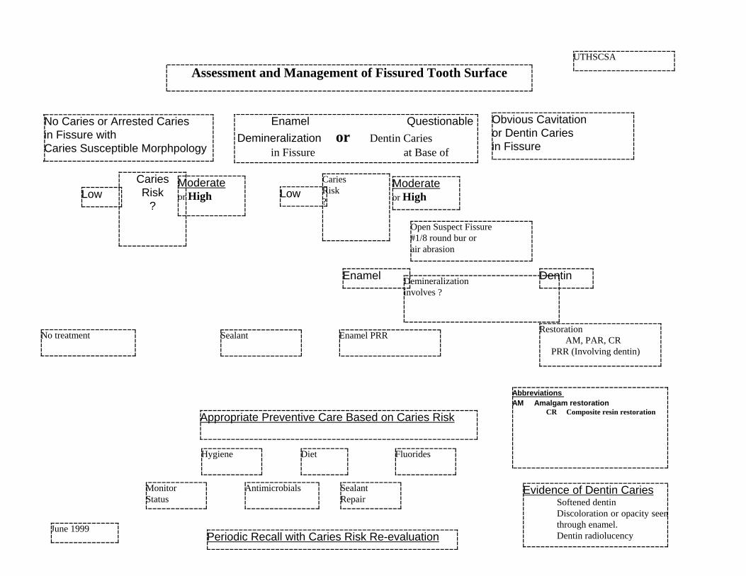
UTHSCSA
Assessment and Management of Fissured Tooth Surface
No treatment
No Caries or Arrested Cariesin Fissure withCaries Susceptible Morphpology
Enamel Questionable Demineralization or Dentin Caries
in Fissure at Base of
Enamel PRRRestoration AM, PAR, CR PRR (Involving dentin)
LowModerateor High
Antimicrobials SealantRepair
DietHygiene Fluorides
Abbreviations AM Amalgam restoration
CR Composite resin restoration
Low
Periodic Recall with Caries Risk Re-evaluation
MonitorStatus
June 1999
Obvious Cavitationor Dentin Cariesin Fissure
Sealant
Demineralizationinvolves ?
DentinEnamel
Open Suspect Fissure#1/8 round bur orair abrasion
CariesRisk?
Appropriate Preventive Care Based on Caries Risk
CariesRisk
?
Evidence of Dentin CariesSoftened dentinDiscoloration or opacity seenthrough enamel.Dentin radiolucency
Moderateor High

Baylor College of DentistryTexas A&M University Health Science Center
1999 National CODE Agenda
1. Vital pulp treatment
a. When, where, and who teaches this topic in your curriculum?In the second year dental student (D-2) class in pre-clinical operative, one of our endodontic professors does onelecture on this topic. He goes into more detail in his D-2 endodontics class. In the third year (D-3) didactic course, Ireview the proper procedures for direct and indirect pulp caps.
My recommendation to students is: use calcium hydroxide on exposures (when indicated) and very near exposuresand cover with a glass ionomer liner. In very deep areas without the danger of exposure, I recommend a glassionomer liner directly on the dentin in that area.
b. Is this treatment utilized in patient care by students?Absolutely.
c. What criteria/protocol are followed in deciding to do or not do vital pulptreatment?If there is a question about the condition of the pulp, we determine the status by performing what we call a S.O.A.P. onthe tooth.
First comes the Subjective information from the patient: i. chief complaintii. thermal sensitivity (as reported by the patient) andiii. chewing sensitivity (as reported by the patient).
Second is the Objective information determined by examination:i. percussionii. palpationiii. any restorations, caries, the presence of a sinus tract, and/or fractures (restoration or tooth) and iv. radiographic findings.
Third is the Assessment based on the above information.
Fourth is formulating a Plan.
Please see the following Addendum #1 for greater details.
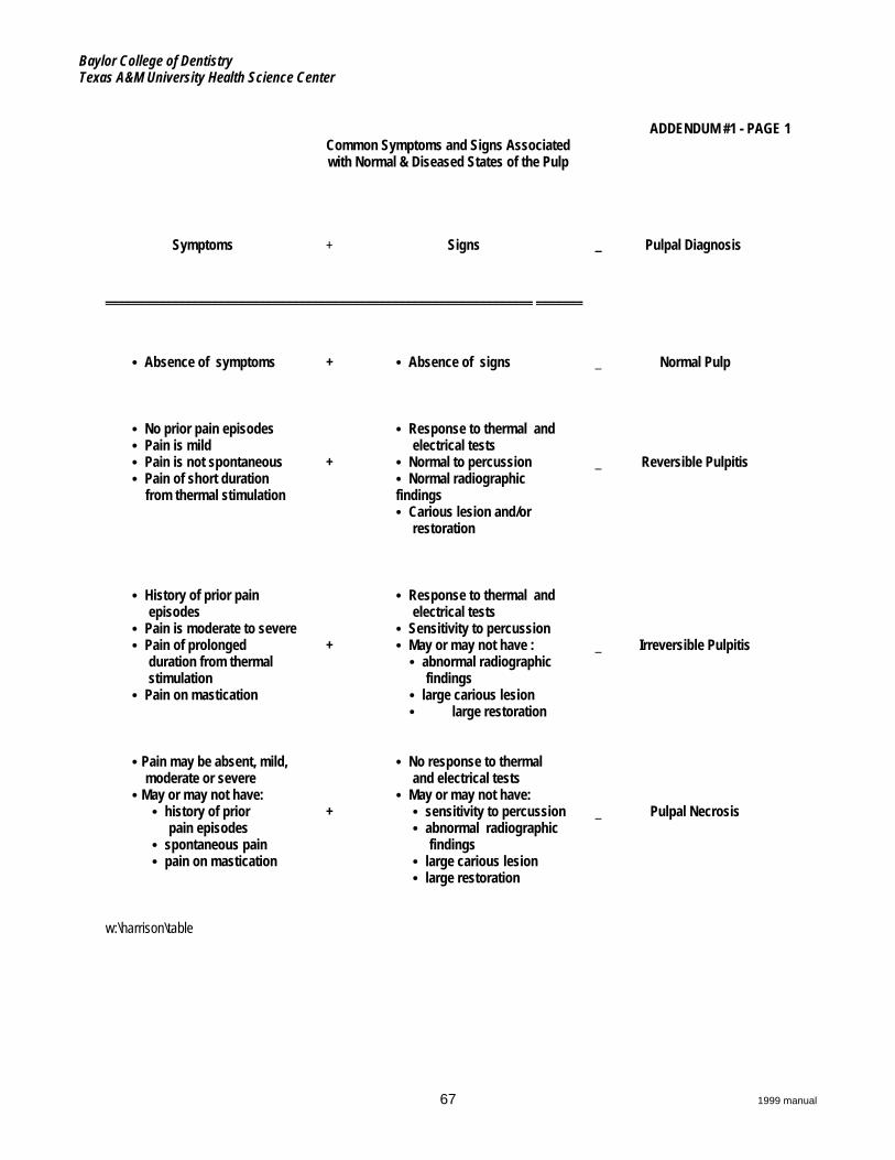
Baylor College of DentistryTexas A&M University Health Science Center
67 1999 manual
ADDENDUM #1 - PAGE 1Common Symptoms and Signs Associatedwith Normal & Diseased States of the Pulp
Symptoms + Signs _ Pulpal Diagnosis
________________________________________________________________ _______
C Absence of symptoms + C Absence of signs _ Normal Pulp
C No prior pain episodesC Pain is mildC Pain is not spontaneousC Pain of short duration from thermal stimulation
+
C Response to thermal and electrical testsC Normal to percussionC Normal radiographicfindingsC Carious lesion and/or restoration
_ Reversible Pulpitis
C History of prior pain episodesC Pain is moderate to severeC Pain of prolonged duration from thermal stimulationC Pain on mastication
+
C Response to thermal and electrical testsC Sensitivity to percussionC May or may not have : C abnormal radiographic findings C large carious lesion C large restoration
_ Irreversible Pulpitis
C Pain may be absent, mild, moderate or severeC May or may not have: C history of prior pain episodes C spontaneous pain C pain on mastication
+
C No response to thermal and electrical testsC May or may not have: C sensitivity to percussion C abnormal radiographic findings C large carious lesion C large restoration
_ Pulpal Necrosis
w:\harrison\table
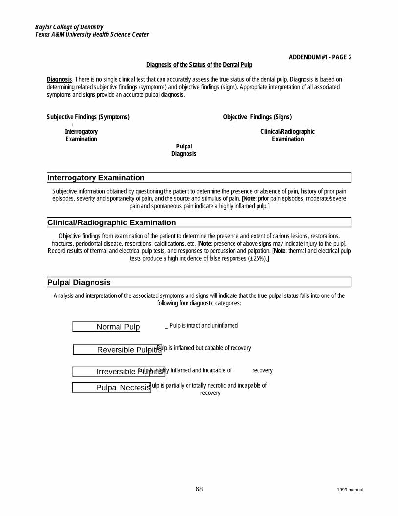
Baylor College of DentistryTexas A&M University Health Science Center
68 1999 manual
Interrogatory Examination
Clinical/Radiographic Examination
Pulpal Diagnosis
Normal Pulp
Reversible Pulpitis
Irreversible Pulpitis
Pulpal Necrosis
ADDENDUM #1 - PAGE 2Diagnosis of the Status of the Dental Pulp
Diagnosis. There is no single clinical test that can accurately assess the true status of the dental pulp. Diagnosis is based ondetermining related subjective findings (symptoms) and objective findings (signs). Appropriate interpretation of all associatedsymptoms and signs provide an accurate pulpal diagnosis.
Subjective Findings (Symptoms) Objective Findings (Signs) W W
InterrogatoryExamination
Clinical/RadiographicExamination
PulpalDiagnosis
Subjective information obtained by questioning the patient to determine the presence or absence of pain, history of prior painepisodes, severity and spontaneity of pain, and the source and stimulus of pain. [Note: prior pain episodes, moderate/severe
pain and spontaneous pain indicate a highly inflamed pulp.]
Objective findings from examination of the patient to determine the presence and extent of carious lesions, restorations,fractures, periodontal disease, resorptions, calcifications, etc. [Note: presence of above signs may indicate injury to the pulp].
Record results of thermal and electrical pulp tests, and responses to percussion and palpation. [Note: thermal and electrical pulptests produce a high incidence of false responses (±25%).]
Analysis and interpretation of the associated symptoms and signs will indicate that the true pulpal status falls into one of thefollowing four diagnostic categories:
_ Pulp is intact and uninflamed
_ Pulp is inflamed but capable of recovery
_ Pulp is highly inflamed and incapable of recovery
_ Pulp is partially or totally necrotic and incapable of recovery
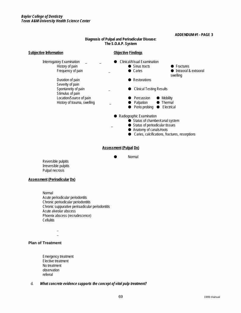
Baylor College of DentistryTexas A&M University Health Science Center
69 1999 manual
ADDENDUM #1 - PAGE 3Diagnosis of Pulpal and Periradicular Disease:
The S.O.A.P. System
Subjective Information Objective Findings
Interrogatory Examination _ _ M Clinical/Visual ExaminationHistory of pain M Sinus tracts M FracturesFrequency of pain _ M Caries M Intraoral & extraoral
swellingDuration of pain M RestorationsSeverity of painSpontaneity of pain _ M Clinical Testing ResultsStimulus of painLocation/Source of pain M Percussion M MobilityHistory of trauma, swelling _ M Palpation M Thermal M Perio probing M Electrical
M Radiographic Examination M Status of chamber/canal system _ M Status of periradicular tissues M Anatomy of canals/roots M Caries, calcifications, fractures, resorptions
Assessment (Pulpal Dx)
M NormalReversible pulpitisIrreversible pulpitisPulpal necrosis
Assessment (Periradicular Dx)
NormalAcute periradicular periodontitsChronic periradicular periodontitisChronic suppurative periraadicular periodontitisAcute alveolar abscessPhoenix abscess (recrudescence)Cellulitis
__
Plan of Treatment
Emergency treatmentElective treatmentNo treatmentobservationreferral
d. What concrete evidence supports the concept of vital pulp treatment?
Baylor College of DentistryTexas A&M University Health Science Center
70 1999 manual
Please site the scientific evidence. Additional comments welcome.
References:Cvek M: Treatment of non-vital permanent incisors with calcium hydroxide, Odontol Rev 23:27, 1972
Cvek M: A clinical report on partial pulpotomy and capping with calcium hydroxide in permanent incisors with complicatedcrown fractures, J Endod 4:232, 1978
Cvek M: Endodontic treatment of traumatized teeth. In Adreason. JO: Traumatic injuries of the teeth, ed 2, Philadelphia, 1981.WB Saunders Co.
Dimaggio JJ, and Hawes RR: Evaluation of direct and indirect pulp capping, J Dent Res 40:24, 1962 (abstract)
Dimaggio JJ and Hawes RR: Continued evaluation of direct and indirect pulp capping, J Dent Res 41:38, 1963 (abstract)
Heys DR, et al: Histological considerations of direct pulp capping agents, J Dent Res 60:1371, 1981
Hirschfeld Z, Sela J, and Ulmansky M: Hydrex and its effect on the pulp, Oral Surg 34:364, 1972
Holland R, et al: Histochemical analysis of the dogs' dental pulp after pulp capping with calcium, barium and strontiumhydroxides, J Endod 8:444, 1982
Horsted P, El Attar K, and Langeland K: Capping of monkey pulps with Dycal and a Ca-eugenol cement, Oral Surg 52:531,1981
Paterson RC, and Watts A: Further studies on the exposed germ-free dental pulp, Int J Endod 20:112, 1987
Pereira JC, and Stanley HR: Pulp capping: influence of the exposure site on pulp healing --- histologic and radiographic studyin dogs' pulp, J Endod 7:213, 1981
Phaneuf RA, Frankl SN, and Ruben M: A comparative histological evaluation of three calcium hydroxide preparations on thehuman primary dental pulp, J Dent Child 35:61, 1968.
Pisanti S, and Sciaky I: Origin of calcium in the repair wall after pulp exposure in the dog, J Dent Res 43:641, 1964
Pitt Ford TR: Pulpal response MPC for capping exposures, Oral Surg 50:81, 1980 Pitt Ford TR, and Roberts GJ: Immediateand delayed direct pulp capping with the use of a new visible-light cured calcium hydroxide preparation, Oral Surg Oral MedOral Pathol 71:338, 1991.
Jaber L, Mascres C, and Donohue WB: Electron microscope characteristics of dentin repair after hydroxlapatite direct pulpcapping in rats, J Oral Pathol Med 20:502, 1991
Jean A, et al: Effects of various calcium phosphate biomaterials on reparative dentin bridge formation, J Endod 14:83, 1988
Kerkhove BC, et al: A clinical and television densitometric evaluation of the indirect pulp capping technique, J Dent Child34:192, 1967
King JB, Crawford JJ, and Lindahl RL: Indirect pulp capping: a bacteriologic study of deep carious dentine in human teeth,Oral Surg 20:663, 1965
2. List five of the most important Operative Dentistry topics which need to be studied via clinical trials.My opinion and the opinions of other of our faculty:a. The success of bonding vs. cementing all-porcelain restorationsb. The effectiveness of conventional cement (zinc phosphate) vs. resin modified cement vs. resin cement for sealing restoration
margins
Baylor College of DentistryTexas A&M University Health Science Center
71 1999 manual
c. Evaluation of condensable (packable) composites (material specific)d. Fracture rate of porcelain onlays (material specific)e. Comparing the effect on the pulp of direct capping with calcium hydroxide vs. glass ionomer or resin modified glass ionomer
vs. etch and bonding agent
3. Calibration of facultya. What is the protocol for calibrating and standardizing your Operative faculty? Address current faculty and new
faculty. Also for pre-clinical and clinical.b. How often is calibration/recalibration done?
i. Two annual events help with calibration of both new and current pre-clinical and clinical third year Operative and fourthyear General Dentistry faculty:(1) Our annual calibration workshops for all faculty which includes Operative dentistry (Class II amalgam, Class II
gold inlay, and MOD gold onlay) and other disciplines(2) Annual Western Regional Examining Board Operative (WREB) calibration workshops for all faculty
Unfortunately, only a limited number of full-time faculty attend this although all full-time and part time faculty are invited.
ii. New faculty are calibrated one-on-one: first by the Operative Dentistry Division director and then by current faculty (inthe clinical setting). New faculty are at first assigned to shadow current faculty.As a reminder to all, we've published the description of "ideal" preparations for the Class II amalgam preparation, theClass III composite preparation, the Class II inlay preparation and the MOD onlay preparation. Those descriptionsfollow in Addendum 2.
iii. Current Operative faculty are encouraged to go to the annual calibration sessions mentioned above. Because so fewattend, periodic review of expectations occurs, usually one-on-one.
c. What evidence supports your protocol for calibration and recalibration? (Please cite.)i. The textbooks:
Fundamentals of Operative Dentistry, A Contemporary Approach, Schwartz RS, Summitt JB, Robbins JW.,Quintessance, 1996
The Art and Science of Operative Dentistry, 3rd Edition, Sturdevant CM, Roberson TM, Heymann HO, Sturdevant, JR. Mosby, 1995
ii. 95% or better passing rate of our students of the WREB licensing examinations
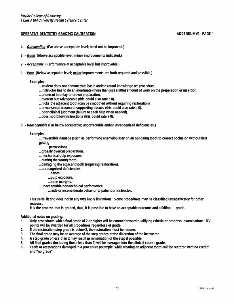
Baylor College of DentistryTexas A&M University Health Science Center
72 1999 manual
OPERATIVE DENTISTRY GRADING CALIBRATION ADDENDUM #2 - PAGE 1
4 - Outstanding (Far above acceptable level; need not be improved.)
3 - Good (Above acceptable level; minor improvements indicated.)
2 - Acceptable (Performance at acceptable level but improvable.)
1 - Poor (Below acceptable level; major improvements are both required and possible.)
Examples:...student does not demonstrate basic and/or sound knowledge re: procedure....instructor has to do an inordinate (more than just a little) amount of work on the preparation or insertion....undercut in onlay or crown preparation....overcut but salvageable (this could also rate a 0)....nicks the adjacent tooth (can be smoothed without requiring restoration)....unwarranted trauma to supporting tissues (this could also rate a 0)....poor clinical judgment (failure to seek help when needed)....does not follow instructions (this could rate a 0).
0 - Unacceptable (Far below acceptable; uncorrectable and/or unrecognized deficiencies.)
Examples: ...irreversible damage (such as performing enameloplasty on an opposing tooth to correct occlusion without firstgetting
permission)...grossly overcut preparation....mechanical pulp exposure....cutting the wrong tooth....damaging the adjacent tooth (requiring restoration)....unrecognized deficiencies
...caries.
...pulp exposure.
...open margins....unacceptable non-technical performance
...rude or inconsiderate behavior to patient or instructor.
This serial listing does not in any way imply limitations. Some procedures may be classified unsatisfactory for otherreasons.It is the process that is graded, thus, it is possible to have an acceptable outcome and a failing grade.
Additional notes on grading: 1. Only procedures with a final grade of 2 or higher will be counted toward qualifying criteria or progress examinations. RV
points will be awarded for all procedures regardless of grade.2. If the restoration step grade is below 2, the restoration must be redone. 3. The final grade may be an average of the step grades at the discretion of the instructor.4. A step grade of less than 2 may result in remediation of the step if possible 5. All final grades (including those less than 2) will be averaged into the clinical course grade..6. Teeth or restorations damaged in a procedure (example: while treating an adjacent tooth) will be restored with no credit”
and “no grade”.

Baylor College of DentistryTexas A&M University Health Science Center
73 1999 manual
THE IDEAL CLASS II AMALGAM PREPARATION ADDENDUM #2 - PAGE 2
Remember: the ideal is seldom found in the mouth.In the clinic we are dealing with real teeth, not typodont teeth. Therefore, the preparation described below may differ somewhat from that taught in the preclinical course.
1. Armamentarium of rotary instrumentsa. #330 bur (to initially establish the 1.5 mm depth of the pulpal floor on the occlusal surface)b. #1556 bur (to slightly deepen and smooth the pulpal floor)c. #4 round bur (for caries removal)
2. The preparationa. Pulpal floor = slightly more than the depth of the #330 bur (approximately 2 mm) and slightly past the dentinoenamel junction
(DEJ) and into the dentinb. Facial and lingual walls, slightly convergent (same as a #330 bur)c. Extension in facial and lingual grooves (molars), slightly divergentd. Mesial or distal wall (next to the marginal ridge), slightly divergente. Proximal box, facial and lingual walls slightly convergent and break contact with the adjacent toothf. It is critical that the facial and lingual box walls be located so there is a 90o angle at the cavosurface margin (may require
reverse curves). It is equally important that neither the mesiofacial, mesiolingual, distofacial or distolingual cusps beobliterated in the process.
g. Retention grooves (optional) in proximal box in dentin at axiofacial and axiolingual line angles, cut facially and lingually (nottowards the pulp)
h. Gingival floor of the box, below the contact area, flat, approximately 1.5 mm. wide mesiodistally with two-thirds of the width inthe dentin parallel to the occlusal plane and the gingival margin breaks contact with the adjacent tooth
i. Axial wall of the box, parallel to the external surface of the toothj. Axiopulpal line angle, rounded or beveledk. Preparation should be free of unsupported enamel and irregularities
For illustrations of the above, see your operative preclinical laboratory manual and your textbooks: Fundamentals of Operative Dentistry, A Contemporary Approach, Schwartz, et al and The Art and Science of Operative Dentistry, Sturdevant, et al, 3rd edition.
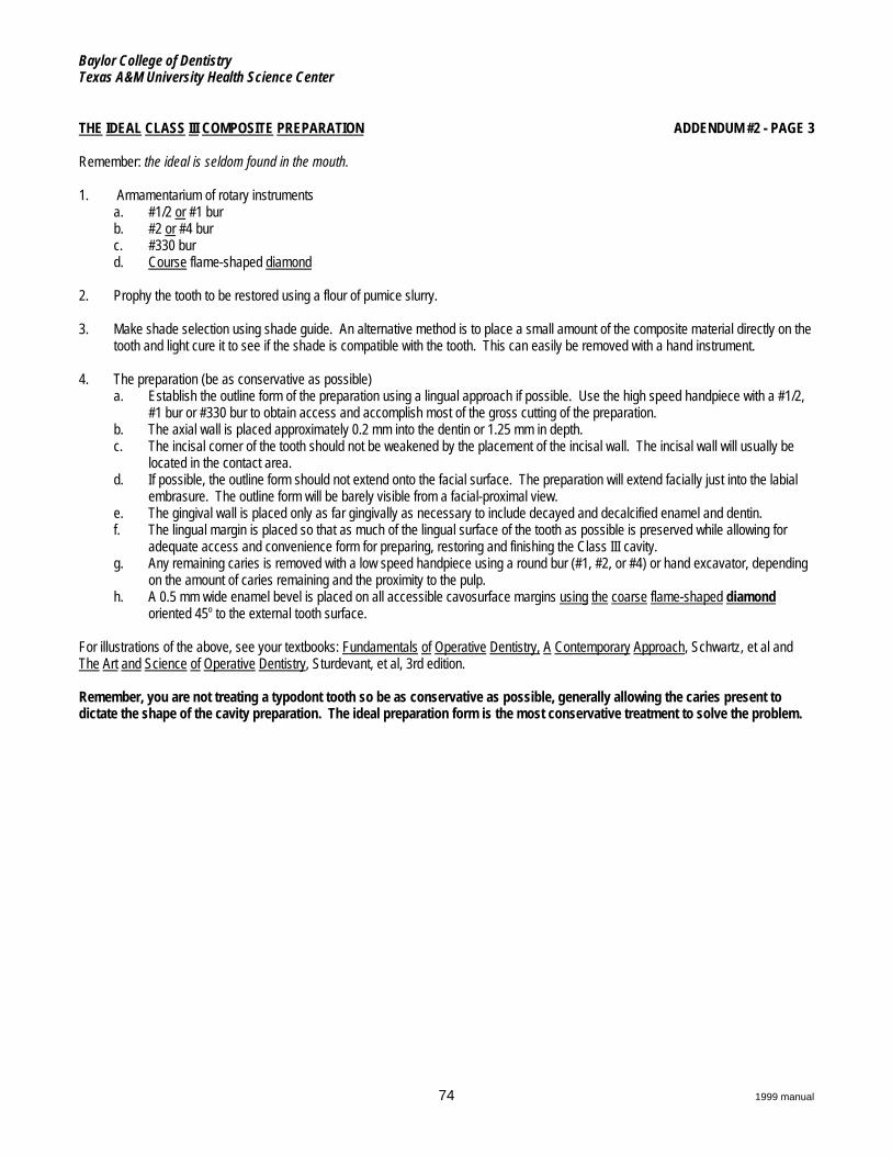
Baylor College of DentistryTexas A&M University Health Science Center
74 1999 manual
THE IDEAL CLASS III COMPOSITE PREPARATION ADDENDUM #2 - PAGE 3
Remember: the ideal is seldom found in the mouth.
1. Armamentarium of rotary instrumentsa. #1/2 or #1 burb. #2 or #4 burc. #330 burd. Course flame-shaped diamond
2. Prophy the tooth to be restored using a flour of pumice slurry.
3. Make shade selection using shade guide. An alternative method is to place a small amount of the composite material directly on thetooth and light cure it to see if the shade is compatible with the tooth. This can easily be removed with a hand instrument.
4. The preparation (be as conservative as possible)a. Establish the outline form of the preparation using a lingual approach if possible. Use the high speed handpiece with a #1/2,
#1 bur or #330 bur to obtain access and accomplish most of the gross cutting of the preparation.b. The axial wall is placed approximately 0.2 mm into the dentin or 1.25 mm in depth.c. The incisal corner of the tooth should not be weakened by the placement of the incisal wall. The incisal wall will usually be
located in the contact area.d. If possible, the outline form should not extend onto the facial surface. The preparation will extend facially just into the labial
embrasure. The outline form will be barely visible from a facial-proximal view.e. The gingival wall is placed only as far gingivally as necessary to include decayed and decalcified enamel and dentin. f. The lingual margin is placed so that as much of the lingual surface of the tooth as possible is preserved while allowing for
adequate access and convenience form for preparing, restoring and finishing the Class III cavity.g. Any remaining caries is removed with a low speed handpiece using a round bur (#1, #2, or #4) or hand excavator, depending
on the amount of caries remaining and the proximity to the pulp.h. A 0.5 mm wide enamel bevel is placed on all accessible cavosurface margins using the coarse flame-shaped diamond
oriented 45o to the external tooth surface.
For illustrations of the above, see your textbooks: Fundamentals of Operative Dentistry, A Contemporary Approach, Schwartz, et al andThe Art and Science of Operative Dentistry, Sturdevant, et al, 3rd edition.
Remember, you are not treating a typodont tooth so be as conservative as possible, generally allowing the caries present todictate the shape of the cavity preparation. The ideal preparation form is the most conservative treatment to solve the problem.
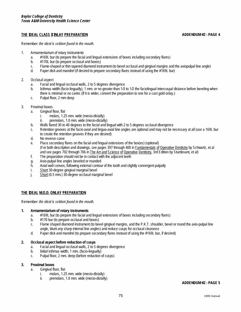
Baylor College of DentistryTexas A&M University Health Science Center
75 1999 manual
THE IDEAL CLASS II INLAY PREPARATION ADDENDUM #2 - PAGE 4
Remember: the ideal is seldom found in the mouth.
1. Armamentarium of rotary instrumentsa. #169L bur (to prepare the facial and lingual extensions of boxes including secondary flares)b. #170L bur (to prepare occlusal and boxes)c. Flame-shaped or thin tapered diamond instrument (to bevel occlusal and gingival margins and the axiopulpal line angle)d. Paper disk and mandrel (if desired to prepare secondary flares instead of using the #169L bur)
2. Occlusal aspecta. Facial and lingual occlusal walls, 2 to 5 degrees divergenceb. Isthmus width (facio-lingually), 1 mm. or no greater than 1/3 to 1/2 the faciolingual intercuspal distance before beveling when
there is minimal or no caries (If it is wider, convert the preparation to one for a cast gold onlay.)c. Pulpal floor, 2 mm deep
3. Proximal boxesa. Gingival floor, flat
i. molars, 1.25 mm. wide (mesio-distally)ii. premolars, 1.0 mm. wide (mesio-distally)
b. Walls flared 30 to 40 degrees to the facial and lingual with 2 to 5 degrees occlusal divergencec. Retention grooves at the facio-axial and linguo-axial line angles are optional and may not be necessary at all (use a 169L bur
to create the retention grooves if they are desired)d. No reverse curvee. Place secondary flares on the facial and lingual extensions of the box(es) (optional)
(For both description and drawings, see pages 397 through 400 in Fundamentals of Operative Dentistry by Schwartz, et aland see pages 702 through 706 in The Art and Science of Operative Dentistry, 3rd Edition by Sturdevant, et al)
f. The preparation should not be in contact with the adjacent teethg. Axio-pulpal line angles beveled or roundedh. Axial wall convex, following external contour of the tooth and slightly convergent pulpallyi. Short 30-degree gingival marginal bevelj. Short (0.5 mm.) 30-degree occlusal marginal bevel
THE IDEAL M.O.D. ONLAY PREPARATION
Remember: the ideal is seldom found in the mouth.
1. Armamentarium of rotary instrumentsa. #169L bur (to prepare the facial and lingual extensions of boxes including secondary flares)b. #170 bur (to prepare occlusal and boxes)c. Flame shaped diamond instrument (to bevel gingival margins, and the P.K.T. shoulder, bevel or round the axio-pulpal line
angle, blunt any sharp internal line angles) and reduce cusps for occlusal clearanced. Paper disk and mandrel (to prepare secondary flares instead of using the #169L bur, if desired)
2. Occlusal aspect before reduction of cuspsa. Facial and lingual occlusal walls, 2 to 5 degrees divergenceb. Initial isthmus width, 1 mm. (facio-lingually)c. Pulpal floor, 2 mm. deep (before reduction of cusps)
3. Proximal boxesa. Gingival floor, flat
i. molars, 1.25 mm. wide (mesio-distally)ii. premolars, 1.0 mm. wide (mesio-distally)
ADDENDUM #2 - PAGE 5
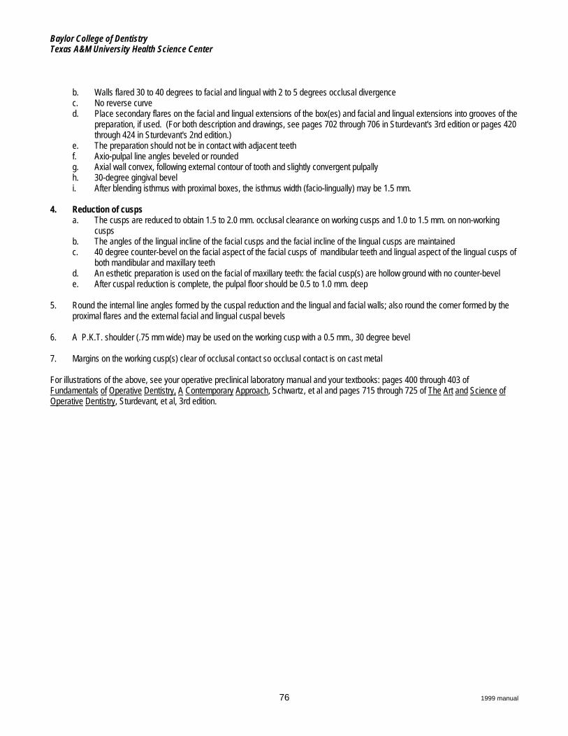
Baylor College of DentistryTexas A&M University Health Science Center
76 1999 manual
b. Walls flared 30 to 40 degrees to facial and lingual with 2 to 5 degrees occlusal divergencec. No reverse curved. Place secondary flares on the facial and lingual extensions of the box(es) and facial and lingual extensions into grooves of the
preparation, if used. (For both description and drawings, see pages 702 through 706 in Sturdevant's 3rd edition or pages 420through 424 in Sturdevant's 2nd edition.)
e. The preparation should not be in contact with adjacent teethf. Axio-pulpal line angles beveled or roundedg. Axial wall convex, following external contour of tooth and slightly convergent pulpallyh. 30-degree gingival beveli. After blending isthmus with proximal boxes, the isthmus width (facio-lingually) may be 1.5 mm.
4. Reduction of cuspsa. The cusps are reduced to obtain 1.5 to 2.0 mm. occlusal clearance on working cusps and 1.0 to 1.5 mm. on non-working
cuspsb. The angles of the lingual incline of the facial cusps and the facial incline of the lingual cusps are maintainedc. 40 degree counter-bevel on the facial aspect of the facial cusps of mandibular teeth and lingual aspect of the lingual cusps of
both mandibular and maxillary teethd. An esthetic preparation is used on the facial of maxillary teeth: the facial cusp(s) are hollow ground with no counter-bevele. After cuspal reduction is complete, the pulpal floor should be 0.5 to 1.0 mm. deep
5. Round the internal line angles formed by the cuspal reduction and the lingual and facial walls; also round the corner formed by theproximal flares and the external facial and lingual cuspal bevels
6. A P.K.T. shoulder (.75 mm wide) may be used on the working cusp with a 0.5 mm., 30 degree bevel
7. Margins on the working cusp(s) clear of occlusal contact so occlusal contact is on cast metal
For illustrations of the above, see your operative preclinical laboratory manual and your textbooks: pages 400 through 403 ofFundamentals of Operative Dentistry, A Contemporary Approach, Schwartz, et al and pages 715 through 725 of The Art and Science ofOperative Dentistry, Sturdevant, et al, 3rd edition.

Baylor College of DentistryTexas A&M University Health Science Center
77 1999 manual
4. Competency education
a. How is competency based Operative education determined?Department Chairs, Division Directors, and course directors were contacted. Expected competencies were requested. Acommittee reviewed these stated competencies. Based on these expected competencies, the first competency documentswas started in 1993 and completed in 1994. The document was reviewed two years later and the current document wasadopted in the Spring of 1997.
b. How is the weak student identified?All students are observed in their daily work to help identify the weak ones. However, when Progress Examinations are given(either in the laboratory or in the clinic) these weak students become much easier to identify since they do not receive aid froma faculty member during these examinations.
c. What measures are taken to upgrade competency of the weak student?If a student consistently fails to successfully complete Progress Examinations, a remediation program is set up and thestudent is tutored by a selected Operative faculty member. During the time that the student is remediating, Operative Clinicprivileges are withdrawn. When that faculty member is satisfied that the student is ready, the student is permitted to retakethe Progress Examination. If a student still cannot satisfactorily complete the Progress Examination, a recommendation todismiss or to repeat the year may be made or additional remediation may be prescribed immediately.
5. Computer based interactive education
a. What interactive computer-based teaching tools are you using to teach Operative Dentistry?None
b. Who developed them? n/a
c. Would your school/region participate in development and in what manner?We would like to think that our school would be willing to participate, however, essential to making a final decision would bethe time and the funds which must be allocated to such a project. The interest is there and we hope we are kept apprised ofany such project.
6. Posterior esthetic restorations - report on direct and indirect.
a. Are they included in the curriculum?
b. Where (pre-clinic and clinic)In the second year pre-clinical Operative course, the students are taught how to do a Class II, Class III, and a Class V directcomposite restoration
In the third year didactic Operative course, we discuss only the direct composite restorations. In the Operative clinic, we doonly direct composite restorations.
In the fourth year the students receive a 2-quarter course from the General Dentistry Department, "Advances in Techniquesand Materials." With the General Dentistry Department they are permitted to do both direct and indirect restorations. Theindirect restorations may be either composite or porcelain.
c. Required experiences? (We like to call them "essential experiences.”)In the pre-clinic course, they are required to do one of each of the three classes mentioned above. In the third year Operativeclinic they are required to do a minimum of two Class III composite restorations plus a Class III composite clinical ProgressExamination. In the fourth year, General Dentistry requires five tooth-colored indirect restorations of either composite orporcelain.

Baylor College of DentistryTexas A&M University Health Science Center
78 1999 manual
d. What materials and/or systems utilized?Composites: Prisma TPH, Herculite XRV, Z-100, Heliomolar, Tetric Flow, SureFil with Composi-Tight rings and matrices forClass II
Resin Modified Glass Ionomer: Dyract (for Class V)
Porcelains: (1) conventional stacked and (2) pressable
e. What are the results? Also cite the evidence for the reported results.Are clinical results during the third year appear to be very good overall. The Chair of General Dentistry reports that they arehaving good results also. Proximal contacts are excellent using the Composi-Tight system for Class II restorations.
These results are determined by observable clinical means. At this time we have no other evidence.
Regional Agenda Items
1. Do you teach and/or are you using air abrasion or laser technology?
We are using air abrasion in the post-graduate A.E.G.D. program. Occasionally the fourth year undergraduate students arepermitted to use the air abrasion technology in the A.E.G.D. clinic.
Laser technology is used in the Graduate Periodontal Clinic. The undergraduate fourth year students are introduced to lasertechnology in a didactic course and they get to do a brief practice on an orange.
2. What is your diagnostic criteria and teaching for fissure caries. What is your criteria for evaluating conservative pit andfissure preparations and restorations?
Most of our faculty are using the explorer and the criteria of "sticks" to "determine" whether or not pit and fissure caries exists. Theyalso look at radiographs to see if anything can be seen at or beyond the dentino-enamel junction.
If the caries is felt to be within the enamel only, a preventive resin restoration (PRR) may be ordered using one of the availablecomposites.
Sealants are also done with Ultraseal XT when it is deemed appropriate, that is, caries is absent or the caries is extremelysuperficial.
Students are taught that an amalgam preparation should be as conservative as possible as long as the smallest condenser fits intothe preparation. Composite preparations are the same, as long as we can get the composite into the preparation. They are taughtto remove all the caries and if a groove is not carious, to seal the groove rather than to prepare and restore it.
3. How do you prepare your students for the National Board Examination?
In my third year didactic Operative course, I incorporate as many of the old National Board questions as possible into myexaminations. Most of the topics are adequately covered in the lectures and assignments.
When these students become fourth year students, I usually will spend one to two hours in review, especially discussing old NationalBoard questions.
The second and third year Operative syllabi and textbooks and the fourth year course are excellent vehicles for review.
Baylor College of DentistryTexas A&M University Health Science Center
79 1999 manual
CODE Questions
1. What is CODE doing well?
The annual regional meetings and the post-meeting handout of collated answers to all the questions as well as what is published bythe National organization are most helpful.
The most helpful of all are the reports given and the exchange of information and discussion which occurs around the table at ourregional meeting. Many times we've made changes based on what was revealed at our annual meeting and for which we are mostthankful.
2. Where do you desire improvement?
I'm all right with the way it is right now.
3. Is $25 US/$30 Canadian school dues adequate. Comments are expected.
Is that enough to do all that CODE wants to do? If so, then the dues is adequate. If CODE has more it wants to do and the duesare not adequate as is, then consideration should be given for a modest raise. All schools are hard pressed for funds now-a-days,so let's not raise the dues unless there's a good reason for a raise.
4. How can participation by faculty in CODE regional meetings be encouraged/improved?
I wish I knew. We had funding allocated for two representatives to participate in this year's regional meeting and I could not get anyother Operative faculty member to attend with me because everyone either has a conflict or other duties which require theirattention.
Perhaps explaining to the Chairman of the department both how important and how beneficial these conferences are and urging theChair persons to encourage or even mandate attendance.
5. Please indicate the office/position with complete mailing address at your school to which regional reports, duesstatements, roster requests, etc. are to be mailed.
Our mailing address is:Baylor College of DentistryTexas A & M University Health Science CenterP.O. Box 660677Dallas, TX 75266-0677
Dues statements:Dr. Amp MillerInterim ChairDepartment of Restorative Sciences
All other mentioned above:Dr. Marvin HirshDirector of Clinical Operative DentistryDepartment of Restorative Sciences
80 1999 manual
University of Mississippi School of Dentistry
1999 National CODE Agenda
1. Vital pulp treatment
a. 1st year students are taught about pulp capping by operative faculty and by endodontic faculty. Also Endodontic course inSophomore year, pulp capping is re-emphasized.
b. Yes; Pulp capping is utilized by students for patient care in the operative clinics and endodontic clinics.c. Criteria for successful pulp capping:
i. Asymptomatic tooth with normal response to thermal testingii. Small exposure site (.5mm or less) in a relatively caries-free area.iii. Hemorrhage from exposure site is easily controlled by sterile cotton pellet (within a minute).iv. Exposure occurred in a clean, uncontaminated field (Rubber dam must be utilized)v. Atraumatic exposure
Pulpcap with a layer of CaOH2 (life) followed by a temporary restoration (IRM) or a resin modified glass ionomer followed by a permanentrestoration.
Evidence:
(1) Harold Stanley symposium article in American Journal of Dentistry Vol 121, Jan 1998.(2) Vital Pulp Capping: a worth while procedure by Lawrence Stockton in the Journal of the Canadian Dental
Association June 1999 Vol 65.(3) Sturdevant text – The Art and Science of Operative Dentistry.
2. List five of the most important Operative Dentistry topics which need to be studied via clinical trials
a. Post and Core Systemsb. Tooth preparation desensitizing agents for pulpal sensitivityc. Long term effects of BIS GMA on the pulpd. A five year study comparing Compomers in Class V restorationse. Wear characteristics of resin modified glass ionomers and Fuji IX
3. Calibration of faculty
a. PreclinicalFaculty are calibrated and standardized during practical examinations at the mid-term practical and final practical. Two facultygrade each practical after written criteria for each exercise are reviewed and agreed upon. If the two faculty are 10 or morepoints apart, a third faculty member determines which of the two grades are closer to matching the written criteria or if anaverage of the two grades is acceptable. The reasons for this final grade are then explained to the two faculty by the thirdarbitrating faculty member who has also been familiarized with the criteria.
ClinicalAll operative faulty are standardized and calibrated during the senior clinical comprehensive examination. Written criteriasimilar to state board exam criteria are reviewed with the faculty before the examination. Two faculty grade each procedureand if they are ten or more points apart, a third faculty must arbitrate the two grades. A failing or passing grade has to beagreed upon by at least two of the three faculty evaluating the procedure. Also all students take clinical competencyexaminations in Class I, Class II and Class III situations. Two clinical faculty grade each competency exam and have to agreeon the final grade within a ten-point range.
b. Calibration/re-calibration is done twice a year during pre-clinical courses, at the mid-term practical and final practicalexaminations. Clinically, calibration is done officially once a year during the senior comprehensive examination. Unstructuredcalibration is an ongoing process during clinical competency examination as described in questions 4.
c. Evidence:Knight G W. Toward faculty Calibration. Journal of Dental Education 61 (12) 941-6. 1997 Dec. Hoelscher DC, Knight GWJournal of Dental Education 1996; 60:221 Assessing criteria formats as part of a faculty development program.
4. Competency education
University of Mississippi School of Dentistry
81 1999 manual
a. Clinical competency based Operative education is determined at University of Mississippi by Class I, Class II, class IIIcompetency exams during the junior and senior years on clinically acceptable patients. Before these competency exams areattempted, a certain number of points or procedures must be completed before the student can apply for the competency. Two faculty evaluate each competency exam. Minimal input is given by the faculty therefore the student must make decisionsindependently. Failure of the exam may result in remedial dentoform exercises before re-examination on patients. Alsoclinical competency is determined by a senior clinical comprehensive exam where dentoform exercises are also required.
b. Weak students-identified by daily grade sheets, end-of-quarter progress grades, tardiness or failure in taking competency exams.
c. Upgrade weak students?Slight failure may only necessitate re-taking another competency on a patient or remedial project in the dentoform or morepatient experiences before re-examination. We are looking into using the DentSim for students requiring remedial workbefore competency re-examination.
5. Computer based interactive education
a. Dent Sim-virtual reality; information management systemsb. Denx America Inc. – DenX Advanced Dental Systemsc. Yes. Need more specifics
6. Posterior esthetic restorations
a. Operative – Caries courses during 2nd year. Fixed Prosthodontics courses during 2nd and 3rd year.
b. Pre-clinici. Operative –
(1) Direct posterior composite, Class I and Class II situation. (2) Indirect
(a) Cerec II porcelain inlay-onlay (elective course)(b) Indirect composite inlay/onlay will be introduced in 2nd year operative course (Belleglass system)
ii. Fixed Prosthodontics –(1) 2nd and 3rd year –
(a) Porcelain/metal posterior crown preparation(b) All porcelain posterior crown preparation (premolar only)
iii. Clinic-(1) Operative (direct) Class I and Class II posterior composite(2) Indirect – Cerec II (in special situation with faculty input)
(a) Starting to teach: posterior Belleglass indirect compositeiv. Fixed Prosthodontics –
(1) Posterior porcelain/metal crowns with porcelain occlusal
c. Required experiences?
i. Operative-(1) 4th Year students are required to do four posterior composite restorations(2) 4th year students can substitute Cerec II inlay/onlays for gold onlays (3)
Fixed Prosthodontics – Minimally, 2 PFM crowns and one posterior esthetic bridge. Students do more posterior PFM’s than full gold toobtain a total of 23 to 26 total units.
d. Materials:Composite: Herculite XRV, Prisma APH, Prisma TPH, expected to begin use of the Belleglass systemPorcelain: CerecII system and PFMs (Finesse porcelain with Argen 57 SF metal)
University of Mississippi School of Dentistry
82 1999 manual
e. Results: When properly placed posterior composites are doing well especially with rubber dam isolation. Case selection mostimportant in determining success. We do not do large intercuspal isthmus width posterior composite restorations (usually 1/4or less) Problems with porcelain occlusals include wearing opposing dentition and adhesive failure of the porcelain to metal. Cerec I had a few problems with restoration fracture in heavy occlusion areas especially in bruxism cases.
Regional Agenda Items
1. We do not teach or use air abrasion or laser technology at this time.
2. Part (A) For diagnosis of caries, in general, we do not use caries disclosing solutions routinely, but rather diagnose pit and fissurecaries by visual, tactile and radiographic examination methods. We use an explorer to check the fissure or pit for softness or anyother break in continuity of the area. A “stick” with an explorer is not by itself an absolute diagnosis of caries but must be used withthe other two methods to obtain a final diagnosis.
Ref: Sturdevant, The Art and Science of Operative Dentistry
Part (B) How are you teaching the restoration of pit and fissure carious lesions?If pit and fissure caries is obvious clinically and /or radiographically, it is not considered minimal and a typical Class Ipreparation for amalgam or composite is performed. If the fissure caries is very minimal or questionable clinically we attemptto excavate with a small round bur (¼ ) in a slow speed handpiece. Carious fissures not penetrating /3 to ½ the depth of theenamel after removal with a round bur are etched and sealed. Minimal carious lesions penetrating to or slightly beyond theDEJ are conservatively restored with a posterior hybrid composite followed by a sealant if other questionable fissures arepresent in the area. Pit and fissure caries undermining enamel in heavy centric occlusion areas are excavated and a typicalClass I amalgam preparation and restoration is performed. If the clinical faculty determines that the occlusion is not heavy inthis area of undermined enamel, a posterior hybrid composite may be utilized. We are looking into using a resin surfacesealant over posterior composites that are in occlusal contact.
Part (C) “Conservative” preparations are considered adequate if they are free of remaining caries and not overextended. Conservative restorations or sealants are adequate if free of voids or other defects, finished to the cavo surface margin, andout of occlusion. NOTE: The teaching of pit and fissure sealant application is currently being re-assigned to the Departmentof Diagnostic sciences.
3. National Board Preparation:As a school, we have a board pre-test to make specific recommendations to each student regarding weak areas. Also reviews aredone by the individual departments (need more time allotted for this). Students are given a week off prior to exam for study andreview. Materials to check out: dental decks, released board exams (also in computer laboratory).
Passing of Part II is a requirement for completing Mississippi licensure. In the restorative department we try to include items on ourexams that have been frequently asked on previously released national board examinations. Also passing Part I is a requirement forpromotion to the next academic year.
CODE Questions
1. CODE enables scheduling of regional meetings annually where operative faculty can interact on a very challenging time consumingagenda that creates much discussion and controversy.
2. No suggestions
3. Yes. This seems adequate.
4. Schools should provide more travel money for more faculty representation at the regional meetings.
5. Dr. Gary W. Reeves, Chairman, Restorative Department, University of Mississippi, School of Dentistry, 2500 North State Street,Jackson, MS 39216-4505, Phone: 601-984-6030, Fax: 601-984-6039, e-mail: [email protected]

University of Mississippi School of Dentistry
83 1999 manual
84 1999 manual
University of Texas Health Science Center Houston, Dental Branch
1999 National CODE Agenda
1. Vital pulp treatment
a. When, where, and who teaches this topic in your curriculum?
! Restorative Dentistry and Biomaterials (Operative I, first year, and Operative II, second year)! Endodontics (second year)! Pediatric Dentistry teaches indirect pulp capping procedures for permanent teeth and leaves the direct pulp capping
procedures to the Endo and Operative courses. They do not teach direct pulp capping procedures for primary teethdue to their observed incidence of internal resorption after this procedure.
b. Is this treatment utilized in patient care by students?
Yes.
c. What criteria/protocol are followed in deciding to do or not do vital pulp treatment?
! Depth of the lesion (estimation of the amount of remaining tooth structure over the pulpal tissue – approximate distancefrom the pulp)
! If exposed pulp – the type and size of exposure (carious or mechanical)! Symptoms reported by the patient (if any)! Results from vitality tests! Amount of caries remaining (if any), prior to pulp capping! Dental history of the patient! Oral condition of the patient including oral hygiene and diet
d. What concrete evidence supports the concept of vital pulp treatment? Please site the scientific evidence. Additionalcomments welcome.
We follow the texts listed below which cite numerous references.
The Art and Science of Operative Dentistry, 3rd Edition, Sturdevant CM, Roberson TM, Heymann HO. JR Mosby, 1995.Fundamentals of Operative Dentistry, A Contemporary Approach, Schwartz RS, Summitt JB, Robbins JW. Quintessance,1996.
2. List five of the most important Operative Dentistry topics which need to be studied via clinical trials
a. Continued materials evaluation, especially studies involving the latest generation of composite resins and bonding systems.
b. Evaluation of the clinical soundness and long term stability and function of tooth structure restored using conservative andultra conservative cavity designs.
c. Continued evaluation of fluoride releasing restorative materials. Evaluate the cariostatic effects of fluoride release and howvarious materials contribute to the prevention or the reduction in the incidence of recurrent decay.
d. Incidence of microleakage and recurrent decay with various bonding systems.
e. Continued evaluation of alternative cavity preparation devices.
3. Calibration of faculty
a. What is the protocol for calibrating and standardizing your Operative faculty? Address current faculty and new faculty. Alsofor pre-clinical and clinical.
! The only calibration occurs with faculty involved with the Senior Mock Board Examination. Calibration here is doneusing inlay, onlay, and Class II amalgam preparation and restoration clinical slides.
University of Texas HSC Houston, Dental Branch
85 1999 manual
b. How often is calibration/recalibration done?
Once per year.
c. What evidence supports your protocol for calibration and recalibration? (Please cite)
No evidence based support.
4. Competency education
a. How is competency based Operative education determined?
! Pre-clinically – competency is determined using various bench-top and manikin examinations.! Clinically - competency is determined using a clinical and a bench-top examination (Third Year Operative Clinical
Competency Examination, Fourth Year Operative Bench Examination).
b. How is the weak student identified?
! By their inability to successfully complete the competency examinations.
c. What measures are taken to upgrade competency of the weak student?
! Pre-clinically – no substantive measures are utilized.! Clinically - additional experiences are assigned to address perceived weaknesses. These may include requiring
additional work utilizing a pre-clinical manikin head or by assigning additional clinical experiences.
5. Computer based interactive educationRegion VI (South) is developing two comprehensive restorative treatment planning cases to be used as problem based learningexercises. It is hoped these cases can be used to develop authoring software that could be used for educational purposes. A jointventure with Dental Interactive Simulation Corporation (DISC) warrants consideration.
a. What interactive computer-based teaching tools are you using to teach Operative Dentistry?
! None at this time
b. Who developed them?
! N/A
c. Would your school/region participate in development and in what manner?
! Yes, in a manner to be determined after a review and analysis of the program. At this point, without an understandingof the program and the needs of the program, it is difficult to envision how our knowledge and resources could fit into oreven benefit the program.
6. Posterior esthetic restorations – report on direct and indirect
a. Are they included in the curriculum? What discipline?
! Yes! Restorative Dentistry and Biomaterials
b. Where (pre-clinic and clinic)
! Clinic : Patient treatment! Pre-clinic: Operative I, first year, and Operative II, second year
c. Required experiences?

University of Texas HSC Houston, Dental Branch
86 1999 manual
! None specifically.
d. What materials and/or systems utilized?
! Direct: HeliomolarSureFilZ-100
! Indirect: Concept system
e. What are the results? Also cite the evidence for the reported results.
! Clinic : Patient treatment utilizing the Concept system. Most of these restorations are not closely followed;however, no significant problems have been realized
! Pre-clinic: In the Operative II course, second year students produce their own indirect resin inlays using natural teethutilizing the Concept system and natural teeth. Fabrication, adjustment, cementation, and finishing andpolishing procedures usually are very successful for most of the students.
Regional Agenda Items
1. Do you teach and/or are you using air abrasion or laser technology?
! Yes, air abrasion in the second year Operative II course. Laser technology is mentioned in various courses.
2. What is your diagnostic criteria and teaching for fissure caries. What is your criteria for evaluating conservative pit andfissure preparations and restorations?
! Diagnostic criteria includes the patient’s previous dental history, oral hygiene, diet, and current caries activity and status. Thetexture and appearance of the fissured enamel (dried), an estimation of the presence and extent of caries, the results of aradiographic analysis, and any symptoms reported by the patient are also considered.
! Clinical evaluation is based on the following criteria: visual inspection for material defects and retention, explorer catches,marginal integrity, evidence of recurrent caries, color stability, anatomic wear.
3. How do you prepare your students for the National Board Examination?
! Preparation involves faculty reviews of restorative information and concepts in a lecture format. The reviews comprise acourse with the grade primarily based on attendance.
CODE Questions
1. What is CODE doing well?
! CODE provides an excellent forum for communication among the Regional schools.
2. Where do you desire improvement?
! Perhaps by considering the implementation of a full, two-day meeting.
3. Is $25 US/$30 Canadian school dues adequate? Comments are expected.
! Without understanding how CODE dues are utilized, and the financial responsibilities of the National CODE office, includingany financed programs, it is difficult to evaluate the adequacy of CODE dues.
4. How can participation by faculty in CODE regional meetings be encouraged/improved?
University of Texas HSC Houston, Dental Branch
87 1999 manual
! Through the years, Region III has enjoyed good response and participation from its member schools. Travel money forfaculty to attend meetings, in addition to the regular allotted departmental travel money, could help to increase individualparticipation.
5. Please indicate the office/position with complete mailing address at your school to which regional reports, duesstatements, roster requests, etc., are to be mailed. NOTE: Regional Director – this information from each school is to betransmitted to the National Director.
Dr. William H. Tate, DDSAssociate ProfessorUniversity of Texas HSC – HoustonDental BranchDepartment of Restorative Dentistry and Biomaterials6516 John Freeman AvenueHouston, Texas [email protected](713) 500-4264 - office
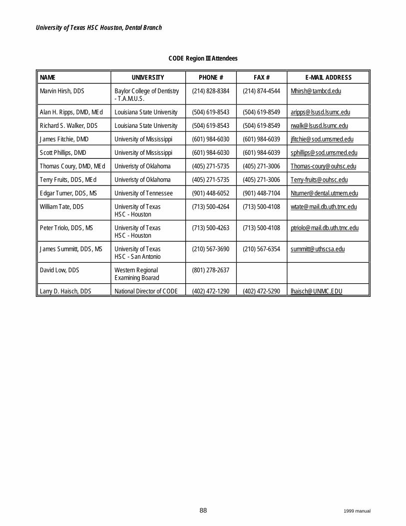
University of Texas HSC Houston, Dental Branch
88 1999 manual
CODE Region III Attendees
NAME UNIVERSITY PHONE # FAX # E-MAIL ADDRESS
Marvin Hirsh, DDS Baylor College of Dentistry- T.A.M.U.S.
(214) 828-8384 (214) 874-4544 [email protected]
Alan H. Ripps, DMD, MEd Louisiana State University (504) 619-8543 (504) 619-8549 [email protected]
Richard S. Walker, DDS Louisiana State University (504) 619-8543 (504) 619-8549 [email protected]
James Fitchie, DMD University of Mississippi (601) 984-6030 (601) 984-6039 [email protected]
Scott Phillips, DMD University of Mississippi (601) 984-6030 (601) 984-6039 [email protected]
Thomas Coury, DMD, MEd Univeristy of Oklahoma (405) 271-5735 (405) 271-3006 [email protected]
Terry Fruits, DDS, MEd Univeristy of Oklahoma (405) 271-5735 (405) 271-3006 [email protected]
Edgar Turner, DDS, MS University of Tennessee (901) 448-6052 (901) 448-7104 [email protected]
William Tate, DDS University of TexasHSC - Houston
(713) 500-4264 (713) 500-4108 [email protected]
Peter Triolo, DDS, MS University of TexasHSC - Houston
(713) 500-4263 (713) 500-4108 [email protected]
James Summitt, DDS, MS University of TexasHSC - San Antonio
(210) 567-3690 (210) 567-6354 [email protected]
David Low, DDS Western RegionalExamining Boarad
(801) 278-2637
Larry D. Haisch, DDS National Director of CODE (402) 472-1290 (402) 472-5290 [email protected]
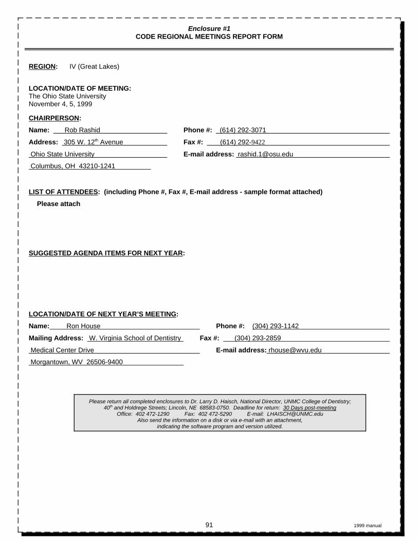
91 1999 manual
Enclosure #1CODE REGIONAL MEETINGS REPORT FORM
REGION: IV (Great Lakes)
LOCATION/DATE OF MEETING:The Ohio State UniversityNovember 4, 5, 1999
CHAIRPERSON:
Name: Rob Rashid Phone #: (614) 292-3071
Address: 305 W. 12th Avenue Fax #: (614) 292-9422
Ohio State University E-mail address: [email protected]
Columbus, OH 43210-1241
LIST OF ATTENDEES: (including Phone #, Fax #, E-mail address - sample format attached)
Please attach
SUGGESTED AGENDA ITEMS FOR NEXT YEAR:
LOCATION/DATE OF NEXT YEAR’S MEETING:
Name: Ron House Phone #: (304) 293-1142
Mailing Address: W. Virginia School of Dentistry Fax #: (304) 293-2859
Medical Center Drive E-mail address: [email protected]
Morgantown, WV 26506-9400
Please return all completed enclosures to Dr. Larry D. Haisch, National Director, UNMC College of Dentistry;40th and Holdrege Streets; Lincoln, NE 68583-0750. Deadline for return: 30 Days post-meeting
Office: 402 472-1290 Fax: 402 472-5290 E-mail: [email protected] send the information on a disk or via e-mail with an attachment,
indicating the software program and version utilized.

Region IV (Great Lakes)
92 1999 manual
1999 National CODE Agenda
1. Vital pulp treatment
General discussion: Need to bring this topic up to boards – discrepancy between what is taught and what is required by boardexams.
a. When, where, and who teaches this topic in your curriculum?
OSUThe subject is taught by the Section of Restorative Dentistry, Prosthodontics, and Endodontics. The dental students get informationon vital pulp treatment over the entire four years they are in dental school. In their first quarter, they are exposed to vital pulptherapy with the overview course about restorative dentistry. They get more of it in the conservative dental amalgam course andcomposite resin course. In the second year, the students are exposed again to the topic in their course covering large amalgamrestorations and restoration of extensively damaged teeth and a laboratory exercise reinforces what was taught in previous coursesand lectures. They are again exposed to this topic in the third and fourth year “co-clinical” and board review courses. These arelecture format and the “mock board” course. The course directors involved are: Robert Seghi, Robert Rashid, Peter Monaghan,Janet Bolina, Stephen Rosenstiel, Harris Bowman, and Al Reader.
IndianaPulp protection and treatment are introduced in the first year in Single Tooth Indirect Restorations. In depth pulp treatment iscovered in the second year Endodontics course. The Restorative department is responsible for all lectures and laboratories.
DetroitEndo does most of the teaching. No strong indications for IPC in vital teeth.
IllinoisRestorative and Endo teach.
Western OntarioTaught in the 2nd year operative dentistry program and repeated in the 3rd year lecture series.
NorthwesternSimilar to most others.
Case WesternPreclinical Courses2nd year courses:Basic Procedures in Restorative Dentistry - (Restorative Dept. Faculty)Preclinical Endodontics (Dept of Endodontics Faculty)
3rd year courses:Operative Dentistry (Restorative Dept. Faculty)Esthetic Dentistry (Restorative Dept. Faculty)
4th year courses:Clinical Dentistry (Dept of General Practice Faculty)Board Review Course (Faculty from Endodontics, Restorative, and General Dentistry departments)
West VirginiaSimilar to OSU.

Region IV (Great Lakes)
93 1999 manual
b. Is this treatment utilized in patient care by students?
OSUThe treatment which is taught is used by the students. All vital teeth get some form of vital pulp therapy even if only copal varnish(Copaliner) is the only material used prior to restoration placement.
IndianaYes
DetroitIllinois
Northwestern
Western OntarioYes
Case WesternYes
West Virginia
c. What criteria/protocol are followed in deciding to do or not do vital pulp treatment?
OSUConservative amalgam restorations receive a copal varnish liner applied in 3 coats. Conservative composite resin restorations arebonded to enamel and dentin using Kerr Optibond Solo according to manufacturer’s directions. Glass ionomer cement liners canalso be used under composite resins. When caries goes beyond minimum extension depth (½ mm pulpally to the DEJ to within ½mm of the dental pulp) two modes of therapy are presented.
Mode 1: Minimum Base Technique, preferred.Base materials up to 1 mm thick are placed when the operator believes there is less than ½ mm of dentin overlying the pulp. These include calcium hydroxide (Dycal) covered with a cement, glass ionomer cement being the preferred, but not onlychoice, or only glass ionomer cement without calcium hydroxide. Copal varnish is also used with this technique.
Mode 2: Board Examination Technique, practiced for board examination competency.Base materials replace dentin to ½ mm pulpally to the DEJ. Materials used in the Mod 1 technique are used here as well. The mode of vital pulp therapy is decided upon by the faculty and the student. Students are encouraged to practice for theboard examinations and the Mode 2 technique is required for the mock board examination.
Pulp Capping
Direct Pulp CapIndications1. Small exposure (0.5 mm, maximum)2. Bleeding subsides quickly3. No prior sensitivity in tooth4. Excellent isolation with rubber dam5. Younger patients6. Tooth can be restored with a direct restoration in one appointment7. Tooth is not a partial denture abutment8. Caries completely removed
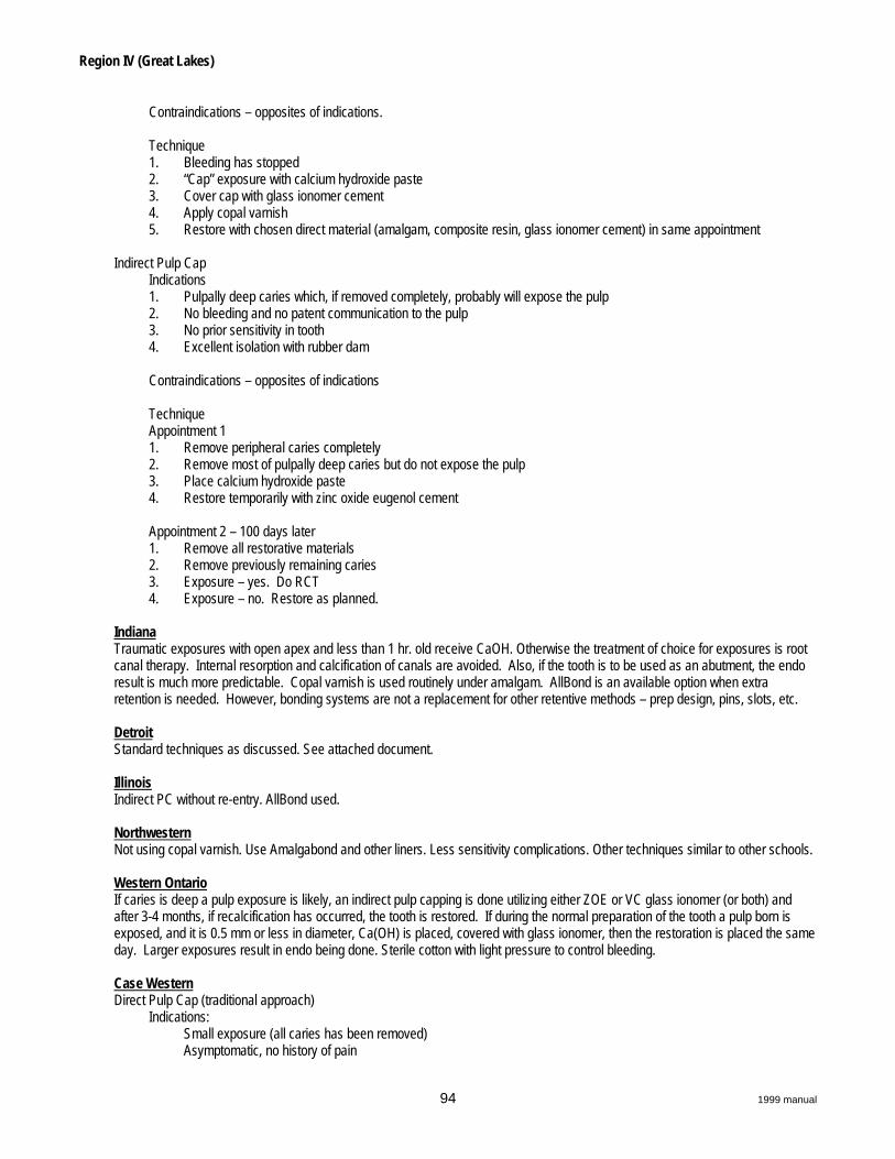
Region IV (Great Lakes)
94 1999 manual
Contraindications – opposites of indications.
Technique1. Bleeding has stopped2. “Cap” exposure with calcium hydroxide paste3. Cover cap with glass ionomer cement4. Apply copal varnish5. Restore with chosen direct material (amalgam, composite resin, glass ionomer cement) in same appointment
Indirect Pulp CapIndications1. Pulpally deep caries which, if removed completely, probably will expose the pulp2. No bleeding and no patent communication to the pulp3. No prior sensitivity in tooth4. Excellent isolation with rubber dam
Contraindications – opposites of indications
TechniqueAppointment 11. Remove peripheral caries completely2. Remove most of pulpally deep caries but do not expose the pulp3. Place calcium hydroxide paste4. Restore temporarily with zinc oxide eugenol cement
Appointment 2 – 100 days later1. Remove all restorative materials2. Remove previously remaining caries3. Exposure – yes. Do RCT4. Exposure – no. Restore as planned.
IndianaTraumatic exposures with open apex and less than 1 hr. old receive CaOH. Otherwise the treatment of choice for exposures is rootcanal therapy. Internal resorption and calcification of canals are avoided. Also, if the tooth is to be used as an abutment, the endoresult is much more predictable. Copal varnish is used routinely under amalgam. AllBond is an available option when extraretention is needed. However, bonding systems are not a replacement for other retentive methods – prep design, pins, slots, etc.
DetroitStandard techniques as discussed. See attached document.
IllinoisIndirect PC without re-entry. AllBond used.
NorthwesternNot using copal varnish. Use Amalgabond and other liners. Less sensitivity complications. Other techniques similar to other schools.
Western OntarioIf caries is deep a pulp exposure is likely, an indirect pulp capping is done utilizing either ZOE or VC glass ionomer (or both) andafter 3-4 months, if recalcification has occurred, the tooth is restored. If during the normal preparation of the tooth a pulp born isexposed, and it is 0.5 mm or less in diameter, Ca(OH) is placed, covered with glass ionomer, then the restoration is placed the sameday. Larger exposures result in endo being done. Sterile cotton with light pressure to control bleeding.
Case WesternDirect Pulp Cap (traditional approach)
Indications:Small exposure (all caries has been removed) Asymptomatic, no history of pain
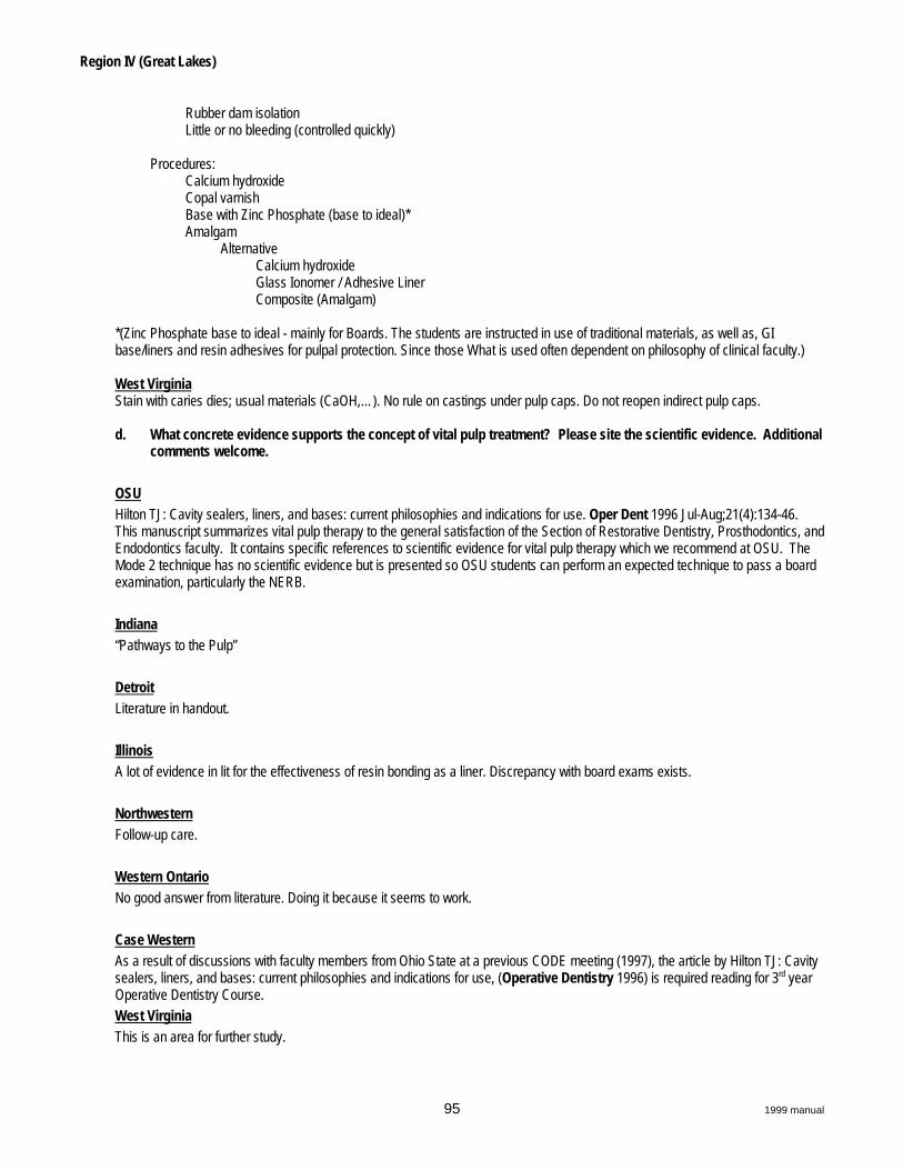
Region IV (Great Lakes)
95 1999 manual
Rubber dam isolationLittle or no bleeding (controlled quickly)
Procedures: Calcium hydroxide Copal varnishBase with Zinc Phosphate (base to ideal)*Amalgam
AlternativeCalcium hydroxideGlass Ionomer / Adhesive LinerComposite (Amalgam)
*(Zinc Phosphate base to ideal - mainly for Boards. The students are instructed in use of traditional materials, as well as, GIbase/liners and resin adhesives for pulpal protection. Since those What is used often dependent on philosophy of clinical faculty.)
West VirginiaStain with caries dies; usual materials (CaOH,…). No rule on castings under pulp caps. Do not reopen indirect pulp caps.
d. What concrete evidence supports the concept of vital pulp treatment? Please site the scientific evidence. Additionalcomments welcome.
OSUHilton TJ: Cavity sealers, liners, and bases: current philosophies and indications for use. Oper Dent 1996 Jul-Aug;21(4):134-46. This manuscript summarizes vital pulp therapy to the general satisfaction of the Section of Restorative Dentistry, Prosthodontics, andEndodontics faculty. It contains specific references to scientific evidence for vital pulp therapy which we recommend at OSU. TheMode 2 technique has no scientific evidence but is presented so OSU students can perform an expected technique to pass a boardexamination, particularly the NERB.
Indiana“Pathways to the Pulp”
DetroitLiterature in handout.
IllinoisA lot of evidence in lit for the effectiveness of resin bonding as a liner. Discrepancy with board exams exists.
NorthwesternFollow-up care.
Western OntarioNo good answer from literature. Doing it because it seems to work.
Case WesternAs a result of discussions with faculty members from Ohio State at a previous CODE meeting (1997), the article by Hilton TJ: Cavitysealers, liners, and bases: current philosophies and indications for use, (Operative Dentistry 1996) is required reading for 3rd yearOperative Dentistry Course. West VirginiaThis is an area for further study.
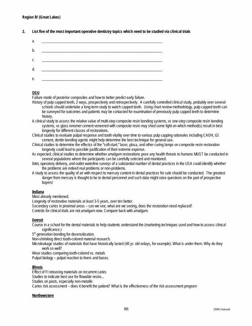
Region IV (Great Lakes)
96 1999 manual
2. List five of the most important operative dentistry topics which need to be studied via clinical trials
a. ________________________________________________________________
b. ________________________________________________________________
c. ________________________________________________________________
d. ________________________________________________________________
e. ________________________________________________________________
OSUFailure mode of posterior composites and how to better predict early failure.History of pulp capped teeth, 2 ways, prospectively and retrospectively. A carefully controlled clinical study, probably over several
schools should undertake a long-term study to watch capped teeth. Using chart review methodology, pulp capped teeth canbe surveyed for outcomes and patients may be contacted for examination of previously pulp capped teeth to determinehistory.
A clinical study to assess the relative value of multi-step composite resin bonding systems, vs one-step composite resin bondingsystems, vs glass ionomer cement veneered with composite resin may shed some light on which method(s) result in bestlongevity for different classes of restorations.
Clinical studies to evaluate pulpal response and tooth vitality over time to various pulp capping rationales including CAOH, GIcement, dentin bonding agents might help determine the best technique for general use.
Clinical studies to determine the effectss of the “soft-start,” laser, plasa, and other curing lamps on composite resin restorationlongevity could lead to possible justification of their extreme expense.
As expected, clinical studies to determine whether amalgam restorations pose any health threats to humans MUST be conducted inseveral populations where the participants can be carefully selected and monitored.
Inlet, operatory delivery, and outlet waterline surveys of a substantial number of dental practices in the USA could identify whetherthe problems are indeed real problems or non-problems.
A study to assess the quality of air with respect to mercury content in dental practices for sale should be conducted. The greatestdanger from mercury is thought to be to dental personnel and such data might raise questions on the part of prospectivebuyers!
IndianaMost already mentioned.Longevity of restorative materials at least 3-5 years, over ten better.Secondary caries in proximal areas – can we see, what are we seeing, does the restoration need replaced?Controls for clinical trials are not amalgam now. Compare back with amalgam.
DetroitCourse in a school for the dental materials to help students understand the (marketing techniques used and how to assess clinical
significance.)5th generation bonding for desensitization.Non-shrinking direct tooth-colored material research.Microleakage studies of materials that have historically lasted (40 yr. old onlays, for example). What is under them. Why do they
work so well?Wear studies comparing tooth-colored vs. metalsPulpal biology – pulpal reaction to liners and bases.
IllinoisEffect of Fl releasing materials on recurrent cariesStudies to indicate best use for flowable resins…Studies on posts, especially non-metallicCaries risk assessment – does it benefit the patient? What is the effectiveness of the risk assessment program
Northwestern

Region IV (Great Lakes)
97 1999 manual
Long-term clinical comparisons of traditional and resin posterion restorations.Bonding agents under amalgam and resins - long term studiesPost and core systems – integrity, fracture of tooth/root. Long term studies to support
Western OntarioEffect on acid etching on dental pulpsToxicity of dental resinsLongevity of Class II composite resin restorationsEffects on dental pulp with the use of laser/plasma arch curingLong term effects of bleaching on enamel plus toxic side effects of H2O2 in this process.Do one-step bonding procedures give as good a bond as separate primer/resin systems.
Case WesternMost topics well covered by others here.Posterior composite recurrent caries.Sealants long term effects.
West VirginiaWhen to place the initial restoration – caries diagnosis. Better evidence based detectionMeans to strengthen teeth. Long-term clinical trials.Look for tooth-colored material that does not have polymerization shrinkageOther modalities for caries detection – induction,..Early remineralization systems.Recall frequency – evidence based using caries risk assessment.
3. Calibration of faculty
a. What is the protocol for calibrating and standardizing your operative faculty? Address current faculty and newfaculty. Also for pre-clinical and clinical.
OSUNew faculty are usually placed in the preclinical courses to familiarize them with the procedures which are taught at OSU. This doestake several years to accomplish. The course directors are responsible for the course content at the discretion of the section chair. The course directors calibrate their faculty as to preparation design, restoration quality, etc. Teaching in the clinic is expected tofollow what is presented in preclinical settings. On entry to the clinic, experienced faculty mentor new faculty with respect to policy,paperwork, evaluation, and other aspects of clinical teaching. There is no formal calibration here. Same forms used pre-clinicallyand clinically.
IndianaAll new and bench instructors attend lectures and have pre-lab meetings to cover criteria for the lab session. Grading done by asmall number (3) of faculty. Biggest problem is with part-time clinical faculty. Pre-clinically, students self-evaluate, but that doesn’tcarry over to the clinic where there are a number of 1 day/week faculty. We are beginning a formal faculty calibration program thatwill include all full-time and part-time faculty.
DetroitPre-clinical course directors calibrate faculty. Clinically there are faculty in-services in am. Little clinical grading at satellite clinic atDetroit Receiving hospital which accounts for almost 50% of clinical training.
IllinoisSimilar to W. Va. Full scale documentation of methods. Phase one of calibration. Standardized philosophy by procedure to establish continuity from pre-clinic to clinc. Finished last
December Phase 2 to establish consistency in grading. One procedure per month. Done by the pre-clinic course directors. 3-5 cases randomly
selected and graded without no communication. Then discussion and numerical values assigned. Goal to have one standardof care and evaluation criteria for pre-clinic and clinic.
Northwestern

Region IV (Great Lakes)
98 1999 manual
No formal program. Closure does not give impetus to do at this time. No separate operative faculty now.
Western OntarioAll faculty meet at beginning of fall term and are briefed on changes to curriculum, grading system is reviewed and protocols. Newfaculty must serve in the preclinical laboratories to become familiar with the current programs before teaching at the clinical level. Allpreclinical grading is done as a group with interaction between instructors in determining grades.
Case WesternNo formal calibration program. Preclinical courses:
The course directors for two courses do all of their own grading so that evaluations are consistent. While other courses have several faculty grading the same criteria for all projects.For two courses: faculty evaluate (grade) the one set of criteria for projects for the entire class, and also be responsible for
evaluating the same criteria for competency examinations. For these courses, new faculty work with course director to review criteria during evaluation sessions.
Clinic Evaluations:No formal calibration program for day to day evaluations.Previous director of clinical faculty had monthly in-service training sessions. Presently searching for new director.
Prior to Mock Boards, and Scheduled Mid-year Competency Exams, the Restorative Department meets to review the criteria,grading scale etc.
Evaluation forms for these exams based on NERB criteria and forms.
West VirginiaCalibration varies – USPHS based criteria used by all faculty. Same forms used pre-clinically and clinically. Biggest problem is whensending students on externships – reinforcing criteria is difficult. Have to go through the three operative pre-clinical courses beforegoing onto the clinic floor.
b. How often is calibration/recalibration done?
OSUCalibration is done as necessary to standardize course faculty appropriately. When new procedures are introduced, it is invaluable. If evaluation of projects becomes inconsistent, recalibration is done at the course directors’ discretion.
IndianaAt the preclinical level, calibration is accomplished prior to each new laboratory project or practical exam. This includes lectures,models, and meetings prior to student contract. We are working on a clincial calibration program at this time.
DetroitDifficult task. Turnover of clinical faculty means it needs to be done frequently.
IllinoisRequired to go through exercises monthly.
NorthwesternMock board every year to standardize or agree on how to look at the preparations/restorations
Western OntarioOverall once a year but usually 3-4 times at the preclinical level (every time there is a practical examination).
Case WesternDepartmental meetings a few times a year.
Re: Clinical EvaluationsThe Restorative Department meets several weeks prior to Mock Boards, and Scheduled Mid-year Competency Examsto review criteria and evaluation sheets. Also a quick review is held the morning of the Exams.
West Virginia
Region IV (Great Lakes)
99 1999 manual
Students grade along with faculty. Continuous re-evaluation of criteria between the two. If large discrepancy exists, then the studentgrade is lowered (promotes effective evaluation).
c. What evidence supports your protocol for calibration and recalibration? (Please site)
OSUWe do not have published data to support our calibration methods. We use the students’ performance on NERB and nationalboards to assess, in part, how our teaching program is working. We have a mock board in which faculty from other dental schoolsact as examiners. Their feedback is quite important.
IndianaA long list of cites are available from the school upon request.
Detroit
IllinoisTry to be within 1/2 grade in the exercises. Also mock board and board exams
Northwestern
Western OntarioAll clinical instructors evaluate with a full time faculty member for a period of time then do it on their own. During practical clinicalexams two (2) instructors grade - usually there is no more than a spread of 5% in the grades given.
Case Western
West Virginia
4. Competency education
a. How is competency based operative evaluation determined?
OSUCompetency exams at the end of the second, third and fourth year over material taught pre-clinically in the first two years, andNERB based for the fourth year. The third year is a typodont amalgam/rubber dam and FPD provisional competency exam. The D2competency is 14 procedures (8 preparations, 6 restorations or provisionals) and is graded by selected faculty. The D3 competencyis graded by 4 faculty and the D4 (mock NERB) competency exam is graded by many faculty in a fashion similar to the way theNERB exam is graded.
IndianaPracticals (competencies) pre-clinically. Clinical operative competencies required. Graded by instructor and clinic director. Must bepassed to clear for graduation. Mock boards required for graduation
DetroitPre-clinical competencies. Standardized exams in simulation lab being started this year.
IllinoisSimilar to the other schools. Performance exams are one means used to measure competency. Senior clinic exam required forgraduation. Includes ‘scripted’ patients. They must pass every station in the exam. If not they go to each department of failedstations to remediate.
Northwestern

Region IV (Great Lakes)
100 1999 manual
Daily graded procedures pre-clinically. Weight is on practical – graded double-blind. Reference scale distribution of class grades(mean is between B and C). Teaching assistants work with weaker students. Clinically, there is a two-year requirement. Mock boardspatterned after NERB and CREDITS. Must be passed for graduation. Informal remediation for failures (on typodont). Writtencompetency exams in the senior year. Required to pass for graduation. Looks to minimal competency to pass.
Western OntarioStudents are given practicals on manikins in the preclinical lab. In the final clinical year all have to pass two (2) practicals, oneamalgam, one composite (Pass = 60% by 2 graders). Students are given only two chances to pass.
Case WesternPreclinical
Several competency examinations throughout the two semester course.For students entering the 3rd year, prior to treating patients in the clinic, a one week series of operative and crown andbridge procedures must be successfully completed. Evaluation is Pass/Fail by clinical preceptor.
Clinical Competencies3rd and 4th year students must Pass (NERB criteria) several typodont and patient competencies each year. Evaluation
of 3rd year competencies by two instructors: their own preceptor and an instructor from Restorative Dept. Theseprocedures include: Class II amalgam restorations, Class II composite restorations, Class III compositerestorations.
These are timed examinations.
Miid-year Competency Exams4th year students: One day exam during finals week (or week prior to finals week) which includes A.M.: typodont Class IIAmalgam and Class III Composite (extracted tooth in typodont)P.M.: 3 unit bridge preparation and provisional3rd year students: Half day examA.M.: typodont Class II Amalgam and Class III Composite (extracted tooth in typodont)
West VirginiaCriteria based competency program. Work of students reviewed prior to externship assignment. Also, current and overall gradescompared to check that students are improving. Procedures complete versus hours in clinic identify the inefficient student.Competency prior to entry into clinic – done on mannikins with natural teeth in some areas.
b. How is the weak student identified?
OSUWeak students are initially identified during preclinical courses by report of individual faculty members to the course directors.Course directors then must determine to magnitude of the problem and can take steps to reduce this problem. Weak students alsotend to receive failing grades in preclinical courses. Prior to entry into the clinic, as described above, the second year students mustdemonstrate competency with a survey of restorative procedures, previously taught to them. Failing a specific number of theseprocedures prevents clinic certification. Weak students who do achieve clinic privileges are identified by clinic director based onobjective feedback from clinical instructors.IndianaWeak students are identified as soon as possible by early evaluation through pre-clinical practical exams and clinical competencies. In the clinics, clinical directors must constantly follow the progress of their assigned students by the monitoring the students clinicalachievement, competencies and mock boards.
DetroitThrough the competency exams and clinic performance.
IllinoisFaculty judgements and performance exam work. Also meet with clinic patient assignment director and clinic director. Also, clinicalexam as described above. Also a promotional committee works with instructor evaluation to determine student progress to the nextyear. Mock board also given.

Region IV (Great Lakes)
101 1999 manual
NorthwesternTeams of students evaluated from day to day contact with team leaders.
Western OntarioUsually through his/her daily performance. Every procedure is marked and all marks are reviewed by the Division Chairman.Instructors also indicate potential weak students to the chair.
Case WesternCompetency exam results and clinical evaluations by preceptors.
West VirginiaWork of students reviewed prior to externship assignment. Also, current and overall grades compared to check that students areimproving. Procedures complete versus hours in clinic identify the inefficient student
c. What measures are taken to upgrade competency of the weak student?
OSUIn individual courses, weak students often receive extra time from course directors ranging from individual to group efforts, designedat improving these students. Students who fail courses are invited to a required remediation program in the summer quarter afterboth first and second years. These programs begin generalized in scope and end as customized programs. Several students whohave achieved clinical competency status but have been identified as less than competent have been actively remediated duringtheir third and fourth years. Such students have had their clinic privileges temporarily suspended and/or been assigned a mentor toassist in their improvement. These efforts are extremely manpower consumptive.
IndianaRemediation can be difficult in our curriculum if the weak student is not identified early in the course. Once a weak student isidentified, intervention is is accomplished “on the run”, i.e. while the course and curriculum continues for the student. Since the nextschool year begins within two weeks of the previous year’s completion, we do not have a summer session to make a “formal”remediation effort. Each remediation is individual and on a case by case basis. This is critical since a failed courses requires therepeat of the year. Clinical remediation is accomplished through the student’s clinic director and the promotion committee.
DetroitVariety of means used. Simulation lab open in the evenings for students. Some personal remediation done.
IllinoisNorthwesternTeaching assistants work with weaker students. Remedial programs for written and practical mock boards.
Western OntarioOne-on-one supervision with demonstrations by supervisors. Extra work in the simulation clinic. Clinically, some students onlyschedule patients under a single faculty to evaluate their performance over time.
Case WesternThe Remediation Program varies, it depends on the needs of the student. Generally a one-to-one supervision. Depending onseverity, students may be asked to not treat patients, until they have improved performance to acceptable level, through intensiveseries of projects and evaluated typodont procedures.
West VirginiaWeak students are assigned restorative advisors. In some cases, the students are restricted to working under only special facultywho can discuss the student’s performance.
Region IV (Great Lakes)
102 1999 manual
5. Computer based interactive education
Regional VI (South) is developing two comprehensive restorative treatment planning cases to be used as problem based learningexercises. It is hoped these cases can be used to develop authoring software that could be used for educational purposes. A jointventure with Dental Interactive Simulation Corporation (DISC) warrants consideration.
a. What interactive computer-based teaching tools are you using to teach operative dentistry?
OSUAnatomy and Histology have courses with interactive computer content. In Restorative Dentistry, Dr. Rosenstiel has developed aninteractive Authorware program that evaluates single tooth restoration using three scenarios (occlusal surface involvement, proximalinvolvement and fractured tooth). The program is an evidenced-based discussion of the different restorative options
IndianaAll entering students have computers. Heavily involved in web-based education. Tooth morphology (Dr. Ed DeSchepper) is allcomputer; no lecture. Drs. Willis and Reifeis have a single tooth restoration course (made with Astound at http://www.astound.com).Used as an adjunct to the lecture course material.
DetroitHope to have Dental Anatomy on web or CD. Nothing interactive in restorative at this time.
IllinoisCariology is on a computer program (used to be in perio. area). Plans to expand on that course and add other courses. Wildfiresimulations is available on CD but has not been evaluated.
NorthwesternNothing at this time for instruction
Western OntarioNone at present although at least two (2) disciplines (Perio and Gross Anatomy) have computer programs and web sites.
Case WesternNothing interactive at this time. School has university commitment and is being wired for fiber-optics and the dental school is startingto put courses online (Med school currently has all courses online).
West VirginiaLooked at some work done at U. Mich. Some done by Oral Pathology
b. Who developed them?
OSUSee above
IndianaSee above. Gross Anatomy, Dental materials also have presentations on the web
Detroit
Region IV (Great Lakes)
103 1999 manual
Illinois
Northwestern
Western OntarioThe two disciplines involved (in-house)
Case Western
West VirginiaOral Pathology; Hygiene
c. Would your school/region participate in development and in what manner?
OSUYes, given faculty time
IndianaYes, in any way possible.
Detroit
Illinois
Northwestern
Western OntarioYes, if we had more faculty, money and time.
Case Western
West Virginia
6. Posterior esthetic restorations – report on direct and indirect
a. Are they included in the curriculum? What discipline?
OSUPosterior esthetic restorations are taught in the preclinical courses. Direct composite resin restorations are taught in the first year, andinclude Class I and Class II restorations. In the second year, ceramic inlay and onlay restorations as well as composite resin inlay andonlay restorations are taught. An elective course in fiber reinforced composite resin restorations is offered to 4th and 3rd year students.These are operative dentistry or restorative dentistry courses. There are no competencies that evaluate indirect tooth-coloredrestorations.
Indiana
Region IV (Great Lakes)
104 1999 manual
Posterior esthetic direct restorations are taught in the pre-clinical Single Tooth Direct Restorations in first year. Laboratory projects arecompleted on extracted and mounted teeth. Emphasis is on small occlusals in molars and small Class II in premolars. Indirect estheticrestorations are introduced in the Single Tooth Indirect Restorations course in first year. An indirect composite resin laboratory projectis completed on an artificial tooth. All phases are included in the second year Dental Sciences II. All posterior esthetic restorationteaching is the responsibility of the Restorative Dentistry department.
DetroitAnterior resin course teaches posterior composites, also.
IllinoisDirect and indirect taught in restoartive 2nd and 4th year. Use natural teeth in typodont and also resin typodont teeth. Fixed pros teachesthe indirect techniques (inlay/onlay and veneers)
NorthwesternOperative pre-clinical taught direct resins using the resin teeth form Kilgore. Some done in the clinic.
Western OntarioYes, operative dentistry and fixed prosthodontics
Case WesternSimilar to others. Direct taught pre-clinical and 3rd year esthetics course. Indirect used to be lab exercise in 3rd year. (Will be again).
West VirginiaIntroduced early in the tooth colored restorative course (direct and indirect). No competencies evaluate these. Esthetics need to beprimary reason for replacement. Rubber dam required. Need enamel walls.
b. Where (pre-clinical and clinical)
OSUThey are taught preclinically and permitted or even encouraged clinically. They are routinely done in the clinic as planned.
IndianaSee above. Clinical experiences are varied and depend upon treatment planning options. However, when indicated, direct and indirectesthetic restorations are generally encouraged.
DetroitPRR’s done, but very few interproximal composites done in the clinic.
IllinoisClinically, it is up to the instructor to ‘promote.’ Pre-clinical 2nd year course.
NorthwesternSee above. Small lesions (molars and premolars) under rubber dam
Western Ontario2nd year preclinical and both clinical years. Operative dentistry and fixed teach (operative teaches the pre-clinical course)
Case WesternClinic has typodont competency for direct, but no requirements. Done with conservative preps.
West VirginiaBoth pre-clinic and clinic
c. Required experiences?
OSU

Region IV (Great Lakes)
105 1999 manual
There are no individual requirements. However, any student whose treatment plan includes them can place them. Our clinic is supposedto be comprehensive in scope so patients requiring these and who should receive these as indicated, will have them done by theirassigned students. However, it is possible for students to graduate without doing these restorations in patients.
IndianaNo clinical requirements. However, every student has some experiences.
DetroitNone
IllinoisNoneNorthwesternNone
Western OntarioNo, we cannot provide enough suitable patients who will pay for indirect esthetic restorations. There is a single crown requirement infixed but not in operative.
Case Western
West VirginiaNot examined with competencies – no requirements clinically, only pre-clinically.
d. What materials and/or systems utilized?
OSUKerr Prodigy and Optibond Solo are the materials used at OSU. Porcelain inlays and onlays and composite resin inlays and onlays arecemented with Kerr Nexus.
IndianaDirect - Herculite, OptiBond Indirect - All materials and techniques that are available through outside laboratories.
DetroitTPH and Prodigy with OptiBond Solo
IllinoisPretty much use ‘everything.’ (All types of composites). Empress, Concept, InCeram, ProCera. (Concept machine in house for studentsto use).
NorthwesternUses many systems.
Western OntarioConcept, Targis, Empress, various other porcelain systems.
Case WesternTPH and OptiBond Solo
West VirginiaJeneric Pentron, hybrid composite materials but not compomers.
e. What are the results? Also site the evidence for the reported results.
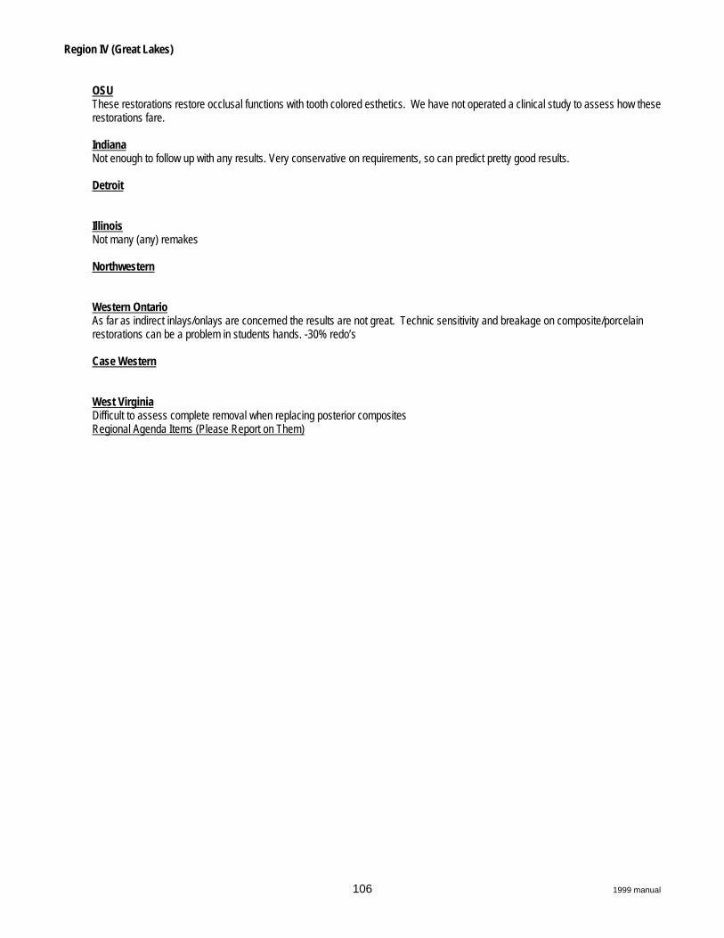
Region IV (Great Lakes)
106 1999 manual
OSUThese restorations restore occlusal functions with tooth colored esthetics. We have not operated a clinical study to assess how theserestorations fare.
IndianaNot enough to follow up with any results. Very conservative on requirements, so can predict pretty good results.
Detroit
IllinoisNot many (any) remakes
Northwestern
Western OntarioAs far as indirect inlays/onlays are concerned the results are not great. Technic sensitivity and breakage on composite/porcelainrestorations can be a problem in students hands. -30% redo’s
Case Western
West VirginiaDifficult to assess complete removal when replacing posterior compositesRegional Agenda Items (Please Report on Them)

Region IV (Great Lakes)
107 1999 manual
CODE Questions
1. What is CODE doing well?
OSUCODE brings together operative dentistry faculty who want to improve their respective teaching programs. CODE allows opendiscussions on a variety of subjects as well as the national agenda. CODE allows for a free exchange of ideas to help improve respectiveprograms.
IndianaEvery time attending Academy of Op. Dent and CODE, there are a number of very useful ‘tidbits’ that can be taken away
DetroitGood opportunity to work on projects together. Much of the subject matter is universal and CODE could be a good way to get togetherand work on some items.
IllinoisAllows us to know what the other schools are doing. Good way to find out where we stand.
NorthwesternBeen great, especially now that many schools have “restorative dentistry” depts., this allows a smaller focus
Western OntarioI believe CODE is doing very well in providing a forum for discussion on what all schools are teaching, why and how each does it.
Case WesternAll been said – CODE regional meetings have been very helpful.
West VirginiaGood opportunity to get together.
2. Where do you desire improvement?
OSUImprovements in CODE would include the development of a web presence with easier access and higher visibility. Since CODE is underthe umbrella of the AADS, this would be the route of such management and the current people who are managing CODE should be givenresources by AADS to allow for this. Better information dissemination through electronic means would also imporve the CODE program.
IndianaDeans need to be convinced of the usefulness of the group.
Detroit
IllinoisReports don’t come to the attendees.
Northwestern
Western Ontario
Case Western
West Virginia

Region IV (Great Lakes)
108 1999 manual
Would be nice to know a little more about CODE before the history is lost. Would be good to have support of the deans in the deansconferences.Firm up schedule so the report is in a timely manner (before the Feb. Operative meeting).
3. Is $24 US/$30 Canadian school dues adequate? Comments are expected.
OSUThe amount of $24 is virtually nothing and any institution should have no problem affording it. A 4-fold increase ($100) would still beworthwhile if improvements to CODE can be realized. Possibly get a sponsor for the schools hosting the regional meetings to cover costs.
IndianaYes. Some increase in dues will be tolerated as needed to maintain the organization.
Detroit
Illinois
Northwestern
Western OntarioI believe so – although I have no idea what the expenses are. Dues should cover expenses. Overall fee is low for those coming to themeeting.
Case Western
West VirginiaVery fair fee. (Been same since ’86?).
4. How can participation by faculty in CODE regional meetings be encouraged/improved?
OSUParticipation in CODE must begin with the member schools providing faculty time and funding. The regional format of the CODE programdoes allow for driving to most meetings in our region. Without such encouragement, CODE will not get better, and, in fact, may becomeless desirable over time
IndianaNothing to add to what others have said.
DetroitAgree with what has been said.
IllinoisBenefit should be realized in terms of improvement in level of operative dentistry education. As a group, we can make recommendationsto schools through the meetings.
Northwestern

Region IV (Great Lakes)
109 1999 manual
Regional meetings really nice. Effort to get ALL schools to attend. Have other regions send representatives to alternate regions meetings.
Western OntarioI think participation in the Great Lakes Region is good. Several schools do not attend but I think with some more effort and remindersthey might be persuaded.
Case WesternInclude information in web format to share with other schools.
West VirginiaOpportunity to develop pride in Operative Dentistry through CODE. With loss of curriculum time, this allows schools to keep in touch withwhat is being taught and bring back to their school. Operative is becoming diluted. CODE should be a more important player in theAcademy of Operative Dentistry.
5. Please indicate the office/position with complete mailing address at your school to which regional reports, dues statements,roster requests, etc. are to be mailed. NOTE: Regional Director - this information from each school is to be transmitted totheNational Director.
OSUDr. Stephen F. Rosenstiel, ChairSection of Restorative Dentistry, Prosthodontics andEndodontics305 West 12th Ave.Columbus, OH 43210-1241(614) [email protected]
IllinoisDr Stephen D. CampbellDepartment of Restorative DentistryThe University of Illinois at Chicago801 South Paulina StreetChicago, IL 60612(312) [email protected]
IndianaDr. Steve DukeChair, Restorative DentistryIndiana UniversitySchool of Dentistry1121 West Michigan St.Indianapolis, IN 46202-5186
NorthwesternDr. James B. Ricker, DirectorDivisiion of Operative DentistryDepartment of Restorative DentistryNorthwestern University Dental School240 East Huron StreetChicago, IL 60611
DetroitDr. Frank E. PinkUniversity of Detroit Mercy8200 West Outer Drive, Box 9Detroit, MI 48219-0900(313) [email protected]
Western OntarioDr. William A. GrayDivision of Operative Dentistry & EndodonticsSchool of DentistryFaculty of Medicine and DentistryUniversity of Western OntarioLondon, Ontario N6A 5C1 Canada
Case WesternDr. Louis CastellarinChair, Restorative DentistryCase Western Reserve University10900 Euclid Ave.Cleveland, OH 44106-4905(216) 368-6736
West VirginiaDr. Ron HouseDirector Restorative DentistryBox 9460Morgantown, WV [email protected]

Region IV (Great Lakes)
110 1999 manual
Regional Questions (as submitted at the Region IV 1998 C.O.D.E.)
1. What constitutes a basic operative curriculum:
a. What do we want our students to know when they graduate?
OSUAs far as operative dentistry is concerned, we want our students to know the indications and contraindications for dental amalgam,including pin retention, composite resin (direct and indirect,) glass ionomer cement, single tooth cast metal restoratives, dentin andenamel bonding, and vital pulp therapy. We want the students to recognize caries and other defects (including esthetic defects) andproperly develop treatment plans to address them. We want the students to be proficient in the restoration of all restorable vital teethand some endodontically treated teeth with these materials. We also want the students to recognize direct gold restorations from ahistorical aspect.
IndianaLike everyone else – morphology, cariology (somewhat), single tooth restorations diagnosis and treatment planning, life-long learning
DetroitEcho what others have said. Partial tooth restoration of any kind should be in bailiwick of operative dentistry. Basic knowledge and skillsof laboratory techniques is appropriate in operative dentistry along with knowledge of occlusion.
IllinoisPrevention, diagnosis and removal of caries – need to understand cariology. Direct restorative procedures and the science of dentalmaterials.
NorthwesternMajor goals:How to do a thorough diagnosis and treatment planManage pts.Deliver sound operative dentistryInteract with dental labs and assess work returnedEvaluate literature and include in practice
Western OntarioAmalgam, direct and indirect composite resins, direct and indirect veneers, indirect and direct pulp capping, tooth bleaching.
Case WesternAll has been said by others here.West VirginiaPrevention, treatment planning and restorations (largely single tooth). Ability to make an ‘educated guess’ in a world of non-evidencebased research..
b. What should be included in the discipline?
OSUIncluded in the clinical discipline should be the integration of basic science which makes the foundation of what we clinically provide.Also, any evidence for what we teach should be provided as well
Indiana

Region IV (Great Lakes)
111 1999 manual
Tooth Morphology, Occlusion, Direct and Indirect Single Tooth Restorations, Dental Materials, Cariology, Preventive Dentistry, BasisScience integration as needed.
Detroit
IllinoisAnything that helps develop competencies in defined operative fields. Any single tooth restoration, cariology and occlusion.
Northwestern
Western OntarioAll single tooth, none-cast, restorations
Case Western
West VirginiaShould include cariology.
c. Should we develop a "Standard of Care" document?
OSUA “Standard of Care” document is a good thing in principle. To make it consistent across all dental schools will necessarily make it verygeneral. Individual schools can attach specifics to such a document. Rubber dam techniques should be included in the standards of care.
IndianaProbably not. This perhaps means a ‘legal’ standard of care. This may limit some specific statements due to the liability of practitioners.
Detroit
IllinoisStandards should be developed for certain proceduresNorthwestern
Western OntarioI am not sure if this would not be just more “paperwork”. At present we have one “Standard of Care” – the standard we would acceptin our own mouths!!
Case Western
West VirginiaNot easily done, but could be done. Should be a long-term goal. If we don’t develop one, who will? (Third party carriers?)
2. How are dental materials courses handled in the curriculum?
Region IV (Great Lakes)
112 1999 manual
OSUAt OSU, we have made a conscious effort to integrate dental materials into the individual preclinical courses. Additionally, the third andfourth year students have didactic courses which discuss dental materials including problem solving as related to the materials. Thetraditional dental materials courses have virtually been eliminated from our teaching program.
IndianaIntegrated. Basic dental materials lectures in first year. When teaching direct and indirect restorations, materials people guest lectureon the appropriate topics. Third year operative lectures revisit materials with more detail on clinical handling and materials updates.
DetroitHad materials scientist who retired. Then individual courses assumed the role of teaching materials. Now have a materials person whoteaches materials in a lecture setting.
IllinoisBasic freshman course. Also incorporated into each operative course as appropriate.
NorthwesternHad a division of biologic materials in a larger department. Handled lecture/lab course for materials. Pre-clinical courses reviewedmaterials. Lab dropped a number of years ago, impacting the pre-clinical courses (the students were less prepared coming in).Now freshman intro lecture, individual courses and then a junior theory course.
Western OntarioIn the past as a separate discipline with individual disciplines reviewing and emphasizing the clinical application. Hoping to get abiomaterials engineer to coordinate courses on materials, again. Currently, materials are handled by each course as appropriate.
Case WesternHad a first-year materials course, discontinued when PhD left. Now the materials are in separate courses. Some impact with facultychanges; hard to determine who teaches what.
West VirginiaOne of the 5 restorative divisions is Dental Materials (chaired by PhD, DDS). Use Anusavich Dental Materials text. First year pre-clinicallab course associated. Good introduction – allows later courses to be clinically oriented.
3. How are instrument-supplying companies selected for the first-year students?
OSUAt OSU, we have a person in charge of procuring instruments for preclinical use. Once the preclinical instrument list is determined, thisperson contacts prospective suppliers and negotiates prices. Often if alternative instruments or supplies are located at better prices,the course directors are contacted and discussed. If appropriate, changes are made to the issue. The preclinical instruments aredetermined for each course by course directors. Instruments used in multiple courses (e.g. explorers, etc) are generally accepted withoutjustification. Unique instruments to a particular course require some justification, depending on how esoteric they are. Substitution ispossible if one instrument is removed for each one substituted. Disposable supplies are usually permitted if the cost is justified.
IndianaHad rented laboratory issue until a few years ago. Now is total purchase. Clinic has rental issue from central sterilization. Go by service,quality and cost. Companies who were deemed good in service and quality had bids requested. Went with Suter.
DetroitPre-clinical course directors define the instruments. Instrument supply companies chosen partly because of ability to assist in financing.
Region IV (Great Lakes)
113 1999 manual
IllinoisInstruments recommendations made by course directors to the pre-doctoral director. Schein is set up in the school to supply materials.Not working out too well.
NorthwesternInstrument/materials/equipment committee. Full time clinic manager tracks inventory and orders. First and second year students getexpendable and transfer issue. Transfer kits are used year-year. In clinics use cassettes with new instruments that are just a few yearsold now. All sections represented on the instrument committee.
Western OntarioThey are selected by Division Chairmen after reviewing the quality of the company product. We attempt to agree on one supplier wherepossible (more committee work!) One person is in charge of ordering.
Case WesternKit committee (dept. chairs with student representation) and separate materials committee (separate). Changes need rationale. Studentsbuy everything.
West VirginiaInstrument and materials committee. Pre-clinical kits purchased and used throughout the 4-year curriculum. Cassette manager for eachof the seven cassettes. Meet quarterly to discuss any changes. Costs challenged on everything. Dental store individual is on thecommittee as non-voting member. Chair of committee is not affiliated with the dental store.
4. How is operative configured in the AADS and how should it be?
This question was not addressed due to time constraints.
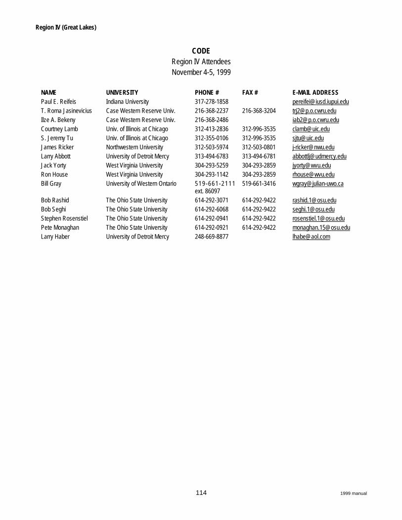
Region IV (Great Lakes)
114 1999 manual
CODERegion IV AttendeesNovember 4-5, 1999
NAME UNIVERSITY PHONE # FAX # E-MAIL ADDRESSPaul E. Reifeis Indiana University 317-278-1858 [email protected] T. Roma Jasinevicius Case Western Reserve Univ. 216-368-2237 216-368-3204 [email protected] Ilze A. Bekeny Case Western Reserve Univ. 216-368-2486 [email protected] Courtney Lamb Univ. of Illinois at Chicago 312-413-2836 312-996-3535 [email protected] S. Jeremy Tu Univ. of Illinois at Chicago 312-355-0106 312-996-3535 [email protected] James Ricker Northwestern University 312-503-5974 312-503-0801 [email protected] Abbott University of Detroit Mercy 313-494-6783 313-494-6781 [email protected] Yorty West Virginia University 304-293-5259 304-293-2859 [email protected] House West Virginia University 304-293-1142 304-293-2859 [email protected] Gray University of Western Ontario 519-661-2111
ext. 86097519-661-3416 [email protected]
Bob Rashid The Ohio State University 614-292-3071 614-292-9422 [email protected] Seghi The Ohio State University 614-292-6068 614-292-9422 [email protected] Rosenstiel The Ohio State University 614-292-0941 614-292-9422 [email protected] Monaghan The Ohio State University 614-292-0921 614-292-9422 [email protected] Haber University of Detroit Mercy 248-669-8877 [email protected]
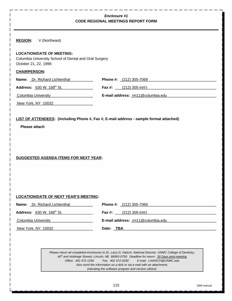
115 1999 manual
Enclosure #1CODE REGIONAL MEETINGS REPORT FORM
REGION: V (Northeast)
LOCATION/DATE OF MEETING:Columbia University School of Dental and Oral SurgeryOctober 21, 22, 1999
CHAIRPERSON:
Name: Dr. Richard Lichtenthal Phone #: (212) 305-7069
Address: 630 W. 168th St. Fax #: (212) 305-8493
Columbia University E-mail address: [email protected]
New York, NY 10032
LIST OF ATTENDEES: (including Phone #, Fax #, E-mail address - sample format attached)
Please attach
SUGGESTED AGENDA ITEMS FOR NEXT YEAR:
LOCATION/DATE OF NEXT YEAR’S MEETING:
Name: Dr. Richard Lichtenthal Phone #: (212) 305-7069
Address: 630 W. 168th St. Fax #: (212) 305-8493
Columbia University E-mail address: [email protected]
New York, NY 10032 Date: TBA
Please return all completed enclosures to Dr. Larry D. Haisch, National Director, UNMC College of Dentistry;40th and Holdrege Streets; Lincoln, NE 68583-0750. Deadline for return: 30 Days post-meeting
Office: 402 472-1290 Fax: 402 472-5290 E-mail: [email protected] send the information on a disk or via e-mail with an attachment,
indicating the software program and version utilized.

Region V (Northeast)
116 1999 manual
Code Meeting - Region V (Northeast) – October 21-22,1999
The Region V meeting was hosted by Columbia University School of Dental and Oral Surgery, Dr. Richard Lichtenthal, Chair, on October21-22,1999.
Schools in Attendance
Columbia University School of Dental and Oral SurgeryMcGill University Faculty of DentistryUniversity of ConnecticutHoward UniversityTufts University School of Dental MedicineBoston University, Goldman School of Dental MedicineUniversity of Maryland Baltimore College of DentistryUMDNJ New Jersey Dental SchoolNew York University College of DentistrySUNY at Stony Brook School of Dental MedicineUniversity pf Pennsylvania School of Dental MedicineTemple University School of Dentistry
Agenda – National CODE Topics
Vital pulp treatment
When, where, and who teaches this topic in your curriculum?
Most of the schools taught this topic in the first two years of their curriculums. This topic is also taught by the Departments of Endodonticsat New York University, Boston University, University of Connecticut, Tufts University, and Temple University.
Is this treatment utilized in patient care by students?
Boston University and New York University refer all cases of vital exposures to their respective Department of Endodontics. TheRestorative or Operative Departments of all of the other schools provide patient.care.
What criteria/protocol are followed in deciding to do or not do vital pulp treatment?
In general, pulp exposures on permanent fully developed teeth are treated with pulpectomy and root canal obturation. Pulpcapping/pulpotomy is used in teeth with incomplete root formation and for socio-economic reasons. Additional determinants includesymtomology, location of the tooth, vitality of the tooth, size of the exposure, radiographic depth, and the number of times the tooth hasbeen restored. All of the schools use a rubber dam and most schools use calcium hydroxide and glass ionomer to treat vital exposures.
What concrete evidence supports the concept of vital pulp treatment? Please site the scientific evidence. Additional comments welcome.
Just about all of the schools present felt that it is difficult to extrapolate evidence-based information. The University of Connecticut, afterconsultation with their Department of Endodontics, reported that “Successful treatment outcome for properly performed pulpectomy withroot canal filling is cited in most publications to exceed 90%. Pulp capping/pulpotomy are normally found to have a successful outcome<75%.
List the most important operative dentistry topics which need to be studied via clinical trials.

Region V (Northeast)
117 1999 manual
5. Question of caries/risk assessments – what is caries and when to treat?6. Biocompatibility of dental materials to pulp, osseous and gingival tissues.7. Restoration of cervical lesions.8. Efficacy of fluoride release and significance to caries.9. Longevity of “tooth colored” restorative materials placed in less than ideal situations.10. Sealing or not sealing/arresting caries.11. Evidence of the use of lasers in cutting hard tissues.12. Comparison of the physical properties of “packable” composites to traditional composites in terms of depth of cure.13. Leakage and sensitivity in posterior composite restorations14. Criteria for the placement or non-placement of restorations.
Calibration of faculty
What is the protocol for calibrating and standardizing your operative faculty? Address current faculty and new faculty. Also for pre-clinicaland clinical.
Most schools utilize manual guides (especially in the pre-clinic) for all faculty especially new faculty. A number of schools hold in-serviceand internal calibration workshops a number of times a year on typodonts for their faculty. Several schools use two or more faculty asgraders in the pre-clinic area and use the Course Director as a “tie breaker”. Additionally, three schools place new faculty in the pre-cliniccourse first, before allowing them to teach in the clinical areas. Most schools use a mentoring system or “shadowing” system in the clinicand use more than one faculty to grade competency examinations. Several schools reported that it is possible to calibrate faculty whenusing typodonts, but difficulties arise when students and patients are thrown into the mix. The schools also reported that it is easier todifferentiate pass and fail procedures; calibration is much more difficult to discern when plus or minus grades are used.
How often is calibration/recalibration done?
Most schools use a random or minimal approach. One or two schools calibrate at scheduled intervals.
What evidence supports your protocol for calibration and recalibration? (please site)
Scant evidence exists regarding calibration and recalibration. A number of schools have difficulties in this area due to the number of parttime faculty in their departments. However, those institutions that maintain a cadre of the same pre-clinical faculty may have an easier time,than clinical faculty where patient interaction plays a role.
Competency education
How is competency-based operative evaluation determined?
Most schools have a minimum number of requirements, but use competency examinations, mock boards, remediation of failures, and dailystudy performance to determine student competency.
How is the weak student identified?
Schools use competency-based examinations, practical examinations, grade sheet evaluations and periodic student profiles.
What measures are taken to upgrade competency of the weak student?
Most schools provide remediation sessions for the weak student. This will include the retaking of failed practical or competencyexaminations. In most cases, students have to redo the failed procedures one or more times before they may retake the failed competency
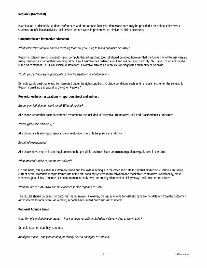
Region V (Northeast)
118 1999 manual
examination. Additionally, student conferences and one-on-one faculty/student workshops may be provided. One school takes weakstudents out of clinical activities until he/she demonstrates improvement on similar manikin procedures.
Computer-based interactive education
What interactive computer-based teaching tools are you using to teach operative dentistry?
Region V schools are not currently using computer-based teaching tools. It should be noted however that the University of Pennsylvania isusing Dent-Sim as part of their teaching curriculum; Columbia has ordered a unit and will be using it shortly. NYU and Boston are involvedin the placement of CAD/CAM clinical restorations. Columbia also has a Web site for diagnosis and treatment planning.
Would your school/region participate in development and in what manner?
Schools would participate and be interested under the right conditions. Outside conditions such as time, costs, etc. enter the picture. IsRegion VI making a proposal to the other Regions?
Posterior esthetic restorations – report on direct and indirect
Are they included in the curriculum? What discipline?
All schools report that posterior esthetic restorations are included in Operative, Restorative, or Fixed Prosthodontic curriculums.
Where (pre-clinic and clinic)?
All schools are teaching posterior esthetic restorations in both the pre-clinic and clinic.
Required experiences?
All schools have set minimum requirements in the pre-clinic and most have set minimum patient experiences in the clinic.
What materials and/or systems are utilized?
On one hand, this question is extremely broad and too wide reaching. On the other, it is safe to say that all Region V schools are usingcurrent dental materials ranging from “state of the art” bonding systems to microhybrid and “packable” composites. Additionally, glassionomers, porcelains (Empress, Cerinate to mention only two) are employed for indirect inlay/onlay and laminate procedures.
What are the results? Also site the evidence for the reported results?
The results should be based on outcomes assessments. However, the assessments for esthetic care are not different from the outcomesassessments for other care. As a result, schools have limited outcomes assessments.
Regional Agenda Items
Question of simulation laboratories – have schools recently installed new Kavo, Adec, or Nevin units?
Schools reported that they have not.
Amalgam repair – can you repair a previously placed amalgam restoration?

Region V (Northeast)
119 1999 manual
Consensus of the schools present is yes.
How do you temporize a preparation assuming one is going to place a “bonded” amalgam restoration?
Schools suggest the use of a compomer such as Dyract, or glass ionomer such as Ketac-silver.
Are schools teaching pins?
Yes, in lecture.
CODE Questions
What is CODE doing well?
Schools in Region V either do not know or feel that CODE is doing nothing for them except collect dues.
Where do you desire improvement?
CODE should establish a Mission statement and a statement of their Goals and Objectives. Additionally, CODE should have a Web sitethat is AADS based. CODE should have some mechanism to set up a body of evidence-based information.
Is $25 US/$30 Canadian school dues adequate? Comments are expected?
Yes. However, no one seems to know where the dues go? Is it for mailings, etc.
How can participation by faculty in CODE regional meetings be encouraged/improved?
Most school faculty feel they have limited time and are not funded to attend such meetings. In many cases they have to pay for themeetings out of their own pockets. This prevents many who want to attend from attending. Several schools also raised the issue of the typeof questions that National CODE put together for discussion. There is a concern that the National questions are not the type of questionsthat those intimately involved in education want to see either from a clinical or philosophical perspective.
Faculty in Attendance at 1999 CODE Meeting
Dr. Richard LichtenthalColumbia University Dental School630 W. 168th StreetNew York, NY 10032
Dr. Amer Abu-HannaDirector of OperationsDepartment of Restorative DentistryUniversity of PennsylvaniaSchool of Dental Medicine4001 Spruce StreetPhiladelphia, PA 19104
Dr. Janis MercerDepartment of Restorative DentistryHoward UniversityCollege of Dentistry600 West Street, NWWashington, DC 20059
Dr. Robert MillerMcGill UniversitySection of Operative DentistryFaculty of Dentistry740 Docteur PenfieldMontreal, P.Q. H3A 1A4Canada
Dr. Mark WolffDepartment of Restorative DentistrySUNY at StonybrookSchool of Dental MedicineStonybrook, NY 11794
Dr. Andrea JacksonDepartment of Restorative DentistryHoward UniversityCollege of Dentistry600 West Street, NWWashington, DC 20059
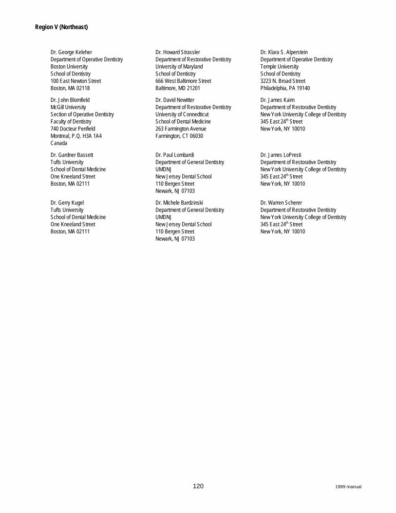
Region V (Northeast)
120 1999 manual
Dr. George KeleherDepartment of Operative DentistryBoston UniversitySchool of Dentistry100 East Newton StreetBoston, MA 02118
Dr. Howard StrasslerDepartment of Restorative DentistryUniversity of MarylandSchool of Dentistry666 West Baltimore StreetBaltimore, MD 21201
Dr. Klara S. AlpersteinDepartment of Operative DentistryTemple UniversitySchool of Dentistry3223 N. Broad StreetPhiladelphia, PA 19140
Dr. John BlomfieldMcGill UniversitySection of Operative DentistryFaculty of Dentistry740 Docteur PenfieldMontreal, P.Q. H3A 1A4Canada
Dr. David NewitterDepartment of Restorative DentistryUniversity of ConnedticutSchool of Dental Medicine263 Farmington AvenueFarmington, CT 06030
Dr. James KaimDepartment of Restorative DentistryNew York University College of Dentistry345 East 24th StreetNew York, NY 10010
Dr. Gardner BassettTufts UniversitySchool of Dental MedicineOne Kneeland StreetBoston, MA 02111
Dr. Paul LombardiDepartment of General DentistryUMDNJNew Jersey Dental School110 Bergen StreetNewark, NJ 07103
Dr. James LoPrestiDepartment of Restorative DentistryNew York University College of Dentistry345 East 24th StreetNew York, NY 10010
Dr. Gerry KugelTufts UniversitySchool of Dental MedicineOne Kneeland StreetBoston, MA 02111
Dr. Michele BardzinskiDepartment of General DentistryUMDNJNew Jersey Dental School110 Bergen StreetNewark, NJ 07103
Dr. Warren SchererDepartment of Restorative DentistryNew York University College of Dentistry345 East 24th StreetNew York, NY 10010
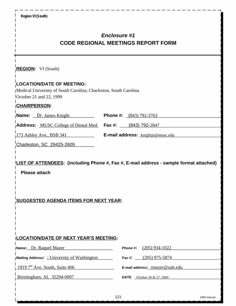
Region VI (South)
121 1999 manual
Enclosure #1CODE REGIONAL MEETINGS REPORT FORM
REGION: VI (South)
LOCATION/DATE OF MEETING:Medical University of South Carolina; Charleston, South CarolinaOctober 21 and 22, 1999
CHAIRPERSON:
Name: Dr. James Knight Phone #: (843) 792-3763
Address: MUSC College of Dental Med. Fax #: (843) 792-2847
173 Ashley Ave., BSB 341 E-mail address: [email protected]
Charleston, SC 29425-2605
LIST OF ATTENDEES: (including Phone #, Fax #, E-mail address - sample format attached)
Please attach
SUGGESTED AGENDA ITEMS FOR NEXT YEAR:
LOCATION/DATE OF NEXT YEAR’S MEETING:
Name: Dr. Raquel Mazer Phone #: (205) 934-1022
Mailing Address: ; University of Washington Fax #: (205) 975-5874
1919 7th Ave. South, Suite 406 E-mail address: [email protected]
Birmingham, AL 35294-0007 DATE: October 26 & 27, 2000

Region VI (South)
122 1999 manual
Please return all completed enclosures to Dr. Larry D. Haisch, National Director, UNMC College of Dentistry;40th and Holdrege Streets; Lincoln, NE 68583-0750. Deadline for return: 30 Days post-meeting
Office: 402 472-1290 Fax: 402 472-5290 E-mail: [email protected] send the information on a disk or via e-mail with an attachment,
indicating the software program and version utilized.
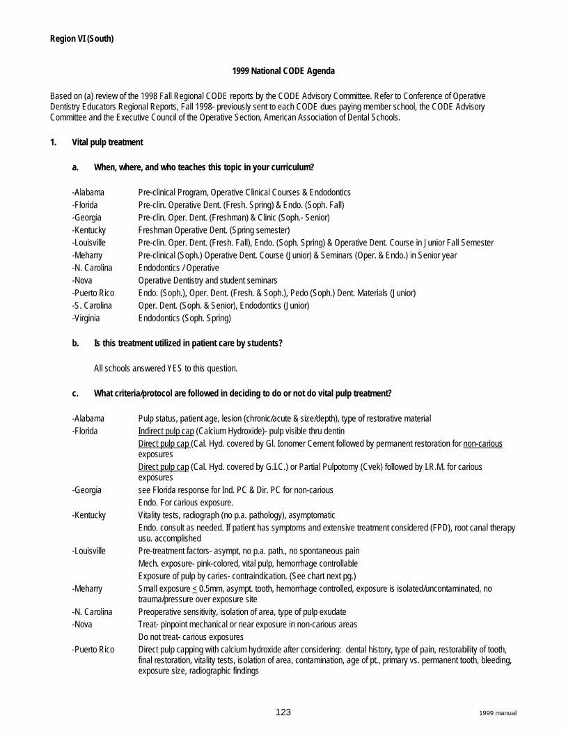
Region VI (South)
123 1999 manual
1999 National CODE Agenda
Based on (a) review of the 1998 Fall Regional CODE reports by the CODE Advisory Committee. Refer to Conference of OperativeDentistry Educators Regional Reports, Fall 1998- previously sent to each CODE dues paying member school, the CODE AdvisoryCommittee and the Executive Council of the Operative Section, American Association of Dental Schools.
1. Vital pulp treatment
a. When, where, and who teaches this topic in your curriculum?
-Alabama Pre-clinical Program, Operative Clinical Courses & Endodontics-Florida Pre-clin. Operative Dent. (Fresh. Spring) & Endo. (Soph. Fall)-Georgia Pre-clin. Oper. Dent. (Freshman) & Clinic (Soph.- Senior)-Kentucky Freshman Operative Dent. (Spring semester)-Louisville Pre-clin. Oper. Dent. (Fresh. Fall), Endo. (Soph. Spring) & Operative Dent. Course in Junior Fall Semester-Meharry Pre-clinical (Soph.) Operative Dent. Course (Junior) & Seminars (Oper. & Endo.) in Senior year-N. Carolina Endodontics / Operative -Nova Operative Dentistry and student seminars-Puerto Rico Endo. (Soph.), Oper. Dent. (Fresh. & Soph.), Pedo (Soph.) Dent. Materials (Junior)-S. Carolina Oper. Dent. (Soph. & Senior), Endodontics (Junior)-Virginia Endodontics (Soph. Spring)
b. Is this treatment utilized in patient care by students?
All schools answered YES to this question.
c. What criteria/protocol are followed in deciding to do or not do vital pulp treatment?
-Alabama Pulp status, patient age, lesion (chronic/acute & size/depth), type of restorative material-Florida Indirect pulp cap (Calcium Hydroxide)- pulp visible thru dentin
Direct pulp cap (Cal. Hyd. covered by Gl. Ionomer Cement followed by permanent restoration for non-cariousexposuresDirect pulp cap (Cal. Hyd. covered by G.I.C.) or Partial Pulpotomy (Cvek) followed by I.R.M. for cariousexposures
-Georgia see Florida response for Ind. PC & Dir. PC for non-carious Endo. For carious exposure.
-Kentucky Vitality tests, radiograph (no p.a. pathology), asymptomatic Endo. consult as needed. If patient has symptoms and extensive treatment considered (FPD), root canal therapyusu. accomplished
-Louisville Pre-treatment factors- asympt, no p.a. path., no spontaneous painMech. exposure- pink-colored, vital pulp, hemorrhage controllableExposure of pulp by caries- contraindication. (See chart next pg.)
-Meharry Small exposure < 0.5mm, asympt. tooth, hemorrhage controlled, exposure is isolated/uncontaminated, notrauma/pressure over exposure site
-N. Carolina Preoperative sensitivity, isolation of area, type of pulp exudate-Nova Treat- pinpoint mechanical or near exposure in non-carious areas
Do not treat- carious exposures-Puerto Rico Direct pulp capping with calcium hydroxide after considering: dental history, type of pain, restorability of tooth,
final restoration, vitality tests, isolation of area, contamination, age of pt., primary vs. permanent tooth, bleeding,exposure size, radiographic findings

Region VI (South)
124 1999 manual
-S. Carolina Vital pulp, asymptomatic, bleeding controllable, tooth history, radiographic appearance, exposure size,contamination, patient ageUltimate plan for the tooth. Consultations with Endo. & Prostho.
-Virginia Used for non-contaminated pulp exposure in permanent fully fully formed teeth. Apexogenesis cases.
d. What concrete evidence supports the concept of vital pulp treatment? Please cite the scientific evidence. Additionalcomments welcome.
Articles- Region VI collective citations, some listed by more than one school-Stanley Harold R. Human Pulp Response to Operative Dental Procedures, UFCD, Gainesville, FL 1982.-Mejare I, Cvek M. Partial pulpotomy in young permanent teeth with deep carious lesions. Endod-Dent-Traumatol. Dec 1993;
9(6):238-242.-Proceedings of Symposium. Current Concepts and Controversies in Vital Pulp Capping. Am. J. Dent. 1998; 11:S17-34.-Stockton LW. Vital pulp capping: a worthwhile procedure. J. Can. Dent. Assoc. 1999; 65(6):328-31.-Olmez A, et al. A histopathologic study of direct pulp-capping with adhesive resins. Oral Surg. Oral Med. Oral Path. Oral
Radiol. Endod. 1998; 86:98-103.-Kitasako Y et al. Effects of direct pulp capping techniques on short-term response of mechanically exposed pulps. J Dent.
1999; 27:257-63.-Tarim B, et al. Pulp response to a resin-modified glass ionomer material on nonexposed and exposed monkey pulps.
Quintessensce Int. 1998; 29:535-42.-Tarim B, et al. Biocompatibility or Optibond and XR-Bond adhesive systems in nonhuman primate teeth. Int J. Periodont.
Restor. Dent. 1998; 1886-99.-Nakanishi MT, et al. A clinical study of direct pulp capping applied to carious-exposed pulps. J Endod 1996; 22:551-6.-Stark M, Nicholson RJ, Soelberg KB. Direct and indirect pulp capping. Dental Clinic of North America. 1976.-Christensen GJ. Pulp capping 1998. Sept. 129(9): 1297-9.
Textbooks-Cohen, Pathways of the Pulp, 7th ed, Chapters 15 and 16.Sturdevant, Art and Science of Operative Dentistry, 4th ed.Weine, Endodontic Therapy, 5th ed. Chapter 16.Ingle, Endodontics, 4th ed. Chapter 19.
2. List five of the most important operative dentistry topics that need to be studied via clinical trials.
The following list represents the collective response of Region VI schools: Numbers after responses indicate number of schools withsame response.
Long term information on bonded direct and indirect restorations including resin inlays, onlays, crowns. Posterior composite(packable) wear and margins. 4Dentin Bonding, Adhesives, Hybridization- longevity of the bond. 3Vital pulp treatment- organized evaluation of various (new) protocols. 4Cariology- Carious lesions: early (noninvasive) detection, study of factors predisposing to carious (& recurrent) lesions andprogression rates.5Remineralization techniques and their efficacy. 3Priniciples of cavity preparation design and their relevance vs. standardized preparation design recommendations for all schools orboards. 2Repair of existing restorations.Viability of bacteria under sealed restorations.Amalgam substitutes and alternatives. Dental applications of new laser/micorabrasion devices. Demonstrate efficacy of caries risk assessment.Cement solubility

Region VI (South)
125 1999 manual
Sealing ability of flowable composites as a liner in posterior comp. restorations.Flowable composites: wear resist. in Class V restorations, longevity, and clinical effectiveness (also- Res. Modif. Gl Ion.) 4Wear resistance and durability of fixed part. dentures constructed from ceromers.Retention / fracture resistance of ceramic & carbon fiber post and core systems. 2Base to ideal depth vs. minimal pulp protection.Dentin bonding agents as cavity sealers.Reduction of post-operative sensitivity.Resin wear vs. enamel wear.Patient perceptions of quality restorative dentistry.Color stability of direct composites. Bleaching of vital tetracylcine stained teeth.Effects of pulp pressure (vitality) in bond strength.Non rotary instrumentation for tooth repair.Ways to improve the bonding of composite to porcelain. Curing lights (lasers, etc.) and resin polymerization variables. Class V restorations. Mechanical retention? Occlu adjust.- abfraction lesions.Efficacy of liners and bases under bonded amalgams on vital teeth.Mercury vapor release from spherical alloys when bleaching with carb. peroxide.
3. Calibration of faculty:
a. What is the protocol for calibrating and standardizing your operative faculty? Address current faculty and newfaculty. Also for pre-clinical and clinical.
-Alabama Two instructors for evaluation of mock board procedures.Faculty meetings using clinical photos. Preclinic calibration via prepared/restored artificial teeth and gradingdiscussions.
-Florida Preclinical- course syllabi with detailed grading criteria. All grading done by two faculty per criteria- must reachconsensus or get third faculty opinion. Same for clinical comp. exams.
-Georgia Rotating new faculty through all restorative courses to learn teaching philosophy. Assigning new faculty to moreexperienced faculty for an “apprenticeship” period. Use standardized manuals.Technique and material use policies from dept. section meetings.
-Kentucky Preclinical- “grade” standard preps before practicals and compare.Clinical- annual RSD off-site to review preclinical curric. All full and part-time faculty attend.
-Louisville Performance criteria for all preps and rest. in preclin. and clinic.Same grade scale in preclinic and clinic. Two faculty grade each comp. exam in preclin. and clinic and grades are compared for consistency. All operativefaculty have taught in preclinic. Each Fall- faculty review/discuss perf. criteria.
-Meharry Using slides, faculty review radiographs, preparations and restor. as a group with discussion of acceptable vs.non-acceptable. Then ea. faculty evaluates procedures independently. All faculty spend a year evaluatingstudents in preclinic before given clinic duties.
-N. Carolina Faculty meetings, classroom assessments, review of faculty grades, and faculty interaction in courses andclinics.
-Nova Faculty attend all lectures. Weekly faculty seminars. Comparison of evaluations within faculty.-Puerto Rico Faculty participation in preclinical courses. All faculty evaluate comp. exams (2 per student). New faculty in
preclinic prior to clinic. Course manuals with explanation of eval. criteria.Calibration course off campus, dept./section meetings.
-S. Carolina One on one discussions, clinical consultations, all full time faculty attend preclinical lectures, and departmentmeetings.
-Virginia Preclinic faculty meet with course director prior to each lab so the course director can standardize instruction forthat period.In-service C.E. Courses held periodically to emphasize teaching protocol for full and part-time employees.
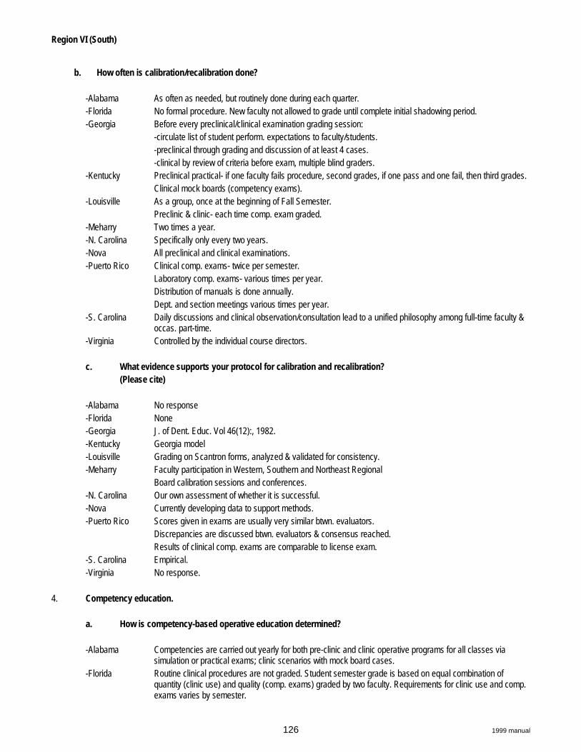
Region VI (South)
126 1999 manual
b. How often is calibration/recalibration done?
-Alabama As often as needed, but routinely done during each quarter.-Florida No formal procedure. New faculty not allowed to grade until complete initial shadowing period.-Georgia Before every preclinical/clinical examination grading session:
-circulate list of student perform. expectations to faculty/students.-preclinical through grading and discussion of at least 4 cases.-clinical by review of criteria before exam, multiple blind graders.
-Kentucky Preclinical practical- if one faculty fails procedure, second grades, if one pass and one fail, then third grades.Clinical mock boards (competency exams).
-Louisville As a group, once at the beginning of Fall Semester.Preclinic & clinic- each time comp. exam graded.
-Meharry Two times a year.-N. Carolina Specifically only every two years.-Nova All preclinical and clinical examinations.-Puerto Rico Clinical comp. exams- twice per semester.
Laboratory comp. exams- various times per year.Distribution of manuals is done annually.Dept. and section meetings various times per year.
-S. Carolina Daily discussions and clinical observation/consultation lead to a unified philosophy among full-time faculty &occas. part-time.
-Virginia Controlled by the individual course directors.
c. What evidence supports your protocol for calibration and recalibration?(Please cite)
-Alabama No response-Florida None-Georgia J. of Dent. Educ. Vol 46(12):, 1982.-Kentucky Georgia model-Louisville Grading on Scantron forms, analyzed & validated for consistency.-Meharry Faculty participation in Western, Southern and Northeast Regional
Board calibration sessions and conferences. -N. Carolina Our own assessment of whether it is successful.-Nova Currently developing data to support methods.-Puerto Rico Scores given in exams are usually very similar btwn. evaluators.
Discrepancies are discussed btwn. evaluators & consensus reached.Results of clinical comp. exams are comparable to license exam.
-S. Carolina Empirical.-Virginia No response.
4. Competency education.
a. How is competency-based operative education determined?
-Alabama Competencies are carried out yearly for both pre-clinic and clinic operative programs for all classes viasimulation or practical exams; clinic scenarios with mock board cases.
-Florida Routine clinical procedures are not graded. Student semester grade is based on equal combination ofquantity (clinic use) and quality (comp. exams) graded by two faculty. Requirements for clinic use and comp.exams varies by semester.
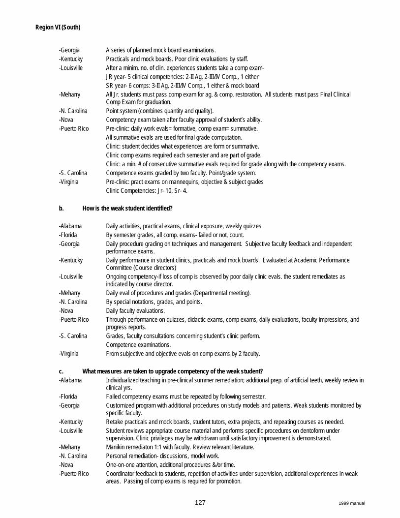
Region VI (South)
127 1999 manual
-Georgia A series of planned mock board examinations.-Kentucky Practicals and mock boards. Poor clinic evaluations by staff.-Louisville After a minim. no. of clin. experiences students take a comp exam-
JR year- 5 clinical competencies: 2-II Ag, 2-III/IV Comp., 1 eitherSR year- 6 comps: 3-II Ag, 2-III/IV Comp., 1 either & mock board
-Meharry All Jr. students must pass comp exam for ag. & comp. restoration. All students must pass Final ClinicalComp Exam for graduation.
-N. Carolina Point system (combines quantity and quality).-Nova Competency exam taken after faculty approval of student’s ability.-Puerto Rico Pre-clinic: daily work evals= formative, comp exam= summative.
All summative evals are used for final grade computation.Clinic: student decides what experiences are form or summative.Clinic comp exams required each semester and are part of grade.Clinic: a min. # of consecutive summative evals required for grade along with the competency exams.
-S. Carolina Competence exams graded by two faculty. Point/grade system.-Virginia Pre-clinic: pract exams on mannequins, objective & subject grades
Clinic Competencies: Jr- 10, Sr- 4.
b. How is the weak student identified?
-Alabama Daily activities, practical exams, clinical exposure, weekly quizzes-Florida By semester grades, all comp. exams- failed or not, count.-Georgia Daily procedure grading on techniques and management. Subjective faculty feedback and independent
performance exams.-Kentucky Daily performance in student clinics, practicals and mock boards. Evaluated at Academic Performance
Committee (Course directors)-Louisville Ongoing competency-if loss of comp is observed by poor daily clinic evals. the student remediates as
indicated by course director.-Meharry Daily eval of procedures and grades (Departmental meeting).-N. Carolina By special notations, grades, and points.-Nova Daily faculty evaluations.-Puerto Rico Through performance on quizzes, didactic exams, comp exams, daily evaluations, faculty impressions, and
progress reports.-S. Carolina Grades, faculty consultations concerning student’s clinic perform.
Competence examinations.-Virginia From subjective and objective evals on comp exams by 2 faculty.
c. What measures are taken to upgrade competency of the weak student?-Alabama Individualized teaching in pre-clinical summer remediation; additional prep. of artificial teeth, weekly review in
clinical yrs.-Florida Failed competency exams must be repeated by following semester.-Georgia Customized program with additional procedures on study models and patients. Weak students monitored by
specific faculty.-Kentucky Retake practicals and mock boards, student tutors, extra projects, and repeating courses as needed.-Louisville Student reviews appropriate course material and performs specific procedures on dentoform under
supervision. Clinic privileges may be withdrawn until satisfactory improvement is demonstrated.-Meharry Manikin remediaton 1:1 with faculty. Review relevant literature.-N. Carolina Personal remediation- discussions, model work.-Nova One-on-one attention, additional procedures &/or time.-Puerto Rico Coordinator feedback to students, repetition of activities under supervision, additional experiences in weak
areas. Passing of comp exams is required for promotion.

Region VI (South)
128 1999 manual
-S. Carolina If fail to pass comp exam after 3 tries mandatory remed = lab exercises 1-on-1 with faculty and additionalhelp in clinic.
-Virginia Remediation is up to the discretion of course director.
5. Computer based interactive education.a. What interactive computer-based teaching tools are you using to teach operative dentistry?
-Alabama None-Florida Dental Anatomy- Web-based info and practice exams.
Data Collection / Treament Planning nearly complete.-Georgia None. Operative Dent Course manual will be on WebCT-Kentucky None-Louisville None-Meharry None-N. Carolina None-Nova None-Puerto Rico None. We are evaluating some initiatives.-S. Carolina None-Virginia None
b. Who developed them?
All schools answering “none” to the previous question are N/A (not applicable) to this question except-
-Florida Dental Anatomy- Dr. Greg Smith, Data Collection- Dr. Doug Benn-Puerto Rico Mathilde Peters (Michigan): glass ionomers to composite resins
c. Would your school/region participate in development and in what manner?
-Alabama Would like to but limited by resources.-Florida Yes- case and mini-course development accompanied by interactive software.-Georgia Yes, Dr. Frank Caughman is a consultant on the DISC project.-Kentucky Yes, grant support would have to be a component of the project.-Louisville-Meharry-N. Carolina-Nova-Puerto Rico We would participate as collaborators at this moment.-S. Carolina Yes willing to explore this.-Virginia
6. Posterior esthetic restorations- report on direct and indirect.
a. Are they included in the curriculum? What discipline?b. Where ? (pre-clinic and clinic)c. Required experiences?d. What materials and/or systems utilized?
a. b. c. d.
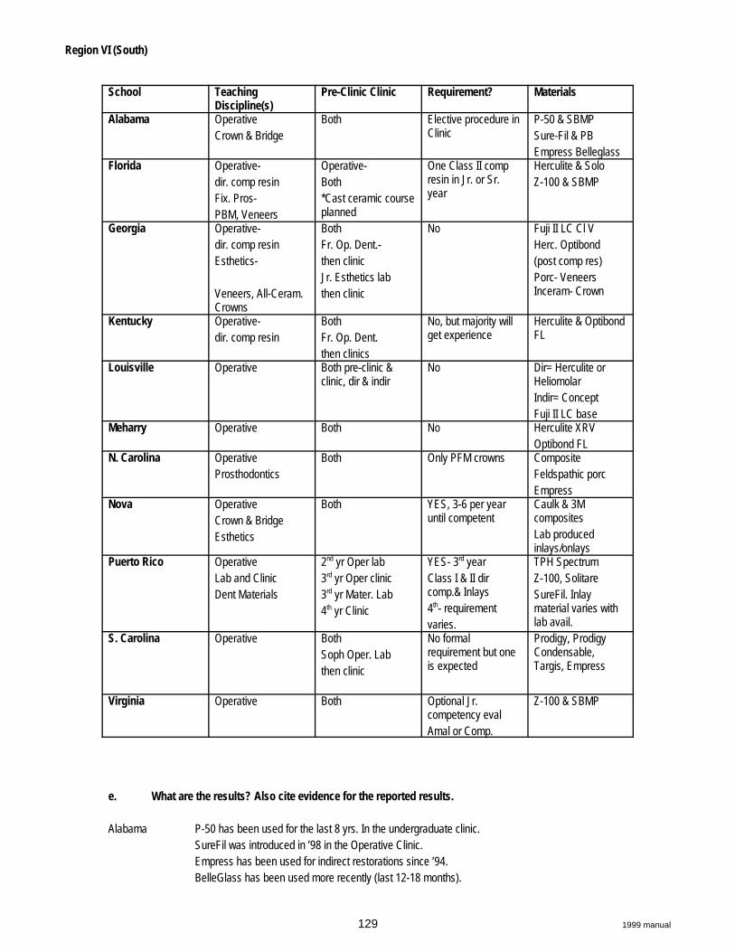
Region VI (South)
129 1999 manual
School TeachingDiscipline(s)
Pre-Clinic Clinic Requirement? Materials
Alabama OperativeCrown & Bridge
Both Elective procedure in Clinic
P-50 & SBMPSure-Fil & PBEmpress Belleglass
Florida Operative-dir. comp resinFix. Pros-PBM, Veneers
Operative-Both*Cast ceramic courseplanned
One Class II compresin in Jr. or Sr.year
Herculite & SoloZ-100 & SBMP
Georgia Operative-dir. comp resinEsthetics-
Veneers, All-Ceram.Crowns
BothFr. Op. Dent.-then clinicJr. Esthetics labthen clinic
No Fuji II LC Cl VHerc. Optibond(post comp res)Porc- VeneersInceram- Crown
Kentucky Operative-dir. comp resin
BothFr. Op. Dent. then clinics
No, but majority willget experience
Herculite & OptibondFL
Louisville Operative Both pre-clinic &clinic, dir & indir
No Dir= Herculite orHeliomolar Indir= ConceptFuji II LC base
Meharry Operative Both No Herculite XRVOptibond FL
N. Carolina Operative Prosthodontics
Both Only PFM crowns CompositeFeldspathic porcEmpress
Nova OperativeCrown & BridgeEsthetics
Both YES, 3-6 per yearuntil competent
Caulk & 3McompositesLab producedinlays/onlays
Puerto Rico OperativeLab and ClinicDent Materials
2nd yr Oper lab3rd yr Oper clinic3rd yr Mater. Lab4th yr Clinic
YES- 3rd year Class I & II dircomp.& Inlays4th- requirementvaries.
TPH SpectrumZ-100, SolitareSureFil. Inlaymaterial varies withlab avail.
S. Carolina Operative Both Soph Oper. Labthen clinic
No formalrequirement but oneis expected
Prodigy, ProdigyCondensable,Targis, Empress
Virginia Operative Both Optional Jr.competency evalAmal or Comp.
Z-100 & SBMP
e. What are the results? Also cite evidence for the reported results.
Alabama P-50 has been used for the last 8 yrs. In the undergraduate clinic.SureFil was introduced in ’98 in the Operative Clinic.Empress has been used for indirect restorations since ’94.BelleGlass has been used more recently (last 12-18 months).

Region VI (South)
130 1999 manual
Florida No formal attempt at evaluation yet, although evaluations to begin shortly in newly formed HealthMaintenance Clinic.
GeorgiaKentucky Evaluated by covering staff on student clinic floor, then at recall.Louisville We have not had to replace any posterior composite resins as far as I know. There is no written evidence of
their performance.Meharry No results at present time; we will recall 3, 6, and 12 months.N. Carolina No responseNova N/A- too soon to know.Puerto Rico Results are acceptable. We are working in the outcome assessment and a recall system to corroborate this
statement. S. Carolina Variable, depending on student operator, case selection, and quality of lab support. Generally, very good
(anecdotal evidence)Virginia No long term follow-up. Anecdotal results.
Region VI (South)
131 1999 manual
CODE QuestionsThe following answers are collective responses from Region VI schools
1. What is CODE doing well?
- Provides friendship-building, hard-working forum for exchange of information, ideas, and results between operative dentistrydepartmts. within/between regions.
- Networking through annual meeting and/or e-mail and phone contacts.- Sharing of ideas with different schools.- Regional meetings are important to gain information about what other schools are doing and the materials/techniques that are
being used.- Communication among all member schools. Review of important operative subjects at meetings.- Keeping schools updated on what everyone is doing.- Meeting regularly. Discussing pertinent issues matter-of-factly.- The organization is doing well and is producing thought provoking agendas and hopefully productive metings.
2. Where do you desire improvement?
- More regionally cooperative education efforts.- More regionally cooperative research efforts.- More proactive efforts nationally on clinical research issues.- More interaction with the Academy of Operative Dentistry and AADS.- Joint national projects involving different regions.- Increase communication with other CODE regions.- More non-operative faculty participation in meetings.- Active participation in calibration of faculty and curriuculum development.- Interact with other regions. - Lengthen meeting time. - Participation at regional meetings needs improvement.
3. Is $25 US / $30 Canadian school dues adequate?
- No, will need to be higher to achieve goals like those stated.- Reasonable. Can be increased if more national projects are planned.- Yes. Seems adequate given the current agenda.- Only the secretary knows for sure.- Yes, until a demonstrated need for increase because of better methods of holding the annual meeting (sites & materials). - OK- OK for what we are receiving now, can be changed.- Increase to $50 and give some of the money to school hosting regional meeting to allow better support.- Information regarding expenses is necessary to determine adequacy of fees.
4. How can participation by faculty in CODE regional meetings be encouraged / improved?
- Suggest faculty exchanges (mini-sabbaticals / 4-6 weeks) between Operative Dentistry departments within a region.- Suggest regional CODE meetings be held on a weekend.- Departmental support.- Rotation of attendees from participating schools.
Region VI (South)
132 1999 manual
- With the budgets as restricted as they are, at least at our school, sending multiple faculty is not possible. Some schools maynot be able to send faculty at all.
- Give CEU credits to all faculty or non-faculty participants. - Invite members of state / regional boards to participate.- CODE agenda covering areas of immediate attention such as interactive teaching, competency-based curriculum.- Ensure relevant issues are discussed. (Make programs interesting and clinically-based.)- Change agenda to encourage discussion of topics of particular interest to attendees. - Be more forward looking: “Where are we going?” What if…?”.- Continuing education credits would be helpful. - Possibly combining this meeting with another might be beneficial.- Having presentations corresponding to the agenda would be good as the agendas typically involve areas of Operative
Dentistry Education that all schools seem to struggle with.

Region VI (South)
133 1999 manual
CODE Region VI AttendeesOctober 20-22, 1999
Charleston, South Carolina
Name University Phone # Fax # E-mailRaquel Mazer University of
Alabama205-934-1022 205-975-5874 [email protected]
Jack Warren University ofFlorida
352-846-1696 352-846-1643 [email protected]
Dan Chan Med Collegeof Georgia
706-721-2881 706-721-8349 [email protected]
Frank Caughman M. C. G. 706-721-2881 706-721-8349 [email protected] Frazier M. C. G. 706-721-2881 706-721-8349 [email protected] Kovarik University of
Kentucky606-257-2147 606-257-1847 [email protected]
Charles Thomas U. of KY 606-323-5486 606-257-1847 [email protected] Osborne U. of KY 606-323-4635 606-257-1847 [email protected] Crim University of
Louisville502-852-1303 502-852-1220 [email protected]
Henry Young Meharry MedCollege
615-327-6359 615-327-6026 [email protected]
Roosevelt Smith M. M. C. 615-327-6359 615-327-6213 [email protected] Yacko M. M. C. 615-327-4751 615-327-6213Timothy Hottel Nova S. E.
University954-262-7349 954-262-1782 [email protected]
James Knight Med Univ ofS. Carolina
843-792-3763 843-792-2847 [email protected]
Dan Sneed M. U. S. C. 843-792-3761 843-792-2847 [email protected] Collins M. U. S. C. 843-792-3766 843-792-2847 [email protected] Rinaudo M. U. S. C. 843-792-3761 843-792-2847Richard Sterne Med College
of Virginia804-828-2977 804-828-3159 [email protected]
Larry Haisch University of Nebraska
402-472-1290 402-472-5290 [email protected]