cx3cr1-microglia mediates neuroinflammation and blood
TRANSCRIPT

RESEARCH Open Access
CX3CR1-microglia mediatesneuroinflammation and blood pressureregulation in the nucleus tractus solitarii offructose-induced hypertensive ratsChiu-Yi Ho1,2†, Yu-Te Lin2,3,4,5†, Hsin-Hung Chen1, Wen-Yu Ho6,7, Gwo-Ching Sun8,9, Michael Hsiao10, Pei-Jung Lu11,Pei-Wen Cheng1,2* and Ching-Jiunn Tseng1,2,12*
Abstract
Background: Inflammation is a common pathophysiological trait found in both hypertension and cardiac vasculardisease. Recent evidence indicates that fractalkine (FKN) and its receptor CX3CR1 have been linked to inflammatoryresponse in the brain of hypertensive animal models. Here, we investigated the role of CX3CR1-microglia in nitricoxide (NO) generation during chronic inflammation and systemic blood pressure recovery in the nucleus tractussolitarii (NTS).
Methods: The hypertensive rat model was used to study the role of CX3CR1-microglia in NTS inflammationfollowing hypertension induction by oral administration of 10% fructose water. The systolic blood pressure wasmeasured by tail-cuff method of non-invasive blood pressure. The CX3CR1 inhibitor AZD8797 was administeredintracerebroventricularly (ICV) in the fructose-induced hypertensive rat. Using immunoblotting, we studied the nitricoxide synthase signaling pathway, NO concentration, and the levels of FKN and CX3CR1, and pro-inflammatorycytokines were analyzed by immunohistochemistry staining.
Results: The level of pro-inflammatory cytokines IL-1β, IL-6, TNF-α, FKN, and CX3CR1 were elevated two weeks afterfructose feeding. AZD8797 inhibited CX3CR1-microglia, which improved the regulation of systemic blood pressureand NO generation in the NTS. We also found that IL-1β, IL-6, and TNF-α levels were recovered by AZD8797addition.
Conclusion: We conclude that CX3CR1-microglia represses the nNOS signaling pathway and promotes chronicinflammation in fructose-induced hypertension. Collectively, our results reveal the role of chemokines such as IL-1β,IL-6, and TNF-α in NTS neuroinflammation with the involvement of FKN and CX3CR1.
Keywords: Nucleus tractus solitarii, Fractalkine, CX3CR1, Hypertension, Inflammation
IntroductionCardiovascular disease is a complicated condition thataffects metabolism and blood pressure [1, 2]. Epidemio-logical study showed that unhealthy dietary habit con-tributes to increased blood pressure, hyperglycemia,hyperlipidemia, and neuroinflammation [3]. Despite re-cent studies showing that excessive fructose
© The Author(s). 2020 Open Access This article is licensed under a Creative Commons Attribution 4.0 International License,which permits use, sharing, adaptation, distribution and reproduction in any medium or format, as long as you giveappropriate credit to the original author(s) and the source, provide a link to the Creative Commons licence, and indicate ifchanges were made. The images or other third party material in this article are included in the article's Creative Commonslicence, unless indicated otherwise in a credit line to the material. If material is not included in the article's Creative Commonslicence and your intended use is not permitted by statutory regulation or exceeds the permitted use, you will need to obtainpermission directly from the copyright holder. To view a copy of this licence, visit http://creativecommons.org/licenses/by/4.0/.The Creative Commons Public Domain Dedication waiver (http://creativecommons.org/publicdomain/zero/1.0/) applies to thedata made available in this article, unless otherwise stated in a credit line to the data.
* Correspondence: [email protected]; [email protected]†Chiu-Yi Ho and Yu-Te Lin indicates co-first authors.1Department of Medical Education and Research, Kaohsiung VeteransGeneral Hospital, Kaohsiung 81300, TaiwanFull list of author information is available at the end of the article
Ho et al. Journal of Neuroinflammation (2020) 17:185 https://doi.org/10.1186/s12974-020-01857-7

consumption leads to pro-inflammatory response andmicroglia activation, the role of pro-inflammatory factorin the nucleus tractus solitarii (NTS) is not well-understood [4, 5]. In addition, we previously showed thatthe brainstem NTS, part of the brain which integratessignals from the peripheral carotid sinus and aortic archto regulate systemic blood pressure, decreases produc-tion of nitric oxide (NO) during fructose-induced hyper-tension [6]. However, the underlying mechanisminvolving NO during chronic inflammation in the NTSis not yet clarified. Therefore, it is essential to investigatethe role of FKN and its receptor CX3CR1 after hyper-tension induced by fructose.C-X3-C motif chemokine receptor 1 (CX3CR1) is a
chemokine receptor that binds to its ligand C-X3-Cmotif chemokine ligand 1 (CX3CL1), also known as frac-talkine (FKN), and is found in the leukocytes, brain,spinal cord, and retina [7, 8]. Both FKN and CX3CR1have been detected in vivo in the brainstem NTS.CX3CR1 is characterized as microglia biomarker. Ac-cording to Bhaskar et al., CX3CR1 is the potential targetfor studying metabolic syndrome [9, 10]. The FKN–CX3CR1 interaction is associated with crosstalk betweenneurons and microglia. Previous study has suggestedthat CX3CR1 may be involved in neurodegenerative dis-eases such as multiple sclerosis [11], Alzheimer’s disease[5], spinal cord injury, and traumatic brain injury [12].Ruchaya et al. reported that FKN microinjection pro-duces cardiovascular response in the NTS of normal rats[13]. However, the function of CX3CR1 during neuro-genic hypertension progression in the NTS is ambigu-ous. On the other hand, NO is a gas molecule that isinvolved in various functions in the dorsal brainstemNTS such as vasodilation, baroreflex, sympathetic nerveactivity, and most importantly blood pressure mainten-ance [14]. It is essential that the autonomic response tocombine afferent signals be integrated from the periph-eral nervous system for optimal adjustment of sympa-thetic activity. In the clinical aspect, overactivity of thesympathetic nervous system contribute to the develop-ment of hypertension [15]. This study aims to examinethe NTS cardiovascular effect and whether increasedpro-inflammatory cytokines such as CX3CR1 has impacton NO production.There is increasing evidence that inflammation in the
brain is associated with excessive fructose intake. Suchinflammatory response poses a threat to the brain andmicroglia cells which reside in the brain that act uponthe threat to clear debris and repair damaged neural tis-sue. However, chronic inflammation in the brain acti-vates microglia cells to release pro-inflammatorycytokines that are harmful to brain tissue [16, 17]. Fur-thermore, fructose, in particular, induces hypertensionin vivo which strongly correlates with chronic brain
inflammation [18–20]. Xu et. al. reported that fructoseincreases blood pressure, and elevates transcription ofpro-inflammatory cytokines, IL-1β, IL-6, and TNF-α inthe brain after 8 weeks of fructose consumption [5].Our previous results suggest that fructose reduces cen-
tral NO production, increases systemic blood pressureand renal sympathetic nerve activity [14, 21, 22]. Thesignaling pathway, PI3K–Akt–ERK1/2–nNOS (neuronalnitric oxide synthase) pathway, is shown to regulatenNOS phosphorylation in the NTS [23]. Additionally,we found that fructose disrupted the Akt–ERK1/2–nNOS signaling pathway [6, 21]. Short-term or 2 weeksof fructose feeding increases TNF-α in the brain [4]. Wepreviously reported that prolonged fructose feeding timeperiod induces blood pressure [22]. These results sup-port the idea that blood pressure increase triggered bydietary fructose is closely linked to pro-inflammatory cy-tokines in the brain. We therefore speculate thatCX3CR1-microglia may affect NO production to modu-late NTS inflammation, and selective CX3CR1 inhibitorAZD8797 may be able to treat the NTS pro-inflammatory response. We observed that pro-inflammatory cytokines and NO production were sup-pressed through abolishing the Akt–ERK1/2–nNOS sig-naling pathway. Finally, microglia activation in the NTSrequires CX3CR1 to promote fructose-induced hyper-tension progression.
MethodsAnimal careAll procedures were reviewed and approved by the Insti-tute of Animal Care and Use Committee at the Kao-hsiung Veterans General Hospital (VGHKS, Kaohsiung,Taiwan). Wistar-Kyoto rats (WKY) were purchased fromNational Laboratory Animal Center (NLAC, Taipei,Taiwan), housed in the VGHKS animal center withlight-controlled cycle of 12-h light and 12-h darkness.
Experimental designWKY animals were divided into five groups. Each con-sisted of six to eight animals in total: (1) control rats re-ceived tap water (Ctrl); (2) hypertensive rats received10% fructose water for 2 or 4 weeks (F2w, F4w); (3)vehicle-control rats were fed with tap water for 2 weeks,followed by osmotic pump implantation (14 days, 0.5 μL/h) filled with 30% 2-hydroxypropyl-β-cyclodextrin (HP-β-CD) (Santa Cruz, Dallas, TX, USA) and continued tapwater feeding simultaneously in the subsequent 2 weeks(V-Ctrl); (4) vehicle-fructose rats received 10% fructosewater for 2 weeks, followed by osmotic pump implant-ation (14 days, 0.5 μL/h) filled with 30% 2-hydroxypropyl-β-cyclodextrin (HP-β-CD) and continued10% fructose water simultaneously in the subsequent 2weeks (V-Fru); (5) finally, the fructose-AZD8797 rats
Ho et al. Journal of Neuroinflammation (2020) 17:185 Page 2 of 12

received 10% fructose water for 2 weeks, followed by os-motic pump implantation (14 days, 0.5 μL/h) filled withAZD8797 (Axon Medchem, Groningen, Netherlands)solution and continued 10% fructose water simultan-eously in the subsequent 2 weeks (Fru-AZD).
Non-invasive blood pressure (NIBP) measurementThe systolic blood pressure (SBP) was measured byusing the tail-cuff method (CODA, Kent Scientific, Tor-rington, CT, USA). Operating procedures were carriedout according to the manufacturer’s manual. Briefly, theWKY rats were trained in a rodent holder and placed onthe warm plate (35 °C) for three times before the experi-ment began. Measurements were taken 15 times, re-stricted to 40min and below for the entire procedure,and systolic blood pressure data were collected at theend of the experiment. To prevent blood pressure fluc-tuation as a result of circadian rhythms, the data werecollected between 09:00–12:00 am, coordinated universaltime with +08:00 offset for Taiwan time zone.
Cerebrospinal fluid (CSF) and serum sample collectionand measurementAfter a 16-hr fast, the rats were anesthetized with iso-flurane (2% mixed with O2), and blood samples werecollected using serum separation tubes. To collectserum, the tubes were centrifuged (3000 × g) at 4 °C for10 min. The CSF was collected in a 1.5-mL Eppendorftube from the fourth ventricle through an insulin needle.The tubes were centrifuged (300 × g) at 4 °C for 10 minto remove residual blood. Immediately, all samples weredispensed and frozen at − 80 °C for biochemical assays.All serum parameters were measured by VITROS® 350Chemistry System (Ortho Clinical Diagnostics, NewBrunswick, NJ, USA).
Intracerebroventricular administration procedureThe three animal groups: V-Ctrl, V-Fru, and Fru-AZDunderwent intracerebroventricular surgery. The osmoticpump (Alzet, Cupertino, CA, USA) which was filled withAZD8797solution, vehicle solution 30% 2-hydroxypropyl-β-cyclodextrin (HP-β-CD), was immersedin normal saline for 16 h at 4 °C prior to surgery. Duringanesthetization, Atropine 0.05 mg/kg, Zolile 40 mg/kg,and Xylazine 10mg/kg were placed on the stereotaxicinstrument; the head was fixed with the cranium ex-posed. The position of the injection was located 1.5 mmlateral and caudal 0.8 mm from the bregma. After sur-gery, the rat tail systolic blood pressure was measuredevery week.
Immunoblot analysisThe brain was excised and the NTS regions (approxi-mately 20 mg) were separated according to the rat
brain in stereotaxic coordinates based on Paxinos andWatson, sixth edition in 2007 [24]. Briefly, the NTSwas dissected by micropunch (1 mm inner diameter)from 1-mm-thick brainstem slice at the level of theobex under the microscope. To collect total protein,NTS tissue was homogenized using T-PER (ThermoFisher, Waltham, MA USA) containing protease andphosphatase inhibitors cocktail at 4 °C. Protein wasquantitatively analyzed with Coomassie R-250 (ThermoFisher, Waltham, MA USA), subjected to 4–20% SDS gradi-ent gel electrophoresis, transferred to PVDF membrane. Themembranes were blocked with 5% BSA in TBS/Tween-20buffer (10 mM Tris, 150 mM NaCl, 0.1% Tween 20, pH 7.4),incubated with anti-p-AktS473 (Cell signaling, 9271), anti-Akt(Cell signaling, 9271), anti-p-eNOSS1177 (BD, 612393), anti-eNOS (BD, 610297), anti-p-nNOSS1416 (Abcam, ab5583),anti-nNOS (Millipore, 07-571), anti-p-ERK1/2T202/Y204 (Cellsignaling, 9101), anti-ERK1/2 (Cell signaling, 9102), or β-actin (Millipore, MAB1501) antibody at 1:1000 dilution inphosphate-buffered saline (PBS) Tween-20 with 5% BSA at4 °C overnight. Peroxidase-conjugated anti-mouse or anti-rabbit antibody (1:5000) was used as the secondary antibody.The proteins were visualized using enhanced chemilumines-cence (ECL), Pico plus detection kit (Thermo Fisher,Waltham, MA, USA), and film. The films were captured byChemiDoc™ MP Imaging System (Bio-Rad, Hercules, CA,USA) and analyzed with Image Lab™ Software (Bio-Rad,Hercules, CA, USA).
Analysis of NO concentration in the NTSTo determine NO concentration in the groups V-Ctrl,V-Fru, and Fru-AZD, the NTS protein lysate was depro-teinized using a Microcon YM-30 filter unit (Millipore,Darmstadt, GmbH) based on a previous method [6]. Thetotal amount of NOx in the samples were determinedusing modified chemiluminescence-based procedure andSievers Nitric Oxide Analyzer purge system (NOA 280i,Sievers Instruments, Boulder, CO, USA) [25].The sample(10 μL) was injected into reflux column containing 0.1mol/L VCl3 in 1 mol/L HCl at 90 °C to reduce nitratesand nitrites into NO. The NOx was then combined withO3 produced by the analyzer to form NO2. The emissionresulting from the excited NO2 was detected by a photo-multiplier tube and digitally recorded (mV). The valueswere then interpolated to standard curve of concurrentlydetermined NaNO2 concentrations. The measurementswere recorded in triplicate for each sample. The NOx
levels measured were corrected for the NTS protein con-centration of the rats.
Enzyme-linked immunosorbent assay (ELISA)The NTS of the brainstem were sectioned and homoge-nized with T-PER (Thermo Fisher, Waltham, MA, USA)containing protease and phosphatase inhibitors cocktail
Ho et al. Journal of Neuroinflammation (2020) 17:185 Page 3 of 12

at 4 °C. The total protein was harvested by grinding andcentrifugation. The total protein content was quantita-tively analyzed by Coomassie R-250 (Thermo Fisher,Waltham, MA, USA). The concentration of IL-1β, IL-6,TNF-α, and fractalkine of serum, CSF, or NTS proteinlysate were measured by ELISA kit, performed accordingto the manufacturer’s instruction (Cloud-Clone Corp,Katy, TX, USA). Expression values were detected byAnthos Zenyth 200rt Microplate Reader (Biochrom,Cambridge, UK). The final values were calculated andnormalized to NTS protein mass.
Immunofluorescent staining analysisThe Ctrl, F2w, and F4w animals were perfused with saline,followed by 4% paraformaldehyde solution. The brainstemwas harvested and immersed in 30% sucrose solution untilit was sunken to the bottom of the tube, and this proced-ure was repeated once. Brain stem sections (5 μm) wereblocked with 5% bovine serum albumin and 0.3% Triton
X-100 for 30min at room temperature, incubated in pri-mary antibody anti-Iba-1 (Wako, 019-19741) for 16 h at4 °C. After PBS wash, the sections were incubated in AlexaFlour 488 Goat anti-rabbit IgG (Thermo Fisher Scientific,Waltham, MA, USA) for 1 h under room temperature.The tissues were mounted in VECTASHIELD mountingmedium containing DAPI (Vector Labs, Burlingham, CA,USA). The sections were analyzed under LSM 800 laserscanning mode of confocal microscope (Carl Zeiss Micro-Imaging, Jena, GmbH). The images were acquired using40 x magnification (objective: Plan-APO 40 x/1.30 Oil DIC(UV) VIS-IR ), image matrix of 1024 × 1024 pixel, pixelscale 0.156 × 0.156 μm, and a depth of 8 bit. Z-stacked im-ages were collected with 0.33-μm slice distance for 15slices in total.
Immunohistochemistry staining analysisThe brain stem sections (5 μm) were blocked in 5% bo-vine serum albumin and 0.3% Triton X-100 for 30 min
Fig. 1 Quantitative immunofluorescent analysis of microglial marker in the NTS following feeding with 10% fructose. a Flowchart presentedanimal experimental design. b In situ qualitative analysis of the microglial marker Iba-1 by immunofluorescent staining. The arrowhead indicatesactivated microglial cells. The scale bar represents 20 μm. c Graphs depicting quantitative analysis of in situ positive cells in the NTS of WKY ratsafter fructose feeding. The percentage of the positive cells was determined by counting positive cells in each hemisphere of the NTS at 400 ×magnification. The values are presented as mean ± SEM. One-way ANOVA with Bonferroni’s post-hoc was performed for statistical analysis.*P <0.05, **P < 0.01 compared to the control group, #P < 0.05 compared to fructose feeding for 4 weeks (n = 6~8 per group)
Ho et al. Journal of Neuroinflammation (2020) 17:185 Page 4 of 12

at room temperature, incubated in primary antibodyanti-IL-1β (Proteintech, 16806-1-AP) and anti-IL-6 (Pro-teintech, 21865-1-AP) in primary antibody diluent (Scy-Tek laboratories, Logan, UT, USA) for 16 h at 4 °C. AfterPBS wash, the sections were incubated in Novolink Poly-mer solution (Leica Biosystems, Nussloch, GmbH) for10 min under room temperature. The tissues werestained in DAB chromogen at room temperature, ana-lyzed by Olympus BX51 microscope (Olympus Tokyo,Japan) and Image Browser (Carl Zeiss, MicroImaging,Jena, GmbH).
Statistical analysisAll data were expressed as mean ± SEM at least threeindependent experiments. IBM SPSS Statistics 20 wasused in this study. The blood pressure (BP) measure-ments (fructose-treated and no-treatment groups)were analyzed by one-way ANOVA for repeatedmeasurements and Bonferroni’s post-hoc tests. One-way ANOVA with Scheffe’s post-hoc comparison wasapplied to immunoblotting and immunohistochemistrystain. P < 0.05 was considered statistically significant.
ResultsFructose consumption leads to higher blood pressure andcentral inflammationFructose feeding induces hypertension in rats, and dataare analyzed at the second and fourth weeks. The sys-tolic blood pressure, fasting blood glucose, high-densitylipoprotein, and triglyceride content were increased inthe fructose group compared to control. Pro-inflammatory cytokines IL-1β and IL-6 and TNF-α sig-nificantly increased in serum and NTS after fructose
administration, but FKN showed no increase in theserum after 4 weeks of fructose feeding. Interestingly,FKN in the CSF and NTS increased after 2 weeks offructose feeding (Table 1). These data demonstrated thatfructose consumption leads to higher blood pressureand central inflammation.
Microglia mediate blood pressure regulation in the NTSin fructose-induced hypertensionPreviously, CX3CR1 has been shown to be a putativemicroglial marker which was implicated to regulate bloodpressure and heart rate [13]. In this study, we determinedmicroglia activation through Iba-1 staining in NTS afterfructose administration. The activated microglia showedstronger IBA-1 signal and shorten processes in the NTSafter 2 or 4 weeks of fructose feeding (Fig. 1a). We quanti-fied the NTS-activated microglia for fructose and controlgroups (Fig. 1b). Based on these observations, we specu-late that microglia may have begun to intervene in theNTS, affecting blood pressure in the early stage offructose-induced hypertension.The fractalkine is a sole ligand that couples to
CX3CR1, existing in either membrane-anchored or sol-uble form. The membrane-anchored FKN (mFKN), sol-uble FKN (sFKN), and CX3CR1 were analyzed byimmunoblotting (Fig. 2a). After 4 weeks of fructose feed-ing, the sFKN was increased in the NTS but not mFKNand CX3CR1. Next, the level of sFKN in the NTS wassignificantly higher compared to control after 4 weeks offructose feeding (Fig. 2b). As a result, we speculate thatsoluble FKN may have begun to stimulate microglia acti-vation in the NTS after fructose consumption, ratherthan mFKN.
Table 1 General characteristics of the fructose-induced WKY rats
Parameter/group Control Fructose 2 weeks Fructose 4 weeks
Systolic blood pressure, mmHg 109.8 ± 1.8 125.1 ± 2.2*** 144.5 ± 1.1***
Fasting serum glucose, mg/dL 85.3 ± 1.8 134.2 ± 4.9*** 147.0 ± 4.0***
Fasting serum triglyceride, mg/dL 82.7 ± 1.2 155.3 ± 18.6*** 177.8 ± 26.6***
Fasting serum dHDL, mg/dL 83.0 ± 1.6 75.5 ± 4.5* 71.5 ± 2.5*
Serum IL-1β, pg/mL 27.93 ± 5.13 49.97 ± 8.59* 74.90 ± 7.07*
Serum IL-6, pg/mL 3.44 ± 0.46 6.21 ± 0.24* 10.02 ± 1.69*
Serum TNF-α, pg/mL 25.82 ± 9.05 74.06 ± 5.62* 202.10 ± 17.82*
Serum fractalkine, ng/mL 0.38 ± 0.06 0.36 ± 0.04 0.43 ± 0.05
CSF fractalkine, ng/mL 1.20 ± 0.04 1.49 ± 0.07* 1.65 ± 0.03*
NTS fractalkine, ng/mg 4.06 ± 0.17 4.80 ± 0.36* 5.98 ± 0.38*
NTS IL-1β, pg/mg 6.53 ± 0.27 9.17 ± 0.32* 12.63 ± 0.83*
NTS IL-6, pg/mg 61.13 ± 6.30 115.48 ± 3.30* 150.87 ± 11.68*
NTSTNF-α, pg/mg 1.15 ± 0.24 4.67 ± 0.75* 12.17 ± 0.41*
Levels of systolic blood pressure; fasting serum glucose; fasting serum triglycerides; fasting serum dHDL; and quantitative ELISA of serum, CSF, or NTS areas forFKN, IL-1β, IL-6, and TNF-α are presented as mean ± SEM. One-way ANOVA with Bonferroni’s post-hoc was performed for statistical analysis.*P < 0.05, ***P < 0.001 compared to control group (n = 6~8 per group)
Ho et al. Journal of Neuroinflammation (2020) 17:185 Page 5 of 12

CX3CR1-microglia mediated systolic blood pressureimprovement in the NTSTo investigate whether CX3CR1-microglia was in-volved in systolic blood pressure regulation infructose-induced hypertensive rats, CX3CR1 wasinhibited by AZD8797 treatment. AZD8797 wasinjected into central intracerebroventricularly via os-motic minipump 2 weeks after hypertensive animalswere established (Fig. 3a). Furthermore, the hyperten-sive animals treated with AZD8797 showed systolicblood pressure decreased to normotensive level 2weeks after treatment began (Fig. 3b).Our previous studies suggested that fructose-fed rats
had reduced NO production in the NTS [21, 22]. There-fore, we measured the NO level and found that NO pro-duction was improved in the NTS of AZD8797-treatedanimals. This suggests that CX3CR1-microglia in theNTS participates in NO and systemic blood pressureregulation (Fig. 3c). The increased NTS microglia activa-tion may be involved in cardiovascular regulation thatelicits a pressure effect and attenuates NO productionthrough CX3CR1-microglia activation.
AZD8797, CX3CR1 inhibitor, restored nNOS pathway inthe NTSIt was previously reported that the cardiovascularregulation effect is mediated by Akt signaling in nor-mal rats [13]. However, our previous study showedthat fructose intake causes defect of the PI3K–Akt–nNOS pathway [6, 21]. To test the pathophysiologicalrole of CX3CR1-microglia in the fructose-fed rat, weanalyzed the Akt–nNOS pathway in the NTS usingimmunoblotting. The level of phosphorylated ERK1/2,Akt, and nNOS proteins may be recovered afterAZD8797 treatment, and phosphorylated eNOS pro-tein did not show any significant difference (Fig. 4).This result suggests that CX3CR1-microglia may beable to inhibit Akt–ERK1/2–nNOS in the NTS offructose-induced hypertensive rats.
AZD8797 treatment decreases the pro-inflammatorycytokines in the NTSTo investigate the effect of AZD8797 on chronic pro-inflammatory environment in the NTS, we analyzed thelevels of IL-1β, IL-6, TNF-α, and fractalkine expression
Fig. 2 Semi-quantitative protein expression of membrane-anchored FKN, soluble FKN, and CX3CR1 in the NTS after fructose feeding for 2 or 4weeks. a The immunoblotting of FKN in the fructose and control animals after 4 weeks of fructose feeding. b Graphs depicting semi-quantitativeanalysis of protein expression level in NTS of WKY rats after fructose feeding. The values are presented as mean ± SEM. One-way ANOVA withBonferroni’s post-hoc was performed for statistical analysis. *P < 0.05 compared to the control group (n = 6~8 per group)
Ho et al. Journal of Neuroinflammation (2020) 17:185 Page 6 of 12

Fig. 4 CX3CR1 inhibits the Akt-nNOS pathway in the NTS of fructose-fed hypertensive rats. a Semi-quantitative immunoblot analysis determinedthe phosphorylation level of ERK1/2, Akt, nNOS, and eNOS protein in the NTS of fructose-fed rats. b The bar graph shows phosphorylated ERK1/2T202/Y204, AktS473, nNOSS1416, and eNOSS1177 in the NTS of hypertensive rats. One-way ANOVA with Scheffe’s post-hoc was performed forstatistical analysis. The values are represented as mean ± SEM. *P < 0.05 compared to the control group. #P < 0.05 compared to fructose-fedrats(n = 6~8 per group)
Fig. 3 Inhibition of CX3CR1 improves systolic blood pressure and NO production. a Time course of systolic blood pressure afterintracerebroventricular administration of the CX3CR1 inhibitor AZD8797 for 2 weeks. b The bar graph presents NO concentration as micromolesnitrate per microgram of the NTS protein. One-way ANOVA with Scheffe’s post-hoc was performed for statistical analysis. The values arerepresented as mean ± SEM. *P < 0.05, **P < 0.01, ***P < 0.001 compared to the control group. #P < 0.05, ##P < 0.01 compared to fructose-fed rats(n = 6~8 per group)
Ho et al. Journal of Neuroinflammation (2020) 17:185 Page 7 of 12
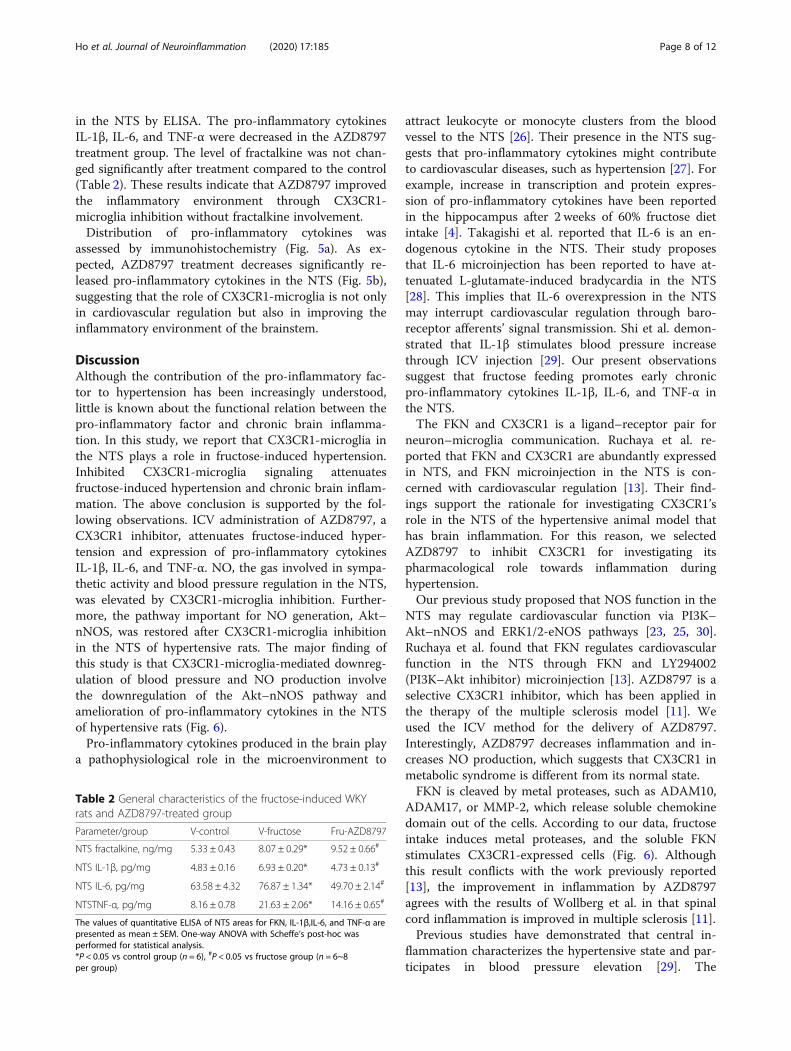
in the NTS by ELISA. The pro-inflammatory cytokinesIL-1β, IL-6, and TNF-α were decreased in the AZD8797treatment group. The level of fractalkine was not chan-ged significantly after treatment compared to the control(Table 2). These results indicate that AZD8797 improvedthe inflammatory environment through CX3CR1-microglia inhibition without fractalkine involvement.Distribution of pro-inflammatory cytokines was
assessed by immunohistochemistry (Fig. 5a). As ex-pected, AZD8797 treatment decreases significantly re-leased pro-inflammatory cytokines in the NTS (Fig. 5b),suggesting that the role of CX3CR1-microglia is not onlyin cardiovascular regulation but also in improving theinflammatory environment of the brainstem.
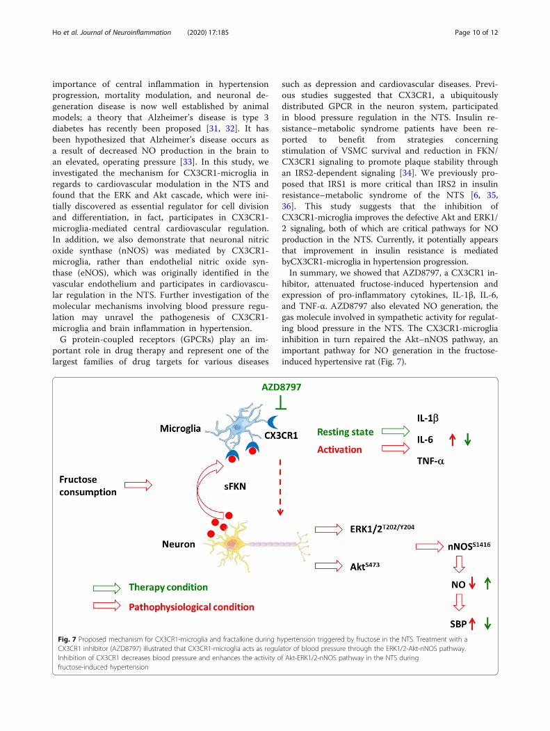
DiscussionAlthough the contribution of the pro-inflammatory fac-tor to hypertension has been increasingly understood,little is known about the functional relation between thepro-inflammatory factor and chronic brain inflamma-tion. In this study, we report that CX3CR1-microglia inthe NTS plays a role in fructose-induced hypertension.Inhibited CX3CR1-microglia signaling attenuatesfructose-induced hypertension and chronic brain inflam-mation. The above conclusion is supported by the fol-lowing observations. ICV administration of AZD8797, aCX3CR1 inhibitor, attenuates fructose-induced hyper-tension and expression of pro-inflammatory cytokinesIL-1β, IL-6, and TNF-α. NO, the gas involved in sympa-thetic activity and blood pressure regulation in the NTS,was elevated by CX3CR1-microglia inhibition. Further-more, the pathway important for NO generation, Akt–nNOS, was restored after CX3CR1-microglia inhibitionin the NTS of hypertensive rats. The major finding ofthis study is that CX3CR1-microglia-mediated downreg-ulation of blood pressure and NO production involvethe downregulation of the Akt–nNOS pathway andamelioration of pro-inflammatory cytokines in the NTSof hypertensive rats (Fig. 6).Pro-inflammatory cytokines produced in the brain play
a pathophysiological role in the microenvironment to
attract leukocyte or monocyte clusters from the bloodvessel to the NTS [26]. Their presence in the NTS sug-gests that pro-inflammatory cytokines might contributeto cardiovascular diseases, such as hypertension [27]. Forexample, increase in transcription and protein expres-sion of pro-inflammatory cytokines have been reportedin the hippocampus after 2 weeks of 60% fructose dietintake [4]. Takagishi et al. reported that IL-6 is an en-dogenous cytokine in the NTS. Their study proposesthat IL-6 microinjection has been reported to have at-tenuated L-glutamate-induced bradycardia in the NTS[28]. This implies that IL-6 overexpression in the NTSmay interrupt cardiovascular regulation through baro-receptor afferents’ signal transmission. Shi et al. demon-strated that IL-1β stimulates blood pressure increasethrough ICV injection [29]. Our present observationssuggest that fructose feeding promotes early chronicpro-inflammatory cytokines IL-1β, IL-6, and TNF-α inthe NTS.The FKN and CX3CR1 is a ligand–receptor pair for
neuron–microglia communication. Ruchaya et al. re-ported that FKN and CX3CR1 are abundantly expressedin NTS, and FKN microinjection in the NTS is con-cerned with cardiovascular regulation [13]. Their find-ings support the rationale for investigating CX3CR1’srole in the NTS of the hypertensive animal model thathas brain inflammation. For this reason, we selectedAZD8797 to inhibit CX3CR1 for investigating itspharmacological role towards inflammation duringhypertension.Our previous study proposed that NOS function in the
NTS may regulate cardiovascular function via PI3K–Akt–nNOS and ERK1/2-eNOS pathways [23, 25, 30].Ruchaya et al. found that FKN regulates cardiovascularfunction in the NTS through FKN and LY294002(PI3K–Akt inhibitor) microinjection [13]. AZD8797 is aselective CX3CR1 inhibitor, which has been applied inthe therapy of the multiple sclerosis model [11]. Weused the ICV method for the delivery of AZD8797.Interestingly, AZD8797 decreases inflammation and in-creases NO production, which suggests that CX3CR1 inmetabolic syndrome is different from its normal state.FKN is cleaved by metal proteases, such as ADAM10,
ADAM17, or MMP-2, which release soluble chemokinedomain out of the cells. According to our data, fructoseintake induces metal proteases, and the soluble FKNstimulates CX3CR1-expressed cells (Fig. 6). Althoughthis result conflicts with the work previously reported[13], the improvement in inflammation by AZD8797agrees with the results of Wollberg et al. in that spinalcord inflammation is improved in multiple sclerosis [11].Previous studies have demonstrated that central in-
flammation characterizes the hypertensive state and par-ticipates in blood pressure elevation [29]. The
Table 2 General characteristics of the fructose-induced WKYrats and AZD8797-treated group
Parameter/group V-control V-fructose Fru-AZD8797
NTS fractalkine, ng/mg 5.33 ± 0.43 8.07 ± 0.29* 9.52 ± 0.66#
NTS IL-1β, pg/mg 4.83 ± 0.16 6.93 ± 0.20* 4.73 ± 0.13#
NTS IL-6, pg/mg 63.58 ± 4.32 76.87 ± 1.34* 49.70 ± 2.14#
NTSTNF-α, pg/mg 8.16 ± 0.78 21.63 ± 2.06* 14.16 ± 0.65#
The values of quantitative ELISA of NTS areas for FKN, IL-1β,IL-6, and TNF-α arepresented as mean ± SEM. One-way ANOVA with Scheffe’s post-hoc wasperformed for statistical analysis.*P < 0.05 vs control group (n = 6), #P < 0.05 vs fructose group (n = 6~8per group)
Ho et al. Journal of Neuroinflammation (2020) 17:185 Page 8 of 12

Fig. 6 CX3CR1 inhibitor does not affect membrane-anchored FKN (mFKN), soluble FKN (sFKN), and ADAM10 levels in the NTS. a Immunoblottingwas performed for mFKN, sFKN, and ADAM10 protein expressions in the NTS. b The bar graph displays the protein content of mFKN, sFKN, andADAM10 in the NTS of hypertensive rats. The values represent mean ± SEM. One-way ANOVA with Scheffe’s post-hoc was performed for statisticalanalysis. *P < 0.05 compared to the control group (n = 6~8 per group)
Fig. 5 In situ quantitative immunohistochemical analysis of pro-inflammatory cytokines in the NTS following feeding with 10% fructose andAZD8797 treatment. a Qualitative analysis of IL-1β and IL-6 were observed by immunohistochemical staining after administration of the CX3CR1inhibitor AZD8797. The arrowhead indicates positive cells as a representative. The scale bar presents 50 μm. b Graphs depicting the quantitativeanalysis of the in situ cytokine-positive cells. The percentage was determined by counting pro-inflammatory positive cells in each hemisphere ofthe NTS at 200 × magnification. One-way ANOVA with Scheffe’s post-hoc was performed for statistical analysis. The values are represented asmean ± SEM. *P < 0.05 compared to control rats and #P < 0.05 compared to fructose-fed rats (n = 6~8 per group)
Ho et al. Journal of Neuroinflammation (2020) 17:185 Page 9 of 12
importance of central inflammation in hypertensionprogression, mortality modulation, and neuronal de-generation disease is now well established by animalmodels; a theory that Alzheimer’s disease is type 3diabetes has recently been proposed [31, 32]. It hasbeen hypothesized that Alzheimer’s disease occurs asa result of decreased NO production in the brain toan elevated, operating pressure [33]. In this study, weinvestigated the mechanism for CX3CR1-microglia inregards to cardiovascular modulation in the NTS andfound that the ERK and Akt cascade, which were ini-tially discovered as essential regulator for cell divisionand differentiation, in fact, participates in CX3CR1-microglia-mediated central cardiovascular regulation.In addition, we also demonstrate that neuronal nitricoxide synthase (nNOS) was mediated by CX3CR1-microglia, rather than endothelial nitric oxide syn-thase (eNOS), which was originally identified in thevascular endothelium and participates in cardiovascu-lar regulation in the NTS. Further investigation of themolecular mechanisms involving blood pressure regu-lation may unravel the pathogenesis of CX3CR1-microglia and brain inflammation in hypertension.G protein-coupled receptors (GPCRs) play an im-
portant role in drug therapy and represent one of thelargest families of drug targets for various diseases
such as depression and cardiovascular diseases. Previ-ous studies suggested that CX3CR1, a ubiquitouslydistributed GPCR in the neuron system, participatedin blood pressure regulation in the NTS. Insulin re-sistance–metabolic syndrome patients have been re-ported to benefit from strategies concerningstimulation of VSMC survival and reduction in FKN/CX3CR1 signaling to promote plaque stability throughan IRS2-dependent signaling [34]. We previously pro-posed that IRS1 is more critical than IRS2 in insulinresistance–metabolic syndrome of the NTS [6, 35,36]. This study suggests that the inhibition ofCX3CR1-microglia improves the defective Akt and ERK1/2 signaling, both of which are critical pathways for NOproduction in the NTS. Currently, it potentially appearsthat improvement in insulin resistance is mediatedbyCX3CR1-microglia in hypertension progression.In summary, we showed that AZD8797, a CX3CR1 in-
hibitor, attenuated fructose-induced hypertension andexpression of pro-inflammatory cytokines, IL-1β, IL-6,and TNF-α. AZD8797 also elevated NO generation, thegas molecule involved in sympathetic activity for regulat-ing blood pressure in the NTS. The CX3CR1-microgliainhibition in turn repaired the Akt–nNOS pathway, animportant pathway for NO generation in the fructose-induced hypertensive rat (Fig. 7).
Fig. 7 Proposed mechanism for CX3CR1-microglia and fractalkine during hypertension triggered by fructose in the NTS. Treatment with aCX3CR1 inhibitor (AZD8797) illustrated that CX3CR1-microglia acts as regulator of blood pressure through the ERK1/2-Akt-nNOS pathway.Inhibition of CX3CR1 decreases blood pressure and enhances the activity of Akt-ERK1/2-nNOS pathway in the NTS duringfructose-induced hypertension
Ho et al. Journal of Neuroinflammation (2020) 17:185 Page 10 of 12

ConclusionThis study provides the fundamental role of CX3CR1-microglia, which can be used for treating cardiovasculardisease because these receptors are involved in variouspathological conditions. Our novel findings suggest thatCX3CR1-microglia may be a potential candidate fortreating hypertension and relieving autonomic nervefunction through reestablishment of normal crosstalkbetween the neuron and microglia.
AbbreviationsBP: Blood pressure; CSF: Cerebral spinal fluid; CX3CL1: C-X3-C motifchemokine ligand 1; CX3CR1: C-X3-C motif chemokine receptor 1;dHDL: Direct high-density lipoprotein; FKN: Fractalkine, another term ofCX3CL1; HP-β-CD: 2-hydroxypropyl-β-cyclodextrin; IL-1β: Interleukin-1β; IL-6: Interleukin-6; TNF-α: Tumor necrosis factor-α; ICV: Intracerebroventricularinjection; mFKN: Membrane-anchored fractalkine; NO: Nitric oxide;nNOS: Neuronal nitric oxide synthase; NTS: Nucleus tractus solitarii;SBP: Systolic blood pressure; sFKN: Soluble fractalkine; WKY: Wistar-Kyoto rat
AcknowledgementsWe thank Dr. Yi-Ling Ma, Ms. Ya-Chu Chuang, Ms. Tzu-Jiun Kuo, and Mr. Jui-Hsiang Tseng for their excellent technical assistance and laboratory house-keeping during the experimental procedures.1) What is new?This study shows that neuroinflammation-regulated neuronal nitric oxidesynthase signaling in the nucleus tractus solitarii of hypertensive animals isstimulated by fructose intake. However, the most interesting result of thiswork is that the CX3CR1 inhibitor reverses the fructose-induced defect in theneuronal nitric oxide synthase signaling pathway by interrupting interactionbetween fractalkine and CX3CR1.2) What is relevant?The crosstalk of neuron-glia involves brainstem autonomic networks duringfructose-induced central inflammation. To understand the neuron–glia com-munication of hypertension, it is critical to develop treatment for neurogenichypertension. Our study reveals that microglial activity is central to neuroin-flammation and neuronal regulation of hypertension, which provides an im-portant aspect to this disease.SummaryThis study provides direct evidence for the CX3CR1 inhibitor in enhancingphosphatidylinositol3-kinase-Akt-neuronal nitric oxide synthase signaling inthe nucleus tractus solitarii to prevent fructose-induced hypertension andcentral inflammation, possibly through the downplay of neuron–glia cross-talk. Our findings provide new insights into the central nervous system inneuron and glia regulations and provide foundation for developing therapiesagainst neurogenic hypertension.
Authors’ contributionsCYH, PWC, YTL, and CJT designed and performed the experiments. PWC,WYH, GCS, YTL, MH, PJL, and CJT funded and supervised the research. CYHand HHC provided technical and material support for the animal surgery.CYH analyzed the data. CYH, PWC, and CJT wrote the manuscript. CHY andYTL have full access to all the data in this study and take responsibility fordata accuracy and integrity. The authors read and approved the finalmanuscript.
FundingThis work was supported by funding from the Ministry of Science andTechnology (MOST104-2320-B-075B-003 and MOST-107-2320-B-075B-002)and Kaohsiung Veterans General Hospital (VGHKS106-149, VGHKS106-G01-3,VGHKS107-168, and VGHKS107-G01-1) (to C.-J. T).
Availability of data and materialsAll data generated or analyzed during this study are included in thispublished article.
Ethics approval and consent to participateThis study was approved by the Institution Care and Use Committees ofKaohsiung Veterans General Hospital (Kaohsiung, Taiwan).
Consent for publicationNot applicable
Competing interestsThe authors declared that they have no competing interests.
Author details1Department of Medical Education and Research, Kaohsiung VeteransGeneral Hospital, Kaohsiung 81300, Taiwan. 2Institute of Biomedical Sciences,National Sun Yat-Sen University, Kaohsiung 80424, Taiwan. 3Section ofNeurology, Kaohsiung Veterans General Hospital, Kaohsiung 81300, Taiwan.4Center for Geriatrics and Gerontology, Kaohsiung Veterans General Hospital,Kaohsiung 81300, Taiwan. 5Shu-Zen Junior College of Medicine andManagement, Kaohsiung 82144, Taiwan. 6Division of General InternalMedicine, Department of Internal Medicine, Kaohsiung Medical UniversityHospital, Kaohsiung Medical University, Kaohsiung 80708, Taiwan. 7Division ofInternal Medicine, School of Medicine, College of Medicine, KaohsiungMedical University, Kaohsiung 80708, Taiwan. 8Department ofAnesthesiology, Kaohsiung Medical University Hospital, Kaohsiung 80708,Taiwan. 9Department of Anesthesiology, Faculty of Medicine, College ofMedicine, Kaohsiung Medical University, Kaohsiung 80708, Taiwan.10Genomics Research Center, Academia Sinica, Taipei 11529, Taiwan.11Institute of Clinical Medicine, National Cheng-Kung University, Tainan70101, Taiwan. 12Department of Medical Research, China Medical UniversityHospital, China Medical University, Taichung 40402, Taiwan.
Received: 27 January 2020 Accepted: 26 May 2020
References1. Hajjar I, Kotchen TA. Trends in prevalence, awareness, treatment, and
control of hypertension in the United States, 1988-2000. JAMA. 2003;290:199–206.
2. Williams B. Treating hypertension in patients with diabetes: when to startand how low to go? JAMA. 2015;313:573–74.
3. Sever PS, Dahlöf B, Poulter NR, Wedel H, Beevers G, Caulfield M, et al.Prevention of coronary and stroke events with atorvastatin in hypertensivepatients who have average or lower-than-average cholesterolconcentrations, in the Anglo-Scandinavian Cardiac Outcomes Trial—LipidLowering Arm (ASCOT-LLA): a multicentre randomised controlled trial.Lancet. 2003;361:1149–58.
4. Cigliano L, Spagnuolo MS, Crescenzo R, Cancelliere R, Iannotta L, Mazzoli A,et al. Short-term fructose feeding induces inflammation and oxidative stressin the hippocampus of young and adult rats. Mol Neurobiol. 2018;55:2869–83.
5. Xu M-X, Yu R, Shao L-F, Zhang Y-X, Ge C-X, Liu X-M, et al. Up-regulatedfractalkine (FKN) and its receptor CX3CR1 are involved in fructose-inducedneuroinflammation: suppression by curcumin. Brain Behav Immun. 2016;58:69–81.
6. Cheng P-W, Chen Y-Y, Cheng W-H, Lu P-J, Chen H-H, Chen B-R, et al. Wntsignaling regulates blood pressure by downregulating a GSK-3β–mediatedpathway to enhance insulin signaling in the central nervous system.Diabetes. 2015;64:3413.
7. Limatola C, Ransohoff RM. Modulating neurotoxicity through CX3CL1/CX3CR1 signaling. Front Cell Neurosci. 2014;8:229.
8. Sheridan GK, Murphy KJ. Neuron-glia crosstalk in health and disease:fractalkine and CX3CR1 take centre stage. Open Biol. 2013;3:130181.
9. Bhaskar K, Konerth M, Kokiko-Cochran ON, Cardona A, Ransohoff RM, LambBT. Regulation of tau pathology by the microglial fractalkine receptor.Neuron. 2010;68:19–31.
10. Hickman SE, Kingery ND, Ohsumi T, Borowsky M, Wang L-C, Means TK, et al.The microglial sensome revealed by direct RNA sequencing. Nat Neurosci.2013;16:1896–905.
11. Ridderstad Wollberg A, Ericsson-Dahlstrand A, Jureus A, Ekerot P, Simon S,Nilsson M, et al. Pharmacological inhibition of the chemokine receptorCX3CR1 attenuates disease in a chronic-relapsing rat model for multiplesclerosis. Proc Natl Acad Sci U S A. 2014;111:5409–14.
12. Poniatowski LA, Wojdasiewicz P, Krawczyk M, Szukiewicz D, Gasik R,Kubaszewski L, et al. Analysis of the role of CX3CL1 (fractalkine) and itsreceptor CX3CR1 in traumatic brain and spinal cord injury: insight into
Ho et al. Journal of Neuroinflammation (2020) 17:185 Page 11 of 12
recent advances in actions of neurochemokine agents. Mol Neurobiol. 2017;54:2167–88.
13. Ruchaya PJ, Paton JF, Murphy D, Yao ST. A cardiovascular role for fractalkineand its cognate receptor, CX3CR1, in the rat nucleus of the solitary tract.Neuroscience. 2012;209:119–27.
14. Tseng CJ, Liu HY, Lin HC, Ger LP, Tung CS, Yen MH. Cardiovascular effects ofnitric oxide in the brain stem nuclei of rats. Hypertension. 1996;27:36–42.
15. Krum H, Schlaich MP, Sobotka PA, Böhm M, Mahfoud F, Rocha-Singh K,et al. Percutaneous renal denervation in patients with treatment-resistanthypertension: final 3-year report of the Symplicity HTN-1 study. Lancet.2014;383:622–9.
16. Bray GA, Popkin BM. Dietary sugar and body weight: Have we reached acrisis in the epidemic of obesity and diabetes? Diabetes Care. 2014;37:950.
17. Tappy L, Lê K-A. Metabolic effects of fructose and the worldwide increase inobesity. Physiol Rev. 2010;90:23.
18. Lopes A, Vilela TC, Taschetto L, Vuolo F, Petronilho F, Dal-Pizzol F, et al.Evaluation of the effects of fructose on oxidative stress and inflammatoryparameters in rat brain. Mol Neurobiol. 2014;50:1124–30.
19. Glushakova O, Kosugi T, Roncal C, Mu W, Heinig M, Cirillo P, et al. Fructoseinduces the inflammatory molecule ICAM-1 in endothelial cells. J Am SocNephrol. 2008;19:1712–20.
20. Oudot C, Lajoix AD, Jover B, Rugale C. Dietary sodium restrictionprevents kidney damage in high fructose-fed rats. Kidney Int. 2013;83:674–83.
21. Yeh TC, Liu CP, Cheng WH, Chen BR, Lu PJ, Cheng PW, et al. Caffeine intakeimproves fructose-induced hypertension and insulin resistance byenhancing central insulin signaling. Hypertension. 2014;63:535–41.
22. Cheng PW, Ho WY, Su YT, Lu PJ, Chen BZ, Cheng WH, et al. Resveratroldecreases fructose-induced oxidative stress, mediated by NADPH oxidasevia an AMPK-dependent mechanism. Br J Pharmacol. 2014;171:2739–50.
23. Huang HN, Lu PJ, Lo WC, Lin CH, Hsiao M, Tseng CJ. In situ Aktphosphorylation in the nucleus tractus solitarii is involved in central controlof blood pressure and heart rate. Circulation. 2004;110:2476–83.
24. Paxinos G, Watson C. The rat brain in stereotaxic coordinates. 6th ed. SanDiego: Academic Press; 2007.
25. Cheng WH, Lu PJ, Ho WY, Tung CS, Cheng PW, Hsiao M, et al. Angiotensin IIinhibits neuronal nitric oxide synthase activation through the ERK1/2-RSKsignaling pathway to modulate central control of blood pressure. Circ Res.2010;106:788–95.
26. Waki H, Hendy EB, Hindmarch CC, Gouraud S, Toward M, Kasparov S, et al.Excessive leukotriene B4 in nucleus tractus solitarii is prohypertensive inspontaneously hypertensive rats. Hypertension. 2013;61:194–201.
27. Shi P, Raizada MK, Sumners C. Brain cytokines as neuromodulators incardiovascular control. Clin Exp Pharmacol Physiol. 2010;37:e52–7.
28. Takagishi M, Waki H, Bhuiyan MER, Gouraud SS, Kohsaka A, Cui H, et al. Il-6microinjected in the nucleus tractus solitarii attenuates cardiac baroreceptorreflex function in rats. Am J Physiol Regul Integr Comp Physiol. 2009;298:R183–R90.
29. Shi P, Diez-Freire C, Jun JY, Qi Y, Katovich MJ, Li Q, et al. Brain microglialcytokines in neurogenic hypertension. Hypertension. 2010;56:297–303.
30. Ho WY, Lu PJ, Hsiao M, Hwang HR, Tseng YC, Yen MH, et al. Adenosinemodulates cardiovascular functions through activation of extracellularsignal-regulated kinases 1 and 2 and endothelial nitric oxide synthase in thenucleus tractus solitarii of rats. Circulation. 2008;117:773–80.
31. de la Monte SM, Wands JR. Alzheimer's disease is type 3 diabetes-evidencereviewed. J Diabetes Sci Technol. 2008;2:1101–13.
32. Mittal K, Mani RJ, Katare DP. Type 3 diabetes: cross talk betweendifferentially regulated proteins of type 2 diabetes mellitus and Alzheimer’sdisease. Sci Rep. 2016;6:25589.
33. Kruyer A, Soplop N, Strickland S, Norris EH. Chronic hypertension leads toneurodegeneration in the TgSwDI mouse model of Alzheimer's disease.Hypertension. 2015;66:175–82.
34. Martinez-Hervas S, Vinue A, Nunez L, Andres-Blasco I, Piqueras L, Real JT,et al. Insulin resistance aggravates atherosclerosis by reducing vascularsmooth muscle cell survival and increasing CX3CL1/CX3CR1 axis. CardiovascRes. 2014;103:324–36.
35. Cheng PW, Kang BH, Lu PJ, Lin SS, Ho WY, Chen HH, et al. Involvement oftwo distinct signalling pathways in IGF-1-mediated central control ofhypotensive effects in normotensive and hypertensive rats. Acta Physiol(Oxf). 2014;212:28–38.
36. Cheng PW, Lin YT, Ho WY, Lu PJ, Chen HH, Lai CC, et al. Fructose inducedneurogenic hypertension mediated by overactivation of p38 MAPK toimpair insulin signaling transduction caused central insulin resistance. FreeRadic Biol Med. 2017;112:298–307.
Publisher’s NoteSpringer Nature remains neutral with regard to jurisdictional claims inpublished maps and institutional affiliations.
Ho et al. Journal of Neuroinflammation (2020) 17:185 Page 12 of 12