cyclodextrin promotes atherosclerosis regression via ... s_sci transl me… · cyclodextrin...
TRANSCRIPT

R E S EARCH ART I C L E
ATHEROSCLEROS I S
http://sD
ownloaded from
Cyclodextrin promotes atherosclerosis regression viamacrophage reprogrammingSebastian Zimmer,1* Alena Grebe,2* Siril S. Bakke,2,3,4 Niklas Bode,1 Bente Halvorsen,5
Thomas Ulas,6 Mona Skjelland,7 Dominic De Nardo,2,8,9 Larisa I. Labzin,2 Anja Kerksiek,10
Chris Hempel,11 Michael T. Heneka,12 Victoria Hawxhurst,13 Michael L. Fitzgerald,13
Jonel Trebicka,14,15 Ingemar Björkhem,16 Jan-Åke Gustafsson,17 Marit Westerterp,18
Alan R. Tall,18 Samuel D. Wright,19 Terje Espevik,4 Joachim L. Schultze,3,6 Georg Nickenig,1
Dieter Lütjohann,10 Eicke Latz2,3,4,20†
Atherosclerosis is an inflammatory disease linked to elevated blood cholesterol concentrations. Despite ongoingadvances in the prevention and treatment of atherosclerosis, cardiovascular disease remains the leading cause ofdeathworldwide. Continuous retention of apolipoprotein B–containing lipoproteins in the subendothelial space causesa local overabundance of free cholesterol. Because cholesterol accumulation and deposition of cholesterol crystals (CCs)trigger a complex inflammatory response, we tested the efficacy of the cyclic oligosaccharide 2-hydroxypropyl-b-cyclodextrin (CD), a compound that increases cholesterol solubility in preventing and reversing atherosclerosis.We showed that CD treatment of murine atherosclerosis reduced atherosclerotic plaque size and CC load andpromoted plaque regression even with a continued cholesterol-rich diet. Mechanistically, CD increased oxysterolproduction in both macrophages and human atherosclerotic plaques and promoted liver X receptor (LXR)–mediated transcriptional reprogramming to improve cholesterol efflux and exert anti-inflammatory effects. In vivo,this CD-mediated LXR agonismwas required for the antiatherosclerotic and anti-inflammatory effects of CD as well asfor augmented reverse cholesterol transport. Because CD treatment in humans is safe and CD beneficially affects keymechanisms of atherogenesis, it may therefore be used clinically to prevent or treat human atherosclerosis.
tm.
on April 7
sciencemag.org/
INTRODUCTION
Atherosclerosis is the underlying pathology that causes heart attacks,stroke, and peripheral vascular disease. Collectively, these conditionsrepresent a common health problem, and current treatments are in-sufficient to adequately reduce the risk of disease development. Phar-macologic reduction (1–3) of high-cholesterol concentrations is amongthe most successful therapeutic approaches to reduce the risk of devel-oping cardiovascular disease and stroke, but adequate reduction of low-density lipoprotein (LDL) cholesterol is not possible in all patients.
1Medizinische Klinik und Poliklinik II, University Hospital Bonn, 53105 Bonn, Germany. 2In-stitute of Innate Immunity, University Hospital Bonn, 53127 Bonn, Germany. 3GermanCenter of Neurodegenerative Diseases (DZNE), 53127 Bonn, Germany. 4Centre of Molec-ular Inflammation Research, Norwegian University of Science and Technology, 7489Trondheim, Norway. 5Research Institute of Internal Medicine, Oslo University HospitalRikshospitalet, 0424 Oslo, Norway. 6Genomics and Immunoregulation, Life and MedicalSciences Institute, University of Bonn, 53115 Bonn, Germany. 7Department of Neurolo-gy, Oslo University Hospital Rikshospitalet, 0424 Oslo, Norway. 8Inflammation Division,The Walter and Eliza Hall Institute of Medical Research, Parkville, Victoria 3052, Australia.9Department of Medical Biology, The University of Melbourne, Parkville, Victoria 3010,Australia. 10Institute of Clinical Chemistry und Clinical Pharmacology, University HospitalBonn, 53105 Bonn, Germany. 11Addi and Cassi Fund, Reno, NV 89511, USA. 12Clinic andPolyclinic for Neurology, University Hospital Bonn, 53105 Bonn, Germany. 13Lipid Metabo-lismUnit, Center for Computational and Integrative Biology, Boston,MA 02114, USA. 14Med-izinische Klinik und Poliklinik I, University Hospital Bonn, 53105 Bonn, Germany. 15Faculty ofHealth Sciences, University of Southern Denmark Campusvej 55, DK-5230 Odense M, Den-mark. 16Division of Clinical Chemistry, Karolinska Institutet, Huddinge University Hospital,141 86 Huddinge, Sweden. 17Center for Nuclear Receptors and Cell Signaling, Universityof Houston, Houston, TX 77004, USA. 18Department of Medicine, Columbia University,New York, NY 10032, USA. 19CSL Behring, King of Prussia, PA 19406, USA. 20Departmentof Infectious Diseases and Immunology, University of Massachusetts Medical School,Worcester, MA 01605, USA.*These authors contributed equally to this work.†Corresponding author. E-mail: [email protected]
www.Sc
, 2016
Atherosclerosis is characterized by arterial wall remodeling, whichis initiated by the retention and accumulation of different classes oflipids in the subendothelial layer. Lipid deposition and the appearanceof cholesterol crystals (CCs) have been associated with the inductionof an inflammatory reaction in the vessel wall, which contributes tothe pathogenesis (4, 5). Patients with increased systemic inflammationhave increased risk of cardiovascular death, and studies are under wayto test whether anti-inflammatory treatment can reduce cardiovascularevent rates (6).
CCs, which can result from excessive cholesterol deposition in ather-osclerotic lesions, are among the proinflammatory triggers that con-tribute to the inflammatory response during atherogenesis (7). CCscan trigger complement activation and neutrophil extracellular trap(NET) formation, as well as induction of innate immune pathways(4, 5, 8–10). Hence, therapeutic strategies aimed at the prevention ofcholesterol phase transition or the removal of CCs could reduce tissueinflammation and disease progression.
Genetic approaches to increase the capacity of macrophages to re-move free cholesterol from atherosclerotic lesions have proven to behighly successful in preclinical trials (11). This prompted us to testwhether pharmacologically increasing cholesterol solubility, clearance,and catabolism can be exploited for the prevention or treatment ofatherosclerosis. 2-Hydroxypropyl-b-cyclodextrin (CD) is a U.S. Foodand Drug Administration (FDA)–approved substance used to solubilizeand entrap numerous lipophilic pharmaceutical agents for therapeuticdelivery in humans (12, 13). Although it has previously been shown thatCD increases cholesterol solubility, promotes the removal of cholesterolfrom foam cells in vitro, and initiates anti-inflammatory mechanisms(14–16), it remains unknown whether CD can exert antiatherogeniceffects in vivo.
ienceTranslationalMedicine.org 6 April 2016 Vol 8 Issue 333 333ra50 1

R E S EARCH ART I C L E
Here, we found that subcutaneous administration of CD pro-foundly reduced atherogenesis and induced regression of establishedatherosclerosis in mouse models. CD augmented dissolution of CCs,reducing their appearance in lesions. Furthermore, CD increased cho-lesterol metabolism and liver X receptor (LXR)–dependent cellular re-programming, which resulted in more efficient reverse cholesteroltransport (RCT) as well as reduced proinflammatory gene expression.The atheroprotective effect of CD was dependent on LXR expressionin myeloid cells transplanted into LDL receptor (LDLR)–deficient mice.These studies suggest that CD mediates atheroprotection by increasingproduction of oxysterols and LXR-dependent cellular reprogrammingand provide preclinical evidence that CD could be developed into aneffective therapy for atherosclerosis in humans.
hD
ownloaded from
RESULTS
CD treatment impairs atherogenesisTo investigate the efficacy of CD treatment in murine atherosclerosis,apolipoprotein E (ApoE−/−) deficient mice were fed a cholesterol-rich dietand concomitantly treated subcutaneously with CD or vehicle control for8 weeks. Although plasma cholesterol, the main driver of atherosclerosis,remained unaffected (Fig. 1A), CD treatment profoundly reduced ather-osclerotic lesions within the aortic root (Fig. 1B). Furthermore, we found
www.Sc
reduced amounts of CCs in atherosclerotic plaques of CD-treated miceas assessed by laser reflection microscopy (Fig. 1, C and D). CD did notinfluence weight gain, blood pressure, heart rate, or the number of bonemarrow–derived or circulating sca1/flk1-positive cells (fig. S1, A to E).Moreover, plasma concentrations of phytosterols, cholestanol, and cho-lesterol precursors were not influenced by CD treatment, indirectlyshowing that CD did not alter enteric cholesterol uptake or overallendogenous biosynthesis (fig. S1F) (17). CD also did not change therelative plaque composition, including cellularity and macrophagecontent (Fig. 1, E and F). However, the production of aortic reactiveoxygen species (Fig. 1G) and plasma concentrations of proinflammatorycytokines were reduced by CD treatment (Fig. 1, H to J), suggesting thatCD may reduce the inflammatory response during atherogenesis.
CD treatment mediates regressionof atherosclerotic plaquesAlthough continuous drug administration in parallel to Western dietfeeding of mice is a standard protocol to investigate potential athero-protective substances (18), patients are generally not treated in earlystages of atherogenesis. Therefore, we tested the effect of CD treatmenton atherosclerosis regression. ApoE−/− mice are hypercholesterolemiceven on normal or lipid-reduced chow, and thus, most murine ather-osclerotic regression models rely on interventional strategies thatnormalize plasma lipids, such as viral gene transfer, transplantation,
on April 7, 2016
ttp://stm.sciencem
ag.org/
A D
B
C
E
F
G
H
I
J
Control CD0
200
400
600
800
Aor
tic R
OS
pro
duct
ion
(RLU
/ s/ m
g)
**
Control CD0
200
400
600
800
1000
Nuc
lei/p
laqu
e ar
ea
0.10
Control CD0.00
0.02
0.04
0.06
0.08
CD
68/p
laqu
e ar
ea
Control0
100
200
300
400
500
IL-1
β (p
g/m
l)
CD
*
Control0
10
20
30
40
IL-6
(pg
/ml)
CD
*
CDControl0
100
200
300
400
TN
F-α
(pg
/ml)
*
Pla
qu
e ar
ea(%
of t
otal
are
a)
***
Control CD0
20
40
60
80
**
Control CD0.0
0.1
0.2
0.3
0.4
Cry
stal
/pla
que
area
Pla
sma
chol
este
rol (
mg/
dl) n.s.
Control CD0
200
400
600
800
1000
Macrophages Nuclei Crystals
Control
CD
Fig. 1. CD treatment impairs murine atherogenesis. ApoE−/−mice werefed a cholesterol-rich diet for 8 weeks and concomitantly treated with CD
CCs; blue, nuclei stained with Hoechst. Enlarged images are the boxed areasin the left images. Scale bars, 500 mm. (E) Plaque cellularity shown as the ratio
(2 g/kg) or vehicle control by subcutaneous injection twice a week (n = 7to 8 per group). (A) Plasma cholesterol concentrations. (B) Atheroscleroticplaque area relative to total arterial wall area. (C) Plaque CC load shown asthe ratio of crystal reflection area to plaque area. (D) Representative imagesof the aortic plaques obtained by confocal laser reflection microscopy. Red,macrophages stained with anti-CD68 antibodies; white, reflection signal of
of nuclei to plaque area. (F) Plaque macrophage load shown as the ratio ofCD68 fluorescence area to total plaque area. (G) Aortic superoxide productiondetermined by L-012 chemiluminescence. ROS, reactive oxygen species; RLU,relative light units. (H to J) Plasma IL-1b, TNF-a, and IL-6 concentrations. Dataare shown as means + SEM. ***P < 0.001, **P < 0.01, and *P < 0.05, controlversus CD (unpaired two-tailed Student’s t test); n.s., not significant.
ienceTranslationalMedicine.org 6 April 2016 Vol 8 Issue 333 333ra50 2

R E S EARCH ART I C L E
Dow
nloa
or infusion of high-density lipoprotein (HDL) particles (19). Weadapted a less invasive regression protocol (20) in which ApoE−/−micewere first fed a cholesterol-rich diet for 8 weeks to induce advancedatherosclerotic lesions and then switched to a normal chow diet foranother 4 weeks during which CD or vehicle control was administered(Fig. 2A). As expected, plasma cholesterol concentrations were de-creased in both groups compared to baseline, but no difference betweencontrol and CD treatment was observed (Fig. 2B and fig. S2A). Al-though switching to a normal chow diet had no effect on athero-sclerotic lesion size in vehicle-treated mice, CD treatment resultedin a regression of atherosclerotic plaques by about 45% (Fig. 2C). Al-though CC load in lesions was already decreased in vehicle-treatedanimals compared to the load before treatment, CC amounts were furtherreduced by CD treatment (Fig. 2D). Because patients with cardiovasculardisease often do not adhere to the recommended lifestyle changes,which include dietary modifications, we next investigated whetherCD treatment can affect atherosclerosis regression during continuousenteric cholesterol challenge. CD or vehicle treatment was started after8 weeks of cholesterol-rich diet, which was continued for the entire12 weeks (Fig. 2E). Although plasma cholesterol and general cholesterol
www.Sc
on April 7, 2016
http://stm.sciencem
ag.org/ded from
metabolism were not altered (Fig. 2F and fig. S2B), atheroscleroticplaque size and CC load were decreased in CD-treated mice on contin-uous cholesterol-rich diet (Fig. 2, G and H). These data demonstratethat CD treatment is effective in reducing established plaques.
CD dissolves extra- and intracellular CCsThere are several possibilities to explain the protective effects of CDtreatment on both atherogenesis and established atherosclerosis. Be-cause CD is known to form soluble inclusion complexes with choles-terol, thereby enhancing its solubility in aqueous solutions by about150,000-fold, we tested whether CD increases the solubility of CCs.Fluorescent CD bound to the surface of CCs (Fig. 3, A and B) andCD mediated the solubilization of CCs in a dose-dependent manner(Fig. 3C). To be effective in atherosclerotic plaques, CD must also act onintracellular CCs. Macrophages rapidly internalized fluorescent CD (Fig.3D) and concentrated it in intracellular compartments (Fig. 3E). Further-more, incubation with 10 mM CD, a subtoxic dose (fig. S3), enhancedthe dissolution of intracellular CCs over time (Fig. 3F and fig. S4).
Metabolism of crystal-derived cholesterol is increased by CDMacrophages within the arterial wall take up excessive amounts ofcholesterol and transform into foam cells, a process that can impairmacrophage function and promote atherogenesis (21). This can bemimicked in vitro by loading macrophages with CCs (fig. S5). Afteruptake of CCs into phagosomes, cholesterol is moved from the lysosomevia the Niemann-Pick type C1 (NPC1) transporter to the endoplasmicreticulum, where acetyl–coenzyme A (CoA) acetyltransferase catalyzesthe formation of cholesteryl esters. This mechanism turns excess freecholesterol, which forms crystals and is cytotoxic, into cholesteryl estersthat can be stored in lipid droplets. A second pathway to metabolizefree cholesterol is the formation of water-soluble oxysterols. Oxysterolscan diffuse across cell membranes and are known to reprogrammacro-phages through activation of LXR, which in turn modulates the inflam-matory response and supports RCT to HDL (22–24). To study how CDinfluences the ability of macrophages to reduce the amount of cholesterolderived from CCs, we incubated macrophages with CCs prepared fromD6-cholesterol (D6-CCs) and followed D6-cholesterol metabolismproducts in cells and cellular supernatants by gas chromatography–massspectrometry selective ion monitoring (GC-MS-SIM) (Fig. 4A). Thisanalysis revealed that CD treatment promoted esterification of crystal-derived D6-cholesterol (Fig. 4B). Furthermore, CD amplified D6-cholesterolconcentrations in supernatants while reducing the overall cellular poolof D6-cholesterol (Fig. 4C). Hence, CD treatment increased the cho-lesterol efflux capacity of macrophages, which represents an importantprotective factor in patients with coronary artery disease (25, 26).Active cholesterol transport is mediated primarily by the adenosine5′-triphosphate–binding cassette transporters A1 and G1 (ABCA1and ABCG1), which transfer free cholesterol to ApoA1 and matureHDL particles, respectively (27). In line with the observed increasein cholesterol efflux capacity, macrophages incubated with CCs hadincreased expression of both ABCA1 and ABCG1, which was evenfurther enhanced by CD treatment (Fig. 4, D to F). Genes involvedin driving cholesterol efflux, including Abca1 and Abcg1, are underthe control of the LXR/retinoid X receptor (LXR/RXR) transcriptionapparatus (22, 28). Because the transcriptional activities of LXRs arepositively regulated by oxysterols, we next analyzed whether CD canpotentiate cholesterol oxidation. We found that CD treatment ofD6-CC–loaded macrophages resulted in a marked 15-fold increase in
F
E
G H
Contro
lCD
0
20
40
60
80 *
Pla
que
area
(% o
f tot
al a
rea)
Contro
lCD
*
0.0
0.1
0.2
0.3
0.4
Cry
stal
/pla
que
area
Contro
lCD
0
500
1000
1500
Pla
sma
chol
este
rol (
mg/
dl)
8 weeksTreatment trial Cholesterol-rich diet
4 weeks
Vehicle or CD
B
A
C D
Baseli
ne
Contro
lCD
0
20
40
60
80
**
***
Pla
que
area
(% o
f tot
al a
rea)
Baseli
ne
Contro
lCD
0
500
1000
1500***
***
Pla
sma
chol
este
rol (
mg/
dl)
8 weeksRegression trial Cholesterol-rich diet Normal chow
4 weeks
Vehicle or CD
Baseli
ne
Contro
lCD
0.0
0.1
0.2
0.3
0.4***
***
*
Cry
stal
/pla
que
area
n.s.
n.s.
n.s.
Fig. 2. CD treatment facilitates regression of murine atherosclerosis.ApoE−/− mice were fed a cholesterol-rich diet for 8 weeks to induce ad-
vanced atherosclerotic lesions. Then, the dietwas either changed to anormalchow (A toD) or the cholesterol-rich diet was continued for another 4weeks (EtoH).Micewere simultaneously treatedwithCD (2g/kg)or vehicle control twicea week (n = 6 to 8 per group). (A and E) Diet and treatment schemes. (B and F)Plasmacholesterol concentrations. (CandG)Atheroscleroticplaquearea relativeto total arterial wall area. (D and H) Plaque CC load shown as the ratio of crystalreflectionarea toplaquearea.Dataare shownasmeans+SEM. ***P<0.001, **P<0.01, and *P < 0.05, control versus CD (unpaired two-tailed Student’s t test).ienceTranslationalMedicine.org 6 April 2016 Vol 8 Issue 333 333ra50 3

R E S EARCH ART I C L E
on April 7, 2016
http://stm.sciencem
ag.org/D
ownloaded from
D6-cholesterol–derived 27-hydroxycholesterol (D5-27-hydroxycholesterol)(Fig. 4G), although the expression of Cyp27a1 was not altered (fig. S6).Unexpectedly, CD also increased 27-hydroxycholesterol productionand secretion from macrophages under normocholesterolemic condi-tions, meaning macrophages not treated with D6-CCs (Fig. 4H). Hence,CD increases the metabolism of free cholesterol and could therebylower the potential for its phase transition into crystals.
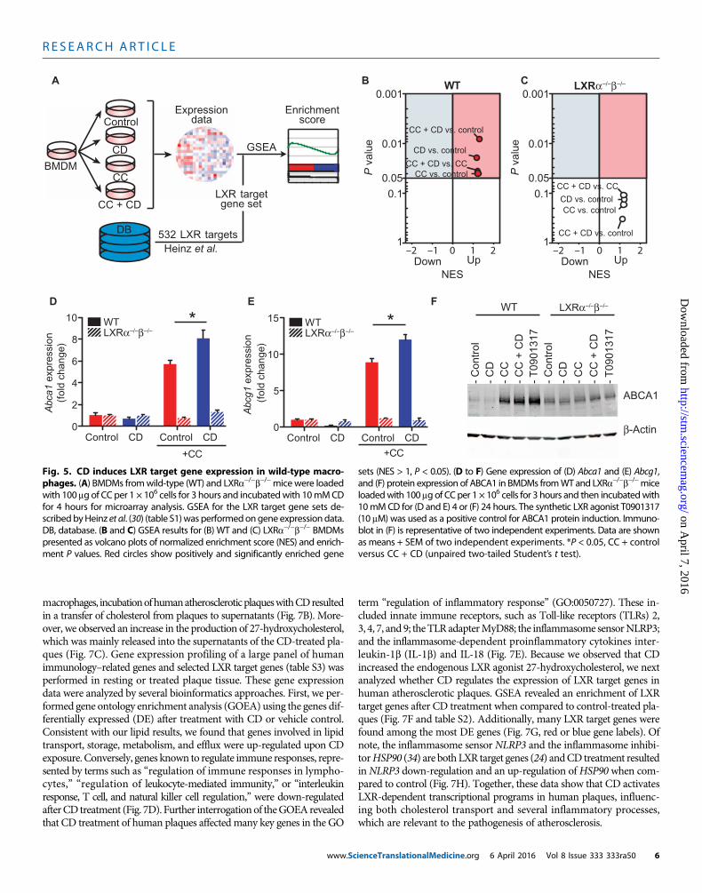
CD induces LXR target gene expression in macrophagesThe drastic CD-mediated increase in oxysterol production upon D6-CCloading and the unanticipated finding that CD can increase oxysterolsin normocholesterolemic macrophages prompted us to comprehensivelyinvestigate whether CD influences the expression profiles of LXR-regulated genes. Wild-type or LXRa−/−b−/− macrophages were ex-posed to CD, CC, or CC and CD, and gene expression was assessed
www.Sc
by genome-wide mRNA profiling. To investigate whether CD changesLXR target gene expression in macrophages, we performed gene setenrichment analysis (GSEA) (29) with a set of 533 of previously iden-tified LXR target genes (30) (Fig. 5A and table S1). Enrichment of LXRtarget gene sets was identified when wild-type macrophages wereincubated with CCs (Fig. 5B), presumably because of cholesterol over-loading of macrophages. Consistent with the strong induction of CC-derived 27-hydroxycholesterol and the observed increase in cholesterolefflux by CD, LXR target gene sets were enriched when CD was addedtogether with CCs (Fig. 5B). CD treatment alone also resulted in LXRgene set enrichment under normocholesterolemic conditions, whichcorrelates with the observed induction of cellular 27-hydroxycholesterol(Fig. 4H). In LXRa−/−b−/−macrophages, none of the conditions resultedin significant enrichments of LXR target gene sets (Fig. 5C). Further-more, these findings could be confirmed for the key LXR target genes
0 1 2 3 4 5 6 7 80
50
100
150
Crystal uptake Treatment
Was
h
Control
CD
Hours
Cry
stal
are
a (%
)
D E F
Per
cent
of m
ax
0 102 103 104 105
Rhodamine CD
0
20
40
60
80
100
Control
Rhodamine CD
CD90 min
0 min
CCs
0 0.01 0.1 1 10 1000
20
40
60
80
100
Filterable
Crystalline
CD (mM)
3 H-c
hole
ster
ol (
% to
tal)
Merge
Bright field
Merge
CD
CC
BA C
0 102 103 104 105
Rhodamine
0
20
40
60
80
100
% o
f max
Control
Rhodamine CD
Fig. 3. CD interactswithanddissolvesextra-and intracellularCCs. (AandB)CCs (1 mg) were incubated with 0.5 mM rhodamine-labeled CD or phosphate-
E) iMacs (immortalized macrophages) were loaded with 200 mg of CC per1 × 106 cells for 3 hours before incubation with 1 mM rhodamine-labeled
buffered saline as control. (A) Representative images obtained by confocallaser reflectionmicroscopy. Scale bar, 20 mm. (B)Quantification of rhodaminefluorescence on CCs by flow cytometry. (C) 3H-CCs were incubated with CDsolutions of the indicated concentrations overnight with shaking at 37°C.Upon filtration through 0.22-mm filter plates, radioactivity was determinedin the filtrate (filterable/solubilized) and the retentate (crystalline). (D and
CD. (D) Quantification of rhodamine fluorescence by flow cytometry. (E) Rep-resentative images obtained by confocal microscopy. Red, rhodamine-labeled CD; green, laser reflection signal. Scale bars, 5 mm. (F) IntracellularCC dissolution in BMDMs treated with 10 mM CD or control for the indicatedtimes determined by polarization microscopy. Data are shown as means± SEM of at least three independent experiments.
ienceTranslationalMedicine.org 6 April 2016 Vol 8 Issue 333 333ra50 4

R E S EARCH ART I C L E
on April 7, 2016
http://stm.sciencem
ag.org/D
ownloaded from
ABCA1 and ABCG1 in wild-type and LXRa−/−b−/− macrophages onthe mRNA and protein levels (Fig. 5, D to F) (31).
CD increases in vivo RCTTo test whether CD-induced LXR reprogramming of macrophages im-proves macrophage cholesterol efflux in vivo, bone marrow–derivedmacrophages (BMDMs) fromwild-type or LXRa−/−b−/−micewere loadedwith D6-CCs ex vivo and injected into the peritoneum of wild-type mice.The mice carrying crystal-loadedmacrophages were then treated with CDorvehicle control, andD6-cholesterol excretion into the feces andurinewasmonitored byGC-MS-SIM(Fig. 6A). CD increasedRCTof crystal-derivedD6-cholesterol fromwild-type and, to a lower extent, LXRa−/−b−/−macro-phages (Fig. 6B). Of note, CD treatment not only induced D6-cholesterolexcretion into the feces but also promoted urinary D6-cholesterol elimina-tion (Fig. 6C), a process that is normally not observed during RCT. Priorwork on NPC disease, a rare genetic disorder in which cholesterol cannotescape the lysosome,has shownthatCDcanmobilize lysosomal cholesterol
www.Sc
and activate LXR-dependent gene expression (32, 33). NPC1-deficientpatients receiveweekly injections of CDwith the aimof overcoming thischolesterol transport defect. To investigatewhetherCDcan also stimulateurinary cholesterol excretion in humans, wemonitored urinary choles-terol excretion of patients with NPC1 mutations after CD infusion overtime. CD, which is primarily excreted through the urinary tract, resultedin a time-dependent cholesterol excretion into the urine (Fig. 6D). Thesedata suggest that CD enhances in vivo RCT frommacrophages, partiallyin an LXR-dependent manner, but can also directly extract and transportcholesterol for excretion.
CD modifies human plaque cholesterol metabolism andgene expressionTo test whether the protective functions of CD onmurinemacrophagesare also exerted in human atherosclerotic plaques, we next performedlipid and genomic analyses on biopsy specimens obtained from carotidendarterectomies (Fig. 7A). Comparable to our findings in murine
G H
Control CD Control CD0
20
40
60
80
27-H
ydro
xych
oles
tero
l (ng
) SupernatantCells
******
Control CD Control CD0
2
4
6
8
1020406080
100 SupernatantCells
D5-2
7-hy
drox
ycho
lest
erol
(ng
)
B CA
Est
erifi
ed D
-ch
oles
tero
l(%
tota
l D -
chol
este
rol)
Con
trol
Med
ium
CD
Con
trol
Med
ium
CD
SupernatantCells
0
5
10
15
20
*n.s.
Con
trol
-
1 m
M C
D -
10 m
M C
D -
Con
trol
-
1 m
M C
D -
10 m
M C
D -
+CCβ-
Act
inA
BC
A1
D E F
Con
trol
Con
trol
CD
0
1
2
3
4
5
+CC
Abc
a 1 e
xpre
ssio
n(f
old
c han
ge)
*
+CC
Con
trol
Con
trol
CD
0
2
4
6
8
10
Abc
g1 e
xpr e
ssio
n(f
old
chan
ge)
*
Con
trol CD
CC
CC
+ C
D
0
2
4
6
8
AB
CA
1/ -a
ctin
(fol
d ch
ange
)
SupernatantCells
*** ***
D6
6
6
-cho
lest
erol
(%
tota
l)
Con
trol
Med
ium
CD
Con
tro l
Med
ium
CD
0
50
1006Free D -cholesterol
D6-cholesteryl ester D5-27-hydroxycholesterol
ACAT-1 Cyp27A1
Storage Passive efflux Active efflux
-CC
AB
CA
1/A
BC
G1
D6-
chol
este
rol
OH
LXR
6D
Fig. 4. CDmediatesmetabolism and efflux of crystal-derived cholesterol.(A) Macrophages loaded with CCs prepared fromD -cholesterol (D -CC) can
macrophages before CD treatment (control bar) and upon 24 hours of CDtreatment. (D to F) Gene expression of Abca1 and Abcg1 andprotein expres-
6 6reduce the amount of free, crystal-derived D6-cholesterol by three mainmechanisms. First, acetyl-CoA acetyltransferase (ACAT-1) can catalyze theformation of D6-cholesteryl esters, the storage form of cholesterol, which aredeposited in lipid droplets. Second, themitochondrial enzyme27-hydroxylase(Cyp27A1) can catalyze the formation of D5-27-hydroxycholesterol, which canpassively diffuse across cell membranes. Third, D5-27-hydroxycholesterol is apotent activator of LXR transcription factors, which in turn mediate the up-regulation of the cholesterol efflux transporters ABCA1 andABCG1. (B andC)iMacs loaded with 200 mg of D6-CC per 1 × 10⁶ cells for 3 hours were treatedwith 10 mM CD or vehicle control before GC-MS-SIM analysis of crystal-derived cholesterol. (B) Percentage of esterified D6-cholesterol in cell andsupernatant fractions before CD treatment (control bar) and after 48 hoursofCD treatment. (C) EffluxofD6-cholesterol into supernatantsofD6-CC–loaded
sion of ABCA1 in BMDMs loadedwith 100 mgof CCper 1×10⁶ cells for 3 hoursand then incubated with 10 mM CD or medium control for (D and E) 4 or (F)24 hours. Immunoblot in (F) is representative of three independent experi-ments, and densitometric analysis of all three experiments is provided for10 mM CD and presented as ABCA1 expression relative to the loading con-trol b-actin. Data are shown as means + SEM of at least three independentexperiments. (G) D5-27-hydroxycholesterol in cell and supernatant fractionsof iMacs loaded with 200 mg of D6-CC per 1 × 10⁶ cells for 3 hours before48 hours of treatment with 10mMCDormedium control, determined by GC-MS-SIM. (H) 27-Hydroxycholesterol in cell and supernatant fractions of iMacsafter 48 hours of treatment with 10mMCD ormedium control. ***P < 0.001and *P < 0.05, medium versus CD (B to C); CC + control versus CC + CD (Dto F); control versus CD (G and H) (unpaired two-tailed Student’s t test).
ienceTranslationalMedicine.org 6 April 2016 Vol 8 Issue 333 333ra50 5
R E S EARCH ART I C L E
on April 7, 2016
http://stm.sciencem
ag.org/D
ownloaded from
macrophages, incubationofhumanatheroscleroticplaqueswithCDresultedin a transfer of cholesterol from plaques to supernatants (Fig. 7B). More-over, we observed an increase in the production of 27-hydroxycholesterol,which was mainly released into the supernatants of the CD-treated pla-ques (Fig. 7C). Gene expression profiling of a large panel of humanimmunology–related genes and selected LXR target genes (table S3) wasperformed in resting or treated plaque tissue. These gene expressiondata were analyzed by several bioinformatics approaches. First, we per-formed gene ontology enrichment analysis (GOEA) using the genes dif-ferentially expressed (DE) after treatment with CD or vehicle control.Consistent with our lipid results, we found that genes involved in lipidtransport, storage, metabolism, and efflux were up-regulated upon CDexposure. Conversely, genes known to regulate immune responses, repre-sented by terms such as “regulation of immune responses in lympho-cytes,” “regulation of leukocyte-mediated immunity,” or “interleukinresponse, T cell, and natural killer cell regulation,” were down-regulatedafterCD treatment (Fig. 7D). Further interrogationof theGOEArevealedthat CD treatment of human plaques affectedmany key genes in the GO
www.Sc
term “regulation of inflammatory response” (GO:0050727). These in-cluded innate immune receptors, such as Toll-like receptors (TLRs) 2,3, 4, 7, and 9; theTLRadapterMyD88; the inflammasome sensorNLRP3;and the inflammasome-dependent proinflammatory cytokines inter-leukin-1b (IL-1b) and IL-18 (Fig. 7E). Because we observed that CDincreased the endogenous LXR agonist 27-hydroxycholesterol, we nextanalyzed whether CD regulates the expression of LXR target genes inhuman atherosclerotic plaques. GSEA revealed an enrichment of LXRtarget genes after CD treatment when compared to control-treated pla-ques (Fig. 7F and table S2). Additionally, many LXR target genes werefound among the most DE genes (Fig. 7G, red or blue gene labels). Ofnote, the inflammasome sensor NLRP3 and the inflammasome inhibi-torHSP90 (34) are both LXR target genes (24) andCD treatment resultedinNLRP3 down-regulation and an up-regulation ofHSP90 when com-pared to control (Fig. 7H). Together, these data show that CD activatesLXR-dependent transcriptional programs in human plaques, influenc-ing both cholesterol transport and several inflammatory processes,which are relevant to the pathogenesis of atherosclerosis.
WT
- C
ontr
ol-
CD
- C
C-
CC
+ C
D-
T09
0131
7-
Con
trol
- C
D-
CC
- C
C +
CD
- T
0901
317
Control CD Control CD0
2
4
6
8
10 WTLXRα–/–β–/–
Abc
a1 e
xpre
ssio
n(f
old
chan
ge)
(fol
d ch
ange
)
+CC
* WT
+CC
Control CD Control CD0
5
10
15
Abc
g1 e
xpre
ssio
n
*D FE
BMDM
CD
CC
CC + CD
Control
GSEA
LXR targetgene set
DB 532 LXR targetsHeinz et al.
Enrichmentscore
Expressiondata
A B CWT
P v
alue
0.05
0.001
0.01
0.1
1
NESUpDown
CC + CD vs. control
CD vs. control
CC + CD vs. CCCC vs. control
–2 –1 0 1 2
LXR
CC + CD vs. control
CD vs. control
CC + CD vs. CC
CC vs. control
P v
alue
0.05
0.001
0.01
0.1
1
NESUpDown
–2 –1 0 1 2
ABCA1
β-Actin
LXRα–/–β–/–
LXRα–/–β–/–
α–/–β–/–
Fig. 5. CD induces LXR target gene expression in wild-type macro-phages. (A) BMDMs fromwild-type (WT) and LXRa−/−b−/−micewere loaded
sets (NES > 1, P < 0.05). (D to F) Gene expression of (D) Abca1 and (E) Abcg1,and (F) protein expression of ABCA1 in BMDMs fromWT and LXRa−/−b−/−mice
with 100 mg of CC per 1 × 10⁶ cells for 3 hours and incubatedwith 10mMCDfor 4 hours for microarray analysis. GSEA for the LXR target gene sets de-scribedbyHeinz et al. (30) (table S1)was performedongene expressiondata.DB, database. (B and C) GSEA results for (B) WT and (C) LXRa−/−b−/− BMDMspresented as volcano plots of normalized enrichment score (NES) and enrich-ment P values. Red circles show positively and significantly enriched gene
loadedwith 100 mg of CC per 1 × 10⁶ cells for 3 hours and then incubatedwith10mMCD for (D and E) 4 or (F) 24 hours. The synthetic LXR agonist T0901317(10 mM) was used as a positive control for ABCA1 protein induction. Immuno-blot in (F) is representative of two independent experiments. Data are shownasmeans + SEM of two independent experiments. *P < 0.05, CC + controlversus CC + CD (unpaired two-tailed Student’s t test).
ienceTranslationalMedicine.org 6 April 2016 Vol 8 Issue 333 333ra50 6

R E S EARCH ART I C L E
on April 7, 2016
http://stm.sciencem
ag.org/D
ownloaded from
Atheroprotection by CD is LXR-dependentWe next tested whether the CD-mediated effects in isolated macro-phages in vitro– or in ex vivo–treated human plaque material reflectthe atheroprotective effects of CD in mice. Because CD treatmentlowered the systemic concentrations of LXR-modulated cytokines[IL-1b, IL-6, and tumor necrosis factor–a (TNF-a)] (Fig. 1, H to J)and also resulted in increased Abca1 and Abcg1 mRNA in the aorticarches of ApoE−/−mice fed a cholesterol-rich diet (fig. S7), we determinedwhether CD-mediated atheroprotection in vivo requires LXR activationand cholesterol efflux from macrophages through ABCA1 and ABCG1.We therefore transplanted wild-type, LXRa−/−b−/−, or macrophage-specificABCA1 and ABCG1 knockout (MAC-ABCDKO) bone marrow intoirradiated LDLR−/− mice. After bone marrow engraftment, the trans-planted mice were fed a cholesterol-rich diet and were concomitantlytreated with CD or vehicle control for 8 weeks. CD treatment did notinfluence plasma cholesterol concentrations in the different transplantgroups (Fig. 8, A to C). The lipoprotein profiles also remained un-changed, except that CD treatment slightly decreased the amount ofHDL in LDLR−/−mice transplanted with MAC-ABCDKO bone marrow(fig. S8). LDLR−/− mice carrying wild-type bone marrow showed re-duced atherosclerotic plaque size, demonstrating that CD is also effec-
www.Sc
tive in the LDLR−/− model of atherosclerosis (Fig. 8D). Of note, CDtreatment did not influence lesion development in LDLR−/− micecarrying LXRa−/−b−/− bone marrow, highlighting that LXR agonismis critical for CD-mediated atheroprotection (Fig. 8E). In contrast, de-ficiency of ABCA1 and ABCG1 in macrophages did not influence theeffectiveness of CD treatment (Fig. 8F), suggesting that CD can bypassthese cholesterol efflux pathways.
To better understand howCD-dependent LXR agonism can amelio-rate atherosclerosis, we performed a genome-wide gene expression anal-ysis on aortic tissue from LDLR−/− mice transplanted with wild-type orLXRa−/−b−/−bonemarrow.GOEAofDEgenesdemonstrated that impor-tant pathways involved in atherogenesis, including lipid metabolism andinflammation, were regulated by CD treatment in an LXR-dependentmanner (Fig. 8G). Similar to our studies on human plaques, LXR targetgeneswere found among the topDE genes uponCD treatment (Fig. 8H).Moreover, we confirmed our observation from human plaques that CDpromoted up-regulation of the NLRP3 inhibitor Hsp90aa1 and down-regulationofNLRP3 inflammasomegenes in anLXR-dependentmanner(Fig. 8I). Together, these data suggest that the CD-mediated athero-protection observed in murine atherosclerosis is dependent on LXRactivation and that CD exertsmultiple anti-inflammatory effects in ath-erosclerotic plaques.
DISCUSSION
Here, we tested the hypothesis that increasing the solubility of cholesterolby pharmacological means can have beneficial effects on diet-inducedatherosclerosis. The large effect observed and the unexpected ability ofCD to promote regression of established atherosclerosis even under theextreme hypercholesterolemic conditions observed during a cholesterol-rich diet cannot be explained by simple mass action of CD alone. Theresults from the lipid and genomic discovery approaches combined within vivo studies in gene-deficient mice suggest that CD exerts its potenteffect mainly by reprogramming cells in atherosclerotic plaques. Byincreasing the amount of endogenous LXR ligands, CD acts akin to a pro-drug except that it is not metabolized itself but rather promotes the me-tabolism of its cargo cholesterol into pharmacologically active metabolites.
It appears that transitory changes in cholesterol metabolism andinflammatory pathways are linked and that the activity of LXR is akey rheostat in this system. For example, innate immune activation bymicrobial components or the acute-phase response can suppress the ex-pression of LXR target genes, such as ABCA1 and ABCG1, causing cho-lesterol retention, which can augment an inflammatory reaction invarious ways (35, 36). It is conceivable that this type of innate immuneamplification could be part of an evolutionarily conserved antimicrobialdefense mechanism (23). The resulting cholesterol accumulation increasesLXR agonists, which in turn can counterbalance the inflammatory re-sponse and increase cholesterol efflux, restoring cholesterol and immunehomeostasis. However, because of the overabundance of proinflamma-tory dietary factors and an excess of cholesterol, this balance may beshifted toward chronic inflammation and cholesterol retention, whichdrive atherogenesis. By promoting cholesterol solubility, enhancing LXRactivity, and mobilizing cholesterol efflux, CD could therefore normal-ize both cholesterol and immune homeostasis in the vasculature.
The effect of CD on macrophages resembles that of the antiathero-genic factor HDL. HDL relieves cells of excess cholesterol throughABC transporters; in addition, HDL can have marked anti-inflammatory
C
A B
C
Vehicle CD Vehicle CD0
100
200
300
AU
C (u
rine
D6-
chol
est e
rol
g/d l
/h)
WT
WT LXR –/– –/–/
Control CD Control CD0
50
100
150
200
250
AU
C (f
ecal
D6-
chol
est e
rol n
g/h)
0 600 1200 18000
100
200
300
400
500
Time after CD infusion start (min)
Patient 3
Patient 2Patient 1
Cho
lest
erol
/cre
atin
ine
(µg/
mg)
D
Ex vivocrystal
loading
In vivotreatment
CD vs. control
GC-MS-SIManalysisin feces
and urine
D6-CC D
6-CC
D6-cholesterol
WT LXRα–/– –/–β
βα
LXR –/– –/–/βα
Fig. 6. CDfacilitatesRCTinvivoandpromotesurinarycholesterolexcretion.(A) BMDMs fromWT or LXRa−/−b−/−mice were loaded with 100 mg of D6-CC
per 1 × 10⁶ cells and injected into the peritoneumofWTmice. Subsequently,mice were treated subcutaneously with CD (2 g kg) or vehicle control (n = 4per group). (B and C) D6-cholesterol content in feces and urine collected ev-ery 3 hours over 30 hours after CD injection. Data are shown as total areaunder the curve (AUC) of excretedD6-cholesterol pooled from themicewith-in a group per time point. (D) Urine samples collected from three individualNPC1 patients upon intravenous application of CD for specific treatment ofNPC. Urine cholesterol concentration was determined by GC-MS-SIM andnormalized to urine creatinine excretion.ienceTranslationalMedicine.org 6 April 2016 Vol 8 Issue 333 333ra50 7

R E S EARCH ART I C L E
on April 7, 2016
http://stm.sciencem
ag.org/D
ownloaded from
D
A
B C
F
H
G
E
Reg
ulat
ion
of in
flam
mat
ory
resp
onse
-4
–6 –4 –2 0 2 4
ITLN1PPBPCDH5
MAPK11S100A8MASP1
CD34CLEC5A
MSR1MBP
CD276TNFRSF14
CD28HSP90AA1
XBP1C9
SREBF1ITGAL
ABCA1CIITA
MAP4K1MME
ABCG1ITGA4
LILRB3CCL26CXCL9PPARGCCL13
Fold change (CD vs. control)
P v
alue
0.05
0.001
0.01
0.1
1
NESUpDown
CD vs. control
–2 –1 0 1 2
NLRP3 ASC
SGT1 HSP90
IL-1βCASP1
CD
vs.
con
trol
–2 2
0
10
20
30
80
90
100
Cho
lest
erol
(%
tota
l)
SupernatantPlaque
Control CD Control CD
*
*
SupernatantPlaque
Control CD Control CD
***
0.00
0.01
0.02
0.03
0.04
0.05
27-H
ydro
xych
oles
tero
l (µg
/mg)
/ c
hole
ster
ol (µ
g/m
g)
Biopsy of human plaque Ex vivo culture Analysis
CD – +1 2
mRNA profiling
GC-MS-SIM
Regulation of phosphorylationand kinase activity
Regulation of cellular lipid transport,storage, metabolism, and efflux
Regulation oflymphocyte differentiation and activation
White blood cellactivation/proliferation/differentiation
Regulation of leukocyte-mediated immunityRegulation of immune response signaling
Regulation of inflammatory,immune, and defense response
Responseto external stimuli
and smooth muscle cell proliferation
Regulation of metabolic processes
Regulation of immune responsein lymphocytes
Metabolic processes, interleukinresponse, and T celland NK cell regulation
Regulation of phospholipid biosynthetic processes
Intracellular signaling pathways
Cholesterol transportand LDL receptor processes
Down-regulationIn CD vs. controlHighest
Highest
Up-regulationIn CD vs. control
EDNRBC5
PTGS2S100A8
IL6CCL24
TNFSF11IL15IL1B
MASP1TLR9TLR3TLR7
SMAD3CFP
TLR2IL23A
IL1RL2S100A9
NT5EBIRC3
CX3CL1TNFAIP3
NFKB1SELE
IL10STAT5B
CCL3SOCS3
TNFRSF1BCTSS
IL18BIRC2CD59
CFBNLRP3IL2RA
PPARDXCL1NOD2CD46
C6CD55IL17FCFH
C2FOXP3MYD88C1QBP
ADACCR7IL6ST
IL20TNFSF4
RELAIL1R1JAK2
APOETNF
NLRP1C7
FCER1GLTA
STAT5AIDO1
PTPN2PTGER4
SERPING1IL1RL1CASP1
BCL6CASP5
CCL5CR1
TLR4TNFRSF11A
HLA-DRB1FCER1APYCARD
PRKCDCD24
CFICD276
CD28C3
NR1H3C9
PPARG
CD vs. control
0 3–3
Fig. 7. CD induces cholester-ol metabolism and an anti-
inflammatory LXR profile inhuman atherosclerotic carotidplaques. (A) Human atheroscle-rotic carotid plaques obtained bycarotid endarterectomy (n = 10)were split into twomacroscopical-ly equal pieces and cultured for24hourswith10mMCDor control.Half of the plaque tissue was usedfor mRNA profiling with nCounterAnalysis System (NanoString Tech-nologies), and the other half andthe culture supernatantwere ana-lyzed by GC-MS-SIM. (B) Cholester-ol efflux from plaque tissue intosupernatants displayed as percentof total cholesterolper sample.C)Dis-tribution of 27-hydroxycholesterolrelative to cholesterol in plaqueand supernatant. (D) GOEA of DEgenes (fold change > 1.3, P < 0.05)visualized as GO network, wherered nodes indicate GO term en-richmentbyup-regulatedDEgenesand blue borders indicate GO termenrichment by down-regulated DEgenes. Node size and border widthrepresent the corresponding falsediscovery rate (FDR)–adjusted en-richment P value (q value). Edgesrepresent the associations betweentwo enriched GO terms based onshared genes, and edge thicknessindicates the overlap of genesbetween neighbor nodes. Highlyconnected terms were groupedtogether and were annotatedmanually by a shared general term.(E) Heat map of genes involved inthe GO term “regulation of inflam-matory response” (GO:0050727).Color bar indicates fold change.(F) Volcano plot of NES and en-richment P values based on GSEAfor the LXR target gene set (tableS2). Red circle indicates positiveand significant enrichment of theLXR target gene set (NES > 1, P <0.05). (G) TopDEgenesdeterminedby three-way analysis of variance(ANOVA) (fold change > 1.5, P <0.05). LXR target genes are coloredin red or blue. (H) The expressionof genes relevant to the NLRP3 in-flammasome pathway. Color barindicates fold change. (B and C)Data are shown as means ± SEM.***P < 0.001 and *P < 0.05, CD ver-sus control (paired two-tailed Stu-dent’s t test).www.ScienceTranslationalMedicine.org 6 April 2016 Vol 8 Issue 333 333ra50 8

R E S EARCH ART I C L E
on April 7, 2016
http://stm.sciencem
ag.org/D
ownloaded from
effects on macrophages (37). However, HDL does not activate LXRbut increases the expression of activating transcription factor 3, akey repressor of innate immune pathways (37). In contrast, althoughCD has the ability to increase HDL-mediated RCT, it can also mobilizecholesterol for direct excretion into the urine and feces. These data sug-gest that CD can bypass ABCA1- and ABCG1-mediated active choles-terol efflux. Consistent with these observations, our bone marrowtransplantation studies indicate that the atheroprotective effects ofCD are independent of the active cholesterol efflux process mediatedby ABCA1 and ABCG1. This is in line with recent findings demonstratingthat synthetic LXR agonists mediate atheroprotection independent of mac-rophage ABCA1 and ABCG1 (38).
There are several limitations to our study. First, although our datademonstrate that CD promotes LXR activation in plaque macro-phages and that LXR is required in myeloid cells for CD-mediatedatheroprotection, we cannot exclude other unidentified pathways. Itis likely that CD-mediated atheroprotection is multifactorial and thatthe differential effects of CD such as physically increasing cholesterolsolubility and promoting cholesterol metabolism and efflux in macro-phages, as well as its anti-inflammatory properties, cannot easily be iso-lated. Second, although our data identified 27-hydroxycholesterol as theprimary LXR agonist upon CD treatment, other endogenous oxysterols
www.Sc
may also contribute to LXR activation. In this context, the functionalactivity of regulatory enzymes such as Cyp27A1 and the differential ef-fects in specific cell types would be interesting. Third, although our exvivo experiments with carotid artery plaques suggest that CD can in-duce atheroprotective pathways in human disease, specific clinical trialsare necessary to validate these findings.
Preclinical models showing the effectiveness of LXR agonism inpreventing murine atherosclerosis (39) and promoting atherosclerosisregression (40) provided promising prospects for clinical use of LXRagonists in atherosclerosis treatment, but the progression of therapeuticmolecules into the clinic was hampered by liver toxicity and lipogeniceffects (41). CD, on the other hand, is already in clinical use in humansfor the delivery of lipophilic drugs and has not shown relevant toxicity.Hence, repurposing CD for the treatment or prevention of atherosclerosiswould be feasible. Our studies provide a proof of principle that therapiesaimed at increasing the solubility and removal of macrophage cholesterolcould be an effective strategy for the treatment of atherosclerosis.
MATERIALS AND METHODS
See the Supplementary Materials.
A
B
C
D
E
F
G
H I
LDLR WT+ +
LDLR–/––/– –/– –/–LXRα β
Negative regulation of NFκBsignaling
Epithelial and endothelial cell differentiation
Immunoglobulinproduction and
secretion
Cell adhesion
Response to lipids
Chemotaxis
Immune response regulating receptor signaling pathways
Regulation of white blood cell activation and proliferation
GTPase signaling
Regulation of cellular movement
Regulation of metabolicand biosynthetic processes
Negative regulation of cell death Metabolic processes and
cell remodeling
Differentiation, proliferation,and morphogenesis
Regulation of cytokine production and cell differentiation
Down-regulationIn CD vs. controlHighest
Highest
Up-regulationIn CD vs. control
–2 –1 0 1 2
S100A9CD79B
POU2F2MBP
PECAM1ITGA6
CXCL12NOTCH1NFKBIA
HSP90AA1ETS1
FKBP5BCL6
PDGFBCDKN1A
CDH5
WT-BMFold change (CD vs. control)
2 –2
NLRP3 ASC
SGT1 HSP90
IL-1βCASP1
NLRP3 ASC
SGT1 HSP90
IL-1βCASP1
CD vs. control
CD vs. control
0
100
200
300
400
500
0
20
40
60
0
100
200
300
400
500
0
20
40
60
0
100
200
300
400
500
0
20
40
60
Control CD Control CD
Control CD Control CD
*
**
Control CD Control CD
Pla
sma
chol
este
rol (
mg/
dl)
Pla
sma
chol
este
rol (
mg/
dl)
Pla
que
area
(%
of t
otal
are
a)P
laqu
e ar
ea (
% o
f tot
al a
rea)
Pla
sma
chol
este
rol (
mg/
dl)
Pla
que
area
(%
of t
otal
are
a)
LDLR
LDLRMAC-ABCDKO
WT LDLR
LDLR
WT
+
+
LDLR
–/–
–/–
–/–
–/–
LXR
αβ
LDLR+–/–
–/–
–/–
–/–
–/– –/–LXRα β
Fig. 8. CD impairs atherogenesis and regulates metabolic and anti-inflammatoryprocesses in anLXR-dependentmanner. LDLR−/−micewere
borders indicateGO termenrichment bydown-regulatedDEgenes. Node sizeand border width represent the corresponding FDR-adjusted enrichment P
transplantedwithWT, LXRa−/−b−/−, orMAC-ABCDKObonemarrow. Theywerethen fed a cholesterol-rich diet for 8 weeks and concomitantly treated withCD (2 g/kg) or vehicle control twice a week (n = 6 to 8 per group). (A to C)Plasma cholesterol concentrations of CD- and vehicle-treated animals. (D to F)Atherosclerotic plaque area relative to total arterial wall area. (G to I) Descend-ing aortas of LDLR−/−mice transplantedwithWT andLXRa−/−b−/−bonemarrowwere used for gene expression analysis bymicroarray,with subsequent filtra-tion for the genes included in the human plaque mRNA profiling. (G) GOEAof DE genes (fold change > 1.3, P<0.05) visualized as GOnetwork, where rednodes indicate GO term enrichment by up-regulated DE genes and blue
value (q value). Edges represent the associations between two enrichedGOterms based on shared genes, and edge thickness indicates the overlap ofgenes between neighbor nodes. Highly connected terms were groupedtogether and were annotatedmanually by a shared general term. NFkB, nu-clear factor kB; GTPase, guanosine triphosphatase. (H) DE genes determinedby three-wayANOVA (foldchange>1.3,P<0.05) inaortasof LDLR−/−mice trans-plantedwithWT bonemarrow. LXR target genes are colored in red or blue.(I) The expression of genes relevant for the NLRP3 inflammasome pathway.Color bar indicates fold change. (A and F) Data are shown as means + SEM;**P<0.01 and *P<0.05, CDversus control (unpaired two-tailed Student’s t test).
ienceTranslationalMedicine.org 6 April 2016 Vol 8 Issue 333 333ra50 9

R E S EARCH ART I C L E
SUPPLEMENTARY MATERIALS
www.sciencetranslationalmedicine.org/cgi/content/full/8/333/333ra50/DC1Materials and MethodsFig. S1. CD treatment does not influence general cardiovascular parameters.Fig. S2. CD treatment does not alter plasma sterol concentrations in atherosclerosis regressiontrials.Fig. S3. CD (10 mM) does not affect the viability of murine macrophages.Fig. S4. CD mediates intracellular CC dissolution.Fig. S5. CC loading of macrophages induces lipid droplet accumulation.Fig. S6. CD does not affect Cyp27a1 expression.Fig. S7. CD treatment induces the expression of cholesterol efflux transporters in aortic archesof atherosclerotic mice.Fig. S8. CD treatment does not alter murine lipoprotein profiles.Table S1. LXR target gene list for GSEA analysis of BMDMs from wild-type and LXRa−/−b−/− mice.Table S2. LXR target gene list for GSEA analysis of human atherosclerotic plaques.Table S3. List of additional metabolic and regulatory genes (nCounter Panel-Plus).Table S4. Original data for all figures (provided as an Excel file).References (42–57)
on April 7, 2016
http://stm.sciencem
ag.org/D
ownloaded from
REFERENCES AND NOTES
1. J. G. Robinson, M. Farnier, M. Krempf, J. Bergeron, G. Luc, M. Averna, E. S. Stroes, G. Langslet,F. J. Raal, M. E. Shahawy, M. J. Koren, N. E. Lepor, C. Lorenzato, R. Pordy, U. Chaudhari,J. J. P. Kastelein; ODYSSEY LONG TERM Investigators, Efficacy and safety of alirocumabin reducing lipids and cardiovascular events. N. Engl. J. Med. 372, 1489–1499 (2015).
2. M. S. Sabatine, R. P. Giugliano, S. D. Wiviott, F. J. Raal, D. J. Blom, J. Robinson, C. M. Ballantyne,R. Somaratne, J. Legg, S. M. Wasserman, R. Scott, M. J. Koren, E. A. Stein; Open-Label Study ofLong-Term Evaluation against LDL Cholesterol (OSLER) Investigators, Efficacy and safety of evo-locumab in reducing lipids and cardiovascular events. N. Engl. J. Med. 372, 1500–1509 (2015).
3. Task Force Members, G. Montalescot, U. Sechtem, S. Achenbach, F. Andreotti, C. Arden,A. Budaj, R. Bugiardini, F. Crea, T. Cuisset, C. Di Mario, J. R. Ferreira, B. J. Gersh, A. K. Gitt,J. S. Hulot, N. Marx, L. H. Opie, M. Pfisterer, E. Prescott, F. Ruschitzka, M. Sabaté, R. Senior,D. P. Taggart, E. E. van der Wall, C. J. M. Vrints; ESC Committee for Practice Guidelines(CPG), J. L. Zamorano, S. Achenbach, H. Baumgartner, J. J. Bax, H. Bueno, V. Dean, C. Deaton,C. Erol, R. Fagard, R. Ferrari, D. Hasdai, A. W. Hoes, P. Kirchhof, J. Knuuti, P. Kohl, P. Lancellotti,A. Linhart, P. Nihoyannopoulos, M. F. Piepoli, P. Ponikowski, P. A. Sirnes, J. L. Tamargo, M. Tendera,A. Torbicki, W. Wijns, S. Windecker; Document Reviewers, J. Knuuti, M. Valgimigli, H. Bueno,M. J. Claeys, N. Donner-Banzhoff, C. Erol, H. Frank, C. Funck-Brentano, O. Gaemperli,J. R. Gonzalez-Juanatey, M. Hamilos, D. Hasdai, S. Husted, S. K. James, K. Kervinen, P. Kolh,S. D. Kristensen, P. Lancellotti, A. P. Maggioni, M. F. Piepoli, A. R. Pries, F. Romeo, L. Rydén,M. L. Simoons, P. A. Sirnes, P. G. Steg, A. Timmis, W. Wijns, S. Windecker, A. Yildirir, J. L. Zamorano,2013 ESC guidelines on the management of stable coronary artery disease: The Task Forceon the management of stable coronary artery disease of the European Society of Cardiology.Eur. Heart J. 34, 2949–3003 (2013).
4. F. J. Sheedy, A. Grebe, K. J. Rayner, P. Kalantari, B. Ramkhelawon, S. B. Carpenter, C. E. Becker,H. N. Ediriweera, A. E. Mullick, D. T. Golenbock, L. M. Stuart, E. Latz, K. A. Fitzgerald, K. J. Moore,CD36 coordinates NLRP3 inflammasome activation by facilitating intracellular nucleation ofsoluble ligands into particulate ligands in sterile inflammation. Nat. Immunol. 14, 812–820(2013).
5. P. Duewell, H. Kono, K. J. Rayner, C. M. Sirois, G. Vladimer, F. G. Bauernfeind, G. S. Abela, L. Franchi,G. Nuñez, M. Schnurr, T. Espevik, E. Lien, K. A. Fitzgerald, K. L. Rock, K. J. Moore, S. D. Wright,V. Hornung, E. Latz, NLRP3 inflammasomes are required for atherogenesis and activated bycholesterol crystals. Nature 464, 1357–1361 (2010).
6. P. M. Ridker, T. Thuren, A. Zalewski, P. Libby, Interleukin-1b inhibition and the prevention ofrecurrent cardiovascular events: Rationale and design of the Canakinumab Anti-inflammatoryThrombosis Outcomes Study (CANTOS). Am. Heart J. 162, 597–605 (2011).
7. A. Warnatsch, M. Ioannou, Q. Wang, V. Papayannopoulos, Neutrophil extracellular trapslicense macrophages for cytokine production in atherosclerosis. Science 349, 316–320 (2015).
8. S. Nymo, N. Niyonzima, T. Espevik, T. E. Mollnes, Cholesterol crystal-induced endothelialcell activation is complement-dependent and mediated by TNF. Immunobiology 219, 786–792(2014).
9. E. O. Samstad, N. Niyonzima, S. Nymo, M. H. Aune, L. Ryan, S. S. Bakke, K. T. Lappegård, O.-L. Brekke,J. D. Lambris, J. K. Damås, E. Latz, T. E. Mollnes, T. Espevik, Cholesterol crystals inducecomplement-dependent inflammasome activation and cytokine release. J. Immunol. 192,2837–2845 (2014).
10. R. Kiyotake, M. Oh-Hora, E. Ishikawa, T. Miyamoto, T. Ishibashi, S. Yamasaki, Human minclebinds to cholesterol crystals and triggers innate immune responses. J. Biol. Chem. 290,25322–25332 (2015).
www.Scie
11. K. J. Rayner, C. C. Esau, F. N. Hussain, A. L. McDaniel, S. M. Marshall, J. M. van Gils, T. D. Ray,F. J. Sheedy, L. Goedeke, X. Liu, O. G. Khatsenko, V. Kaimal, C. J. Lees, C. Fernandez-Hernando,E. A. Fisher, R. E. Temel, K. J. Moore, Inhibition of miR-33a/b in non-human primates raisesplasma HDL and lowers VLDL triglycerides. Nature 478, 404–407 (2011).
12. S. Gould, R. C. Scott, 2-Hydroxypropyl-b-cyclodextrin (HP-b-CD): A toxicology review. FoodChem. Toxicol. 43, 1451–1459 (2005).
13. T. Loftsson, P. Jarho, M. Másson, T. Järvinen, Cyclodextrins in drug delivery. Expert Opin.Drug Deliv. 2, 335–351 (2005).
14. S. M. Liu, A. Cogny, M. Kockx, R. T. Dean, K. Gaus, W. Jessup, L. Kritharides, Cyclodextrinsdifferentially mobilize free and esterified cholesterol from primary human foam cellmacrophages. J. Lipid Res. 44, 1156–1166 (2003).
15. L. Kritharides, M. Kus, A. J. Brown, W. Jessup, R. T. Dean, Hydroxypropyl-b-cyclodextrin-mediated efflux of 7-ketocholesterol from macrophage foam cells. J. Biol. Chem. 271,27450–27455 (1996).
16. V. M. Atger, M. de la Llera Moya, G. W. Stoudt, W. V. Rodrigueza, M. C. Phillips, G. H. Rothblat,Cyclodextrins as catalysts for the removal of cholesterol from macrophage foam cells. J. Clin.Invest. 99, 773–780 (1997).
17. D. S. Mackay, P. J. H. Jones, Plasma noncholesterol sterols: Current uses, potential andneed for standardization. Curr. Opin. Lipidol. 23, 241–247 (2012).
18. G. S. Getz, C. A. Reardon, Animal models of atherosclerosis. Arterioscler. Thromb. Vasc. Biol.32, 1104–1115 (2012).
19. B. Hewing, E. A. Fisher, Preclinical mouse models and methods for the discovery of thecauses and treatments of atherosclerosis. Expert Opin. Drug Discov. 7, 207–216 (2012).
20. K. J. Rayner, F. J. Sheedy, C. C. Esau, F. N. Hussain, R. E. Temel, S. Parathath, J. M. van Gils,A. J. Rayner, A. N. Chang, Y. Suarez, C. Fernandez-Hernando, E. A. Fisher, K. J. Moore,Antagonism of miR-33 in mice promotes reverse cholesterol transport and regressionof atherosclerosis. J. Clin. Invest. 121, 2921–2931 (2011).
21. I. Tabas, Consequences of cellular cholesterol accumulation: Basic concepts and physio-logical implications. J. Clin. Invest. 110, 905–911 (2002).
22. J. J. Repa, D. J. Mangelsdorf, The liver X receptor gene team: Potential new players inatherosclerosis. Nat. Med. 8, 1243–1248 (2002).
23. A. R. Tall, L. Yvan-Charvet, Cholesterol, inflammation and innate immunity. Nat. Rev. Immunol.15, 104–116 (2015).
24. A. Reboldi, E. V. Dang, J. G. McDonald, G. Liang, D. W. Russell, J. G. Cyster, 25-Hydroxycholes-terol suppresses interleukin-1–driven inflammation downstream of type I interferon. Science345, 679–684 (2014).
25. A. V. Khera, M. Cuchel, M. de la Llera-Moya, A. Rodrigues, M. F. Burke, K. Jafri, B. C. French,J. A. Phillips, M. L. Mucksavage, R. L. Wilensky, E. R. Mohler, G. H. Rothblat, D. J. Rader,Cholesterol efflux capacity, high-density lipoprotein function, and atherosclerosis. N. Engl. J.Med. 364, 127–135 (2011).
26. A. Rohatgi, A. Khera, J. D. Berry, E. G. Givens, C. R. Ayers, K. E. Wedin, I. J. Neeland, I. S. Yuhanna,D. R. Rader, J. A. de Lemos, P. W. Shaul, HDL cholesterol efflux capacity and incidentcardiovascular events. N. Engl. J. Med. 371, 2383–2393 (2014).
27. D. J. Rader, E. Puré, Lipoproteins, macrophage function, and atherosclerosis: Beyond thefoam cell? Cell Metab. 1, 223–230 (2005).
28. B. A. Janowski, P. J. Willy, T. R. Devi, J. R. Falck, D. J. Mangelsdorf, An oxysterol signallingpathway mediated by the nuclear receptor LXRa. Nature 383, 728–731 (1996).
29. A. Subramanian, P. Tamayo, V. K. Mootha, S. Mukherjee, B. L. Ebert, M. A. Gillette, A. Paulovich,S. L. Pomeroy, T. R. Golub, E. S. Lander, J. P. Mesirov, Gene set enrichment analysis: A knowledge-based approach for interpreting genome-wide expression profiles. Proc. Natl. Acad. Sci. U.S.A.102, 15545–15550 (2005).
30. S. Heinz, C. Benner, N. Spann, E. Bertolino, Y. C. Lin, P. Laslo, J. X. Cheng, C. Murre, H. Singh,C. K. Glass, Simple combinations of lineage-determining transcription factors prime cis-regulatory elements required for macrophage and B cell identities. Mol. Cell 38, 576–589(2010).
31. J. J. Repa, S. D. Turley, J.-M. A. Lobaccaro, J. Medina, L. Li, K. Lustig, B. Shan, R. A. Heyman,J. M. Dietschy, D. J. Mangelsdorf, Regulation of absorption and ABC1-mediated efflux ofcholesterol by RXR heterodimers. Science 289, 1524–1529 (2000).
32. B. Liu, H. Li, J. J. Repa, S. D. Turley, J. M. Dietschy, Genetic variations and treatments thataffect the lifespan of the NPC1 mouse. J. Lipid Res. 49, 663–669 (2008).
33. A. M. Taylor, B. Liu, Y. Mari, B. Liu, J. J. Repa, Cyclodextrin mediates rapid changes in lipidbalance in Npc1−/− mice without carrying cholesterol through the bloodstream. J. Lipid Res.53, 2331–2342 (2012).
34. A. Mayor, F. Martinon, T. De Smedt, V. Pétrilli, J. Tschopp, A crucial function of SGT1 andHSP90 in inflammasome activity links mammalian and plant innate immune responses.Nat. Immunol. 8, 497–503 (2007).
35. A. Castrillo, S. B. Joseph, S. A. Vaidya, M. Haberland, A. M. Fogelman, G. Cheng, P. Tontonoz,Crosstalk between LXR and toll-like receptor signaling mediates bacterial and viral antagonismof cholesterol metabolism. Mol. Cell 12, 805–816 (2003).
36. K. R. Feingold, C. Grunfeld, The acute phase response inhibits reverse cholesterol transport.J. Lipid Res. 51, 682–684 (2010).
nceTranslationalMedicine.org 6 April 2016 Vol 8 Issue 333 333ra50 10

R E S EARCH ART I C L E
on April 7, 2016
http://stm.sciencem
ag.org/D
ownloaded from
37. D. De Nardo, L. I. Labzin, H. Kono, R. Seki, S. V. Schmidt, M. Beyer, D. Xu, S. Zimmer, C. Lahrmann,F. A. Schildberg, J. Vogelhuber, M. Kraut, T. Ulas, A. Kerksiek, W. Krebs, N. Bode, A. Grebe,M. L. Fitzgerald, N. J. Hernandez, B. R. G. Williams, P. Knolle, M. Kneilling, M. Röcken, D. Lütjohann,S. D. Wright, J. L. Schultze, E. Latz, High-density lipoprotein mediates anti-inflammatory reprogram-ming of macrophages via the transcriptional regulator ATF3. Nat. Immunol. 15, 152–160 (2014).
38. M. S. Kappus, A. J. Murphy, S. Abramowicz, V. Ntonga, C. L. Welch, A. R. Tall, M. Westerterp,Activation of liver X receptor decreases atherosclerosis in Ldlr−/− mice in the absence ofATP-binding cassette transporters A1 and G1 in myeloid cells. Arterioscler. Thromb. Vasc.Biol. 34, 279–284 (2014).
39. S. B. Joseph, E. McKilligin, L. Pei, M. A. Watson, A. R. Collins, B. A. Laffitte, M. Chen, G. Noh,J. Goodman, G. N. Hagger, J. Tran, T. K. Tippin, X. Wang, A. J. Lusis, W. A. Hsueh, R. E. Law,J. L. Collins, T. M. Willson, P. Tontonoz, Synthetic LXR ligand inhibits the development ofatherosclerosis in mice. Proc. Natl. Acad. Sci. U.S.A. 99, 7604–7609 (2002).
40. J. E. Feig, I. Pineda-Torra, M. Sanson, M. N. Bradley, Y. Vengrenyuk, D. Bogunovic, E. L. Gautier,D. Rubinstein, C. Hong, J. Liu, C. Wu, N. van Rooijen, N. Bhardwaj, M. Garabedian, P. Tontonoz,E. A. Fisher, LXR promotes the maximal egress of monocyte-derived cells from mouse aorticplaques during atherosclerosis regression. J. Clin. Invest. 120, 4415–4424 (2010).
41. X. Li, V. Yeh, V. Molteni, Liver X receptor modulators: A review of recently patentedcompounds (2007–2009). Expert Opin. Ther. Pat. 20, 535–562 (2010).
42. V. Hornung, F. Bauernfeind, A. Halle, E. O. Samstad, H. Kono, K. L. Rock, K. A. Fitzgerald, E. Latz,Silica crystals and aluminum salts activate the NALP3 inflammasome through phagosomaldestabilization. Nat. Immunol. 9, 847–856 (2008).
43. S. Wassmann, A. T. Bäumer, K. Strehlow, M. van Eickels, C. Grohé, K. Ahlbory, R. Rösen, M. Böhm,G. Nickenig, Endothelial dysfunction and oxidative stress during estrogen deficiency in spon-taneously hypertensive rats. Circulation 103, 435–441 (2001).
44. D. Lütjohann, C. Hahn, W. Prange, T. Sudhop, M. Axelson, T. Sauerbruch, K. von Bergmann,C. Reichel, Influence of rifampin on serum markers of cholesterol and bile acid synthesis inmen. Int. J. Clin. Pharmacol. Ther. 42, 307–313 (2004).
45. D. Lütjohann, A. Brzezinka, E. Barth, D. Abramowski, M. Staufenbiel, K. von Bergmann, K. Beyreuther,G. Multhaup, T. A. Bayer, Profile of cholesterol-related sterols in aged amyloid precursor proteintransgenic mouse brain. J. Lipid Res. 43, 1078–1085 (2002).
46. S. Zimmer, M. Steinmetz, T. Asdonk, I. Motz, C. Coch, E. Hartmann, W. Barchet, S. Wassmann,G. Hartmann, G. Nickenig, Activation of endothelial toll-like receptor 3 impairs endothelialfunction. Circ. Res. 108, 1358–1366 (2011).
47. J. Spandl, D. J. White, J. Peychl, C. Thiele, Live cell multicolor imaging of lipid droplets witha new dye, LD540. Traffic 10, 1579–1584 (2009).
48. D. Lütjohann, M. Stroick, T. Bertsch, S. Kühl, B. Lindenthal, K. Thelen, U. Andersson, I. Björkhem,K. von Bergmann, K. Fassbender, High doses of simvastatin, pravastatin, and cholesterol re-duce brain cholesterol synthesis in guinea pigs. Steroids 69, 431–438 (2004).
49. D. Lütjohann, O. Breuer, G. Ahlborg, I. Nennesmo, A. Sidén, U. Diczfalusy, I. Björkhem, Cholesterolhomeostasis in human brain: Evidence for an age-dependent flux of 24S-hydroxycholesterolfrom the brain into the circulation. Proc. Natl. Acad. Sci. U.S.A. 93, 9799–9804 (1996).
50. S. Maere, K. Heymans, M. Kuiper, BiNGO: A Cytoscape plugin to assess overrepresentationof gene ontology categories in biological networks. Bioinformatics 21, 3448–3449 (2005).
51. D. Merico, R. Isserlin, O. Stueker, A. Emili, G. D. Bader, Enrichment map: A network-basedmethod for gene-set enrichment visualization and interpretation. PLOS One 5, e13984(2010).
52. L. Oesper, D. Merico, R. Isserlin, G. D. Bader, WordCloud: A Cytoscape plugin to create avisual semantic summary of networks. Source Code Biol. Med. 6, 7 (2011).
53. T. G. Brott, J. L. Halperin, S. Abbara, J. M. Bacharach, J. D. Barr, R. L. Bush, C. U. Cates, M. A. Creager,S. B. Fowler, G. Friday, V. S. Hertzberg, E. B. McIff, W. S. Moore, P. D. Panagos, T. S. Riles,R. H. Rosenwasser, A. J. Taylor, 2011 ASA/ACCF/AHA/AANN/AANS/ACR/ASNR/CNS/SAIP/SCAI/SIR/SNIS/SVM/SVS Guideline on the Management of Patients With ExtracranialCarotid and Vertebral Artery Disease.A Report of the American College of CardiologyFoundation/American Heart Association Task Force on Practice Guidelines, and the
www.Scie
American Stroke Association, American Association of Neuroscience Nurses, AmericanAssociation of Neurological Surgeons, American College of Radiology, American Soci-ety of Neuroradiology, Congress of Neurological Surgeons, Society of AtherosclerosisImaging and Prevention, Society for Cardiovascular Angiography and Interventions,Society of Interventional Radiology, Society of NeuroInterventional Surgery, Societyfor Vascular Medicine, and Society for Vascular Surgery. Circulation 124, e54–e130(2011).
54. D. S. Mackay, P. J. H. Jones, S. B. Myrie, J. Plat, D. Lütjohann, Methodological considerationsfor the harmonization of non-cholesterol sterol bio-analysis. J. Chromatogr. B Analyt. Technol.Biomed. Life Sci. 957, 116–122 (2014).
55. P. Pehkonen, L. Welter-Stahl, J. Diwo, J. Ryynänen, A. Wienecke-Baldacchino, S. Heikkinen,E. Treuter, K. R. Steffensen, C. Carlberg, Genome-wide landscape of liver X receptor chromatinbinding and gene regulation in human macrophages. BMC Genomics 13, 50 (2012).
56. E. B. Mathiesen, K. H. Bønaa, O. Joakimsen, Echolucent plaques are associated with highrisk of ischemic cerebrovascular events in carotid stenosis: The tromsø study. Circulation103, 2171–2175 (2001).
57. P. S. Olofsson, K. Jatta, D. Wågsäter, S. Gredmark, U. Hedin, G. Paulsson-Berne, C. Söderberg-Nauclér,G. K. Hansson, A. Sirsjö, The antiviral cytomegalovirus inducible gene 5/viperin is expressed inatherosclerosis and regulated by proinflammatory agents. Arterioscler. Thromb. Vasc. Biol. 25,e113–e116 (2005).
Acknowledgments: Weappreciate the great technical assistanceof C. Lahrmann, A. Glubokovskih,G. Hack and S. Bellinghausen (University of Bonn), A. Marstad (Norwegian University of Science andTechnology), Z. Ali (Karolinska Institutet), and K. Krohg-Sørensen (Oslo University Hospital). We thankC. Hastings (Children’s Hospital & Research Center Oakland) for help with the acquisition of samplesfrom NPC patients and greatly appreciate the contribution of the Hadley Hope Fund (Medford). CDand rhodamine-labeled CD were provided by CTD Holdings Inc. LD540 was provided by C. Thiele(University of Bonn). Funding: Thisworkwas funded byNIH grants R01-HL093262, R21-HL113907-01(to E.L.), HL112661-01 (toM.L.F. and E.L.), HL101274 (toM.L.F.), and HL107653 (to A.R.T.); the ResearchCouncil of Norway through its Centres of Excellence funding scheme project no. 223255/F50 (toS.S.B., E.L., and T.E.); BONFOR (to S.Z. andN.B.); the Robert A.Welch Foundation (E-0004; to J.-Å.G.); theSwedish Science Council (H2416223; to J.-Å.G.); and grants from the Deutsche Forschungsgemeinschaft(DFG; SFB645, SFB670, SFB704, TRR83, and TRR57) (to E.L., J.L.S., and J.T.). E.L., M.T.H., and J.L.S. aremembers of the Excellence Cluster ImmunoSensation (Exc1023) funded by the DFG. Authorcontributions: S.Z. and A.G. designed, performed, and analyzed experiments. N.B. assisted withatherosclerosis mouse models and data analysis. S.S.B., L.I.L., B.H., M.S., J.T., A.K., and V.H. per-formed experiments. S.S.B., T.U., and J.L.S. performed bioinformatic analyses. J.-Å.G., M.T.H., I.B.,M.W., and A.R.T. provided knockout mice. D.D., G.N., T.E., M.L.F., S.D.W., and D.L. analyzed dataand provided critical suggestions and discussions throughout the study. C.H. provided the initialidea for the study. S.Z., A.G., and E.L. designed the study and wrote the manuscript. Competinginterests: S.D.W. is a full-time employee of CSL Limited, which does not work with CD. All theother authors declare that they have no competing interests. Data and materials availability:Accession codes for data in Gene Expression Omnibus: GSE67014 includes GSE67011 andGSE67013.
Submitted 11 October 2015Accepted 18 February 2016Published 6 April 201610.1126/scitranslmed.aad6100
Citation: S. Zimmer, A. Grebe, S. S. Bakke, N. Bode, B. Halvorsen, T. Ulas, M. Skjelland, D. De Nardo,L. I. Labzin, A. Kerksiek, C. Hempel, M. T. Heneka, V. Hawxhurst, M. L. Fitzgerald, J. Trebicka,I. Björkhem, J.-Å. Gustafsson, M. Westerterp, A. R. Tall, S. D. Wright, T. Espevik, J. L. Schultze,G. Nickenig, D. Lütjohann, E. Latz, Cyclodextrin promotes atherosclerosis regression viamacrophage reprogramming. Sci. Transl. Med. 8, 333ra50 (2016).
nceTranslationalMedicine.org 6 April 2016 Vol 8 Issue 333 333ra50 11

10.1126/scitranslmed.aad6100] (333), 333ra50. [doi:8Science Translational Medicine
Nickenig, Dieter Lütjohann and Eicke Latz (April 6, 2016) Samuel D. Wright, Terje Espevik, Joachim L. Schultze, GeorgBjörkhem, Jan-Åke Gustafsson, Marit Westerterp, Alan R. Tall,
IngemarVictoria Hawxhurst, Michael L. Fitzgerald, Jonel Trebicka, Larisa I. Labzin, Anja Kerksiek, Chris Hempel, Michael T. Heneka,Halvorsen, Thomas Ulas, Mona Skjelland, Dominic De Nardo, Sebastian Zimmer, Alena Grebe, Siril S. Bakke, Niklas Bode, Bentemacrophage reprogrammingCyclodextrin promotes atherosclerosis regression via
Editor's Summary
for the treatment of atherosclerosis.already known to be safe in humans, this drug is now a potential candidate for testing in human patients cholesterol crystals, and successfully treating atherosclerosis in a mouse model. Because cyclodextrin isdiscovered that cyclodextrin can also solubilize cholesterol, removing it from plaques, dissolving
. haveet alalready used as a solubilizing agent to improve delivery of various drugs. Now, Zimmer effectively treated with existing approaches. Cyclodextrin is a common FDA-approved substance that isworldwide, and additional therapies for this disease are greatly needed because not all patients can be
Cardiovascular disease resulting from atherosclerosis is one of the most common causes of deathDissolving away cholesterol
This information is current as of April 7, 2016. The following resources related to this article are available online at http://stm.sciencemag.org.
Article Tools
http://stm.sciencemag.org/content/8/333/333ra50article tools: Visit the online version of this article to access the personalization and
MaterialsSupplemental
http://stm.sciencemag.org/content/suppl/2016/04/04/8.333.333ra50.DC1"Supplementary Materials"
Related Content
http://stm.sciencemag.org/content/scitransmed/5/196/196ra100.fullhttp://stm.sciencemag.org/content/scitransmed/6/239/239sr1.fullhttp://stm.sciencemag.org/content/scitransmed/7/275/275fs7.fullhttp://stm.sciencemag.org/content/scitransmed/7/275/275ra20.full
's sites:ScienceThe editors suggest related resources on
Permissionshttp://www.sciencemag.org/about/permissions.dtlObtain information about reproducing this article:
is a registered trademark of AAAS.MedicineScience TranslationalAssociation for the Advancement of Science; all rights reserved. The title
Science, 1200 New York Avenue, NW, Washington, DC 20005. Copyright 2016 by the Americanweekly, except the last week in December, by the American Association for the Advancement of
(print ISSN 1946-6234; online ISSN 1946-6242) is publishedScience Translational Medicine
on April 7, 2016
http://stm.sciencem
ag.org/D
ownloaded from