cynara scolymus for relieving on nonalcoholic steatohepatitis
TRANSCRIPT

Research Article
CYNARA SCOLYMUS FOR RELIEVING ON NONALCOHOLIC STEATOHEPATITIS INDUCED IN RATS
SAFAA H MOHAMED1 HANAA H AHMED1 ABDEL RAZIK H FARRAG 2 NAHLA S ABDEL-AZIM3 ABDELAATY A SHAHAT34
1Department of Hormone 2 Department of Pathology 3Phytochemisrty Department National Research Centre El Bohous Street 12311 Dokki Cairo Egypt 4Medicinal Aromatic and Poisonous Plants Research Center College of Pharmacy King Saud University PO Box 2457
Riyadh 11451 Saudi Arabia Email aashahathotmailcom ashahatksuedusa
Received 12 Sep 2012 Revised and Accepted 29 Oct 2012
ABSTRACT
Objective The current study was undertaken to evaluate the efficacy of the total crude aqueous methanolic extract of Cynara scolymus and its fraction against high fat diet-induced of nonalcoholic steatohepatitis (NASH) in adult female rats Methods Forty adult female Sprague Dawley rats were classified into 4 groups The first group was kept on standard rodent chow and served as healthy control The other groups received high fat diet (HFD) for 32 weeks for NASH induction These animals were assigned as NASH-induced group Cynara scolymus (CSM) extract-treated group and purified fraction (CSF) -treated group Results The results revealed significant increase in serum ALT activity cholesterol LDL and triglycerides levels as well as leptin and resistin levels Additionally serum NF-κB TNF-α Cox-2 CD 40 and HGF levels have been increased significantly while serum HDL and adeponectin levels have been decreased significantly in NASH-induced group compared with healthy control group Conversely treatment with CSM or CSF resulted in significant decrease in serum ALT activity cholesterol LDL and triglycerides levels as well as leptin and resistin levels Serum NF-κB TNF-α Cox-2 CD40 and HGF levels also showed significant decrease While serum HDL and adiponectin levels were significantly increased as a consequence of treatment with either CSM or CSF as compared to the untreated NASH-induced rats The photomicrogrophs of liver section of rats treated with CSM or CSF extract confirmed the present improvement in the studied biomarkers The results suggested that Cynara scolymus extract or its purified fraction possess hepatoprotective activity hypolipidemic effect and anti-inflammatory property Conclusion Thus our findings reinforce current advice recommending the consumption of natural products to modulate nonalcoholic steatohepatitis and its metabolic complications
Keywords Cynara scolymus Nonalcoholic steatohepatitis Insulin resistance Inflammation Hyperlipidemia Rats
INTRODUCTION
Non-alcoholic fatty liver disease (NAFLD) is a clinic pathologic entity increasingly recognized as a major health burden in developed as well as in developing countries It includes a spectrum of liver damage ranging from simple steatosis to nonalcoholic steatohepatitis (NASH) advanced fibrosis and probable progression to cirrhosis [1] The presence of NASH with cirrhosis has been documented in large series Cirrhosis occurs in a minority of NASH patients but the overall incidence has been reported to be as high as 26 Progression of fibrosis as detected by liver biopsy has been reported to occur in 43 of NASH patients while 54 of patients remained unchanged and 3 showed histologic improvement during a follow-up from 1 to 7 years [2] In general 30-50 of individuals with NASH will develop fibrosis 15 will develop cirrhosis and 3 will progress to terminal liver failure [3] Among the many causative factors of NASH oxidative stress lipid peroxidation and inflammation are considered the most probable causative factors [4] NASH is believed to be a feature of metabolic syndrome because it is closely associated with visceral obesity dyslipidaemia insulin resistance and type 2 diabetes mellitus [5]
Artichoke (Cynara scolymus L) Asteraceae family (Compositae) is a plant that is widely grown in Mediterranean countries and is rich in natural antioxidants It is not only a good food known for its pleasant bitter taste but also an interesting and widespread herbal drug [6] Artichoke leaf contains up to 2 phenolic acids mainly 3-caffeoylquinic acid (chlorogenic acid) plus 13-di-O-caffeoylquinic acid (cynarin) and caffeic acid 04 bitter sesquiterpene lactones of which 47-83 is cynaropicrin 0110 flavonoids including the glycosides luteolin-7-β-rutinoside (scolymoside) luteolin-7- β-D-glucoside and luteolin-4-β-D-glucoside phytosterols (taraxasterol) sugars inulin enzymes and a volatile oil consisting mainly of the sesquiterpenes β-selinene and caryophyllene [7 8]
The artichoke leaf extract has been used as hepatoprotective [9] antimicrobial [10] and cholesterol reducing purposes [11] Artichoke has been found to decrease the production of reactive oxygen species the oxidation of low-density lipoproteins [12] lipid peroxidation [9] and protein oxidation and increase the activity of glutathione peroxidase [13]
The aim of the present article is to investigate the efficacy of Cynara scolymus total methanolic extract (CSM) and its fraction (CSF) against high fat diet-induced NASH in adult female rats in attempt to understand their mechanisms of action which may pave the way for possible therapeutic applications This could be achieved through conducting routine biochemical analysis for liver functions estimating the circulating levels of insulin resistance indices evaluating serum levels of inflammatory markers Histopathological investigation of liver sections was also carried out to confirm the biochemical analyses
MATERIALS AND METHODS
Plant materials
Preparation of Cynara scolomus total extracts (CSM)
The leaves of Cynara scolomus were collected from the experimental farm at Nubaria Alexandria Egypt on October 2009 air dried (3 kg) and extracted with 80 methanol at room temperature for three times followed by the removal of solvent under reduced pressure to obtain the crude aqueous methanolic extract (CSM) (26 from the dried leaves)
Preparation of Cynara scolomus fraction (CSF)
300 g of CSM was subjected to silica gel column chromatography and eluted with solvent of increasing polarity (hexaneethylacetatemethanol) The fractions eluted with ethyl acetatemethanol (11) were collected together to give a purified fraction (CSF) (120 g)
Animals
The present study was conducted on forty adult female Sprague Dawley rats weighing 120-150g obtained from the Animal House Colony of the National Research Centre Cairo Egypt The animals were maintained on standard laboratory diet and water ad libitum for two weeks before starting the experiment All animals received human care and use according to the guide lines for Animal Experiments which were approved by the Ethical Committee of Medical Research National Research Centre Egypt Steatohepatitis (NASH) was induced in rats by using high fat diet which provided
International Journal of Pharmacy and Pharmaceutical Sciences
ISSN- 0975-1491 Vol 5 Suppl 1 2013
AAccaaddeemmiicc SScciieenncceess
Shahat et al Int J Pharm Pharm Sci Vol 5 Suppl 1 57-66
58
30 of its energy from fat 35 from carbohydrate and 35 from protein (casein) for 32 weeks Supplements of vitamins and minerals were also included [14]
Experimental set-up
The animals were classified into four groups with ten animals in each (1) Healthy control group which was fed ad-libitum with an isocaloric regular rat chow [15] (2) Steatohepatitis (NASH) - induced group which was fed ad-libitum with high fat diet [14] (3) NASH -induced group orally treated with 150 mg kg bwt of CSM daily for 8 weeks This dose was calculated from the chronic toxicity study for CSM (data not shown) and (4) NASH -induced group orally treated with 150 mg kg bwt of CSF daily for 8 weeks This dose was calculated from the chronic toxicity study for CSF (data not shown)
At the end of the experimental period the rats were fasted overnight and the blood samples were collected from the retro orbital plexus under diethylether anaesthesia [16] The blood samples were left to clot and then centrifuged using cooling centrifuge at 1800 xg for ten minutes to obtain sera The clear serum samples were stored at -20 ordmC until analysis After blood collection all animals were rapidly killed and the liver tissues were dissected washed in isotonic saline then cut into small pieces (05x05cm) and fixed in 10 saline buffered formalin overnight for histological examination
Biochemical assays
Serum alanine transaminase (ALT) activity was estimated colorimetrically using kit purchased from Quimica Clinica Aplicada SA Co Spain according to the method of Reitman and Frankel [17] Serum cholesterol (Chol) concentration was determined colorimetrically using kit purchased from Stanbio Laboratory Boerne Texas USA according to the method of Allain et al [18] Serum LDL-cholesterol (LDL) concentration was assayed colorimetrically using kit purchased from Quimica Clinica Aplicada SA Co Spain according to the method of Assman et al [19] Serum HDL-cholesterol (HDL) concentration was measured colorimetrically using kit purchased from Stanbio Laboratory Boerne Texas USA according to the method of Lopez-Virella et al [20] Serum triglycerides (TG) level was determined colorimetrically using kit purchased from Stanbio Laboratory Boerne Texas USA according to the method of Fassati and Prencipe [21] Serum adiponectin concentration was measured by enzyme-linked immunosorbent assay (ELISA) technique using kit purchased from AssayPro USA according to the method of Pannacciulli et al [22] Serum leptin level was measured by ELISA procedure using kit purchased from Ray Biotech Co Georgia USA according to the method described by Petridou et al [23] Serum resistin
concentration was determined by ELISA technique using kit purchased from Glory Science Co Ltd Veterans Blvd Suite USA according to the method of Schaffler et al [24] Serum NF-κB p56 concentration was determined by ELISA technique using kit purchased from Glory Science Co Ltd Veterans Blvd Suite USA according to the manufacturerrsquos instructions Serum Cox-2 concentration was determined by ELISA technique using kit purchased from Glory Science Co Ltd Veterans Blvd Suite USA according to the manufacturerrsquos instructions Serum TNF-α concentration was measured by ELISA procedure using kit purchased from Ray Biotech Co Georgia USA according to the method of Brouckaert et al [25] Serum CD40 concentration was measured by ELISA technique using kit purchased from Glory Science Co Ltd Veterans Blvd Suite USA according to the manufacturerrsquos instructions Serum hepatocyte growth factor (HGF) level was quantified by ELISA procedure using kit purchased from Glory Science Co Ltd Veterans Blvd Suite USA according to the method of Plum et al [26]
Histopathological examination
Fragments of liver tissue previously fixed in 10 formalin saline were processed and submitted to hematoxilin and eosin (HampE) stain SCHARLACH Rs stain was used for a more precise identification of fatty change Histological variables were semiquantitated from 0 to 4+ including macro-and microvesicular fatty change the foci of necrosis portal and perivenular fibrosis as well as the inflammatory infiltrate
Statistical Analysis
In the present study all results were expressed as Mean + SE of the mean Data were analyzed by one way analysis of variance (ANOVA) using the Statistical Package for the Social Sciences (SPSS) program version 11 followed by least significant difference (LSD) to compare significance between groups [27] Difference was considered
significant when P value was gt 005 The percent difference was
calculated according to the following equation
difference = Treated group value ndash Control group value ∕ Control group value X 100
RESULTS
(Table 1) showed the effect of treatment with CSM and CSF on serum ALT activity and lipid profile in NASH-induced rats The NASH-induced group showed significant increase in serum ALT activity (608 ) in comparison with the healthy control group Conversely treatment with CSM or with CSF produced significant decrease in serum ALT activity (-409 and -396 respectively) in comparison with the untreated NASH-induced group
Table 1 Table shows the effect of treatment with CSM and CSF on serum ALT activity and lipid profile in NASH - induced rats
Triglycerides (mgdL) LDL (mgdL)
HDL (mgdL)
Cholesterol (mgdL)
ALT (UL)
Parameters Groups
648 plusmn 31 99 plusmn 02 413 plusmn 28 707 plusmn 17 354plusmn 32 Healthy control group 958 plusmn 30a
(478 ) 188 plusmn 11a
(8989 ) 208plusmn 11a
(-496 ) 1247 plusmn 37a
(763 ) 608plusmn17a (717)
NASH ndash induced group
758 plusmn 37b
(-208 ) 128 plusmn 05
(-319 ) 309 plusmn 25b
(485) 787 plusmn 25b
(-368 ) 409plusmn17b
(-327) NASH +CSM treated group
725 plusmn 34b
(-24369 )
105plusmn 05b
(4414)
337plusmn09b
(6201)
7547 plusmn 37b
(-354 )
386plusmn21b
(-365 )
NASH +CSF-treated group
a Significant change at P lt 005 in comparison with the healthy control group
b Significant change at P lt 005 in comparison with NASH-induced group
() percent difference with respect to the corresponding control value
The induction of NASH produced significant elevation in serum cholesterol LDL and triglycerides levels (1247 898 and 958 respectively) associated with significant decline in serum HDL level (-208) in comparison with the healthy control group On the other hand treatment of NASH-induced group with CSM resulted in significant depletion in serum cholesterol triglycerides levels and insignificant decrease in serum LDL level (-787 -758 and -
319 respectively) accompanied with significant rise in serum HDL level (485) in comparison with the untreated NASH-induced group Serum cholesterol LDL and triglycerides levels were significantly decreased by -754 -4414 and -725 respectively while serum HDL level was significantly increased by 6201 in NASH-induced group treated with CSF as compared to untreated NASH-induced group
Shahat et al Int J Pharm Pharm Sci Vol 5 Suppl 1 57-66
59
(Table 2) showed the effect of treatment with CSM and its fraction (CSF) on serum adiponectin leptin and resistin levels in NASH-induced rats Significant increase in serum leptin and resistin levels
(121 and 792) accompanied with significant decrease in serum adiponectin level (-336) were observed in NASH-induced group in comparison with the healthy control group
Table 2 Table shows the effect of treatment with CSM and CSF on serum adiponectin leptin and resistin levels in NASH-induced rats
Resistin (pgmL)
Leptin (pgmL)
Adiponectin (ngmL)
Parameters Groups
308 plusmn 0 5 3432 plusmn 26 104 plusmn 042 Healthy control group 552 plusmn 037a
(792 ) 7612 plusmn 25a
(121 ) 69 plusmn 02a
(-336 ) NASH ndash induced group
328 plusmn 037b
(-405 ) 5808 plusmn 24b
(-236 ) 88 plusmn 03b
(275) NASH + CSM-treated group
30 plusmn 039b
(-456 ) 5764 plusmn 28 b
(-242 )
93 plusmn 02b
(347 )
NASH +CSF-treated group
a Significant change at P lt 005 in comparison with the healthy control group
b Significant change at P lt 005 in comparison with NASH-induced group
() percent difference with respect to the corresponding control value
In contrast treatment of NASH-induced group with CSM or CSF resulted in significant decrease in serum leptin level (-236 and -242 respectively) and resistin level (-405 and -456 respectively) in concomitant with significant increase in serum adiponectin level (275 and 347 respectively) as compared to untreated NASH-induced group
(Table 3) showed the effect of treatment with CSM and CSF on serum NF-κBp56 TNF-α levels and Cox-2 activity in NASH-induced
rats Significant increase in serum NF-κBp56 TNF-α levels and Cox-2 activity (1031 676 and 903 respectively) was recorded in NASH-induced group in comparison with the healthy control group Conversely the treatment of NASH-induced group with CSM or CSF caused significant decrease in serum NF-κB p56 level (-446 and -476 respectively) TNF-α levels (-242 and -289 respectively) and Cox-2 activity (-25 and -651 respectively) as compared to the untreated NASH-induced group
Table 3 Table shows the effect of treatment with CSM and CSF on serum NF-κB TNF-α levels and Cox-2 activity in NASH-induced rats
Cox-2 (UL)
TNF-α (PgmL)
NF-κB (ngmL)
Parameters Groups
1303 plusmn 04 581 plusmn 18 064 plusmn 004 Healthy control group 248 plusmn 11a
(903 ) 974plusmn 12a
(676 ) 13 plusmn 01a
(1031 ) NASHndashinduced group
186 plusmn 03b
(-25 ) 738 plusmn 15b
(-242 ) 072plusmn 002b
(-446 )
NASH +CSM-treated group
162 plusmn 05b
(-343 )
692 plusmn 12b
(-289 )
068plusmn 003b
(-476) NASH+CSF-treated group
a Significant change at P lt 005 in comparison with the healthy control group
b Significant change at P lt 005 in comparison with NASH-induced group
() percent difference with respect to the corresponding control value
The effect of treatment with CSM or CSF on serum CD40 and HGF levels in NASH -induced rats was illustrated in (Table 4) The data revealed that the NASHndashinduced group showed significant increase in CD40 and HGF levels (954 and 885 respectively) in comparison with the
healthy control group Meanwhile treatment of NASH-induced group with CSM or CSF resulted in significant decrease in serum CD 40 (-31 and -342 respectively) and HGF levels (-234 and-281respectively) as compared to the untreated NASH-induced group
Table 4 Table shows the effect of treatment with CSM and CSF on serum CD40 and HGF levels in NASH-induced rats
HGF (ngL)
CD40 (ngL)
Parameters Groups
10240 plusmn 16 3772 plusmn 18 Healthy control group 19305 plusmn 14a (885 ) 7372 plusmn 29a (954 ) NASH ndash induced group 14770 plusmn 16b (-234 ) 5084 plusmn 26b (-31) NASH +CSM-treated group 13870 plusmn 21b (-281 ) 4850plusmn 14b (-342) NASH +CSF-treated group
a Significant change at P lt 005 in comparison with the healthy control group
b Significant change at P lt 005 in comparison with NASH-induced group
() percent difference with respect to the corresponding control value [[
Our histological study showed that there is no specific findings were observed during the hepatohistological examination of the healthy control rats (Fig1-A) Histopathological investigation of liver tissue slides stained with HampE in rats fed with high fat diet for induction of
NASH showed moderate to severe macrovesicular fatty changes which were diffusely distributed throughout the liver lobule Parenchymal inflammation with both acute and chronic inflammatory cells accompanying focal necrosis was also observed (Fig 1-B and 1-C)
Shahat et al Int J Pharm Pharm Sci Vol 5 Suppl 1 57-66
60
Fig 1A It shows liver section of healthy control rat showing intact histological structure of the liver Notice the central veins (CV) hepatocytes and blood sinusoids
Fig 1B It shows liver section of NASH induced rat showing a high degree of hepatocellular cytoplasmic vacuolation (macrovesicular and microvesicular steatosis)
Fig 1c It shows liver section of NASHndashinduced rat showing parenchymal inflammation with both acute and chronic inflammatory cells accompanying focal necrosis
Histological examination of liver tissues of NASH-induced group treated with CSM showed significant reduction in fatty infiltration as compared with that in the untreated NASH-induced group (Fig 1-D) Interestingly
histological investigation of liver tissues of NASH-induced group treated with the CSF revealed significant improvement in the degree of liver fatty changes which appeared like the healthy control group (Fig 1-E)
Fig 1D It shows liver section of NASH-induced rat treated with CSM showing significant reduction in fat deposits in liver tissues
Shahat et al Int J Pharm Pharm Sci Vol 5 Suppl 1 57-66
61
Fig 1E It shows liver section of NASH ndashinduced rat treated with CSF showing that the degree of liver steatosis was improved and the histologic feature was restored to nearly normal (H amp E x 300)
Histopathological examination of liver sections of healthy control rats stained with CSHARLACH Rs stain showed negative stain (Fig 2-A) Moderate macro- and microvesicular fatty changes in the periportal zone in the liver of NASHndashinduced rats were detected
(Fig 2-B) whereas in NASH-induced rats treated with CSM few of macro and microvesicular fatty changes were observed (Fig 2-C) Meanwhile no fatty infiltration was seen in liver of NASH-induced rats treated with CSF (Fig 2-D)
Fig 2A It shows liver section of healthy control rat showing normal histological structure of the liver The reaction is negative and the hepatocytes are slightly swollen with centrally placed nuclei No fatty change is seen
Fig 2B It shows liver section of NASHndashinduced rat showing the positive reaction in the macro and microvesicular fatty infiltration
Fig 2C It shows liver section of NASHndashinduced rat treated with the CSM showing significant reduction in fatty deposits in liver tissues and the reaction is negative in most areas of the lobules
Shahat et al Int J Pharm Pharm Sci Vol 5 Suppl 1 57-66
62
Fig 2D It shows liver section of NASHndashinduced rat treated with CSF showing that the reaction is negative indicating the improvement of fatty infiltration (SCHARLACH Rs x 300)
Mean fatty infiltration in the NASH-induced group was 3 (Table 5) Fat deposit in this group was classified as macrovesicular Mean fatty infiltration in the NASH-induced group treated with CSM or CSF
was 1 and fat deposit was mixed Fatty infiltration in the treated groups was significantly lower than that in the untreated NASH-induced group (P lt 005)
Table 5 Grades of fatty infiltration in the different studied groups
Groups Rats (n) Steatosis grades 0 1 2 3
Healthy control group 10 10 - - - NASH ndash induced group 10 - - 3 8 NASH +CSM-treated group 10 8 - 2 - NASH +CSF-treated group 10 9 - 1 -
DISCUSSION
The result of the present study revealed marked increase in serum ALT activity in NASH group which is in agreement with Hooper et al [28] Both aminotransferases (AST and ALT) are highly concentrated in the liver and the increasing serum ALT activity is considered a consequence of hepatocyte damage in NASH patients [29] A growing body of evidence supports the possibility that insulin resistance associated with adipose tissue inflammation and hepatic microvascular dysfunction as shown in our histological findings might actually contribute to the development andor progression of ALT activity in serum [30]
Treatment of NASH group with CSM extract or CSF fraction induced remarkable depletion in serum ALT activity In addition both of these treatments led to an improvement in the histological feature of the liver of the treated rats as shown in our results These effects could be attributed to the active ingredients in Cynara scolymus crude extract and fraction which are known as caffeoylquinic acid derivatives (cynarin and chlorogenic acid) These compounds have been proved to be effective in decreasing serum ALT activity [31] via their strong hepatoprotective effect and antioxidant capacity
The current results showed marked increase in serum cholesterol triglycerides and LDL-cholesterol in concomitant with significant decrease in serum HDL level in NASH group These results coincide with Adams et al [32] Cholesterol metabolism was associated with liver fat content independent on body weight implying that the more fat the liver contains the higher is cholesterol synthesis [33] Cellular cholesterol synthesis is regulated by activation of membrane bound transcription factors designated sterol regulatory element-binding proteins (SREBPs) which are the most abundant in the liver [34] and the excess of cellular cholesterol is esterified by the acyl CoA-cholesterol acyltransferase (ACAT) [35] The high level of cholesterol synthesis and the increased SREBP-2 activity has paradoxically been shown in subjects with NASH [36]
In NASH disease the ability of insulin to inhibit the production of very low density lipoproteins (VLDL) is impaired [37] This results in hyperinsulinemia and hypertriglyceridemia which in turn lead to lower HDL cholesterol concentration [38] This explains the diminished HDL serum level and the high triglycerides level in NASH
group in the current study The histopathological results of the present study showed macrovesicular and microvesicular steatosis Hepatic accumulation of triglycerides has been associated with the development of macrovesicular steatosis of the liver Since the inhibition of mitochondrial fatty acid metabolism is considered to result in microvesicular steatosis [39] secondary accumulation of cytosolic triglycerides and phospholipids in the presence of initial mitochondrial damage may explain the development of a mixed type of liver steatosis over time
The insufficient elimination of triglycerides probably caused by hepatic insulin resistance [40] may also contribute to the development of NASH Triglycerides are progressively reduced by the action of lipoprotein lipase (LPL) eventually resulting in intermediate-density lipoproteins (IDLs) and low-density lipoproteins (LDL) with relatively high cholesterol content [41] LDL circulates and is absorbed by the liver through binding of LDL to LDL receptor [42] In addition NAFLD ranging from simple steatosis to nonalcoholic steatohepatitis (NASH) is strongly associates with insulin resistance which caused inflammatory cytkokine tumor necrosis factor-alpha (TNF-α) to be over expressed in the liver TNF-α activates cholesterol synthesis and inhibits cholesterol elimination through bile acids which together contribute to increase LDL-cholesterol and decrease HDL-cholesterol [37]
Treatment of NASH group with CSM or CSF produced marked decrease in serum cholesterol triglycerides and LDL levels accompanied with significant increase in serum HDL Additionally histopathological investigation of liver tissue of the treated groups indicated a reduction in macrovesicular steatosis and microvesicular steatosis These results coincide with Lattanzio et al [43] who declared that the active compunds in Cynara scolomus extract represented by caffeic acid chlorogenic acid cynarin cynaroside scolymoside and have been found to affect cholesterol metabolism Daniel [44] reported that Cynara scolomus extract has anticholesterolemic action by decreasing rate of cholesterol synthesis in the liver and other tissue and this may be due to that Cynara scolomus contains some constituents as cynarin and luteolin which play a crucial role in inhibiting cholesterol and triglycerides synthesis Luteolin by beta glucosidase in digestive tract could cause
inhibition up to 60 of cholesterol synthesis [45] However highly
Shahat et al Int J Pharm Pharm Sci Vol 5 Suppl 1 57-66
63
significant decrease of plasma LDL and an increase of HDL in the treated groups are agreed with Cieslik et al [46] who reported decline tendency in total cholesterol LDL and VLDL when diets were supplemented with Cynara scolomus flour Moreover Taylor [47]
showed a decrease from 10 to 15 in total cholesterol LDL and
ratio of LDL to HDL cholesterol in serum due to treatment with Cynara scolomus leaves extract This could be explained as this extract contains active compounds as flavonoids and caffeoylquinic acid which have hypolipidemic effect These compounds could not only increase the breakdown of cholesterol to bile salts and enhance their elimination through increased bile production and flow but they also inhibit the internal production of cholesterol in liver [48] Furthermore Cynara scolomus extract may work through the indirect inhibition of enzyme hydroxyl methyle glutryle ndash CoA (HMG-CoA) which avoid problems occur with strong direct inhibitors of HMG-coA reductase during long treatment The indirect inhibition was supported by the fact that Cynara scolomus extract effectively blocked insulin-dependent stimulation of HMG-coA-reductase a key enzyme in cholesterol synthesis and HMG-coA reductase inhibitors generally reduce cholesterol LDL and triglycerides levels in serum [49]
The present data showed marked decrease in serum adiponectin level in NASH group It has been shown that adiponectin is found in relatively high circulating levels but it is decreased in patients with NASH and in clinical manifestations associated with insulin resistance such as metabolic syndrome (MS) and type 2 diabetes mellitus [50] In addition plasma adiponectin levels correlated inversely with the markers of systemic oxidative stress and oxidative stress is known to be a feature of liver disease Many studies hypothesized that oxidative stress has been demonstrated in conditions such as NAFLD and NASH due to the increased levels of free fatty acids and consequent increased levels of free radicals [51] In cultured adipocytes under oxidative stress condition the suppressed mRNA expression and secretion of adiponectin were detected This could be attributed to the decreased gene expression of adiponectin under this condition [52]
Treatment of NASH group with CSM or CSF showed marked increase in serum adiponectin level It has been demonstrated that Cynara scolomus extract contains natural antioxidants such as caffeoylquinic acid derivatives and flavonoids [53] that can regulate mRNA expression and secretion of adiponectin [52]
Serum leptin level showed significant increase in NASH group in the present study Leptin is released into the circulation by mature adipocytes in response to changes in body fat mass and nutritional status It has varied metabolic effects with the most significant of these being related to body weight and energy expenditure [54] In NASH patients leptin levels are elevated and are directly correlated with the severity of steatosis [55] The presence of hepatic steatosis despite the presence of hyperleptinemia suggests the development of leptin resistance [56] In addition leptin levels have been reported to be associated with oxidative stress conditions which enhance reactive oxygen species (ROS) formation in accumulated fat This leads to the elevated adipose nicotinamide adenine dinucleotide phosphate (NADPH) oxidase that leads to dysregulated production of leptin [52]
Treatment of NASH group with CSM or CSF resulted in appreciable decrease in serum leptin level as compared to the untreated NASH group Cynara scolomus active constituents (caffeic acid and chlorogenic acid) could reduce plasma cholesterol and triglycerides levels and this leads to a decrease in plasma leptin and an increase in adiponectin levels [57]
Serum resistin level in NASH group showed significant increase in comparison with the healthy control group This result is in agreement with Pagano et al [58] who reported that patients with NASH are characterised by high serum resistin level A major target organ of resistin is the liver where resistin induces insulin resistance and increases glucose production Resistin is related to hepatic fat content and insulin resistance [59] It has been suggested that resistin may contribute to hepatic steatosis by promoting insulin resistance and the increased resistin levels in NASH patients
are related to histological severity of the disease [60] Underlying liver damage and the progression of pure fatty liver to NASH and fibrosis the hepatic stellate cells produce a variety of cytokines including resistin Daniel reported that a genetic polymorphism in the promoter region of the resistin gene may be an independent predictor of circulating resistin level Hence it is possible that a gene polymorphism(s) may be responsible for the high resistin levels in NASH disease [58]
Treatment of NASH group with CSM or CSF produced remarkable decrease in serum resistin level Hepatoprotective effect of Cynara scolomus leaves extract may be assumed to be related to inducing glutathione peroxidase besides its direct antioxidant properties which may be useful for the prevention of oxidative stress that exerts an impact on endogenous expression of resistin in the adipocyte [31] Polyphenolic compounds in Cynara scolomus extract may be responsible for the suppression of hydrogen peroxide-induced oxidative stress [61] By this way CSM and its fraction (CSF) might reduce serum resistin level
The present results showed significant increase in serum NF-κB p56 level in NASH group High oxidative stress status in the liver of NAFLD patients with steatohepatitis may lead to modulation of Kupffer cell function through activation of transcription factors such as NF-κB [62] NF-κB then translocates from the cytoplasm to the nucleus to activate the inflammatory cytokines perturbing the inflammatory cycle [63]
Treatment of NASH group with CSM or CSF recorded marked decrease in serum NF-κB p56 level The inhibition of NF-κB activation correlated with suppression of inhibitor of NF-κB (IκBα) phosphorylation and degradation p65 nuclear translocation and NF-κB-dependent reporter gene transcription Cynara scolomus components mainly luteolin and apignen have been found to block IκBα phosphorylation and degradation [64] and in turn it could reduce NF-κB level Serum TNF-α level showed significant elevation in NASH group as compared to the healthy control group This could be attributed to the oxidative stress and stimulation of kupffer cell as well as stellate cell to secrete inflammatory cytokines such as TNF-α [65] Moreover it has been found that NAFLD patients have elevated plasma levels of lipopolysaccharide-binding protein (LBP) which are further increased in patients with NASH This increase is related to a rise in TNF-α gene expression in the hepatic tissue which supports a role of endotoxemia in the development of steatohepatitis [66]
Treatment of NASH group with CSM or CSF led to significant decrease in serum TNF-α in comparison with the untreated NASH group This effect could be attributed to the presence of luteolin and apignen in Cynara scolomus extract which could inhibit the inflammatory cytokine production in lipopolysaccharide-induced TNF- production [67]
Significant increase in serum Cox-2 activity was recorded in NASH group in the current work This could be explained as the oxidative stress which triggers lipid peroxidation and cytokines production such as TNF-α and interleukin (IL)-6 in the steatotic liver could mediate inflammatory recruitment directly or indirectly via activating NF-κB with upstream consequences that include cyclooxygenase-2 activity [68]
Treatment of NASH group with CSM or CSF produced significant decline in serum Cox-2 activity Cynara scolomus extract with its active consitutent (luteolin and apignen) has been found to block NF-κB expression [64] COX-2 which mediates prostaglandin synthesis during inflammation is induced by NF-κB [69] Thus the inhibition of NF-κB by Cynara scolomus extracts contributes in the inhibition of Cox-2 activity
The present data showed significant increase in serum CD40 level in NASH group This result is in agreement with Ercin et al [70] Soluble CD40 was not only correlated with BMI but was also more strongly related to lipid peroxidation [71] Circulating sCD40 was believed to derive predominantly from platelets associated with platelet activation and lipid peroxidation during oxidative stress conditions Thus oxidative stress plays a role in increasing platelet
Shahat et al Int J Pharm Pharm Sci Vol 5 Suppl 1 57-66
64
CD40 expression [72] Besides that the upregulation of CD40 is mediated by TNF-α which stimules platelet activation via interaction with its platelet receptors TNF-α has been shown to enhance oxidative stress via NADPH oxidase activation and TNF-α upregulated platelet CD40 via arachidonic acid-mediated oxidative stress [73] Treatment of NASH group with CSM or CSF resulted in significant deplation in serum CD40 Luteolin in Cynara scolomus extract may be responsible for this effect Luteolin could inhibit CD40 ligand expression by activated basophils [74]
The present results showed significant increase in serum hepatocyte growth factor (HGF) level in NASH group This result is in consistent with that of Koutsogiannis et al [75] It has been demonstrated that HGF mRNA produced by nonparenchymal cells increases in NASH patients [76] In NASH the activation of Kupffer cells and macrophages within liver tissue increased the production of NF-κB which induced the expression of HGF and consequently its level [4]
Treatment of NASH group with CSF resulted in marked decrease in serum HGF as compared to the untreated NASH group Luteolin and apignen in Cynara scolomus have been found to block NF-κB expression [64] and in turn they could indirectly reduce the stimulant of HGF expression and consequently its level [4]
The current study shed lights on the potential role of CSM and CSF in management of nonalcoholic steatohepatitis The active contituents of Cynara scolomus namely flavonoids and caffeoylquinic acid may be responsible for this effect These compounds have been proved to have hepatoprotective activity hypolipidemic effect antioxidant capacity and antiinflammatory property Beside that these compounds could modulate insulin resistance status associated with nonalcoholic steatohepatitis Therefore Cynara scolomus could have possible therapeutic application in chronic diseases accompanied with insulin resistance and severe inflammation
ACKNOWLEDGEMENTS
Work is partially supported by Science and Technology Development Fund (STDF) Egyptian Academy of Scientific Research and Technology ldquoID 245rdquo
REFERENCES
1 Paschos P Paletas K Non-alcoholic Fatty Liver Disease and Metabolic Syndrome Hippokratia 2009 1 9-19
2 Brunt ME Non-alcoholic Steatohepatitis Definition and Pathology Semin Liver Dis 2001 21 3-16
3 Fassio E Alvarez E Dominguez N Landeira G Longo C Natural History of Non-alcoholic Steatohepatitis a Longitudinal Study of Repeat Liver Biopsies Hepatolology 2004 40 820-26
4 Preiss D Sattar N Non-alcoholic Fatty Liver Disease an Overview of Prevalence Diagnosis Pathogenesis and Treatment Considerations Clinical Science 2008 115 141ndash50
5 Hůlek P Dresslerovaacute I Metabolic Syndrome and Liver (NAFLDNASH) Vnitr Lek 2009 55 646-9
6 Mulinacci N Prucher D Peruzzi M Romani A Pinelli P Giaccherini C Vincieri FF Commercial and Laboratory Extracts from Artichoke Leaves Estimation of Caffeoyl Esters and Flavonoidic Compounds Content Journal of Pharmaceutical and Biomedical Analysis 2004 34349-57
7 Leung AY Foster S Encyclopedia of Common Natural Ingredients Used in Food Drugs and Cosmetics 2nd ed New York John Wiley amp Sons Inc 1996 4244
8 Newall CA Anderson LA Phillipson JD Herbal Medicines A Guide for Health-Care Professionals London The Pharmaceutical Press 1996 3738
9 Speroni E Cervellati R Govoni P Guizzardi S Renzulli C Guerra MC Efficiency of Different Cynara Scolymus Preparations Liver Complaints J Ethnopharmacol 2003 86203ndash11
10 Zhu X Zhang H Lo R Phenolic Compounds from the Leaf Extract of Artichoke (Cynara Scolymus) and Their Antimicrobial Activities J Agric Food Chem 2004 527272ndash78
11 Kuumlskuuml-Kiraz Z Mehmetccedilik G Dogru-Abbasoglu S Uysal M Artichoke Leaf Extract Reduces Oxidative Stress and Lipoprotein Dyshomeostasis in Rats Fed on High Cholesterol Diet Phytother Res 2010 24565-70
12 Zapolska-Downar D Zapolski-Downar A Naruszewicz M Siennicka A Krasnodebska B Kolodziej B Protective Properties of Artichoke (Cynara Scolymus) against Oxidative Stress Induced in Cultured Endothelial Cells and Monocytes Life Science 2002 712897ndash908
13 Jimenez-Escrig A Dragsted LO Daneshvar B Pulido R Saura-Calixto F Invitro Antioxidant Activities of Edible Artichoke (Cynara Scolymus L) and Effect on Biomarkers of Antioxidants in Rats J Agric Food Chem 2003 515540ndash45
14 Tipoe GL Ho CT Liong EC Leung TM Lau TYH Fung ML Nanji AA Voluntary Oral Feeding of Rats not Requiring a Very High Fat Diet is a Clinically Relevant Animal Model of Non-alcoholic Fatty Liver Disease (NAFLD) Histol Histopathol 2009 241161-69
15 Reeves PG Nielsen FH Fahey GC AIN-93 Purified Diets for Laboratory Rodents Final Report of The American Institution Ad Hoc Writing Committee on The Reformulation of The AIN-76A Rodent diet J Nutr 1993 1231939-51
16 Sandford HS Method for Obtaining Venous Blood from The Orbital Sinus of The Rat or Mouse Science 1954 119100
17 Reitman S Frankel S a Colorimetric Method for The Determination of Serum Glutamic Oxalacetic and Glutamic Pyruvic Transaminases Am J Clin Path 1957 2856-63
18 Allain CC Poon LS Chan CS Richmond W Fu PC Enzymztic Determination of Total Serum Cholesterol Clin Chem 1974 20470 -75
19 Assman G Jabs HU Kohnert U Nolte W Schriewer H LDL-Cholesterol Determination in Blood Serum following Precipitation of LDL with Polyvinylsulfate Clin Chim Acta 1984 14077-83
20 Lopez ndash Virella MF Stone P Elli S Golwell JA Cholesterol Determination in HDL-Chol Separated by Three Different Methods Clin Chem 1977 23882-84
21 Fassati P Prencipe L Serum Triglycerides Determined Colorimetrically with an Enzyme That Produces Hydrogen Peroxide Clin Chem 1982 282077-80
22 Pannacciulli N Vettor R Milan G Granzotto M Catucci A Federspil G De Giacomo P Giorgino R De Pergola G Anorexia Nervosa is Characterized by Increased Adiponectin Plasma Levels and Reduced Non-oxidative Glucose Metabolism J Clin Endocrinol Metab 2003 881748-52
23 Petridou E Mantzoros CS Belechri M Skalkidou A Dessypris N Papathoma E Salvanos H Lee JH Kedikoglou S Chrousos G Trichopoulos D Neonatal Leptin Levels are Strongly Associated with Female Gender Birth Length IGF-I Levels and Formula Feeding Clin Endocrinol (Oxf) 2005 62366-71
24 Schaumlffler A Buumlchler C Muumlller-Ladner U Herfarth H Ehling A Paul G Schoumllmerich J Zietz B Identification of Variables Influencing Resistin Serum Levels in Patients with Type 1 and Type 2 Diabetes Mellitus Horm Metab Res 2004 36702-7
25 Brouckaert P Libert C Everaerdt B Takahashi N Cauwels A Fiers W Tumor Necrosis Factor-α Its Receptors and The Connection with Interleukin-1 and Interleukin-6 Immunobiology 1993187317-29
26 Plum L Lin HV Dutia R Tanaka J Aizawa KS The Obesity Susceptibility Gene Carboxypeptidase E Links FoxO1 Signaling in Hypothalamic Prondashopiomelanocortin Neurons with Regulation of Food Intake Nature Med 2009 151195-201
27 Armitage P Berry G Comparison of Several Groups In Statistical Method in Medical Research 2nd Ed Blockwell Significant Publication Oxford 1987186
28 Hooper AJ Adams LA Burnett JR Genetic Determinants of Hepatic Steatosis in Man J Lipid Res 2011 52593-617
29 Yang RZ Park S Reagan WJ Goldstein R Zhong S Lawton M Rajamohan F Qian K Liu L Gong DW Alanine Aminotransferase Isoenzymes Molecular Cloning and Quantitative Analysis of Tissue Expression in Rats and Serum Elevation in Liver Toxicity Hepatology 2009 49598ndash607
30 Jacobs M van Greevenbroek MM van der Kallen CJ Ferreira I Feskens EJ Jansen EH Schalkwijk CG Stehouwer C The Association between The Metabolic Syndrome and Alanine Amino Transferase is Mediated by Insulin Resistance via Related Metabolic Intermediates Metabolism 2011 60 969-75
Shahat et al Int J Pharm Pharm Sci Vol 5 Suppl 1 57-66
65
31 Mehmetccedilik G Ozdemirler G Koccedilak-Toker N Cevikbaş U Uysal M Effect of Pretreatment with Artichoke Extract on Carbon Tetrachloride Induced Liver Injury and Oxidative Stress Exp Toxicol Pathol 2008 60475-80
32 Adams LA Lymp JF St Sauver J Sanderson SO Lindor KD Feldstein A Angulo P The Natural History of Non-alcoholic Fatty Liver Disease Population Based Cohort Study Gastroenterology 2005 129113-21
33 Simonen P Kotronen A Hallikainen M Sevastianova K Makkonen J Hakkarainen A Lundbom N Miettinen TA Gylling H Yki-Jaumlrvinen H Cholesterol Synthesis is Increased and Absorption Decreased in Non-alcoholic Fatty Liver Disease Independent of Obesity Journal of Hepatology 2010 54153-9
34 Brown MS Goldstein JL The SREBP Pathway Regulation of Cholesterol Metabolism by Proteolysis of Membrane Bound Transcription Factor Cell 1997 89331ndash40
35 Ikonen E Cellular Cholesterol Trafficking and Compartmentalization Nat Rev Mol Cell Biol 2008 9125ndash38
36 Caballero F Fernandez A De Lacy AM Fernandez-Checa JC Caballeria J Garcia-Ruiz C Enhanced Free Cholesterol SREBP-2 and StAR Expression in Human NASH J Hepatolology 2009 50789ndash96
37 Tacer FK Rozman D Non-alcoholic Fatty Liver Disease Focus on Lipoprotein and Lipid Deregulation J Lipids 2011 2011 783976
38 Yki-Jaumlrvinen H Fat in The Liver and Insulin Resistance Ann Med 2005 37 347ndash56
39 Fromenty B Pessayre D Inhibition of Mitochondrial Beta-oxidation as Mechanism of Hepatotoxicity Pharmacolology Therapy1995 67 101ndash54
40 Tsochatzis EA Papatheodoridis GV Archimandritis AJ Adipokines in Non-alcoholic Steatohepatitis from Pathogensis to Implications in Diagnosis and Therapy Mediators of inflammation 2009
41 Tulenko N Sumner AE The Physiology of Lipoproteins Journal of Nuclear Cardiology 2002 9 638ndash49
42 Brown S Goldstein JL A receptor-mediated Pathway for Cholesterol Homeostasis Science 1986 23234ndash47
43 LattanzioV Kroon PA Linsalata V Cardinali A Globe artichoke A Functional Food and Source of Nutraceutical Ingredients Journal of Functional Foods 2009 1131-44
44 Daniel M Herbal Tonic Therapies Ed Keats Publishing USA 1993
45 Gebhardt R Inhibition of Cholesterol Biosynthesis in HepG2 Cells by Artichoke Extracts is Reinforced by Glucosidase Pretreatment Phytotherapy Research 2002 368ndash72
46 Cieslik E Kopec A Praznik W Healthy Properties of Jerusalem Artichoke Flour (Helianthus Tuberosus L) El J Polish Agric Univ Food Sci Technol vol 82art-37 2005 wwwejpaumediaplvolume8issue2art-37html
47 Taylor L Tropical Plants The Healing Power of Rainforest Herbs Garden City Park NY Square One Publishers 2005ISBN 0-7570-0144-0
48 Gebhardf R Inhibition of Cholesterol Biosynthesis in Primary Culture Rat Hepatocytes by Artichoke (Cynava Scolymnus L) Extracts J pharmacolology Exp Therapy 1998 2863
49 Gebhardt R Antioxidative and Protective Properties of Extract from Leaves of The Artichoke (Cynara Scolymnus L) against Hydro-Peroxids Induced Oxidative Stress in Cultured rat Hepatocytes Toxicol Appl pharmacol 1997 144 279-86
50 Pagano C Soardo G Pilon C Milocco C Basan L Milan G Donnini D Faggian D Mussap M Plebani M Avellini C Federspil G Sechi LA Vettor R Increased Serum Resistin in Non-alcoholic Fatty Liver Disease is Related to Liver Disease Severity and not to Insulin Resistance J Clin Endocrinol Metab 2006 911081ndash6
51 Lewis GF Carpentier A Adeli K Giacca A Disordered Fat Storage and Mobilization in The Pathogenesis of Insulin Resistance and Type 2 Diabetes Endocr Rev 2002 23 201ndash29
52 Furukawa S Fujita T Shimabukuro M Iwaki M Yamada Y Nakajima Y Nakayama O Makishima M Matsuda M Shimomura L Increased Oxidative Stress in Obesity and Its Impact on Metabolic Syndrome J Clin Invest 2004 114 1752ndash61
53 Joy JF Haber SL Clinical Uses of Artichoke Leaf Extract Am J Health Syst Ph 2007 64 1906ndash9
54 Friedman JM Leptin Receptors and The Control of Body Weight Nutr Rev 1998 56 38ndash46
55 Margetic S Gazzola C Pegg GG Hill RA Leptin Review of Its Peripheral Actions and Interactions Int J Obes Relat Metab Disord 2002 261407ndash33
56 Clark JM Brancati FL Diehl AM The Prevalence and Etiology of Elevated Aminotransferase Levels in the United States Am J Gastroenterol 2003 98960ndash7
57 Cho AS Jeon SM Kim MJ Yeo J Seo K Choi MS Lee MK Chlorogenic Acid Exhibits Anti-obesity Property and Improves Lipid Metabolism in High-Fat Diet-Induced-Obese Mice Food and Chemical Toxicology 2010 48937-43
58 Pagano C Soardo G Pilon C Milocco C Basan L Milan G Donnini D Faggian D Mussap M Plebani M Avellini C Federspi G Sechi LA Vettor R Increased Serum Resistin in Non-alcoholic Fatty Liver Disease is Related to Liver Disease Severity and not to Insulin Resistance J Clin Endocrinol Metab 2006 911081ndash6
59 Bajaj M Suraamornkul S Hardies LJ Pratipanawatr T DeFronzo RA Plasma Resistin Concentration Hepatic Fat Content and Hepatic and Peripheral Insulin Resistance in Pioglitazone-Treated Type II Diabetic Patients Int J Obes Relat Metab Disord 2004 28 783ndash9
60 Murad A Hassan H Husein H Ayad A Serum Resistin Levels in Non-alcoholic Fatty Liver Disease and Their Relationship to Severity of Liver Disease Journal of Endocrinology 20101553-6
61 Hashim MS Lincy S Remya V Teena M Anila L Effect of Polyphenolic Compounds from Coriandrum Sativum on H2O2-Induced Oxidative Stress in Human Lymphocytes Food Chemistry 200593653-66
62 Baeuerle PA Henkel T Function and Activation of NF-kappa B in The Immune System Ann Rev Immunol 199412 141
63 Tian N Moore RS Braddy S Rose RA Gu JW Hughson MD Manning Jr RD Interactions between Oxidative Stress and Inflammation in Salt-Sensitive Hypertension Am J Physiol Heart Circ Physiol 2007 293 3388ndash95
64 Manna SK Mukhopadhyay A Van NT Aggarwal BB Silymarin Suppresses TNF-Induced Activation of NF-κB c-Jun N-Terminal Kinase and Apoptosis Journal of Immunology 1999 1636800-9
65 Wigg AJ Roberts-Thomson IC Dymock RB McCarthy PJ Grose RH Cummins AG The Role of Small Intestinal Bacterial Overgrowth Intestinal Permeability Endotoxaemia and Tumor Necrosis Factor α in The Pathogenesis of Non-alcoholic Steatohepatitis Gut 2001 48 206ndash11
66 Ruiz AG Casafont F Crespo J Cayoacuten A Mayorga M Estebanez A Fernadez-Escalante JC Pons-Romero F Lipopolysaccharide Binding Protein Plasma Levels and Liver TNF-alpha Gene Expression in Obese Patients Evidence for The Potential Role of Endotoxin in The Pathogenesis of Non-alcoholic Steatohepatitis Obes Surg 2007171374-80
67 Kotanidou A Xagorari A Bagli E Kitsanta P Fotsis T Papapetropoulos A Roussos C Luteolin Reduces Lipopolysaccharide-induced Lethal Toxicity and Expression of Proinflammatory Molecules in Mice Am J Respir Crit Care Med 200265818-23
68 Yu J Ip E dela Pec na A Hou JY Sesha J Pera N Hall P Kirsch R Leclercq I Farrell JC Steatohepatitis Role as Pro-inflammatory Mediator Hepatology 200643826-36
69 Lee YS Song YS Giffard RG Chan PH Biphasic Role of Nuclear Factor-kappa B on Cell Survival and COX-2 Expression in SOD1 Tg Astrocytes after Oxygen Glucose Deprivation J Cereb Blood Flow Metab 2006 261076-88
70 Ercin CN Dogru T Tapan S Karslioglu Y Haymana C Kilic S Sonmez A Yesilova Z Uygun A Gulsen M Bagci S Erbil MK Levels of Soluble CD40 Ligand and P-Selectin in Non-alcoholic Fatty Liver Disease Dig Dis Sci 2010 551128ndash34
71 Desideri G Fer C Effects of Obesity and Weight Loss on Soluble CD40L Levels JAMA 20032891781ndash82
72 Unek IT Bayraktar F Solmaz D Ellidokuz H Sisman AR Yuksel F Yesil S The Levels of Soluble CD40 Ligand and C-Reactive
Shahat et al Int J Pharm Pharm Sci Vol 5 Suppl 1 57-66
66
Protein in Normal Weight Overweight and Obese People Clinical Medicine and Research 2005 889 -95
73 Pignatelli P Cangemi R Celestini A Carnevale R Polimeni L Martini A Ferro D Loffredo L Violi F Tumor Necrosis Factor-α Upregulates Platelet CD40L in Patients With Heart Failure Cardiovasc Cardiovasc Res 2008 78 515
74 Hirano T Arimitsu J Higa S NakaT Ogata A Shima Y Fujimoto M Yamadori T OhkawaraT Kuwabara Y Kawai M
Kawase I Tanaka T Luteolin a Flavonoid Inhibits CD40 Ligand Expression by Activated Human Basophils Int Arch Allergy Immunol 2006140150
75 Koutsogiannis D Summers K George B Adams P Marotta P Chakrabarti S Identification of Serum Biomarkers in End Stage Liver Disease The Open Biomarkers Journal 2010 31-6
76 Fausto N Laird AD Webber EM Role of Growth Factors and Cytokines in Hepatic Regeneration FASEB J 1995 91527-36

Shahat et al Int J Pharm Pharm Sci Vol 5 Suppl 1 57-66
60
Fig 1A It shows liver section of healthy control rat showing intact histological structure of the liver Notice the central veins (CV) hepatocytes and blood sinusoids
Fig 1B It shows liver section of NASH induced rat showing a high degree of hepatocellular cytoplasmic vacuolation (macrovesicular and microvesicular steatosis)
Fig 1c It shows liver section of NASHndashinduced rat showing parenchymal inflammation with both acute and chronic inflammatory cells accompanying focal necrosis
Histological examination of liver tissues of NASH-induced group treated with CSM showed significant reduction in fatty infiltration as compared with that in the untreated NASH-induced group (Fig 1-D) Interestingly
histological investigation of liver tissues of NASH-induced group treated with the CSF revealed significant improvement in the degree of liver fatty changes which appeared like the healthy control group (Fig 1-E)
Fig 1D It shows liver section of NASH-induced rat treated with CSM showing significant reduction in fat deposits in liver tissues
Shahat et al Int J Pharm Pharm Sci Vol 5 Suppl 1 57-66
61
Fig 1E It shows liver section of NASH ndashinduced rat treated with CSF showing that the degree of liver steatosis was improved and the histologic feature was restored to nearly normal (H amp E x 300)
Histopathological examination of liver sections of healthy control rats stained with CSHARLACH Rs stain showed negative stain (Fig 2-A) Moderate macro- and microvesicular fatty changes in the periportal zone in the liver of NASHndashinduced rats were detected
(Fig 2-B) whereas in NASH-induced rats treated with CSM few of macro and microvesicular fatty changes were observed (Fig 2-C) Meanwhile no fatty infiltration was seen in liver of NASH-induced rats treated with CSF (Fig 2-D)
Fig 2A It shows liver section of healthy control rat showing normal histological structure of the liver The reaction is negative and the hepatocytes are slightly swollen with centrally placed nuclei No fatty change is seen
Fig 2B It shows liver section of NASHndashinduced rat showing the positive reaction in the macro and microvesicular fatty infiltration
Fig 2C It shows liver section of NASHndashinduced rat treated with the CSM showing significant reduction in fatty deposits in liver tissues and the reaction is negative in most areas of the lobules
Shahat et al Int J Pharm Pharm Sci Vol 5 Suppl 1 57-66
62
Fig 2D It shows liver section of NASHndashinduced rat treated with CSF showing that the reaction is negative indicating the improvement of fatty infiltration (SCHARLACH Rs x 300)
Mean fatty infiltration in the NASH-induced group was 3 (Table 5) Fat deposit in this group was classified as macrovesicular Mean fatty infiltration in the NASH-induced group treated with CSM or CSF
was 1 and fat deposit was mixed Fatty infiltration in the treated groups was significantly lower than that in the untreated NASH-induced group (P lt 005)
Table 5 Grades of fatty infiltration in the different studied groups
Groups Rats (n) Steatosis grades 0 1 2 3
Healthy control group 10 10 - - - NASH ndash induced group 10 - - 3 8 NASH +CSM-treated group 10 8 - 2 - NASH +CSF-treated group 10 9 - 1 -
DISCUSSION
The result of the present study revealed marked increase in serum ALT activity in NASH group which is in agreement with Hooper et al [28] Both aminotransferases (AST and ALT) are highly concentrated in the liver and the increasing serum ALT activity is considered a consequence of hepatocyte damage in NASH patients [29] A growing body of evidence supports the possibility that insulin resistance associated with adipose tissue inflammation and hepatic microvascular dysfunction as shown in our histological findings might actually contribute to the development andor progression of ALT activity in serum [30]
Treatment of NASH group with CSM extract or CSF fraction induced remarkable depletion in serum ALT activity In addition both of these treatments led to an improvement in the histological feature of the liver of the treated rats as shown in our results These effects could be attributed to the active ingredients in Cynara scolymus crude extract and fraction which are known as caffeoylquinic acid derivatives (cynarin and chlorogenic acid) These compounds have been proved to be effective in decreasing serum ALT activity [31] via their strong hepatoprotective effect and antioxidant capacity
The current results showed marked increase in serum cholesterol triglycerides and LDL-cholesterol in concomitant with significant decrease in serum HDL level in NASH group These results coincide with Adams et al [32] Cholesterol metabolism was associated with liver fat content independent on body weight implying that the more fat the liver contains the higher is cholesterol synthesis [33] Cellular cholesterol synthesis is regulated by activation of membrane bound transcription factors designated sterol regulatory element-binding proteins (SREBPs) which are the most abundant in the liver [34] and the excess of cellular cholesterol is esterified by the acyl CoA-cholesterol acyltransferase (ACAT) [35] The high level of cholesterol synthesis and the increased SREBP-2 activity has paradoxically been shown in subjects with NASH [36]
In NASH disease the ability of insulin to inhibit the production of very low density lipoproteins (VLDL) is impaired [37] This results in hyperinsulinemia and hypertriglyceridemia which in turn lead to lower HDL cholesterol concentration [38] This explains the diminished HDL serum level and the high triglycerides level in NASH
group in the current study The histopathological results of the present study showed macrovesicular and microvesicular steatosis Hepatic accumulation of triglycerides has been associated with the development of macrovesicular steatosis of the liver Since the inhibition of mitochondrial fatty acid metabolism is considered to result in microvesicular steatosis [39] secondary accumulation of cytosolic triglycerides and phospholipids in the presence of initial mitochondrial damage may explain the development of a mixed type of liver steatosis over time
The insufficient elimination of triglycerides probably caused by hepatic insulin resistance [40] may also contribute to the development of NASH Triglycerides are progressively reduced by the action of lipoprotein lipase (LPL) eventually resulting in intermediate-density lipoproteins (IDLs) and low-density lipoproteins (LDL) with relatively high cholesterol content [41] LDL circulates and is absorbed by the liver through binding of LDL to LDL receptor [42] In addition NAFLD ranging from simple steatosis to nonalcoholic steatohepatitis (NASH) is strongly associates with insulin resistance which caused inflammatory cytkokine tumor necrosis factor-alpha (TNF-α) to be over expressed in the liver TNF-α activates cholesterol synthesis and inhibits cholesterol elimination through bile acids which together contribute to increase LDL-cholesterol and decrease HDL-cholesterol [37]
Treatment of NASH group with CSM or CSF produced marked decrease in serum cholesterol triglycerides and LDL levels accompanied with significant increase in serum HDL Additionally histopathological investigation of liver tissue of the treated groups indicated a reduction in macrovesicular steatosis and microvesicular steatosis These results coincide with Lattanzio et al [43] who declared that the active compunds in Cynara scolomus extract represented by caffeic acid chlorogenic acid cynarin cynaroside scolymoside and have been found to affect cholesterol metabolism Daniel [44] reported that Cynara scolomus extract has anticholesterolemic action by decreasing rate of cholesterol synthesis in the liver and other tissue and this may be due to that Cynara scolomus contains some constituents as cynarin and luteolin which play a crucial role in inhibiting cholesterol and triglycerides synthesis Luteolin by beta glucosidase in digestive tract could cause
inhibition up to 60 of cholesterol synthesis [45] However highly
Shahat et al Int J Pharm Pharm Sci Vol 5 Suppl 1 57-66
63
significant decrease of plasma LDL and an increase of HDL in the treated groups are agreed with Cieslik et al [46] who reported decline tendency in total cholesterol LDL and VLDL when diets were supplemented with Cynara scolomus flour Moreover Taylor [47]
showed a decrease from 10 to 15 in total cholesterol LDL and
ratio of LDL to HDL cholesterol in serum due to treatment with Cynara scolomus leaves extract This could be explained as this extract contains active compounds as flavonoids and caffeoylquinic acid which have hypolipidemic effect These compounds could not only increase the breakdown of cholesterol to bile salts and enhance their elimination through increased bile production and flow but they also inhibit the internal production of cholesterol in liver [48] Furthermore Cynara scolomus extract may work through the indirect inhibition of enzyme hydroxyl methyle glutryle ndash CoA (HMG-CoA) which avoid problems occur with strong direct inhibitors of HMG-coA reductase during long treatment The indirect inhibition was supported by the fact that Cynara scolomus extract effectively blocked insulin-dependent stimulation of HMG-coA-reductase a key enzyme in cholesterol synthesis and HMG-coA reductase inhibitors generally reduce cholesterol LDL and triglycerides levels in serum [49]
The present data showed marked decrease in serum adiponectin level in NASH group It has been shown that adiponectin is found in relatively high circulating levels but it is decreased in patients with NASH and in clinical manifestations associated with insulin resistance such as metabolic syndrome (MS) and type 2 diabetes mellitus [50] In addition plasma adiponectin levels correlated inversely with the markers of systemic oxidative stress and oxidative stress is known to be a feature of liver disease Many studies hypothesized that oxidative stress has been demonstrated in conditions such as NAFLD and NASH due to the increased levels of free fatty acids and consequent increased levels of free radicals [51] In cultured adipocytes under oxidative stress condition the suppressed mRNA expression and secretion of adiponectin were detected This could be attributed to the decreased gene expression of adiponectin under this condition [52]
Treatment of NASH group with CSM or CSF showed marked increase in serum adiponectin level It has been demonstrated that Cynara scolomus extract contains natural antioxidants such as caffeoylquinic acid derivatives and flavonoids [53] that can regulate mRNA expression and secretion of adiponectin [52]
Serum leptin level showed significant increase in NASH group in the present study Leptin is released into the circulation by mature adipocytes in response to changes in body fat mass and nutritional status It has varied metabolic effects with the most significant of these being related to body weight and energy expenditure [54] In NASH patients leptin levels are elevated and are directly correlated with the severity of steatosis [55] The presence of hepatic steatosis despite the presence of hyperleptinemia suggests the development of leptin resistance [56] In addition leptin levels have been reported to be associated with oxidative stress conditions which enhance reactive oxygen species (ROS) formation in accumulated fat This leads to the elevated adipose nicotinamide adenine dinucleotide phosphate (NADPH) oxidase that leads to dysregulated production of leptin [52]
Treatment of NASH group with CSM or CSF resulted in appreciable decrease in serum leptin level as compared to the untreated NASH group Cynara scolomus active constituents (caffeic acid and chlorogenic acid) could reduce plasma cholesterol and triglycerides levels and this leads to a decrease in plasma leptin and an increase in adiponectin levels [57]
Serum resistin level in NASH group showed significant increase in comparison with the healthy control group This result is in agreement with Pagano et al [58] who reported that patients with NASH are characterised by high serum resistin level A major target organ of resistin is the liver where resistin induces insulin resistance and increases glucose production Resistin is related to hepatic fat content and insulin resistance [59] It has been suggested that resistin may contribute to hepatic steatosis by promoting insulin resistance and the increased resistin levels in NASH patients
are related to histological severity of the disease [60] Underlying liver damage and the progression of pure fatty liver to NASH and fibrosis the hepatic stellate cells produce a variety of cytokines including resistin Daniel reported that a genetic polymorphism in the promoter region of the resistin gene may be an independent predictor of circulating resistin level Hence it is possible that a gene polymorphism(s) may be responsible for the high resistin levels in NASH disease [58]
Treatment of NASH group with CSM or CSF produced remarkable decrease in serum resistin level Hepatoprotective effect of Cynara scolomus leaves extract may be assumed to be related to inducing glutathione peroxidase besides its direct antioxidant properties which may be useful for the prevention of oxidative stress that exerts an impact on endogenous expression of resistin in the adipocyte [31] Polyphenolic compounds in Cynara scolomus extract may be responsible for the suppression of hydrogen peroxide-induced oxidative stress [61] By this way CSM and its fraction (CSF) might reduce serum resistin level
The present results showed significant increase in serum NF-κB p56 level in NASH group High oxidative stress status in the liver of NAFLD patients with steatohepatitis may lead to modulation of Kupffer cell function through activation of transcription factors such as NF-κB [62] NF-κB then translocates from the cytoplasm to the nucleus to activate the inflammatory cytokines perturbing the inflammatory cycle [63]
Treatment of NASH group with CSM or CSF recorded marked decrease in serum NF-κB p56 level The inhibition of NF-κB activation correlated with suppression of inhibitor of NF-κB (IκBα) phosphorylation and degradation p65 nuclear translocation and NF-κB-dependent reporter gene transcription Cynara scolomus components mainly luteolin and apignen have been found to block IκBα phosphorylation and degradation [64] and in turn it could reduce NF-κB level Serum TNF-α level showed significant elevation in NASH group as compared to the healthy control group This could be attributed to the oxidative stress and stimulation of kupffer cell as well as stellate cell to secrete inflammatory cytokines such as TNF-α [65] Moreover it has been found that NAFLD patients have elevated plasma levels of lipopolysaccharide-binding protein (LBP) which are further increased in patients with NASH This increase is related to a rise in TNF-α gene expression in the hepatic tissue which supports a role of endotoxemia in the development of steatohepatitis [66]
Treatment of NASH group with CSM or CSF led to significant decrease in serum TNF-α in comparison with the untreated NASH group This effect could be attributed to the presence of luteolin and apignen in Cynara scolomus extract which could inhibit the inflammatory cytokine production in lipopolysaccharide-induced TNF- production [67]
Significant increase in serum Cox-2 activity was recorded in NASH group in the current work This could be explained as the oxidative stress which triggers lipid peroxidation and cytokines production such as TNF-α and interleukin (IL)-6 in the steatotic liver could mediate inflammatory recruitment directly or indirectly via activating NF-κB with upstream consequences that include cyclooxygenase-2 activity [68]
Treatment of NASH group with CSM or CSF produced significant decline in serum Cox-2 activity Cynara scolomus extract with its active consitutent (luteolin and apignen) has been found to block NF-κB expression [64] COX-2 which mediates prostaglandin synthesis during inflammation is induced by NF-κB [69] Thus the inhibition of NF-κB by Cynara scolomus extracts contributes in the inhibition of Cox-2 activity
The present data showed significant increase in serum CD40 level in NASH group This result is in agreement with Ercin et al [70] Soluble CD40 was not only correlated with BMI but was also more strongly related to lipid peroxidation [71] Circulating sCD40 was believed to derive predominantly from platelets associated with platelet activation and lipid peroxidation during oxidative stress conditions Thus oxidative stress plays a role in increasing platelet
Shahat et al Int J Pharm Pharm Sci Vol 5 Suppl 1 57-66
64
CD40 expression [72] Besides that the upregulation of CD40 is mediated by TNF-α which stimules platelet activation via interaction with its platelet receptors TNF-α has been shown to enhance oxidative stress via NADPH oxidase activation and TNF-α upregulated platelet CD40 via arachidonic acid-mediated oxidative stress [73] Treatment of NASH group with CSM or CSF resulted in significant deplation in serum CD40 Luteolin in Cynara scolomus extract may be responsible for this effect Luteolin could inhibit CD40 ligand expression by activated basophils [74]
The present results showed significant increase in serum hepatocyte growth factor (HGF) level in NASH group This result is in consistent with that of Koutsogiannis et al [75] It has been demonstrated that HGF mRNA produced by nonparenchymal cells increases in NASH patients [76] In NASH the activation of Kupffer cells and macrophages within liver tissue increased the production of NF-κB which induced the expression of HGF and consequently its level [4]
Treatment of NASH group with CSF resulted in marked decrease in serum HGF as compared to the untreated NASH group Luteolin and apignen in Cynara scolomus have been found to block NF-κB expression [64] and in turn they could indirectly reduce the stimulant of HGF expression and consequently its level [4]
The current study shed lights on the potential role of CSM and CSF in management of nonalcoholic steatohepatitis The active contituents of Cynara scolomus namely flavonoids and caffeoylquinic acid may be responsible for this effect These compounds have been proved to have hepatoprotective activity hypolipidemic effect antioxidant capacity and antiinflammatory property Beside that these compounds could modulate insulin resistance status associated with nonalcoholic steatohepatitis Therefore Cynara scolomus could have possible therapeutic application in chronic diseases accompanied with insulin resistance and severe inflammation
ACKNOWLEDGEMENTS
Work is partially supported by Science and Technology Development Fund (STDF) Egyptian Academy of Scientific Research and Technology ldquoID 245rdquo
REFERENCES
1 Paschos P Paletas K Non-alcoholic Fatty Liver Disease and Metabolic Syndrome Hippokratia 2009 1 9-19
2 Brunt ME Non-alcoholic Steatohepatitis Definition and Pathology Semin Liver Dis 2001 21 3-16
3 Fassio E Alvarez E Dominguez N Landeira G Longo C Natural History of Non-alcoholic Steatohepatitis a Longitudinal Study of Repeat Liver Biopsies Hepatolology 2004 40 820-26
4 Preiss D Sattar N Non-alcoholic Fatty Liver Disease an Overview of Prevalence Diagnosis Pathogenesis and Treatment Considerations Clinical Science 2008 115 141ndash50
5 Hůlek P Dresslerovaacute I Metabolic Syndrome and Liver (NAFLDNASH) Vnitr Lek 2009 55 646-9
6 Mulinacci N Prucher D Peruzzi M Romani A Pinelli P Giaccherini C Vincieri FF Commercial and Laboratory Extracts from Artichoke Leaves Estimation of Caffeoyl Esters and Flavonoidic Compounds Content Journal of Pharmaceutical and Biomedical Analysis 2004 34349-57
7 Leung AY Foster S Encyclopedia of Common Natural Ingredients Used in Food Drugs and Cosmetics 2nd ed New York John Wiley amp Sons Inc 1996 4244
8 Newall CA Anderson LA Phillipson JD Herbal Medicines A Guide for Health-Care Professionals London The Pharmaceutical Press 1996 3738
9 Speroni E Cervellati R Govoni P Guizzardi S Renzulli C Guerra MC Efficiency of Different Cynara Scolymus Preparations Liver Complaints J Ethnopharmacol 2003 86203ndash11
10 Zhu X Zhang H Lo R Phenolic Compounds from the Leaf Extract of Artichoke (Cynara Scolymus) and Their Antimicrobial Activities J Agric Food Chem 2004 527272ndash78
11 Kuumlskuuml-Kiraz Z Mehmetccedilik G Dogru-Abbasoglu S Uysal M Artichoke Leaf Extract Reduces Oxidative Stress and Lipoprotein Dyshomeostasis in Rats Fed on High Cholesterol Diet Phytother Res 2010 24565-70
12 Zapolska-Downar D Zapolski-Downar A Naruszewicz M Siennicka A Krasnodebska B Kolodziej B Protective Properties of Artichoke (Cynara Scolymus) against Oxidative Stress Induced in Cultured Endothelial Cells and Monocytes Life Science 2002 712897ndash908
13 Jimenez-Escrig A Dragsted LO Daneshvar B Pulido R Saura-Calixto F Invitro Antioxidant Activities of Edible Artichoke (Cynara Scolymus L) and Effect on Biomarkers of Antioxidants in Rats J Agric Food Chem 2003 515540ndash45
14 Tipoe GL Ho CT Liong EC Leung TM Lau TYH Fung ML Nanji AA Voluntary Oral Feeding of Rats not Requiring a Very High Fat Diet is a Clinically Relevant Animal Model of Non-alcoholic Fatty Liver Disease (NAFLD) Histol Histopathol 2009 241161-69
15 Reeves PG Nielsen FH Fahey GC AIN-93 Purified Diets for Laboratory Rodents Final Report of The American Institution Ad Hoc Writing Committee on The Reformulation of The AIN-76A Rodent diet J Nutr 1993 1231939-51
16 Sandford HS Method for Obtaining Venous Blood from The Orbital Sinus of The Rat or Mouse Science 1954 119100
17 Reitman S Frankel S a Colorimetric Method for The Determination of Serum Glutamic Oxalacetic and Glutamic Pyruvic Transaminases Am J Clin Path 1957 2856-63
18 Allain CC Poon LS Chan CS Richmond W Fu PC Enzymztic Determination of Total Serum Cholesterol Clin Chem 1974 20470 -75
19 Assman G Jabs HU Kohnert U Nolte W Schriewer H LDL-Cholesterol Determination in Blood Serum following Precipitation of LDL with Polyvinylsulfate Clin Chim Acta 1984 14077-83
20 Lopez ndash Virella MF Stone P Elli S Golwell JA Cholesterol Determination in HDL-Chol Separated by Three Different Methods Clin Chem 1977 23882-84
21 Fassati P Prencipe L Serum Triglycerides Determined Colorimetrically with an Enzyme That Produces Hydrogen Peroxide Clin Chem 1982 282077-80
22 Pannacciulli N Vettor R Milan G Granzotto M Catucci A Federspil G De Giacomo P Giorgino R De Pergola G Anorexia Nervosa is Characterized by Increased Adiponectin Plasma Levels and Reduced Non-oxidative Glucose Metabolism J Clin Endocrinol Metab 2003 881748-52
23 Petridou E Mantzoros CS Belechri M Skalkidou A Dessypris N Papathoma E Salvanos H Lee JH Kedikoglou S Chrousos G Trichopoulos D Neonatal Leptin Levels are Strongly Associated with Female Gender Birth Length IGF-I Levels and Formula Feeding Clin Endocrinol (Oxf) 2005 62366-71
24 Schaumlffler A Buumlchler C Muumlller-Ladner U Herfarth H Ehling A Paul G Schoumllmerich J Zietz B Identification of Variables Influencing Resistin Serum Levels in Patients with Type 1 and Type 2 Diabetes Mellitus Horm Metab Res 2004 36702-7
25 Brouckaert P Libert C Everaerdt B Takahashi N Cauwels A Fiers W Tumor Necrosis Factor-α Its Receptors and The Connection with Interleukin-1 and Interleukin-6 Immunobiology 1993187317-29
26 Plum L Lin HV Dutia R Tanaka J Aizawa KS The Obesity Susceptibility Gene Carboxypeptidase E Links FoxO1 Signaling in Hypothalamic Prondashopiomelanocortin Neurons with Regulation of Food Intake Nature Med 2009 151195-201
27 Armitage P Berry G Comparison of Several Groups In Statistical Method in Medical Research 2nd Ed Blockwell Significant Publication Oxford 1987186
28 Hooper AJ Adams LA Burnett JR Genetic Determinants of Hepatic Steatosis in Man J Lipid Res 2011 52593-617
29 Yang RZ Park S Reagan WJ Goldstein R Zhong S Lawton M Rajamohan F Qian K Liu L Gong DW Alanine Aminotransferase Isoenzymes Molecular Cloning and Quantitative Analysis of Tissue Expression in Rats and Serum Elevation in Liver Toxicity Hepatology 2009 49598ndash607
30 Jacobs M van Greevenbroek MM van der Kallen CJ Ferreira I Feskens EJ Jansen EH Schalkwijk CG Stehouwer C The Association between The Metabolic Syndrome and Alanine Amino Transferase is Mediated by Insulin Resistance via Related Metabolic Intermediates Metabolism 2011 60 969-75
Shahat et al Int J Pharm Pharm Sci Vol 5 Suppl 1 57-66
65
31 Mehmetccedilik G Ozdemirler G Koccedilak-Toker N Cevikbaş U Uysal M Effect of Pretreatment with Artichoke Extract on Carbon Tetrachloride Induced Liver Injury and Oxidative Stress Exp Toxicol Pathol 2008 60475-80
32 Adams LA Lymp JF St Sauver J Sanderson SO Lindor KD Feldstein A Angulo P The Natural History of Non-alcoholic Fatty Liver Disease Population Based Cohort Study Gastroenterology 2005 129113-21
33 Simonen P Kotronen A Hallikainen M Sevastianova K Makkonen J Hakkarainen A Lundbom N Miettinen TA Gylling H Yki-Jaumlrvinen H Cholesterol Synthesis is Increased and Absorption Decreased in Non-alcoholic Fatty Liver Disease Independent of Obesity Journal of Hepatology 2010 54153-9
34 Brown MS Goldstein JL The SREBP Pathway Regulation of Cholesterol Metabolism by Proteolysis of Membrane Bound Transcription Factor Cell 1997 89331ndash40
35 Ikonen E Cellular Cholesterol Trafficking and Compartmentalization Nat Rev Mol Cell Biol 2008 9125ndash38
36 Caballero F Fernandez A De Lacy AM Fernandez-Checa JC Caballeria J Garcia-Ruiz C Enhanced Free Cholesterol SREBP-2 and StAR Expression in Human NASH J Hepatolology 2009 50789ndash96
37 Tacer FK Rozman D Non-alcoholic Fatty Liver Disease Focus on Lipoprotein and Lipid Deregulation J Lipids 2011 2011 783976
38 Yki-Jaumlrvinen H Fat in The Liver and Insulin Resistance Ann Med 2005 37 347ndash56
39 Fromenty B Pessayre D Inhibition of Mitochondrial Beta-oxidation as Mechanism of Hepatotoxicity Pharmacolology Therapy1995 67 101ndash54
40 Tsochatzis EA Papatheodoridis GV Archimandritis AJ Adipokines in Non-alcoholic Steatohepatitis from Pathogensis to Implications in Diagnosis and Therapy Mediators of inflammation 2009
41 Tulenko N Sumner AE The Physiology of Lipoproteins Journal of Nuclear Cardiology 2002 9 638ndash49
42 Brown S Goldstein JL A receptor-mediated Pathway for Cholesterol Homeostasis Science 1986 23234ndash47
43 LattanzioV Kroon PA Linsalata V Cardinali A Globe artichoke A Functional Food and Source of Nutraceutical Ingredients Journal of Functional Foods 2009 1131-44
44 Daniel M Herbal Tonic Therapies Ed Keats Publishing USA 1993
45 Gebhardt R Inhibition of Cholesterol Biosynthesis in HepG2 Cells by Artichoke Extracts is Reinforced by Glucosidase Pretreatment Phytotherapy Research 2002 368ndash72
46 Cieslik E Kopec A Praznik W Healthy Properties of Jerusalem Artichoke Flour (Helianthus Tuberosus L) El J Polish Agric Univ Food Sci Technol vol 82art-37 2005 wwwejpaumediaplvolume8issue2art-37html
47 Taylor L Tropical Plants The Healing Power of Rainforest Herbs Garden City Park NY Square One Publishers 2005ISBN 0-7570-0144-0
48 Gebhardf R Inhibition of Cholesterol Biosynthesis in Primary Culture Rat Hepatocytes by Artichoke (Cynava Scolymnus L) Extracts J pharmacolology Exp Therapy 1998 2863
49 Gebhardt R Antioxidative and Protective Properties of Extract from Leaves of The Artichoke (Cynara Scolymnus L) against Hydro-Peroxids Induced Oxidative Stress in Cultured rat Hepatocytes Toxicol Appl pharmacol 1997 144 279-86
50 Pagano C Soardo G Pilon C Milocco C Basan L Milan G Donnini D Faggian D Mussap M Plebani M Avellini C Federspil G Sechi LA Vettor R Increased Serum Resistin in Non-alcoholic Fatty Liver Disease is Related to Liver Disease Severity and not to Insulin Resistance J Clin Endocrinol Metab 2006 911081ndash6
51 Lewis GF Carpentier A Adeli K Giacca A Disordered Fat Storage and Mobilization in The Pathogenesis of Insulin Resistance and Type 2 Diabetes Endocr Rev 2002 23 201ndash29
52 Furukawa S Fujita T Shimabukuro M Iwaki M Yamada Y Nakajima Y Nakayama O Makishima M Matsuda M Shimomura L Increased Oxidative Stress in Obesity and Its Impact on Metabolic Syndrome J Clin Invest 2004 114 1752ndash61
53 Joy JF Haber SL Clinical Uses of Artichoke Leaf Extract Am J Health Syst Ph 2007 64 1906ndash9
54 Friedman JM Leptin Receptors and The Control of Body Weight Nutr Rev 1998 56 38ndash46
55 Margetic S Gazzola C Pegg GG Hill RA Leptin Review of Its Peripheral Actions and Interactions Int J Obes Relat Metab Disord 2002 261407ndash33
56 Clark JM Brancati FL Diehl AM The Prevalence and Etiology of Elevated Aminotransferase Levels in the United States Am J Gastroenterol 2003 98960ndash7
57 Cho AS Jeon SM Kim MJ Yeo J Seo K Choi MS Lee MK Chlorogenic Acid Exhibits Anti-obesity Property and Improves Lipid Metabolism in High-Fat Diet-Induced-Obese Mice Food and Chemical Toxicology 2010 48937-43
58 Pagano C Soardo G Pilon C Milocco C Basan L Milan G Donnini D Faggian D Mussap M Plebani M Avellini C Federspi G Sechi LA Vettor R Increased Serum Resistin in Non-alcoholic Fatty Liver Disease is Related to Liver Disease Severity and not to Insulin Resistance J Clin Endocrinol Metab 2006 911081ndash6
59 Bajaj M Suraamornkul S Hardies LJ Pratipanawatr T DeFronzo RA Plasma Resistin Concentration Hepatic Fat Content and Hepatic and Peripheral Insulin Resistance in Pioglitazone-Treated Type II Diabetic Patients Int J Obes Relat Metab Disord 2004 28 783ndash9
60 Murad A Hassan H Husein H Ayad A Serum Resistin Levels in Non-alcoholic Fatty Liver Disease and Their Relationship to Severity of Liver Disease Journal of Endocrinology 20101553-6
61 Hashim MS Lincy S Remya V Teena M Anila L Effect of Polyphenolic Compounds from Coriandrum Sativum on H2O2-Induced Oxidative Stress in Human Lymphocytes Food Chemistry 200593653-66
62 Baeuerle PA Henkel T Function and Activation of NF-kappa B in The Immune System Ann Rev Immunol 199412 141
63 Tian N Moore RS Braddy S Rose RA Gu JW Hughson MD Manning Jr RD Interactions between Oxidative Stress and Inflammation in Salt-Sensitive Hypertension Am J Physiol Heart Circ Physiol 2007 293 3388ndash95
64 Manna SK Mukhopadhyay A Van NT Aggarwal BB Silymarin Suppresses TNF-Induced Activation of NF-κB c-Jun N-Terminal Kinase and Apoptosis Journal of Immunology 1999 1636800-9
65 Wigg AJ Roberts-Thomson IC Dymock RB McCarthy PJ Grose RH Cummins AG The Role of Small Intestinal Bacterial Overgrowth Intestinal Permeability Endotoxaemia and Tumor Necrosis Factor α in The Pathogenesis of Non-alcoholic Steatohepatitis Gut 2001 48 206ndash11
66 Ruiz AG Casafont F Crespo J Cayoacuten A Mayorga M Estebanez A Fernadez-Escalante JC Pons-Romero F Lipopolysaccharide Binding Protein Plasma Levels and Liver TNF-alpha Gene Expression in Obese Patients Evidence for The Potential Role of Endotoxin in The Pathogenesis of Non-alcoholic Steatohepatitis Obes Surg 2007171374-80
67 Kotanidou A Xagorari A Bagli E Kitsanta P Fotsis T Papapetropoulos A Roussos C Luteolin Reduces Lipopolysaccharide-induced Lethal Toxicity and Expression of Proinflammatory Molecules in Mice Am J Respir Crit Care Med 200265818-23
68 Yu J Ip E dela Pec na A Hou JY Sesha J Pera N Hall P Kirsch R Leclercq I Farrell JC Steatohepatitis Role as Pro-inflammatory Mediator Hepatology 200643826-36
69 Lee YS Song YS Giffard RG Chan PH Biphasic Role of Nuclear Factor-kappa B on Cell Survival and COX-2 Expression in SOD1 Tg Astrocytes after Oxygen Glucose Deprivation J Cereb Blood Flow Metab 2006 261076-88
70 Ercin CN Dogru T Tapan S Karslioglu Y Haymana C Kilic S Sonmez A Yesilova Z Uygun A Gulsen M Bagci S Erbil MK Levels of Soluble CD40 Ligand and P-Selectin in Non-alcoholic Fatty Liver Disease Dig Dis Sci 2010 551128ndash34
71 Desideri G Fer C Effects of Obesity and Weight Loss on Soluble CD40L Levels JAMA 20032891781ndash82
72 Unek IT Bayraktar F Solmaz D Ellidokuz H Sisman AR Yuksel F Yesil S The Levels of Soluble CD40 Ligand and C-Reactive
Shahat et al Int J Pharm Pharm Sci Vol 5 Suppl 1 57-66
66
Protein in Normal Weight Overweight and Obese People Clinical Medicine and Research 2005 889 -95
73 Pignatelli P Cangemi R Celestini A Carnevale R Polimeni L Martini A Ferro D Loffredo L Violi F Tumor Necrosis Factor-α Upregulates Platelet CD40L in Patients With Heart Failure Cardiovasc Cardiovasc Res 2008 78 515
74 Hirano T Arimitsu J Higa S NakaT Ogata A Shima Y Fujimoto M Yamadori T OhkawaraT Kuwabara Y Kawai M
Kawase I Tanaka T Luteolin a Flavonoid Inhibits CD40 Ligand Expression by Activated Human Basophils Int Arch Allergy Immunol 2006140150
75 Koutsogiannis D Summers K George B Adams P Marotta P Chakrabarti S Identification of Serum Biomarkers in End Stage Liver Disease The Open Biomarkers Journal 2010 31-6
76 Fausto N Laird AD Webber EM Role of Growth Factors and Cytokines in Hepatic Regeneration FASEB J 1995 91527-36
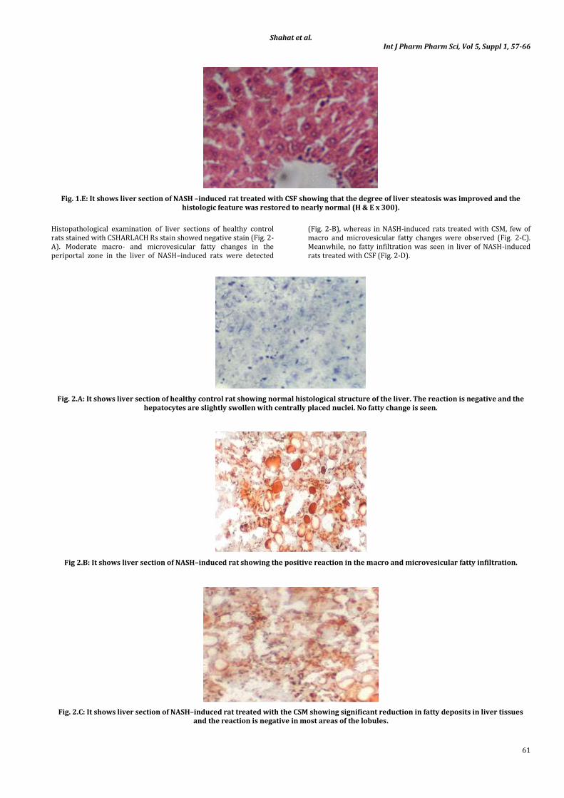
Shahat et al Int J Pharm Pharm Sci Vol 5 Suppl 1 57-66
66
Protein in Normal Weight Overweight and Obese People Clinical Medicine and Research 2005 889 -95
73 Pignatelli P Cangemi R Celestini A Carnevale R Polimeni L Martini A Ferro D Loffredo L Violi F Tumor Necrosis Factor-α Upregulates Platelet CD40L in Patients With Heart Failure Cardiovasc Cardiovasc Res 2008 78 515
74 Hirano T Arimitsu J Higa S NakaT Ogata A Shima Y Fujimoto M Yamadori T OhkawaraT Kuwabara Y Kawai M
Kawase I Tanaka T Luteolin a Flavonoid Inhibits CD40 Ligand Expression by Activated Human Basophils Int Arch Allergy Immunol 2006140150
75 Koutsogiannis D Summers K George B Adams P Marotta P Chakrabarti S Identification of Serum Biomarkers in End Stage Liver Disease The Open Biomarkers Journal 2010 31-6
76 Fausto N Laird AD Webber EM Role of Growth Factors and Cytokines in Hepatic Regeneration FASEB J 1995 91527-36