cytogenetic characterization of the genome of mealybug ... · cytogenetic characterization of the...
TRANSCRIPT

CARYOLOGIA Vol. 51, n. 1: 37-49,1998
Cytogenetic characterization of the genome of mealybug Planococcus citri(Homoptera, Coccoidea)
MARINA FERRARO, CHIARA EPIFANI, SILVIA BONGIORNI * , ANNA MARIA NARDONE, SAVIANAPARODI-DELFINO * and GIORGIO PRANTERA *Dipartimento di Genetica e Biologia Molecolare, Universitil "La Sapienza", Roma; * Dipartimento di Agrobiologia eAgrochimica, Universitil della Tuscia, Viterbo.
SUMMARY - Mealybugs although being agriculturally harmful insects have been very poorlystudied by modern cytogenetics techniques, and no cytotaxonomic criteria to distinguish betweenclosely related species is available yet. In the mealybug Planococcus citri (2n = 10) male and femaleindividuals are both diploid, however in males, at the stage of blastula, the haploid chromosome set ofpaternal origin becomes heterochromatic, even though its complete inertia has been consideredquestionable. Here we present data on the cytogenetic characterization of the chromosomes ofPlanococcus citri. We report on (i) the fluorescence karyotype (D287/170), which to our knowledgeis the first banded karyotype of a mealybug to be described; (ii) the chromosome localization ofconstitutive heterochromatin; (iii) the chromosome localization of rDNA sites; (iv) NORs activity.Our data also show, for the first time, that in the heterochromatic chromosome set ribosomal genesare still active.
INTRODUCTION
Mealybugs (Homoptera, Coccoidea, Pseudococcidae) are agriculturally harmful insects which arepresent all over the world with more than 2000 species. Nevertheless, they have been very poorlystudied by modern cytogenetics techniques possibly because of their very small and holocentricchromosomes. No chromosome banding of any species of Coccids nor cytotaxonomic criteria todistinguish between closely related species has been available yet. Their cytogeneticcharacterization is therefore of most importance as the preliminary step toward the application ofcytogenetics to the problem of cytotaxonomy of these species. In the mealybug Planococcus citrimale and female individuals are both diploid, however in males, at the stage of blastula, oneentire haploid set becomes heterochromatic in most of the tissues. Males also present an invertedmeiotic sequence with euchromatic chromosomes segregating from their homologs, and onlythose cells receiving the euchromatic set proceed to form sperm. (HUGHES-SHRADER 1948).Some Authors (BROWN and NUR 1964) have suggested the possibility of some residual activityin the

38 FERRARO, EPIFANI, BONGIORNI, NARDONE, PARODI-DELFINO and PRANTERA
inactivated chromosome set, however further investigations have never been attempted.Interestingly, the heterochromatized chromosome set is invariably that of paternal origin(BROWN and NELSON-REES 1961), implying that, at the blastula stage, the paternal and thematernal sets should be differentially imprinted. Mealybugs should therefore provide plenty ofscope for further investigations on such fundamental biological phenomena asheterochromatization mechanisms and chromosome imprinting. In this paper we report for thefirst time on the banded karyotype of the mealybug P. citri (2n = 10) after chromosome stainingwith the A T -specific compound D287/170. Moreover, we describe the chromosomelocalization, by C-banding, of constitutive heterochromatin and its characterization by fluorescentbanding with A T - and GCspecific fluorochromes, and by H33258-induced site-specificchromosome undercondensation. Finally, we report on the localization of ribosomal genesobtained by fluorescent in situ hybridization of a rDNA probe from Drosophila melanogaster,and about the activity of r-gene clusters and demonstrate that, in males, also the ribosomal geneslocated in the heterochromatic chromosome set are active.
MATERIALS AND METHODS
Chromosome preparations - The chromosome spreads were obtained by air drying according tothe technique described by ODIERNA et al. (1993) with minor modifications. Ovaries containingfertilized eggs were removed from females dissected in 0,9% NaCl and placed in a drop ofcolchicine 10-JM in H2O for 2 hours. Successively they were transferred to an Eppendorf-liketube and centrifuged at 5000 rpm for 5 min. After discarding the supernatant, 200 ìl of fixative(methanol: acetic acid, 3: 1) was added and the tissues homogenized through a 20G syringegauge. The material was centrifuged again at 7000 rpm for 8 min, and after the supernatant wasdiscarded and freshly made fixative added, it was passed again repeatedly through the syringegauge. 60- 70 ìl of the suspension were dropped on each slide. In some experiments, immediatelyafter dissection, the material was placed in a drop of Hoechst 33258 (Sigma) 50 ìg/ml in 0,7%NaCl at 26°C for 10-30 hours.
C-banding- Slides were treated at 30°C with a saturated solution of Ba(OH)2 for time lengthsranging from 10 to 30 min. After washing in distilled water the slides were incubated in 2xSSC at60°C for 60 min, then stained in a 5% Giemsa solution.
Fluorochrome staining - DAPI. Slides were hydrated in 2xSSC for 5 min, stained in DAPI(Boehringer) 0.2 ìg/ml in 2xSSC for 10 min in the dark, then washed in 2xSSC. Hoechst 33258(H33258). Slides were hydrated in 0.15M NaCl, 0.03M KC1, 0.01M disodium phosphate, pH 7,for 5 min and stained in Hoechst 33258 (Sigma) 0.5 ìg/ml in the above solution for 5 min.Chromomycin A3 (CMA3). Slides were hydrated in 0.14M phosphate buffer, pH 6.8, containing500 ìM MgCL, for 10 min. Then, 3-4 drops of CMA3 (Boehringer) (0,5mg/ml), made inphosphate buffer without MgCL2

CYTOGENETIC CHARACTERIZAnON OF THE GENOME OF PLANOCOCCUS CITRI 39
were placed on the slides. After 1 hour of staining the slides were washed in 0.15M NaCl/0.005MHepes buffer (GIBCO) and counter stained in 100~M methyl green (Merck) dissolved in theabove buffer, for 15 min. D287/170. A few drops of D287/170 staining solution (50 ìg/ml inMcIlvaine buffer) were placed over slides for 20-30 min at room temp. in the dark, then rinsed indeionized water (SCHNEDL et al. 1981).
"In situ hybridization" - rDNA 28S: a plasmid containing a 1.3 kb BgI II-Hind II fragment fromthe 28S rDNA gene of Drosophila melanogaster (LOHE and ROBERTS 1990); this fragmentcrosshybridizes in Southern hybridization with P. citri DNA, giving a 1.8 kb single band (notshown). Biotynilated dUTP was incorporated into probe DNA by nick-translation. Slides weretreated with 0.08 ìg/ml Proteinase K, at 37°C, for time lengths of 5-10 min, and RNase(200~gfml) at 37°C for 1 hour. Chromosomal DNA was denaturated by incubation in 70%formamide in 2xSSC at 70°C for two min. Hybridization was carried out overnight at 37°C in50% formamide in 2xSSC. Post-hybridization washes were 3x5 min at 37°C in 50% formamidein 2xSSC and 3x5 min at 42°C in 2xSSC. For the detection the slides were incubated withblocking solution (4xSSC, 30 mgfml BSA, 0.1% Tween 20) at 37°C for 30 min, then withAvidine-FITC (Vector laboratories) diluted 1:300 in 4xSSC, 10 mg/ml BSA, 0.1% Tween 20, at37°C for 30 min. After counterstaining in DAPI fluorescent images were captured with a CCDcamera (Photometrics 01) using IPLab software (Signal Analytic Corporation) and processedwith a Macintosh Power 7100 using Adobe Photoshop software.
NOR-banding. Slides were treated with a mixture of gelatin solution/AgNO3 solution (50%) at70°C for 2-3 min, rinsed with distilled water, then stained with Giemsa (HOWELL and BLACK1980).
RESULTS
Our initial effort was to obtain preparations with a high number of mitotic figures with elongatedchromosomes showing a good morphology. In Figure la, prophase male cells are shown withdifferent degrees of condensation between the maternal, euchromatic (E), and the paternal,heterochromatic (H), chromosome sets. As indicated in the diagram, the difference decreases asthe cell proceeds towards metaphase, where all chromosomes are equally condensed. In Fig. 1b,two female cells at mid and late prophase but showing no difference between the two haploid setsare shown.
Chromosome distribution of AT- and GC-rich DNA regions. - In order to identify the five pairs ofhomologs and to characterize the chromosomes on the base of composition along their length,chromosome preparations were stained with A T -specific (H33258, DAPI, D287/170) and GC-specific (CMA3) fluorochromes, or gently dissected ovaries were treated, before fixing, with asolution of Hoechst 33258. When added to "living" cells H33258 binds to ATrich DNA regionsinhibiting their condensation at mitosis.

40 FERRARO, EPIFANI, BONGIORNI, NARDONE, PARODl-DELFINO and PRANTERA
Figs. la,b. - Morphological differences between male and female cells chromosomes. (a) male: the chromosome sets ofmaternal (E) and paternal (H) origin show quite different degrees of condensation. (b) female cells: all thechromosomes always show the same degree of condensation. The variations of male and female chromosomecondensation from interphase to metaphase are schematized at far right of the figure, I = interphase, p = prophase, PM= prometaphase, M = metaphase.
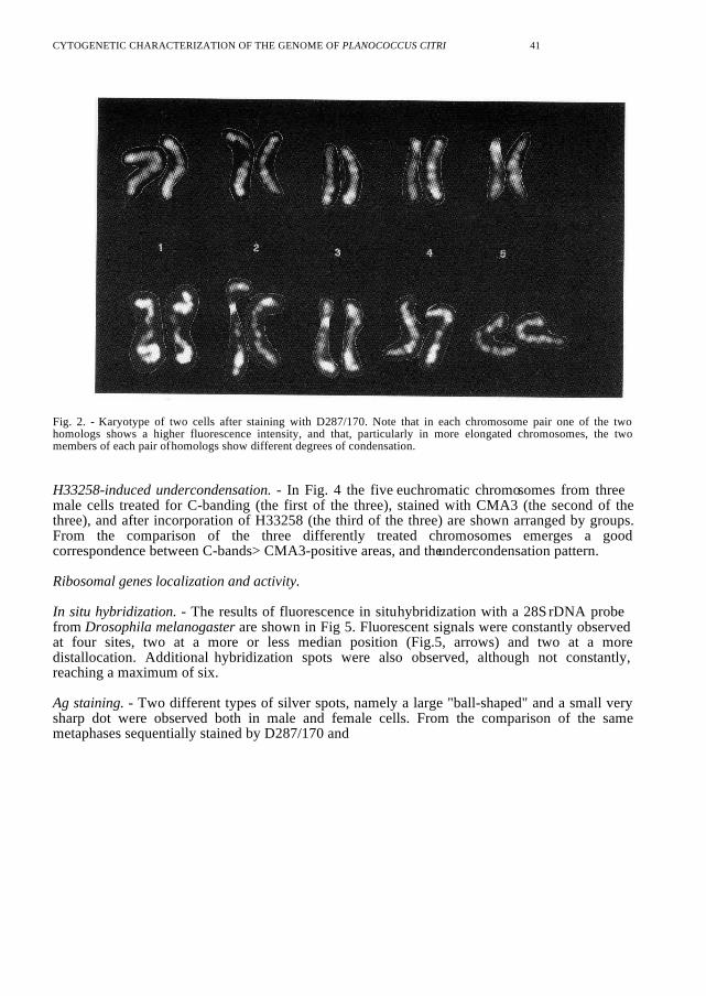
Fluorochrome staining. - After H33258 and DAPI staining the chromosomes appeared roughlyuniformly fluorescent (not shown). However, after staining with D287/170, a clear bandingpattern was obtained which allowed the unequivocal identification of homologous chromosomes(Fig. 2). In each chromosome pair, a difference in the fluorescence intensity between the twohomologs was appreciable. Furthermore, in metaphases with more elongated chromosomes adifference also in the degree of condensation was evident. In addition, CMA3 produced a bandingpattern, also if less detailed than the one obtained after D287/170, that was quite similar to theone observed after Cbanding.
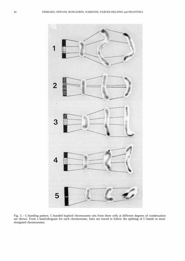
C-banding. - Higly condensed chromosomes have C-bands mainly confined to the telomericregions while in more elongated chromosomes some intercalary C-bands become evident. This isclearly shown in Fig. 3 where three haploid chromosome sets, at different degrees ofchromosome condensation are vertically arranged. Corresponding-number chromosomes fromeach metaphase are horizontally aligned and lines are traced to follow the splitting of C-bands inmore elongated chromosomes.
CYTOGENETIC CHARACTERIZATION OF THE GENOME OF PLANOCOCCUS CITRI 41
Fig. 2. - Karyotype of two cells after staining with D287/170. Note that in each chromosome pair one of the twohomologs shows a higher fluorescence intensity, and that, particularly in more elongated chromosomes, the twomembers of each pair of homologs show different degrees of condensation.
H33258-induced undercondensation. - In Fig. 4 the five euchromatic chromosomes from threemale cells treated for C-banding (the first of the three), stained with CMA3 (the second of thethree), and after incorporation of H33258 (the third of the three) are shown arranged by groups.From the comparison of the three differently treated chromosomes emerges a goodcorrespondence between C-bands> CMA3-positive areas, and the undercondensation pattern.
Ribosomal genes localization and activity.
In situ hybridization. - The results of fluorescence in situ hybridization with a 28S rDNA probefrom Drosophila melanogaster are shown in Fig 5. Fluorescent signals were constantly observedat four sites, two at a more or less median position (Fig.5, arrows) and two at a moredistallocation. Additional hybridization spots were also observed, although not constantly,reaching a maximum of six.
Ag staining. - Two different types of silver spots, namely a large "ball-shaped" and a small verysharp dot were observed both in male and female cells. From the comparison of the samemetaphases sequentially stained by D287/170 and
42 FERRARO, EPIFANI, BONGIORNI, NARDONE, PARODl-DELFINO and PRANTERA
Fig. 3. - C-banding pattern. C-banded haploid chromosome sets from three cells at different degrees of condensationare shown. From a band idiogram for each chromosome, lines are traced to follow the splitting of C-bands in moreelongated chromosomes.

CYTOGENETIC CHARACTERIZATION OF THE GENOME OF PLANOCOCCUS CITRI 43
Fig. 4 - Comparison of patterns obtained for each chromosome after C-banding (first), CMA3 staining (second), andH33258 incorporation (third). In each group the three patterns show a good correspondence.
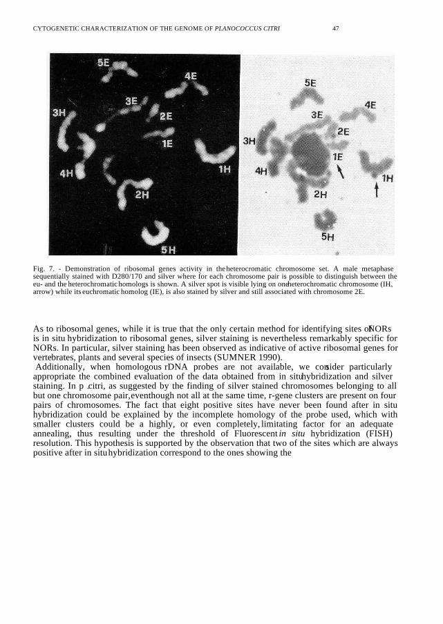
by silver (Fig. 6) it was possible to check the location of the two different types of silver spots.The large spots (arrows) were always located at a more or less median position of the first pair ofhomologs at the site on the chromosomes corresponding to a faintly fluorescent band afterstaining with D287/170. The small spots (arrowheads) were observed at subterminal position inthe chromosome pairs number 2, number 3 and number 4. Occasionally, a silver spot wasobserved at telomeric position of a chromosome identified as chromosome number 5. Thenumber of silver spots observed in a metaphase cell varied from a minimum of 2 to a maximumof 5. Two silver stained chromosomes were usually present in prometaphase male cells, thosebeing chromosomes number 1 and number 4 (Fig. 6 upper row). Moreover, in cells where it waspossible to distinguish between the euchromatic and the heterochromatic homologs, it has beenfound that both the eu- and the heterochromatic homologs could be silver stained (Fig. 7).
DISCUSSION
The chromosomes of Planococcus citri, like all other Coccid species, are very small andholocentric. This feature has made their identification quite difficult and it is probably the mainreason why a banded karyotype of none of these species has been described yet. A modificationof the air-drying technique for insects described by ODlERNA et al. (1993) allowed us to obtainmetaphases showing well elongated chromosomes and the use of both A T specific (H33258,DAPI, D287/170) and GC-specific (CMA3) fluorochromes allowed us to characterize DNA basecomposition variations along P. citri chromosomes, and to describe its banded karyotype byD287/170 (Fig. 2) . This

44 FERRARO, EPIFANI, BONGIORNI, NARDONE, PARODI-DELFINO and PRANTERA
Fig. 5 - Localization of 28S rDNA by fluorescence in situ hybridization. The two fluorescent spots at the more medianposition (arrows) and two of those more distally located are always present. Six hybridization spots, as seen in thefigure, are only seldom visible. Note that the hybridization spots are more often seen as a single lateral signal.
is a compound derived from a modification of DAPI considered as having affinity for A T -richDNA, as suggested by the Q-banding pattern produced on mouse chromosomes when used at lowconcentrations. Moreover, the restricted distribution of positive sites when used at highconcentration suggests that this fluorochrome does not simply show specificity for A T -richDNA, but may also show sequence specificity (SCHNEDL etal. 1981a). Indeed, it has been used

CYTOGENETIC CHARACTERIZATION OF THE GENOME OF PLANOCOCCUS CITRI 45
to distinguish certain classes of heterochromatin in different species (SCHNEDL et al. 1981b;BABU and VERMA 1986; MAYR et al. 1986). Our results therefore both confirm theindications about the peculiarity of this compound - see the lack of chromosome differentiationafter H33258 and DAPI staining - and suggest the existence of discrete stretches of specific AT -rich sequences along P. citri chromosomes.The analysis of karyotypes evidenced that homologous chromosomes consistently show differentfluorescence intensities, and that, especially in metaphases with more elongated chromosomes,the two members of each chromosome pair show a different degree of condensation (Fig. 2,bottom row). This could possibly be caused by differences in the level of genetic activity betweenthe two homologs, the different degree of compaction being a way of dosage compensation infemale cells in response to the heterochromatization of a whole haploid set in males. In anotherexample of dosage compensation, namely the X-chromosome inactivation in mammalian femalecells, the phenomenon is also accompanied by a higher degree of compaction of the inactivehomolog (LATT 1973; KEREM et al. 1983).The application of the C-banding technique evidenced a defined pattern of bands on all the fivechromosome pairs (Fig.3). The staining pattern obtained after CMA3-Methyl Green, showingthat the more fluorescent regions correspond to C-band positive areas, indicated that in P. citri theconstitutive heterochromatin is at least partially GC-rich. This suggestion was confirmed by theexperiments of H33258 binding to "living" chromosomes. It is known that, when added to culturecells, H33258 binds to AT-rich DNA before mitotic condensation thus inducing the appearance,along mitotic chromosomes, of these regions as undercondensed areas (HILWIG and GROPP1973, 1975; ROCCHI et al. 1976; PIMPINELLI et al., 1976). Indeed, the incorporation ofH33258 resulted in a banding pattern of chromosomes close to that obtained after C-banding(Fig. 4). The failure to find a dull appearance of these regions after staining with the A T -specificfluorochromes, could be explained by the fact that in the more condensed constitutiveheterocromatin there is more DNA per unit length than in the euchromatin. However, some of thetelomeric regions being positive to D287/170, we can hypotesize the presence of AT-richsequences embedded in predominantly GC-rich areas.
Fig. 6. - Identification of the chromosomes showing active ribosomal gene clusters. Three karyotypes from cellssequentially stained with D280/170 and silver are shown. The first karyotype is unequivocally from a male cell whilefor the two others it is not possible to say if they are made from a male- or a female-embryo cell since both are presentin the ovary, and at metaphase all chromosomes are equally condensed also in males. Chromosomes of the first pair(always Ag + ) show a large dot (arrows), while dots on other chromosomes of the same cell, when present, are alwaysquite smaller (arrowheads).

46 FERRARO, EPIFANI, BONGIORNI, NARDONE, PARODI-DELFINO and PRANTERA
CYTOGENETIC CHARACTERIZATION OF THE GENOME OF PLANOCOCCUS CITRI 47
Fig. 7. - Demonstration of ribosomal genes activity in the heterocromatic chromosome set. A male metaphasesequentially stained with D280/170 and silver where for each chromosome pair is possible to distinguish between theeu- and the heterochromatic homologs is shown. A silver spot is visible lying on one heterochromatic chromosome (IH,arrow) while its euchromatic homolog (IE), is also stained by silver and still associated with chromosome 2E.
As to ribosomal genes, while it is true that the only certain method for identifying sites of NORsis in situ hybridization to ribosomal genes, silver staining is nevertheless remarkably specific forNORs. In particular, silver staining has been observed as indicative of active ribosomal genes forvertebrates, plants and several species of insects (SUMNER 1990). Additionally, when homologous rDNA probes are not available, we consider particularlyappropriate the combined evaluation of the data obtained from in situ hybridization and silverstaining. In p .citri, as suggested by the finding of silver stained chromosomes belonging to allbut one chromosome pair, eventhough not all at the same time, r-gene clusters are present on fourpairs of chromosomes. The fact that eight positive sites have never been found after in situhybridization could be explained by the incomplete homology of the probe used, which withsmaller clusters could be a highly, or even completely, limitating factor for an adequateannealing, thus resulting under the threshold of Fluorescent in situ hybridization (FISH)resolution. This hypothesis is supported by the observation that two of the sites which are alwayspositive after in situ hybridization correspond to the ones showing the

48 FERRARO, EPIFANI, BONGIORNI, NARDONE, PARODl-DELFINO and PRANTERA
large "ball-shaped" silver spots, constantly found in all metaphases. The comparison of the samemetaphases first stained with Giemsa and then with silver (not shown) , supports this hypothesissince the silver stained chromosomes are very often the ones associated with the nucleolus.Finally, in male cells, positive silver staining was clearly detected both on euchromatic andheterochromatic chromosomes therefore demonstrating that in the heterochromatic set of malecells at least the ribosomal genes are active. The fact that r-gene clusters in a heterochromaticbackground can still function is nevertheless known for many species, like Drosophilamelanogaster and man, where the NORs indeed map in constitutive heterocromatic regions.In conclusion, (i) to our knowledge, this is the first time when a banded karyotype of this or anyother coccid species has been made. The banded karyotype of these species could be a first steptoward the application of cytogenetics to the problem of taxonomy which is particularly relevantfor this family. (ii) The data reported here about ribosomal genes, gives the first directdemonstration that, indeed, in P. citri the heterochromatic set is not completely inactivated, asotherwise suggested by indirect observations by NELSON-REES (1962). Finally, we would like to stress that, chromosome imprinting being a phenomenon whoserelevance is becoming apparent in most organisms> including man, mealybugs could be a usefulsystem to study fundamental biological phenomena as the heterochromatization process (see theindications coming from the differences both in fluorescence intensity and condensation of homo-logous chromosomes) and the chromosome imprinting.
Acknowledgements. - We are deeply indebted to the entomology group of Prof. A. Tranfaglia at the University ofPotenza, in particular to Dr. S. Marotta, for giving us the Planococcus citri cultures, and for their help and advice inculturing and disseecting mealybugs. We like also to thank Dr. S. Bonaccorsi for providing us the Drosophila 28SrDNA probe.
REFERENCES
BABU A. and VERMA R.S., 1986. - Characterization of human chromosomal constitutive heterochromatin. Canad. J.Genet. Cytol., 28: 631-644.BROWN S.W. and NELSON-REES W.A., 1961. - Radiation analysis of a lecanoid genetic system. Genetics, 46: 983-1007.BROWN S.W. and NUR U., 1964. - Heterochromatic chromosomes in the Coccids. Science, 145: 130136.HILWIG I. and GROPP A. , 1973. - Decondensation of constitutive heterochromatin in L cell chromosomes by abenzimidazole compound (Hoechst 33258). Exp. Cell Res., 81: 474-477.HOWELL V.M. and BLACK D.A., 1980. - Controlled silver staining of nucleolus olganizer regions with a protectivecolloidal developer: a 1-step method. Experientia, 36: 1014.HUGHES-SCHRADER S. 1948. - Cytology of Coccids (Coccoidea-Homoptera). Adv. Genet., 2: 127-203. KEREM B-S., GOITEIN R., RICHLER C., MARCUS M. and CEDAR H., 1983. - In situ nick-translation distinguishes betweenactive and inactive X chromosomes. Nature, 304: 88-90.
CYTOGENETIC CHARACTERIZATION OF THE GENOME OF PLANOCOCCUS CITRI 49
LATT S.A., 1973. - Microfluorimetric detection of deoxyribonucleic acid replication in human metaphasechromosomes. Proc. Natl. Acad. Sci. USA, 70: 3395-3399.LOHE A.R. and ROBERTS P.A., 1990. - An unusual y chromosome of Drosophila simulans carrying amplified rDNAspacer without rRNA genes. Genetics, 125: 399-406.MAYR B., GEBER G., AUER H., KALAT M. and SCHLEGER W., 1986. - Heterochromatin composition andnucleolus olganizer activity in four canid species. Canad. J. Genet. Cytol., 28: 744- 753. NELSON-REES W.A., 1962.- The effects of radiation damaged heterochromatic chromosomes on male fertility in the Mealybug, Planococcus citri(Risso). Genetics, 47: 661-683.NUR U., 1990. - Heterochromatization and euchromatization of whole genome in scale insects (Coccoi-dea:Homoptera). Development, Suppl.: 29-34.ODIERNA G., BALDANZA F., APREA G. and OLMO E., 1993. - Occurrence of G-banding in metaphasechromosomes of Encarsia berlesei (Hymenoptera'Aphelinidae). Genome, 36: 662-667.PIMPINELLI S., PRANTERA G., ROCCHI A. and GATTI M., 1976. - Effects of Hoechst 33258 on human leukocytesin vitro. Cytogenet. Cell Genet., 17: 114-121.ROCCHI A., PRANTERA G., PIMPINELLI S. and DI CASTRO, M. 1976. - Effect of Hoechst 33258 on Chinesehamster chromosomes. Chromosoma, 56: 41-46.SCHNEDL W. , ABRAHAM R. , DANN O. , GEBER G. and SCHWEIZER D. , 1981a. - Preferential fluorescentstaining of heterochromatic regions in human chromosomes 9, 15, and y by D287/170. Hum. Genet., 59: 10-13.SCHNEDL W., ABRAHAM R., FORSTER M. and SCHWEIZER D., 1981b. - Differential fluorescent staining ofporcine heterochromatin by Chromomycin A3/Distamycin A/DAPI and D287/170. Cytogenet. Cell Genet., 31: 249-253.SUMNER A. T., 1990. - Chromosome banding. Academic Division of Unwin Hyman Ltd, London.
Received 20 October 1997; accepted 18 December 1997