cytological and transcriptomic analyses reveal and transcriptomic analyses reveal ... significantly...
TRANSCRIPT
Cytological and Transcriptomic Analyses RevealImportant Roles of CLE19 in Pollen Exine Formation1[OPEN]
Shuangshuang Wang,a Jianan Lu,a Xiu-Fen Song,b Shi-Chao Ren,b Chenjiang You,a Jie Xu,c
Chun-Ming Liu,b,d Hong Ma,a and Fang Changa,2
aState Key Laboratory of Genetic Engineering and Collaborative Innovation Center for Genetics andDevelopment, Ministry of Education Key Laboratory of Biodiversity Sciences and Ecological Engineering andInstitute of Biodiversity Sciences, Institute of Plant Biology, School of Life Sciences, Fudan University,Shanghai 200438, ChinabKey Laboratory of Plant Molecular Physiology, Institute of Botany, Chinese Academy of Sciences, Beijing 100093, ChinacCollaborative Innovation Center for Genetics and Development, School of Life Sciences and Biotechnology,Shanghai Jiao Tong University, Shanghai 200240, ChinadInstitute of Crop Science, Chinese Academy of Agricultural Science, Beijing 100081, China
ORCID IDs: 0000-0001-5293-3302 (S.W.); 0000-0002-7271-362X (C.-M.L.); 0000-0001-8717-4422 (H.M.); 0000-0003-3378-8829 (F.C.).
The CLAVATA3/ESR-RELATED (CLE) peptide signals are required for cell-cell communication in several plant growth anddevelopmental processes. However, little is known regarding the possible functions of the CLEs in the anther. Here, we show that aT-DNA insertional mutant, and dominant-negative (DN) and overexpression (OX) transgenic plants of the CLE19 gene, exhibitedsignificantly reduced anther size and pollen grain number and abnormal pollen wall formation in Arabidopsis (Arabidopsis thaliana).Interestingly, the DN-CLE19 pollen grains showed a more extensively covered surface, but CLE19-OX pollen exine exhibited clearlymissing connections in the network and lacked separation between areas that normally form the lacunae. With a combination of cellbiological, genetic, and transcriptomic analyses on cle19, DN-CLE19, and CLE19-OX plants, we demonstrated that CLE19-OX plantsproduced highly vacuolated and swollen aborted microspores (ams)-like tapetal cells, lacked lipidic tapetosomes and elaioplasts, and hadabnormal pollen primexine without obvious accumulation of sporopollenin precursors. Moreover, CLE19 is important for the normalexpression of more than 1,000 genes, including the transcription factor gene AMS, 280 AMS-downstream genes, and other genesinvolved in pollen coat and pollen exine formation, lipid metabolism, pollen germination, and hormone metabolism. In addition, theDN-CLE19(+/+) ams(2/2) plants exhibited the ams anther phenotype and ams(+/2) partially suppressed the DN-CLE19 transgene-induced pollen exine defects. These findings demonstrate that the proper amount of CLE19 signal is essential for the normal expressionof AMS and its downstream gene networks in the regulation of anther development and pollen exine formation.
Pollen grains are generated in the male reproductiveorgan anther of flowering plants and are essential forplant fertility. In the model plant Arabidopsis (Arabidopsisthaliana), pollen grains are ellipsoidal, and the pollensurface is covered by a reticulate exine. In contrast, rice
(Oryza sativa) pollen grains are globular and have asmooth surface without the reticulate structure. Awell-organized pollen wall structure is essential for thephysical and chemical stability of the mature pollen byproviding protection for pollen grains from environ-mental stresses, such as desiccation and microbial attacks(Scott et al., 2004). The pollen wall is composed mainly ofthree layers: the intine layer, the exine layer, and thepollen coat (Zinkl et al., 1999; Edlund et al., 2004;Blackmore et al., 2007). The intine is the innermost layerof the pollen wall immediately adjacent to the plasmamembrane of the pollen vegetative cell and is composedmainly of pectin, cellulose, hemicellulose, hydrolyticenzymes, and hydrophobic proteins (Scott et al., 2004).Both the exine layer and the pollen coat layer are basi-cally of a lipidic nature. The exine is largely formed fromacyl lipid and phenylpropanoid precursors, which to-gether form the mixed stable biopolymer known assporopollenin (Guilford et al., 1988; Bedinger et al., 1994;Thom et al., 1998). The pollen exine has many importantfunctions in the prevention of dehydration of male ga-metophytes, the facilitation of pollen dispersal, and
1 Thisworkwas supported by grants from theNationalNatural ScienceFoundation of China (31130006, 31470282, 31670316, and 31370029) andthe State Key Laboratory of Genetic Engineering at Fudan University.
2 Address correspondence to [email protected] author responsible for distribution of materials integral to the
findings presented in this article in accordance with the policy de-scribed in the Instructions for Authors (www.plantphysiol.org) is:Fang Chang ([email protected]).
S.W., H.M., and F.C. designed the research; S.W. performed theRNA in situ hybridization analysis, the phenotypic observation, thereal-time PCR experiments, the scanning electron microscopy andtransmission electron microscopy observations, and tissue collectionfor the transcriptome; S.-C.R., X.-F.S., and C.-M.L. generated theDN-CLE transgenic mutants; J.L. and C.Y. together carried out thetranscriptome analysis; S.W. and F.C. analyzed the data and wrotethe article; F.C. and H.M. revised the manuscript.
[OPEN] Articles can be viewed without a subscription.www.plantphysiol.org/cgi/doi/10.1104/pp.17.00439
1186 Plant Physiology�, November 2017, Vol. 175, pp. 1186–1202, www.plantphysiol.org � 2017 American Society of Plant Biologists. All Rights Reserved. www.plantphysiol.orgon May 20, 2018 - Published by Downloaded from
Copyright © 2017 American Society of Plant Biologists. All rights reserved.

pollen-stigma recognition and adhesion (Zinkl et al.,1999). The pollen coat fills the cavities of the pollenexine during the late stages of pollen development, andprevious studies have demonstrated that develop-mental defects of pollen wall structure or lack of pollencoat lipids in Arabidopsis lead to a failure or delay ofpollen hydration and subsequent male sterility (Preusset al., 1993; Mayfield and Preuss, 2000; Wilson andZhang, 2009; Li and Zhang, 2010).Anther development is precisely regulated tempo-
rally by transcriptional networks, receptor-like proteinkinase-mediated signal pathways, and other path-ways (Ma, 2005; Ge et al., 2010; Chang et al., 2011). Thebasic helix-loop-helix (bHLH) transcriptional factorDYSFUNCTIONAL TAPETUM1 (DYT1) is among theearliest acting male-specific regulators and is requiredfor the normal development of tapetum by regulatingthe normal expression of more than 1,000 anther genes,and dyt1mutants form abnormal prematurely vacuolatedtapetal cells and lack mature pollen (Zhang et al., 2006;Feng et al., 2012; Gu et al., 2014; Zhu et al., 2015). In ad-dition, bHLH010, bHLH089, and bHLH091 together arerequired for normal tapetum development and tran-scriptome and pollen fertility, forming both feed-forwardand feedback loops with DYT1 (Zhu et al., 2015; Cui et al.,2016). Another bHLH transcription factor, ABORTEDMICROSPORES (AMS), is a key regulator of anthertranscriptome, sporopollenin biosynthesis and secretion,and pollen wall formation (Sorensen et al., 2003; Xu et al.,2010; Feng et al., 2012; Ma et al., 2012; Xu et al., 2014).DEFECTIVE IN TAPETAL DEVELOPMENT ANDFUNCTION (TDF1)/MYB35 encodes a putative R2R3MYB transcription factor, and loss of TDF1 functiondue to mutation or transgene causes tapetal hyper-trophy extending into the locule and results in sporo-phytic male sterility (Zhu et al., 2008; Feng et al., 2012).MALE STERILITY1 (MS1) is a PHD-finger motif-containing nuclear protein and is essential for tapetumdevelopment at the postmeiotic phase and regulatesgenes important for exine formation (Wilson et al.,2001; Ito et al., 2007). In addition, another R2R3 MYBgene, MS188/MYB103/MYB80, is required for sporo-pollenin biosynthesis and sexine formation (Phan et al.,2011; Xiong et al., 2016). Further molecular genetic andbioinformatics studies revealed that these transcriptionfactors likely form a genetic pathway as follows: DYT1,MYB35/TDF1, AMS, MS188/MYB103/MYB80, MS1,and MYB99 from upstream to downstream during an-ther/pollen development (Ito et al., 2007; Feng et al.,2012).Extracellular ligands and their receptor-like protein
kinase (RLK)-mediated signaling pathways play im-portant roles in the regulation of anther development.In particular, the RLKs EMS1/EXS and SERK1/2 andthe secreted peptide TPD1 are required for tapetumformation and function (Canales et al., 2002; Zhao et al.,2002; Yang et al., 2003; Albrecht et al., 2005; Colcombetet al., 2005). Furthermore, the TPD1 protein from mi-crosporocyte precursors/microsporocyte interacts withthe Leu-rich repeat domain of the RLK EMS1/EXS
expressed in the tapetal precursor/tapetal cells to pro-mote the phosphorylation of EMS1, thereby regulatinganther tapetum development and function (Canaleset al., 2002; Zhao et al., 2002; Yang et al., 2003, 2005; Jiaet al., 2008; Huang et al., 2016; Li et al., 2017). In addi-tion, brassinosteroids and the brassinosteroid cell sur-face receptor BRASSINOSTEROID INSENSITIVE1 arerequired for pollen exine formation and pollen releasethrough the regulation of key genes in tapetum andpollen development, such as AMS, MYB103, MS1, andMS2 (Ye et al., 2010). In addition, several other RLKs arerequired for normal anther development, but theirligands remain unknown. For instance, ERECTA,ERECTA-LIKE1 (ERL1), and ERL2 are important foranther lobe formation and normal cell patterning(Torii et al., 1996; Shpak et al., 2003; Hord et al., 2008).BARELY ANY MERISTEM1 (BAM1) and BAM2 reg-ulate the formation of anther somatic cell layers, andthe bam1 bam2 double mutant anther forms manypollen mother-like cells but lacks the three subepi-dermal somatic cell layers (DeYoung et al., 2006; Hordet al., 2006). RECEPTOR-LIKE PROTEIN KINASE2 isessential for the formation of both the tapetum and themiddle layer (Mizuno et al., 2007). On the other hand,besides TPD1, no other peptide signals have been impli-cated in anther or pollen development.
CLAVATA3 (CLV3)/EMBRYO SURROUNDINGREGION (ESR)-RELATED (CLE) genes encode a familyof putative peptide ligands, with at least 32 CLEmembers in Arabidopsis (Cock and McCormick, 2001).Mature CLE peptide signals are 12 to 14 amino acidslong, and many of them play important roles in variousplant developmental processes. For instance, CLV3 isnecessary to restrict stem cell numbers in the shootapical meristem (Fletcher et al., 1999) by promoting theformation of the CLV1/CLV2 protein complex to in-hibit the expression of WUSCHEL in the organizingcenter of the shoot apical meristem (Laux et al., 1996;Fiers et al., 2004). CLE8 regulates the size of embryosand seeds and is expressed in the endosperm and theearly embryo to promoteWOX8 expression (Fiume andFletcher, 2012). CLE40 regulates the activity of the rootapical meristem and is expressed in differentiating cellsin roots and likely acts through the receptor-like kinaseCRINKLY4 to restrict the expression and position ofWOX5 (Stahl and Simon, 2009). The CLE19 peptide trig-gers root meristem consumption in a CLV2-dependentmanner (Casamitjana-Martínez et al., 2003; Fiers et al.,2005; Al-Refu et al., 2009). However, whether the CLEgenes are required for normal anther development stillremains unknown.
One difficulty in studying the endogenous functionsof these small CLE genes is the shortage of genetic mate-rials, namely the mutant plants. In addition, previousstudies revealed that T-DNA insertion mutants of CLE1,CLE7, CLE10, CLE16, CLE18, or CLE19 in Arabidopsisexhibited novisible abnormal phenotype (Fiers et al., 2004;Jun et al., 2010), but overexpression of the CLE genesCLE2,CLE3,CLE4,CLE5,CLE6,CLE7,CLE10,CLE11, andCLE13 resulted in pleiotropic phenotypes similar to those
Plant Physiol. Vol. 175, 2017 1187
CLE19 Regulates Pollen Wall Formation
www.plantphysiol.orgon May 20, 2018 - Published by Downloaded from Copyright © 2017 American Society of Plant Biologists. All rights reserved.
of CLV3 or CLE40 overexpression plants, and in vitroapplication or overexpression of one of the CLE genesCLE19, CLE21, CLE25, CLE42, and CLE44 caused similardwarf and short-root phenotypes (Fiers et al., 2004, 2005;Strabala et al., 2006), suggesting a high level of functionalredundancy or overlap amongCLEmembers. Fortunately,an antagonistic peptide technology (Song et al., 2013) wasdeveloped recently as an effective tool to investigate theendogenous functions of these functionally redundant se-creted peptides. Using CLV3 as a test case, Song et al.(2013) examined the antagonistic peptide technology bytransformations of wild-type Arabidopsis with constructscarrying the full-length CLV3 with every residue in thepeptide-coding region replaced one at a time by Ala toprobe the effectiveness of each mutation. They found thatthe Gly-to-Ala substitution in the core CLE motif caused adominant-negative (DN) clv3-2-like phenotype. Furthersubstitutions of the Gly residue individuallywith the other18 possible proteinaceous amino acids determined that theGly-to-Thr substitution resulted in the strongest antago-nistic effect in the wild type (Song et al., 2012, 2013). Withthis strategy, the peptide signal CLE19 was revealed to beimportant for cotyledon establishment in embryos andendosperm development (Xu et al., 2015).
Here, we tested CLE genes for expression in the an-ther and show that CLE9,CLE16,CLE17,CLE19,CLE41,CLE42, and CLE45 were preferentially expressed intapetal and reproductive cells in the anther. Using theantagonistic peptide technology, we generated DNmutants for these seven genes, and our analyses ofthese mutant transgenic plants indicated that they areimportant for normal pollen development, especiallyfor the development of pollen exine. Using CLE19 as arepresentative, we further characterized its function in theregulation of pollen exine formation using a combinationof morphological molecular and transcriptomic analysesof cle19, DN-CLE19, and CLE19-OX mutants. We finallyrevealed that CLE19 acts as a negative regulatory elementin tapetum development, sporopollenin biosynthesis, andexine formation, likely through the regulation of thetranscription factor AMS and downstream genes. Thus,we propose a novel signaling pathway starting from anextracellular peptide signal and eventually affecting theintracellular transcriptional network that is crucial for theregulation of pollen wall development.
RESULTS
Seven CLE Genes Are Expressed Preferentially inArabidopsis Tapetal and Reproductive Cells
The expression of CLE genes in Arabidopsis was firstexamined by searching public microarray databases(http://bar.utoronto.ca/efp/cgi-bin/efpWeb.cgi andhttp://www.arabidopsis.org/portals/expression/microarray/ATGenExpress.jsp) and using quantitativereal-time PCR and RNA in situ hybridization analyses todetect the expression of these CLE genes in the devel-oping anther.Among the 14CLEgenes thatwere includedin the microarray expression databases, CLE9, CLE10,
CLE21, CLE26, CLE40, CLE44, and CLV3 exhibited rel-atively high expression levels in the anther, whereasCLE6 andCLE12were relatively highly expressed in themature pollen (Supplemental Fig. S1A). Quantitativereal-time PCR analyses using Columbia-0 (Col-0) in-florescence and stage 4 to 12 anthers revealed thatCLE9, CLE16, CLE17, CLE25, CLE27, CLE41, CLE42,and CLE45 were relatively highly expressed in bothinflorescence and stage 4 to 12 anthers (SupplementalFig. S1B), suggesting that they might be involved inmale reproductive development.
Further RNA in situ hybridization analyses wereperformed to investigate the expression patterns ofthese anther-expressedCLE genes. StrongCLE9,CLE16,CLE17, CLE19, CLE41, CLE42, and CLE45 signals weredetected in the anther at various developmental stages(Fig. 1). For instance,CLE9 andCLE16were both expressedin the tapetum andmicrospores at anther stages 7 to 10 butnot in either cell layer at stage 5 (Fig. 1, A–H). CLE19 wasexpressed preferentially in both tapetal and male repro-ductive cells at anther stages 5 to 7, and CLE17, CLE41,CLE42, and CLE45 were expressed preferentially in bothtapetal and male reproductive cells at anther stages 5 to10 (Fig. 1, I–AB), suggesting their involvement in regulatingtapetum function and pollen development. In addition,their highly similar spatial-temporal expression pattern alsosuggested that these seven CLE genes possibly participatein similar anther developmental processes.
DN Mutant Transgenic Plants of CLE9, CLE16, CLE17,CLE19, CLE41, CLE42, and CLE45 Exhibited AbnormalAnther Development
To further investigate the functions of the CLE genesin anther development, the T-DNA insertional mutantsof CLE9, CLE17, CLE19, or CLE42 and the RNA in-terference (RNAi) mutant of CLE16 were obtained(Supplemental Fig. S2), and the transcript levels of thesegenes in mutant plants were analyzed using real-timequantitative PCR. The cle9-1, cle16-1, cle17-1, cle19-1 (inthe qrt1-2 background), cle19-2, and cle42-1 mutantsexhibited drastically reduced expression of the relevantgenes (Supplemental Fig. S2) and were further exam-ined phenotypically. Both cle19-1 qrt1-2 and cle19-2produced abnormally short siliques and had slightlydecreased anther size and some aborted pollen grains(Fig. 2, A–E, J–N, S–W, and AB–AF). Considering thatcle19-1 was in the qrt1-2 background, we observed thephenotypes of silique, flower, anther, and pollen inthe qrt1-2 plants and did not find any defects except forthe pollen phenotype (Fig. 2, C, L, U, and AD). Theabove results suggested thatCLE19 is possibly involvedin anther development. In contrast, the cle9-1, cle16-1,cle17-1, and cle42-1 single mutants all exhibited nor-mal plant growth and flower and anther development(Supplemental Fig. S3; Supplemental Table S1).
Based on the high amino acid similarity between thefunctional domains of these Arabidopsis CLE members(Ni and Clark, 2006) and the no/weak defects in anther
1188 Plant Physiol. Vol. 175, 2017
Wang et al.
www.plantphysiol.orgon May 20, 2018 - Published by Downloaded from Copyright © 2017 American Society of Plant Biologists. All rights reserved.

development observed in the available cle single mu-tants, we proposed a possible functional redundancyamong these anther-expressed CLE genes in the regu-lation of anther development. Therefore, we generateddouble and triple mutants of these CLE genes bycrossing cle16-1, cle17-1, cle19-1, and cle42-1. The qrt1-2mutation in cle19-1 plants segregated away during thisprocess. Compared with the cle19-1 mutant, our phe-notypic and statistical analyses showed that the cle16cle19, cle17 cle19, and cle19 cle42 double mutants hadfewer pollen grains in the anther (Supplemental Fig. S4,A–AA; Supplemental Table S1). Moreover, the cle17cle19 cle42 triple mutant showed more barren siliques,less pollen amount, and a larger fraction of defectivepollen in the anther (Supplemental Fig. S4, A–AA;Supplemental Table S1). In addition, we generated DNmutant plants for CLE9, CLE16, CLE17, CLE19, CLE41,CLE42, and CLE45 by transforming wild-type plantsseparately with fusions of the CLE promoter and thecoding region for the CLEG6T point mutation (DN-CLE).
The DN-CLE transgenic plants exhibited obviouslyreduced silique length (Fig. 2, F and G; SupplementalFig. S5, B-G). Alexander staining and scanning elec-tron microscopy experiments revealed abnormal an-ther development with aborted pollen in the anthersof these DN-CLE plants (Fig. 2, X, Y, AG, and AH;Supplemental Fig. S5, H–U). These results suggestedthat the normal functions of these genes are importantfor anther development and that the anther-expressedCLE9, CLE16, CLE17, CLE19, CLE41, and CLE42mightfunction in a redundant manner. Although the DNtransgenic plants of all seven CLE genes exhibited ab-normal anther development, only the cle19 knockoutmutants showed weak anther developmental defects;therefore, we focused the subsequent detailed functionalanalyses on CLE19 to gain insights into its role in anther/pollen development.
CLE19 Is Required for Normal Pollen Development, PollenGermination, and Pollen Tube Elongation
In addition to the above-mentioned cle19 and DN-CLE19 mutants, we also wanted to investigate whetheran increase of CLE19 expression affects male reproduc-tive development, using the 35S promoter-driven CLE19overexpression transgenic lines (CLE19-OX; Fiers et al.,2004). Two CLE19-OX transgenic lines with differentCLE19 expression levels, CLE19-OX-S and CLE19-OX-M(Fig. 3D), were selected for the analysis of anther de-velopmental phenotypes. Both lines exhibited obviouslyreduced fertility, including aborted siliques, small an-thers, and abnormal pollen (Fig. 2, H, I, Q, R, Z, AA, AI,and AJ).
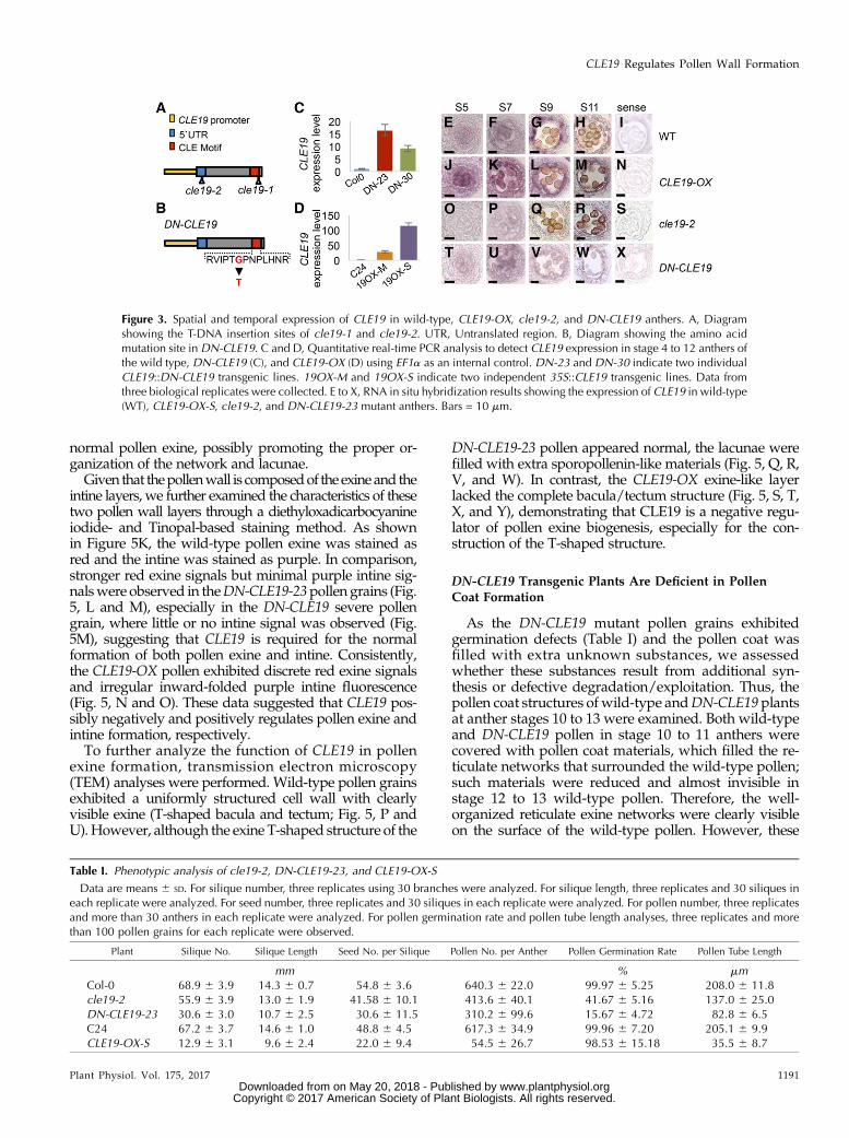
As detected using real-time PCR, the expression levelof CLE19 was found to be elevated up to over 100-foldin theCLE19-OX-S line (Fig. 3D) and to a lesser extent inDN-CLE19 anthers (Fig. 3C). The anther expressionpattern of CLE19 in the transgenic plants was examinedfurther using RNA in situ hybridization analysis (Fig. 3,E–X). Consistently, the expression of CLE19 was obvi-ously increased inCLE19-OX-S and decreased in cle19-2in both tapetal and male reproductive cells (Fig. 3, J–S).However, the expression in DN-CLE19 anthers showedno obvious difference from that in the wild type (Fig. 3,T–X), suggesting that the 35S promoter is not highlyactive in the tapetum and that in situ hybridizationmight not be quantitative enough to detect relativelysmall differences in expression level.
We further performed statistical analysis for variousmale fertility processes of the cle19-2, DN-CLE19-23,and CLE19-OX-S plants, including the pollen numberin each anther, pollen germination and pollen tubeelongation, silique length and silique number in eachmain stem, and seed number in each silique. Comparedwith the wild type, DN-CLE19-23 produced obviouslyfewer pollen grains (48.4% of that in each wild-typeanther), a reduced pollen germination rate (15.7% ofthe rate for the wild type), defective pollen tube elon-gation, a decreased number of siliques from one re-productive shoot, reduced silique length, and a lower
Figure 1. Spatial and temporal expression analyses of CLE genes. Ex-pression patterns are shown for CLE9 (A–D), CLE16 (E–H), CLE17 (I–L),CLE19 (M–P), CLE41 (Q–T), CLE42 (U–X), and CLE45 (Y–AB) in wild-type anthers by RNA in situ hybridization. Sections are from anthers ofstage 5 (A, E, I, M, Q, U, and Y), stage 7 (B, F, J, N, R, V, and Z), stage10 (C, G, K, O, S, W, and AA), and controls (D, H, L, P, T, X, and AB)using sense probe on wild-type stage 7 anthers. Ms, Microsporocytes;Msp, microspores; T, tapetum; Tds, tetrads. Bars = 10 mm.
Plant Physiol. Vol. 175, 2017 1189
CLE19 Regulates Pollen Wall Formation
www.plantphysiol.orgon May 20, 2018 - Published by Downloaded from Copyright © 2017 American Society of Plant Biologists. All rights reserved.

seed number in each silique (Table I). The cle19-1 andcle19-2 plants exhibited similar but weaker phenotypescompared with those of DN-CLE19, suggesting a func-tional redundancy among the anther-expressed CLE fam-ily members, as other members also might be inhibited bythe mutated CLE peptide. The CLE19-OX-S anther pro-duced only 9% of the amount of pollen in one wild-typeanther (Table I). All these results demonstrated the im-portant function of CLE19 in male reproductive processesand that the correct level of CLE19 expression is importantfor normal function.
To further investigate the function of CLE19 in antherdevelopment, particularly to identify the earliest stageswhen morphological differences can be observed, an-ther transverse semithin sections of cle19-2, DN-CLE19-23, and CLE19-OX mutants were examined. At antherstage 5, the wild-type anther formed well-organized five-cell layers in each anther lobe, the epidermis, endothe-cium, middle layer, tapetum, and microsporocytes, fromouter to inner. Afterward, the microsporocytes under-wentmeiosis and formed themicrospores that developedfurther into the pollen in the wild type at stages 6 to 13.
Compared with the wild type (Fig. 4, A–E), the cle19-2mutant anther showed no obvious cytological defectsduring anther stages 5 to 10, except for fewer pollengrains in the anther at stage 12 (Fig. 4, F–J). However,the DN-CLE19-23 anthers tended to produce more thannormal tapetum cells surrounding the microsporocytesat stage 5, and aborted pollen was observed at stage10 and onward (Fig. 4, K–O). In comparison, change of thetapetal cell numberwas not obvious in eitherCLE19-OX-Sor CLE19-OX-M anthers; nevertheless, most of the tapetal
cells were enlarged and excessively vacuolated at stages5 to 7, and the CLE19-OX-S tapetum soon degenerated atstage 8 (Fig. 4, P–R). Therefore, the development of mostpollen grains was defective (Fig. 4, S and T). The CLE19-OX-M tapetum exhibited a weaker vacuolated morphol-ogy compared with CLE19-OX-S but also disappeared atstage 10. A small portion of defective pollenwas observedat stage 10 and onward (Fig. 4, U–Y). These results furtherstrengthened the idea that a proper amount of CLE19 isrequired for normal anther development.
Pollen Exine Formation Was Affected in CLE19 Mutants
We then further investigated whether CLE19 is in-volved in pollen exine formation. The morphologicalpollen exine structure of wild-type, DN-CLE19-23, andCLE19-OX-S plants was first observed using scanningelectron microscopy. Pollen grains with either moderateor severe defectswere analyzed in bothDN-CLE19-23 andCLE19-OX-S plants. The results showed that the externalsurface of wild-type pollen exine had a network-likestructure formed with well-organized lacunae and threenarrow apertures (Fig. 5, A and F). In comparison, boththemoderately defective and the severely defective pollenof DN-CLE19-23 exhibited more extensively covered sur-faces with smaller lacunae filled with additional materials(Fig. 5, B, C, G, and H). In contrast, the CLE19-OX pollenexine exhibited clearlymissing connections in the networkand lack of separation between the spaces that wouldform the lacunae (Fig. 5, D, E, I, and J), suggesting that theproper amount of CLE19 is required for the formation of
Figure 2. Phenotypic analyses of wild-type, cle19 T-DNA insertion mutant, DN-CLE19 transgenic, and CLE19-OX transgenicplants. A to I, Phenotypic analysis of plant growth of wild-type, cle19 T-DNA insertionmutant,DN-CLE19 transgenic, andCLE19-OX transgenic plants. J to R, Phenotypic analysis of flowers of wild-type, CLE19 T-DNA insertion mutant,DN-CLE19 transgenic,andCLE19-OX transgenic plants. S to AA, Phenotypic analysis of anthers of wild-type,CLE19 T-DNA insertionmutant,DN-CLE19transgenic, and CLE19-OX transgenic plants. AB to AJ, Phenotypic analysis of pollen grains of wild-type, CLE19 T-DNA insertionmutant,DN-CLE19 transgenic, andCLE19-OX transgenic plants. Red arrowheads indicated aborted pollen grains. Bars = 2 cm forplants, 500 mm for flowers, 200 mm for anthers, and 25 mm for pollen grains.
1190 Plant Physiol. Vol. 175, 2017
Wang et al.
www.plantphysiol.orgon May 20, 2018 - Published by Downloaded from Copyright © 2017 American Society of Plant Biologists. All rights reserved.
normal pollen exine, possibly promoting the proper or-ganization of the network and lacunae.Given that thepollenwall is composedof the exineand the
intine layers, we further examined the characteristics of thesetwo pollen wall layers through a diethyloxadicarbocyanineiodide- and Tinopal-based staining method. As shownin Figure 5K, the wild-type pollen exine was stained asred and the intine was stained as purple. In comparison,stronger red exine signals but minimal purple intine sig-nalswere observed in theDN-CLE19-23pollen grains (Fig.5, L and M), especially in the DN-CLE19 severe pollengrain, where little or no intine signal was observed (Fig.5M), suggesting that CLE19 is required for the normalformation of both pollen exine and intine. Consistently,the CLE19-OX pollen exhibited discrete red exine signalsand irregular inward-folded purple intine fluorescence(Fig. 5, N and O). These data suggested that CLE19 pos-sibly negatively and positively regulates pollen exine andintine formation, respectively.To further analyze the function of CLE19 in pollen
exine formation, transmission electron microscopy(TEM) analyses were performed. Wild-type pollen grainsexhibited a uniformly structured cell wall with clearlyvisible exine (T-shaped bacula and tectum; Fig. 5, P andU).However, although the exine T-shaped structure of the
DN-CLE19-23 pollen appeared normal, the lacunae werefilled with extra sporopollenin-like materials (Fig. 5, Q, R,V, and W). In contrast, the CLE19-OX exine-like layerlacked the complete bacula/tectum structure (Fig. 5, S, T,X, and Y), demonstrating that CLE19 is a negative regu-lator of pollen exine biogenesis, especially for the con-struction of the T-shaped structure.
DN-CLE19 Transgenic Plants Are Deficient in PollenCoat Formation
As the DN-CLE19 mutant pollen grains exhibitedgermination defects (Table I) and the pollen coat wasfilled with extra unknown substances, we assessedwhether these substances result from additional syn-thesis or defective degradation/exploitation. Thus, thepollen coat structures of wild-type andDN-CLE19plantsat anther stages 10 to 13 were examined. Both wild-typeand DN-CLE19 pollen in stage 10 to 11 anthers werecovered with pollen coat materials, which filled the re-ticulate networks that surrounded the wild-type pollen;such materials were reduced and almost invisible instage 12 to 13 wild-type pollen. Therefore, the well-organized reticulate exine networks were clearly visibleon the surface of the wild-type pollen. However, these
Figure 3. Spatial and temporal expression of CLE19 in wild-type, CLE19-OX, cle19-2, and DN-CLE19 anthers. A, Diagramshowing the T-DNA insertion sites of cle19-1 and cle19-2. UTR, Untranslated region. B, Diagram showing the amino acidmutation site in DN-CLE19. C and D, Quantitative real-time PCR analysis to detect CLE19 expression in stage 4 to 12 anthers ofthe wild type, DN-CLE19 (C), and CLE19-OX (D) using EF1a as an internal control. DN-23 and DN-30 indicate two individualCLE19::DN-CLE19 transgenic lines. 19OX-M and 19OX-S indicate two independent 35S::CLE19 transgenic lines. Data fromthree biological replicates were collected. E to X, RNA in situ hybridization results showing the expression of CLE19 in wild-type(WT), CLE19-OX-S, cle19-2, and DN-CLE19-23 mutant anthers. Bars = 10 mm.
Table I. Phenotypic analysis of cle19-2, DN-CLE19-23, and CLE19-OX-S
Data are means 6 SD. For silique number, three replicates using 30 branches were analyzed. For silique length, three replicates and 30 siliques ineach replicate were analyzed. For seed number, three replicates and 30 siliques in each replicate were analyzed. For pollen number, three replicatesand more than 30 anthers in each replicate were analyzed. For pollen germination rate and pollen tube length analyses, three replicates and morethan 100 pollen grains for each replicate were observed.
Plant Silique No. Silique Length Seed No. per Silique Pollen No. per Anther Pollen Germination Rate Pollen Tube Length
mm % mmCol-0 68.9 6 3.9 14.3 6 0.7 54.8 6 3.6 640.3 6 22.0 99.97 6 5.25 208.0 6 11.8cle19-2 55.9 6 3.9 13.0 6 1.9 41.58 6 10.1 413.6 6 40.1 41.67 6 5.16 137.0 6 25.0DN-CLE19-23 30.6 6 3.0 10.7 6 2.5 30.6 6 11.5 310.2 6 99.6 15.67 6 4.72 82.8 6 6.5C24 67.2 6 3.7 14.6 6 1.0 48.8 6 4.5 617.3 6 34.9 99.96 6 7.20 205.1 6 9.9CLE19-OX-S 12.9 6 3.1 9.6 6 2.4 22.0 6 9.4 54.5 6 26.7 98.53 6 15.18 35.5 6 8.7
Plant Physiol. Vol. 175, 2017 1191
CLE19 Regulates Pollen Wall Formation
www.plantphysiol.orgon May 20, 2018 - Published by Downloaded from Copyright © 2017 American Society of Plant Biologists. All rights reserved.

pollen coat materials were still present on DN-CLE19mutant pollen at anther stages 12 to 13 (Supplemental Fig.S6C), suggesting that the defect was in the degradation orremoval of these materials.
The Expression of AMS and Downstream TranscriptionFactor Genes Was Affected in cle19 Mutants
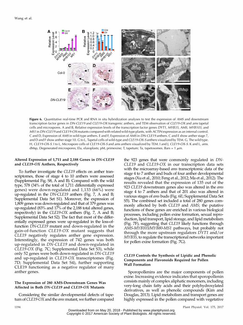
As the transcription factor genes DYT1,MYB35/TDF,AMS, MS188/MYB103/MYB80, and MS1 are requiredfor normal anther development in a DYT1-MYB35/TDF1-AMS-MS188/MYB103/MYB80-MS1 regulatorypathway (Ge et al., 2010; Chang et al., 2011; Zhu et al.,
2011) and AMS was reported to act as a master regu-lator coordinating pollen wall development (Xu et al.,2014), we further examined the expression levels ofgenes encoding these five transcription factors in DN-CLE19 and CLE19-OX anthers using quantitative real-time PCR analyses. The expression levels of AMS,MS188/MYB103/MYB80, and MS1 showed over 2-foldincreases in DN-CLE19 but over 2-fold decreases inCLE19-OX anthers (Fig. 6, A and B), suggesting thatthese genes are negatively regulated by CLE19. How-ever, the expression of eitherDYT1 orMYB35 exhibitedno obvious difference between theDN-CLE19 andwild-type anthers, but both showed 40% reductions in
Figure 4. Cell biological analyses of wild-type, cle19-2, DN-CLE19, and CLE19-OX anthers. A to E, Wild-type (WT) anthermorphology using semithin transverse sections. F to J, Morphology of cle19-2 anthers using semithin transverse sections. K to O,Morphology ofDN-CLE19 anthers using semithin transverse sections. P to T, Morphology of CLE19-OX-S anthers using semithintransverse sections. U to Y,Morphology ofCLE19-OX-M anthers using semithin transverse sections. Z to AA, Statistical analyses ofthe tapetal or reproductive cell numbers inDN-CLE19, CLE19-OX, and wild-type anthers, respectively. *P, 0.05 by Student’st-test. AB, How the numbers of tapetal or reproductive cells in each lobe in transverse sections were calculated. DPG,Degenerated pollen grains; Ms, meiocytes; PG, pollen grains; T, tapetum. Bars = 20 mm.
1192 Plant Physiol. Vol. 175, 2017
Wang et al.
www.plantphysiol.orgon May 20, 2018 - Published by Downloaded from Copyright © 2017 American Society of Plant Biologists. All rights reserved.

CLE19-OX anthers (Fig. 6, A and B), suggesting that theexpression of these two genes possibly was not or wasonly slightly affected byCLE19. These results suggestedthat CLE19 probably functions as a negative regulatorof the AMS-dependent transcriptional networks fornormal pollen wall formation.The expression of AMS in DN-CLE19 and CLE19-OX
anthers was tested further using RNA in situ hybridi-zation analysis. The results showed that theAMS signalwas hardly seen at anther stage 5 (Fig. 6, C and D) andwas detected in both the tapetum and microsporocytesat stages 7 to 10, albeit weakly (Supplemental Fig. S7, Band C); the signal then disappeared at anther stage12 (Supplemental Fig. S7, B and C). In comparison,AMS expression was obviously increased in the DN-CLE19 transgenic anthers at anther stages 7 to 10 (Fig. 6,E and F), suggesting that inhibition of the CLE19 functioncaused an increase inAMS expression. However, noAMSsignal was detected in CLE19-OX transgenic anthers at allexamined stages, similar to the results in the ams mutant(Supplemental Fig. S7, G–P), revealing that the increasedCLE19 function suppresses AMS expression. These datademonstrated that CLE19 acts as a negative upstreamregulator of AMS.
CLE19-OX Anthers Had ams-Like Swollen Tapetal Cells
It is known that tapetal cells secrete sporopolleninprecursors onto the primexine of the developing pollenfor the formation of the pollen exine and that AMS playsan important role in this process, with ams showing se-verely swollen tapetal cells with large vacuoles and fewlipidic tapetosomes and elaioplasts (Xu et al., 2014). Wepostulated that, if CLE19 acts as a negative upstreamregulator of AMS, tapetal cells of the CLE19-OX antherwould exhibit similar abnormal phenotypes to that of ams.Therefore, we performed TEM analysis of CLE19-OXtapetal cells. As expected, in contrast to the condensed anddegenerated cytoplasm of wild-type tapetal cells, withevident disintegration of cell membrane and accumula-tion of lipidic tapetosomes and elaioplasts (Fig. 6G),CLE19-OX tapetal cells were highly vacuolated andswollen, with large vacuoles and lack of lipidic tapeto-somes and elaioplasts (Fig. 6H). In addition, sporopolleninprecursors on the primexine were clearly observed on theouter surface of wild-type pollen grains; however, noobvious accumulation of sporopollenin precursors wasseen on the primexine of theCLE19-OX (Fig. 6, I and J) andams (Fig. 6, K and L) pollen grains.
Figure 5. Microscopic analyses of wild-type, DN-CLE19, and CLE19-OX pollenexine. A to J, Pollen grains from wild-type, DN-CLE19, and CLE19-OX plants vi-sualized by scanning electronmicroscopy. Kto O, Cytochemical staining of semithinsections of wild-type, DN-CLE19, andCLE19-OX pollen. P to Y, Wild-type,DN-CLE19, and CLE19-OX pollen vi-sualized by TEM. A, F, K, P, and U,Wild-type pollen. B, G, L, Q, and V,DN-CLE19-M pollen. C, H, M, R, andW,DN-CLE19-S pollen. D, I, N, S, and X,CLE19-OX-M pollen. E, J, O, T, and Y,CLE19-OX-S pollen. Ex, Exine; In, intine.Bars = 10 mm for A to E, 2 mm for F to J,5 mm for K to O, 2 mm for P to T, and 0.5mm for U to Y.
Plant Physiol. Vol. 175, 2017 1193
CLE19 Regulates Pollen Wall Formation
www.plantphysiol.orgon May 20, 2018 - Published by Downloaded from Copyright © 2017 American Society of Plant Biologists. All rights reserved.
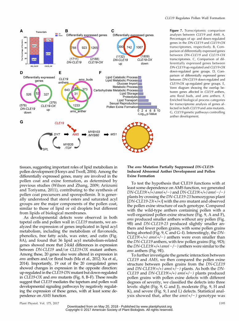
Altered Expression of 1,711 and 2,188 Genes in DN-CLE19and CLE19-OX Anthers, Respectively
To further investigate the CLE19 effects on anther tran-scriptomes, those of stage 4 to 10 anthers were assessed(Supplemental Fig. S8, A and B). Compared with the wildtype, 578 (34% of the total of 1,711 differentially expressedgenes) were down-regulated and 1,133 (66%) wereup-regulated in the DN-CLE19 anthers (Fig. 7, A and B;Supplemental Data Set S1). Moreover, the expression of1,809 genes was down-regulated and that of 379 genes wasup-regulated (83% and 17% of the 2,188 total altered genes,respectively) in the CLE19-OX anthers (Fig. 7, A and B;Supplemental Data Set S2). The fact that most of the differ-entially expressed genes were up-regulated in the loss-of-function DN-CLE19 mutant and down-regulated in thegain-of-function CLE19-OX mutant suggests thatCLE19 negatively regulates anther gene expression.Interestingly, the expression of 742 genes was bothup-regulated in DN-CLE19 and down-regulated inCLE19-OX (Fig. 7C; Supplemental Data Set S3), whileonly 52 genes were both down-regulated in DN-CLE19and up-regulated in CLE19-OX transcriptomes (Fig.7D; Supplemental Data Set S4), further supportingCLE19 functioning as a negative regulator of manyanther genes.
The Expression of 280 AMS-Downstream Genes WasAffected in Both DN-CLE19 and CLE19-OX Mutants
Considering the similar developmental defects of tape-tumofCLE19-OXand the amsmutant,we further compared
the 923 genes that were commonly regulated in DN-CLE19 and CLE19-OX in our transcription data setswith the microarray-based ams transcriptomic data of thestage 4 to 7 anther and buds of four anther developmentalstages (Xu et al., 2010; Feng et al., 2012;Ma et al., 2012). Theresults revealed that the expression of 135 out of the923 CLE19 downstream genes also was altered in the amsstage 4 to 7 anthers and that of 201 also was altered invarious stages of ams buds (Fig. 6E; Supplemental Data SetS5). The combined set included a total of 280 genes com-monly affected by both CLE19 and AMS; the putativefunctions of these genes are enriched in various biologicalprocesses, including pollen exine formation, sexual repro-duction, lipid transport, lipid storage, and lipidmetabolism(Fig. 7F), suggesting that CLE19 likely functions throughAMS-MYB103/MYB80-MS1 pathways, but probably notthrough the more upstream regulators DYT1 and/orMYB35, to regulate the transcriptional networks importantfor pollen exine formation (Fig. 7G).
CLE19 Controls the Synthesis of Lipidic and PhenolicComponents and Flavonoids Required for PollenWall Formation
Sporopollenins are the major components of pollenexine. Increasing evidence indicates that sporopolleninconsistsmainly of complex aliphaticmonomers, includingvery-long chain fatty acids and their polyhydroxylatedderivatives, as well as phenolic compounds (Kim andDouglas, 2013). Lipidmetabolism and transport genes arehighly expressed in the pollen compared with vegetative
Figure 6. Quantitative real-time PCR and RNA in situ hybridization analyses to test the expression of AMS and downstreamtranscription factor genes in DN-CLE19 and CLE19-OX transgenic anthers, and TEM observation of CLE19-OX and ams tapetalcells and microspores. A and B, Relative expression levels of the transcription factor genes DYT1, MYB35, AMS, MYB103, andMS1 inDN-CLE19 andCLE19-OXmutants comparedwith relatedwild-type plants, withACTIN expression as an internal control.C and D, Expression of AMS in wild-type anthers. E and F, Expression of AMS inDN-CLE19 anthers. C and E show anther stage 7,andD and F showanther stage 10. G to L, Tapetal cells of wild-type andCLE19-OX-S anthers visualized by TEM.G, Thewild type.H, CLE19-OX-S. I to L, Microspore cells of CLE19-OX-S and ams anthers visualized by TEM. I and J, CLE19-OX-S. K and L, ams.dMsp, Degenerated microspores; Ela, elaioplasts; pM, primexine; T, tapetum; Ta, tapetosomes. Bars = 1 mm.
1194 Plant Physiol. Vol. 175, 2017
Wang et al.
www.plantphysiol.orgon May 20, 2018 - Published by Downloaded from Copyright © 2017 American Society of Plant Biologists. All rights reserved.
tissues, suggesting important roles of lipid metabolism inpollen development (Honys and Twell, 2004). Among thedifferentially expressed genes, many are involved in thepollen coat and exine formation, as determined byprevious studies (Wilson and Zhang, 2009; Ariizumiand Toriyama, 2011), contributing to the synthesis ofpollen coat precursors and sporopollenin. It is gener-ally understood that sterol esters and saturated acylgroups are the major components of the pollen coat,similar to those of lipid or oil droplets but differentfrom lipids of biological membranes.As developmental defects were observed in both
tapetal cells and pollen wall in CLE19 mutants, we an-alyzed the expression of genes implicated in lipid acylmetabolism, including the metabolism of flavonoids,phenolics, free fatty acids, wax ester, and cutin (Fig.8A), and found that 36 lipid acyl metabolism-relatedgenes showed more that 2-fold differences in expressionbetween DN-CLE19 and/or CLE19-OX mutant anthers.Among these, 20 genes also were altered in expression inams anthers and/or floral buds (Ma et al., 2012; Xu et al.,2014). Importantly, 19 out of the 20 coregulated genesshowed changes in expression in the opposite direction:up-regulated in theCLE19-DNmutant but down-regulatedin CLE19-OX and ams mutants (Fig. 8, B–F). These resultssuggest that CLE19 mediates the tapetum and pollen walldevelopmental signaling pathways by negatively regulat-ing the expression of these genes, with at least some de-pendence on AMS function.
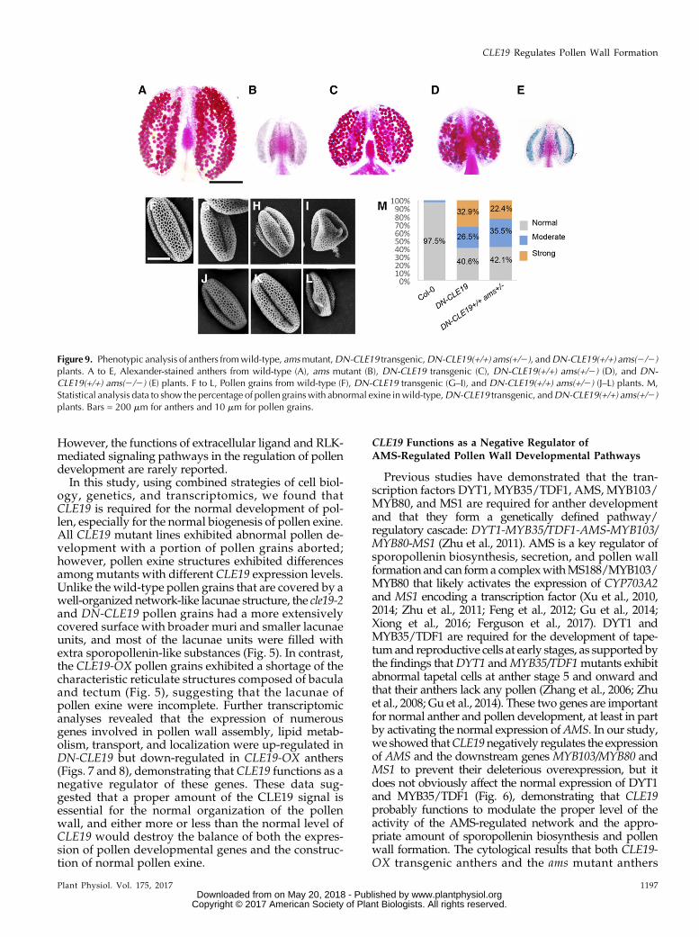
The ams Mutation Partially Suppressed DN-CLE19-Induced Abnormal Anther Development and PollenExine Formation
To test the hypothesis that CLE19 functions with atleast some dependence on AMS function, we generatedDN-CLE19(+/+) ams(+/2) andDN-CLE19(+/+) ams(2/2)plants by crossing theDN-CLE19-23 homozygous plant[DN-CLE19-23(+/+)] with the amsmutant and observedthe pollen exine structure of each genotype. Comparedwith the wild-type anthers containing pollen with awell-organized pollen exine structure (Fig. 9, A and F),ams produced smaller anthers without any pollen (Fig.9B) and DN-CLE19-23 produced slightly smaller an-thers and fewer pollen grains, with some pollen grainsbeing aborted (Fig. 9, C and G–I). Interestingly, theDN-CLE19(+/+) ams(+/2) anthers were even smaller thantheDN-CLE19 anthers, with few pollen grains (Fig. 9D);theDN-CLE19(+/+) ams(2/2) anthers were similar to theams anthers (Fig. 9E).
To further investigate the genetic interaction betweenCLE19 and AMS, we then compared the pollen exinestructure between pollen grains from the DN-CLE19and DN-CLE19(+/+) ams(+/2) plants. As both the DN-CLE19 and DN-CLE19(+/+) ams(+/2) plants producedpollen grains with pollen exine defects with differentdegrees of severity, we classified the defects into threelevels: slight (Fig. 9, G and J), moderate (Fig. 9, H andK), and severe (Fig. 9, I and L) defects. Statistical anal-ysis showed that, after the ams(+/2) genotype was
Figure 7. Transcriptomic comparisonanalyses between CLE19 and AMS. A,Percentages of up- and down-regulatedgenes in the DN-CLE19 and CLE19-OXtranscriptomes, respectively. B, Com-parison of differentially expressed genesbetween DN-CLE19 and CLE19-OXtranscriptomes. C, Comparison of dif-ferentially expressed genes betweenDN-CLE19 up-regulated and CLE19-OXdown-regulated gene groups. D, Com-parison of differentially expressed genesbetween DN-CLE19 down-regulated andCLE19-OX up-regulated gene groups. E,Venn diagram showing the overlap be-tween genes affected in CLE19 anthers,ams floral buds, and ams anthers. F,Enriched biological process categoriesfor transcriptome analysis of genes af-fected in both CLE19 and ams mutants.G, CLE19 genetic pathways controllinganther development.
Plant Physiol. Vol. 175, 2017 1195
CLE19 Regulates Pollen Wall Formation
www.plantphysiol.orgon May 20, 2018 - Published by Downloaded from Copyright © 2017 American Society of Plant Biologists. All rights reserved.

introduced into theDN-CLE19 plants, the percentage ofseverely defective pollen was obviously reduced (from32.9% to 22.4%; Fig. 9M) and the percentages of pollenwithmoderate defects and slight defects were increased(from 26.5% to 35.5% and from 40.6% to 42.1%, re-spectively; Fig. 9M), demonstrating that CLE19 worksin a way dependent on the AMS function.
DISCUSSION
A Proper Amount of CLE19 Is Important for PollenWall Formation
Cell-cell communication is often mediated by the ex-tracellular ligands and their receptors and is essential forthe development of multicellular organisms, includinganther development in angiosperms (Zhao et al., 2002;Yang et al., 2003, 2016; Jia et al., 2008; Huang et al., 2016).During pollen development, the adjacent tapetal layerprovides metabolites and sporopollenin precursors forpollen coat and exine formation as well as signals es-sential for pollen development (Mariani et al., 1990;
Goldberg et al., 1993; Sanders et al., 1999). The pollenmother cells surrounded by the tapetum undergomeiosis and form a tetrad enveloped by a thick pro-tective callose (1,3-b-glucan) wall that is digested sub-sequently by glucanases secreted from the tapetum,resulting in the release of microspores. Then, the mi-crospores develop into mature pollen grains with thedeposition of lipidic precursors/components secreted bythe tapetal cells onto the pollen surface to form the pollenexine and pollen coats (Mascarenhas, 1975; Stieglitz, 1977;Pacini and Juniper, 1979). The signaling communicationbetween tapetal cells and the reproductive cells (includingpollen mother cells and developing pollen grains) is im-portant for normal anther development and pollen forma-tion. For instance, Arabidopsis reproductive cell-preferentialTPD1 and the tapetum-expressed EMS1 and SOMATICEMBRYOGENESIS RECEPTOR-LIKE KINASE1/2(SERK1/2) complex interact to regulate normal ta-petum differentiation and pollen development, andplants with mutations in TPD1, EMS1, or SERK1/2 pro-duce no microspores (Zhao et al., 2002; Yang et al., 2003;Albrecht et al., 2005; Huang et al., 2016; Li et al., 2017).
Figure 8. Transcriptomic analyses ofgenes involved in the metabolism offlavonoids, phenolics, wax, cutin, andfree fatty acids in the wild type and theCLE19 mutant. A, Metabolic processesof the main compounds of the pollenwall. B to F, Fold changes in expressionlevels of genes involved in flavonoidmetabolic, cutin metabolic, wax estermetabolic, free fatty acidmetabolic, andphenolic metabolic processes.
1196 Plant Physiol. Vol. 175, 2017
Wang et al.
www.plantphysiol.orgon May 20, 2018 - Published by Downloaded from Copyright © 2017 American Society of Plant Biologists. All rights reserved.
However, the functions of extracellular ligand and RLK-mediated signaling pathways in the regulation of pollendevelopment are rarely reported.In this study, using combined strategies of cell biol-
ogy, genetics, and transcriptomics, we found thatCLE19 is required for the normal development of pol-len, especially for the normal biogenesis of pollen exine.All CLE19 mutant lines exhibited abnormal pollen de-velopment with a portion of pollen grains aborted;however, pollen exine structures exhibited differencesamong mutants with different CLE19 expression levels.Unlike the wild-type pollen grains that are covered by awell-organized network-like lacunae structure, the cle19-2and DN-CLE19 pollen grains had a more extensivelycovered surface with broader muri and smaller lacunaeunits, and most of the lacunae units were filled withextra sporopollenin-like substances (Fig. 5). In contrast,the CLE19-OX pollen grains exhibited a shortage of thecharacteristic reticulate structures composed of baculaand tectum (Fig. 5), suggesting that the lacunae ofpollen exine were incomplete. Further transcriptomicanalyses revealed that the expression of numerousgenes involved in pollen wall assembly, lipid metab-olism, transport, and localization were up-regulated inDN-CLE19 but down-regulated in CLE19-OX anthers(Figs. 7 and 8), demonstrating that CLE19 functions as anegative regulator of these genes. These data sug-gested that a proper amount of the CLE19 signal isessential for the normal organization of the pollenwall, and either more or less than the normal level ofCLE19 would destroy the balance of both the expres-sion of pollen developmental genes and the construc-tion of normal pollen exine.
CLE19 Functions as a Negative Regulator ofAMS-Regulated Pollen Wall Developmental Pathways
Previous studies have demonstrated that the tran-scription factors DYT1, MYB35/TDF1, AMS, MYB103/MYB80, and MS1 are required for anther developmentand that they form a genetically defined pathway/regulatory cascade: DYT1-MYB35/TDF1-AMS-MYB103/MYB80-MS1 (Zhu et al., 2011). AMS is a key regulator ofsporopollenin biosynthesis, secretion, and pollen wallformation and can forma complexwithMS188/MYB103/MYB80 that likely activates the expression of CYP703A2and MS1 encoding a transcription factor (Xu et al., 2010,2014; Zhu et al., 2011; Feng et al., 2012; Gu et al., 2014;Xiong et al., 2016; Ferguson et al., 2017). DYT1 andMYB35/TDF1 are required for the development of tape-tumand reproductive cells at early stages, as supported bythe findings thatDYT1 andMYB35/TDF1mutants exhibitabnormal tapetal cells at anther stage 5 and onward andthat their anthers lack any pollen (Zhang et al., 2006; Zhuet al., 2008; Gu et al., 2014). These two genes are importantfor normal anther and pollen development, at least in partby activating the normal expression ofAMS. In our study,we showed thatCLE19 negatively regulates the expressionof AMS and the downstream genes MYB103/MYB80 andMS1 to prevent their deleterious overexpression, but itdoes not obviously affect the normal expression of DYT1and MYB35/TDF1 (Fig. 6), demonstrating that CLE19probably functions to modulate the proper level of theactivity of the AMS-regulated network and the appro-priate amount of sporopollenin biosynthesis and pollenwall formation. The cytological results that both CLE19-OX transgenic anthers and the ams mutant anthers
Figure 9. Phenotypic analysis of anthers fromwild-type, amsmutant,DN-CLE19 transgenic,DN-CLE19(+/+) ams(+/2), andDN-CLE19(+/+) ams(2/2)plants. A to E, Alexander-stained anthers from wild-type (A), ams mutant (B), DN-CLE19 transgenic (C), DN-CLE19(+/+) ams(+/2) (D), and DN-CLE19(+/+) ams(2/2) (E) plants. F to L, Pollen grains from wild-type (F), DN-CLE19 transgenic (G–I), and DN-CLE19(+/+) ams(+/2) (J–L) plants. M,Statistical analysis data to show the percentage of pollen grainswith abnormal exine inwild-type,DN-CLE19 transgenic, andDN-CLE19(+/+) ams(+/2)plants. Bars = 200 mm for anthers and 10 mm for pollen grains.
Plant Physiol. Vol. 175, 2017 1197
CLE19 Regulates Pollen Wall Formation
www.plantphysiol.orgon May 20, 2018 - Published by Downloaded from Copyright © 2017 American Society of Plant Biologists. All rights reserved.

showed similar abnormal tapetum cells and pollenprimexine developmental defects, and the genetic re-sult that ams(+/2) partially suppressed the DN-CLE19transgene-induced pollen exine defects (Fig. 9), to-gether support the ideas that AMS works downstreamof the CLE19 signaling pathway and that the full effectof CLE19 depends on the normal level of AMS.
Therefore, we propose that the CLE19 peptide, proba-bly through unknown plasma membrane-localizedreceptor(s), negatively regulates the AMS-MYB103-MS1transcriptional cascades to maintain their functional ho-meostasis and ensure the normal development of pollen(Fig. 10A). However, in the DN-CLE19 mutants, the re-duced function of CLE19 releases the transcriptional in-hibition and causes the deleterious overexpression ofAMS and downstream networks; such overexpressionthen subsequently affects the normal organization ofpollen exine (Fig. 10B). In contrast, in the CLE19-OX an-thers, CLE19 synthesized at abnormally high levels ex-cessively inhibits the expression and function ofAMS anddownstream networks, thereby inducing abnormal pol-len development (Fig. 10C).
Functional Redundancy between Anther-ExpressedCLE Genes
As an extracellular peptide hormone, CLE19 wasexpressed at relatively low levels in the inflorescence and
stage 4 to 12 anthers in our quantitative real-time PCRanalysis using EF1a as the internal control (SupplementalFig. S1B). Nevertheless, both CLE19-OX and DN-CLE19transgenic plants showed obviously male fertility defects,even though the cle19 singlemutants showedweak antherdevelopmental defects (Fig. 2). We would like to offer thefollowing thoughts. First, RNA in situ hybridizationanalysis results showed thatCLE19was expressed only inthe tapetal and male reproductive cells at anther stages5 to 7 (Fig. 1, M–P), suggesting strong cell specificityand a short duration ofCLE19 expression in the anther.We believe that both the high cell specificity and theshort duration of CLE19 expression contribute to itssignal being diluted by nonexpressing cells in the inflo-rescence or stage 4 to 12 anthers. Second, as supportedby the results here, CLE19 acts as a negative regulator ofAMS and additional downstream genes, which are re-quired for normal tapetum function and pollen devel-opment. IfCLE19 is allowed to be expressed at very highlevels, normal pollen development would be inhibited,as seen in the CLE19-OX plants.
It is challenging to obtain mutants of CLE genes becausethey are all small genes; in addition, the anther-expressedCLE genes might have overlapping (redundant) functions.In this study, knockout mutants of five anther-expressedCLE genes, CLE9, CLE16, CLE17, CLE19, and CLE42, werecharacterized briefly, and only CLE19 knockout mutantsexhibited veryweak anther developmental defects,whereas
Figure 10. Proposed working model for CLE19 in the regulation of anther development. A, In wild-type anthers, CLE19 peptides interact with theirunknown receptor to negatively regulate the AMS-MYB103-MS1 transcriptional cascade to prevent their deleterious overexpression, which triggers thenormal expression of downstream exine formation-related genes, and subsequently regulate the formation of normally well-organized pollen exine. B, InDN-CLE19 anthers, CLE19 peptides without function competitively interact with the unknown CLE19 receptor, which blocks or obviously reduces signaltransduction from the functional CLE19 to its downstream molecules, leading to the deleterious overexpression of the AMS-MYB103-MS1 transcriptionalcascade and downstream exine formation-related genes, and subsequently affect normal pollen exine formation. C, InCLE19-OX anthers, overexpressionof CLE19 peptide strengthens the suppression of transcription factor gene expression, resulting in abnormal pollen exine formation.
1198 Plant Physiol. Vol. 175, 2017
Wang et al.
www.plantphysiol.orgon May 20, 2018 - Published by Downloaded from Copyright © 2017 American Society of Plant Biologists. All rights reserved.
mutants of the other four genes showed no obvious devel-opmental or fertility defects. Nevertheless, our statisticalanalysis results showed that, compared with the wild-typeplants, their double and triple mutants showed several de-tectable defects: increased barren silique number, reducedtotal pollen number but increased aborted pollen number ineach anther, and increased percentage of pollen with ab-normal pollen exine structure (Supplemental Fig. S4;Supplemental Table S1). In addition, the DN transgenicmutants of these seven anther-expressedCLEmembers,DN-CLE9, DN-CLE16, DN-CLE17, DN-CLE19, DN-CLE41,DN-CLE42, and DN-CLE45, all showed severe antherdevelopmental and pollen exine formation defects(Supplemental Fig. S5). All these results suggested thatthese anther-expressedCLEmemberspossibly function in aredundantway in the regulation ofmale fertility, especiallyin the regulation of pollen exine formation.We believe thatfurther studieswith variousmultiplemutantsmayuncoverthe details of redundant and distinctive functions on dif-ferent developmental aspects between theseCLEmembers.In addition, further studies also are needed to uncover
additional signaling components in the complex networkthat regulates pollen wall formation. For example, whatis the RLK that perceives the CLE peptide signal andtransduces the signal into the cytoplasm? What are thedownstream effectors, such as kinase and other factors,that respond to the CLE-RLK signalingmodule, and howdo they activate the AMS transcription factor?What is theproper amount of CLE peptide? Further investigationsusing a combination of omics, biochemistry, cell biology,and genetic strategies may answer these questions andfurther advance the study of male fertility.
CONCLUSION
In summary, our observations demonstrated that CLE19is essential for normal anther development and pollen wallformation, likely and in large part by negatively regulatingthe expressionofAMSand thedownstreamgenesMYB103/MYB80 and MS1 to prevent their deleterious over-expression, and to modulate the proper level of the ac-tivity of the AMS-regulated network and the appropriateanther development and pollen wall formation.
MATERIALS AND METHODS
Plant Growth and Genotyping
Arabidopsis (Arabidopsis thaliana) plants were grown on soil in greenhouseconditions (21°C) under long-day conditions (16 h of light/8 h of dark). T-DNAinsertion mutant alleles of cle19 (CS879453 and CS321816), cle9 (SALK_018122C),cle17 (SALK_103714C), and cle42 (SALK_057407C) mutants and RNAi plants ofCLE16 (CS201209) in the Col-0 background were all obtained from ArabidopsisBiological Resource Center stocks. The T-DNA insertion of CLE19 alleles wasverified using the T-DNA border primer SALKLB1.3 in combination with thegene-specific primers listed in Supplemental Table S2.
Molecular Cloning and Generation of Transgenic Plantsand Genotyping
Genomic sequences of CLE9, CLE16, CLE17, CLE19, CLE41, CLE42, andCLE45 were cloned into the pDONR221 vector (Life Technologies) with the
primers list in Supplemental Table S3.Using aPCR-based site-directedmutagenesis kit(Transgene) and specific primers (Supplemental Table S3), the Gly codon at the sixthpositionof theCLEmotifof theseCLEgeneswas replacedby theThrcodon,producingpDONR221-CLE9G6T, pDONR221-CLE16G6T, pDONR221-CLE17G6T, pDONR221-CLE19G6T, pDONR221-CLE41G6T, pDONR221-CLE42G6T, and pDONR221-CLE45G6Tentry clones. These entry cloneswere then subcloned into the pBGWFS7 binary vector(Karimi et al., 2002) to produce CLE19G6T, CLE19G6T, CLE16G6T, CLE17G6T, CLE41G6T,CLE42G6T, and CLE45G6T. Transformation was performed via an Agrobacterium tume-faciens-mediated floral dip method (Clough and Bent, 1998). Transgenic plants wereobtained under the selection of 25 mg of Basta (Sigma-Aldrich).
Quantitative Real-Time PCR Analysis
To analyze the expression levels of the CLEs and other downstream genes,stage 1 to 11 flower buds of Col-0, each related T-DNA or RNAi mutant, andDN-CLE transgenic plants were collected, and total RNA was extractedaccording to a Trizol-based (Sigma-Aldrich) method. After DNase I digestionand first-strand cDNA synthesis, quantitative real-time PCR was performedwith SYBR premix Ex Taq II (Takara) on the ABI StepOnePlus real-time system(Life Technologies) using the primers listed in Supplemental Table S4 and withEF1a (AT5G60390) as the internal control.
RNA in Situ Hybridization
RNA in situ hybridization analysis was performed using the DigoxigeninRNALabeling Kit (Roche) and the PCRDIG Probe Synthesis Kit (Roche). cDNAfragments of target genes about 400 bpwere amplified using the specific primerslisted in Supplemental Table S5. PCR products were cloned into the pGEM-Tvector and confirmed by sequencing. Completely digested plasmid DNAswereused as the template for transcription with T7 or SP6 RNA polymerase.
Phenotypic Analysis of Flowers and Anthers
Plants were photographed with a digital camera (Canon). Flower imageswere taken with the SPOT FLEX digital camera (Diagnostic Instruments) usingthe SteREO Discovery V8 dissecting microscope (Carl Zeiss Microimaging).
Stage 12 anthers were collected and stained overnight at room temperatureusing the Alexander solution prepared following a published protocol(Alexander, 1969), and additional anthers were pressed to release the stainedpollen grains and photographed by an AXIO ScopeA1 microscope (Carl ZeissMicroimaging) with a Axio Cam HRc camera (Carl Zeiss Microimaging).
Light and Electron Microscopy Observation
Inflorescences of wild-type and mutant plants were collected and fixed inFAA (formaldehyde–acetic acid–ethanol) solution and embedded in Technovit7100 resin (Heraeus Kulzer) as described (Jin et al., 2013). Semithin sectionswere prepared by cutting inflorescence materials into 1-mm-thick sections usinga motorized RM2265 rotary microtome (Leica Microsystems) and then werestained with 0.05% Toluidine Blue O for 15 to 30 min and photographed bybright-field microscopy.
For pollen wall observation, sections were stained with Toluidine Blue for5 min (10 mg mL21), Tinopal for 15 min (10 mg mL21; Sigma-Aldrich), anddiethyloxadicarbocyanine iodide for 5 min (5 mL mL21; Sigma-Aldrich), andsample photographs were taken using an AXIO ScopeA1 microscope with a390- to 440-nm excitation filter and a 478-nm blocking filter.
For scanning electron microscopy examination, fresh pollen grains releasedfrom stage 13 anthers were coated with gold and observed with an SU8010microscope (Hitachi).
For TEM observation, different stage buds were fixed in glutaric dialdehydebuffer andembedded into the freshmixed resin.Ultrathin sections (70-100nmthick)wereobservedwithaTecnaiG2SpiritTWINtransmissionelectronmicroscope(FEI).
RNA-seq and Data Analysis
More than 1,000 stage 4 to 10 anthers fromwild-type andmutant plantswerecollected and immediately frozen in liquid nitrogen. Total RNA from eachsamplewas extracted and purified using the ZRPlant RNAMiniprepKit (ZymoResearch). Two micrograms of total RNA of each sample was used for deepsequencingbyan IlluminaHiSeq2000 system.All sequencingdataweremappedand analyzed following the previously reported method (Zhu et al., 2015).
Plant Physiol. Vol. 175, 2017 1199
CLE19 Regulates Pollen Wall Formation
www.plantphysiol.orgon May 20, 2018 - Published by Downloaded from Copyright © 2017 American Society of Plant Biologists. All rights reserved.
Accession Numbers
The original RNA-seq data from this article have been submitted to the NationalCenter for Biotechnology Information Gene Expression Omnibus database under ac-cession number GSE94607. Sequence data from this article can be found in the Ara-bidopsis Genome Initiative database under the following accession numbers: CLE9(At1g26600), CLE16 (At2g01505), CLE17 (At1g70895), CLE19 (At3g24225), CLE42(At2g34925),CLE45 (At1g69588),DYT1 (At4g21330),AMS (At2g16910),MYB35/TDF1(At3g28470), MS1 (At5g22260), MYB103 (At1g63910), EF1a (At5g60390), and ACT2(At3g18780). Germplasm used included cle9-1 (SALK_018122C), cle16-1 (CS201209),cle17-1 (SALK_103714C), cle19-1 (CS879453), qrt1-2 (SALK_CS8846), cle19-2(CS321816), and cle42-1 (SALK_057407C).
Supplemental Data
The following supplemental materials are available.
Supplemental Figure S1. Expression patterns of CLE genes in variousArabidopsis organs.
Supplemental Figure S2. T-DNA locations or RNAi targeting sites andrelative expression of detected genes in the cle9-1, cle16-1, cle17-1, qrt1-2,cle19-1 qrt1-2, cle19-2, and cle42-1 mutants.
Supplemental Figure S3. Phenotypic analyses of plant growth, anthers,and pollen of wild-type and cle9-1, cle16-1, cle17-1, qrt1-2, cle19-1 qrt1-2,cle19-2, and cle42-1 plants.
Supplemental Figure S4. Phenotypic analyses of siliques, flowers, anthers, andpollen grains in the wild type and the cle single, double, and triple mutants.
Supplemental Figure S5. Phenotypic analyses of plant growth, anthers,and pollen grains of wild-type and DN-CLE transgenic plants.
Supplemental Figure S6. Comparison of exine patterns among cle19-2,DN-CLE19, DN-CLE41, DN-CLE42, and the wild type.
Supplemental Figure S7. RNA in situ hybridization analysis to detect thespatial and temporal expression of the AMS gene in wild-type, ams,DN-CLE19, and CLE19-OX transgenic anthers.
Supplemental Figure S8. Scatterplots for replicates of transcriptome data.
Supplemental Table S1. Statistical phenotypic analysis results of the clesingle, double, and triple mutants.
Supplemental Table S2. Primers used for genotyping.
Supplemental Table S3. Primers used for DN-CLE transgenic constructs inthis work.
Supplemental Table S4. Primers used for quantitative real-time PCR analysis.
Supplemental Table S5. Primers used for in situ constructs in this work.
Supplemental Data Set S1. List of 578 genes down-regulated and 1,133 genesup-regulated in DN-CLE19-23 transcriptomic analysis in this study.
Supplemental Data Set S2. List of 1,809 genes down-regulated and 379 genesup-regulated in CLE19-OX-S transcriptomic analysis in this study.
Supplemental Data Set S3. List of 742 genes both up-regulated in DN-CLE19-23 and down-regulated in CLE19-OX-S transcriptomic data.
Supplemental Data Set S4. List of 52 genes both down-regulated in DN-CLE19-23 and up-regulated in CLE19-OX-S transcriptomic data.
Supplemental Data Set S5. List of overlap genes differentially expressed inboth DN-CLE19-23 and CLE19-OX-S transcription data and the microarray-based transcriptomic data of the ams stage 4 to 7 anthers and 404 RNA-seq-based transcriptomic data of ams buds.
ACKNOWLEDGMENTS
We thank the Salk Institute Genomic Analysis Laboratory and theOhio StateUniversity Arabidopsis Biological Resource Center for providing the sequence-indexed Arabidopsis T-DNA insertion mutants.
Received March 30, 2017; accepted September 12, 2017; published September15, 2017.
LITERATURE CITED
Albrecht C, Russinova E, Hecht V, Baaijens E, de Vries S (2005) TheArabidopsis thaliana SOMATIC EMBRYOGENESIS RECEPTOR-LIKEKINASES1 and 2 control male sporogenesis. Plant Cell 17: 3337–3349
Alexander MP (1969) Differential staining of aborted and nonabortedpollen. Stain Technol 44: 117–122
Al-Refu K, Edward S, Ingham E, Goodfield M (2009) Alterations in thebasement membrane zone in cutaneous lupus erythematosus (CLE)as demonstrated by immunohistochemistry. Arch Dermatol Res 301:47
Ariizumi T, Toriyama K (2011) Genetic regulation of sporopollenin syn-thesis and pollen exine development. Annu Rev Plant Biol 62: 437–460
Bedinger PA, Hardeman KJ, Loukides CA (1994) Travelling in style: thecell biology of pollen. Trends Cell Biol 4: 132–138
Blackmore S, Wortley AH, Skvarla JJ, Rowley JR (2007) Pollen wall de-velopment in flowering plants. New Phytol 174: 483–498
Canales C, Bhatt AM, Scott R, Dickinson H (2002) EXS, a putative LRRreceptor kinase, regulates male germline cell number and tapetalidentity and promotes seed development in Arabidopsis. Curr Biol 12:1718–1727
Casamitjana-Martínez E, Hofhuis HF, Xu J, Liu CM, Heidstra R, ScheresB (2003) Root-specific CLE19 overexpression and the sol1/2 suppressorsimplicate a CLV-like pathway in the control of Arabidopsis root meri-stem maintenance. Curr Biol 13: 1435–1441
Chang F, Wang Y, Wang S, Ma H (2011) Molecular control of microspo-rogenesis in Arabidopsis. Curr Opin Plant Biol 14: 66–73
Clough SJ, Bent AF (1998) Floral dip: a simplified method for Agrobacterium-mediated transformation of Arabidopsis thaliana. Plant J 16: 735–743
Cock JM, McCormick S (2001) A large family of genes that share homologywith CLAVATA3. Plant Physiol 126: 939–942
Colcombet J, Boisson-Dernier A, Ros-Palau R, Vera CE, Schroeder JI(2005) Arabidopsis SOMATIC EMBRYOGENESIS RECEPTOR KINASES1and 2 are essential for tapetum development and microspore matura-tion. Plant Cell 17: 3350–3361
Cui J, You C, Zhu E, Huang Q, Ma H, Chang F (2016) Feedback regulationof DYT1 by interactions with downstream bHLH factors promotesDYT1 nuclear localization and anther development. Plant Cell 28:1078–1093
DeYoung BJ, Bickle KL, Schrage KJ, Muskett P, Patel K, Clark SE (2006)The CLAVATA1-related BAM1, BAM2 and BAM3 receptor kinase-likeproteins are required for meristem function in Arabidopsis. Plant J45: 1–16
Edlund AF, Swanson R, Preuss D (2004) Pollen and stigma structure andfunction: the role of diversity in pollination. Plant Cell (Suppl) 16: S84–S97
Feng B, Lu D, Ma X, Peng Y, Sun Y, Ning G, Ma H (2012) Regulation of theArabidopsis anther transcriptome by DYT1 for pollen development.Plant J 72: 612–624
Ferguson AC, Pearce S, Band LR, Yang C, Ferjentsikova I, King J, Yuan Z,Zhang D, Wilson ZA (2017) Biphasic regulation of the transcriptionfactor ABORTED MICROSPORES (AMS) is essential for tapetum andpollen development in Arabidopsis. New Phytol 213: 778–790
Fiers M, Golemiec E, Xu J, van der Geest L, Heidstra R, Stiekema W, LiuCM (2005) The 14-amino acid CLV3, CLE19, and CLE40 peptides triggerconsumption of the root meristem in Arabidopsis through a CLAVATA2-dependent pathway. Plant Cell 17: 2542–2553
Fiers M, Hause G, Boutilier K, Casamitjana-Martinez E, Weijers D,Offringa R, van der Geest L, van Lookeren Campagne M, Liu CM(2004) Mis-expression of the CLV3/ESR-like gene CLE19 in Arabidopsisleads to a consumption of root meristem. Gene 327: 37–49
Fiume E, Fletcher JC (2012) Regulation of Arabidopsis embryo and endo-sperm development by the polypeptide signaling molecule CLE8. PlantCell 24: 1000–1012
Fletcher JC, Brand U, Running MP, Simon R, Meyerowitz EM (1999)Signaling of cell fate decisions by CLAVATA3 in Arabidopsis shootmeristems. Science 283: 1911–1914
Ge X, Chang F, Ma H (2010) Signaling and transcriptional control of re-productive development in Arabidopsis. Curr Biol 20: R988–R997
Goldberg RB, Beals TP, Sanders PM (1993) Anther development: basicprinciples and practical applications. Plant Cell 5: 1217–1229
Gu JN, Zhu J, Yu Y, Teng XD, Lou Y, Xu XF, Liu JL, Yang ZN (2014) DYT1directly regulates the expression of TDF1 for tapetum development andpollen wall formation in Arabidopsis. Plant J 80: 1005–1013
1200 Plant Physiol. Vol. 175, 2017
Wang et al.
www.plantphysiol.orgon May 20, 2018 - Published by Downloaded from Copyright © 2017 American Society of Plant Biologists. All rights reserved.

Guilford WJ, Schneider DM, Labovitz J, Opella SJ (1988) High resolutionsolid state C NMR spectroscopy of sporopollenins from different planttaxa. Plant Physiol 86: 134–136
Honys D, Twell D (2004) Transcriptome analysis of haploid male game-tophyte development in Arabidopsis. Genome Biol 5: R85
Hord CL, Chen C, Deyoung BJ, Clark SE, Ma H (2006) The BAM1/BAM2receptor-like kinases are important regulators of Arabidopsis early antherdevelopment. Plant Cell 18: 1667–1680
Hord CL, Sun YJ, Pillitteri LJ, Torii KU, Wang H, Zhang S, Ma H (2008)Regulation of Arabidopsis early anther development by the mitogen-activated protein kinases, MPK3 and MPK6, and the ERECTA and re-lated receptor-like kinases. Mol Plant 1: 645–658
Huang J, Zhang T, Linstroth L, Tillman Z, Otegui MS, Owen HA, Zhao D(2016) Control of anther cell differentiation by the small protein lig-and TPD1 and its receptor EMS1 in Arabidopsis. PLoS Genet 12:e1006147
Ito T, Nagata N, Yoshiba Y, Ohme-Takagi M, Ma H, Shinozaki K (2007) Arabi-dopsis MALE STERILITY1 encodes a PHD-type transcription factor andregulates pollen and tapetum development. Plant Cell 19: 3549–3562
Jia G, Liu X, Owen HA, Zhao D (2008) Signaling of cell fate determinationby the TPD1 small protein and EMS1 receptor kinase. Proc Natl Acad SciUSA 105: 2220–2225
Jin Y, Yang H, Wei Z, Ma H, Ge X (2013) Rice male development underdrought stress: phenotypic changes and stage-dependent transcriptomicreprogramming. Mol Plant 6: 1630–1645
Jun J, Fiume E, Roeder AH, Meng L, Sharma VK, Osmont KS, Baker C, Ha CM,Meyerowitz EM, Feldman LJ, et al (2010) Comprehensive analysis of CLEpolypeptide signaling gene expression and overexpression activity in Arabi-dopsis. Plant Physiol 154: 1721–1736
Karimi M, Inzé D, Depicker A (2002) GATEWAY vectors for Agrobacterium-mediated plant transformation. Trends Plant Sci 7: 193–195
Kim SS, Douglas CJ (2013) Sporopollenin monomer biosynthesis in Ara-bidopsis. J Plant Biol 56: 1–6
Laux T, Mayer KF, Berger J, Jürgens G (1996) The WUSCHEL gene is re-quired for shoot and floral meristem integrity in Arabidopsis. Develop-ment 122: 87–96
Li H, Zhang D (2010) Biosynthesis of anther cuticle and pollen exine in rice.Plant Signal Behav 5: 1121–1123
Li Z, Wang Y, Huang J, Ahsan N, Biener G, Paprocki J, Thelen JJ, Raicu V,Zhao D (2017) Two SERK receptor-like kinases interact with EMS1 tocontrol anther cell fate determination. Plant Physiol 173: 326–337
Ma H (2005) Molecular genetic analyses of microsporogenesis and micro-gametogenesis in flowering plants. Annu Rev Plant Biol 56: 393–434
Ma X, Feng B, Ma H (2012) AMS-dependent and independent regulationof anther transcriptome and comparison with those affected by otherArabidopsis anther genes. BMC Plant Biol 12: 23
Mariani C, Beuckeleer MD, Truettner J, Leemans J, Goldberg R (1990)Induction of male sterility in plants by a chimaeric ribonuclease gene.Nature 347: 737–741
Mascarenhas JP (1975) The biochemistry of angiosperm pollen develop-ment. Bot Rev 41: 259–314
Mayfield JA, Preuss D (2000) Rapid initiation of Arabidopsis pollinationrequires the oleosin-domain protein GRP17. Nat Cell Biol 2: 128–130
Mizuno S, Osakabe Y, Maruyama K, Ito T, Osakabe K, Sato T, ShinozakiK, Yamaguchi-Shinozaki K (2007) Receptor-like protein kinase 2 (RPK 2)is a novel factor controlling anther development in Arabidopsis thaliana.Plant J 50: 751–766
Ni J, Clark SE (2006) Evidence for functional conservation, sufficiency, and pro-teolytic processing of the CLAVATA3 CLE domain. Plant Physiol 140: 726–733
Pacini E, Juniper BE (1979) The ultrastructure of pollen-grain developmentin the olive (Olea europaea). 2. Secretion by the tapetal cells. New Phytol83: 165–174
Phan HA, Iacuone S, Li SF, Parish RW (2011) The MYB80 transcriptionfactor is required for pollen development and the regulation of tapetalprogrammed cell death in Arabidopsis thaliana. Plant Cell 23: 2209–2224
Preuss D, Lemieux B, Yen G, Davis RW (1993) A conditional sterile mu-tation eliminates surface components from Arabidopsis pollen and dis-rupts cell signaling during fertilization. Genes Dev 7: 974–985
Sanders PM, Bui AQ, Weterings K, Mcintire KN, Hsu YC, Pei YL, MaiTT, Beals TP, Goldberg RB (1999) Anther developmental defects inArabidopsis thaliana male-sterile mutants. Plant Reprod 11: 297–322
Scott RJ, Spielman M, Dickinson HG (2004) Stamen structure and func-tion. Plant Cell (Suppl) 16: S46–S60
Shpak ED, Lakeman MB, Torii KU (2003) Dominant-negative receptoruncovers redundancy in the Arabidopsis ERECTA leucine-rich repeatreceptor-like kinase signaling pathway that regulates organ shape. PlantCell 15: 1095–1110
Song XF, Guo P, Ren SC, Xu TT, Liu CM (2013) Antagonistic peptidetechnology for functional dissection of CLV3/ESR genes in Arabidopsis.Plant Physiol 161: 1076–1085
Song XF, Yu DL, Xu TT, Ren SC, Guo P, Liu CM (2012) Contributions ofindividual amino acid residues to the endogenous CLV3 function inshoot apical meristem maintenance in Arabidopsis. Mol Plant 5: 515–523
Sorensen AM, Kröber S, Unte US, Huijser P, Dekker K, Saedler H (2003)The Arabidopsis ABORTED MICROSPORES (AMS) gene encodes aMYC class transcription factor. Plant J 33: 413–423
Stahl Y, Simon R (2009) Is the Arabidopsis root niche protected by se-questration of the CLE40 signal by its putative receptor ACR4? PlantSignal Behav 4: 634–635
Stieglitz H (1977) Role of b-1,3-glucanase in postmeiotic microspore re-lease. Dev Biol 57: 87–97
Strabala TJ, O’Donnell PJ, Smit AM, Ampomah-Dwamena C, Martin EJ,Netzler N, Nieuwenhuizen NJ, Quinn BD, Foote HC, Hudson KR(2006) Gain-of-function phenotypes of many CLAVATA3/ESR genes,including four new family members, correlate with tandem variationsin the conserved CLAVATA3/ESR domain. Plant Physiol 140: 1331–1344
Thom I, Grote M, Abraham-Peskir J, Wiermann R (1998) Electron andx-ray microscopic analyses of reaggregated materials obtained afterfractionation of dissolved sporopollenin. Protoplasma 204: 13–21
Torii KU, Mitsukawa N, Oosumi T, Matsuura Y, Yokoyama R, WhittierRF, Komeda Y (1996) The Arabidopsis ERECTA gene encodes a putativereceptor protein kinase with extracellular leucine-rich repeats. Plant Cell8: 735–746
Wilson ZA, Morroll SM, Dawson J, Swarup R, Tighe PJ (2001) The Ara-bidopsis MALE STERILITY1 (MS1) gene is a transcriptional regulator ofmale gametogenesis, with homology to the PHD-finger family of tran-scription factors. Plant J 28: 27–39
Wilson ZA, Zhang DB (2009) From Arabidopsis to rice: pathways in pollendevelopment. J Exp Bot 60: 1479–1492
Xiong SX, Lu JY, Lou Y, Teng XD, Gu JN, Zhang C, Shi QS, Yang ZN, ZhuJ (2016) The transcription factors MS188 and AMS form a complex toactivate the expression of CYP703A2 for sporopollenin biosynthesis inArabidopsis thaliana. Plant J 88: 936–946
Xu J, Ding Z, Vizcay-Barrena G, Shi J, Liang W, Yuan Z, Werck-ReichhartD, Schreiber L, Wilson ZA, Zhang D (2014) ABORTED MICROSPORESacts as a master regulator of pollen wall formation in Arabidopsis. PlantCell 26: 1544–1556
Xu J, Yang C, Yuan Z, Zhang D, Gondwe MY, Ding Z, Liang W, Zhang D,Wilson ZA (2010) The ABORTED MICROSPORES regulatory network isrequired for postmeiotic male reproductive development in Arabidopsisthaliana. Plant Cell 22: 91–107
Xu TT, Ren SC, Song XF, Liu CM (2015) CLE19 expressed in the embryoregulates both cotyledon establishment and endosperm development inArabidopsis. J Exp Bot 66: 5217–5227
Yang L, Qian X, Chen M, Fei Q, Meyers B, Liang W, Zhang D (2016)Regulatory role of a receptor-like kinase in specifying anther cell iden-tity. Plant Physiol 171: 2085–2100
Yang SL, Jiang L, Puah CS, Xie LF, Zhang XQ, Chen LQ, Yang WC, Ye D(2005) Overexpression of TAPETUM DETERMINANT1 alters the cellfates in the Arabidopsis carpel and tapetum via genetic interaction withEXCESS MICROSPOROCYTES1/EXTRA SPOROGENOUS CELLS. PlantPhysiol 139: 186–191
Yang SL, Xie LF, Mao HZ, Puah CS, Yang WC, Jiang L, Sundaresan V, YeD (2003) Tapetum determinant1 is required for cell specialization in theArabidopsis anther. Plant Cell 15: 2792–2804
Ye Q, Zhu W, Li L, Zhang S, Yin Y, Ma H, Wang X (2010) Brassinosteroidscontrol male fertility by regulating the expression of key genes involvedin Arabidopsis anther and pollen development. Proc Natl Acad Sci USA107: 6100–6105
Zhang W, Sun Y, Timofejeva L, Chen C, Grossniklaus U, Ma H (2006)Regulation of Arabidopsis tapetum development and function byDYSFUNCTIONAL TAPETUM1 (DYT1) encoding a putative bHLHtranscription factor. Development 133: 3085–3095
Zhao DZ, Wang GF, Speal B, Ma H (2002) The excess microsporocytes1 geneencodes a putative leucine-rich repeat receptor protein kinase that
Plant Physiol. Vol. 175, 2017 1201
CLE19 Regulates Pollen Wall Formation
www.plantphysiol.orgon May 20, 2018 - Published by Downloaded from Copyright © 2017 American Society of Plant Biologists. All rights reserved.

controls somatic and reproductive cell fates in the Arabidopsis anther.Genes Dev 16: 2021–2031
Zhu E, You C, Wang S, Cui J, Niu B, Wang Y, Qi J, Ma H, Chang F (2015) TheDYT1-interacting proteins bHLH010, bHLH089 and bHLH091 are redun-dantly required forArabidopsis anther development and transcriptome. Plant J83: 976–990
Zhu J, Chen H, Li H, Gao JF, Jiang H, Wang C, Guan YF, Yang ZN (2008)Defective in Tapetal development and function 1 is essential for anther
development and tapetal function for microspore maturation in Arabi-dopsis. Plant J 55: 266–277
Zhu J, Lou Y, Xu X, Yang ZN (2011) A genetic pathway for tapetumdevelopment and function in Arabidopsis. J Integr Plant Biol 53: 892–900
Zinkl GM, Zwiebel BI, Grier DG, Preuss D (1999) Pollen-stigma adhesionin Arabidopsis: a species-specific interaction mediated by lipophilicmolecules in the pollen exine. Development 126: 5431–5440
1202 Plant Physiol. Vol. 175, 2017
Wang et al.
www.plantphysiol.orgon May 20, 2018 - Published by Downloaded from Copyright © 2017 American Society of Plant Biologists. All rights reserved.