cytological observations on endamoeba blattae ·...
TRANSCRIPT


B RA RYOF THE
U N 1 VERS ITYOF 1 LLI NOI5
570.5ILL
V.\7
Cop. 7.
7)
HISTORY

Return this book on or before the
Latest Date stamped below. Acharge is made on all overdue
books. . _ ..
University of Illinois Library
OCT 2 2 952
11953


ILLINOIS BIOLOGICALMONOGRAPHS
Volume XVII
PUBLISHED BY THE UNIVERSITY OF ILLINOIS
URBANA, ILLINOIS

Digitized by the Internet Archive
in 2011 with funding from
University of Illinois Urbana-Champaign
http://www.archive.org/details/cytologicalobser174megl

ILLINOIS BIOLOGICAL MONOGRAPHS
Vol. XVII No. 4
Published by the University of Illinois
Under the Auspices of the Graduate School
Urbana, Illinois

EDITORIAL COMMITTEE
John Theodore BuchholzFred Wilbur Tanner
Harley Jones Van Cleave
1000—12-40—19253
CYTOLOGICAL OBSERVATIONS ONENDAMOEBA BLATTAE
WITH EIGHT PLATES
BY
Paul A. Meglitsch
Contributions from the Zoological Laboratory of the
University of Illinois
No. 556
THE UNIVERSITY OF ILLINOIS PRESSURBANA
1940

CONTENTS
I. Introduction 7
II. Acknowledgments 11
- III. Material 11
IV. Methods 12
A. Feulgen's Nucleal Reaction 15
B. Heidenhain's Haematoxylin 15
C. Safranin 16-
V. General Morphology 18
VI. Cytoplasmic Inclusions 22
A. Mitochondria .23B. Osmiophilic and Argentophilic Inclusions ... 24
C. Neutral Red-Stainable Inclusions 25
D. Inferences 27
VII. Terminology Used for Nuclear Description ... 28
VIII. The Trophic Nucleus; Historical Review . ... 31
A. Interphase 31
B. Kinetophase 34
IX. The Nucleus in Life 36
X. Nuclear Activities During the Trophic Stage . . 39
A. The Interphase 39
B. The Kinetophase 45
XI. Effects of Fixation on the Nuclear Elements . . 52
A. Simple, Uncombined Fixatives 53
B. Compound Fixatives 74
XII. Nature of the Elements of the Trophic Nucleus . 82
XIII. Nature of the Chromatin 92

XIV. The Precystic Period 105
XV. The Transformation from Trophic to Precystic
Period 106
XVI. The Living Precystic Amoeba 109
XVII. The Early Precystic Phenomena Ill
XVIII. Nuclear Transformation 113
XIX. The Late Precystic Phenomena 115
XX. The Cystic Period; Historical Review . . . .116
XXI. Appearance of Fresh Cysts 118
XXII. Study of Fixed and Stained Cysts 119
XXIII. Summary 122
Bibliography 127
Plates 131

I. INTRODUCTION
Cell structure, and especially nuclear structure, is similar in the major-
ity of metazoan groups. Detailed morphological differences are numerous,
of course, even within comparatively restricted systematic limits, but
these differences are confined to relatively unimportant minutae, and do
not appear to be associated with far-reaching distinctions of taxonomic
and phylogenetic importance. The basic morphological pattern of meta-
zoan cells includes a vesicular nucleus with basophilic material sus-
pended in a reticular or structureless matrix of karyolymph. Insofar as
the writer is aware, the distribution of basophilic material coincides, or
practically coincides, with the distribution of the material which reacts
positively to the Feulgen nucleal test in all cases which have been
studied. The majority of cytologists consider the basophilic nucleic acid-
containing material as chromatin. During division almost all metazoan
cells undergo mitotic phenomena which, but for slight variations in detail,
follow the same course in all types of organisms. Meiosis, likewise, is
similar in all Metazoa in its more general aspects. Thus we may con-
sider the metazoan nucleus a comparatively fixed structural and physio-
logical unit.
Such uniformity does not prevail among the Protozoa. Not only do
distantly associated groups have nuclei with fundamentally different
structure, as, for example, the vesicular nucleus of the Sarcodina and
the massive macronucleus of the Ciliata, but relatively closely related
forms, particularly among the Sarcodina, have nuclear patterns with
significant differences. Although nuclear phenomena are but incompletely
understood it appears that the divergence from the typical metazoan
pattern, as observed in some groups, is quite fundamental.
Several factors appear to be important in bringing about nuclear
diversity in Protozoa. One of the most important is the tendency
toward nuclear specialization. In many of the protozoan groups nuclei
are set aside for the consummation of specialized functions. Nearly all
ciliates have macronuclei and micronuclei, apparently differentiated pri-
marily for metabolic and reproductive functions respectively. A some-
what similar nuclear specialization is found in the trypanosomes, where
a so-called kinetonucleus is distinguished from a trophonucleus, the
former appearing to be associated with locomotive phenomena and the
initiation of division, while the latter is associated with metabolic func-
tions. Specialization of vegetative and generative nuclei is found among
some of the Sporozoa. In the Myxosporidia nuclear specialization is car-
ried still further. The developing pansporoblast contains shell nuclei
and capsulogenous nuclei which appear to govern the differentiation of
the spore membrane and polar capsule respectively. That so many cases
8 ILLINOIS BIOLOGICAL MONOGRAPHS
of nuclear specialization are associated with a segregation of reproductive
from metabolic functions suggests that we have to deal with a segrega-
tion of reproductive chromatin or idiochromatin from somatic chromatin
or trophochromatin. This concept was extremely popular at the turn of
the present century and has since been utilized to interpret many nuclear
phenomena among the various types of Protozoa. Since this possible
separation of a "germ line" from a "somatic line" is not accompanied
by histological differentiation as it is in the Metazoa, the nucleus itself
must bear the brunt of the specialization.
Instead of specialization of whole nuclei the Amoebaea present a
long series of slightly differing nuclear patterns which may be extremely
simple, as in Yahlkampfia, or highly complicated, as in some of the
Endamoebidae. This high degree of variability may be an expression
of the tendency to segregate trophochromatin from idiochromatin. Mor-ris ( 1936) suggests that this may be the case in Endamocba blattae.
Whatever the causes underlying the diversity of nuclear structure in the
amoebae, the fact remains that nuclear morphology is an important tax-
onomic character. Relationships are postulated through the comparison
of interphase and kinetophase nuclei. As might be anticipated the diver-
sity of nuclear structure is accompanied by differences in the pattern
which appears during division. A series extends from the simple, almost
amitotic division of Vahlkampfia to highly complicated division types
closely resembling metazoan mitosis, as in Entamoeba histolytica, and
to equally complicated karyokinetic figures, as in Endamocba blattae.
So striking are the differences in these division figures that some investi-
gators have hoped to learn something of the development of mitosis
through the study of primitive types of amoebae. Most of the phyloge-
netic speculation dealing with the amoebae has been based primarily on a
comparison of interphase and dividing nuclei.
As a result of the variation in the location and appearance of the
nuclear elements of the amoebae, the distribution of the basophilic ma-terial, commonly known as chromatin, is extremely varied. The identi-
fication of chromatic elements in the nucleus is difficult in some cases,
for the Feulgen nucleal preparation which is, to some extent, a test for
thymonucleic acids, fails to react with the basophilic material found in
the nucleus. Thus Morris (1936) and Sassuchin (1936) report that the
nucleus of E. blattae was not colored by the Feulgen nucleal reaction,
indicating that if any thymonucleic acids were present they were either
chemically combined in such a way that the color reaction did not occur
with the Feulgen reagents as applied, or that normal nucleic acids were
present, but in quantities too minute to call forth a visible color reaction.
Chalkley (1936) found that in Amoeba proteus the peripheral baso-
philic material was not colored by the Feulgen reaction, but that a cen-

ENDAMOEBA BLATTAE—MEGLITSCH 9
trally placed karyosome reacted positively. The morphological relation-
ship of chromatin—or at least the basophilic constituents of the nucleus
—
to nucleic acids is not understood in most members of the group.
Critical, detailed studies on the morphology of the interphase and
dividing nuclei of different amoebae are essential if generic relationships
and the implications of the differing structural patterns are to be under-
stood. Such studies may reveal fundamental correlations between the
distribution of basophilic constituents and of thymonucleic acids, and
may shed some light on the functional significance they have in the
consummation of nuclear activities during the life cycle of the organism.
It is with the problems outlined above that the present investigation
deals. The large unusual nucleus of E. blattac, while attracting the
attention of a number of investigators for the ease with which it can be
studied, is still but incompletely known. A number of past investigations
have resulted in contradictory results, and much work of confirmation
alone is needed. The failure of the Feulgen nucleal reaction to reveal
nucleic acid in the nucleus during the interphase is a striking example
of the unusual conditions prevailing in this species. Sassuchin ( 1936)
reported that he had failed to obtain a positive response to the Feulgen
reaction, but suggested that studies of different stages in division and in
the life cycle might reveal the presence of nucleic acid at particular
stages.
Since the writer has used the Feulgen nucleal reaction a great deal in
the present investigation, an explanation of his use and interpretation of
this reaction may be valuable. It is an obvious criticism that this reaction
is not a specific microchemical test of known validity for thymonucleic
acids, as it can also react with other substances which are present in the
cell. For example, plasmogen in the cytoplasm reacts positively. This
substance was removed by exposing the slides to 95 per cent alcohol for
from 12 to 24 hours. In some cases this substance was still present in
small quantities, and the cytoplasm was colored lightly by the reaction.
Since it occurs in the cytoplasm, however, it could scarcely be mistaken
for nucleic acids occurring in the nucleus. A plasmogen reaction was
observed in amoebae in which no reaction could be found in the nucleus,
and in other amoebae in which no cytoplasmic reaction had occurred there
was a nuclear reaction. Other cytoplasmic substances may react posi-
tively, but insofar as the writer is aware, the only substance which might
react positively in the nucleus are the nucleic acids. Additional evidence
in favor of the view that the reacting substance was nucleic acid was
supplied by fixation in absolute alcohol and subsequent washing. Accord-
ing to Fischer (1899) and Mann (1902) nucleic acid is precipitated by
alcohol. Slides of E. blattac, with control slides of the ciliates Nycto-
therus oralis and Balantidium praenucleatum, were fixed in absolute

10 ILLINOIS BIOLOGICAL MONOGRAPHS
alcohol and were found to react positively to the Feulgen test if there
was a minimum exposure to water. The deep coloration of the macro-
nuclear spherules of Nyctotherus ovalis was typical in all respects.
These spherules are thought to contain nucleic acid (see Kudo, 1936).
But nucleic acid precipitated by absolute alcohol is soluble in water,
according to Mann and Fischer. Slides fixed in absolute alcohol and
washed for 48 hours in running tap water failed to show a positive
reaction for ciliates or amoebae in thin sections. Similar smear prepara-
tions were negative for the amoebae, and for the ciliates a faint colora-
tion in the center of the macronucleus occasionally occurred in a few
organisms lying in the thicker portions of the smear, while the majority
failed to show any reaction. Just such a response to the technique used
would be expected from nucleic acids. According to Mann and Fischer,
nucleoproteins are also precipitated by absolute alcohol, but are insoluble
in water after precipitation. This seems to offer some experimental proof
in favor of the view that it was nucleic acid which reacted with the
Feulgen reagents in the present stud}', and not nucleoprotein. With these
few indications, and willing to admit that the specificity of the Feulgen
reaction may be considered as doubtful, the writer considers the material
in the nucleus of E. blattae which reacts positively to the Feulgen test to
be, in all probability, nucleic acid. Until the development of new micro-
chemical tests of greater validity for checking our results we are forced to
work with whatever means are at hand. It is with this in mind that the
present problem has been undertaken. The writer has interpreted the
material reacting positively, then, throughout the present paper, as
nucleic acid, not in the more strict chemical sense, but in a loose,
cytological sense.
In this study a comparison of results proceeding from the use of
basic dyes and the Feulgen reaction were made in order to determine the
relationship of basophilic material to nucleic acids, as demonstrated by
the Feulgen reaction, throughout the life cycle up to the formation of the
mature cyst, following here the suggestion made by Sassuchin (1936)
mentioned above, although when the work was undertaken the writer
was unfamiliar with Sassuchin's publication. At the same time it was
possible to search for new information concerning the morphology and
life cycle of the amoeba, and to confirm some of the observations of
earlier investigators. As a result of the study, in addition to details of
nuclear division during the trophic and cystic stages, it has been possible
to observe a cyclical variation in the quantity and distribution of nucleic
acid, as determined by the Feulgen test, correlated with the processes
of nuclear division and encystment. Several unusual relationships between
the basophilic material and the nucleic acid-containing material have also
been observed.

ENDAMOEBA BLATTAE—MEGLITSCH 11
II. ACKNOWLEDGMENTS
The writer wishes to express his deep appreciation of the aid given
him by Professor R. R. Kudo, at whose suggestion the problem was
undertaken. His encouraging and helpful suggestions have unfailingly
proved stimulating and without them the work could never have been
completed. His thanks are also due Professor W. Shumway and Profes-
sor H. J. Van Cleave for suggestions and advice. To various other mem-
bers of the staff of the Department of Zoology at the University of
Illinois the writer wishes to express his regard for their criticisms and
suggestions. The writer also wishes to express his thanks to Mrs. Alison
Meglitsch for preparing the plates and text figures.
III. MATERIAL
Endamoeba blattae occurs in the anterior dilated portion of the colon
of at least three species of cockroaches, Blatta oriental-is, Periplaneta
americana, and P. australasiae. The oriental cockroach was the source
of the amoebae used for this investigation. It is very abundant on the
University of Illinois campus and may be collected in large numbers
from early spring to late fall. During the day the insects retreat into
crevices in the walls of the buildings. At night they emerge to feed
and can be captured easily with the aid of a flashlight.
They were kept in battery jars, from fifty to a hundred roaches
living well in a single container. A variety of foods were tried, including
Fleischman's yeast cakes, moistened soybean meal, potatoes, and apples.
As reported by Kudo (1926) the yeast cakes form a most satisfactory
diet, not because the insects fail to thrive on the other diets tried, but
because the protozoan fauna respond unusually well to the yeast cake diet.
In cockroaches kept in a relatively crowded condition for several
weeks in the laboratory the incidence of infection rose to almost 100
per cent. In nature, of course, such high incidence is not usually ob-
served. Kudo ( 1926) reported that the percentage of infection is lowest
in nature in spring and late fall when it falls to about 5 per cent. It is
highest during the summer and early fall, reaching a peak of about 50
per cent in July, August, and September. Similar seasonal variation in
incidence of infection of cockroaches just captured is recorded by the
author. The incidence of infection was maintained at a high level
throughout the winter in the animals kept in the laboratory, but for some
reason it fell appreciably during early spring every year, although con-
ditions under which the host animals were kept remained constant
throughout. This was the more inexplicable since the large ciliate Nycto-
therus ovalis was as numerous in these hosts as it had been during the

12 ILLINOIS BIOLOGICAL MONOGRAPHS
winter, and the smaller ciliate Balantidium praenucleatum appeared to be
increasing in number at this time.
In immature hosts there is no appreciable difference in the incidence
of infection between male and female insects. But a small percentage
of adult males harbor amoebae, however, and a heavy infection in such
hosts is extremely rare. The mature females, on the other hand, are as
heavily infected as the immature insects and approach 100 per cent
in incidence of infection. One factor concerned in this difference ap-
pears to be the small amount of food consumed by winged males, whose
colons are frequently almost empty and greatly shrunken, in contrast to
the large amount of food consumed by the voracious females, which
have a very large and well filled intestinal tract.
IV. METHODS
Locke's solution proved to be a very satisfactory medium for main-
taining amoebae for periods long enough to complete the necessary pro-
cedures. The Locke solution was made with a reduced NaCl content
(0.45 per cent) and other compounds reduced proportionately. This was
the optimum concentration for survival as revealed by a series of tests
for maximum survival time in varying dilutions. When observations
were to be made in vivo the amoebae were kept in small (20 mm.)
petri dishes in sterilized Locke plus 0.5 per cent to 1 per cent Difco
albumin. They remained alive and active in this medium for several days
if a part of the dissected colon was left in the culture. Many of the
amoebae died after from two days to a week. The survivors appeared
to adapt themselves to their environment and remained alive and more or
less active after a month in many instances. Subculture was not effective,
however, as multiplication was quite rare among the cultured amoebae.
Depression slide mounts were used only when a single organism was
to be studied for an extensive period of time. Usually the amoebae were
kept in petri dishes, removed for observation whenever necessary, and
then replaced in the petri dishes. This method allowed greater freedom
and proved to be less laborious than the depression slide method. It was
especially well adapted to study with vital dyes, where it was not neces-
sary to observe the same specimen continuously in some experiments.
Vital dyes were kept in 1-1000 and 1-10000 solutions in Locke, and were
diluted with Locke-Albumin to the desired concentration just before use.
Of a number of vital dyes tried, Janus green B, Bismarck brown, bril-
liant cresyl blue and neutral red were the most satisfactory. The optimum
strength for these dyes varied between 1-50000 and 1-100000. When a
large amount of debris was present in the teased colon a larger amount
of dve was needed.

ENDAMOEBA BLATTAE—MEGL1TSCH 13
Table I
Summary of Nuclear Fixatives Used
Simple (Uncombined) Fixatives Compound Fixatives
Strong Strong
1. Absolute Alcohol* 1. Gilson Carnoy*2. Glacial Acetic Acid* 2. Schaudinn*
3. 10% Acetic Acid* 3. Carnoy*
4. 40% Formaldehyde* 4 Gilson*
5. Saturated Aqueous Mercuric 5. Sublimate Alcohol*
Chloride* 6. Sublimate Acetic*
6. Saturated Aqueous Picric Acid*
7. 1% Chromic Acid*
Medium Medium
8. 5% Acetic Acid* 7. Zenker*
9. 2% Acetic Acid 8. Bouin*
10. 3% Mercuric Chloride
11. 1% Acetic Acid
Weak
12. 4% Formaldehyde13. 0.5% Acetic Acid
14. 2% Osmic Acid
15. Osmic Vapor16. 70% Alcohol
17. 30% Alcohol
18. Dioxane
Weak
9. Flemming with Acetic
10. Flemming without Acetic
11. Champy12. Altmann13. Regaud
•Denotes that the fixatives were used at room temperature and at 45-50° C.
All others were used only at room temperature.
Permanent mounts for the study of cytoplasmic inclusions were made
in toto and in sections 3 to 8 /t thick. Altmann, Regaud, Champy and
Flemming without acetic were used as mitochondrial fixatives and were
followed by Heidenhain's haematoxylin, Regaud's haematoxylin, Ben-
sley's copper haematoxylin, Altmann's fuchsin-picric acid, Bensley's
fuchsin-methyl green, Kull's fuchsin-aurantia-toluidine blue or Benda's
alizarin-crystal violet. By far the most satisfactory stained mounts were
obtained in material in which the whole colon was fixed with Altmann
or Champy and sectioned at 3 to 5 p., followed by staining in Benda's
technique. Golgi material was studied with Kopsch and Kolatchev
osmium methods and the da Fano silver technique.
Studies of the nucleus were made with living amoebae under oil im-
mersion in bright and dark field illumination. Permanent mounts were
made in toto and in sections cut from 2 to 10 p. in thickness. Except
for the finer details of nuclear structure the thick sections proved more
practicable.
A number of fixatives were used, chosen for the satisfactory quality
of results obtained in preliminary trials. A summary of the fixatives used
is given in Table I. The compound fixatives most used were numbers

14 ILLINOIS BIOLOGICAL MONOGRAPHS
1, 2, 3, 7, 8, and 9. A number of others not mentioned were tried and
gave unsatisfactory results or were not used to any great extent because
the results were not significantly different from those obtained with the
standard protozoological fixatives.
Fixation and staining so completely alter the appearance and struc-
ture of the nucleus of E. blattae that it is impossible to describe the fixed
and living nucleus in similar terms without very careful study. So manystructures can be seen only in fixed nuclei that it was necessary to de-
termine their position in the living nucleus before their interpretation
was possible. The correlation between living and fixed nuclei was ob-
tained by a study of the phenomena accompanying fixation. This study
included all simple or uncombined fixatives shown in Table I, and num-
bers 1, 2, 3, 6, 7, 8, and 9 of the compound fixatives.
x\moebae were kept in Locke solution without albumin or were washed
before use, for when fixative was added to a solution with albumin in
it, the precipitation of the albumin obscured the amoebae. The organism
selected was put on a slide in a small drop of Locke solution and a
cover glass applied. By using the less viscous mineral oil in place of
cedar oil it was possible to carry on observation under oil immersion.
A piece of paper toweling was placed in contact with the cover slip at
one side of the slide, to draw off the fluid until the amoeba was com-
pressed, but still alive and able to move slightly. This compression was
necessary, for amoebae not treated in this way were either carried awaywith the fixative, or were rendered too opaque for accurate observation.
The nucleus was usually slightly compressed also, but insofar as could
be determined the structure was not changed, and the parts retained
their normal relationships.
The compressed nucleus was studied briefly under oil immersion.
When the position of the various regions and structures had been de-
termined, a drop of the fixing solution was placed in contact with the
cover glass opposite the paper toweling, which drew it under the cover
glass very rapidly. By this method the fixation could be watched under
high magnifications and the results determined. Several times dark field
illumination was used, but the precipitation of the protoplasm caused so
much refraction that it was not continued as a routine practice. Indeed,
even in the living nucleus nothing could be seen in dark field that could
not also be seen in bright field illumination. When fixation was slow,
a very rare condition, the sequence of the appearance of nuclear struc-
tures invisible in life was determined. Usually the process reached com-
pletion almost instantaneously, or at most in a few seconds, so that it
was necessary to repeat many times, fixing the attention successively on

ENDAMOEBA BLATTAE—MEGLITSCH 15
first one and then another region of the nucleus before the complete
process could be ascertained. In order to identify the nuclear structures
found, their affinity for basic dyes was determined at the end of the ex-
periment by adding acetocarmine or methyl green to the preparation,
after appropriate washing. During the whole procedure the nucleus was
kept under constant observation.
This study of the process of fixation was very helpful in determining
the relationship of the structures in the fixed nucleus to those observed
in the living nucleus, and aided in obtaining a reasonably complete under-
standing of the morphological effects of fixation. By combining the study
of the structure immediately following fixation with that of similarly
fixed nuclei in stained permanent mounts, some idea of the effects of
dehydration were also gained. Each fixative, simple or compound, so
studied was used in preparing permanent mounts in toto and in sections,
stained with Feulgen's nucleal reaction, Heidenhain's haematoxylin, and
Safranin light green, following a rigidly standardized staining procedure.
A. Feulgen's Nucleal Reaction
After washing for 12 to 24 hours in 95 per cent alcohol the smears
and sections were hydrated and passed into the hydrolizing solution
where they were held for 5 minutes at 60° C, rinsed in cold hydrolizing
solution 1 minute, put into the fuchsin sulphurous solution for one and a
half hours, washed in 3 changes of sulphurous acid wash water, 10
minutes each, and in running dechlorinated tap water for 30 minutes.
Following dehydration to 95 per cent alcohol the slides were lightly
counterstained with light green (10 seconds in a slightly basic 0.5 per cent
solution in 95 per cent alcohol) and mounted in balsam after clearing in
xylol.
B. Heidenhain's Haematoxylix
Slides were mordanted for 5 minutes at 45° C. after which they were
rinsed a total of 5 minutes in 3 changes of distilled water. They were
stained in 0.5 per cent haematoxylin for 5 minutes at 45° C, washed in
running dechlorinated tap water for 20 minutes, and differentiated in
large lots in saturated aqueous picric acid. The destaining process could
not be standardized perfectly because slight variations in thickness were
sufficient to alter the time required to reach maximum contrast. A partial
standardization was effected by differentiating large numbers of slides
simultaneously, a very effective method for materials sectioned at the
same thickness. The differentiation of smear preparations, naturally,
could be but incompletely standardized even by this method.

16 ILLINOIS BIOLOGICAL MONOGRAPHS
C. Safranin
After postosmication in used Flemming fixative for 15 minutes, and
a wash of equal duration the slides were placed in Zwaardemaker's
Safranin for one hour and washed in distilled water for 5 minutes. The
preparations were then placed in 70 per cent alcohol for 30 seconds,
95 per cent alcohol plus 1/2 per cent light green S. F. yellowish to
deepest contrast (30 seconds to 1 minute according to fixation), rinsed
by dipping into 95 per cent alcohol, left in absolute for 30 seconds, and
passed into xylol.
Before the warmed haematoxylin method was settled upon for the
standardized technique it was compared carefully with the cold method,
using both mordant and stain for 12 hours. In all essentials the results
were exactly comparable, differing only in the tint of the result. The
warmed haematoxylin tended to give deep violet-black nuclear structures,
while the cold method produced an even deeper brownish black. Similarly
various agents for differentiating haematoxylin were tried. Picric acid
gave the sharpest differentiation, and iron alum was next best.
For purposes of comparison a number of other staining techniques
were tried. Paralleling the standardized techniques used with the simple
fixatives a series of slides stained with Giemsa were made. Occasionally
other stains were tried with the simple fixatives, but none were used
consistently enough to allow any interpretation of results. Insofar as
could be seen with the incomplete data from the other stains, no further
structure could have been identified with the other stains tried. With the
compound fixatives, however, many combinations were used. These are
summarized in Table II.
The large number of stains and fixatives used made it possible to
check the results of nearly all the earlier investigators with preparations
treated with the same reagents they had used, or with comparable tech-
niques, as determined by a comparison of the effects of different fixa-
tives and stains. This has led to the explanation of some of the dis-
crepancies found in their work.
It became apparent from the study of the material that the nuclear
morphology was sometimes altered strikingly by the fixative and stain
used. The important differences observed in nuclear appearance after
different commonly used fixatives had been employed led to the stud}'
of the effects of the simple fixatives, in the hope that some explanation
for these discrepancies might be gained. In many cases the action of
compound fixatives could be explained from the study of the reagents
composing them. The simple fixatives tried are summarized in Table I.
Each simple fixative was studied in the same way. The immediate
effects of fixation were determined by observation of the process of fix-

ENDAMOEBA BLATTAE—MEGL1TSCH 17
Table II
Summary of Stain-Fixative Techniques Used
Nuclear Studies
Stain Fixatives*
1. Delafield's haematoxylin 1, 2, 3, 4, 5, 6, 8, 9.
2. Ehrlich's haematoxylin 1, 2, 3, 4.
3. Heidenhain's haematoxylin 1,2,3,4,5,6,7,8,9,10,11.
4. Borax Carmine 1, 2, 3, 4.
5. Paracarmine 1, 2, 3, 4.
6. Giemsa 1, 2, 3, 4, 5, 6, 7, 8, 9.
7. Safranin 3,5,6,8,9,11.
8. Safranin-light green 3, 5, 6, 7, 8, 9, 10, 11.
9. Safranin-orange G 9, 11.
10. Flemming's Triple 3, 6, 8, 9, 11.
11. Feulgen's nucleal reaction 1, 2, 3, 4, 5, 6, 7, 8, 9, 10, 11.
Cytoplasmic Studies
12. Heidenhain's haematoxylin 10, 11, 12, 13.
13. Regaud's haematoxylin 11, 12, 13.
14. Bensley's copper haematoxylin 11.
15. Altmann's Fuchsin-Picric Acid 10, 11, 12, 13.
16. Bensley's Fuchsin-Methyl Green 10, 11, 12, 13.
17. Kull's Fuchsin-Aurantia-Toluidin Blue.. 10,11,12,13.
18. Benda's Alizarin-Crystal Violet 10,11,12,13.
*The numbers refer to the numbers found in Table I in the CompoundFixative column.
ation under oil immersion, using the technique described above for com-
pound fixatives. Smear preparations fixed with the simple fixatives
were made and stained with the three standardized procedures outlined
above. Sections 10 jx thick were also stained with these procedures.
These permanent mounts were divided into two lots. The first lot was
made with a view toward exposing the preparations to a minimum con-
tact with water, at least before the staining process was inaugurated.
The second lot was washed for prolonged periods of time in running
dechlorinated tap water before staining. Smears were all washed for
24 hours. At first an attempt was made to wash sections for 24 hours
also, but later the whole colons were washed for two days before section-
ing, and the sections were washed for an additional 12 hours just before
they were stained.
As had been expected from the available knowledge concerning the
effects of fixation on certain of the more common chemical substances
occurring in the nucleus, some of the structures precipitated by simple
fixatives were soluble in water, while others were not. By comparing the
results obtained from the study of washed and unwashed preparations
with the tables of soluble and insoluble substances precipitated by the
various fixatives in Mann (1902) and Fischer (1899), it was possible to
come to some preliminary and tentative conclusions concerning the com-

18 ILLINOIS BIOLOGICAL MONOGRAPHS
position of certain of the nuclear elements. These results were also used
to explain the effects of some of the compound fixatives.
V. GENERAL MORPHOLOGY
Endamoeba blattae is one of the largest species of endozoic amoebae
known. It attains a maximum size of over 200 ,a and a minimum size of
about 45 p. in the trophic stage. Size distribution varies markedly between
these extremes and is affected significantly by the diet of the host. Star-
vation of the host reduces the number and size of the amoebae. An apple
diet also causes a reduction of size, although to a lesser degree. In either
case there is a distinct reduction in the number of food inclusions, result-
ing in beautifully transparent forms. Under such conditions the mean
size distribution may shift from 10 to 15 /x or more. When precystic and
cystic amoebae are not present the number of smaller trophic forms is
usually distinctly reduced. The highest size frequency usually occurs
between 80 and 100 p. in well fed hosts.
Before the work of Lucas (1927) all amoebae in the cockroach
colon were considered as belonging to a single species, E. blattae. Thus
the size range of this species had always been extended to include the
much smaller forms, Endamoeba thomsoni and Endolimax blattae. Grassi
( 1882), for example, mentions a number of small amoebae 4.4 to 6.6 /j.
in diameter. From his brief description of these tiny organisms it is
apparent that he must have observed Endolimax blattae. Other early in-
vestigators record similar minute dimensions. Some of these investigators
believed that the tiny amoebae were recently excysted Endamoeba blattae,
a view which may have had some basis in fact according to the results
of Morris (1936), who describes extremely small, transparent amoebae
which emerge from the cyst. Morris was the first to record the size
range of the trophic amoebae which has been observed in this study,
setting the lower limit of size for the trophic forms at about 50 p..
Precystic amoebae are much smaller than trophic forms, ranging from
about 20 to 50 u, with the largest number between 30 and 40 p in
diameter. The cysts measure from 15 to 35 /.i in diameter, with the meanaround 25 p..
Cytoplasmic differentiation is apparently of two kinds. A clear, hya-
line ectoplasm contrasts with a more granular vacuole-filled endoplasm.
A plasmasol-plasmagel differentiation may also be observed. In addition
to these, Schubotz (1905) described a so-called "light" and "dark" plasma
differentiation. From his descriptions and figures it appears that the dark
plasma was the plasmagel and the light plasma the plasmasol, for the
distribution of the two types of plasma appears to be identical, as Morris
(1936) was led to conclude. Biitschli (1878) and Schubotz (1905) were
ENDAMOEBA BLATTAE—MEGLITSCH 19
unable to distinguish a distinct ectoplasmic layer. Most of the other
investigators have observed an ectoplasmic differentiation, however, and
Kudo ( 1926) discusses this point thoroughly. As he points out, a distinct
ectoplasmic sheet is but rarely visible and occurs only in comparatively
sluggish or inactive amoebae. Small regions of ectoplasm may occur in
the anterior or anterolateral margins of active amoebae, but a distinct
ectoplasmic layer is not found.
Under high magnifications the plasmasol-plasmagel differentiation is
distinctly visible. The plasmagel appears as a more or less continuous
reticulum throughout the amoeba in active forms. It is somewhat lighter
in color than the plasmasol in life, and stains more intensely with acid
dyes, as can be seen in sectioned preparations (Fig. 65). Within the
interstices of the plasmagel reticulum lies the plasmasol, a rather gran-
ular substance in life, which stains less intensely with acid dyes. Not
infrequently a posterior region containing much plasmagel and very little
plasmasol can be distinguished in actively motile forms.
During active forward movement striae frequently appear in the
cytoplasm. They appear only in active amoebae, and run parallel to the
direction of flow of the cytoplasmic currents. These have been described
by almost every investigator, most of whom have offered some explana-
tion for their occurrence. Biitschli (1878) and Schubotz (1905) believed
that the striae are fibrils formed by longitudinally oriented alveolar walls.
Kudo (1926) postulated a stretching of the alveoli in the direction of
flow caused by the active endoplasmic streaming which resulted in the
striated appearance. Morris (1936) suggested that the plasmasol and
plasmagel are immiscible and that the striae are produced by streamers
of plasmagel extending into the plasmasol. No observations have been
made which will support any of these contentions directly. Concerning
the last mentioned possibility, however, it may be noted that streamers
of plasmagel do sometimes reach far forward from the posterior gelated
region of the amoeba. These have been observed many times in actively
moving amoebae, even when striae, which can be seen under quite low
magnifications, unlike the plasmagel streamers, could not be found. In
other cases striae have been observed with the streamers of plasmagel
extending forward between them. The writer is, therefore, of the
opinion that the very conspicuous striae are distinct from the delicate
streamers of plasmagel. Biitschli (1878) noted that the striae tend to
disappear when the amoeba is slightly compressed, which has been
amply confirmed during the present study. This observation seems to
be inexplicable on the basis of any theory advanced so far, and until
further information is available the problem of the nature and mode of
formation of the striae must be considered as unsolved.
Locomotion and endoplasmic streaming are very striking in E. blattae,

20 ILLINOIS BIOLOGICAL MONOGRAPHS
and have been the subject of considerable study. As early as 1898
Rhumbler studied some of the phenomena associated with pseudopodial
formation in E. blattae. He described cytoplasmic currents accompany-
ing locomotion as "fountain-streaming," characterized by a strong back-
ward flow of plasmasol beneath the relatively thin outer shell of plas-
magel. Pseudopodial formation, he noted, is often almost explosive, the
current being very rapid and strong.
The speed of forward progression may be quite rapid. Kudo (1926)
noted an amoeba which traversed twice its body length in one minute,
and several which travelled distances well over one body length per
minute. Similar speeds were observed during the course of this study.
It appears that active amoebae usually average about one body length
per minute, unless very large or hampered by much debris. During
active movement the shape assumed is similar to that of Vahlkampfia
Umax. There is a single broad pseudopod, the advancing edge of which
may be tipped by a narrow band of ectoplasm or have small antero-
lateral margins of ectoplasm. Occasionally two pseudopods are formed.
Among active amoebae this indicates a sudden change in the direction of
movement. Large numbers of lobose pseudopodia, as are formed by
Amoeba proteus, have not been observed.
In actively moving individuals endoplasmic streaming involves, as ex-
plained by Rhumbler, axial and superficial cytoplasmic currents. The
axial current advances, moving from the posterior region of plasmagel
toward the anterior margin of the pseudopod. There it breaks, fountain-
like, to form superficial currents which run posteriorly just under the thin
outer layer of plasmagel. A very little of the protoplasm streaming back-
wards may sometimes return directly into the axial stream, forming
an eddy. These eddies, which are comparatively rare, are very strik-
ing in amoebae stained with neutral red. The great majority of the
plasmasol flowing toward the posterior end of the body is converted
into plasmagel at the middle of the body or somewhat anterior to it.
Morris (1936, p. 231) says, "Although forward movement of the ani-
mal masks the fact during locomotion, it can clearly be seen, when the
amoeba is attached by the uroid, that the material which streams forward
through the center turns at the anterior end and streams backward along
the periphery. There is apparently no anterolateral gelation, nor cor-
responding reliquefaction in E. blattae." The writer cannot confirm this
point for amoebae moving actively in a relatively clear field, although the
opinion was maintained during the early part of the work. During the
observations made on vitally stained amoebae it was possible to trace
the course of single granules in the endoplasm during their circuit
through the cytoplasm. Granules always showed approximately the
ENDAMOEBA BLATTAE—MEGLITSCH 21
same course. They flowed forward from the posterior end until they
reached the anterior tip, where they turned and started to flow back-
ward. The backward flow continued from one-third to one-half of the
length of the amoeba during very active movement, and somewhat less
during slower movement. At this time the granules ceased to move back-
ward. Using slides with scratches on the surface it was possible to dem-
onstrate that the granules did not change in relation to the substrat from
this time until they began to flow forward again, which seems to indicate
that a lateral gelation had taken place. It appeared to the writer that at the
point at which the supposed gelation occurred there was a reduced amount
of Brownian movement. The only exceptions to this were the few granules
which became caught in the cytoplasmic eddies which occasionally are
found at the anterior end. These enter almost immediately into the
anteriorly directed current without continuing backward to the region of
gelation. After this had been observed it was sought for in amoebae
which were not vitally stained, lest the gelation was an effect of the stain-
ing process. With some difficulty the observations were tentatively con-
firmed in unstained amoebae, although the difficulties in observing un-
stained small granules made it impossible to be entirely certain of the
results.
Not all amoebae show currents which are exactly like those just
described. If an amoeba is caught in a mass of detritus, or if it is
attached to the slide at one end by the "uroid," streaming without gela-
tion occurs. This confirms the description given by Morris (1936),
cited above. It is not a normal locomotory current, however, in that it
does not lead to forward progression. When organisms undergo this
peculiar type of streaming it sometimes occurs that the superficial back-
ward current flows along only one side of the organism. This results in
displacing the usually central axial stream toward the opposite side, and
gives the amoeba a peculiar spiral appearance. As when attached by the
uroid, this type of streaming apparently does not lead to forward pro-
gression. It appears to be identical with the so-called "spiral streaming"
described by Sassuchin (1930). It has been observed that during "spiral
streaming" gelation in the lateroposterior region of the amoeba occasion-
ally occurs, although it is usually repressed.
Rhumbler's explosive pseudopodial formation in relatively inactive
amoebae has been observed frequently. It involves a rapidly moving cur-
rent of endoplasm breaking through the comparatively rigid ectoplasmic
sheet. It is interesting to compare this phenomenon with the formation
of pseudopodia in Amoeba proteus as described by Mast (1926). Hedescribes a hyaline cap, which is formed as the endoplasm breaks through
to the ectoplasmic layer. This seems to be directly analogous to the mode

22 ILLINOIS BIOLOGICAL MONOGRAPHS
of formation of the "explosive pseudopodia" in E. blattae. It is even
more striking in the latter, however, because of the stronger endoplasmic
currents and the heavier layer of ectoplasm.
Inactive amoebae are frequently rounded, and bear a large number of
small papilliform ectoplasmic protuberances which appear to be formed
by the explosive process. In these cases the plasmasol which burst
through the gelated region has become gelated and shows no further
streaming. Not infrequently one or more of these may be drawn out into
a very long pseudopodium containing only hyaline ectoplasm. These are
formed very slowly and appear to have no association with locomotion,
for the organisms lie quiescent during the whole process. Actively mov-
ing amoebae never possess these long hyaline pseudopodia, which persist
for but a short time after the organisms bearing one or more of them
begin to undergo progressive locomotion. These long pseudopodia appear
to be identical in structure with the small ones. Kudo (1926) gives a very
accurate description of the small pseudopodia.
VI. CYTOPLASMIC INCLUSIONS
The protozoan cytologist has much difficulty in understanding and in-
terpreting cytoplasmic inclusions. So much cytoplasmic differentiation has
occurred that many structures not homologous to any found in metazoan
cells occur. Some of these structures show affinity for osmium and silver,
which is the most important test for Golgi material in metazoan cells.
In some cases, as the silver line system of ciliates, no difficulty is met
with in distinguishing cytoplasmic inclusions from the unusual specializa-
tions of the protozoans. In other cases, particularly in some of the
kinetic elements of the flagellates, there has been no unanimity in in-
terpretation, and many discrepancies appear in the literature. Wherethe cytoplasmic inclusions resemble those found in the metazoan cells
which have been studied, interpretation is not so difficult. The osmio-
philic material in the cytoplasm of the Sporozoa does resemble osmio-
philic material found in invertebrate cells, and it is not difficult to sup-
pose that the two are homologous. But the osmiophilic material around
the contractile vacuole of some ciliates is quite different in all respects.
Nevertheless it is interpreted by some investigators as being homologous
with metazoan Golgi. An added complication has developed from the
study of the neutral red vacuome, declared by some cytologists to be the
Golgi material and by others to be distinct from it. As a result of these
various difficulties there is much confusion prevailing among protozo-
ologists with respect to the Golgi material.
In two groups of Protozoa, the Sporozoa and Sarcodina, the identifi-
cation of Golgi material seems to be less troublesome than in the other

ENDAMOEBA BLATTAE—MEGL1TSCH 23
groups. The morphological similarity of the osmiophilic and the argento-
philic inclusions to those found in the metazoan cells aid a great deal in
interpreting results. A further aid is found in the fact that there is almost
a complete absence of other structures which resemble the Golgi material
only in their ability to reduce osmium or silver.
In spite of, or perhaps because of, the relative ease with which
Golgi material can be studied in these two groups, it has been rather
neglected. A considerable amount of work has been completed on the
Sporozoa, but studies on Sarcodina are very few. Causey (1925) studied
Entamoeba gingivalis. Brown (1930) studied Amoeba proteus. Hirsch-
ler (1927) made observations on Endamoeba blattae. A few added ob-
servations have been made by Mast and Doyle (1935), on Amoebaproteus. Otherwise the amoebae are wholly unknown. In view of our in-
complete knowledge of the Golgi material in the amoebae, E. blattae has
been restudied, using osmium and silver techniques, with an attempt to
correlate the results with those obtained with neutral red and other
vital dyes. Some observations were also made on the mitochondria.
A. Mitochondria
In amoebae stained vitally with Janus green B the mitochondria are
stained a light green. They appear as slender rods, usually rather short,
which flow about freely in the cytoplasm during the streaming which
accompanies movement. The mitochondria appear to be rather adhesive.
They are often found adhering to other cytoplasmic granules, and in
amoebae stained with Janus green B and neutral red the granules stained
red often adhered to the mitochondria. In view of these observations
the fact that the mitochondria are occasionally found adhering to the
wall of the food vacuoles does not appear to be significant. In manycases there were no mitochondria found on small vacuoles, and many of
the mitochondria were not associated with vacuoles. It appears to be
safe to conclude that no direct relationship between mitochondria and
food vacuoles has been observed for E. blattae.
After fixation the mitochondria are shorter and heavier. The)' usually
lie in a small clear area in the cytoplasm. This may be a shrinkage
phenomenon. Although no dividing mitochondria were found in vitally
stained material, in permanent slides evidences of division have been
found. Dumbbell-shaped mitochondria have been found ; in several in-
stances two small mitochondria were found in a single clear area. This
seems to indicate that the mitochondria divide. No relationship whatever
could be found between the occurrence of division in the mitochondria
and the stage of nuclear division. During the early part of the precystic
period the mitochondria appear to be as numerous as, and morphologically

24 ILLINOIS BIOLOGICAL MONOGRAPHS
identical with, the inclusions found in the trophic amoebae. Later pre-
cystic and cystic stages have not been observed. Early observations seem
to indicate a diminution in number and size of mitochondria in the
mature cysts.
B. OSMIOPHILIC AND ArGENTOPHILIC INCLUSIONS
Studies on the osmiophilic inclusions of E. blattae were first made by
Hirschler (1927). He found two types of osmiophilic inclusions. The
larger inclusions were composed of two types of material. An outer
osmiophilic region, circular or crescentic in optical section, was con-
trasted with a more or less spherical inner region of osmiophobic ma-
terial in the larger type of inclusion. These inclusions measured about
3 ju in diameter. In addition to the larger inclusions there were a number
of smaller inclusions which were quite variable in shape and were
restricted to the plasmasol or the dark plasma of Schubotz.
Granules and larger inclusions, apparently identical with those de-
scribed by Hirschler, were found. His descriptions and figures leave no
doubt that the inclusions found were the same as those he had observed.
According to Hirschler the smaller granules were lighter than the osmio-
philic shell of the larger inclusions. In the material prepared for this
study the two osmiophilic components—the smaller inclusions and the
osmiophilic shell of the larger inclusions—were approximately equally
dark. The difference noted here may be explained by the fact that
Hirschler appears to have differentiated further than was the practice in
this study.
Argentophilic material appears to be identical in all respects with the
osmiophilic material. No difference in appearance or distribution could
be found.
A comparison of the osmiophilic material found in E. gingivalis as
described by Causey (1925) with those found in E. blattae shows rather
distinctive differences. Causey found a large reticulum which apparently
originated from smaller crescentic bodies. These smaller crescentic bodies
seem to be similar to the larger inclusions found in E. blattae. Small
granules which Causey found associated with the food vacuole led him
to conclude that these small granules are the primordia of the Golgi
material, which is formed from them. Causey's work has been questioned
by several investigators, however, who feel that his methods and
techniques were not standardized. Thus Nigrelli and Hall (1930, p. 21)
say: "The technique used by Causey was not one of the methods used
commonly for demonstration of Golgi material and Bowen (1928) has
pointed out that Causey's method ordinarily would not be expected to
demonstrate elements of the Golgi apparatus. Thence, the occurrence of a

ENDAMOEBA BLATTAE—MEGLITSCH 25
filamentous or net-like 'Golgi apparatus' in Entamoeba gingivalis cannot
be accepted definitely until Causey's observations have been checked by
some of the more commonly used methods for demonstration of Golgi
material." Nigrelli and Hall have studied Arcella, and found in that
member of the Sarcodina a group of bodies not unlike the larger struc-
tures found in E. blattae. Brown (1930) found a number of osmiophilic
and argentophilic granules accompanied by a smaller number of larger
bodies composed of an osmiophilic cortex and an osmiophobic core in
Amoeba proteus. The larger bodies described by Brown appear to be
morphologically similar to those found in E. blattae, and the smaller
granules also seem similar to the smaller inclusions found in this species.
Exclusive of the work of Causey, then, Golgi material, or, at least,
osmiophilic material appears to be generally distributed in two forms.
Smaller granules of osmiophilic substances are dispersed in the cytoplasm
in large numbers, and larger structures consisting of osmiophilic and
osmiophobic parts occur in smaller numbers. The smaller granules may be
lacking in Arcella, as Nigrelli and Hall do not mention them. The ex-
ceptional case of E. gingivalis requires further study before any interpre-
tation can be made. It is interesting to note that the osmiophilic material
in the Sporozoa which have been studied is likewise composed of two
types of inclusions similar to those found in the Sarcodina (see Hirschler.
1927 and Bowen, 1928).
C. Neutral Red-Stainable Inclusions
The use of neutral red as a vital dye for the demonstration of Golgi
material has met with some opposition. Neutral red was originally
thought to demonstrate the vacuome, supposedly distinct from the Golgi
material. The fact that the vacuome, which stains with neutral red,
is able to bring about a reduction of osmium has led to the formulation
of the opinion that Golgi and the vacuome are either closely related or
identical. Hall and his students have found that the neutral red-staining
inclusions are identical with the osmiophilic material in a number of
protozoan species, including Arcella.
In the case of E. blattae no report has been found in the literature
concerning the results of intra-vitum staining with neutral red. In
dilutions of 1-50000 to 1-100000 it was found that a number of small
granules were specifically stained. These small granules were floating
freely in the endoplasm. In addition to the granules a number of larger
spherical bodies measuring from 3 to 3.5 jx in diameter were stained,
although less intensely than the smaller granules. The granules usually
acquire a red-violet color in fifteen to twenty minutes exposure, while

26 ILLINOIS BIOLOGICAL MONOGRAPHS
after this period the spherical bodies are an orange red. This intensity
of color may represent a greater dilution of the stain in the larger bodies,
or may possibly indicate a different pH in the two types of inclusions.
Almost every one of the larger spherical inclusions have from one to five
or six of the smaller granules attached to the periphery. A spherule
without at least one granule has never been found. The difference in
staining intensity makes it very easy to determine whether or not there
are granules adhering to the surface of the spherical bodies.
The spherical bodies are not stained permanently by the neutral red.
Their color reaches its greatest intensity in from 15 to 30 minutes after
the dye is first applied to the amoeba, when they are a deep orange red
shade. This typical coloration persists for several hours, and then gradu-
ally becomes less intense. In from 6 to 24 hours colorless spherical
inclusions may be found with several intensely stained granules adhering
to them. It is possible that this is due to the oxidation of the stain to
a colorless leucobase. The smaller granules retain their deep red-violet
coloration until after the death of the amoeba.
In the precystic amoebae the conditions are quite different from those
just described. The smaller granular inclusions are present in great
numbers, distributed at random throughout the cytoplasm. The larger
spherical inclusions, however, are not to be found, at least after the
nuclear transformation which accompanies the precystic development
is completed. During the study of the vitally stained amoebae it was
believed that some connection might exist between the larger spherical
inclusions and the food vacuoles. This was especially noticeable in several
well-fed amoebae from potato-fed hosts. In these specimens a large
number of the spherical inclusions were found adhering to the walls of
the food vacuoles containing grains of potato starch. The complete re-
lationship of vacuole and inclusion is not as yet understood. It is interest-
ing in this connection to observe that during the precystic period nearly
all and finally all of the food vacuoles disappear at the same time that
the spherical inclusions disappear. This appears to strengthen somewhat
the inference that there is some relationship between the vacuoles and the
inclusions.
Granules and spherules, apparently identical with those described
above, were also stained with brilliant cresyl blue and Bismarck brown.
These two dyes were so much less specific than neutral red, and the
amoebae die so much more rapidly that few observations were made with
them. No decolorization of the spherical inclusions was observed in
amoebae stained with these dyes, but whether this depended on the
greater toxicity of the stains or a difference in the chemical nature of the
dve could not be determined. A number of other stains were tried.

ENDAMOEBA BLATTAE—MEGLITSCH 27
Unfortunately they were either ineffective or stained so many structures
that it could not be determined whether they stained the neutral
red-stainable inclusions.
D. Inferences
The similarity in size, appearance, and general distribution of the
inclusions staining with neutral red and those which were impregnated
with osmium and silver appears to be in accord with the observations of
Hall and his students on various species of Protozoa. Insofar as this
writer is aware, this information was previously available for but one
member of the Sarcodina, namely Arcella. A number of times confirma-
tion was obtained by impregnation of vitally stained amoebae with
osmium. In all cases the stained inclusions were darkened with reduced
osmium. Insofar as could be determined such preparations did not differ
from the usual Golgi slides in general appearance and distribution of the
impregnated inclusions.
The fact that there were two distinct types of inclusions, both of
which were stained by neutral red and impregnated with osmium and
silver, made it extremely difficult to interpret the results. It has been felt
that until some additional information is available regarding the osmio-
philic inclusions it would be impossible to determine accurately which
type of inclusion is to be considered as a homologue of the metazoan
Golgi material. Certainly not all osmiophilic inclusions are Golgi material,
and supplementary work must be carried out before any conclusions can
be reached.
It is extremely interesting, however, to observe a similarity between
the larger type of osmiophilic inclusion and the secretory granules found
in some metazoan gland cells. Sharp, in his Introduction to Cytology
(1934), shows figures of secretory granules from various sources (Fig.
37, p. 76) which appear to be identical insofar as structure is con-
cerned with the larger inclusions occurring in E. blattae, Amoeba proteus,
and Arcella. Some of the Sporozoa, also, have osmiophilic inclusions
which are similar to the secretory granules. The question as to whether
this is merely a coincidental resemblance, or is significant, cannot be
settled on the basis of the present data. If, as it appears possible, some
relationship between the food vacuole and osmiophilic inclusion can be
found, it would seem that there would be an even greater resemblance of
the secretory granule to the larger osmiophilic inclusion.
The smaller type of osmiophilic inclusion of E. blattae appears to cling
to the outside of the larger spherical bodies in amoebae stained with
neutral red. The writer believes it possible, at least, that the osmiophilic
crescent of the larger spherules may be no more than the accumulation
and imperfect preservation of the small granules upon the larger inclu-
28 ILLINOIS BIOLOGICAL MONOGRAPHS
sion. This, again, appears to afford another resemblance of the larger
inclusion to secretory granules, for it has been supposed that the
osmiophilic portion of the secretory granules represents a portion of the
Golgi network which remained in contact with the secretory granule
after it had been formed. If this relationship of osmiophilic granule to
the larger spherical inclusion should prove, on further work, to be correct,
it appears that the small granules would be the true Golgi homologue.
It is, of course, impossible to conclude, on the basis of the evidence at
hand, that the larger inclusions are secretory granules or Golgi material,
or that the smaller inclusions are the Golgi homologue, and we await
further investigations of the functional relationships of the granules to the
spherical inclusions and of the spherical inclusions to the food vacuoles
which may possibly throw some light on the question.
VII. TERMINOLOGY USED FOR NUCLEAR DESCRIPTION
In order to facilitate interpretation of the descriptive text a diagram-
matic representation of the trophic interphase and kinetophase nuclei are
given in Text Figs. I and II, with the structures fully labelled. The terms
selected, because of the structural differences between the nucleus of
E. blattae and that of other parasitic amoebae, are in some cases inap-
plicable if strict homology of similarly designated structures is expected.
To preserve the customary terminology as much as possible some con-
cepts have been rather strained. Indeed, in many of the nuclear struc-
tures of the Sarcodina great confusion prevails insofar as homology is
concerned, and few investigators have attempted to erect a consistent
terminology. In the case of E. blattae, the previous investigators have
frequently referred to the same structures with very diverse terms.
Whenever feasible the terms employed by the majority of previous
workers are used. Definitions and remarks concerning the terms selected
are given below to facilitate comparisons with the descriptions of previous
investigators.
Central ground-plasm.—This substance fills the inner part of the intra-
nuclear space. Although not technically a ground-plasm, since the chro-
mosomes are formed from it, its clear, transparent optical character in
life and its homogeneous or delicately reticulated, undifferentiated ap-
pearance in the fixed condition during interphase give the term some
descriptive value. It forms the homogeneous, granular or reticular
region which lies within the endosomal girdle in fixed material. It is
optically differentiated from the peripheral ground-plasm, with which it
is connected at a few points by radii-like extensions in fixed material, or
is in contact along the whole margin in material fixed with weak fixatives
or in life (Text Fig. I).

ENDAMOEBA BLA TTAE—MEGLITSCH 29
Text Fig. I. Schematized Diagram of Nucleus of Trophic E. blattae
(1) Nuclear membrane; (2) peripheral zone; (3) peripheral ground-plasm;
(4) endosome; (5) central region; (6) centriole; (7) central ground-plasm;
(8) peripheral granule; (9) peripheral spherule.
Text Fig. II. Schematized Drawing of Dividing Nucleus of
Trophic E. blattae
(1) Nuclear membrane; (2) endosomal spherule; (3) peripheral spherule;
(4) peripheral granule; (5) chromosome.
Central region.—The region lying within the endosomal girdle in the
interphase nucleus. This is identical with the central zone of Sassuchin
(1936) (Text Fig. I).
Centriole.—The small basophilic granules which may sometimes be
seen at the center of the central ground-plasm in the interphase nucleus
and at the poles in the kinetophase nucleus. Although it could not be

30 ILLINOIS BIOLOGICAL MONOGRAPHS
traced accurately throughout karyokinesis, its position and appearance
invariably resembled that of a centriole. This is identical with the central
granule of Kudo (1926) and the karyosome of Morris (1936) (Text
Figs. I and II).
Chromosome.—The strands which appear at the poles of dividing
nuclei, and whose anlage may be seen in the central region during early
division stages. They contain free thymo-nucleic acid as determined
by the Feulgen nucleal reaction, but are not intensely basophilic. Their
failure to stain deeply with basic dyes and the fact that they never pass
through a metaphase plate stage shows that they are not entirely homol-
ogous in their reactions to the metazoan chromosomes, but since they
appear to be functionally indentical they merit the term chromosome.
Morris (1936) and Janicki (1909) also use the term chromosome for
these structures (Text Fig. II).
Chromosomal strands.—Slender strands of discrete granules which
appear in the central ground-plasm during early division stages. They
appear to be the anlage of the chromosomes (Fig. 25).
Endosomes.—Large basophilic bodies lying between the peripheral
and central regions. The question of their relation to nucleolar substance
is discussed elsewhere. They have been termed pseudochromosomes
(Morris, 1936, and Mercier (?) 1910). Kirby (1927) terms similar
structures in amoebae from termites nucleoli (Text Fig. I).
Endosomal anlage.—The elongate heterophilic strands from which the
endosomes develop during early interphase. They appear during very
late telophase ( Figs. 1, 2, 3 ; see p. 132).
Endosomal spherules.—The small spherules of basophilic material
found in dividing nuclei. They occupy the median part of the constrict-
ing nuclei and are frequently arranged in irregular rows (Text Fig. II).
Hyaline body.—The mass of hyaline material lying at the poles of
late kinetophase and early interphase nuclei, associated with the dediffer-
entiating chromosomes. This structure seems to be distinctive, no such
nuclear element being described in other parasitic amoebae. It appears
to be identical with the karyosome of Janicki (1909) (Figs. 9, 10).
Interphase.—The metabolic period during which no reaction with the
Feulgen reagents can be demonstrated, indicating that there is no free
thymo-nucleic acid, or that it is present in but very small amounts.
Kinetophase.—The division period. Lack of a metaphase makes it in-
advisable to attempt to differentiate prophase and anaphase, so the whole
division period is considered together under the term kinetophase. Early
kinetophase signifies the period before the typical concentric arrange-
ment of the nuclear elements is disrupted. Late kinetophase signifies the
ENDAMOEBA BLATTAE—MEGLITSCH 31
period following the clumping of the chromosomes at the poles of the
nucleus. Middle kinetophase refers to the period between these two
stages. The kinetophase is considered as completed when the reaction to
the Feulgen reagents is no longer demonstrable.
Peripheral granules.—The basophilic granules lying in the peripheral
ground-plasm between the nuclear membrane and the endosomes (Text
Fig. I).
Peripheral ground-plasm.—The ground substance between the endo-
somes and the nuclear membrane, containing the peripheral granules and
peripheral spherules (Text Fig. I).
Peripheral spherules.—The refractive spheres which lie in the periph-
eral ground-plasm in living nuclei. They are resistant to staining with
acid or basic dyes (Text Figs. I and II).
Peripheral zone.—The region between the nuclear membrane and the
endosomes (Text Fig. I).
VIII. THE TROPHIC NUCLEUS; HISTORICAL REVIEW
A. Interphase
Nuclear membrane.—The unusually heavy nuclear membrane is re-
ported by all investigators as measuring from 1 to 2 u. in thickness. Arefractive, homogeneous structure is described by these investigators for
both living and fixed nuclear membranes. Biitschli (1878), who gave the
first description of E. blattae from living material, described a delicate
membrane which invested the nuclear membrane, separating it from the
cytoplasm. This has never been confirmed. According to Schubotz
(1905) the nuclear membrane appears to be bilamellar in some cases. Heobserved a dead organism in which the membrane had separated into a
thin inner and heavier outer layer. This is unconfirmed by later workers.
A distinct beak-like projection has been seen protruding from the nucleus
by all the previous investigators. Janicki (1909) observed that the
membrane is thinner at that point. Although no one has since definitely
confirmed this, the illustrations in several publications by others have
shown clearly that the projection has a thinner membrane than the
remainder of the nucleus. Xo further contributions to the morphology of
the nuclear membrane appeared until 1930, when Sassuchin reported
that it had a striated structure in fixed and stained preparations. Hebelieved that he could demonstrate a comparable physical structure in
living nuclei studied with dark field illumination. These striations. he
suggested, mav represent a system of canals connecting the cytoplasm and
the nucleoplasm. Xo investigator has confirmed his observations.

32 ILLINOIS BIOLOGICAL MONOGRAPHS
Peripheral zone.—Biitschli described the peripheral zone as a finely
granular-reticular region. Schubotz (1905), who was the first to give a
detailed account of the morphology of the nucleus in fixed and stained
condition, made a careful search for nuclear structures and attempted to
determine the nature of the elements he saw with artificial digestion
experiments. He described the refractive peripheral spherules, whose
rapid dissolution during artificial digestion led him to believe that they
contained very little chromatin. The pheripheral ground-plasm appeared
as an orange reticulum, bearing a large number of orange granules whenstained by the Flemming tricolor technique. Elmassian ( 1909) considered
the peripheral reticulum as a continuation of the central reticulum. Therefractive peripheral spherules, according to this investigator, were of a
plasto-chromatin nature and were identical with the basophilic granules
which he found imbedded in the reticulum of the fixed and stained
nuclei. Janicki (1909), on the other hand, reported that the peripheral
spherules were completely dissolved during the preparation of balsam
mounts, unless dehydration was extremely rapid, when they were but
partly dissolved. He was of the opinion that the spherules represented
reserve food material for the nuclear elements. In 1910 Mercier observed
that the granules comprising the peripheral zone were uniform in size.
Kudo (1926) reported that the refractive spherules were of more or
less uniform dimensions. They were resistant to staining with Lugol's
solution, methylene blue, neutral red, Bismarck brown, and osmic acid.
They disappeared on fixation, being replaced by a fine reticulum to which
chromatin granules were attached. Morris (1936) described the
pheripheral zone as granular in the living and fibrous in the fixed state.
He says (p. 232), "The peripheral zone contains granules which are
highly refractive in living and chromatic in the fixed state." In this
interpretation he agrees with Elmassian and disagrees with Janicki and
Kudo. Sassuchin (1936) observed that the peripheral granules could be
divided into two groups depending on their reactions to stains. Whether
or not he means here that the spherules can be distinguished, or that the
granules are of two kinds is not clear. At times during the life cycle
the peripheral granules were found by Sassuchin to disappear.
Endosomes.—According to Schubotz (1905) there were a number of
bodies, varying in size, shape, and number, which lay between the
peripheral and central zones. They were not always visible in living
nuclei. Spherical in shape, they measured from 2 to 5 /j, in diameter. In
Flemming triple preparations they were stained a light red. The rapid
destruction of the endosomes during artificial digestion experiments
indicated a small chromatin content. According to Elmassian (1909) the
endosomes were composed partly of chromatin and partly of plastin. He

ENDAMOEBA BLATTAE—MEGLITSCH 33
observed the very considerable variations in the shapes of the endosomes
and attributed it to active movements of the nucleus. Janicki, in the
same year, expressed the opinion that the endosomes underwent a growth
period during the interphase, when chromatin material originating in the
central region was deposited on them. In larger endosomes Janicki found
transparent vacuoles. Kudo (1926) found the endosomes to be irregular
in outline and usually more or less rounded. They were less basophilic
than the peripheral granules. Sassuchin (1930) contended that the
endosomes were nucleolar in nature. Morris (1936) observed that the
endosomes were formed during the early interphase from the peripheral
chromatin granules. He described them as hollow bodies with a more
lightly-staining core and a more basophilic shell. In number they approxi-
mated the number of chromosomes, varying between 12 and 18 or more.
The endosomes were destroyed during the late interphase, forming manysmall spheres and granules. Sassuchin (1936) observed that the endo-
somes varied a great deal in number, size, and structure, sometimes
appearing to be spherical, sometimes oval, and again composed of a series
of small spherules.
Central region.—The central region was first described by Butschli
(1878) as a cavity, probably filled with liquid, which sometimes contained
a dark body. Schubotz (1905, p. 18) stated, "Schon an frischen Kernen
lasst sich in diesen Centrum bei starker Vergrossung ein femes Waben-werk wahrnehmen." The central region was a weak gray or yellow in
Flemming triple preparations. During the course of his artificial digestion
experiments almost everything in the nucleus was dissolved by the
reagents except a few granules grouped in the center of the nucleus.
These granules were specifically stained by Delafield's haematoxylin, and
were all of the chromatin in the nucleus, according to Schubotz. Dia-
metrically opposed to this opinion was that of Elmassian (1909), whoconsidered the peripheral and central ground-plasms as continuous, and
composed entirely of achromatic material. Janicki (1909) described the
central region as an area containing many delicate granules, which he
believed to be chromatic in nature. He also mentioned an eccentric
karyosome which lay in the central region, and, according to his illustra-
tions, was comparatively large in size. Mercier (1910) disputed the
presence of a karyosome. Kudo (1926) found that the central region
was more coarsely reticular than the peripheral zone, and was usually
lacking in chromatin granules. No karyosome such as that described by
Janicki could be found, but he did see an occasional small basophilic
granule in the center of the central region, surrounded by achromatic
substance. Sassuchin (1930) believed that he could see the karyosome
described by Janicki in the living nucleus. Morris (1936, p. 232) states,

34 ILLINOIS BIOLOGICAL MONOGRAPHS
"Within this granular (peripheral) zone is a central reticulum which is
clear in the living and densely reticular in the fixed nuclei, and in which
a dot-like karyosome ma)' sometimes be seen." This karyosome is not
identical with the structure of that name described by Janicki, but
appears to be the same as the central granule mentioned by Kudo.
Sassuchin (1930) describes a cellular structure for the central region.
A karyosome was found in some nuclei. This structure, also, appears to
be identical with the central granule of Kudo.
B. KlNETOPHASE
Mercier (1908-1910) was one of the first to give an account of
karyokinesis in E. blattae. According to his observations the first indica-
tion of approaching nuclear division was the destruction of the endosomes
and the formation of a number of fine chromatic granules. These were
arranged in a longitudinal series on an achromatic ribbon which was
formed in the central region. From the long beaded strand a more or less
homogeneous ribbon which was uniformly basophilic was developed.
Until this time there were no visible changes in the rest of the nucleus.
Through fragmentation of the long "spireme" 4, 5, or 6 pseudochromo-
somes were formed. Because of the variation in number of these struc-
tures, Mercier came to believe that they could not be true chromosomes,
hence his term pseudochromosome. As these pseudochromosomes began
to migrate toward the poles the nucleus began to elongate. Ultimately a
constriction across the longitudinal axis separated the two daughter
nuclei. During reconstruction after division the pseudochromosomes
became clumped in the center of the nucleus forming a reticulum from
which the endosomes were derived.
Elmassian (1909, p. 52) remarked concerning the description of divi-
sion offered by Mercier, "D'apres cette description il nous es difficile de
voir la un division mitotique, il s'agit tout simplement d'un changement
du noyaux dan lequel les quelques variations de la chromatine ne sont
peut-etre autre chose que la modification du noyaux au stade vegetativ
dont il a ete question plus haute." Thus relegating Mercier's observations
to changes in the interphase, Elmassian described the reconstruction of
the nucleus immediately after division. This was the only stage of
karyokinesis that he had seen. In the recently divided nuclei the mem-brane was appreciably thinner than usual. The whole nucleus was at
first filled with fine chromatic granules which gradually aggregated to
form the definitive endosomes.
Janicki (1909) offered a contrasting interpretation of karyokinesis.
He believed that he had seen two kinds of division. An amitotic type
division involved elongation of the karyosome and its subsequent division,

ENDAMOEBA BLATTAE—MEGLITSCH 35
followed by a simple constriction of the nucleus, dividing the peripheral
zone and central region into two parts. Elongation of the karyosome
likewise initiated mitotic division. It first elongated at right angles to the
long axis of the nucleus and later rotated through 90 degrees to coincide
with the nuclear axis. A spindle was formed from the karyosomal
material. There were more than 6 chromosomes which migrated to the
poles of the nucleus without going through an equatorial plate stage. At
anaphase they became arranged at the poles of the constricting nucleus,
where they formed a rosette, surrounded by transparent karyolymph.
The polar clump of dedifferentiating chromosomes became twisted and
irregular and finally formed a mass of chromatin at the poles of the
nucleus. Nuclear constriction was not completed until the chromosomes
were at least partially dedifferentiated. Janicki reported that it required
15 minutes for nuclear constriction to occur in a dividing nucleus
observed in life.
Kudo (1926), who, like Janicki, observed division in living and fixed
amoebae, reported that just before division began the refractive peripheral
spherules began to undergo a very marked Brownian movement. As the
living nucleus elongated the clear central region also became oval, and
then bilobed as the nuclear constriction began. The peripheral spherules
were concentrated in the center of the nucleus as the clear central
material migrated to the poles, where, as can be seen clearly in his
figures, there remained a small area free from the spherules. This clear
area appears to be the divided central region. A long intradesmose
connected the two daughter nuclei until the constriction cut through it.
One division required 10 minutes while another took 60. One was fol-
lowed immediately by cytokinesis, and the other was not. In fixed and
stained nuclei the early stages of division were characterized by an
elongation of the central region and enlargement of the peripheral
chromatin granules. These migrated toward the central region and
formed several lines, extending from one pole to another, investing the
central region. They appeared to increase in number, possibly by division,
at this time. Then they were grouped in two masses which movedtoward the two poles where they were linked together in rod-like struc-
tures. Nuclear constriction separated the two daughter nuclei, after
which the chromatin bodies assumed a rounded compact form and were
scattered over the achromatic network.
Morris ( 1936) described mitotic division for E. blattae. According to
him the first indication of approaching division was the disruption of the
typical concentric arrangement of the nucleus and destruction of the
endosomes. This produced a sponge-like reticulum which carried
chromatin in the form of net-knots. As the nucleus began to elongate

36 ILLINOIS BIOLOGICAL MONOGRAPHS
fine filiform chromosomes emerged from the reticulum. These were
stained but lightly and were masked by the reticulum up to this time.
As the chromosomes migrated to the poles, the nucleus continued to
elongate. The chromosomes formed a clump at the poles as the constric-
tion of the nucleus began, after which they became ragged and vesicular,
losing their identity in the formation of a new central region. The
chromatin spherules derived from the peripheral region were located
at the antipolar end of the telophase nucleus, but migrated around the
developing central region, thus reestablishing the concentric arrangement
typical for the interphase nucleus.
IX. THE NUCLEUS IN LIFE
Even under low magnifications the nucleus is very prominent in E.
blattac because of its large size and highly refractive nuclear membrane.
It is usually about 20 p. in diameter but may be appreciably larger in some
cases. A number of nuclei between 20 and 25 p. in diameter were seen.
The largest nucleus recorded in which no indication of parasitism or
abnormalcy could be found was just over 30 /i in diameter. Whenparasitized by Nucleophaga or when undergoing degenerative changes the
nucleus increases in size, sometimes reaching almost double normal size.
During the early stages of the development of Nucleophaga the nucleus
remains about normal (Fig. 56), but as the later stages are reached
hypertrophy of the nucleus begins. The nucleus appears to reach an
ultimate size of nearly twice its normal diameter.
The thickness of the nuclear membrane appears to vary from just less
than 1 fx to about 2 /i. The delicate cytoplasmic membrane investing the
nuclear membrane described by Butschli (1878) was not seen. The most
careful search for any indication of a bilamellar structure of the nuclear
membrane such as that described by Schubotz (1905) has remained un-
successful. In bright and dark field illumination the nuclear membraneremains clear and hyaline, without any sign of division. Occasionally one
or several bright lines can be seen in bright field, but from their reactions
to focussing and shifting of light they seemed to be diffraction images with
no structural basis. A beak-like projection at one pole of the nucleus is
common. This has been observed by all previous investigators, and
generally interpreted as a remnant of the internuclear strand connecting
daughter nuclei during division since Janicki (1909) first suggested this
explanation. As Janicki observed, the membrane is thinner at this point.
The projection appears to gradually decrease in size, with an accompany-
ing increase in the thickness of the membrane. Ultimately it remains as
a small, rounded prominence on the nuclear membrane.

ENDAMOEBA BLATTAE—MEGLITSCH 37
The most conspicuous element of the peripheral zone is the large
number of highly refractive peripheral spherules (Fig. 38) which are
suspended in the almost transparent karyolymph. They vary greatly in
size and number in different nuclei, but are comparatively uniform in
size and regularly distributed in any one nucleus. The spherules are
collected at one side of the nucleus in most cases. This appears to be
the result of a slow migration of the spherules through the ground-
plasm during nuclear reconstruction following division. Nuclei with the
spherules distributed equally throughout the peripheral zone are quite
rare. Observed in white light, the spherules have a slightly yellowish
tinge. In bluish light they are greenish.
Kudo (1926) reported that the spherules were less numerous in small
amoebae with little food in the cytoplasm than in large, well-fed speci-
mens. The same tendency was noticed in the present study. This seems to
be explained by the effects of early development leading ultimately to the
precystic condition, to be discussed in greater detail subsequently. As the
amoebae begin to approach encystment, the division rate increases, and
the cytoplasm becomes increasingly clear as the food vacuoles and other
included material become less abundant. As a result of the increase in
rate of division, the peripheral material of the nucleus becomes greatly
diminished (see p. 108). This is true, not only for the peripheral
spherules, but for the ground-plasm and endosomes as well. The increase
in division rate seems to be the factor most important in bringing about
a reduction in the size of the amoebae as well. These facts lead the
writer to the opinion that Kudo observed trophic amoebae which were
approaching a precystic state. Janicki's suggestion that the peripheral
spherules represent reserve food material for the nucleus might be sup-
ported by these observations were it not for the fact that the reduction
of peripheral spherules seems to be a normal part of the development of
precystic forms. It is still possible, of course, that the disappearance of
the food vacuoles cuts off the supply of material from which the
peripheral spherules are formed. Against this possibility some evidence
may be adduced. A study of amoebae from starved cockroaches which
contained no food vacuoles and had cytoplasm as clear and transparent as
precystic amoebae showed no decrease in the number of peripheral
spherules, nor in the size of those present, insofar as could be determined.
At least in the larger trophic amoebae, then, the disappearance of food
vacuoles is not associated with a decrease in the number or size of
peripheral spherules.
Small, inconspicuous granules also occur in the peripheral zone. These
bodies, from their distribution and size, appear to be identical with the
basophilic peripheral granules. They are never distinct except in nuclei

38 ILLINOIS BIOLOGICAL MONOGRAPHS
in which the peripheral spherules are clumped at one side of the
peripheral zone. In such cases the small granules can be seen clearly in
the opposite side of the zone. Previous investigators have apparently
failed to distinguish these granules in living amoebae. Once they have
been seen, many of the contradictory statements concerning the staining
reactions and relation of the peripheral spherules to the basophilic
granules maj' be understood (see p. 85).
The ground-plasm in which the spherules and granules are suspended
is normally in the gel phase, as is shown by the absence of Brownian
movement in the peripheral granules. It is not easy to see in the living
nucleus, and even in the most favorable conditions appears only as an
indistinct outline. In nuclei with the peripheral spherules collected at one
pole of the peripheral zone, the ground-plasm can sometimes be seen
indistinctly at the other. In such nuclei, even though the substance of
the ground-plasm cannot itself be seen, the distribution of the peripheral
granules makes it possible to determine the outlines of the peripheral
ground-plasm. No Brownian movement was observed in the peripheral
granules when the nucleus was compressed, nor during the early degen-
erative processes occurring in free nuclei outside of the amoebae. Appar-
ently solation of the peripheral ground-plasm occurs only during division.
Lying between the central region and the peripheral zone there is a
girdle of large endosomes. These are but rarely visible in life, and, whenseen appear as indistinct hyaline bodies without clear outlines. Attempts
to determine the differences in nuclei in which the endosomes were
visible in life have been unsuccessful so far.
Within the endosomes lies the clear, transparent central ground-plasm.
According to Schubotz (1905) this region is reticular in living amoebae.
Sassuchin (1936) also believed that he could demonstrate a cellular
structure in the central region of living amoebae. The writer has been
unable to confirm these reports. With the optical equipment used there
was visible a pattern of bright lines through the center of the nucleus.
These were visible only in areas covered by the peripheral spherules, and
appeared to be produced by diffraction of light through the spherules.
Dark field studies, likewise, failed to reveal a reticular structure.
Stages of late nuclear reconstruction were studied in life. At this
time the Brownian movement of the peripheral spherules described by
Kudo had ceased, and the peripheral region had an uneven distribution of
the various elements. The spherules were clumped at one pole, and very
gradually moved around the central region. This was the only change
that could be followed in life in the material studied.

ENDAMOEBA BLATTAE—MEGLITSCH 39
X. NUCLEAR ACTIVITIES DURING THETROPHIC STAGE
It is not easy to determine which structures are artifacts and which are
truly reflections of structures present in the living nucleus in the case of
E. blattac. The large number of nuclear elements, some of which are
invisible in life but visible in the fixed nuclei and others of which are
visible in living nuclei but usually absent in fixed nuclei, serve to com-
plicate the problem. In the following account of the nuclear appearance
and nuclear changes during interphase and kinetophase the interpreta-
tions made are not explained in all cases. All interpretations were based
on the study of the effects of fixatives on the nucleus, and the bases for
them are treated in subsequent sections. Detailed and exhaustive accounts
of slight variations in appearance due to fixation and staining are not
given here. These, too, can be found in succeeding sections. This discus-
sion, then, represents the generalized sum total of the experiences of the
writer, critically considered, with the view of describing and interpreting
the nuclear activities of the trophic amoeba.
A. The Interphase
In the nuclei of most organisms the interphase condition is a morpho-
logically static stage during which the metabolic activities are carried out
with a minimum of visible changes. The chromatin remains in a dispersed
state throughout the interphase, or is centered in a definite endosomal
body with a greater or lesser amount scattered between the endosome and
the nuclear membrane. This appears to be quite unlike the condition in
E. klattae. Definite morphological alterations accompany the interphase
nuclear activities. When the division rate is high the nucleus has an
appearance distinctly unlike that observed in cases where the division
rate is low. In hosts in which a large number of division figures were
found interphase reorganization was restricted. When the division rate
is low the nuclei remain in the interphase condition for a relatively
long period of time, and the organization and appearance of the nucleus
reflect the long duration of the metabolic activities without nuclear
reorganization.
The interphase proper begins when the migration of peripheral ele-
ments around the central region produces a continuous peripheral zone
around the central chromosomal material, which is at this time negative to
the Feulgen reaction. The central region is granular, and in some cases
contains a few indications of the old dedifferentiating chromosomes. Theendosomes are present as series of basophilic granules or spherules con-
nected by a lighter-staining substance, or as discrete small spherules. The

40 ILLINOIS BIOLOGICAL MONOGRAPHS
ground-plasm is rilled with a large number of peripheral spherules and
granules. The spherules remain clumped at one pole of the nucleus,
while the granules are more or less equally distributed throughout the
whole peripheral ground-plasm (Fig. 4).
The reorganization of the central region during early interphase
entails the disappearance of granules associated with the chromosomes, an
almost complete loss of affinity for basic dyes, and a loss of the positive
reaction to the Feulgen nucleal test. At the late kinetophase period,
the chromosomes are found in a clump at one end of the nucleus, more
or less fused together, and already partially or almost wholly lacking in
basophilic material (Figs. 1,2). Nuclei, unless fixed with Flemming, do
not show a hyaline body. After Flemming fixation, however, a hyaline
body may be found (Fig. 4). This structure, becoming less basophilic
as the interphase advances, is characterized by its constant polar position
adhering to the central region, and its clear hyaline appearance. It
remains for part of the interphase period (Fig. 15). The central region
gradually comes to occupy a position in the center of the nucleus, mi-
grating slowly from the pole as the peripheral ground-plasm, carrying
peripheral granules with it, moves down and around it. Apparently the
central ground-plasm, during this period of development, is unusually
dispersed, for it is usually shrunk more during fixation than at later
stages, drawing away from the peripheral ground-plasm to a marked
degree (cf. Figs. 4 and 9). After the central region has come to occupy
a central position, or is approaching it, the central and peripheral ground-
plasms usually are connected by radii-like strands of the central ground-
plasm (Figs. 5, 6), unless fixed in a fixative which entails little or no
shrinkage of the ground-plasm, in which case it is attached to the
peripheral region along its whole margin (Figs. 9, 10). The hyaline body
is at first slightly basophilic, but as the peripheral material gradually
invests the central material the hyaline bod)' becomes less basophilic and
stains rather deeply with the acid dyes. Shortly after the central ground-
plasm has attained its final appearance and is no longer colored by the
Feulgen reaction, the hyaline body disappears, to be seen no more until
the late kinetophase of the next division. Insofar as the central ground-
plasm is concerned, there are no marked changes in its appearance from
this time until the chromosomes begin to appear during the following
division. It is at this time either a recticular or amorphous region of
acidophilic material, which is occasionally found to contain small granules
of more intensely acidophilic substance, especially after fixation in Gilson-
Carnoy. Attempts to correlate the appearance of these granules with
endosomal appearance have, thus far, been unsuccessful.
At the time that the central ground-plasm is forming in the chromo-

ENDAMOEBA BLATTAE—MEGLITSCH 41
somal clump at the pole of the nucleus, the peripheral ground-plasm,
migrating along the nuclear membrane to the pole of the nucleus, con-
tains the deeply basophilic endosomal bodies and the peripheral granules,
slightly less basophilic. Ultimately a more or less even coating of the
nuclear membrane is formed, and from this time on the peripheral
ground-plasm retains its structure and appearance. It is usually reticular,
especially after the less vigorous fixatives, but occasionally develops an
odd fibrous appearance (Fig. 14), in which the alveolar walls tend to
be stretched at right angles to the nuclear membrane. The endosomes
likewise tend to become more evenly distributed around the central
ground-plasm, but the peripheral spherules lag behind the other peripheral
elements, usually retaining their uneven distribution until the next divi-
sion. Nuclei in which the peripheral spherules are evenly distributed are
extremely rare.
Once the typical concentric arrangement of the various nuclear parts
has appeared, with the central ground-plasm occupying a central position,
surrounded by a sub-equal layer of the peripheral ground-plasm contain-
ing spherules and endosomes more or less equally distributed throughout,
very little change occurs in the nuclear elements, exclusive of the endo-
somes, until the next division. These undergo a series of gradual changes
which comprise the most striking feature of interphase activities. In the
early interphase there is a great diversity of appearance in the endosomal
material which appears to be reflected in later stages. The diversity of
appearance is such that there can be no doubt that the endosomal changes
do not follow a rigid course of development. The variation in endosomal
appearance in late interphase stages appears not to be a random one,
however, and the writer believes that there is a seriation of indefinite
morphological types.
The endosomal development begins during the late kinetophase. At
this time endosomal anlage are formed from basophilic endosomal
spherules derived from the endosomal substance of the parent nucleus
during the early kinetophase stage (Figs. 1, 2, 3). The endosomal anlage
appear as strands of endosomal spherules, which are connected by a
more lightly-staining substance. Not all of the spherules are fused to
form the endosomal anlage at this time, however, and a greater or smaller
number of them remain as discrete bodies at the pole of the nucleus lying
opposite to the dedifferentiating chromosomes. At first the endosomal
anlage tend to lie in straight lines, but after the peripheral ground-plasm
has finished its migration around the central region, the endosomal anlage
assume characteristic sinuous shapes (Figs. 7, 12). The original beaded
appearance of the endosomal strands gradually disappears and the endo-
somes appear as winding bars of deeply basophilic material. In nuclei

42 ILLINOIS BIOLOGICAL MONOGRAPHS
which have undergone a greater amount of differentiation after staining,
however, the spherules composing the anlage can still be seen (Figs. 5, 9,
11). The spherules are more distinct in nuclei which have been stained
with safranin than in nuclei which have been stained with haematoxylin
in these later stages, unless differentiation is carried far beyond the normal
point. This indicates that the lighter-staining substance which originally
connected the endosomal spherules develops an affinity for haematoxylin
at a more rapid rate than it develops an affinity for safranin. The endo-
somes later lose their beaded appearance even when destaining is almost
complete, and at this time show a definite tendency to become shorter and
heavier (Fig. 13). During this stage of their development the resemblance
of the endosomes to metaphase or late prophase chromosomes of meta-
zoan nuclei is very striking, which probably accounts for the term pseudo-
chromosome which has been used by Mercier (1910) and Morris (1936)
to indicate these middle interphase endosomes. It is interesting to observe
that the endosomes tend to assume angular shapes after Gilson-Carnoy
fixation (Fig. 16), but tend to be spherical after Schaudinn or Flemming
fixation (Fig. 17). Zenker fixation, likewise, favors the spherical shape.
It appears that most nuclei do not progress beyond this stage but undergo
a division before later developments occur. This, then, is the typical inter-
phase condition (Figs. 15. 16). If the division rate is extremely low,
later endosomal changes may occur. To what extent these are degenera-
tive and to what extent normal is not known at this time. The endosomes
become larger and develop a vacuolated appearance. Even in the middle
interphase condition a differentiation of a cortical deep-staining and an
inner lightly-staining portion is frequently observed, but in these very late
stages the endosomes show this characteristic even more clearly (Fig. 18),
and a small central granule of basophilic material can be seen. It is not
uncommon for the lightly-staining material in the endosome to become
distributed in small vacuoles during further development, which entails
a reduction in the number of endosomes and an increase in size. It was
thought that this might be an indication of the beginning of the disin-
tegration of the endosomes preparatory to the next division, but since
most nuclei divide long before this stage is reached, it seems improbable
that it is a regular occurrence.
In one host, especially, and more rarely in several other hosts, the
endosomes were sometimes found in a very characteristic orientation.
Endosomes in the short rod condition, approaching the cuboidal shape,
became arranged in a more or less parallel pattern forming a girdle of
endosomes about the nucleus (Fig. 13). The significance of this type of
development is not known.
This endosomal development has not entirely escaped the attention of
ENDAMOEBA BLATTAE—MEGLITSCH 43
earlier investigators. Mercier (1910) described part of this process (see
p. 34). The process involved, as he described it, the formation of
beaded strands which became homogeneously basophilic and assumed
sinuous shapes. These strands formed a spireme thread which broke up
into pseudochromosomes. The writer has observed several nuclei in which
the endosomal substance was almost entirely gathered in one long sinuous
endosome. Mercier reported that there were usually from 4 to 6 pseudo-
chromosomes. In the material used for this study there were usually
a dozen or more. This may be an indication of a racial difference in the
materials studied. At the present time there is no information on the
variations which occur in E. blattae in different species of cockroaches
growing under various environmental conditions, so that no positive evi-
dence can be adduced for this point. The later stages of division described
by Mercier are not very clear. Amoebae have occasionally been found
which resemble certain of his figures, but it is not as yet completely
understood. This might, again, be associated with racial, environmental,
or host differences. Mercier, then, has described some of the steps in the
development of the endosomes during the interphase, although he believed
that they were a part of the division phenomena.
Earlier work of Mercier (1907, 1909) in which the same division
cycle was described in practically the same words brought forth a com-
ment from Elmassian (1909) that he believed it probable that Mercier
had observed vegetative rather than division phenomena (see p. 34).
Elmassian appears to have noted some of the endosomal changes men-
tioned and interpreted them as vegetative in nature. He is thus
the first to hint at interphase nuclear changes. Janicki (1909) also
mentioned endosomal variation and described the growth of the endo-
somes during interphase. Morris (1936, p. 233) mentions the variation
in endosomal appearance and says, "They exhibit a gradual change, from
late telophase to early prophase, seeming to alternate in period of activity
with the true chromosomes." Unfortunately he does not elaborate on
this statement insofar as the changes he had noticed are concerned.
The endosomal activity is quite unlike that found in most of the
other members of the genus Endamoeba. In the Endamoeba described
by Kirby (1927) from termites a more or less similar nuclear appearance
during interphase was noticed, although he did not describe any trends
of endosomal changes similar to those mentioned here. Most striking
was the occurrence of the elongate sinuous endosomes in E. disparata,
very similar in all respects to those found in E. blattae. In Euglypha sp.
Belaf (1926) described endosomes which were, in many respects, similar
to the elongated endosomes found in E. blattae. These, however, lay in
the center of the nucleus instead of the periphery. They were broken

44 ILLINOIS BIOLOGICAL MONOGRAPHS
down during early division stages, and the chromosomes were formed
from peripheral material and not from endosomal material. This appears
to be a case in which the arrangement of the nuclear material is the
exact reverse of that observed in E. blattae, although the conditions
appear to be analogous in most respects.
The functional significance of the endosomal alteration during inter-
phase is not understood completely. The changes suggest that the endo-
somes may be extremely active during the metabolic period. Morris
(1936, p. 245) has considered the possibility that the peripheral chromatin
of E. blattae represents trophochromatin. He says: "A plausible explana-
tion of the behavior of the peripheral chromatin of the adult E. blattae is
not easy to reach. Its apparent quiescence during the mitotic portion of
the nuclear cycle and notable activity during the interphase seem to set
this species in a group by itself. The nearest approach to this phenomenon
appears to be the case of the opalinid, Zelleriella, described recently by
Chen (1932), but even this does not present a perfect analogy. Metcalf
(1909) and, later Tonniges (1919) and Belaf (1926) suggested the
similarity of the large chromatic bodies in the opalinid nucleus to the
macronuclear material of such infusoria as Paramecium, and this maywell be so, regardless of the fact that these writers failed to describe the
chromosomes correctly and therefore misinterpreted much of what they
saw.
"If the same analogy holds true for the trophochromatin of E. blattae,
although this must still be considered hypothetical, then it may well be
that the peripheral chromatin of all the Endamoebae should be considered
in this way, for the pseudochromosomes of E. blattae give every indica-
tion of being homologous to the trophochromatin of other members of the
genus."
The writer wishes to subscribe to the opinion of Morris. As will be
seen in later sections of this study, there is a great difference in the stain-
ing reactions of the peripheral and central "chromatin" of the nucleus
of E. blattae. This, with the difference in their reaction with fixatives,
appears to indicate definite chemical differences (see p. 87 ff.). It is prob-
able, although yet hypothetical, that the endosomes contain a nucleo-
protein, while the central region contains, at division stages, nucleic acid,
and during the interphase a nucleoprotein, which, in chemical composition
or in physical state, is distinct from the nucleoprotein of the endosomes.
The paradoxical situation in which the most prominently basophilic
nuclear elements, the endosomes, do not show a direct genetic relationship
to the chromosomes is not unlike the situation in Amoeba protcus as
described by Chalkley (1936). At the present time no definite conclusions
can be drawn, but it becomes increasingly possible that, as in the ciliates,

ENDAMOEBA BLATTAE—MEGLITSCH 45
there is a segregation of trophic and kinetic chromatin which may express
itself in the nuclear structure of the organisms and in the relationship of
the nuclear elements composed of "chromatin" to the true chromosomes.
B. The Kinetophase
Kinetophase changes may begin at any point in the interphase cycle,
depending on the division rate. The chromosomal cycle appears to be the
same, regardless of the division rate, but the activities of the peripheral
material depends to a certain extent on the length of the interphase pre-
ceding the division and the appearance of the endosomes at the time
division begins.
The central region of the late interphase nucleus is composed of a
reticular or homogeneous region in permanent mounts, in the center of
which a centriole can be seen in many nuclei. At this time the ground-
plasm is more or less acidophilic, depending to some extent on the fixa-
tive, and invariably negative to the Feulgen nucleal test. The first
noticeable step towards division is observable in the central region, which
becomes somewhat more reticular, and begins to show slight affinities for
the basic dyes. At this time a slight tint of violet is observed in nuclei
subjected to the Feulgen reaction. This positive reaction with the
Feulgen reagents occurs diffusely in the whole ground-plasm of the
central region. This is followed by the appearance of delicate granules,
which are especially well demonstrated after fixation in Gilson-Carnoy
and Flemming (Figs. 20, 21). These granules appear to be more
basophilic than the surrounding ground-plasm, and in rare instances
seemed to be more intensely colored by the Feulgen reaction than the
ground-plasm. This last observation is not made with a great degree of
certainty, however, as the reaction is too light at this time to make
comparisons certain. The granules are soon afterwards arranged on
strands which lie twisted and coiled together in the mass of central
ground-plasm, which, at this time, becomes noticeably smaller in amount.
These strands, like the granules, show a greater affinity for basic dyes
than does the surrounding ground-plasm and appear to react more inten-
sively with the Feulgen reagents. The strands assume a beaded appear-
ance as the granules are collected along them. At this time the centriole,
when visible, is double (Fig. 21). It is at this time that the nucleus sud-
denly enters into a "dedifferentiated phase" which disrupts the normal
concentric arrangement of the nuclear elements. The peripheral material
is greatly altered in appearance as the ground-plasm becomes very
irregular in distribution, and the endosomes are broken down into small
spherules (Figs. 22, 23). At this time it becomes impossible to differen-
tiate the central region, and the whole nucleus becomes filled with a

46 ILLINOIS BIOLOGICAL MONOGRAPHS
spongy mass of material with the endosomal substance distributed at
random throughout the nucleus. This period appears to coincide with
the period of active Brownian movement described by Kudo (1926), whowatched living nuclei in the process of division. The endosomal spherules
and the peripheral ground-plasm gradually come to occupy the central
part of the nucleus, investing the central region, which becomes visible
once again beneath the girdle of peripheral material (Figs. 24, 25, 26).
The central region is now distinctly elongated or bilobed, and the chro-
mosomal strands are much more distinct, while the centrioles show a
distinct tendency to be much farther apart than before the "dedifferen-
tiated phase." The nucleus is usually beginning to elongate at this time.
The most careful search has failed to show any stage comparable to a
metaphase. As soon as the peripheral material has arranged itself in a
girdle about the central part of the nucleus, the chromosomes, still twisted
and coiled, begin to migrate toward the poles, and appear protruding
from the edges of the peripheral girdle. The central ground-plasm is not
entirely gone at this point, and usually assumes a bilobed appearance
during the first part of the migration. It is during this migration that
the chromosomes undergo their final development and appear as homoge-
neous strands, or, in some cases, as comparatively heavy strands of light-
staining substance containing small granules of basophilic material in
them. As the nucleus begins to constrict, or, sometimes, before constric-
tion begins (Figs. 27, 28, 29) the chromosomes have completed their
migration to the poles, and may be seen in a rosette at the poles of the
nucleus. During the last portion of their movement to the pole they
assume a characteristic V-shape, similar to that of chromosomes during
anaphase in metazoan mitosis. At this time they react very distinctly
to the Feulgen technique, and show some affinity for basic dyes. The
endosomal substance, gathered in small spherules after the "dedifferen-
tiated phase," remains very intensely basophilic, much more so than the
chromosomes, but does not react with the Feulgen reagents. Nuclear
constriction appears to occur rather more slowly than the changes
preceding it, and long intradesmoses sometimes persist for some time
after division appears to be complete in other respects. As the nucleus
constricts, and the chromosomes reach their polar position, the peripheral
material comes to occupy the center of the elongated nucleus. The
endosomal spherules and the refractive peripheral spherules tend to lie
in straight lines at this time (Fig. 29), although the linear arrangement
is frequently quite irregular, and at least partially disrupted by the
tendency of the endosomal spherules to form a coarse reticulum (Fig.
30). The spherules of refractive substance so characteristic of the living
nucleus cannot be studied advantageously in permanent mounts made

ENDAMOEBA BLATTAE—MEGLITSCH 47
from material fixed with compound fixatives, but were observed in
amoebae fixed with formalin. In these nuclei the tendency of the
peripheral spherules to lie in straight lines during the constriction of
the nucleus was very marked, even more so than was the case with the
endosomal spherules. After nuclear constriction is complete the two
daughter nuclei reorganize themselves into typical interphase nuclei.
During the reorganization of the interphase nuclei from the daughter
nuclei, the endosomal spherules form in lines, and become connected
by a light-staining substance, described with the interphase nucleus.
These endosomal anlage gradually become homogeneous in their staining
reactions, and migrate with the peripheral ground-plasm about the central
region, which gradually comes to assume its definitive central position.
In safranin, particularly, the chromosomes appear as a row of baso-
philic granules arranged on an acidophilic strand. The granules are
conspicuous during the time that they form a rosette at the poles (Fig.
28), but as the interphase reorganization occurs the granules become less
prominent and ultimately disappear entirely. As the clump of chromo-
somes begins to undergo its transition into the central region, a mass
of hyaline material appears at the pole of the nucleus. This is quite
basophilic at the time that the granules are visible (Fig. 4), but after-
wards gradually loses its affinity for basic dyes, and ultimately stains
almost exclusively with the acid dyes. As the hyaline body appears, the
chromosomes fuse together, lose their individuality insofar as can be
determined visuallv, and form the central ground-plasm of the daughter
nuclei.
The endosomal material lying in the constricted region of the new
daughter nucleus is less easily followed. The endosomes are destroyed
in the early kinetophase, and are not present as such during division.
In their place a large number of small endosomal spherules are found.
The method of endosomal destruction and the relationship of the old and
new endosomes to the endosomal spherules appears to have been a source
of much difficulty in descriptions of the division of E. blattac.
Kudo (1926, p. 147) says, "When the nucleus starts to divide the
chromatin granules become larger in number and size The chro-
matin granules become grouped into a number of somewhat larger
bodies, spherical or rounded-oval, which become arranged in several lines
between the poles over the achromatic reticulum." This would indicate
that there was a fusion of the peripheral granules to form the endosomal
spherules. Morris (1936, p. 233) differs in his interpretation, saying,
"They (the late interphase endosomes) appear to be hollow, or composed
of lightly-staining material surrounded by a more heavily-staining zone.
This condition terminates with a breakdown into spheres and granules as

48 ILLINOIS BIOLOGICAL MONOGRAPHS
the prophase approaches." Earlier he says (p. 233), "During the late
telophase-early interphase period the granules of peripheral chromatin are
usually in the form of connected series like strings of beads. These
coalesce to form large chromosome-like bodies or pseudochromosomes,
intermingled with small dense chromatic granules." The relationship of
the material formed from the old endosomes to the new endosomes is
not made clear. If the peripheral chromatin granules form the newendosomes, the fate of the old endosomal material is still unexplained.
Certainly the two most probable methods of endosomal disruption are
gradual disintegration by vacuolization and loss of basophilic material,
or disruption into endosomal spherules directly. During the late inter-
phase period the endosomes are fewer in number and larger in size.
These larger endosomes are vacuolated, and in some cases show by irregu-
lar contours that they may be produced by fusion of smaller elements.
The development of vacuoles may possibly be taken as a sign of loss of
basophilic material by slow dissolution or chemical change resulting from
use. Fusion in itself may be a sign of early disintegration. In some of
the larger endosomes there is no sign of fusion, indicating that the
endosomes may have grown directly before beginning, or while in the
process of, vacuolation. These observations seem to indicate that
the destruction of the old endosomes simultaneously involves fusion,
vacuolation, and dissolution of the basophilic material. On the other
hand the nuclei found in hosts showing a high division rate show no
indication of vacuolation of endosomes. In such nuclei the endosomes
seem to break down directly into smaller basophilic elements, and since
they appear at this time, the endosomal spherules are the most probable
products. Indeed, during the early kinetophase "dedifferentiated stage,"
the formation of the spherules from endosomes can be seen directly. Acombination of the two methods, with the emphasis on vacuolation when
the division rate is low, and on a general physical disruption when the
rate is high, seems to be the most satisfactory explanation with the data
now at hand.
In case the endosomal material is largely dissolved the endosomal
spherules of the dividing nucleus must be developed from some existing
nuclear element other than the endosomes. This seems to devolve on the
peripheral granules, which, as Kudo says, increase in size and become
grouped together to form the larger spherules. It has not been possible
to demonstrate fusion of the peripheral granules, and to what extent they
enter into the formation of endosomal spherules has not yet been
determined.
Once the endosomes have disintegrated and the endosomal spherules
have formed, the nucleus enters the so-called "dedifferentiated phase."

ENDAMOEBA BLATTAE—MEGUTSCH 49
This is characterized by the coarse reticulum carrying a large number of
basophilic elements, completely surrounding and obscuring the central
region. The probable significance of this has never been completely
understood. Morris suggests, and with the probability of being correct,
that it is associated with the Brownian movement of the peripheral
spherules observed by Kudo during division of living nuclei. The fact
that the reticulum formed by the peripheral ground-plasm is so irregular
at this time may possibly indicate that there has been a shift from gel to
sol phase in the material comprising the peripheral region.
The spherules then become arranged in rows extending from pole to
pole of the elongating nucleus, as described by Kudo. These rows are
superficial, lying around the central region, as can be plainly seen in rare
favorable cases where the peripheral material does not wholly obscure
the central region (Figs. 25, 26). The constriction of the nucleus divides
the endosomal spherules between the two daughter nuclei, but there is no
indication of a true quantitative division. No mechanism for accomplish-
ing an even distribution has been found, unless the tendency toward
forming rows, extending almost from one pole of the constricting nucleus
to the other, be considered as having this function.
The breakdown of the endosomal substance and the formation of new
endosomes with each division of the nucleus is an extremely interesting
phenomenon. If the peripheral basophilic material is considered as
trophochromatin, it acquires some significance. In the macronucleus
of several ciliates reorganization bands have been described. Turner
(1930), Kidder (1933), and Summers (1935), for example, have de-
scribed such phenomena in Euplotes patella, Conchophthirius mytili, and
Aspidisca lynceus respectively. Occasionally this is accompanied by the
discarding of a part of the macronuclear chromatin, as described by
Kidder for Conchophthirius. This may be paralleled in Endamoeba
blattac, as Kudo (1926, 1939) points out. In the division of a living
nucleus Kudo observed the formation of a small bulb in the connecting
strand, which may be compared to the chromatin cast out of the macro-
nucleus of Conchophthirius. The writer has observed similar bulges in
the connecting strand extending to daughter nuclei in fixed preparations.
How frequently or regularly they occur is not yet known, but it seems
certain that they are not of universal occurrence, for in many division
figures the bulb is wholly lacking.
The parallelism of the endosomal changes of E. blattac and the macro-
nuclear changes of these ciliates is in many ways most striking. The
ciliate reorganization band appears to involve a shift from gel to sol
phase in the region of the clear solution plane. In E. blattae there is a
shift from gel to sol phase in the peripheral ground-plasm, which

SO ILLINOIS BIOLOGICAL MONOGRAPHS
apparently accompanies the breaking up of the endosomes. The bulb is
composed entirely of peripheral material, since the peripheral material
comes to occupy the central part of the constricting nucleus. Thus the
endosomal material must be represented in the discarded material, and
appears in it in fixed preparations. But the significance of this parallelism
cannot be determined at the present time. The mechanisms involved are
quite dissimilar. In addition, there is some uncertainty which we neces-
sarily feel concerning the question of whether the endosomal substance
can be reasonably called trophochromatin, at least in the same sense as
can the ciliate macronucleus. The writer, however, does wish to empha-
size the fact that the parallelism in the activities of the peripheral
chromatin of E. blattae and macronuclear organization of some ciliates
serves to support, in some degree, the idea that the peripheral chromatin
of E. blattae may be a type of trophochromatin.
When the constriction is completed the daughter nuclei have a very
characteristic appearance (Fig. 31). At the pole lie a number of
chromosomes in the form of a rosette. The whole antipolar region is
packed with the endosomal spherules and the peripheral spherules, which,
like the former, were passively distributed to the two daughter nuclei in
about equal numbers. The concentric arrangement is restored by the
migration of the peripheral material about the developing central region.
The endosomal spherules begin to fuse together during this migration,
forming the endosomal anlage (Figs. 1, 2, 3), composed of the intensely
basophilic endosomal spherules connected by strands of more lightly-
staining material. The endosomal material sometimes appears to precede
the peripheral ground-plasm in the migration about the central region.
As the ground-plasm reaches the polar end of the nucleus, completely
investing the central region, which, by this time, has completely or almost
completely lost its ability to react with the Feulgen reagents, the
interphase begins.
Formation of new endosomes appears to involve a fusion of the endo-
somal spherules rather than the peripheral granules, as suggested by
Morris. The early endosomal anlage appear to be composed of basophilic
structures too large to be identified as peripheral granules, and more
basophilic than the granules. Indeed, it is possible to decolorize the
granules entirely, while leaving the endosomal spherules still quite deeply
stained (Fig. 5). It seems possible that at least some of the endosomal
spherules are partly formed from the peripheral granules during the
early division stages, and also that the gradual development of a
homogeneous staining reaction in the endosomal anlage entails an accumu-
lation of the peripheral granules in the lightly-staining portion of the
anlage, but this has not been definitely demonstrated. It is certain, how-

ENDAMOEBA BLATTAE—MEGLITSCH 51
ever, that there is an increased amount of basophilic material in the
endosomes, developed during early interphase, which may quite possibly
arise from the lightly-staining substance composing the connecting strands
of the anlage, or may be obtained from the peripheral granules.
Division, as it occurs in E. blattae, is quite unlike that described for
Entamoeba histolytica by Kofoid and Swezy (1925) or by previous inves-
tigators for that species. The work of Cleveland and Saunders (1930)
only serves to make the difference between these two species of amoebae
even more striking. The close similarity of the division of the termite
Endamoebae, E. disparata and E. simulans, described by Kirby (1927)
supports the idea that these organisms are very closely related and are
rather distantly related to other forms of parasitic amoebae.
The termite amoebae and E. blattae present a very characteristic type
of division, entailing the absence of a metaphase plate, and the presence
of a comparatively large number of chromosomes which appear in the
center of the nucleus as beaded strands. Kirby did not use the Feulgen
reaction, so that the question as to whether or not there is a variation in
the demonstrable nucleic acid of the nucleus of E. disparata or E. simu-
lans during division, similar to that observed in E. blattae, is not known.
The activities of the endosomes during division are similar in these forms
also. They are broken down at the time of division and do not appear to
contribute directly to the formation of the chromosomes.
There appears to be a growing tendency toward the separation of the
several types of Endamoebae into subgroups. Morris (1936) suggested
that the genus Endamoeba be divided into three subgenera. One of these,
sub-genus Endamoeba of which the type is blattae, is suggested for the
cockroach and termite amoebae. Two other subgenera, Placoidia with
type E. (P.) minchini and Poneramoeba with type E. (P.) histolytica
were used for the other types of amoebae occurring in the genus
Endamoeba. The writer agrees with Morris in feeling that some sort of
a separation is necessary. However, in spite of the ruling of the Interna-
tional Commission on Zoological Nomenclature (Opinion 99) that End-
amoeba and Entamoeba are synonyms, the writer feels that it would be
more logical to retain the names Endamoeba and Entamoeba, and limit
the former to the cockroach and termite amoebae, and other forms that
present the same characteristics of a thick-walled nucleus with a number
of prominent endosomes and nuclear division lacking a metaphase plate
stage. Under the latter the two subgenera suggested by Morris, Placoidia
and Poneramoeba, could be used. This opinion is based on the fact that
in general characteristics and in the activities of the nucleus during inter-
phase and division, there is a more fundamental difference between the
Endamoeba type and the other two than is consistent with a subgeneric

52 ILLINOIS BIOLOGICAL MONOGRAPHS
grouping. In view of the fact that later evidence has appeared, which
shows quite clearly the difference between the types of amoebae, and
since it seems undesirable to alter the name of the human parasites which
have the great mass of literature concerning them, this division seems
convenient and helpful. Dobell (1919) and Kudo (1931, 1939) have
advanced this suggestion. Kudo (1939, p. 312) expresses this point of
view very aptly, saying, "The generic differentiation is based upon
morphological characteristics of the nucleus. Summary No. 99 of 'Opin-
ions Rendered' by the International Commission of Zoological Nomencla-
ture (1928) holds that Entamoeba is a synonym of Endamoeba; in the
present work, however, Endamoeba and Entamoeba are separated, since
the two groups of species placed under them possesses different nuclear
characteristics and since it is not advisable to establish another generic
name in place of Entamoeba which has been so frequently and widely
used throughout the world."
XL EFFECTS OF FIXATION ON THENUCLEAR ELEMENTS
When a nucleus is exposed to reagents which precipitate the substances
which compose it in a form sufficiently insoluble so that technical pro-
cedures, such as sectioning and staining, can be performed, it goes with-
out saying that many profound changes of a chemical and physical
nature have ensued. A comparison of the living nucleus with the fixed
and stained nucleus in permanent mounts emphasizes the importance of
these reactions. In the case of E. blattae it appears that the changes
accompanying fixation and staining are more complicated and numerous
than is the case with many other types of nuclei. To make them more
difficult to understand, they vary significantly with different fixatives.
Generally speaking, the effects involve the solution and total degeneration
of the peripheral spherules, and a great increase in the visibility of the
peripheral granules, the endosomes, and the central ground-plasm. Other
differences of a more detailed nature are noticed when a close comparison
is carried out.
The utility of any fixed and stained preparation is in proportion to
the adequacy with which one can interpret the results in terms of the
living nucleus. Until the question of artifact formation is understood no
thorough interpretation is possible. Change of visibility in the nuclear
elements is helpful, for it makes a more complete view possible, if it is
certain that the final image is not too aberrant. Indeed, were it not for
the changes in visibility there would be no need nor use for permanent
preparations. It seems probable that any structure which appears after

ENDAMOEBA BLATTAE—MEGLITSCH 53
fixation with several different fixatives, which shows a constancy in its
position, and which is consistent in its staining reactions actually exists
in the living nucleus, either as a distinct structural unit or as a region of
differentiated protoplasm. Before these structures, visible only in fixed
nuclei, can be interpreted in terms of their function, however, it is
necessary to know their exact relation to the structures found in the
living nuclei.
In order to bridge the gap between the living nucleus and the nucleus
as seen in the fixed and stained mounts a series of observations were
made on the process of fixation and the effects of dehydration and stain-
ing. Every fixative used for important and extensive observations and
all the reagents which composed those fixatives were studied in the
same way. The living amoebae were compressed slightly beneath a
cover slip and studied under oil immersion before, during, and after the
application of the fixing reagent. In order to facilitate a comparison
with permanent mounts the nucleus was stained, after appropriate wash-
ing, with acetocarmine or methyl green. In all cases observation of
the nuclei, from the beginning of the experiment until its conclusion, was
continuous. The results of this preliminary study were followed by a
careful comparison of the immediate effects of fixation with those occa-
sioned by dehydration and staining. Knowing what the effects of the fixa-
tive were, the differences between the nuclei immediately after fixation
and those in permanent mounts represented the effects of the later
technical processes. With the hope of gaining some information as to the
relative solubility, and through this, of the probable nature of the various
precipitated elements, two types of permanent mounts were made with
each fixative. One set of sections and smears was stained with a mini-
mum amount of contact with water. Another set was washed for long
periods of time in running water. The differences observed represented
solution of precipitated nuclear elements in water, and an idea of the
relative solubility of the various nuclear structures after treatment of
the fixing reagent was obtained.
A. Simple, Uncombined Fixatives
Ethyl alcohol.—It is generally observed in most discussions of tech-
nique that different dilutions of alcohol cause different effects during
fixation. Low grades of alcohol are said to cause much more serious
malformation of the cell during fixation than absolute alcohol. In order
to observe these differences, absolute alcohol, 70 per cent alcohol, and 30
per cent alcohol were used. The absolute alcohol was used at roomtemperature and warmed to 45-50° C. The lower grades were used only
at room temperature.

54 ILLINOIS BIOLOGICAL MONOGRAPHS
The effects of alcohol as a fixative have been studied by several
previous investigators. Baker (1933) describes a great deal of shrink-
age, both nuclear and cytoplasmic. He also reports a tendency toward
the pushing of cell and nuclear contents to one side of the cell. This was
especially marked in the peripheral parts of the tissue. Strangeways and
Canti (1927) report that there is no very great disturbance of cell
morphology during fixation. It is interesting to note here that Baker's
study was of tissue sectioned and stained after alcohol fixation, and
Strangeways and Canti (1927) observed the process of fixation in
isolated cells studied under dark field illumination.
With regard to the physico-chemical effects of alcohol as a fixative,
there are several notes available. Fischer (1899) reports that albumins
are precipitated in a form which is insoluble in water, while nucleic
acids are soluble. Mann (1902) reports that nucleo-albumins and
nucleins are insoluble after alcohol fixation. The precipitation of proteins,
called "denaturation," is explained by Baker (1933) as one in which the
protein linkages are affected, altering the solubility of the compounds
and their affinity for acids and bases, without appreciably affecting their
chemical nature.
In the study of the immediate effects of fixation the alcohol was ap-
plied to the edge of the cover slip and drawn under it rapidly with a
piece of paper toweling applied to the opposite edge. It must be noted
here that some dilution of the alcohols must have occurred during this.
The small amount of water and the large amount of alcohol used indi-
cates that the dilution was not great. Fixation with alcohol was slow,
requiring from 2 to 3 minutes for the visible alterations of nuclear ap-
pearance to cease, although the animal was killed almost instantaneously.
Strangeways and Canti (1927) reported that alcohol fixation was instan-
taneous. At the moment of fixation the nuclear membrane became
thinner, probably at least partially the result of the lifting of the cover
slip by the advancing wave of the fixative. No other visible change
affected the nuclear membrane. No change in the position, shape or size
of the peripheral granules was observed, but they became much more re-
fractive. The peripheral ground-plasm became much darker and was pre-
cipitated in very fine granules. There was no effect on the peripheral
spherules. This failure of the alcohol to dissolve the peripheral spherules
during fixation seems to indicate that the belief of Janicki ( 1909) that the
spherules were dissolved by the alcohols during dehydration is probably
incorrect. The endosomes, sometimes partially hidden by the peripheral
spherules, became slightly more refractive the instant that the alcohol
struck the animal. After this there was no further visible change in the
endosomes. The central region, transparent in life, underwent the most

ENDAMOEBA BLATTAE—MEGL1TSCH 55
striking series of alterations. A number of fine strands appeared first,
several seconds after the alcohol came in contact with the amoeba. These
became more prominent, and about a minute after fixation had begun
a number of fine granules appeared on the strands. Several moments
after the strands became prominent the central ground-plasm began to
precipitate. The precipitate was in the form of extremely small particles
which gradually increased in numbers and ultimately obscured the strands
and granules. When fixation occurred in the proper kinetophase stage
a hyaline body could be observed. When this homogeneous structure was
noticed at one pole of the nucleus, it was accompanied by a number of
delicate granules in the ground-plasm of the central region which ap-
peared just before the ground-plasm was precipitated. These were not
observed in interphase nuclei.
The most characteristic phenomenon observed in alcohol fixation was
the gradual appearance of the various nuclear elements. In almost every
other fixative tried fixation was instantaneous. In every experiment the
elements formed slowly and in all cases the various nuclear elements
appeared following the same temporal sequence. The addition of methyl
green to the fixed nucleus showed that the peripheral granules were baso-
philic, the endosomes somewhat more so, and the hyaline body somewhat
less so. After some time the whole nucleus became lightly stained with
the green dye.
WTiile still lying in the alcohol the nuclei were very accurately and
well preserved. Shrinkage was much less than had been anticipated,
especially in the lower grades of alcohol. The nuclear elements appeared
to be preserved in approximately natural conditions, and there was no
observed tendency of the cell or nuclear contents to draw to one side of
the cell. The results of observations on the immediate effects of fixation
appear to confirm Strangeways' and Canti's (1927s
! report that alcohol
does not cause any striking alteration of form.
It was only in permanent preparations that aberrations and other
indications of poor fixation were found, as will be noted below. The
nuclear membrane was homogeneous and about the same regardless
of the percentage of alcohol or temperature. It was acidophilic, and re-
sisted staining with basic dyes. The membrane was greatly wrinkled in
slides fixed in 70 per cent alcohol, due to shrinkage of the nucleus. In
many cases the uneven shape was so disturbing that the nuclear elements
within could not be studied satisfactorily. This effect was much less in-
tense in absolute or in 30 per cent alcohol. The low grade of alcohol,
insofar as nuclear shape and amount of shrinkage was concerned, was
superior to 70 per cent, and very little inferior to absolute. Few nuclei
were misshapen, and. based on comparative measurements, seemed to

56 ILLINOIS BIOLOGICAL MONOGRAPHS
show but little shrinkage. There was no indication that the substance
composing the membrane was poorly preserved. The peripheral ground-
plasm was precipitated as a homogeneous or very finely granular region,
distinct from the central ground-plasm. Again the comparative shrink-
age effects could be noted by comparing the amount of separation of
peripheral and central ground-plasms. With fixation in hot absolute, the
amount of shrinkage was less than with cold absolute. It was very great
with 70 per cent alcohol. In 30 per cent, however, the shrinkage was
reduced to a minimum. The central and peripheral regions were in con-
tact along the whole periphery (cf. Figs. 44, 45"). The substance of the
peripheral ground-plasm was in finer particles in the slides fixed with
30 per cent than with absolute alcohol. The peripheral ground-plasm
was acidophilic but did not resist staining with basic dyes. The periph-
eral granules were found in a comparatively natural condition in nuclei
fixed with absolute alcohol, and exposed to little contact with water, but
were dissolved away or rendered much less conspicuous by washing
(cf. Figs. 44 and 45). Reduction of the percentage of alcohol reduced the
resistance of the granules to solution, so that after fixation in 30 per cent
alcohol they were usually dissolved or modified even when contact with
water was slight. The peripheral spherules were found after fixation in
all grades of alcohol. They were usually regular in shape, but occasionally
irregularities in outline and signs of fusion were observed (Fig. 47). The
irregularities were never great, and were not increased by exposure to
water. In many fixed nuclei the spherules were clumped near one pole
and filled the whole peripheral region at that point (Fig. 43). During
kinetophase they were found between the two poles, and often showed
a tendency to form irregular rows at that stage (Fig. 47). It was in-
teresting to observe that fixation in 30 per cent alcohol made the spherules
more resistant to washing than fixation in absolute. This was thought at
first to indicate solution of the spherule substance in alcohol, but after
several days in absolute alcohol, no modifications beyond that of normal
fixation were observed. In all cases the spherules were resistant to a high
degree to staining with acid and basic dyes. In rare cases—and these
usually near a division—the spherules were tinted very lightly by orange
G and light green. They were never tinted by eosin. The endosomes
were precipitated by all grades of alcohol, but the higher grades gave a
more perfect picture of them. They were invariably stained with basic
dyes, but never with the Feulgen reaction. After washing they were
sometimes partially fused together ( Figs. 45, 46) and always showed
clear evidences of ragged and tattered edges. This effect was much more
clearly indicated in elongated than in spherical endosomes. Lying
between the chromosomes at the poles were the endosomal spherules in

ENDAMOEBA BLATTAE—MEGLITSCH 57
dividing nuclei. These were usually invisible in nuclei fixed in low grades
of alcohol, and very imperfectly preserved in higher grades (Fig. 47).
There was no difference in their resistance to washing and their general
staining reactions between hot and cold absolute fixation. The central
region appeared as a reticulum or as a homogeneous region (Figs. 44, 45).
In unwashed nuclei a very irregular and indistinct region at one pole
of the central region was occasionally found. It had a higher affinity
for basic dyes than the remainder of the central region, and it is thought
that it represented a remnant of the hyaline body. It was never observed
in washed nuclei. The ground-plasm itself showed no particular dif-
ferences between fixation in low and high grades of alcohol, except that
the particles appeared to be slightly smaller in nuclei fixed in hot absolute
and 30 per cent alcohols. Shrinkage of the central ground-plasm appeared
to parallel that of the peripheral ground-plasm, being lowest in hot
absolute and 30 per cent alcohol. The ground-plasm was almost neutral
in staining reactions, showing affinities for neither acidic nor basic dyes,
nor resistance to staining with them. On occasions the central region was
eccentric, suggesting the condition mentioned by Baker (1933). In nuclei
in early kinetophase, strands appeared which seem to be identical with
the chromosomal strands observed in compound fixatives (see below).
In middle kinetophase, the chromosomes were found clumped together
at the poles (Figs. 47, 48). In all cases they were greatly fused together,
with indistinct outlines, showing clearly that they had been partially dis-
solved. This was most clearly observed in nuclei which had undergone
prolonged washing. Lower grades of alcohol preserved the chromosomal
substance less perfectly than higher grades. The chromosomes showed
little affinity for basic dyes, and were but very slightly colored by the
Feulgen reaction, or, more frequently, and always in the washed prepara-
tions, were completely negative to the Feulgen test. The chromosomal
substance was colored rather deeply by light green and orange G.
After prolonged washing nuclei fixed with alcohol were sometimes
entirely empty of all the nuclear elements. Only the nuclear membrane,
sometimes retaining its spherical shape, and. at other times greatly
wrinkled and misshapen, remained. This occurred after all grades of
alcohol, and, it may be noted here, after all other fixatives. It appears
that this is due either to some abnormal condition of the nucleus
itself or to uneven fixation. The fact that it occurred in all fixa-
tives suggests that probably some abnormal condition of the nucleus
itself occasioned the solution of all of the inner elements.
The results of alcohol fixation may be summarized in the following
terms. Nuclear shrinkage is greatest in the medium grade of alcohol.
The substance of the nuclear membrane is well preserved and resistant

58 ILLINOIS BIOLOGICAL MONOGRAPHS
to washing. The substance of the peripheral ground-plasm is most satis-
factorily preserved in high and low grades and is resistant to washing.
The peripheral spherules are well preserved and resistant to washing.
The peripheral granules are imperfectly preserved, best by the highest
grades, and are more or less soluble in water. The endosomal substance
is imperfectly preserved, especially in lower grades, and especially in
nuclei which are dividing or are in the early interphase stages. It is less
soluble than the substance composing the peripheral granules, but shows
distinct signs of dissolving, even in nuclei fixed in hot absolute alcohol.
The central ground-plasm is preserved rather well in interphase nuclei,
and is not dissolved during after-treatments. The chromosomes are very
imperfectly preserved, especially in the lower grades of alcohol, and are
clumped and partially dissolved during after-treatments even when fixed
in absolute alcohol and exposed as little as possible to water. They are
but slightly positive or wholly negative to the Feulgen nucleal test.
The hyaline body, although well preserved in nuclei still lying in the
fixative, is extremely soluble, and usually disappears almost entirely even
when the contact with water is reduced to a minimum. On the whole,
fixation appears to be very good while the nucleus still lies in the fixative,
but during after-treatments aberrations of various kinds occur, which
frequently produce extremely imperfect and unnatural images. Surpris-
ingly enough the best fixation was obtained with the highest and lowest
grades of alcohol, and the poorest by the medium grade. No explanation
of this has been adduced.
Dioxane.—The use of dioxane for cytological technique is so recent
that little is known of its effects on tissues and cells. It has replaced
alcohol for dehydration to no small extent, especially before imbedding
in paraffin, and since it was used for dehydration during part of this
study, its effects on the nucleus were studied in order to determine if it
was likely to affect the solubility of compounds precipitated by other
fixatives.
The writer is not aware of any information concerning the chemical
compounds precipitated by dioxane, nor their solubility after such precipi-
tation. It is recommended by some cytologists to replace alcohol in certain
of the alcoholic fixatives, and new formulae using dioxane as a fixative
have been devised (see McClung, 1938).
In observations of the immediate effects of fixation, the death of the
organism was instantaneous, and the nuclear changes also were instan-
taneous, resembling other fixatives more than alcohol in this particular.
Cytoplasmic shrinkage was very small, but nuclear shrinkage was quite
noticeable, and was estimated at about 20 per cent. The nucleus was
drawn out of shape in many cases, usually becoming oval or indented

ENDAMOEBA BLATTAE—MEGLITSCH 59
during the shrinking process. The nuclear membrane was poorly pre-
served. It was noticeably thinner, and in many cases the material com-
posing the membrane ran into the cytoplasm in the form of irregular,
short filaments of clear, hyaline material (Fig. 36). A profound altera-
tion of the peripheral zone accompanied fixation. The ground-plasm was
no longer distinctly differentiated from the spherules and granules. The
spherules were partially dissolved and ran together confusedly, resulting
in a very characteristic appearance. This may have resulted partially
from a loss in distinctness of outline resulting from a change in refrac-
tivity, and results obtained in permanent preparations seemed to indicate
that this was the case. The peripheral granules were apparently precipi-
tated, but were little, if any, more prominent than in the living nuclei,
and the indistinct nature of the background made it extremely difficult to
observe them. The endosomes were distinct, regular in outline, and ap-
peared to be well preserved. The)' were much more prominent in fixed
than in the living nuclei. The central region contained a coarse precipitate
in which neither hyaline bod} - nor centriole was ever observed.
In permanent preparations the nucleus was deformed and had under-
gone a great deal of shrinkage. The homogeneous nuclear membrane
showed a slight affinity for basic dyes, especially for haematoxylin. The
peripheral spherules were present in large numbers in almost all nuclei,
although in a few, rare cases the)' had been completely dissolved. Except
for these exceptional nuclei, they appeared to be well preserved. They
were resistant to staining with dyes, but occasionally were very lightly
tinted by light green or orange G. The spherules were found in the
median part of elongated dividing nuclei, where they were arranged in
irregular rows and could be distinguished from the cndosomal spherules
by their staining reactions. The peripheral granules were present in
nuclei which were exposed to very little water, but dissolved out very
readily. Two hours of washing was sufficient to completely dissolve the
peripheral granules. When present, they were basophilic. The endosomes
were poorly preserved, usually being quite misshapen. They were dis-
tinctly less basophilic after washing, and in washed nuclei showed
evidences of fusion and partial solution. In dividing nuclei the endoso-
mal spherules were quite indistinct and frequently had lost their affinity
for basic dyes to a considerable extent. Like the endosomes, they were
often partially fused. The central region was not drawn away from
the peripheral region, indicating very little shrinkage. It was somewhat
more basophilic than the peripheral ground-plasm in unwashed nuclei,
but this distinction was lost in washed nuclei. In unwashed nuclei the
central ground-plasm showed a light, diffusely positive reaction to the
Feulgen reagents during early and late kinetophase stages. In no case

60 ILLINOIS BIOLOGICAL MONOGRAPHS
was a hyaline body or centriole observed. Chromosomes were never
found, although in several dividing nuclei a homogeneous region of
differentiated material which, from its position, was apparently derived
from the chromosomes was found. In unwashed material this region
was somewhat more basophilic than the surrounding material.
The effects of dioxane may be summarized as follows. The process
of fixation entails a considerable nuclear shrinkage, and appears to in-
volve a poor preservation of the substance of the nuclear membrane. The
apparent partial solution of the peripheral spherules is not observed in
nuclei in permanent preparations, so it appears that this, and other
observations of nuclei lying in the fixative, are partially determined by
alterations in the refractive index resulting from the dioxane. The endo-
somes are apparently well preserved in nuclei still lying in the fixative,
but show signs of solubility by fusion and loss of basophilic material in
permanent preparations. The central ground-plasm is well preserved,
insofar as shrinkage is concerned, but the basophilic elements are lost
during extended periods of washing. The chromosomes are very poorly
preserved, and the hyaline body and centriole seem to be completely lost,
even in unwashed nuclei. Comparing this with the results obtained with
alcohol, it is evident that the two reagents cannot be considered as identi-
cal in their effects. Neither reagent is sufficient, alone, to give good fixa-
tion, but the}' fail for different reasons. It is worthy of note that of all
the reagents tried, dioxane was the only one to fix the nuclear membrane
poorly, and there is some evidence that the substance of the nuclear
membrane is soluble in dioxane.
Acetic acid.—Acetic acid is widely used as a nuclear fixative. It is
not considered a satisfactory fixative alone, for it does not fix the cyto-
plasm and its effect on the chromatin is too violent. Baker (1933, p. 36)
states, "Acetic acid is an excellent fixative for showing up nuclei clearly
for histological work, but for a cytological study of the interphase nucleus
it is useless. Chromosomes, however, it preserves excellently, and acetic
acid is a component of all fixatives for chromosome studies Never-
theless it is not proper to use acetic acid (except perhaps very dilute) in
studying the condensation of the chromatin to form chromosomes, nor the
telophase transformation of the chromosomes into the interkinetic nu-
cleus." Very little information is available concerning the chemistry in-
volved in acetic fixation. Baker explains that its swelling action depends
on the absorption of water because of an increase in the osmotic pres-
sure in the fixed cell. The osmotic change is the result of the formation
of a salt of the protein and the anion, which dissociates as a non-diffusible
protein cation and an acetate anion, which is held to the cation by electro-
static attraction. The precipitate formed by acetic acid and albumin is
ENDAMOEBA BLATTAE—MEGLITSCH 61
soluble in an excess of acetic acid. Nucleic acids, nucleoproteins, and
nucleins are precipitated, the first two, at least, being insoluble in water.
Acetic acid was used as a fixative at room temperature and warmed to
45-50° C. in this study. Glacial acetic, 10 per cent acetic, and 5 per cent
acetic were used at both temperatures. Only room temperature was tried
for solutions of 2 per cent, 1 per cent, and 0.5 per cent acetic.
Death was instantaneous with the acetic acid solutions used. A coarse
precipitate of the cytoplasm formed immediately in all cases except fixa-
tion with 1 per cent and 0.5 per cent acetic. In these last mentioned
dilutions the cytoplasm did not appear to be precipitated, and would
disintegrate shortly after fixation, usually floating away and carrying
the nucleus with it. For this reason, the observations of the nuclear
effects with 1 per cent and 0.5 per cent acetic are less complete than in the
case of the more concentrated solutions. Xuclear and cytoplasmic swelling
occurred with fixation. The nuclear swelling was greater than the cyto-
plasmic swelling, reaching a maximum of about 76 per cent with glacial
acetic. Even the lowest grades of acetic caused some swelling.
As a result of fixation, the nuclear membrane became much thinner.
This appeared to be at least partially the result of the great swelling of
the nucleus. It was homogeneous and somewhat less refractive than in
life. The peripheral ground-plasm was precipitated as a finely granular
region, with a number of prominent peripheral granules imbedded in it.
The spherules were dissolved rapidly in the lower grades of acetic acid,
but in the higher grades dissolved very slowly, or not at all. The endo-
somes were usually more prominent than in the living nucleus after fixa-
tion, except in the case of glacial acetic, when they were very little more
refractive than in life. Usually homogeneous, the endosomes occasionally
showed evidences of vacuolation. The central region was partially ob-
scured by fixation, but appeared to be precipitated in very fine particles.
Neither centriole nor hyaline body was ever observed in it. Acetocarmine
and methyl green stained the peripheral granules and the endosomes, but
not very selectively. All of the nucleus would gradually become stained,
and frequently the cytoplasm, too, would stain with the basic dye. Thefirst to stain, however, were the peripheral granules and endosomes. Noevidence of a distinct basophilic nature could be detected in the central
region.
In permanent preparations the nuclear membrane was homogeneous,
staining quite deeply with the acid dye. After fixation in hot acetic acid,
it showed a slightly higher affinity for basic dyes than in the other cases.
In the lower grades of acetic, the nuclear membrane appeared to be
thinner than in the higher grades (cf. Figs. 49, 53). Nuclei fixed in
grades lower than 5 per cent frequently appeared greatly wrinkled, ap-

62 ILLINOIS BIOLOGICAL MONOGRAPHS
parently a result of incomplete fixation of the material within the nucleus
as well as of the nuclear membrane.
The appearance of the peripheral region depended to a great extent
on the grade of acetic acid used. The ground-plasm was homogeneous
in nuclei fixed in glacial acetic acid, sometimes indistinguishable from
the central ground-plasm, and sometimes clearly separate (cf. Figs. 51
and 53). With increased contact with water the differentiation of the
central and peripheral ground-plasms decreased, but this may have been
partially an effect of the destruction of the endosomes and the peripheral
granules during washing. In 10 per cent acetic the peripheral ground-
plasm was usually an irregular reticulum. In 5 per cent acetic, the periph-
eral ground-plasm was more or less dispersed if fixation was carried
out at 45° C. At room temperature with 5 per cent acetic and all of
the lower grades, there was no peripheral ground-plasm, as such, in the
nucleus. All of the nuclear contents were clumped together in the center
or at one side of the nucleus. This clumping of substances was probably
at least partially caused by the absence of a supporting peripheral ground-
plasm. It seems, from the differences between fixation of the peripheral
ground-plasm in hot and cool 5 per cent acetic acid that the precipitation
of the peripheral ground-plasm in the hot 5 per cent acetic was due to
the heat and not the action of the acetic. The peripheral spherules were
partially destroyed in hot glacial acetic acid (Fig. 51), and were increas-
ingly dissolved as the percentage of acetic acid was reduced. They were
more satisfactorily preserved in nuclei fixed with hot acetic than cold
acetic. Even with the warmed fixative, they were completely destroyed
in nuclei fixed with lower grades of acetic. The peripheral granules were
most prominent in nuclei fixed with 10 per cent acetic (Fig. 49). In
higher and lower grades they were rarely found, except in occasional
nuclei in the unwashed preparations (Fig. 53). In washed slides in all
percentages of acetic acid, used either hot or cold, the granules were dis-
solved (Fig. 52).
The endosomes were poorly preserved by acetic acid in most cases.
With glacial acetic fixation they are quite basophilic, but tend to fuse
together, especially after washing (Figs. 52, 53). When the endosomes
have not fused together the differentiation of a darkly-staining cortex
and lightly-staining core is commonly observed (Fig. 51). Not uncom-
monly the endosomes are completely dissolved in preparations washed for
prolonged periods. Insofar as the endosomes were concerned fixation
in 10 per cent acetic was like the glacial acetic fixation. The results of
cold 10 per cent acetic fixation were similar to those observed in hot
5 per cent, and in cold 5 per cent acetic and all lower grades, the
endosomes were quite soluble. The tendency toward fusion of the

ENDAMOEBA BLATTAE—MEGLITSCH 63
endosomes in nuclei fixed in 10 per cent acetic acid was less marked
than in nuclei fixed with glacial acetic, especially when the nuclei were
in the early interphase condition. Hot fixation usually favored endosomal
fusion. The observations of endosomal spherules closely paralleled those
on the endosomes, whenever they were observed. In the glacial acetic
preparations no dividing nuclei were found, so that the observations are
incomplete. Similarly none were found in the lowest grades, and all
observations are on dividing nuclei which were encountered in slides
fixed in either 10 per cent or 5 per cent acetic.
The central region appeared to be homogeneous after fixation in glacial
acetic, and usually merged with the peripheral zone. In some cases the
central region was more basophilic than the peripheral zone (Fig. 51),
and in early or late kinetophase a diffusely positive Feulgen reaction was
observed, even in washed nuclei. Although the central region was homo-
geneous after fixation in hot 10 per cent acetic, the cold 10 per cent fixa-
tion gave it a granular appearance (Fig. 49). With hot 5 per cent acetic
the central region was fibrillar and granular, and several nuclei in which
remnants of a hyaline body could be seen were found. Fixation in cold
5 per cent, or in lower grades was so incomplete that no observations
could be made successfully. So much of the nuclear substance was dis-
solved that identification of particular elements was uncertain. In several
nuclei fixed with glacial acetic, remnants of the centriole were found,
and one nucleus seemed to contain a dividing centriole (Fig. 53). Chromo-
somes were well preserved by acetic acid, especially in 10 per cent. Their
contours were regular, and appeared quite precise. The staining reaction
with Feulgen appeared to be positive, although so few dividing nuclei
were present in the Feulgen material that a definite statement cannot be
made. With haematoxylin, the chromosomes showed a slight affinity
for the basic dye.
Summarizing the results of the observations on acetic acid fixation,
the following conclusions may be drawn. Acetic fixation involves a con-
siderable swelling of the nucleus and an accompanying attenuation of the
nuclear membrane. The substance of the membrane is usually well pre-
served, except in low grades of acetic. The peripheral ground-plasm is
precipitated in an insoluble form by the high grades of acetic, but in the
lower grades the solubility of the substances precipitated increases. Periph-
eral granules are never well preserved, but are found most satisfac-
torily preserved in the highest grades of acetic only. The peripheral
granules are preserved most satisfactorily by 10 per cent acetic acid, and
with an increase or decrease are usually absent, or are less basophilic.
Endosomes are poorly preserved by acetic acid, usually fusing together,
or dissolving completely during staining procedures. Immediately after

64 ILLINOIS BIOLOGICAL MONOGRAPHS
fixation, however, before exposed to water for any length of time, they
appear to be comparatively well preserved. The central region contains
a centriole in some nuclei, although this is not invariably true. The
hyaline body is not preserved. The ground-plasm is precipitated with
little shrinkage in high grades of acetic, but as the percentage of acetic
acid decreases, the fixation is progressively poorer, the precipitate formed
becoming more soluble in water. Chromosomes are well preserved,
especially in 10 per cent acetic, and a positive reaction with the Feulgen
test is suggested.
Mercuric chloride.—A saturated solution (about 6.5 to 7 per cent) of
mercuric chloride was used for fixation at room temperature and at 45-
50° C. A 3 per cent solution was also employed at room temperature.
The detailed effects of fixation were somewhat different in the three
procedures. In the study of the immediate effects of fixation, a saturated
solution was used.
The action of mercuric chloride during fixation is not very completely
understood. Mann (1902, p. 77), writes, "With ammonia it [mercuric
chloride] forms the mercuriammonium chloride or infusible white pre-
cipitate HgCl 2 plus 2 NH 3 = NHgH 2Cl plus Nl^Cl. Analogously to this
equation it probably combines with the nitrogenous constituents of the
cell." This long-accepted view of sublimate action was denied by the
work of Thomas and Norris (1925) who found that the coagulation of
albumin produced by ferric chloride in dilute solutions was soluble in an
excess of ferric chloride, but that a new precipitate appeared in more
concentrated solutions. Baker (1933) believes that sublimate acts in the
same way as ferric chloride. The precipitate formed in dilute solutions is
a compound, a mercury or ferric albuminate, during the formation of
which the albumin is acting as an anion because the salt is on the basic
side of the isoelectric point. But as the concentration is increased the
pH of the fluid gradually goes toward the acid side due to the ionization
of the mercuric chloride to hydrogen and chloride ions plus (HgCl) 2
or HgClOH. Mann's statement that it ionizes to HgOH and 2 HC1 was
disproved by Luther (1904). As the concentration of the fixative is in-
creased a precipitate of denatured albumin forms for entirely different
reasons than the first. This denatured protein, different from the de-
natured protein resulting from precipitation by alcohol, carries a varying
amount of the mercury salt with it. No union between these occurs,
however, for all the mercury can be washed out. Thomas and Norris
consider that this precipitation with ferric chloride is identical with heat
coagulation. This view depends on the fact that salts are invariably
necessary for heat coagulation and that salts of heavy metals are the
only ones which will cause precipitation of proteins at room temperatures.

ENDAMOEBA BLATTAE—MEGL1TSCH 65
It is worthy of note that the heat coagulations of albumins in general has
been known to require hydrogen ions for a long while. Mann (1902,
p. 67) says, "Any factor which tends to prevent the formation of hydro-
gen ions will also prevent coagulations." Any salt of a heavy metal
favors the liberation of hydrogen ions. This may be an important factor
in producing the coagulation resulting from mercuric chloride solutions.
Fischer (1899, p. 23) reports, "Wie diese [Peptone, Deuteralbumose,
and Protalbumose], werden auch Albumin, Globulin, Casein, und Conglu-
tin, Hamoglobin, Nuclein und Xucleinsaure as alkalischer und saurer
I^osung unloslich ausgefallt."
Not much of importance has been found concerning the desirability
of mercuric chloride as a fixative. Strangeways and Canti (1927) report
that a coarse precipitate is formed in nucleus and cytoplasm by this salt.
Baker (1933) accuses it of causing a very unlifelike fixation, and says
that the good stainability of material after sublimate fixation is its chief
advantage.
Death in mercuric chloride is instantaneous, although several moments
are required for fixation to go to completion. Cytoplasmic shrinkage is
considerable, but nuclear shrinkage is small. The nuclear membrane was
homogeneous after fixation, but a distinct loss in refractivity was ob-
served. In the peripheral zone the ground-plasm was precipitated in small
particles. The whole of the ground-plasm appeared to be homogeneous
after fixation. Peripheral granules were not visibly altered during fixa-
tion, and did not increase in refractivity or numbers. The peripheral
spherules were not visibly altered when the sublimate first came in con-
tact with the nucleus, but in about a minute they became noticeably
smaller, and were usually completely dissolved in about two minutes. Novisible changes other than a gradual decrease in size accompanied their
solution. The endosomes appear to be homogeneous after fixation, and
much more refractive than in life. The central region was almost homo-
geneous after fixation in some nuclei, and was finely granular in other
nuclei. In a few cases a central granule could be observed in the center
of the central region. In nuclei in the correct stages a hyaline body could
be observed. It was indistinguishable from the endosomes insofar as
shape or refractivity were concerned, but occupied its characteristic
position, and was stained much more lightly with methyl green than the
endosomes.
In permanent preparations the nuclear membrane was homogeneous
and distinctly acidophilic. It was consistently thinner than in permanent
preparations fixed with compound fixatives, but there was no indication of
solution of the substance composing it even after prolonged washing.
The peripheral ground-plasm was a homogeneous region, composed of

66 ILLINOIS BIOLOGICAL MONOGRAPHS
fine granules. It was acidophilic, although in a few nuclei it was tinted
lightly by the basic dye, especially in nuclei fixed with the cold saturated
solution. The peripheral spherules were excellently fixed by the hot
saturated mercuric chloride, almost filling the whole peripheral region,
and obscuring any peripheral granules which might have been present.
Peripheral spherules were but very occasionally found in nuclei fixed
in the cold saturated, and never in the 3 per cent, solutions. Hot and cold
saturated solutions caused a slight separation of the central and peripheral
regions, but this effect was little noticed in nuclei fixed with 3 per cent
sublimate. Peripheral granules were occasionally found in the nuclei
fixed with a cold saturated solution, but were much less prominent in
material fixed in a 3 per cent solution.
The endosomes were essentially the same in all of the sublimate-fixed
nuclei. They were distinctly basophilic, but remained uncolored by the
Feulgen reaction. The larger spherical endosomes were quite frequently
vacuolated, but the elongated endosomes never showed this structure.
The endosomes were always sharply outlined, and the substance, except
for the vacuolated ones, was homogeneous throughout.
The central region was finely granular and reticular in saturated
solutions, and homogeneous in 3 per cent solution. In nuclei fixed in
3 per cent sublimate, the differentiation of the peripheral and central
ground-plasms was very difficult, for they were not drawn away from
each other, and both were homogeneous and acidophilic to very slightly
basophilic. After fixation in cold sublimate only was a hyaline body ob-
served. It was most prominent in Feulgen-light green preparations, where
it was distinguished by its affinity for light green and its clear hyaline
appearance.
Two interesting points appear in the results of mercuric chloride
fixation. The first is the differences observed in the size of the particles
of the precipitated ground-plasm after treatment with dilute and satu-
rated solutions of sublimate. The fine particles produced during fixation
with the 3 per cent solutions cause a homogeneous appearance which con-
trasts very strongly with the finely granular appearance resulting from
fixation with saturated solutions. When albumin is precipitated in test-
tube experiments, active precipitants have a tendency to form flocculent
precipitates, while less active ones show a light opalescent coloration.
These opalescent colors in the test-tube experiments appear to be compa-
rable to the homogeneous appearance resulting from fixation in the dilute
solution. The coarser precipitate forms as a result of stronger concen-
trations in both nucleus and cytoplasm. If the coagulation of albumins
by mercuric chloride is comparable to heat coagulation, an increased
effect should be apparent in the heated solutions. No such sum-

ENDAMOEBA BLATTAE—MEGL1TSCH 67
mation effect was found, except for the preservation of the peripheral
spherules, which were preserved only in the heated solution. In other
simple fixatives the heated solutions frequently showed a more accurate
and complete preservation of the peripheral spherules whether the cold
fixative did or did not preserve them, which seems to indicate that the
peripheral spherules are preserved by heat. The peripheral spherules,
however, are soluble in cold mercuric chloride, as was observed in studies
of the immediate effects of fixation. A greater amount of detailed work
is necessary before any conclusions can be drawn, but the inference seems
clear that, regardless of the way that albumins are precipitated by mer-
curic chloride, the mercuric chloride fixation of the spherules does not
resemble heat coagulation. It appears that in the case of man)^ fixatives
their total effect is very incompletely known, and they may react very
differently with different elements.
Another interesting observation was made on mercuric chloride
fixation. The nuclei were extremely lifelike in appearance, particularly
after fixation in a hot saturated solution. This appears to be different
than is the case with most other types of nuclei, which are said to be
abominably fixed by sublimate. In the case of E. blattae, the nucleus is
extremely lifelike, marred only by the very finely granular appearance of
the peripheral ground-plasm, and the slight increase in refractivity of
the central ground-plasm. Even shrinkage is extremely small.
Formaldehyde.—Undiluted formalin (40 per cent formaldehyde) was
used for fixation at room temperature and at 45-50° C. A 10 per cent
solution of formalin (4 per cent formaldehyde) was used at room
temperature.
Most of the observations on fixation processes were made with un-
diluted formalin. A few trials with the 10 per cent solution were under-
taken, but the results of the first few coincided with the observations
made with formalin, and no great number were attempted. Not enough
work was done with the 10 per cent solution to draw any valid conclusions.
Cytoplasmic shrinkage was very small, and the nuclear shrinkage was
so slight that none could be measured. Death was instantaneous and all
of the visible changes accompanying fixation were completed in one
minute. At the moment of fixation the nuclear membrane appeared to
become somewhat thinner. The substance of the membrane remained
homogeneous, and no alteration of refractivity was observed. The periph-
eral zone underwent very little visible alteration during fixation. The
peripheral ground-plasm remained homogeneous, and no precipitate was
formed. The peripheral spherules became slightly smaller, and appeared
to be a little less refractive, but no other changes could be observed. The
peripheral granules were no more distinct after fixation than before

68 ILLINOIS BIOLOGICAL MONOGRAPHS
fixation. The endosomes gradually became more prominent, until about a
minute after the fixative was applied. They appeared to be homogeneous,
and vacuolation was not observed. The central region remained hyaline
after fixation. No visible precipitation occurred, and the central ground-
plasm remained invisible.
In permanent preparations the nuclear membrane appeared to be
homogeneous and acidophilic. It was usually most deeply stained with
light green. In some cases where shrinkage had occurred during dehy-
dration the membrane was greatly wrinkled. This was most noticeable in
sectioned amoebae which had been subjected to prolonged periods of
washing in running water. In unwashed preparations this was very rarely
noticed. There were no obvious visible differences in the fixation with
undiluted and 10 per cent formalin.
The peripheral zone was very characteristic in appearance after
formalin fixation. The ground-plasm was homogeneous and showed some
affinity for both acid and basic dyes. It was difficult to observe because
of the large numbers of peripheral spherules which were invariably
present (Fig. 54). Hot and cold fixatives appeared to preserve the
spherules equally well, and they showed no tendency to dissolve after
fixation in the 10 per cent solution. They were invariably resistant to
staining with both acid and basic dyes, but were occasionally very lightly
tinted with the acid dyes. No evidences of fusion or partial disintegra-
tion were ever observed. The peripheral spherules were present in such
large numbers that they usually obscured the peripheral granules, just
as they do in living nuclei. In some cases a few granules, rather baso-
philic, could be found.
Tn all preparations the endosomes were homogeneous unless very
large, when vacuoles were occasionally observed. They were stained with
basic dyes, but were also deeply colored by acid dyes in wholly destained
preparations and in Feulgen-light green preparations. They were not
colored by the Feulgen reaction. The basic dyes appeared to stain the
endosomes less deeply than after some of the other fixatives. Occasion-
all)' a dark basophilic granule was observed in the center of larger, non-
vacuolated endosomes. No alteration of the endosomes resulted from
prolonged washing.
The central region was homogeneous, and indistinguishable from the
peripheral ground-plasm. In no case were the central and peripheral
ground-plasms separated by fixation shrinkage, either in washed or
unwashed preparations (Fig. 54). In most nuclei the central ground-
plasm was stained very lightly with the acid dye, or occasionally, very
lightly with the basic dye. Centriole chromosomal strands and hyaline
bodies were sought in vain. During division the central region changes

ENDAMOEBA BLATTAE—MEGLITSCH 69
could not be studied, and the chromosomes were not distinct (Fig. 55),
but formed a homogeneous region at the pole of the dividing nuclei,
slightly tinted with purple in Feulgen preparations. These nuclei were
almost identical in appearance with the descriptions given by Kudo
(1926) of living nuclei during division, except that the clump of ma-
terial at the pole of the nuclei which was not invaded by the peripheral
granules and spherules was tinted with the basic dye or with the Feulgen
reaction.
It was interesting to observe that nuclei parasitized by Nucleophaga,
described by Merrier (1907, 1910) and Kirby (1927), appeared in nuclei
fixed with formalin in an extremely lifelike condition (Fig. 56). These
nuclei were characterized by the lack, or reduced amount, of endosomal
substance, and the presence of the large spherical parasites.
As can be seen from the above description, the action of formaldehyde
leaves the nucleus in a most lifelike condition, insofar as appearance is
concerned. This is especially unexpected when one considers the large
number of chemical reactions which supposedly accompany fixation with
formalin (see Mann, 1902). Fixation of most of the substances entails
the formation of additive compounds with formaldehyde, with the release
of water. The structures are apparently coagulated without causing any
great alteration of the physical state of the nucleus, resulting in a very
slight alteration of the refractivity of the parts. This is not a new ob-
servation, for Noel and Mangenot (1922), after using formaldehyde on
a large number of different kinds of plant and animal nuclei, came to
the conclusion that formalin, diluted, is an excellent fixative. Baker
(1933, p. 32), remarks, "Noel and Mangenot (1922) claim that the use
of formaldehyde gives a very lifelike fixation of the nucleus, unlike
standard fixatives. For some reason it is an atrocious fixative for the
mammalian testis. Shrinkage is so great that sections are scarcely recog-
nizable." Fixation of E. blattae appears to resemble that reported
by Noel and Magenot, especially for organisms fixed by the smear
technique. In sectioned preparations, especially if any prolonged period
of washing is allowed, considerable shrinkage occurs.
Indeed, formalin fixation is so lifelike that it is hardly more ad-
vantageous for study than the living nucleus, except for the clearer image
of the endosomes made possible by staining, and the permanence of the
preparations. In only one respect does E. blattae differ from the material
studied by Noel and Mangenot. They report that chromosomes are ex-
cellently preserved, while in E. blattae they are not distinct.
Chromic acid.—The study of chromic acid fixation was made with a
one per cent solution, used at room temperature and at 45-50° C. for
permanent slides, and at room temperature for the observation of the
immediate effects of fixation.

70 ILLINOIS BIOLOGICAL MONOGRAPHS
Death was instantaneous in chromic acid, but the visible alteration
of the nuclear elements was not complete for several minutes. Cytoplas-
mic and nuclear shrinkage was very slight. The nuclear membrane
became somewhat thinner at the moment of fixation, but remained homo-
geneous and highly refractive. The peripheral ground-plasm was precipi-
tated in fine granules which clumped together in indefinite aggregations
and usually obscured the peripheral granules to some extent. The granules,
insofar as they could be observed, underwent no visible changes, and were
not more refractive after fixation. At the moment of fixation the periph-
eral spherules became darker and noticeably less refractive. In a few
moments they began to dissolve, and gradually disappeared. There was
no evidence of fusion of the spherules while they dissolved. The endo-
somes were well preserved. They were distinctly outlined, and were
composed of two types of substances which were visibly differentiated.
The outer layer of the endosomes was more dense and refractive. The
inner portion was composed of material apparently less dense and less
refractive. The larger endosomes showed a vacuolated structure with the
less refractive material collected in the vacuoles. The central ground-
plasm was precipitated in fine particles which tended to aggregate into
clumps similar to those observed in the peripheral ground-plasm. The
hyaline body and centriole were never observed.
In permanent mounts made with material fixed in chromic acid, the
nuclear membrane retained its homogeneous and refractive nature. The
membrane was invariably acidophilic, although in a few cases some
affinity for the basic dyes was also noticed. The peripheral ground-plasm
was precipitated as a coarsely granular region which usually contained a
combination of the acid and basic dyes (Fig. 58). Scattered throughout
the peripheral region were the basophilic peripheral granules. No indica-
tions of solution of the peripheral granules were noticed in the slides
washed for prolonged periods. The peripheral spherules were not found,
even in slides in which contact with water had been reduced to a mini-
mum. The endosomes usually appeared to consist of an outer, more
basophilic cortex, and an inner, less basophilic core. The outer material
was sharply outlined, and distinctly differentiated from the inner material.
In some cases the endosomes were vacuolated. There was no evidence of
solution of the material composing the endosomes in the washed slides.
The endosomes were not colored by the Feulgen reaction. The central
ground-plasm formed a coarse precipitate which was not visibly differ-
entiated from the peripheral ground-plasm, and reacted similarly to it
with basic and acidic dyes. Nuclei in which the chromosomal anlage were
visible, however, showed a distinctive differentiation of central and
peripheral regions, with a more homogeneous appearance of the central
region and slightly basophilic strands scattered throughout the central

ENDAMOEBA BLATTAE—MEGLITSCH 71
region (Fig. 57). The hyaline body was never observed. The centriole,
likewise, was not visible. No dividing nuclei were found in the prepara-
tions made with chromic acid.
Although chromic acid has been used on animal substances in tanning
for many years its action is not completely understood. When it strikes
the cell, denaturation of the protein is thought to be the first step in
fixation. A slower hardening action is also involved, according to Berg,
who attributes this to the anion HCr0 4 . The fixation process seems to
be accompanied by the formation of compounds of the protein with the
chromic acid, but the manner of its formation is not very clearly under-
stood. Berg (1927) believes that this compound is linked with the oxi-
dizing powers of the acid. Baker (1933, p. 42) says, "Exactly what hap-
pens is not known, but part of the process is an oxidation, resulting in the
formation of Cr2 3 , which gives a greenish color to the tissue. This is
certainly not all that happens, for oxidizing agents are by no means
necessarily fixatives." Its action may be summarized by saying that it is
a precipitant for almost all proteins. It is thought by Baker to have no
effect on the fat or mitochondrial substance. In view of the incomplete
knowledge of the effects of chromic fixation it is impossible to draw any
immediate conclusions from the observed morphological changes in the
cell which it causes.
Picric acid.—Picric acid fixation for permanent mounts was done with
a saturated solution used at room temperature and at 45-50° C. Observa-
tions on the immediate effects of fixation were made with a saturated
solution used at room temperature.
Fixation with picric acid was very rapid, occurring in less than a
minute, insofar as visible changes are concerned. Death was instantaneous,
and cytoplasmic shrinkage was very great. Nuclear shrinkage was some-
what smaller than cytoplasmic shrinkage. The nuclear membrane was
homogeneous after fixation. It did not undergo shrinkage, and remained
homogeneous and refractive. The peripheral ground-plasm was formed
as a rather coarse yellow precipitate. The spherules disappeared dur-
ing fixation, and the granules were usually obscured by the precipi-
tated ground-plasm. The endosomes were homogeneous, and appeared
to be well preserved. The central ground-plasm was precipitated as a
coarse material, usually drawn away from the peripheral region rather
considerably. This tendency towards separation of the central and
peripheral regions, noticed in many other fixatives, was accompanied by
an unusual condition. The endosomes, which usually remained at the
surface of the peripheral region when the two were separated in other
fixatives, consistently clung to the central region and were drawn away
from the peripheral zone in picric acid.
In permanent preparations the nuclear membrane was homogeneous

72 ILLINOIS BIOLOGICAL MONOGRAPHS
and refractive, and invariably acidophilic. The membrane appeared
to be thicker in nuclei fixed with picric acid than with other fixatives.
The peripheral ground-plasm was drawn away from the central region,
and appeared to be finely granular. Imperfectly preserved peripheral
spherules were occasionally found imbedded in the ground-plasm, but
they were never present in large numbers as in living nuclei, and usually
were absent. Basophilic granules were present, staining darkly with
haematoxylin and safranin. The endosomes were well preserved. They
were homogeneous, distinctly outlined, and almost hyaline when com-
pletely decolorized. In many cases a more basophilic cortical substance
was observed in the endosomes. The central ground-plasm was separated
from the peripheral region, and appeared as an acidophilic, reticular
region. A centriole was occasionally observed. The hyaline body was
never prominent, but traces of that structure were occasionally found.
When present it tended to be slightly acidophilic. Dividing forms were
not numerous, so the structure of the dividing nuclei could not be studied
very thoroughly. The chromosomes appeared to be rather poorly pre-
served, with indistinct outlines and evidences of fusion, particularly in the
washed preparations. They were acidophilic, being rapidly decolorized.
No dividing nuclei were found in the Feulgen preparations.
Picric acid precipitates proteins in the same way that other compounds
containing complex anions act. The anion forms a chemical compound
with the protein ; in the case of picric acid, a protein picrate. Picric acid is
said to have no effect on lipides, but to preserve albumin, globulin,
nuclein, and nucleic acid. The precipitate of nucleic acid is soluble in
water, according to Mann (1902). Jones (1920) says that picric acid
precipitates only protein from the nuclein solutions, leaving the nucleic
acid in solution. The failure to precipitate nucleic acid may very possibly
be involved in the apparent poor preservation of the chromosomes by
picric acid. Since the endosomes appear to be very well preserved, it
seems to indicate that they are precipitated entirely, and not in protein
and nucleic acid fractions. If they are precipitated in protein and nucleic
acid fractions, the nucleic acid must dissolve very quickly, for no indica-
tions of a positive Feulgen reaction was obtained in the endosomes, even
when the contact with water was reduced to a minimum.
Osmium.—Fixation for permanent mounts was accomplished with
osmic vapor used for 45 seconds to one minute, and a 2 per cent solution
of osmium tetroxide at room temperature. For the study of the
immediate effects of fixation the 2 per cent solution was used at room
temperature.
At the moment the fixative was applied the first group of visible
alterations occurred instantaneously. There was no noticeable shrinkage

ENDAMOEBA BLATTAE—MEGLITSCH 73
of cytoplasm or nucleus. The cytoplasm remained very lifelike, and the
nucleus retained its normal size and shape. The nuclear membranebecame noticeably darker and was less refractive. It gave an added indi-
cation of an alteration in its substance by a marked affinity for methyl
green. It remained homogeneous, however. The peripheral ground-plasm
remained homogeneous after fixation. For about ten minutes the
spherules were visible, not altering their appearance in any way. After
this they gradually disappeared, and finally were entirely dissolved.
The peripheral granules became a little more prominent and refractive
during fixation. For the final appearance of the nucleus to be attained
(Fig. 37) required, on the average, about fifteen minutes. The central
region was precipitated in a finely granular condition in all cases. Acentriole was never observed, possibly because of the granular nature
of the precipitated central ground-plasm. The hyaline body was distinct
after fixation of nuclei in the late kinetophase or early interphase (Fig.
37). It was homogeneous and retained its characteristic position at one
pole of the central region. Unlike its appearance in many other fixatives,
especially when studied in permanent mounts, its outlines were regular
and distinct. It had a slight affinity for the basic dyes. The endosomes
were coagulated by osmium, usually appearing to be perfectly
homogeneous, but in rare cases of large endosomes, showing a vacuolated
inner structure. They were invariably distinctly basophilic, staining
deeply with methyl green.
In permanent preparations the nuclear membrane remained homo-
geneous and basophilic. The peripheral ground-plasm was homogeneous,
and stained lightly with basic dyes before differentiation. The basic
dyes were rapidly extracted, and the ground-plasm stained darkly with
acid dyes. Peripheral spherules were not found in nuclei fixed with
osmium tetroxide solution, but occasionally could be found very im-
perfectly preserved and much misshapen in preparations fixed with the
osmium vapor. The peripheral granules were invariably basophilic, but
never so deeply basophilic as the endosomes. The endosomes were not
very well preserved, and in many cases the outlines were rather indistinct,
especially in nuclei fixed with osmium vapor. The remnants of such
endosomes stained with the basic dye. In endosomes fixed with the solu-
tion, the shape was frequentlv well preserved, and the endosomes showed
a distinct differentiation of an inner less basophilic material and an outer
more basophilic material. The inner substance was not infrequently
gathered in vacuoles in larger endosomes. The central region was well
preserved. The ground-plasm was precipitated in fine particles which
did not form a reticulum. In some nuclei the basophilic centriole could
be seen. A hyaline body was not observed in most nuclei, but in some
74 ILLINOIS BIOLOGICAL MONOGRAPHS
cases it was visible in an incomplete form. Its reaction to stains varied
somewhat in different nuclei, ranging from slightly basophilic to some-
what acidophilic.
Osmic fixation is thought to occur in two steps. The primary action,
described by Berg (1927), involves a combination of the molecule with
the protein amino-groups. The second action is associated with a gradual
oxidation of the compound formed and is accompanied by a blackening
of the tissues and a reduction in their stainability. Osmium preserves
things in a very lifelike state, especially the fats and lipides. Baker
(1933) reports that cells are more lifelike after osmium fixation than any
other simple fixative. It was the more remarkable in view of these
statements, to observe that the nucleus was not as lifelike after fixation
with osmium as after formaldehyde. Cytoplasmic fixation was superior to
that observed for any other simple fixative. Except for the refractive
peripheral spherules and the central region, osmium fixation is rather
lifelike, while the nucleus is still lying in the fixative. New unlifelike
appearances creep in during the washing and staining procedures, causing
the final appearance in permanent mounts to be quite different from that
of the living amoeba.
B. Compound Fixatives
As most of the changes in nuclear appearance resulting from fixation
with the compound fixatives survived the washing, dehydration, and stain-
ing procedures involved in making permanent slides, only the observations
made on nuclei during fixation are given below. A few detailed differ-
ences may be found in the discussion of nuclear morphology in the
trophic nucleus in a later section. Many of the effects of fixation with
compound fixatives showed rather clearly a summation of the effects of
the reagents composing them.
A variety of compound fixatives was tried, but for most of the
descriptive work, at least, six were used almost to the exclusion of the
others. These were Gilson-Carnoy, Schaudinn, Carnoy, Bouin, Zenker
and Flemming. A brief discussion of these will precede the observations
of fixation.
Gilson-Carnoy, a powerful fixative composed of a saturated solution
of mercuric chloride in six parts of absolute alcohol, three parts of
chloroform, and one part of glacial acetic acid, is the most vigorous
fixative used in this study. It is generally recognized as an extremely
penetrating fixative, and is not recommended for cytological work on
delicate structures. The chief objection to it given in most technique
manuals is the shrinkage which it is reputed to cause. It is said to
leave the nucleus and cytoplasm in a most unlifelike condition.

ENDAMOEBA BLATTAE—MEGLITSCH 75
Schaudinn is composed of 2 parts of a saturated solution of mercuric
chloride and one part of absolute alcohol, plus one to five per cent of
glacial acetic acid. In this study the acetic acid was varied between
one and two per cent. This rather strong and penetrating fixative is an
active precipitant of almost all the substances found in the cytoplasm
and nucleus. Although it is widely used among protozoologists it is not
popular with cytologists. Baker (1933) does not even mention it. Mc-Clung (1938) recommends it as a standard routine fixative for protozoan
work, but not to be used for special investigation without supplementation
with other fixatives. Langeron (1925) similarly relegates its use primarily
to protozoan work, but recommends either alcoholic or aqueous Bouin's
as being equally satisfactory.
Carnoy is composed of 3 parts of absolute alcohol and one part of
glacial acetic acid. It is a strong precipitant and penetrates rapidly.
Baker (1933, p. 65) says, "The fluid is a rational one, for the acetic
prevents the shrinkage and extreme hardening which would be caused
by the alcohol. The alcohol fixes the cytoplasm and the acetic acid the
nucleoproteins." According to Baker it dissolves lipides, and precipitates
glycogen. It is a good fixative for chromosomes but cannot be used for
Golgi and mitochondria. Most of the remainder of the handbooks on
microscopical technique recommend Carnoy as a rather good fixative for
nuclear detail if accompanied by fixatives of other types.
Aqueous Bouin, composed of 25 parts of 40 per cent formaldehyde, 5
parts of glacial acetic acid, and 75 parts of a saturated solution of picric
acid has long been recognized as one of the most dependable of general
fixatives. Baker ( 1933, pp. 65, 66) remarks, concerning the general
properties of Bouin as a fixative, "Mitochondria are not usually fixed.
The fluid penetrates quickly and fixes evenly The chromosomes
are no doubt fixed mainly by the acetic and the formaldehyde probably
restrains the too coarse precipitation of the cytoplasm by the picric and
of the nucleoproteins by the acetic, while the picric gives a sufficiently
soft consistency for easy sectioning and makes staining easy."
Zenker is not a standard cytological fixative. It is not mentioned by
Baker (1933) and is but briefly mentioned in most other handbooks of
cytological technique. It is composed of a solution of 2.5 per cent
potassium bichromate, 1 per cent sodium sulphate, 5 per cent mercuric
chloride, and 5 per cent acetic acid. It has not been used extensively in
studies on the nucleus in Sarcodina, but has been highly recommended as
a fixative for studies of the cytoplasmic fibrils and other structures in
ciliates.
Flemming, composed of 15 parts of a 1 per cent solution of chromic
acid, 4 parts of a 2 per cent solution of osmium tetroxide, and 1 part of

76 ILLINOIS BIOLOGICAL MONOGRAPHS
glacial acetic acid is one of the most widely used and best liked fixatives.
Baker (1933) observes that it frequently does not fix evenly. This
difficulty was encountered in colons preserved whole for sectioning, when
the amoebae along the margin of the colon were much more satisfactorily
preserved than those in the center. Baker explains its action in the follow-
ing way (p. 68), "The osmium gives a homogeneous fixation of the
cytoplasm, prevents too crude a precipitation of the nucleoproteins of the
interkinetic nucleus, and preserves and blackens fats, while the acetic acid
and chromic preserve the chromosomes admirably. The chromic further
gives sufficiently firm consistency and facilitates the staining of chromo-
somes with the basic dyes." In preparations made for this study Flemming
fixation was exceptionally good in most respects.
With all of these fixatives death was instantaneous. The effects of
fixation on the various nuclear elements, with very few exceptions,
reached completion so rapidly that each structure had to be studied
separately and observed many times in order to gain a complete view
of what happened during fixation. Visible alterations were usually com-
plete after the first five to fifteen seconds, except in Zenker and
Flemming, when some of the effects were quite deliberate.
The strong fixatives, Gilson-Carnoy, Schaudinn, and Carnoy, form
a coarse cytoplasmic precipitate. Alveolation is somewhat less marked in
Carnoy than in the other two strong fixatives. Cytoplasmic alveolation
was still less developed in Bourn. A very coarse cytoplasmic precipitate
was formed during fixation with Zenker, but alveolation was very slight.
Cytoplasmic fixation was most nearly lifelike with Flemming, where a
fine precipitate and almost no alveolation was the rule. Cytoplasmic
shrinkage was very great with Gilson-Carnoy and Schaudinn. Carnoy
and Bourn bring about much shrinkage, although less than the preceding
two fixatives. Shrinkage in Flemming and Zenker is extremely small. It
appears from these results that there is very little correlation between
the size of the particles precipitated and the degree of shrinkage and
aveolation. As might be anticipated, however, there is a rather direct
correlation between the amount of cytoplasmic shrinkage and the degree
of alveolation.
Nuclear shrinkage is not directly correlated with the cytoplasmic
shrinkage. The greatest shrinkage of the nucleus was observed con-
sistently in Gilson-Carnoy, Schaudinn, and Flemming. Carnoy caused
marked shrinkage, but less than the above-mentioned fixatives. Thesmallest nuclear shrinkage was caused by Bouin and Zenker fixation. The
large amount of nuclear shrinkage during Flemming fixation was sur-
prising in view of its accepted excellence as a nuclear fixative. Although
repeated many times, the same amount of shrinkage was invariably

ENDAMOEBA BLATTAE—MEGEITSCH 77
found. In spite of this, the spatial relationship of the various nuclear
elements remained unchanged, and accurate presentation of nuclear
structure invariably resulted from Flemming fixation.
Visible alterations in the nuclear membrane were very slight during
fixation. The advancing wave of fixative always elevated the cover slip
slightly, so that the nucleus was less compressed and the membrane
appeared somewhat thicker. Because of this it was impossible to estimate
accurately the shrinkage or swelling of the nuclear membrane during
fixation. The highly refractive membrane always appeared somewhat
less refractive after fixation with Gilson-Carnoy, Schaudinn, and Flem-
ming fixatives. Part of this loss in refractivity of the membrane un-
doubtedly came as a result of the increased refractivity of the cytoplasmic
substances. However, the other fixatives used, which also caused a
change in the refractivity of the cytoplasmic substances, left the nuclear
membrane almost unchanged. For this reason it appears probable that
there was some direct effect of the first three fixatives on the nuclear
membrane. A definite precipitation of the substances composing the
nuclear membrane was never observed. The membrane remained clear
and hyaline in all cases. During the shrinkage of the nucleus it was hoped
that some evidence of the hyaline cytoplasmic membrane investing the
nucleus, described by Butschli (1878), might be found. Careful observa-
tion failed to reveal any trace of this structure. The nuclear membrane
remained homogeneous in all fixatives, and the bilamellar structure of
the membrane described by Schubotz (1905) was never observed.
Striations, possibly canals extending through the nuclear membrane, as
described by Sassuchin (1930) were also looked for in vain. The nuclear
membrane in the fixed state was invariably acidophilic, or, at least,
showed no affinity for methyl green or acetocarmine beyond that showed
by the cytoplasm, unless fixed with Flemming. After Flemming fixation
the membrane was distinctly colored by methyl green, and somewhat less
so by acetocarmine. Man}- times the stained membrane was almost as
dark as the endosomes. A similar affinity for basic dyes was observed
in the nuclear membrane of nuclei fixed in Flemming and Champy in
permanent slides. Among the simple fixatives, osmium showed a very
distinct tendency to cause the membrane to become basophilic, and
chromic acid, also, gave the membrane a less marked tendency to be
stained with haematoxylin. Baker (1933) suggests that the chromic
acid causes an increased affinity of tissue for haematoxylin by a mordant-
ing action. This is even more marked with mitochondria and other
cytoplasmic structures. In view of the mordanting effect of chromic acid
which has been postulated, the writer is inclined to explain the basophilic
affinities of the Flemming-fixed nuclear membrane as being caused by

78 ILLINOIS BIOLOGICAL MONOGRAPHS
osmic acid. It seems probable that it depends on the nature of the
additive compounds formed by osmium and the substances composing the
nuclear membrane, which Baker (1933) believes are formed.
Fixation of the peripheral zone causes very profound changes in its
appearances. The strong alcohol-acetic-sublimate fixatives precipitate the
peripheral ground-plasm, almost invisible in the living nucleus, in fine
particles which gather in irregular, flocculent masses. Only rarely is a
reticulum formed. In all cases observed, this tendency was most marked
in Gilson-Carnoy and Schaudinn, followed by Zenker, Bouin, Carnoy,
and Flemming in approximately that order. These effects, although very
obvious and easy to observe, probably involve many diverse chemical and
physical reactions of fixative and substances comprising the ground-
plasm. Certainly the original colloid is greatly altered even in Flemming,
when a coarse precipitate is formed which is almost evenly distributed
throughout the peripheral region. To what extent the results observed
can be traced back to the effects as seen in the observations of the
effects of simple fixatives is difficult to say. Those substances which
cause the greatest degree of accumulation of particles in flocculent
masses in the fixatives studied must be sought in the components of
Gilson-Carnoy, Schaudinn, and Zenker, and should be absent in Flemming.
No such substance can be found, for there is no single substance present
in the first three and absent in the last fixatives. It seems probable that
mercuric chloride, present in the vigorous fixatives, does not bring about
the formation of the flocculent masses, since when used alone it leaves
the peripheral ground-plasm in a comparatively homogeneous condition.
Similarly alcohol, which is found in the first two fixatives, did not cause
such a flocculent mass of substance to be formed when used alone. Acetic
acid, however, when used in higher concentrations, did bring about the
appearance in question, and may be the most important factor in causing
it in Schaudinn and Gilson-Carnoy. Probably in Zenker fixation, the
acetic effect is aided, at least, if not replaced, by the effect of the potas-
sium bichromate, which in several attempts also caused a vigorous
precipitation, which tended to be accompanied by the arrangement of the
particles in flocculent masses. The action of the acetic ma}' be hindered
in Carnoy by the greater proportion of alcohol and by the absence of
water. The Flemming effect may be caused by the low percentage of
acetic, while in Bouin it is possible that the formaldehyde hinders the
acetic effect. Until further work of a more detailed nature can be under-
taken, the complete explanation of the effects of the various compound
fixatives cannot be satisfactorily explained, and the above must be classed
as suggestion rather than explanation.
Lying imbedded in the precipitated peripheral ground-plasm are the

ENDAMOEBA BLATTAE—MEGHTSCH 79
peripheral spherules and granules. The spherules are dissolved, partially
or entirely, by most of the fixatives, or are imperfectly preserved.
Gilson-Carnoy and Schaudinn appeared to cause the spherules to dissolve,
or, perhaps, completely obscured the remnants of the spherules in the
mass of ground-plasm. Carnoy caused a partial solution of the spherules.
Flemming fixation was accompanied by a loss in refractivity of the
spherules, and in the unstained nucleus the spherules were almost
obscured. Even Flemming fixation failed to preserve the spherules
entirely. In Bouin fixation the spherules disappeared very rapidly, so
that in less than a quarter of a minute after the fixative had begun to
act the spherules were completely dissolved. With Zenker the spherules
also dissolved, but in a very deliberate fashion. When the fixative first
came in contact with the nucleus the spherules became less refractive.
Then, after from five to eight minutes a diminution in the diameter of the
spherules became noticeable. In about ten minutes the spherules were
almost completely dissolved and shortly thereafter they were no longer
visible. Attempts to understand the action of the compound fixatives with
the use of the observations made on the action of the simple fixatives
has not been entirely satisfactory in the case of the peripheral spherules.
Mercuric chloride, osmium tetroxide. and chromic acid cause the solution
of the spherules, as do the high grades of acetic acid. The other fixatives
either preserve them imperfectly or leave them in a condition in which
the}' may be partially destroyed by the technical procedures followed
during the preparations of permanent mounts. Formalin alone causes a
lifelike preservation of the peripheral spherules. In view of the above, it
is easy to explain why the various compound fixatives dissolve, or fail
to fix, the spherules, for they all contain at least one substance inimical
to the spherules. The real difficulty comes in explaining the action of
Flemming (Fig. 39), which, although it contains chromic acid and
osmium tetroxide, does not dissolve the spherules. This seems to indicate
that the spherules are perhaps precipitated by low grades of acetic in an
insoluble form, an idea which the study of the action of acetic alone does
not entirely controvert. If this is the case, however, it is not clear why
the same effect does not occur in Zenker fixation. At present, no
explanation can be offered for these results.
Observations on the peripheral granules can be made very briefly.
With the compound fixatives, generally speaking, the granules become
more refractive during fixation. This is, in the case of the alcohol-acetic-
sublimate fixatives, at least partially obscured by the heavy precipitate of
the ground-plasm. Careful study, however, almost invariably shows the
granules to be lying interspersed throughout the ground-plasm. In all
cases the granules are lightly stained by methyl green and acetocarmine.

SO ILLINOIS BIOLOGICAL MONOGRAPHS
The fact that the granules are visible, although with difficulty, in the
living nucleus, and that the same granules have been observed to become
more numerous and refractive during fixation while the peripheral
spherules disappear indicates very clearly the fact that there are two
definitely different types of inclusions in the peripheral region of the
nucleus, confirming the observations of Janicki (1909) and Kudo (1926).
This point has been the source of some difficulty in the past (see p. 32).
Janicki contended that there were two types of inclusions, one of which
dissolved during dehydration. Kudo likewise considered that there were
two types of inclusions, one of which dissolved during fixation. Baso-
philic ones, invisible during life, were believed to appear during fixation
to give the peripheral region its characteristic stippled appearance in
permanent mounts. Elmassian (1909), Mercier (1910), and Morris
(1936) appear to have noticed only one kind of peripheral inclusion,
believing that the spherules and the granules were identical. Sassuchin
(1936) mentions two types of inclusions distinguishable on the basis of
staining reaction, probably referring to the peripheral spherules and
granules. Since it has been shown in the present study that (1) the
peripheral granules and spherules are both visible in life in favorable
nuclei, (2) the peripheral spherules are resistant to staining when present
in fixed nuclei, (3) the peripheral granules are smaller than the spherules,
more irregular in shape, basophilic, and distributed differently in living
and fixed nuclei, and (4) the peripheral spherules have been observed
during fixation in the process of dissolving in nuclei in which the granules
were clearly visible, it seems that the thesis of Kudo can be confirmed
completely. It may be appended that the spherules may dissolve, at
least partially, during the preparation of the permanent slides, but that
this appears to occur in water and not in alcohol, as suspected by
Janicki.
The endosomes, invisible in life or visible as indefinitely outlined
shapes, become much more refractive during fixation with all compound
fixatives. They are invariably precipitated almost instantaneously and
undergo no further visible changes. The amount of shrinkage could be
determined in but one nucleus in which the endosomes were distinctly
visible in the living state, and therefore was not typical. This nucleus,
fixed in Zenker, showed very little shrinkage, and the endosomes under-
went no measurable shrinkage. The endosomes, homogeneous and hyaline
after all fixatives when comparatively small, were frequently divided
into two types of substances in the nuclei in which large endosomes
prevailed. In this case the cortical portion of the endosomes was more
dense and refractive, and the inner portion more dispersed and less
refractive. The inner substance was sometimes collected in vacuoles in

ENDAMOEBA BLATTAE—MEGLITSCH 81
the largest endosomes. The endosomes were invariably basophilic, stain-
ing rather deeply with methyl green and acetocarmine. In nuclei fixed
with Zenker the cortical and inner types of substances were particularly
well shown, and the denser substance appeared to be more basophilic,
even in the temporary slides stained with acetocarmine. Endosomes
fixed in the elongate strand condition showed no inner structure. Since
there were so few visible differences in endosomes fixed by the various
simple fixatives it is not possible to speculate on the part the simple
fixatives play in bringing about the precipitation of the endosomal
substances.
Because of the increased opacity of the peripheral material resulting
from precipitation of the ground-plasm, the central region could be
studied in detail in but a few favorable cases. The central ground-plasm
appeared to be precipitated as a homogeneous region by Gilson-Carnoy
and Zenker. Carnoy-fixed nuclei had a finely granular, amorphous, or
indistinctly reticular central region. Schaudinn contracts the central
region into a dense mass of rather coarse particles in which a hyaline
body may occasionally be seen. Flemming, like Schaudinn, induced a
considerable amount of shrinkage of the central region, which although
less marked than in Schaudinn, separated the two ground-plasms at the
margin of the peripheral region. The central ground-plasm precipitate
was rather coarse. When fixed in the late kinetophase or early interphase
condition the Flemming-fixed nuclei frequently show well-preserved
hyaline bodies at one pole of the central ground-plasm (cf. Figs. 38 and
39). The hyaline body, invisible in life, is homogeneous and refractive in
the fixed nuclei, with smooth outlines. It was always found at one pole
of the ground-plasm, and with some practice could be distinguished from
the endosomes without staining with methyl green or acetocarmine. In
stained nuclei it was invariably more slightly basophilic than the endo-
somes. The ground-plasm was always acidophilic, although during early
interphase, when the ground-plasm lay near one pole of the nucleus
with the hyaline body attached to it, small granules could be seen which
appeared to be slightly basophilic. The centriole could be seen so rarely
in nuclei immediately after fixation that no systematic observations were
attempted for it. As with the endosomes the central region presents so
few differences in appearance which are characteristic in the studies
with simple fixatives, it is impossible to speculate concerning the part
the various simple fixatives play in bringing about the action of the
compound fixatives. The hyaline body, however, can be analyzed to a
certain extent. It is fixed by osmium, and is visible after fixation with
alcohol and mercuric chloride. In the case of alcohol and mercuric
chloride, the precipitated hyaline body appears to be readily soluble in

82 ILLINOIS BIOLOGICAL MONOGRAPHS
water, for it disappears during the preparation of permanent slides. In
the compound fixatives, it was seen only in nuclei fixed with Schaudinn
and Flemming. With Schaudinn fixation, it is not insoluble, and so
disappears during the after-treatments leading to the preparation of
permanent slides. In Flemming-fixed nuclei, however, it is present in
permanent mounts. This parallels closely the observations made with
simple fixatives. It would appear that acetic acid is distinctly inimical
to the precipitation of the hyaline body in larger quantities, for in Gilson-
Carnoy, containing the alcohol and sublimate necessary to precipitate it
in a soluble form it was never seen. It was never found in Zenker-fixed
nuclei, which also suggests that it may possibly be destroyed by potassium
dichromate. Not enough work was done with fixation resulting from po-
tassium bichromate alone to make a comparison on this point, although a
hyaline body was never seen in dichromate-fixed nuclei. It may be said
that of the various simple fixatives tried, osmium is the only one to pre-
cipitate the hyaline body in an insoluble form, and that compound fixa-
tives which contain osmium are most likely to preserve it in a recognizable
form. In several nuclei fixed with Champy the hyaline body was present,
which serves to strengthen this supposition.
XII. NATURE OF THE ELEMENTS OF THETROPHIC NUCLEUS
It appears from the preceding descriptions that there are a number of
distinctive elements occurring in the nucleus of E. blattae, visible in
living or in fixed amoebae. In order to summarize the results of the ob-
servations made on the various types of material, the following con-
clusions and inferences may be given.
Nuclear membrane.—The nuclear membrane is a homogeneous struc-
ture in both living and fixed conditions. In rare cases after Gilson-
Carnoy fixation it may appear superficially to be bilamellar in structure,
but this condition is encountered so rarely that it may be assumed with
safety that it is abnormal, even though it is unaccompanied by definite
morphological evidences of degeneration. It is not probable that it is
entirely an effect of fixation. The fact that Schubotz (1905) noticed in
an unpreserved, dead organism a double nuclear membrane appears to
indicate that the bilamellar condition may exist without fixation, and the
rarity of its occurrence even in material fixed in Gilson-Carnoy suggests
that it was not the fault of the fixative entirely. A stratified appearance
may be observed in almost all nuclei either in life or in fixed condition.
This appears to be due entirely to the diffraction of light, and is not
considered to be an indication of structural lamination. In no case has a

ENDAMOEBA BLATTAE—MEGLITSCH 83
delicate membrane between the cytoplasm and the nucleus, such as that
described by Butschli (1878), been observed. No suggestions concerning
the structural basis of Butschli's description can be made at this time. In
many amoebae the nuclear membrane is drawn out into a beak-like
projection, as observed by all previous investigators. This appears during
the late division stages as a remnant of the inter-nuclear strand, and
gradually disappears after division, as stated by Janicki (1909). Janicki's
statement that the nuclear membrane is thinner at the locus of the ex-
tension appears to be true during the time it is prominent, but as it
becomes smaller it forms a small rounded prominence on the membrane
in which the wall of the nucleus is thickened. Concerning the striations
of the nuclear membrane, described by Sassuchin (1930), who interpreted
the striations as canals leading through the nuclear membrane, little can
be said at the present. At no time have such striations been seen in bright
or in dark field. Fixed nuclei, also, are without such striations, insofar
as could be determined with the optical equipment used. In a few nuclei
stained with safranin, striations were produced by the arrested diffusion
of the stain through the membrane, but these do not appear to be
analogous to those described by Sassuchin. In its reactions to stains the
nuclear membrane is almost invariably acidophilic, although it acquires a
slight affinity for basic dyes after treatment with osmic fixatives. In the
latter case this is most marked with haematoxylin fixation, but may also
be noticed after staining with methyl green, acetocarmine, and to a
slighter degree, with safranin. The basophilic properties are apparently
the direct result of the formation of additive compounds with the osmium,
or possibly with chromic acid occurring in the fixative and the substance
of the membrane. The fact that all osmium fixatives used gave this
effect strengthens the view that it is osmium which forms the additive
compounds.
The nuclear membrane yields typical protein reactions to the various
treatments to which it was subjected. It was precipitated in an insoluble
condition by all of the simple fixatives tried, except, possibly, with
dioxane, which appears to have caused a partial dissolution of the mem-brane. It was slightly soluble after fixation in dilute acetic acid, in which
case heat apparently aided in the precipitation, since hot acetic gave a
more complete precipitation than cold acetic among the lower grades.
In its general reactions it closely resembled the complex cytoplasmic
proteins. That it is unlike any of these appears certain, although the
difference may conceivably be a physical rather than a chemical one. It
is interesting to note that the membrane never lost its characteristic
hyaline appearance even in the most vigorous fixatives. This suggests that
the membrane is not a pure protein, or that it may possibly be a gelated

84 ILLINOIS BIOLOGICAL MONOGRAPHS
colloid of very dense structure. With the present knowledge of the physi-
cal condition of the precipitated proteins and protein compounds, no more
definite conclusions can be drawn.
Peripheral ground-plasm.—In the living state the peripheral ground-
plasm is hyaline and clear, containing numerous peripheral spherules and
peripheral granules. Butschli described it as a finely granular-reticular
region. His work was done with living nuclei only. The reticular ap-
pearance which he mentioned was not demonstrable in the living nucleus.
The reticular appearance frequently seen in fixed preparations is thought
to be the result of fixation shrinkage, and a general relationship of the
size of the interstices of the reticulum and the amount of shrinkage of
the ground-plasm appears to support this view. Baker (1933) also sup-
ports the view that the reticular appearance of the karyolymph in meta-
zoan cells is caused by fixation. He says (p. 15), "His [Tellyesniczky]
reasons for disbelieving in the meshwork in the nucleus were ( 1 ) that it
is invisible during life; (2) it does not appear after fixation with those
fixatives that are not protein precipitants; (3) that it does appear after
fixation with protein precipitants ; and (4) that in appearance it resembles
a protein coagulum There seems little doubt, however, that the
ground substance of the nucleus is structureless, and that the network or
spongework is an artifact produced by fixation." Sharp (1934) appears
to agree with Baker on this point. After most fixatives the peripheral
ground-plasm is acidophilic, but after Zenker fixation, there is a much
heightened affinity of the ground-plasm for the basic dyes. This is noted
also, although to a somewhat slighter degree, after fixation in Flemming
and Champy. This may possibly be traced to the chrome compounds
which may have a mordanting effect, especially for haematoxylin.
It may be mentioned here that in no case do the peripheral and
central ground-plasms appear to be continuous. They may be in contact
for a lesser or greater portion of their margins, but invariably present
some visible signs of differentiation. This may appear as a physical
difference in the precipitated particles with respect to size, as in Gilson-
Carnoy fixation, in which the central ground-plasm is composed of very
fine particles and the peripheral ground-plasm of coarser ones. It ma)'
appear as a difference in stainability, as in Carnoy, when the peripheral
net is acidophilic, and the central net somewhat basophilic, or in the size
of the meshes of the nets, as is the case in Flemming, Carnoy, Schaudinn,
etc. All of these point to the conclusion that the peripheral and central
ground-plasms are to be considered as distinct substances, differing in
physical, if not in chemical, nature, and in no case are to be considered
as continuous, as suggested by Elmassian (1909).
The peripheral ground-plasm appears to be of protein nature, since it

ENDAMOEBA BLATTAE—MEGLITSCH 85
is precipitated with protein precipitants, and shows other typical protein
reactions. Its failure to react with basic dyes suggests that it is not
combined with nucleic acid in the form of a nuclein or a nucleoprotein.
This interpretation is further suggested by the fact that the peripheral
ground-plasm is very rapidly dissolved with pepsin-hydrochloric acid.
At present it appears impossible to come to any conclusions concerning
its chemical nature, but it is inferred that the ground-plasm is a rather
complex mixture of substances, with the basic material being a protein
which is normally in the gel phase during interphase, and appears to
shift to the sol phase during kinetophase. This protein material appears
to be similar to, but not identical with, the substances of like nature oc-
curring in the cytoplasm, for there is a consistent visible differentiation
between these two protein substances.
Peripheral spherules.—In the living nucleus the peripheral ground-
plasm contains large numbers of highly refractive spherules. These are
clumped at one end of the nucleus during the early interphase where they
frequently remain long after the other nuclear elements have regained
their original concentric organization. During fixation the spherules react
in extremely diverse ways. With some, Bouin's, for example, the
spherules rapidly dissolve, with others, such as Zenker, they dissolve very
slowly, and in a few cases they are not changed in appearance, as in
formaldehyde. They are quite resistant to staining with all stains. In
safranin preparations, following a Flemming fixation, the remains of the
spherules are colored a very light pink shade. This is the only knowncase where they react positively with a basic dye. Occasionally they are
lightly stained with acid dyes. It is not impossible that these observed
staining reactions are actually accumulations of stain at the surface of the
spherules. The spherules have been the source of much difficulty in the
past. Schubotz (1905), who was the first to describe them, was uncertain
as to whether or not they were visible in the fixed nucleus. Elmassian
(1909) believed that they were identical with the granules he observed in
the fixed nucleus. Janicki (1909) stated that they were soluble in
alcohols and appeared only in preparations which had undergone rapid
dehydration and then were present in a partially dissolved condition.
Mercier (1910) described them from the living nuleus but did not defi-
nitely link them with the granules seen in fixed nuclei. Kudo (1926)
stated that they dissolve in the process of fixation and are replaced by
chromatin granules in the fixed nucleus. Morris (1936) stated that the
refractive spherules in the living nucleus were identical with the chro-
matic ones of the fixed and stained nuclei.
On the basis of the observations made on the fixation of nuclei, it
seems evident that the peripheral spherules are soluble in some fixatives,

86 ILLINOIS BIOLOGICAL MONOGRAPHS
as stated by Kudo, but not in all of them. In those fixatives in which
they are not dissolved, some preserve them well, and others do so but
poorly. The spherules do not appear to be soluble in alcohol, as sug-
gested by Janicki, but are actually preserved, although not perfectly, by
absolute alcohol. It appears that during the dehydration process the
spherules, if partially destroyed, are dissolved by the water rather than by
the alcohol. It can be stated without reservation that the spherules and
granules are not identical. In favorable cases both are visible in the living
nucleus, and in a few cases, both are visible in fixed nuclei. When they
are both present in the fixed nucleus, the spherules are resistant to stain-
ing, while the granules are basophilic. It seems evident that the con-
clusions reached by Kudo and Janicki were essentially correct.
In their reactions to most of the techniques tried, the peripheral
spherules gave results which were rather dubious. They were coagulated
by heat, it appears, since in some cases the fixation of the spherules was
improved by heating the fixative. They were also partially coagulated
by alcohol. Although seemingly partially soluble in glacial acetic acid,
there was no evidence to show that they were soluble in dilute acetic
acid, and appeared to be insoluble in alcohol following treatment with
dilute acetic acid. They were insoluble in picric acid and soluble in
chromic acid. Mercuric chloride left them in a rather insoluble condition
in saturated solution, insofar as water and alcohol are concerned, but the
spherules dissolved in the saturated sublimate solution if left in it long.
These reactions do not fit perfectly with any type of chemical substance
with which the writer is familiar. In general, however, they appear to
show some relationship to the phospho-globulins, but until more infor-
mation is available they cannot be placed in this group. It is interesting
to observe that if they should belong to some chemical group similar to
the phospho-globulins they might serve as a source of paranucleic acid
and albumins, and thus supply material for the rest of the nuclear ele-
ments. Janicki (1909) has suggested, without offering any significant
evidence for its support, that they represent a reserve food supply for
the nucleus. If the spherules are acting as a source of phosphorous-
containing compounds to be utilized in the production of nucleic acid
this would support his ideas. It is interesting in this respect, to observe
that during the early precystic development, when the spherules are dis-
appearing (see p. 114), there is a distinct increase in the amount of nucleic
acid present in the nucleus, as measured by the Feulgen nucleal reaction.
There is no evidence, however, that the appearance and disappearance
of the Feulgen-positive substances in the central region of the nucleus
during division in the trophic stage is aided in any way by the refrac-
tive spherules.

ENDAMOEBA BLATTAE—MEGLITSCH 87
Peripheral granules.—Although never prominent, the peripheral
granules may be observed in favorable living nuclei. They are usually
obscured partially or wholly by the peripheral spherules. In fixed nuclei
the granules are basophilic, but remain uncolored by the Feulgen reaction.
Schubotz (1905) reported that they were colored orange in Flemming
triple preparations, but in such preparations made for this study they
were colored a light red, indicating basophilic properties.
Up to the present time there is little evidence to indicate the function
of the peripheral granules in nuclear activities. Janicki ( 1909) suggested
that the peripheral granules served as a supply for the endosomes and
that the endosomes grew by the addition of basophilic substances from the
granules to their surface. The present study has failed to gain any
evidence either for or against this interpretation of the granules' function.
The peripheral granules are precipitated by nearly all reagents used.
They were poorly preserved by low grades of alcohols and acetic acid,
and osmic tetroxide fixation leaves them in a somewhat indeterminate
state. They are digested in pepsin-hydrochloric acid and are soluble in
10 per cent solutions of sodium and potassium chloride. They are baso-
philic, but less so than the endosomal material. In their general reactions
they appear to be closely allied to the nucleins. The uncertain knowledge
of nucleins, due to the great variation in their chemical make-up, makes
it impossible to go beyond this point in their analysis at this time.
Endosomes.—In the living nucleus the endosomes cannot be studied,
as they are visible as indefinite shadows, or wholly invisible. Fixed
nuclei are characterized by more prominent endosomes. As they are
basophilic they show up well in permanent preparations. In haematoxy-
lin or safranin they stain very dark, and after strong alcoholic fixatives,
sublimate fixatives, or Zenker, the inner structure occasionally becomes
apparent. The larger endosomes present a typical appearance. There is a
dark-staining region in the cortical zone, which represents a shell of baso-
philic material. This contains a more lightly-staining substance which
may fill the whole interior of the endosome, or be lodged in vacuoles. In
many cases a central basophilic granule lying at the very center of the
endosome can be observed. Janicki (1909) observed that large endosomes
are frequently built as vacuolated structures, and Mercier (1910) and
Kudo (1926) observed similar structure in some endosomes. Morris
(1936) pointed out that there was usually a basophilic cortex and a
more lightly-staining interior. The central granule of basophilic material
does not appear to have been mentioned, and its significance is not under-
stood. The dual nature of the endosomal material seems quite evident,
although all of its implications are not yet understood. The lightly-
staining material may be formed from the basophilic portion, which may

88 ILLINOIS BIOLOGICAL MONOGRAPHS
account for the growth of the endosomes during the late interphase
period, but the evidence for this point is very slight. There is no reason
to believe with Janicki (1909) that the endosomal growth during in-
terphase occurs as a result of the endosome taking up substances from
the central region, since in reaction to fixatives and stains the substances
are distinctly different. Although intensely basophilic, the endosomes
do not react with the Feulgen reagents at any time during interphase or
kinetophase.
The endosomal variation in shape, size, and number is very striking.
Shape varies from elongate to the predominantly spherical types, with
various intermediate shapes. The number of endosomes varies from about
five or six to twenty-four or more. Morris (1936) points out that the
endosomal number approximates the chromosomal number. The wide
range of variation and the fact that there is some indication of a reduc-
tion of endosome number during the late interphase period suggests that
this similarity between average number of endosomes and approximate
number of chromosomes is coincidental rather than indicative of a true
correlation.
The cortical, basophilic, portion of the endosomes is well preserved
by mercuric chloride, chromic acid, and 10 per cent acetic acid. It is less
well preserved by alcohol, osmium, and formaldehyde, and appears to
be irregular and doubtfully preserved in glacial acetic acid. The substance
is rather resistant to digestion when put in pepsin-hydrochloric acid
solution, but is broken down after some time. It is soluble in the 10
per cent solutions of sodium and potassium chlorides. These reactions
suggest that we have to do with a nucleoprotein. The poor preservation
of the endosomes in stronger acetic acid solutions is believed to be pri-
marily the result of the action of the acetic on the inner, more lightly-
staining material. The basophilic part of the endosomes appears to agree
in all particulars with the expected reactions of nucleoproteins if this
view is maintained.
The inner endosomal material is less easily determined. It is pre-
served very well in picric acid, chromic acid, and mercuric chloride, and
less well by alcohol, glacial acetic acid, osmium, and formaldehyde. It is
preserved in dilute acetic acids, however. It seems to be dissolved by 10
per cent solutions of sodium and potassium chlorides, and is rapidly at-
tacked by pepsin-hydrochloric acid digestion. It seems possible that it ma}'
represent a product of the disintegration of the basophilic outer portion of
the endosomes, which might suggest that it is a nuclein of some kind.
However, the acetic acid reaction argues against this interpretation.
Mann (1902) showed that nuclein is precipitated by acetic acid, which
will not account for the poor preservation of this material by glacial
ENDAMOEBA BLATTAE—MEGLITSCH 89
acetic acid. The reaction of protalbumose is more nearly identical with
the observed reactions. Protalbumose is precipitated by alcohol, but
in a soluble form, according to Mann, accounting for the comparatively
poor fixation with alcohol. Osmium and formaldehyde might be expected
to fix protalbumose poorly according to Mann's table. The fact that
protalbumose is soluble in high grades of acetic, but insoluble after treat-
ment with the low grades also suggests that this may be the material
found in the center of the endosomes. Like many other types of sub-
stances, protalbumoses are well preserved by chromic acid and sublimate.
Artificial digestion experiments support this view. On the basis of the
present information, the writer is inclined to the opinion that the inner
endosomal substance is composed of protalbumose, or some closely allied
substance, although much more work is necessary before any definite
conclusion is possible.
Central ground-plasm.—The ground-plasm of the central region is
transparent and structureless in living nuclei. Schubotz (1905) believed
that he could see a fine reticulum in the central region of living nuclei,
but in the material studied for this investigation, this was not observed.
The central region, like the peripheral region, is essentially acidophilic in
its reactions to stains, but in other respects it is distinctly different. It
appears that the acidophilic reaction of the central ground-plasm reaches
its height in the middle of the interphase period, and the substance
becomes more basophilic as division approaches, although it never becomes
intensely so.
Many amoebae do not react positively to the Feulgen test for nucleic
acids. E. blattae has been thought to belong to this group. Morris ( 1936,
p. 230) says, "Although tried in many variations the Feulgen reaction
gave uniformly negative results, although ciliates on the same slides
stained well." The results of this study do not confirm his results, except
during the interphase of the trophic stage. Sassuchin (1936) also re-
ports that the nucleus of E. blattae does not react with the Feulgen
reagents, but suggests that at other parts of its life cycle, it may show a
positive reaction. As Sassuchin suggested, the nucleus does not remain
negative to the Feulgen reaction throughout the life cycle. Although in-
variably negative to the Feulgen reaction during the interphase, the
central region gradually becomes somewhat diffusely positive during the
early division stages, and the chromosomes which are formed from the
substance of the central ground-plasm are distinctly positive. During
the reorganization of the nucleus after division, the chromosomes break
down, and the formation of a new central region from the chromosomal
substance is accompanied by the gradual disappearance of the positive
reaction with the Feulgen reagents. While the Feulgen reaction is quite

90 ILLINOIS BIOLOGICAL MONOGRAPHS
possibly not specific for nucleic acids, it is the best test which we have
at present for them. The fact that after fixation with alcohol the nucleic
acids are precipitated, but in a soluble condition, and that nuclei fixed in
alcohol showed chromosomes which were poorly preserved and failed to
react well with Feulgen reagents, although the techniques used were
the same as those which gave good reactions during similar division
stages after fixation in compound fixatives, supports the idea that the re-
acting substance was nucleic acid. The results of fixation of the chro-
mosomes and the central region during stages just prior to or just after
division in various simple fixatives shows that the Feulgen-positive sub-
stances and the chromosomes reacted as one would expect nucleic acid
to react according to the table shown by Mann (1902) and the similar
work of Fischer (1899). In view of these facts it is believed that the
Feulgen-positive material in E. blattae is a thymo-nucleic acid, and that
the nucleic acid content of the central region fluctuates during the division
cycle, reaching a peak during the anaphase-like stage while the chromo-
somes are clumped at the poles of the dividing nucleus, and disappearing
during the reconstruction of the nucleus. It appears probable that the
disappearance of the nucleic acids involves a combination of the nucleic
acid into a nuclein or nucleoprotein. During the interphase the substance
composing the central ground-plasm appears to react, in general, similarly
to nucleoproteins. It shows considerable resistance to digestion with
pepsin-hydrochloric acid, and in other respects appears to show some
similarities to nucleoproteins. Although it usually shows affinities for
acid dyes, it seems probable that the central ground-plasm during the in-
terphase stage is composed of a nucleoprotein, different, certainly from
the one noticed in the endosomal cortex. It is most probable that the
nucleoproteins of the central ground-plasm are associated with various
other compounds, of which the majority appear to be of a protein nature.
Imbedded in the central ground-plasm are delicate granules in some
nuclei. They are quite invisible in the living nucleus, but the regularity
with which they appear in fixed nuclei in which diverse techniques have
been used suggest that they are not artifacts. Schubotz (1905) observed
such granules, and from artificial digestion experiments came to the con-
clusion that they were the only chromatin in the nucleus. This does not
agree with the results of the present study with respect to artificial di-
gestion experiments (see p. 32), and it is possible that Schubotz used
different concentrations of hydrochloric acid or pepsin. Janicki (1909)
also described chromatic central granules. Kudo (1926) states that the
central region usually contained no chromatin granules except during
early division stages, when he believed that the chromatin granules maymigrate into the central region. In the material studied it seems probable
that, as Kudo observed, the chromatin granules occur in the central

ENDAMOEBA BLATTAE—MEGLITSCH 91
region only in stages approaching division and that the central region
granules are peculiar to early kinetophase nuclei. No evidence in favor
of the migration of peripheral granules into the central region could be
found, however, and it seems probable that the granules are associated
with the chromosomes, for when they first appear the chromosomes
assume a beaded appearance. These granules are somewhat basophilic,
but not distinctly so, and are not specifically positive to the Feulgen test.
They appear to be equivalent to the granules described on the chromo-
somes of E. disparata by Kirby (1927). It is possible that these granules
represent the collected nucleoprotein of the central region, but although
early observations point to this inference, not enough work has been
completed on nuclei in this stage to make a definite conclusion possible.
Hyaline body.—After Flemming or Champy fixation a hyaline struc-
ture may be found in the central region. It was found in fixed nuclei
only, but Sassuchin (1930) reports having seen a body in the central
region of living nuclei which he believes is identical with the "karyosome"
of Janicki (1909). Similarly Biitschli (1S7S) reported that there was a
dark body visible in the central region of some nuclei. Merrier (1910)
and Kudo (1926) did not observe this structure in living or fixed nuclei,
but Janicki (1909) gives a good description, although his figures are not
very clear. According to Janicki it is homogeneous and lies at one pole
of the central region. It is almost constant in position. It initiates
karyokinesis by its division and the formation of a spindle from its
substance. Although his description of its position and general appear-
ance seems to fit the hyaline body, no relationship to division as reported
by Janicki has been observed. He termed the structure a karyosome, but
it seems that since it does not appear to be homologous structurally or
functionally to the structure ordinarily called a karyosome in other
amoebae, another term should be used.
In nuclei stained with safranin the hyaline body is most prominent.
It is much more difficult to distinguish in haematoxylin preparations. It is
less basophilic than the endosomes and its constant position at one pole
of the central zone is so characteristic that it can usually be recognized
easily. It is most evident during the late kinetophase and early inter-
phase period, when it lies at the pole of the dedifferentiating chromo-
somes as a distinctly basophilic body. During later interphase develop-
ment it is no longer basophilic, and finally disappears altogether. Nospindle has been observed in the dividing nuclei, and a division of the
hyaline body has never been witnessed. It first appears when the chro-
mosomes are clumped at the poles of the nucleus, and at the present time
its mode of formation is wholly unknown. It appears to be associated
with the reorganization of the nucleus after division.
Concerning its reactions with simple fixatives, it is interesting to note

92 ILLINOIS BIOLOGICAL MONOGRAPHS
that it may possibly consist of a material closely allied to a protamine.
Protamine is precipitated in an insoluble form by chromic acid and mer-
curic chloride, according to Mann (1902). It is not precipitated by acetic
acid nor formaldehyde. It is precipitated in a difficultly soluble form by
alcohol. The hyaline body is found after mercuric chloride fixation in
prepared mounts. It is observed in cells immediately after fixation in
alcohol, but not after washing for long periods of time. It is worthy of
note that while protamines are insoluble in absolute alcohol, they are
soluble in the lower grades. It was not observed after acetic acid fixation,
nor after formaldehyde nor chromic acid. In osmium-fixed material it is
incompletely preserved. The absence of the hyaline body in chromic acid
material seems to deny that it is a protamine, but it may be pointed
out that while it is incompletely preserved by osmium, it is well fixed by
the osmic-chromic combination of Flemming's fluid. This may indicate
that the chromic acid had some effect on the hyaline body during Flem-
ming fixation.
It is interesting to observe in this connection that the hyaline bodies
appear to be very closely associated with the chromosomes during the
reconstruction of the central region after division. This is accompanied
by a definite shift in the reaction of the substance composing the chromo-
somes in the staining reactions. Up to the time of the appearance of the
hyaline body the chromosomes react positively to the Feulgen test.
While it is present this positive reaction is lost, and when it disappears,
the central region is composed of Feulgen-negative substance. That
protamine has a strong affinity for nucleic acids has been known for a
long time, as pointed out by Mann (1902). It is a characteristic substance
in the sperm of lower animals, where it appears to be connected with the
development of large amounts of nucleic acid found in the mature sperm.
It is suggested by the above that the hyaline body may possibly have some
similar functional importance in the nucleus of E. blattae, if it is indeed
composed of a substance resembling protamine. It may be associated
with the reduction in nucleic acid content of the chromosomes during
their dedifferentiation. This may account for the shift in stainability of
a hyaline body, as the nucleic acid from the chromosomes forms a baso-
philic compound with the substance composing the hyaline body.
XIII. NATURE OF THE CHROMATIN
The exact definition of chromatin is somewhat uncertain at the present
time. It has altered a great deal in its significance since the term wasfirst coined to indicate the dark-staining material in the nucleus. It became
increasingly apparent as the nuclear activities were more completely
understood that the dark- and light-staining materials might be more

ENDAMOEBA BLATTAE—MEGLITSCH 93
closely related functionally than was originally believed. The concept
of oxychromatin and basichromatin, developed by Flemming, defined the
dark-staining material as basichromatin because it stained with basic
dyes, and the light-staining material oxychromatin because it stained with
acid dyes. There was an attempt to distinguish between achromatin,
the so-called "linin" or nuclear reticulum from the oxychromatin, but this
was very indefinite, and, as stated by Sharp (1920, p. 64), "As used
by many writers the term oxychromatin includes also the linin, so
that in much of the cytological literature linin and oxychromatin are
more or less interchangeable terms, while "chromatin" refers to the
basichromatin."
Belaf (1926) summed up some of the objections to the older concep-
tion of chromatic elements in the nucleus. He found the term "achro-
matin," as distinguished from oxychromatin, undesirable. He says (p.
243), "Es erscheint daher immer nach vorderhand zweckmassiger, solche
'achromatischer' Strukturen, denen vitale Realitat nicht aberkannt werden
darf, einfach als Differenzierung der amorphen Kerngrundsubstanz zu
bezeichnen." He expresses the opinion that the term chromatin is a
functional rather than a chemical one. Thus he says (p. 243), "In den-
jenigen—noch dazu recht seltenen—Fallen, wo an chromatischen Struk-
turelementen, deren vitale praexistenz feststeht, eine Unterscheidung von
acidophiler und basophiler Substanz moglisch erscheint (z. B. die sogen-
naneten Chromomeren und das chromatische Chromosomen 'skelett'), sei
jedoch daran errinnert, dass der chromatin begriff letzten Endes ein
morphologischer ist und dass wir nicht selten aus neutral oder gar acido-
phil reagierenden Strukturen Chromosomen hervorgehen sehen." Follow-
ing this line of thought the chromatin cannot be analyzed chemically,
since (p. 241), "Eben diese morphogenetische Analyse zwingt uns aber
andererseits, in vielen Fallen Strukturen, die sich farberisch entgeg-
engesetzt oder neutral verhalten, ebenfalls als Chromatin zu bezeichnen,
sobald namlich der Nachweis erbracht ist, dass sie genetisch mit den
Chromosomen zusammenhangen. Die Ausdrucksweise 'chromatinarmer'
und 'chromatinreicher' Kern is somit als Unfug zu bezeichnen." Thus
Belaf's concept appears to have diverged far from the classical basi-
chromatin-oxychromatin concept.
Gutherz (1927) differs in his interpretation of chromatin. He says
(p. 332), "Im folgenden wird 'Chromatin' ausschleisslich zur Bezeichnung
basophiler, d. h. mit basischen Farbstoffen elektiv farbbarer Substanzen
innerhalb des Zellkernes bzw. in den ausgebildeten Chromosomen ver-
wendet werden Der Begriff 'Oxychromatin' scheidet definitions-
gemass fur unsere Betrachtung aus." He, thus, disposes of the term oxy-
chromatin by considering only basophilic material as chromatic.
Sharp (1934) refers the question of the nature of the difference

94 ILLINOIS BIOLOGICAL MONOGRAPHS
between basic and acidophilic materials in the nucleus to either a physi-
cal difference, a chemical difference, or a combination of the two. Hesays (p. 55), "It has been suggested that if there are two elements,
chromatin and linin, they are not so distinct morphologically as the earlier
workers supposed, the chromatin existing rather as a thin fluid impreg-
nating the linin substratum. The chromatic lumps are often not sharply
set off from the rest of the thread but taper off gradually. In such cases
it has been found that purposes of cytological description are well served
by the conception of a reticulum composed of a single complex substance
which stains variously in different regions and at different stages of the
nuclear cycle, according to the size of the strands, their physico-chemical
state, and the technical procedure employed. This one substance is loosely
spoken of as chromatin, but because of the long application of this term
to a supposedly distinct component of the reticulum it is advisable to use
Lundegardh's (1910) term karydtin for the reticular substance as a
whole. Only future research can decide whether karyotin ('chromatin'
in the wide sense) is a true chemical compound or a looser combination
of two or more constituents, only one of which is 'chromatin' (in the
narrow sense)."
In the present study, it would have been possible to term chromatin,
following Gutherz's basophilic concept, to apply to the endosomes and
peripheral granules only, except for the very light basophilic reaction of
the chromosomes, which might be included as chromatin-containing
structures. By using Belaf's functional definition, the comparatively non-
basophilic material of the central region would be termed chromatin, since
it is from this substance that the chromosomes are formed. The broad
sense suggested by Sharp would lead to the whole nuclear contents, ex-
cept, perhaps, the peripheral spherules, to be considered as chromatin.
Of course, not all of the points of view expressed about chromatin
have been cited, and still other interpretations might be made, based on
other uncited and unmentioned opinions. This great confusion makes it
quite difficult to use the term chromatin until some future studies and
discussions have unified the concept.
The variety of nuclear elements in this organism makes it doubly
difficult to reach a definition of chromatin. It was decided that some at-
tempt to study the nucleus with the so-called specific chromatin tests
might lead to a possible solution. In this attempt to determine the relation
of chromatin and the various nuclear elements the following tests were
applied: the Feulgen nucleal reaction, acidified methyl green, 10 per
cent NaCl solution, comparison of the action of 10 per cent KC1 and
CaCl 2 with the XaCl solution, and artificial digestion experiments.
The development of a technique by Feulgen and Rossenbeck (1924)

ENDAMOEBA BLATTAE—MEGLITSCH 95
which could be applied to cytological material for the demonstration of
free nucleic acid made it possible to determine the distribution and rela-
tionship of nucleic acid to the nuclear elements. It has been pointed out
by numerous investigators that the Feulgen nucleal reaction is not to be
considered as specific for nucleic acids. There is the substance plasmogen,
in the cytoplasm, which gives a positive reaction, unless removed by
solution in 95 per cent alcohol, and in addition there are other compounds
which may occur in the cytoplasm of various types of cells which will
react positively. In spite of all these various objections the Feulgen
nucleal reaction is the best that we have at the present time, and the
writer is of the opinion that for substances within the nuclear membrane
it is most advantageous to consider it specific for nucleic acids until
further microchemical tests have made it possible to refine our present
concept. For that reason, and with these reservations, the writer has used
the terms nucleic acid, and Feulgen-positive material interchangeably.
Only substances within the nucleus are considered here, and the positive
reaction in the cytoplasm is not referred to. By comparing the intensity
of the reaction of the nuclear substances with the Feulgen reagents as
indicated by a deeper violet color, it was possible to estimate comparative
amounts of nucleic acid present at different periods. The writer wishes
to point out that although this is distinctly a qualitative test, it may yield
a rough quantitative measure of a substance or group of substances
which, for lack of a more accurate term, have been called nucleic acids,
not in the strict chemical sense, but in a loose cytological sense. Obser-
vations of the effects of the nucleal reaction at various times during the
trophic phase of the life cycle have revealed a distinct cyclical variation in
the amount of nucleic acid present in the nucleus of E. blattae, associated
with the division cycle.
During the middle and late interphase periods there is no reaction
demonstrable in the nucleus of E. blattae. Because of the comparatively
short time required for the nucleus to complete its division, this includes
well over 95 per cent of all the nuclei seen in the present study. At this
time the whole nucleus, following the standardized Feulgen technique
cited previously, gives no reaction which is visible with the Feulgen
reagents. The nucleus stains with the acid dyes. As the kinetophase ap-
proaches, however, the central region of the nucleus begins to show a
very light violet reaction. This was so weak that it was not noticed in the
first preparations, and it was not until later stages were studied in which
a more decided reaction was obtained that the earlier stages were found
to be positive. As the kinetophase advances the original diffuse reaction
becomes more intense and the anlage of the chromosomes appear. At this
time the chromosomal strands do not appear to be positive to p. greater

96 ILLINOIS BIOLOGICAL MONOGRAPHS
degree than the surrounding material. Granules appear in the central
region at this time, and the chromosomal strands, the granules, and the
ground-plasm are all positive to approximately the same degree. Follow-
ing the so-called "dedifferentiated" stage of nuclear division, when a re-
organization of the peripheral material is completed, the amount of
nucleic acid present as indicated by an increased deepening of the Feulgen
reaction increases quite rapidly. As the chromosomes migrate to the poles
of the nucleus they enter a massive condition in which the granules
cannot be distinguished readily in most nuclei. At this time they are
colored most intensely by the reaction. Even at this time they are by no
means dark, and, as compared to the macronucleus of the ciliates Nyc-
totherus ovalis and Balantidium praenucleatiim which occur on the same
slides, the coloration would be termed quite light.
Until the chromosomes begin to dedifferentiate the Feulgen reaction
continues to be comparatively intense. As soon as the chromosomes
begin to fuse together and form the new central region of the daughter
nucleus, the amount of nucleic acid present appears to decrease rapidly.
At this time the hyaline body, lying very closely pressed against the
clumped chromosomes, occasionally shows a very light positive reaction.
This, however, is not sufficiently regular to permit considering it a
normal part of the division cycle. It does, however, normally present a
somewhat basophilic aspect at this time. As the chromosomes alter their
shape, and finally form a new central region, the violet color disappears
entirely. After the endosomes have attained the sinuous strand stage
(Fig. 1), the central region no longer reacts positively to the Feulgen test.
In two nuclei of the thousands observed the endosomal material was
colored a very light violet shade by the Feulgen reaction. Both of these
nuclei were fixed in Gilson-Carnoy. This appears, from its extreme
rarity, to be an atypical condition, for in no other case was a positive
reaction with endosomal material observed. It is possible, of course, that
there is a minimal reaction so slight that it cannot be seen, but with the
present development of the Feulgen technique, the endosomes must be
considered as negative to the test.
Taken as a specific test for chromatin, the nucleus appears to be
wholly lacking in chromatin at times, and never liberally supplied with it.
Of course, as with the endosomes, it is possible that there is a submini-
mal reaction which is not visible, but until further tests are possible this
must remain a somewhat dubious possibility. Whatever chromatin nor-
mally occurs in the nucleus appears to be present in the central region
in normal nuclei. This material, it may be pointed out, is identical with
the material which forms the chromosomes, and more or less closely
parallels the interpretation of chromatin advanced by Belaf (1926), in-
ENDAMOEBA BLATTAE—MEGLITSCH 97
sofar as division stages themselves are concerned. The long interphase
condition would be a period in which the chromosomal material would not
be chromatic, however, if the Feulgen test be considered specific for
chromatin.
Although approaching a test for chromatin as defined by Belaf, the
Feulgen reaction fails utterly as a chromatin test if any other definition
than Belaf's be accepted. It does not react with the peripheral basophilic
elements, and thus falls short of demonstrating the supposed chromatic
elements if any connection between affinity for basic dyes and chromatin
be postulated.
To some extent the Feulgen reaction might be considered as specific
for the "chromatin in the strict sense" mentioned by Sharp (1934), and
would fail absolutely to demonstrate the "chromatin in the wide sense."
It fails to demonstrate the former in the interphase state, however, and
thus stands in the same relation to Sharp's "chromatin in the strict sense"
as it does to Belaf's chromatin determined by its relation to the
chromosomes.
As a result of this, the conclusion must be reached that chromatin is
not demonstrated specifically by Feulgen's nucleal reaction. Regardless
of its value or lack of value as a specific test for nucleic acids, it is not
sufficient in itself to determine the chromatic elements of the nucleus,
insofar as E. blattae is concerned, unless we are willing to admit that
during the interphase condition E. blattae is lacking enough chromatin
to give a positive reaction.
Methyl green has been known as a specific test for chromatin for a
long time. It is used as a temporary stain, applied directly to the living
organism in a solution acidified with 1 per cent acetic acid. The 1 per
cent solution of the dye, which has been widely recommended was found
to be too strong for E. blattae, and a 0.25 per cent was found to be muchmore efficacious. The same technique was used for the study of the
effects of methyl green as were used for the observation of the immediate
effects of fixation.
As the acidified methyl green struck the organism death occurred
instantaneously. It was accompanied by a slight swelling of the nucleus.
The membrane was more sharply outlined after fixation, and appeared to
be somewhat thinner than in life. It acquired a very light green tint. The
peripheral ground-plasm was also tinted a light green color at first, but
this gradually changed to a violet color, during the first eleven minutes
or so, due to the metachromatic effect of the methyl violet present as an
impurity in the stain. The peripheral granules became very slightly more
prominent during fixation, and were stained a somewhat darker green
than the peripheral ground-plasm. The endosomes became more promi-

98 ILLINOIS BIOLOGICAL MONOGRAPHS
nent after fixation and were stained a rather dark green. They were ap-
proximately as dark as the most intensely stained peripheral granules
(Fig. 35). Sometimes in the larger endosomes the cortical and medullar
parts of the endosomes could be identified. The central region was not
very clear, but appeared to be stained a very light green, approximately
the same as the peripheral ground-plasm and the cytoplasm.
Using methyl green as an indicator of chromatic material the chro-
matin would appear to be centered in the peripheral granules and the
endosomes. This coincides with the basophilic material, and would thus
be specific for chromatin as interpreted by Gutherz (1927). In spite of
this correlation of the methyl green test and the basophilic material, the
writer feels that methyl green cannot be accepted as a specific chromatin
test in E. blattae. Methyl green, being a basic stain, might be expected to
demonstrate the basophilic elements of the nucleus, and can be considered
no more specific than is haematoxylin, and possibly less so, since the dif-
ferentiation between the stained and unstained parts of the amoeba are
much less obvious. An obvious difficulty is that if we accept the baso-
philic definition of chromatin we are led to believe that the central region,
which forms the chromosomes, are achromatic in the interphase, and
contain very little chromatin during division, a condition which would
hardly be anticipated from our concept of the close relationship of chro-
matin and hereditary units as observed in Metazoa. For that reason the
writer feels justified in considering the acidified methyl green and other
basic dyes as not demonstrating all of the chromatin in the nucleus and
therefore not a specific chromatin test.
Zacharias (1881) found that chromatin was swollen, and finally
dissolved by a 10 per cent solution of sodium chloride. This method has
been used occasionally as a specific chromatin test. It was applied, using
the same method as that employed for the study of the immediate effects
of fixation, except that because of the greatly increased density of the
organism resulting from the dehydration caused by the high concentration
of the salt, it was necessary to compress the animal more than was neces-
sary for the study of fixatives.
Except for a slight darkening of all of the components, the nucleus
seemed unchanged when the solution was first added. There was a slight
swelling of the whole nucleus, but no apparent focus of increase in size.
The most careful attempts failed invariably to reveal a swelling of any
single component. A few moments after the solution of NaCl was added
there was a slight Brownian movement of the peripheral spherules. This
continued to a slight degree throughout the remainder of the experiment.
The nuclear contents had contracted away from the membrane slightly
within three or four minutes. At this time the central region was probably

ENDAMOEBA BLATTAE—MEGLITSCH 99
at least partially dissolved, for it was very clear, and in some cases there
were a few peripheral spherules moving into it. The peripheral ground-
plasm also went into solution partially, leaving the peripheral spherules
and granules visible. The peripheral granules were the first to go into
solution, after which the spherules gradually disappeared. It required
from IS to 20 minutes for the peripheral spherules to disappear entirely.
After the spherules had disappeared a number of extremely delicate
strands were revealed. These had been obscured by the spherules and
remained invisible until the spherules were almost completely gone. They
were very thin, just within the limits of visibility. These strands were
not dissolved in 30 minutes, and later experiments, not involving constant
observation, indicated that they were present even after longer periods.
These strands seem to represent the "achromatin" of the nucleus.
The possibility that these strands represent the base on which the reticu-
lum is formed cannot be entirely discounted, but the fact that they cannot
be seen in the living nucleus makes it impossible at the present time to
determine what their nature is. It is possible that they were formed as a
result of the treatment with sodium chloride, and represent artifacts.
Their minute dimensions and the fact that they are obscured in the ex-
perimental nuclei until the spherules are almost completely dissolved
make it uncertain whether they could be seen in the living nucleus if
present.
The solubility of the components would suggest that all of the re-
mainder of the material is chromatic in nature if solubility in 10 per cent
NaCl be considered a test for chromatin. This interpretation would differ
widely from the more specific tests, which isolate one or another struc-
ture from the remaining nuclear parts. At the present time the writer is
inclined to believe that the method used here cannot be utilized as a
specific chromatin test, but might possibly be developed as a test for
the "chromatin in a wide sense" as used by Sharp (1934). It seems
almost impossible at the present time to speculate on the nature of the
strands demonstrated by this technique. One obvious possibility is that
they may represent the "linin" of the nucleus. In this case solution with
10 per cent NaCl might be developed in E. blattae as a means of sepa-
rating the "karyotin" from the "linin."
Organisms were also exposed to solutions of 10 per cent KC1 and
CaCl2 using the same methods. With the potassium solution the results
were identical with those obtained with solutions of NaCl (Figs. 40, 41).
The same structures were dissolved and fine strands were left in the
nucleus at the end of the experiment. The potassium chloride appeared to
react a little more rapidly than the sodium chloride, the whole process
being completed in about 10 minutes.

100 ILLINOIS BIOLOGICAL MONOGRAPHS
Calcium chloride was not at all similar in its effects. As the solution
struck the organism the nucleus underwent a great deal of shrinkage,
accompanied by a great wrinkling of the membrane and indentation of
the nucleus. The nuclear membrane disappeared rather rapidly, and the
various components became less refractive. The peripheral spherules
became quite indistinct. The central region disappeared when the indenta-
tion of the nucleus occurred, appearing to be crowded out of the picture.
Addition of water caused the nucleus to swell, but when an indentation
had formed, as was usually the case, it remained present. The peripheral
spherules were found to be somewhat indistinct and were apparently
partially fused. Prolonged action of the original calcium chloride solution
did not bring about their solution nor otherwise alter their appearance.
It was concluded from this that the potassium chloride solution was
as good for dissolving material from the nucleus as the sodium chloride
in solutions of the same percentage. Whether either of them can be con-
sidered as specific tests for chromatin in E. blattae is extremely dubious,
and is contraindicated by most of the modern conceptions of chromatin.
On one point it seemed that results of some importance were obtained.
In spite of the fact that the peripheral ground-plasm reacts to stains and
fixatives in much the same way as the cytoplasm does, and is apparently
basically a protein colloid, possibly containing other compounds in it,
it reacts very differently than the cytoplasm to the sodium and potassium
chloride solutions. This seems to indicate rather forcibly that the cyto-
plasmic and nuclear proteins are not identical in either physical or chemi-
cal states, or both. It was also concluded that calcium chloride could not
be used as a nuclear solvent, and did not act in any way that was compa-
rable to the action of sodium and potassium chlorides.
Resistance to digestion with pepsin-hydrochloric acid solutions has
been used for many years as a test for chromatin. Schubotz (1905) is the
only previous investigator to report the action of this solution on the
nucleus of E. blattae. His results indicated that the chromatin occurring
in the nucleus was restricted to small granules which gathered in the
center of the nucleus. The results of the digestion carried on in this study
differ somewhat from those reported by Schubotz. A 2 per cent solution
of pepsin in 0.5 per cent hydrochloric acid was used at 37° C. By using
a warm stage it was possible to keep the experimental amoebae under
observation continually.
As the pepsin-hydrochloric acid came in contact with the organism,
death was instantaneous. The cytoplasm became much darker and the
nucleus was swollen. The nuclear membrane almost disappeared. The
peripheral spherules were very rapidly dissolved, as were the peripheral

ENDAMOEBA BLATTAE—MEGLITSCH 101
granules. The peripheral ground-plasm disappeared in from 15 to 20
minutes, leaving only the endosomes and the central zone in the nucleus.
The endosomes became more prominent as the solution first came in
contact with the organism. They were more refractive and homogeneous
at first. Shortly after this first effect was consummated the endosomes
began to alter in appearance. In the center of the endosomes a small
refractive granule appeared, while a less refractive area surrounded it.
The cortex of the endosomes, like the granule in the center, remained
highly refractive. This endosomal structure, coinciding with the struc-
ture observed in fixed nuclei, seemed to indicate the early solution of the
more lightly-staining inner substance of the endosomes, and a greater re-
sistance of the basophilic outer material. After the inner structure became
apparent the endosomes changed but little in the next hour. About an
hour and a half after the pepsin-hydrochloric acid was first applied the
endosomes showed signs of further dissolution. A distinct increase in
the size of the vacuoles was noticed and there was a loss in refractivity
of the whole structure. Partial fusion of the endosomes was a further
indication that they were beginning to go into solution. By about three
hours after the application of the pepsin-hydrochloric acid solution the
endosomes were dissolved.
The central region was the most resistant to the digestion experi-
ments. No alteration in the appearance of the central region had occurred
in two hours except that the material composing it was precipitated and
much darker than in the living nucleus. This change had occurred whenthe solution first struck the organism, and seemed to be an effect of the
acid rather than of the pepsin. After three hours slight indications of
disintegration had appeared, but these were very limited in extent.
Accepting the resistance to digestion as an indication of chromatic
nature, the central region is the most chromatic region of the nucleus.
The cortex and the dark-staining central granule of the endosomes repre-
sent the remainder of the chromatin. The endosomes, since they dissolve
more rapidly than the central region, seem to be less chromatic than the
central region. One of the most interesting points was the rapid solution
of the basophilic peripheral granules. This may have been due to their
small size as compared to the endosomes, or may possibly indicate a dis-
tinct chemical difference in the composition of the granules and endo-
somes. The writer is inclined to favor the latter interpretation, since the
granule lying in the center of the endosomes was of about the same size
as the peripheral granules, and persisted for several hours.
It is evident from this that the pepsin-hydrochloric acid digestion
does not follow any staining reaction in its effect on the nucleus of E.
blattae. It indicates a chromatic nature for the basophilic cortex of the

102 ILLINOIS BIOLOGICAL MONOGRAPHS
endosomes and a non-chromatic nature for the basophilic peripheral
granules. It indicates the chromatic nature of the central region and
the non-chromatic nature of the peripheral ground-plasm, both of which
are more or less acidophilic in their staining reactions. In this respect
it fails to follow precisely the definition of chromatin as presented by any
of the previous writers with which the author is acquainted, and as such
might be considered as non-specific for chromatin in the accepted sense
of the term, whatever that may be.
For aid in comparison of the effects of the various tests that were
attempted, a summary of the results may be gathered as follows:
Nuclear membrane.—Invariably negative to all tests, except that it
stains with basic dyes after osmium fixation.
Peripheral ground-plasm.—Negative to Feulgen ; usually unstained by
basic dyes ; negative to methyl green ; dissolved by 10 per cent sodium and
potassium chloride, but revealing a number of delicate strands of ma-
terial resistant to the action of the solutions; dissolved by artificial di-
gestion very rapidly. Thus the ground-plasm is achromatic in all tests
except those with sodium and potassium chlorides, in which it appears to
be composed of chromatic and achromatic parts, the latter being
represented by a number of delicate strands.
Peripheral spherules.—Negative to Feulgen ; usually resistant to all
stains, except, perhaps, safranin after osmic fixation ; dissolve in acidified
methyl green ; dissolve in 10 per cent sodium and potassium chlorides
;
dissolve in pepsin-hydrochloric acid. The spherules are therefore nega-
tive to all tests except solution in sodium and potassium chloride, in which
they give a positive reaction to the chromatin test.
Peripheral granules.—Negative to Feulgen; stained by basic dyes;
stained by acidified methyl green; dissolved by 10 per cent sodium and
potassium chlorides ; dissolved by pepsin-hydrochloric acid. These are
inconsistent, being negative to Feulgen and artificial digestion experiment,
but positive to the others.
Endosomes, cortical layer, and central granule.—Negative to Feulgen
;
positive to all other reactions. The}' are digestible in pepsin-hydrochloric
acid, but very slowly so.
Endosomes, inner vacuoles.—Negative to all tests but solubility in 10
per cent sodium and potassium chlorides, in which they apparently
dissolve, and to basic dyes, with which they show a slight reaction.
Central ground-plasm.—Negative to Feulgen, except immediately be-
fore and immediately after division ; usually negative to basic dyes, but
always more basophilic than the cytoplasm ; unstained by acidified methyl
green ; soluble in 10 per cent solutions of sodium and potassium chlorides;

ENDAMOEBA BLATTAE—MEGLITSCH 103
resistant to artificial digestion. The alteration in nucleic acid content as
measured by the Feulgen reaction makes it periodically positive and
negative to that test. Otherwise the central region is uniformly positive
for chromatin. The poor stainability of the central ground-plasm to basic
dyes as compared to the endosomes is offset to some extent by the fact
that it is somewhat more basophilic than the cytoplasm.
The diversity of the results need not be pointed out. As mentioned
above, there are difficulties in accepting any of the above as a specific
chromatin test, and the choice of any seems to be left up to the discretion
of the individual. Since arbitrarily accepting the Feulgen reaction as a
specific chromatin test involves the contention that for the greater part
of the interphase period the nucleus is composed entirely of achromatic
material, or contains subminimal amounts of chromatin, it seems unten-
able. That nucleic acid is released from nucleoprotein or some similar
material at the time of division appears to be quite probable in the case of
E. blattae. That this same nucleic acid is recombined into more complex
nucleoproteins or similar substances is an almost inseparable corollary,
to account for the disappearance of the Feulgen-positive material after
division. The close relationship of nucleic acid and nucleoprotein whenconsidered from a morphological and functional point of view make it
undesirable to differentiate between them, calling one chromatin and the
other achromatin. Such functional inconsistencies may be expected from
any concept of chromatin as a static chemical compound.
Similarly it seems undesirable to interpret all of the basophilic as
chromatin and none of the acidophilic material as chromatin. The tech-
nique employed in preparing a slide is often very important in determining
what will be basophilic and what acidophilic in reaction. In this respect
the fixative is of greatest importance. For example, it is quite improbable
that the nuclear membrane is chromatic, and yet it reacts as a basophilic
structure after osmic fixation. Furthermore the acceptance of the baso-
philic point of view would require the belief that the chromosomes and
the material from which they arise during division is largely non-
chromatic, and that the endosomes, having no relationship to genetic
factors insofar as can be found by analogy with other cytological
observations are the most chromatic elements of the nucleus.
Belaf's point of view avoids some of the more obvious difficulties. Byidentifying the chromatin as those materials which are genetically asso-
ciated with the chromosomes, he develops a functional point of view
which makes the actual chemical nature of the material unimportant. Toa great extent, this is an interpretation which eliminates all of the
objections of the tests mentioned above. If in some organisms, like
E. blattae, and some of the other Protozoa, as mentioned by Sassuchin

104 ILLINOIS BIOLOGICAL MONOGRAPHS
(1936) which show a positive reaction to the Feulgen test at restricted
points in their life cycle, there is a chemical change in the material asso-
ciated with the chromosomes, Belaf's concept of chromatin will include
them readily enough.
On one point, however, the interpretation of chromatin as stated by
Belaf fails to wholly satisfy the conditions observed in E. blattae. It
seems logical that if the term chromatin is to have a functional signifi-
cance as he suggested, it should include not only that material which is
most active during the division of the nucleus in distributing genetic
factors, but that the active material of the interphase period should also
be included. Since the mere distribution of genetic factors is unim-
portant unless they do function during the interphase condition, the
functional interpretation should include the structures which appear to
undergo the greatest activity during the interphase. In most nuclei there
is no morphogenetic difference between the structures which are derived
from the chromosomes and the structures functioning during the
metabolic activities of the nucleus in interphase, insofar as we are aware
at the present time. In E. blattae, however, activity in the interphase
appears to center in the endosomes, which, insofar as can be determined,
are not derived from the chromosomes at each division and receive no
material from the chromosomes during interphase. This leads to the
point of view that the endosomes, as well as the central region material
associated with the chromosomes, should be considered as chromatic.
Since the inner light-staining material seems to be developed during the
interphase period, and since it is possibly developed from the outer baso-
philic portion of the endosomes, the basophilic material is believed to be
the active part of the endosomes.
The results of the study of the possible chemical composition of the
nuclear elements show that the endosomal material and the chromosomal
material are the only elements which appear to be composed of nucleic
acid or nucleoprotein at the present time. This functional point of view
as outlined above does not diverge an excessive amount from a chemical
view, although it seems quite true that the nucleoproteins composing the
central region and the endosomes, from their staining reactions, are
probably chemically different, or, at least, in a different physical state.
The closest approximation to a specific chromatin test, if the above
point of view be maintained, was obtained with the pepsin-hydrochloric
acid. Realizing that future work may greatly alter our present concepts,
it is impossible to offer any static and final concept, but, with the present
information, it seems possible to infer that pepsin-hydrochloric acid
digestion does isolate and differentiate between the chromatic and non-
chromatic material of the nucleus, and, further, offers an opportunity

ENDAMOEBA BLATTAE—MEGHTSCH 105
to differentiate between the morphological structures which appear to be
most active in the interphase and in the dividing stages of the nucleus,
at least for E. blattae.
XIV. THE PRECYSTIC PERIOD
The precystic organisms may be distinguished from the trophic organ-
isms by their smaller size, their clearer cytoplasm, and the presence of
two or more nuclei. They are typically quite active until just before
the formation of the cyst wall, when they become quiescent and round up
into a spherical shape.
Butschli (1878) was the first to describe the precystic amoebae. Henoticed that in addition to large uninucleate individuals there were a
number of smaller organisms with several nuclei. Nuclear size was
inversely proportional to the number of nuclei, and the organisms with
more nuclei were usually somewhat smaller. The ones with manynuclei were frequently quiescent and rounded in shape. Nuclei were
typically spherical, but sometimes were irregularly elongated or spindle-
shaped. The appearance of the nucleus differed from that of the larger
forms in that the interior was a large fluid-filled cavity, and nuclear
contents were restricted to a narrow band at the edge of the nucleus in
contact with the nuclear membrane. Butschli's observations were made
on living amoebae.
Schubotz (1905) saw amoebae with up to 20 nuclei. They were
regularly smaller than the trophic forms, and he believed them to be
precystic. In the multinucleate amoebae the nuclear membrane was much
thinner, appearing as a single line instead of a double-contoured struc-
ture. In the living condition they were quite similar to the nuclei of the
trophic stages, except that there was a smaller number of peripheral
granules and nucleoli.
Elmassian (1909) described the formation of two kinds of cysts. Hetermed these dark and light cysts. The precystic stage of the dark cyst
was an amoeba with a single large typical nucleus, with endosomes.
It was indistinguishable from the trophic amoeba. e>;cept for its clearer
cytoplasm and somewhat smaller size. As the organisms secreted a
gelatinous coat the nucleus exploded, releasing secondary nuclei, which
developed into the cyst nuclei. The light cysts were developed from
amoebae containing a single large nucleus which had undergone a
morphological transformation involving the total destruction of the
endosomes and the formation of a large number of tiny chromatic
granules. The nucleus divided and a gelatinous cyst wall was formed,
which ended the precystic development.
Mercier (1910) believed that the first step towards encystment was

106 ILLINOIS BIOLOGICAL MONOGRAPHS
extrusion of chromatin from the trophic nucleus, after which the nucleus
divided. The peripheral and central zones of the two nuclei resulting
from this division were poorly defined. During the division of these
nuclei a centrosome occurred at each pole, and extending between them
was an achromatic spindle. The chromatin occurring on the spindle was
in the form of small spherical chromosomes. These divisions were not
always synchronous, resulting in the production of amoebae with 3, 4, 5,
6, 7, or 8 nuclei. In the interphase the central region contained an area
of achromatic ground-plasm in which large chromatic chromosomes and
small achromatic granules could be found. The peripheral zone was finely
granular. Nuclear division was initiated by division of a small granule
found in the central region, which he termed the centrosome. The finely
granular central zone divided, and a spindle extending from pole to pole
was formed. The chromosomes became arranged on the spindle and
moved to the two poles. The number of chromosomes was not determined
in dividing forms, but appeared to be about 8 in the interphase nucleus.
At the time of the formation of the cyst wall the nuclei were invariably
clumped at the center of the cyst. This occurred when there were 8
nuclei in the amoeba in most cases.
Although Kudo (1926) did not study the precystic stage specifically
he described an interesting migration of nuclei in an amoeba apparently
preparing to encyst. There were 4 nuclei which moved to the periphery,
and at one time a superficial resemblance to budding resulted. Later they
were drawn back into the cytoplasm, however.
Morris ( 1936) found that precystic amoebae were characterized by
nuclei which had a reduced amount of peripheral chromatin. The
"karyosome" or central granule lying in the central region was much
easier to observe in the precystic stage. Nuclear division of a type
essentially like that of the trophic nucleus continued until 8 or 16 nuclei
were formed. After these divisions a reorganization of the nucleus
occurred during which the nuclear wall was reduced in thickness and the
chromatin was arranged along the nuclear wall at regular intervals. Anumber of achromatic radii connected the central region to the nuclear
wall. After the nuclear transformation the amoebae rounded up and
the nuclei migrated to the center of the body, where they were arranged
in a compact clump during the formation of the cyst wall.
XV. THE TRANSFORMATION FROM TROPHIC TOPRECYSTIC PERIOD
The cycle of an infection with E. blattae is but little known. Cleveland
and Saunders (1930) found that infections with E. histolytica follow a
regular course, involving a gradually accelerated division rate inversely
ENDAMOEBA BLATTAE—MEGL1TSCH 107
proportional to a gradual decrease in the size of the amoebae. Morris
(1936, p. 234) remarks concerning E. blattae, "Where numbers in infec-
tions are comparatively small, the individuals are habitually larger and
show fewer indications of recent fission, while the approach of encyst-
ment is uniformly accompanied by increasing numbers of smaller animals
which are usually in some stage of division or show signs of recent
completion of this process."
Several questions are raised by a consideration of these statements.
Are the trophic amoebae smaller in heavy infections, and if so, do they
show signs of a more rapid division rate? Is a high division rate always
accompanied by smaller size and encystment, or is it associated with the
density of population? Are cysts produced only in heavily infected cock-
roaches, and if not, does the division rate increase in amoebae approach-
ing encystment ?
Cyst production was observed in both light and heavy infections. In
either case, the amoebae which, as shown by their smaller size and less
abundant food vacuoles, were approaching the precystic condition showed
a definite increase in division rate. This increase in frequency of division
was apparently independent of the density of the amoebae population of
the host. A larger number of precystic individuals were found in the more
heavily infected cockroaches, but not necessarily a higher percentage of
precystic individuals. The indications of a higher division rate in the
trophic forms in heavily infected hosts were not very clearly expressed,
and the large trophic forms of heavily and lightly infected hosts appeared
to average about the same size. It becomes rather clearly indicated by
these points that the rapid divisions leading to the formation of cysts are
independent of population factors in the case of E. blattae, at least in
some cases, but that the transformation from the trophic to the precystic
condition is accompanied by a definite increase in division rate independ-
ent of the population density. The increase in division rate leads to a
change in the nuclear appearance, which makes it possible to recognize
which are developing toward the precystic condition.
The early stages of the transition cannot be followed, for the series of
rapid divisions which end in the precystic stage are not unlike those of the
trophic part of the life cycle. As the cytoplasm becomes less filled with
food vacuoles, however, the amoebae begin to decrease in size as a result
of the divisions and lack of food and can be distinguished with com-
parative ease. The nucleus is smaller and the nuclear membrane is
thinner, although still heavy enough to form a double-contoured image
under oil immersion. The division of these nuclei is still typical until the
late kinetophase is reached. When the chromosomes are clumped at the
poles of the daughter nuclei the segregation of the peripheral and
chromosomal material is verv distinct. The chromosomes do not undergo

108 ILLINOIS BIOLOGICAL MONOGRAPHS
the rapid clumping which occurs in the division of the trophic individual,
and the migration of the peripheral material around the chromosomal
mass is delayed (Figs. 59, 60, 61). The segregation of the two nuclear
regions is maintained for a longer time, and the chromosomes often form
a loose reticulum instead of massing together (Fig. 62). The reticulum
formed by the peripheral and endosomal material is much more slow to
migrate around the central region and is noticeably smaller in amount in
these stages. By the time that several such divisions occur the amount of
endosomal and peripheral material is greatly reduced. Interphase reor-
ganization is rather restricted, as the next division begins very soon after
the last is completed. These interphase nuclei are characterized by the
much smaller proportion of endosomal and peripheral material to central
region than is the case with the trophic amoebae. This appears to result
from the failure of the nucleus to form new endosomal material and
new peripheral ground-plasm as rapidly as the nuclei divide. Since there
is some evidence that this synthesis of peripheral material occurs at least
partially in the interphase, the shortening of the interphase period is
believed to be a possible factor in bringing about this reduction. The
restricted amount of food may be another indirectly operating factor.
Living amoebae during this period are characterized by the more
dense and clearer cytoplasm. The food material is reduced in amount and
there are few or no food vacuoles. They are extremely active, however,
undergoing locomotion which, size considered, is much more rapid than
is the case of the trophic amoebae. The nucleus is smaller and the
peripheral spherules fewer in number. The endosomal material is often
more prominent as a result of the reduction in the peripheral spherules.
The central region occupies a relatively larger proportion of the nucleus.
Fixed nuclei are characterized by a thin acidophilic membrane within
which lies a narrow band of acidophilic peripheral ground-plasm bearing
peripheral granules and endosomal spherules staining deeply with
haematoxylin and safranin but negative to the Feulgen test. The
granules are relatively few in number. The endosomal material is in the
form of small spherules, which are smaller in size, although they lose none
of their affinity for basic dyes. They no longer appear to be composed
of a basophilic shell and a less basophilic core. The central region is
usually acidophilic in its reactions to haematoxylin and, to a smaller
extent, to safranin. A basophilic centriole may occasionally be demon-
strated. In Flemming-safranin preparations the hyaline body is more
prominent than in the trophic nucleus. Its division during the early
kinetophase, as described by Janicki (1909), has not been observed,
although it can be found in comparatively later stages of nuclear reor-
ganization following division than is the case in the ordinary trophic

ENDAMOEBA BLATTAE—MEGLITSCH 109
amoebae. The kinetophase begins when the central region appears to
increase in its affinity for basic dyes. The chromosomal material moves
away from the nuclear membrane and forms a coarsely reticular, dediffer-
entiated stage, which is followed by the arrangement of the peripheral
spherules and peripheral granules in rows between the poles of the
elongating nucleus. The chromosomes migrate to the two poles, without
apparently undergoing a metaphase stage, where they form a rosette of
strands, which often may be replaced by a loose reticulum of tangled
chromosomal strands. The endosomal spherules become arranged on the
reticulum formed by the peripheral ground-plasm in the median part of
the nucleus, and shortly thereafter the nucleus begins to constrict. In
the daughter nuclei which are formed, the isolation of the peripheral and
central zone material is more complete than in trophic individuals. As
the hyaline body appears at the poles of the daughter nuclei associated
with the chromosomal mass, the definitive central zone is formed. The
amount of peripheral material is reduced to a small vestige remaining
at the pole opposite the chromosomes. The hyaline body becomes some-
what more resistant at this time and may be located after fixations which
destroy it in the normal trophic individuals. Glacial acetic acid will
preserve the hyaline body of such amoebae, approaching the precystic
condition. It appears to be distinctly more basophilic at this time than is
the case in the trophic nucleus.
XVI. THE LIVING PRECYSTIC AMOEBA
The precystic stage begins when a suppression of cytoplasmic division
after nuclear division brings about a binucleate condition. Nuclear
divisions continue until between 8 and 16 nuclei are present, when a
nuclear transformation occurs. Following the nuclear transformation a
period occurs during which there is active locomotion, but no food is
engulfed and no visible alterations in the nuclei occur. This appears to
be a comparatively long period, as pointed out by Morris (1936). The
rounding up of the organism is followed by the formation of the cyst wall.
Nuclear changes accompany the formation of the cyst.
The early precystic period, extending from the binucleate stage to
nuclear transformation, involves several nuclear divisions which have not
been observed in life. The active amoebae are about 25 to 35 p. in largest
diameter. Few food vacuoles are present, and these are almost invariably
incompletely digested. The cytoplasm appears to be somewhat more dense
than in the trophic amoebae, but this density is not accompanied by any
decrease in activity, for the organisms are always actively motile. They
usually move in a "limax" form, with but one broad pseudopodium

110 ILLINOIS BIOLOGICAL MONOGRAPHS
being formed. Cytoplasmic striation during locomotion is not infrequent.
Cytoplasmic inclusions have been studied in some individuals in permanent
mounts and with vital dyes. Mitochondria apparently do not undergo any
significant alteration. The neutral red-stainable inclusions are present.
The granules are present in large numbers (Fig. 74), but the larger
spherules are greatly reduced in number. In many precystic amoebae the
spherical inclusions are entirely lacking. The granules are osmiophilic,
as in the trophic amoebae.
The nuclei are variable in number. They are usually rather small,
the size being reduced as the number of nuclei increase. The nuclear
membrane is relatively thin, and the transparent central region is rela-
tively much larger than in the trophic amoebae. The number of periph-
eral spherules is much smaller than in the trophic nucleus. They lie in
the peripheral zone, imbedded in the peripheral ground-plasm which is
also greatly reduced in amount. Endosomal spherules, somewhat less
refractive than the peripheral spherules, are usually more numerous than
in the trophic nuclei, and are much smaller.
During nuclear transformation no great alteration in the cytoplasm
occurs. The amount of food decreases, and the cytoplasm is usually
completely free from food vacuoles by the end of the nuclear transforma-
tion. The nuclear appearance alters comparatively little insofar as the
living nuclei are concerned. The more refractive peripheral spherules
disappear, but are replaced by endosomal spherules. These are usually
quite deliberate in their migration around the central region after division,
and are frequently seen surrounding half of the nucleus in a "crescentic"
area. The alterations in the central region, as seen in fixed nuclei, are
apparently not visible in life. The nuclear membrane becomes muchthinner.
After transformation of the nuclei into their new structure, the
amoebae remain actively motile for a time. This is followed by a round-
ing up of the organism, during which the cytoplasm becomes noticeably
more dense. At this time the larger inclusions stained by neutral red
disappear, although granules appear to occur. There are indications of
a reduction in the number of the smaller inclusions, also, but this is not
so striking as the disappearance of the larger spherical inclusions. Theformation of the cyst wall appears to be a rather slow process during
which some cytoplasmic streaming occurs. No other visible activities
occurred. It may be that it occurs more rapidly in hosts than it does
in depression slide mounts, but no means of determining its speed in the
host has been devised. On slides it requires from one to several days
for completion.

ENDAMOEBA BLATTAE—MEGLITSCH 111
XVII. THE EARLY PRECYSTIC PHENOMENA
The appearance of the nuclei in the earl}7 precystic amoeba is quite like
that of the nuclei seen in the rapidly dividing "precystic" organisms.
Each nucleus is smaller after each division, although the ratio of nuclear
mass to cytoplasmic mass appears to be increased during the divisions.
The nuclear membrane is rather thin, but reacts to stains and fixatives
exactly as the nuclear membrane of the trophic amoeba. It is homogeneous
after all fixatives, and, in all but osmic fixations, is acidophilic.
The peripheral zone is restricted in extent. There is a much reduced
amount of ground-plasm, which is usually homogeneous or finely reticular
after fixation. As in the trophic nucleus, fixation with low grades of
acetic fails to preserve the ground-plasm. The basophilic peripheral
granules are much smaller in number than in the trophic nucleus. The}'
are present in small numbers during the early precystic period, however,
although they appear to become less numerous as the number of nuclei
increases. There are a few peripheral spherules in most nuclei, visible
after alcoholic or formaldehyde fixation. The}' are resistant to staining.
The endosomal material is present in the form of tiny spherules which
are unstained in Feulgen preparations, but are distinctly basophilic. These
small spherules, visible in life, are very rarely fused into true endosomes,
although this does occasionally occur in early stages. The occasional
nuclei in which endosomes are formed from the endosomal spherules
never contain elongated strands, but instead the spherules mass to-
gether into spherical clumps which rapidly become smoothly rounded
(Fig. 65). The endosomes and the endosomal spherules are well pre-
served by all of the compound fixatives. Although the spherules are
usually attached to the nuclear wall they are sometimes clumped around
the central region (Fig. 70). They are sometimes irregular and appear
to be less basophilic after alcohol fixation. Although the endosomes were
rather poorly fixed in trophic amoebae by glacial acetic acid, this is less
noticeable in the precystic amoebae. They are well preserved by glacial
acetic acid. The visible differentiation of light-staining and dark-staining
substances is not found in the precystic endosomal material. The fact that
all of the endosomal material appears to be equally well preserved in
glacial acetic acid and 10 per cent acetic acicl when the light-staining
material is absent supports the view that it is the light-staining material
which was poorly preserved in the trophic endosomes by acetic acid. The
shorter interphase period may account for the failure of the less baso-
philic material to form, if the basophilic material is indeed broken down
in the interphase stage into the less basophilic vacuolar material.
The central region of the early precystic amoeba is but little altered.

112 ILLINOIS BIOLOGICAL MONOGRAPHS
Radii connect the central region to the peripheral region, and in some
cases the radii appear to penetrate through the peripheral material and
attach to the nuclear membrane. The number of radii is not constant in
the precystic amoebae, nor in the trophic amoebae, thus contrasting with
the constancy in the number of radii in Entamoeba histolytica, reported
by Kofoid and Swezy (1925). The central region is usually homogeneous,
but in some cases may appear somewhat fibrous. The fibrous consistency
is seen most frequently in material fixed in chromic acid or Zenker's
fluid. The central region is usually acidophilic, but following a chromic
acid or Zenker fixation, it is quite basophilic. This appears to be the
result of a mordanting action of the chromic acid rather than the presence
of basophilic material not fixed by other reagents. The actual amount of
central region material appears to decrease during the several nuclear
divisions, but much less than the peripheral material. It is little more
conspicuous than in the trophic amoebae, in spite of the smaller amount
of obscuring peripheral material. The hyaline body can be found after
appropriate fixation. It is more prominent than in trophic nuclei, and
is somewhat more resistant to deleterious fixations. Although it cannot
be traced throughout the whole interphase, it appears to last during a
greater portion of the interphase than is the case in the trophic nuclei.
Early precystic divisions are typically like the trophic divisions. The
first division producing four nuclei is rarely exactly synchronous, so that
a trinucleate stage is rather common. In these trinucleate forms one
nucleus is usually in the early kinetopha.se condition, while the other two
are in the early interphase. The first division seems to be somewhat
atypical, especially in some amoebae. Mercier (1910) described a clump-
ing of chromatic constituents together in the binucleate stage to form a
transitory karyosome. The further changes of this structure are described
as follows (p. 158), ''Le caryosome se decompose et donne d'une part
les nucleolus et cl'autre part un appareil centrosomien (centrosome et
sphere) qui est situe dans la zone claire du noyau." This karyosome
described by Mercier has not been found, unless he refers to the hyaline
body. Occasionally a large endosome formed from several endosomal
spherules occurs, but in no case can this be considered as similar to the
karyosome mentioned by Mercier. Mercier figures (Fig. 24) a division
from the binucleate to the trinucleate stage. He finds a centrosomal
granule at each pole and a number of chromatic bodies which extend
between the granules. In the material of the present investigation these
atypical divisions have occasionally been observed. The normal division
figures are characterized by their comparatively round shape and bluntly
rounded poles (Fig. 67). No chromosomes could be found in these
nuclei. These may have represented nuclei in the so-called "dedifferen-

ENDAMOEBA BLATTAE—MEGLITSCH 113
tiated" stage which were beginning to elongate. In other cases, nuclei
dividing in a typical fashion were found.
The tetranucleate organism is capable of undergoing nuclear trans-
formation, but this is apparently a rare occurrence, the change in nuclear
structure usually occurring in the 8-nucleated stage. Since the divisions
are not wholly synchronous, the number of nuclei do not coincide with
the powers of two. In rare cases a full 16 nuclei are formed before
transformation occurs.
The precystic divisions following the first one are typical. Endosomal
spherules lie in the median part of the elongated nucleus, and the
chromosomes are found at the poles (Fig. 68), where they are sometimes
clumped in a slightly basophilic mass. In the constricted daughter nuclei,
the typical telephase appearance is usually observed (Fig. 69). There are
some indications that a reduction in chromosomal material has occurred,
for the chromosomal clump is noticeably smaller in size, and attempts to
count the chromosomes give consistently lower numbers. Until the num-ber of chromosomes has been definitely determined in the trophic stage,
however, no definite meiotic processes can be recognized.
Constriction of the nucleus leads to the late kinetophase reconstruc-
tion. The basophilic endosomal spherules, imbedded in the peripheral
ground-plasm, lie in a crescentic mass at one pole of the daughter
nucleus after the nucleus has rounded up (Fig. 73). At the opposite pole
are the chromosomal strands which are slightly basophilic in haematoxylin
preparations, but are very clearly basophilic in safranin preparations. Thechromosomes are positive to the Feulgen test. The hyaline body maybe found at the pole of the dedifferentiating chromosomes, but it is smaller
than in the trophic nucleus. The karyolymph sometimes forms a spherical
mass between the chromosomes and the peripheral material.
XVIII. NUCLEAR TRANSFORMATION
The end of the early precystic development during which the increase in
number of nuclei was accomplished is marked by a complete alteration
in nuclear morphology. The nuclear transformation appears to begin
with a gradual increase in the nucleic acid content of the central region,
as indicated by the Feulgen reaction. At first the central region is stained
a diffuse violet color in Feulgen preparations, but the shade gradually
becomes more intense as the transformation of the remaining components
advances. It appears that the peripheral spherules disappear at this time,
and it is suggested that if they are truly a phosphoglobulin-like substance,
they may supply the central region with a phosphorous compoundutilized in producing nucleic acid. At one pole of the central region a

114 ILLINOIS BIOLOGICAL MONOGRAPHS
small basophilic granule appears. Its position is such that it seems to
have been derived from the hyaline body, but it is entirely unlike the
hyaline body in its reactions to stains and fixatives. It is clearly distinct
from the central granule found in the trophic and precystic nucleus, for
this nuclear element can be seen in the center of the central region at the
same time that the larger eccentric granule is present (Fig. 71). Morris
(1936) says that the "karyosome" or centriole is more conspicuous in
the precystic amoeba than in the trophic form. This does not seem to be
the case, for the centriole is quite difficult to demonstrate in the precystic
amoebae. Morris may possibly have mistaken the eccentric body for the
central granule or centriole. The eccentric granule increases in size (Fig.
72) and becomes still more noticeably positive to the Feulgen test at this
time. The peripheral material is not greatly altered. The ground-
plasm is slowly decreasing in amount and the basophilic endosomes
and endosomal material are concentrated into smaller granules which
finally become arranged along the inner surface of the nuclear membrane
at relatively regular intervals. This occurs at different speeds, so that
no definite time for the completion of the redistribution of the peripheral
material can be specified. It is usually completed before the central region
alterations are completed, but this is not invariably the case.
The eccentric basophilic body becomes altered by its disruption into
several distinct granules during later development. This seems to differ
somewhat in different fixatives, but the reason for this variation is not
wholly understood. With strong alcoholic fixatives the body generally
forms a small basophilic circlet (Fig. 76), which is at first undifferen-
tiated and homogeneous along its whole length, but later forms four or
five distinct granules, connected by more lightly-staining material (Fig.
77). The circlet is less prominent after Zenker fixation, but usually can
be found in favorable cases. In osmic-containing fixatives, however, the
circlet is almost invariably lacking. In its place there is a larger eccentric
body, from which the four or five granules are formed directly.
The appearance after transformation approaches the morphology of
the cyst nuclei, but is very unlike that of the trophic nucleus. The
nuclear membrane is comparatively thin. Along its inner surface there
are a number of small basophilic granules developed from the endosomal
spherules, which are arranged at almost regular intervals. They are nega-
tive to the Feulgen test, and have been derived from the peripheral
material of the trophic nucleus. The central ground-plasm is acidophilic,
and contains a small central granule which is quite inconspicuous,
although basophilic. The eccentric basophilic circlet and associated
granules are positive to the Feulgen test, and after the completion of the
nuclear transformation appear to contain the majority of the nucleic acid
found in the nucleus.

ENDAMOEBA BLATTAE—MEGLITSCH 115
XIX. THE LATE PRECYSTIC PHENOMENA
The nuclei may divide after the nuclear transformation, before the
formation of the cyst wall, but this does not appear to occur in all cases.
This division, when it does occur, is atypical. It is initiated by a trans-
verse division of the basophilic, eccentric circlet (Figs. 77, 78). After
division of the circlet, the two halves each unite to form a smaller circlet
(Fig. 80) which migrate to opposite poles of the central region. Thenext step has not been observed, but apparently consists of an elongation
of the nucleus and the migration of the daughter circlet to the pole of
the nucleus. Once arrived at the poles, the circlets remain in this posi-
tion, while the elongated nucleus constricts (Fig. 81), forming two
daughter nuclei. Chromosomes have never been seen in this type of
division. The rarity of the dividing figures suggests that division may be
very rapidly completed, or that it is quite rare. It occurs most frequently
in precystic amoebae with few nuclei. It is probable that, since nuclei
have been seen in the early division stages more frequently than in the
later stages, the actual elongation and constriction of the nucleus occurs
in a comparatively short space of time. Reconstruction after division
leaves the nucleus with a small circlet, about half the size of the normal
circlet (Fig. 80).
The nuclei undergo another morphological transition as the cyst wall
formation approaches, and the amoeba rounds up and becomes inactive.
The cytoplasm acquires an odd striated appearance which has been
described by Mercier (1910) and Morris (1936). These investigators
also observed that the nuclei migrate to the center of the organism where
they form a small clump. The nuclear structure is wholly altered. Theinterior of the nucleus is filled with a spongy reticulum which bears a
large number of granules. In its general appearance it is quite similar
to the dedifferentiated stage of the dividing trophic nuclei, from which it
differs primarily in that the particles occurring on the spongy net are
deeply basophilic and positive to the Feulgen test (Fig. 82). As the
nuclei acquire irregular shapes the granules which occur on the reticulum
become quite darkly colored by the Feulgen reaction, and the nucleic
acid content appears to reach its peak. At no other time during the life
cycle, from trophic amoeba to mature cyst, does there appear to be such
a high concentration of the nucleic acid. During the succeeding activities
the nucleic acid content is rapidly reduced, and it is believed that the
nuclei are active in some way during the formation of the cyst wall. Their
irregular shape, and their indistinct outlines appear to support this view.
Later changes, to be described with the cystic phenomena, suggest that
these changes may be considered as early kinetophase activities in part.

116 ILLINOIS BIOLOGICAL MONOGRAPHS
XX. THE CYSTIC PERIOD; HISTORICAL REVIEW
When the deposition of the cyst wall occurring at the end of the pre-
cystic period is completed, the amoebae pass into the cystic stage. The
wall is thick and hyaline, and quite conspicuous. At this time they begin
to pass down to lower parts of the alimentary tract.
Biitschli (1878) was the first to describe the cysts. He mentions
thick-walled multinucleate cysts which were probably a part of the life
cycle of the amoeba. He noted that the nuclei were much smaller
in the cysts, ranging from about 3 to 8 /a.
Schubotz (1905) described the cyst nuclei as varying from about 4 to
6 ix. Each cyst contained from 20 to 30 nuclei. The nuclei were spherical
to elliptical, and the central region was much less prominent than in the
trophic forms. The cyst nuclei appeared to be finely granular, and few
refractive granules occurred along the nuclear membrane. The nuclei
usually lay in the hyaline part of the cytoplasm, which became divided
into two zones, a finely granular darker part and a hyaline lighter part.
He noted that in some cysts nuclei were found lying side by side, with
adjacent parts of the nuclear membrane flattened. He believed that this
might represent division or copulation. Since there was no reduction in
number of nuclei in the older cysts, Schubotz came to the conclusion
that these paired nuclei were more probably dividing than copulating. Aweek in a moist chamber did not bring about any alterations, but cysts
from faeces which had been dried showed distinctive differences. The
cyst wall, while as thick as in the fresh cysts, was often irregular. The
cytoplasm was uniformly granular throughout, as the hyaline protoplasm
had disappeared. The nuclei were irregularly distributed over the whole
cyst, but differed little or none from the nuclei in the fresh cysts. Al-
though he was unable to bring about development from cysts in feeding
experiments he observed a number of very small amoebae (6 to 8 ,u)
which were characterized by their hyaline cytoplasm and active move-
ments. He believed that these were young E. blattae.
Elmassian (1909) studied encystment in the colon of the host and
in material forced to encyst by leaving the trophic amoebae in solutions
of salt containing the teased apart host colon at 12° C. and at room
temperature. At room temperature encystment was rather capricious, but
at low temperatures the course of the encystment was slow and manyamoebae were still motile after 9 days in salt solution. He found two
kinds of cysts, light and dark. The cyst wall was laid down in the
uninucleate stage in the case of the dark cyst. Secondary nuclei developed
into the cyst nuclei. At the time of their release into the cytoplasm, the
secondary nuclei were nearing division, and they formed a primitive
spindle on which fine chromatic granules were distributed. The bursting

ENDAMOEBA BLATTAE—MEGLITSCH 117
of the nucleus left a fine chromidial residue, which formed a reticulum
throughout the cytoplasm. The secondary nuclei underwent several
divisions until their number was considerably increased. In some of the
nuclei a halo of chromatic material lay around them which was at first
crescentic in shape and then formed distinct granules. He believed that
this was a process of chromatin diminution. At this time the nuclei often
appeared to be in pairs, which he described as a fusion of gametic nuclei.
The contents of the cyst assumed a new form at this time. The nuclei
divided, increasing in numbers, but in doing so, decreasing in size, while
the cytoplasm separated into light and dark plasms. The granular dark
plasm was differentiated from the hyaline light plasm by its affinity for
basic dyes, which he considered to be due to the remains of the chromidial
net. The cytoplasmic differentiation was lost by the time that the cysts
were expelled from the colon. As many as 72 nuclei were found in one
cyst. Small chromatic granules occurred at the periphery of the small
nuclei found in the ripe cyst. Elmassian believed that there were two
divisions involved in cyst formation, the first of which was a true mitosis.
The clear cysts had fewer nuclei. As described in the precystic dis-
cussion, they were derived from a binucleate precystic form, which
secreted a cyst wall. The two nuclei were characterized by a very thin
nuclear membrane enclosing a fine reticulum on which there were a large
number of fine chromatic granules. The two nuclei prepared to divide,
elongating and assuming a characteristic spindle shape. A chromatic
mass occurred at each pole of the elongated nuclei. The two spindles
constricted in the center, producing four spindles, which changed shape
somewhat, and then divided into 8 nuclei. Two more divisions took place,
producing 32 nuclei in the mature light cysts. The spindles observed
during the divisions involved in the cyst development were characterized
by small granular chromosomes. There was no centriole. The irregular
chromatin occurring at the poles of these spindles, Elmassian believed
to be something other than chromosomes.
Elmassian was of the opinion that the light cysts represented a
schizogonic cycle, while the dark cysts, in which the paired nuclei fuse,
represented a sporogony, similar to that occurring in the Sporozoa.
Janicki (1909) noted that cysts were usually formed from small
amoebae with 8 nuclei. Mitotic divisions similar to those found in the
trophic amoebae occurred in precystic development, but cyst divisions
were different. The karyosome initiated division by elongating and
becoming slender. Two centrioles appeared at the poles of the elongated
karyosome, which Janicki was inclined to believe originated by the
division of a single centriole. No structure connecting the two cen-
trosomes was present. A small amount of indistinct chromatin was

118 ILLINOIS BIOLOGICAL MONOGRAPHS
present at that time. Within the nuclear membrane a spindle was formed.
An equatorial plate stage was easily found at the time that the nucleus
was spindle-shaped. There were six spherical chromosomes which moved
to the poles and gathered together in rather indistinct masses. The
karyosome reappeared in the resting stage, surrounded by granular
chromatin. Up to the stage with 8 or 16 nuclei the divisions were more
or less synchronous. Cysts with up to 30 nuclei were found.
Mercier (1910) considered encystment as a process allied with
gametogenesis. The cyst stage was reached when the amoebae were in
the 8-nucleated stage in most cases. As the cyst wall began to form the
nuclei were clumped in the center of the organism. The cytoplasm was
distinctly striated at that time, but the striae disappeared very soon after
the cyst wall was completed. When the cytoplasm regained its homo-
geneous condition the 8 nuclei divided more or less synchronously. The
divisions were mitotic, but the nuclear membrane persisted through this
and succeeding divisions. The later divisions were more difficult to
follow because of the small size of the amoebae. Chromidia occurred
in the cytoplasm, derived from the precystic reduction process. Cysts
so formed were passed out with the faeces.
Morris (1936) was the last to undertake a study of encystment. Heobserved that the nuclei of the precystic amoebae migrated to the center
of the body at the time of cyst wall formation. The cytoplasm became
more dense as the cyst wall was secreted. After the cyst wall was formed
one nuclear division followed immediately. The small size of the nuclei
made it impossible to study the division satisfactorily but he observed
that there were indications that it was atypical. After this division no
further nuclear changes occurred, and the two cytoplasmic phases
separated into a denser, hyaline material and a more fluid granular
material occurring in the form of large vacuoles. The ripe cysts passed
out of the host in the faeces and infected new cockroaches.
XXI. APPEARANCE OF FRESH CYSTS
The cysts vary in size from about 20 /i to almost 50 /t. They are very-
easy to recognize because of their refractivity, and may be located readily
even under low magnifications. The cyst wall is relatively thick, some-
times attaining a thickness of 4 to 5 p., but usually measuring 2.5 to 3.5 /j.
It is refractive and hyaline, and is closely pressed against the cytoplasm
lying within it. The outer surface of the cyst wall is relatively smooth
in fresh cysts, but, as pointed out by Schubotz (1905), it becomes quite
irregular in cysts which have been dried in faeces. The differentiation of
the hyaline and granular plasma is quite difficult in living cysts but can

ENDAMOEBA BLATTAE—MEGL1TSCH 119
be made out with careful study. The nuclei are quite small, and in many
cysts are invisible in life. The smallness of the nuclei, and the fact that
their refractive index approaches that of the cytoplasm makes it impos-
sible to study them satisfactorily in life.
XXII. STUDY OF FIXED AND STAINED CYSTS
The fixed and stained cysts undergo some shrinkage during treatment,
but fall in the same size range as the living ones. The cytoplasm is fre-
quently somewhat basophilic, and it is extremely difficult to accomplish
satisfactory differentiation.
The cyst wall is quite heavy and shows indications of stratification in
many cases, especially after sectioning. The cyst wall is quite resistant to
staining with basic dyes, and is lightly stained or unstained with the
cytoplasmic dyes. The cytoplasm at the time the cyst wall is formed is
striated. As described by Morris, the cytoplasm becomes homogeneous
after the deposition of the cyst wall. At this time it is hyaline and
appears to be quite dense. During later development vacuoles of a
granular, more basophilic plasm appear in it. These seem to appear
near the periphery first, and fuse together, later migrating to the center
of the cyst. The mature cyst usually contains from one-half to one-third
granular plasm. In older cysts this may be even more pronounced, and
Schubotz (1905) found that in week-old dried cysts all of the cytoplasm
is granular. This suggests that Elmassian's view that the granular part
of the cytoplasm represents chromidia is very doubtful. Another view-
point is expressed by Morris (1936, p. 235) : "At the close of the cystic
nuclear division, the two cytoplasmic phases, described earlier in the
active adult amoeba, separate from each other ; the denser, less fluid
portion forming a continuous matrix wherein the nuclei rest, while more
fluid material forms a series of large vacuoles at the periphery of the
cyst." It seems much more probable, in view of the fact that the whole of
the cytoplasm is at first homogeneous, and gradually becomes divided into
a hyaline and a granular type of plasma, that the two were not present at
first as two distinctly different types of plasma. The writer is inclined
to believe that there is a gradual formation of the granular from the
hyaline plasma, which may, possibly, result from certain metabolic
activities which occur. At any rate, the hyaline plasma may be entirely
converted into granular plasma after a period of drying. As observed
by all previous investigators who have studied the cysts, the cyst nuclei
appear to prefer the hyaline plasma.
During the late stages in the cyst wall formation the nuclei are irreg-
ular and somewhat elongate. In a few cases the nuclei begin to become

120 ILLINOIS BIOLOGICAL MONOGRAPHS
regular in outline while the cyst wall is quite thin, but this does not
appear to be general. After the cyst wall is completely formed, the nuclei
regain their regular outline, and appear as somewhat elongated, usually
bluntly pointed nuclei (Fig. 83). The thin acidophilic nuclear membrane
contains an acidophilic ground-plasm, which in some cases appears to
have formed a spindle. In no case have spindle fibres been seen, however,
and if the spindle occurs it is quite indefinite. Arranged in a single girdle
around the center of the nucleus lie a number of granules which are
intensely basophilic and react well to the Feulgen test. These granules
are arranged in rows parallel to the long axis of the nucleus. Although
they are not exactly like the chromosomes in the trophic nucleus in their
derivation or in their appearance, their obvious relation to division
processes has induced the writer to apply the term chromosome to them.
They divide into two groups which move toward the poles. Whether
this is accompanied by a splitting of each granule has not been deter-
mined. During the migration period the rows of granules are irregular
and become indistinguishable. Janicki's (1909) metaphase of the cyst
nuclei probably corresponds to these atypical spindles and chromosomal
granules. The spindle is elongated during the migration of the granules
to the poles, and a constriction divides the spindle into two daughter
nuclei. Division of spindles into daughter spindles, as described by
Elmassian (1909) has not been observed.
The cyst nuclei arising from these spindles are characteristic in shape
and structure. They are spherical and rather small, averaging between
4 and 5 p. in diameter. The nuclear membrane is quite thin, and within it
lies an area of basophilic material which is in contact with the membrane
along its whole periphery. In some cases this is irregular in distribution,
forming a crescentic mass at one side of the nucleus. This seems to be
identical with the "halo" mentioned by Merrier (1910) and Elmassian
(1909). Insofar as could be determined the nucleus does not extrude
chromatin into the cytoplasm. Within the basophilic area there are usually
several discrete chromatic granules which appear to be derived from the
endosomal granules of the precystic nucleus (Fig. 90). Within the periph-
eral basophilic zone there is a narrow light-staining region containing
a small basophilic granule in the center of it (Fig. 90). This is ap-
parently identical with the centriole observed in the trophic nuclei.
Two other types of nuclear division were found. In several cysts a
division typically like the trophic divisions was found. In these nuclei the
chromosomes are apparently quite similar in nature to those found in the
trophic form. They migrate to the poles of the elongated nucleus, and
between the two clumps of chromosomes there is an area of dark-staining
granules (Fig. 84). The question as to whether the division figure repre-

ENDAMOEBA BLATTAE—MEGLITSCH 121
sents a second cystic division will be discussed later. The chromosomes
found at the poles of such dividing nuclei are more basophilic than the
trophic ones. Definite basophilic granules stained with haematoxylin
occur in them. Like the first division, it is synchronous.
There is a third type of division figure which is quite rare. It wasobserved in two small cysts. The nuclei were much elongated and had
begun to constrict in the median line. There were vague signs of a
spindle, as in the division figures first mentioned in this section, but no
visible spindle fibres occurred. Basophilic structures in these nuclei were
limited to four granules at the poles of the nuclei, which were almost
exactly like the atypical precystic division (Figs. 85, 86). Chromosomesand endosomal spherules appeared to be wholly absent. The possible
significance of this type of division is not known.
The question as to whether there is more than one division in cystic
development has not yet been definitely settled. It appears certain that
there is more than one division, for although but few of the precystic
amoebae have more than 16 nuclei in them, and most contain about 8
nuclei, the cysts almost invariably contain at least 32 nuclei, and mayhave as many as 72. Morris (1936) says that the precystic amoebae
contain from 4 to 16 nuclei and that the cysts may contain from 64 to
72 nuclei. As to the methods of increase of nuclei after the first cystic
division, Morris leaves us in doubt, and although a single nuclear division
will not suffice to increase the nuclei from the average precystic to the
average cystic numbers, he says (p. 235), "A final nuclear division im-
mediately follows encystment," and again "Mercier believed that more
than one postencystment division might occur, but the present observa-
tions do not confirm this view." It seems quite possible that the first
nuclear division is followed by the type of division described as similar to
the trophic divisions. It was interesting to notice that although the
chromosomes could not be accurately counted because of their small size
and the poor differentiation which was frequently obtained, there seems
to be more than half as many as in trophic divisions. This may indicate
that no meiotic phenomena have occurred, or that nuclear fusion occurs
in the cyst as believed by Elmassian. No evidence on either point is at
hand.
Some evidence for an amitotic division of cyst nuclei has been dis-
covered. In several cysts a few nuclei were found in which the nuclei
were pear-shaped and appeared to have recently constricted (Fig. 88).
There were no signs of chromosomes or spindle, and it resembled an
amitotic division in all respects. Variable size among the cystic nuclei
has been observed in many cases, suggesting that there may be occasional
nuclear divisions following the synchronous mitotic ones (Fig. 87).

122 ILLINOIS BIOLOGICAL MONOGRAPHS
At the time of cyst wall formation the nucleic acid content of the
cystic nucleus is very high. During the first division of the cystic nucleus
the granules lying on the spindle are colored a deep violet red by the
Feulgen test. The second type of division is also a period when the
amount of nucleic acid is quite high. After this second division, however,
the nuclei gradually become less deeply colored, and by the time that the
cyst has reached maturity the nuclei are almost completely negative to the
Feulgen test.
XXIII. SUMMARY
(1) The cytoplasm of Endamoeba blattae is differentiated into ecto-
plasm and endoplasm. The relative amount of the former is usually
greater in less active organisms. The endoplasmic streaming is fountain-
like in active amoebae, the central axial stream breaking anteriorly into
superficial currents which stream back beneath the surface. In at least
some amoebae there appears to be a gelation about one-third to one-
fourth of the way back from the anterior end, and a posterior region
where solation occurs. When the organism is among detritus, and in
other cases, gelation and solation may not occur. Other types of pseudo-
podial formation occur and are discussed briefly.
(2) The cytoplasm contains a number of small thread-like mitochon-
dria which may be related to the food vacuoles. These are not noticeably
altered during the development of the precystic amoebae.
(3) Inclusions which are osmiophilic and argentophilic occur and are
stained by neutral red. They fall into two groups, one larger type con-
sisting of a chromophobic core and a chromophilic cortex. The cortical
portion, when vitally stained, consists of a number of small granules,
apparently identical with the second type of inclusion occurring in the
cytoplasm ; small, intensely chromophilic granules, almost uniform in size.
The spherules are sometimes in contact with the food vacuole wall. The
function of these inclusions is discussed briefly, without drawing any
definite conclusions. The spherules are reduced in number in the precystic
amoebae, although the granules are apparently not affected.
(4) The trophic interphase nucleus is a large heavy-walled structure
with a characteristic concentric arrangement of parts, involving a periph-
eral zone and a central region. In living nuclei the peripheral region
consists of a homogeneous ground-plasm enclosing larger peripheral
spherules which are highly refractive, and smaller less refractive granules.
In fixed nuclei the peripheral ground-plasm appears as a finely reticular
or homogeneous region which contains smaller basophilic granules and
larger spherules which are resistant to staining with acid and basic dyes.
At the inner margin of the peripheral zone there are a variable number of

ENDAMOEBA BLATTAE—MEGLITSCH 123
endosomes composed of a basophilic outer and a lightly-staining inner
region, containing a small basophilic central granule. The acidophilic
central mass is transparent in living nuclei and is usually reticular, but
may be homogeneous, in fixed nuclei. At the center of this central region
a basophilic centriole is found in many nuclei.
(5) During division the central region becomes more basophilic and
a small amount of nucleic acid, as determined by the Feulgen nucleal
reaction, appears in it. At this time the centriole is frequently double.
The nucleic acid content of the central region increases as the chromo-
somes appear. The chromosomes are beaded strands which shorten and
become condensed into homogeneous strands which have a high nucleic
acid content. Just before the chromosomes migrate to the poles the
endosomes are broken down and the remaining basophilic material forms
small basophilic endosomal spherules. Some of the peripheral granules
may also form similar endosomal spherules. The concentric arrangement
is disrupted as the endosomal spherules migrate to the center of the
elongating nucleus, where they become arranged in longitudinal rows.
The chromosomes migrate to the poles and form a rosette, after which
the nucleus constricts and forms two daughter nuclei. The chromosomes
mass together as they dedifferentiate and form a new central region
which rapidly loses its nucleic acid and affinity for basic dyes. The
chromosomal dedifferentiation is associated with the appearance of a
hyaline body which is at first basophilic, but gradually becomes less baso-
philic after the new central region is formed, finally disappearing during
the middle interphase.
(6) The interphase nucleus undergoes an alteration with respect to
endosomal appearance, thought to be associated with metabolic activities.
The early elongated endosomal anlage are formed from the endosomal
spherules and a light-staining substance, the derivation of which is
unknown, at the time that the chromosomes dedifferentiate. They migrate
around the central region with the peripheral ground-plasm to renew
the concentric arrangement of the nucleus. The early beaded appearance
is supplanted by a homogeneous condition, and the strands first become
sinuous, and then shorten and become straighter, finally assuming a
cuboidal or spherical shape. After the spherical shape is acquired the
endosomes differentiate into a basophilic cortex and light-staining inner
region.
(7) The nuclear membrane is composed of a protein substance which
is apparently in the form of a rather concentrated colloid, possibly mixed
with a non-protein substance. The chemical nature of the nuclear ele-
ments is quite uncertain, but an attempt to throw some light on their
composition is made.

124 ILLINOIS BIOLOGICAL MONOGRAPHS
(8) A series of specific chromatin tests were applied to the nucleus
with varied results. It is suggested that chromatin may be considered
as the active material during the interphase and kinetophase phenomena,
which would be the chromosomal material of the central region and the
outer basophilic part of the endosomes. This is isolated from the re-
maining nuclear material by pepsin-hydrochloric acid digestion, and
probably represents all of the nucleic acid and nucleoprotein found in the
nucleus.
(9) A series of rapid divisions of the trophic amoeba leads to the
development of precystic amoebae. The divisions are typical and result
in a decrease in the quantity of peripheral ground-plasm and in the
number of peripheral spherules and peripheral granules. The endosomal
material usually remains in the form of small spherules instead of uniting
to form true endosomes.
(10) The early precystic development includes several divisions
which may be more or less synchronous, and which end in a nuclear
transformation. From 4 to 16 nuclei may be present at transformation,
but there are usually from 8 to 12. The early precystic divisions are
quite like the trophic divisions, the only important difference being the
clear appearance of a basophilic granule at each pole of the division
figures. It is thought that this may be identical with the centriole, but
exact information is lacking.
(11) Nuclear transformation follows the early divisions. It involves
the appearance of a basophilic body at one pole of the central region,
which develops into four or five granules after passing through a circlet
stage. The four granules are arranged in the shape of a circle, and are
usually connected by areas of more lightly-staining substance. The periph-
eral material disappears except for a few small endosomal spherules
and a very small amount of peripheral ground-plasm. The endosomal
spherules become distributed over the nuclear membrane at more or less
regular intervals, where they remain. As the peripheral spherules disap-
pear the nucleic acid content of the central region increases. This maybe associated with the disintegration of the phospho-globulin thought
to compose the spherules. The nucleic acid is at first distributed diffusely
throughout the central region, but finally becomes concentrated in the
circlet of granules.
(12) Division after nuclear transformation may occur but does not
seem to be common. When it does occur it is atypical and no chromo-
somes can be found. Division of the circlet initiates total division, and the
daughter circlets at the poles of the elongated nucleus are the only baso-
philic parts of the division figure.
(13) The nuclei migrate to the center of the amoeba at the time of

ENDAMOEBA BLATTAE—MEGL1TSCH 125
the formation of the cyst wall, when the organism becomes rounded and
the cytoplasm assumes a characteristic striated appearance. The nuclear
structure is broken down into a coarse reticulum on which a number
of chromatic granules occur. At this time the nucleic acid content
reaches its peak, and the nuclei appear to be affected by the secretion
of the cyst wall, and become quite irregular.
(14) The nuclei regain their regular outline after the cyst wall is
formed and develop directly into division figures containing a poorly
differentiated spindle on which small granules are arranged in rows.
These move toward the poles, where they form an indistinguishable
mass and develop into cyst nuclei. During this division the nuclei are
quite positive to the Feulgen test, and the cytoplasm appears homogeneous.
(15) Two other types of division figures are described from cysts.
Both are synchronous divisions, and one is like the early precystic di-
visions, while the other resembles the division of the precystic amoebae
after nuclear transformation. The former type, which occurs morefrequently, is considered as a possible second cystic nuclear division.
(16) The mature cysts gradually become less homogeneous, and de-
velop a granular cytoplasm. The nuclei are very small, with a narrow
band of basophilic material along the nuclear membrane, and a compara-
tively large, light-staining central region. The nucleic acid rapidly di-
minishes in amount after the last nuclear division, and very little is
present in mature cysts.


BIBLIOGRAPHYBaker, J. R.
1933. Cytological technique. London, 1933.
Belar, K.1926. Der Formwechsel der Protistenkerne. Ergebn. u. Fortschr. der Zool.
6:232-654.
Berg.
1927. Articles "Chromsaure" and "Osmiumsaure" in Krause's Enzyklopadieder mikroskopische Technik.
Bowen, R. H.1928. The methods for the demonstration of Golgi apparatus. VI. Protozoa.
The vacuome of plant tissues. Anat. Rec. 40:225-276.
Brown, V. E.
1930. Golgi apparatus of Amoeba proteus. Biol. Bull. 59:240-246.
Butschli, O.
1878. Beitrage zur Kenntniss der Flagellaten und einiger verwandten Organ-ismen. Zeitschr. f. wiss. Zool. 30:205-281.
Causey, D.
1925. Mitochondria and Golgi bodies in Entamoeba gingivalis. Univ. Calif.
Publ. Zool. 28:1-18.
Chalkley, H. W.1936. The behavior of the karyosome and the peripheral chromatin during
mitosis and interkinesis in Amoeba proteus with particular reference
to the morphologic distribution of nucleic acid as indicated by the
Feulgen reaction. Jour. Morph. and Phys. 60:13-29.
Cleveland, L. R., and Saunders, R. P.
1930. Encystation, multiple fission without encystment, excystation, metacystic
development and variation in a pure line and nine strains of
Entamoeba histolytica. Arch. Protsitenk. 70:223-266.
DOBELL, C.
1919. The amoebae living in man. London, 1919.
Elmassian, M.1909. Sur VAmoeba blattae. Morphologie Generation. Arch. Protistenk. 16:
143-163.
Feulgen, R., and Rossenbeck, H.1924. Mikroskopich-chemischer Nachweis einer Nucleisaure vom Typus der
Thymonucleinsaure und die darauf beruhende elektive Farbung vonZellkernen in mikroskopischen Praparaten. Zeitschr. f. Physiol.
Chemie 135:203-248.
Fischer, A.
1899. Fixirung, Farbung und Bau des Protoplasmas. Jena, 1899.
Grassi, B.
1882. Sur quelques protistes endoparasites appartinant aux classes des Flagel-
lata, Lobosa, Sporozoa, et Ciliata. Arch. Ital. Biol. 2:402-444; 3:23-37.
GUTHERZ.1927. Article on "Chromatin" in Krause's Enzyklopadie der mikroskopisches
Technik.
Hall, R. P.
1929. Modifications for demonstration of Golgi apparatus in free-living Proto-
zoa. Trans. Am. Micr. Soc. 48:443-444.
HlRSCHLER, J.
1927. Studien iiber die sich mit osmium Schwarzenden Plasmakomponenten( Golgi-Apparat, Mitochondrien) einiger Protozoenarten, nebst Bem-erkungen iiber die Morphologie der Ersten von Ihnen im Tierreiche.
Zeitschr. f. Zellforsch. u. mikr. Anat. 5:704-786.
127

128 ILLINOIS BIOLOGICAL MONOGRAPHS
Janicki, C. v.
1908. Contribucion alle conscenza di alcuni Protozoi parassiti della Peri-
planeta oricntalis (Loplwmonas blattarum Stein, L. striata Biitschli,
e. Amoeba blattae Biitschli). Atti della Reale Accad. dei Lincei.
Rendiconti 17:140-151.
1909. Uber Kern und Kernteilung bei Entamoeba blattae Biitschli. Biol.
Centralbl. 29:381-393.
Jones, W.1920. Nucleic acids, their chemical properties and physiological conduct. 2nd
ed., London, 1920.
Kidder, G. W.1933. Studies on Conchophthirius mytili De Morgan. I. Morphology and Divi-
sion. Arch. Protistenk. 79:1-24.
Kirby, H., Jr.
1927. Studies on some amoebae from the termite Microtermes, with notes onsome other protozoa from the Termitidae. Quat. Jour. Micr. Soc.
71 : 189-222.
Kofoid, C. A., and Swezy, O.
1924. The cytology of Endamaeba gingivalis Brumpt compared with that of
E. dysenteriae with special reference to the determination of the
amoebas in bone marrow in arthritis deformans of Ely's secondtype. Univ. Calif. Publ. Zool. 26:165-198.
1925. On the number of chromosomes and the type of mitosis in Endamoebadysenteriae. Univ. Calif. Publ. Zool. 26:331-352.
Kudo, R. R.
1926. Observations on Endamoeba blattae. Am. Jour. Hyg. 6:139-152.
1926a. Observations on Loplwmonas blattarum, a flagellate inhabiting the
colon of the cockroach, Blatta orientalis. Arch. Protistenk. 53:191-214.
1931. Handbook of protozoology. Springfield, 1931.
1936. Studies on Nyctotherus ovalis Leidy with special reference to its nu-
clear structure. Arch. Protistenk. 87:10-42.
1939. Protozoology. Springfield, 1939.
Langeron, M.1925. Precis de Microscopic Paris, 1925.
Leidy, J.
1879. On Amoeba blattae. Proc. Acad. Nat. Sci. Phila. 31:204-205.
Lucas, C. L. T.
1927. Two new species of amoebae found in cockroaches, with notes on the
cysts of Nyctotherus ovalis Leidy. Parasit. 19:223-235.
Luther, R.
1904. Die Hydrolyse des Quecksilberchloride. Chem. Centralbl. 8:1:571-572.
McClung, C. E.
1926. Handbook of microscopical technique. New York, 1926.
1938. Handbook of microscopical technique. 2nd ed., New York, 1938.
Mann, G.
1902. Physiological histology. Oxford, 1902.
1906. Chemistry of the proteids. London, 1906.
Mast, S. O.
1926. Structure, movement, locomotion and stimulation in Amoeba. Jour.
Morph. and Phys. 41:347-426.
Mast, S. O., and Doyle, W. L.
1935. Structure, origin, and function of cytoplasmic constituents in Amoebaproteus with special reference to mitochondria and Golgi substance.
II. Origin and function based on experimental evidence; effect ofcentrifuging on Amoeba proteus. Arch. f. Protistenk. 86:278-306.

ENDAMOEBA BLATTAE—MEGLITSCH 129
Mercier, L.
1907. Un parasite A'Amoeba blattae. C. R. Soc. Biol. (Nancy) 62:1132-1134.
1909. Le cycle evolutif d'Amoeba blattae Biitschli Arch. f. Protistenk. 16:
165-169.
1910. Contribution a l'etude de l'amibe de la blatte (Entamoeba blattae
Biitschli). Arch. f. Protistenk. 20:143-175.
Morris, S.
1936. Studies of Endamoeba blattae (Biitschli). Jour. Morp. 59:225-263.
Nigrelli, R. F., and Hall, R. P.
1930. Osmiophilic and neutral red-stainable inclusions of Arcella. Trans. Am.Micr. Soc. 49:18-25.
Noel, R., and Mangenot, G.
1922. Le Formol, fixateur nucleaire. C. R. Soc. Biol. 87:1130-1132.
Rhumbler, L.
1898. Physikalische Analyse von Lebenserscheinungen der Zelle. I. Bewe-gungen, Nahrungsaufnahme, Defakation, Vacuolen-Pulsation undGehausebau bei lobosen Rhizopoden. Arch. Entwicklungsm. d. Org.
7:103-350.
Rosenthaler.1914. Der Nachweis organischer Verbindungen. Die Chemische Analyse.
Vols. 19, 20. Stuttgart, 1914.
Sassuchin, D. N.
1930. Lebensbedingungen, Cytologie, und Entwicklung von Endamoeba blattae.
Arch. f. Protistenk. 70:681.
1936. Zum Studium der Protisten- und Bakterienkerne. Mitteilung I. Uberdie Nuklealreaktion und ihre Anwendung bei protozoologischen undbakteriologischen Untersuchungen. Arch. Protistenk. 84:186-196.
Schryver, S. B., and Buston, H. W.1930. The proteins. Allen's commercial organic analysis. Vol. 8. Phila-
delphia, 1930.
SCHUBOTZ, H.
1905. Beitrage zur Kenntnis der Amoeba blattae (Biitschli) und Amoebaproteus (Pall.). Arch. f. Protistenk. 6:1-47.
SlEBOLD, TH. V.
1839. Beitrage zur Naturgeschicht der Wirbellosen Thiere. Danzig, 1839.
(Quoted by Biitschli, 1878).
Sharp, L.
1920. An introduction to cytology. New York, 1920.
1934. An introduction to cytology, 2nd ed. New York, 1934.
Strangeways, T. S. P., and Canti, R. G.
1927. The living cell in vitro as shown by dark ground illumination and the
changes induced in such cells by fixing reagents. Quat. Jour. Micr.
Soc. n. s. 71:1-14.
Summers, F. M.1935. The division and reorganization of the macronuclei of Aspidisca
lynceus, Diophrvs appendiculata and Stylonychia pustulata. Arch. f.
Protistenk. 85:173-210.
Thomas, A. W., and Norris, N. G.
1925. The "irregular series" in the precipitation of albumins. Jour. Am.Chem. Soc. 47:501-513.
Turner, J. P.
1930. Division and conjugation in Euplotes patella. Univ. Calif. Publ. Zool.
33:193-258.
Zacharias.1881. Uber die chemische Beschaffenheit des Zellkernes. Bot. Zeit. 39:169-176.


PLATES
Note: All figures are made from camera lucida drawings, andare magnified 1450 times. Leitz oil immersion 1/12; ocular 4.
131
132 ILLINOIS BIOLOGICAL MONOGRAPHS
PLATE I
Late kinetophase. Endosomal anlage migrating around developing central
chromosomal mass. Gilson-Carnoy, haematoxylin, orange G.
Late kinetophase. Slightly later stage than Fig. 1. Gilson-Carnoy, haema-toxylin, orange G.
Early interphase. Continuous peripheral region ; endosomal anlage becominghomogeneous. Gilson-Carnoy, haematoxylin, orange G.
Early interphase. Hyaline body at pole of developing central region. Flem-ming, safranin.
Early interphase. Destained more than usual. Note rows of endo-
somal spherules and absence of peripheral granules. Gilson-Carnoy,
haematoxylin.
Early interphase. Endosomal material not formed in straight lines. Gilson-
Carnoy, haematoxylin.
Middle interphase. Endosomes in the form of sinuous strands. Surface
view of nucleus. Gilson-Carnoy, haematoxylin.
Middle interphase. Endosomes contracting. Gilson-Carnoy, haematoxylin.
Middle interphase. Endosomal spherules still visible, due to transparency
of stain. Hyaline body becoming less basophilic. Flemming, safranin.
. Middle interphase. Sinuous endosomes. Flemming, haematoxylin.
. Middle interphase. Endosomes gathering at margin of the peripheral zone.
Gilson-Carnoy, haematoxylin.
Fig. 12. Middle interphase. Sinuous endosomes, homogeneous and at their defini-
tive position. Hyaline body gone. Flemming, safranin.
Fig. 13. Middle interphase. Endosomes oriented in parallel direction. Gilson-
Carnoy, haematoxylin.
Fig. 14. Late interphase. Differentiated less than usual. Note the granular
peripheral region and irregular endosomes. Gilson-Carnoy, haema-toxylin.
Fig. IS. Late interphase. Endosomes spherical ; hyaline body still present. Flem-ming, safranin.
Fig. 16. Late interphase. Endosomes cuboidal. Gilson-Carnoy, haematoxylin.
Fig. 17. Late interphase. Endosomes spherical ; hyaline body not present. Flem-ming, safranin.
Fig. 18. Late interphase. Endosomes spherical, showing division into two types of
substance. Carnoy, haematoxylin.
Fig. 1.
Fig. 2.
Fig. 3.
Fig. 4.
Fig. S.
Fig. 6.
Fig. 7.
Fig. 8.
Fig. 9.
Fig. 10
Fig. 11

*<,-'•'*'
:
%
9%.% -~ .
16
8
.1
'vO
11
» '1';-*
-TO"
IS
O
,=•
18

134 ILLINOIS BIOLOGICAL MONOGRAPHS
PLATE II
Fig. 19. Late interphase. Note poor preservation of endosomes. Carnoy, haema-toxylin.
Late interphase. Schaudinn, haematoxylin.
Early kinetophase. Chromosomal strands appearing in the central region
;
centriole double. Gilson-Carnoy, haematoxylin.
Early kinetophase. The "dedifferentiated stage." Endosomes breaking
down to form endosomal spherules. Gilson-Carnoy, haematoxylin.
Early kinetophase. Later stages of endosomal disruption. Gilson-Carnoy,
haematoxylin.
Middle interphase. Endosomal spherules median; chromosomes appearing
at edges of girdle of endosomal material. Gilson-Carnoy, haematoxylin.
Middle interphase. Chromosomal strands in central region; centriole
double. Gilson-Carnoy, haematoxylin.
Middle kinetophase. Peripheral view of nucleus shown in Fig. 25. Girdle
of endosomal spherules. Gilson-Carnoy, haematoxylin.
Middle kinetophase. Chromosomes at poles; endosomal material forming
a basophilic reticulum. Gilson-Carnoy, haematoxylin.
Middle kinetophase. Chromosomes at poles; endosomal spherules discrete,
not forming a reticulum. Flemming, safranin.
Late kinetophase. Beginning of nuclear constriction. Gilson-Carnoy,
haematoxylin.
Late kinetophase. Slightly later than Fig. 29. Gilson-Carnoy, haema-toxylin.
Late kinetophase. Daughter nucleus after constriction. Gilson-Carnoy,
haematoxylin.
Late kinetophase. Daughter nucleus beginning to develop toward the inter-
phase. Beginning of the peripheral migration.
Nucleus immediately after fixation with Flemming's fixative.
Nucleus treated with metlvyl green.
Nucleus immediately after fixation with dioxane.
Nucleus immediately after fixation with osmic tetroxide.
Living nucleus, early interphase.
Same nucleus as Fig. 37, immediately after treatment with absolute alcohol.
Note hyaline body at pole.
Fig. 20.
Fig. 21.
Fig. 22.
Fig. 23.
Fig. 24.
Fig. 25.
Fig. 26.
Fig. 27.
Fig. 28.
Fig. 29.
Fig. 30.
Fig. 31.
Fig. 32.
Fig. 35.
Fig. 34.
Fig. 35.
Fig. 36.
Fig. 37.
Fig, 38.

•* v' '.
23
20 9 O
.'•£*AV*, ^XSa^
Ȥ|I53 25 26
29
28
3132
33
36
34
.
37
35..
38
PLATE II

136 ILLINOIS BIOLOGICAL MONOGRAPHS
PLATE III
Fig. 39. Section through whole amoeba. Gilson-Carnoy, haematoxylin.
Fig. 40. Nucleus 7 minutes after being placed in 10 per cent potassium chloride.
Fig. 41. Same nucleus after 12 minutes in 10 per cent potassium chloride.
Fig. 42. Nucleus immediately after fixation with Gilson-Carnoy and staining with
methyl green.
Fig. 43. Peripheral region of nucleus fixed in cold absolute alcohol. Unstained
peripheral spherules present. Haematoxylin.
Fn,. 44. Early kinetophase nucleus fixed in cold absolute alcohol. Chromosomalstrands visible in central region. Haematoxylin.
Fig. 45. Middle interphase nucleus fixed in hot absolute alcohol. Endosomes poorly
preserved ; central region homogeneous. Haematoxylin.
Fig. 46. Peripheral region of nucleus shown in Fig. 45.
FlG.47. Dividing nucleus fixed in 70 per cent alcohol. Shape irregular due to
shrinkage. Haematoxylin.
Fig. 48. Late kinetophase nucleus fixed in hot absolute alcohol. Chromosomesunstained. Feulgen.
Fig. 49. Nucleus fixed in 10 per cent acetic acid. Endosomes and peripheral
granules well preserved. Haematoxylin.
Fig. 50. Reorganizing nucleus fixed in 10 per cent acetic acid. Chromosomes well
preserved. Haematoxylin.

'
45
40
SL?
46
Sf
44•
> - ??
41 .
I
f <
j
47
48
.
39
49
v V-
50
PLATE III

138 ILLINOIS BIOLOGICAL MONOGRAPHS
PLATE IV
Fig. 51. Nucleus fixed in hot glacial acetic acid. Endosomes incompletely pre-
served. Haematoxylin.
Fig. 52. Nucleus fixed in cold glacial acetic acid. Centriole present in homo-geneous central region. Haematoxylin.
Fig. 53. Nucleus fixed in cold glacial acetic acid. Endosomes fused. Haematoxylin.
Fig. 54. Nucleus fixed in hot formalin. Peripheral spherules and endosomes well
preserved. Haematoxylin.
Fig. 55. Dividing nucleus fixed in hot formalin. Chromosomes indistinguishable
as such, but region well stained. Feulgen.
Nucleus parasitized by Nucleophaga fixed in hot formalin. Haematoxylin.Early kinetophase nucleus fixed in 1 per cent chromic acid. Haematoxylin.Interphase nucleus fixed in 1 per cent chromic acid. No differentiation of
peripheral and central ground-plasms. Haematoxylin.Late kinetophase, trophic nucleus. Gilson-Carnoy, haematoxylin.
Late kinetophase, trophic nucleus. Gilson-Carnoy, haematoxylin.
Late kinetophase, trophic nucleus approaching the precystic condition.
Gilson-Carnoy, haematoxylin.
Fig. 62. Late kinetophase, trophic nucleus nearing precystic condition. Notechromosomes forming" reticulum and segregation of peripheral andcentral materials. Gilson-Carnoy, haematoxylin.
Fig. 63. Late kinetophase nucleus just before the precystic condition is reached.
Very small amount of peripheral material. Gilson-Carnoy, haema-toxylin.
Fig. 64. Late kinetophase, precystic amoeba, binucleate. Gilson-Carnoy, haema-toxylin.
Fig. 65. Interphase, binucleate precystic amoeba. Endosomal material scarce.
Small amount of food in cytoplasm. Gilson-Carnoy, haematoxylin.
Fig. 66. Trinucleate precystic amoeba. Nuclei preparing to divide; almost no foodin cytoplasm. Gilson-Carnoy, haematoxylin.
Fig. 67. Dividing precystic nucleus. Mercuric chloride, haematoxylin.
Fig. 68. Dividing precystic nucleus. Bouin, haematoxylin.
Fig. 56.
Fig. 57.
Fig. 58.
Fig. 59.
Fig. 60.
Fig. hi

67
64
68
65
PLATE IV

140 ILLINOIS BIOLOGICAL MONOGRAPHS
PLATE V
Fig. 69. Early precystic divisions, late kinetophase. Gilson-Carnoy, haematoxylin.
Fig. 70. Precystic interphase, before nuclear transformation. Gilson-Carnoy,
haematoxylin.
Fig. 71. Beginning of nuclear transformation. Appearance of eccentric granule.
Gilson-Carnoy, haematoxylin.
Fig. 72. Nuclear transformation. Enlargement of the eccentric granule. Flemming,safranin.
Fig. 73. Nuclear reconstruction before transformation. Flemming, safranin.
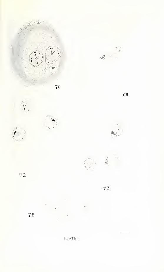
•
*•.
70
.v..
69
t
-:
. <
72
71
73
PLATE V

142 ILLINOIS BIOLOGICAL MONOGRAPHS
PLATE VI
Fig. 74. Precystic amoeba stained with neutral red. Chromophilic granules present,
but none of the larger spherical inclusions. Food vacuoles lacking.
Fie. 75. Nuclear transformation. Formation of the circlet. Flemming, safranin.
Fig. 76. Precystic amoeba after completion of nuclear transformation. Gilson-
Carnoy, haematoxylin.
Fig. 77. Division of the circlet. Gilson-Carnoy, haematoxylin.
Fig. 78. Division of the circlet. Zenker, haematoxylin.

75
77
76
74
78
*
••-
• •
PLATE VI

144 ILLINOIS BIOLOGICAL MONOGRAPHS
PLATE VII
Fig. 79. Circlet migrating to poles of central region. Gilson-Carnoy, haematoxylin.
Fig. 80. Nuclei after division in late precystic period. Gilson-Carnoy, haematoxylin.
Fig. 81. Late precystic division figure. No chromosomes, circlet at pole. Gilson-
Carnoy, haematoxylin.
Fig. 82. Nucleus at time of cyst wall formation.
Fig. 83. Nuclear appearance at time of cyst wall formation. Flemming, safranin.

83
4
: a<9.
.-»
.
.
r,
80
%2
79
.' '
.
'
J
\&i
81°:
PLATE VII
146 ILLINOIS BIOLOGICAL MONOGRAPHS
PLATE VIII
Fig. 84. Nuclear division immediately after cyst wall formation. Gilson-Carnoy,
Feulgen.
Fig. 85. Cystic division resembling trophic division. Gilson-Carnoy, haematoxylin.
Fig. 86. Cystic division resembling late precystic division. Bouin, haematoxylin.
Fig. 87. Next section of same cyst. Bouin, haematoxylin.
Fig. 88. Cyst with different-sized nuclei. Mercuric chloride, haematoxylin.
Fig. 89. Mature cyst nuclei. Amitotic (?) division of nuclei. Gilson-Carnoy,
haematoxylin.
Fig. 90. Mature cyst. Hot glacial acetic acid, haematoxylin.
Fig. 91. Mature cyst. Gilson-Carnoy, haematoxylin.

.:s
>
to
'
84
5
1
86
85
87 88
&^m
89
©0'
•.
90 91
PLATE VIII

WHVB8ITY OF HlMWW
URBAN*

7*
CYTOLOGICAL OBSERVATIONSON ENDAMOEBA BLATTAE
BY
Paul A. Meglitsch
Price $1.00
THE UNIVERSITY OF ILLINOIS PRESS
URBANA, ILLINOIS
1940

ILLINOIS BIOLOGICAL MONOGRAPHSis a general title used to comprehend a series
of monographs on subjects in botany, ento-
mology, zoology, and allied fields. Each issue
is an independent publication. For the conven-
ience of bibliographers and librarians, each
year's output is called a volume. Each volume
consists of approximately 450 pages, priced at
four dollars. A volume may consist of two,
three, or four individual monographs. Prices
of individual numbers are indicated in the list
of titles given on the back cover. Requests for
exchanges should be addressed to the Exchange
Department, University of Illinois Library,
Urbana, Illinois. All communications concern-
ing sale or subscription should be addressed to
the Director, University of Illinois Press,
Urbana, Illinois.

ILLINOIS BIOLOGICAL MONOGRAPHSVol. I
Nos. 1-2. A Revision of the Cestode Family Proteocephalidae. With 16 plates. By G. R.La Rue. $2.00.
No. 3. Studies on the Cestode Family Anoplocephalidae. With 6 plates. By H. Douthitt.80 cts.
No. 4. Some North American Larval Trematodes. With 8 plates. By W. W. Cort. $1.20.
Vol. II
No. 1. Classification of Lepidopterous Larvae. With 10 plates. By S. B. Fracker. $1.50.No. 2. On the Osteology of Some of the Loricati. With 5 plates. By J. E. Gutberlit. 50 cts.
No. 3. Studies on Gregarines. With 15 plates. By Minnie E. Watson. $2.00.No. 4. The Genus Meliola in Porto Rico. With 5 plates. By F. L. Stevens. (Out of print).
Vol. Ill
No. I. Studies on the Factors Controlling the Rate of Regeneration. By C. Zeleny. $1.25.No. 2. The Head-capsule and Mouth-parts of Diptera. With 25 plates. By A. Peterson.
$2.00.
No. 3. Studies on North American Polystomidae, Aspidogastridae, and Paramphistomidae.With 11 plates. By H. W. Stunkard. $1.25.
No. 4. Color and Color-pattern Mechanism of Tiger Beetles. With 29 black and 3 coloredplates. By V. E. Shelford. $2.00.
Vol. IVNo. 1. Life History Studies on Montana Trematodes. With 9 plates. By E. C. Faust. $2.00.
No. 2. The Goldfish (Carassius carassius) as a Test Animal in the Study of Toxicity. ByE. B. Powers. $1.00.
No. 3. Morphology and Biology of Some Turbellaria from the Mississippi Basin. With 3plates. By Ruth Higley. $1.25.
No. 4. North American Pseudophyllidean Cestodes from Fishes. With 13 plates. By A. R.Cooper. $2.00.
Vol. VNo. 1. The Skull of Amiurus. With 8 plates. By J. E. Kindred. $1.25.
No. 2. Contributions to the Life Histories of Gordius robustus Leidy and Paragordius varius(Leidy). With 21 plates. By H. G. May. $1.50.
Nos. 3-4. Studies on Myxosporidia. A synopsis of Genera and Species of Myxosporidia.With 25 plates and 2 text-figures. By R. Kudo. $3.00.
Vol. VINo. 1. The Nasal Organ in Amphibia. With 10 plates. By G. M. Higgins. $1.00.
Nos. 2-3. Revision of the North American and West Indian Species of Cuscuta. With 13plates. By T. G. Yuncker. $2.00.
No. 4. The Larvae of the Coccinellidae. With 6 plates. By J. H. Gage. 75 cts.
Vol. VIINo. 1. Studies on Gregarines, II. Synopsis of the Polycystid Gregarines. With 4 plates.
By Minnie Watson Kamm. $1.00.
No. 2. The Molluscan Fauna of the Big Vermilion River, Illinois. With 15 plates. By F. C.Baker. $1.25.
No. 3. North American Monostomes. With 9 plates. By E. C. Harrah. $1.25.
No. 4. A classification of the Larvae of the Tenthredinoidea. With 14 plates. By H. Yuasa.$2.00.
Vol. VIII
No. 1. The Head-capsule of Coleoptera. With 26 plates. By F. S. Stickney. $2.00.
No. 2. Comparative Studies on Certain Features of Nematodes and Their Significance. With4 plates. By D. C. Hetherington. $1.00.
No. 3. Parasitic Fungi from British Guiana and Trinidad. With 19 plates. By F. L. Stevens.
$1.25.
No. 4. The External Morphology and Postembryology of Noctuid Larvae. With 8 plates.
By L. B. Ripley. $1.25.
Vol. IXNo. 1. The Calciferous Glands of Lumbricidae and Diplocardia. With 12 plates. By Frank
Smith. $1.25.
Nos. 2-3. A Biologic and Taxonomic Study of the Microsporidia. With 27 plates and 9 text-
figures. By R. Kudo. $3.00.
No. 4. Animal Ecology of an Illinois Elm-maple Forest. With 7 plates. By A. O. Weese.$1.25.
[Continued on back cover.]

Vol. XNo. I. Studies on the Avian Species of the Cestode Family Hymenolepididae. With g plates
and 2 text-figures. By R. L. Mayhew. $1.50.
No. 2. Some North American Fish Trematodes. With 6 plates, 2 charts, and 1 text-figure.
By H. W. Manter. $1.50.
No. 3. Comparative Studies on Furcocercous Cercarie. With 8 plates and 2 text-figures. ByH. M. Miller. $1.25.
No. 4. A Comparison of the Animal Communities of Coniferous and Deciduous Forests.
With 16 plates. By I. H. Blake. $1.50.
Vol. XI
No. 1. An Ecological Study of Southern Wisconsin Fishes. With 16 plates. By A. R. Cahn.$1.50.
No. 2. Fungi from Costa Rica and Panama. With 18 plates. By F. L. Stevens. $1.25.
No. 3. The Structure and Development of Corallobothrium. With 5 plates. By H. E. Essex.$1.00.
No. 4. Caryophyllaeidae of North America. With 7 plates. By G. W. Hunter, III. $1.25.
Vol. XII
No. 1. Morphological Studies of the Genus Cercospora. With 4 plates. By W. G. Solheim.$1.00.
No. 2. Morphology, Taxonomy, and Biology of Larval Scarabaeoidea. With 15 plates. ByW. P. Hayes. $1.25.
No. 3. Sawflies of the Sub-Family Dolerinae of America North of Mexico. With 6 plates.
By H. H. Ross. $1.25.
No. 4. A Study of Fresh-water Plankton Communities. By Samuel Eddy. $1.00.
Vol. XIII
No. 1. Studies on Some Protozoan Parasites of Fishes of Illinois. With 8 plates. By R. R.Kudo. 75 cts.
No. 2. The Papillose Allocreadiidae: A Study of Their Morphology, Life Histories, andRelationships. With 4 plates and 6 text-figures. By S. H. Hopkins. $1.00.
No. 3. Evolution of Foliar Types, Dwarf Shoots, and Cone Scales of Pinus. With 32 text-
figures. By C. C. Doak. $1.50.
No. 4. A Monographic Rearrangement of Lophodermium. With 5 plates. By L. R. Tehon.$2.00.
Vol. xrvNo. 1. Development of the Pectoral Limb of Necturus maculosus. With 11 plates. By H. K.
Chen. $1.00.
No. 2. Studies on North American Cercariae. With 8 plates. By E. L. Miller. $1.50.
No. 3. Studies on the Morphology and Life History of Nematodes in the Genus Spironoura.With S plates and 2 text-figures. By J. G. Mackin. $1.00.
No. 4. The Life History of Cotylophoron cotylophorum, a Trematode from Ruminants.With 9 plates. By H. J. Bennett. $1.50.
Vol. XVNo. 1. Experimental Studies on Bckinostoma revolutum (Froelich), a Fluke from Birds and
Mammals. With 3 plates. By P. C. Beaver. $1.00.
No. 2. Generic Classification of Nearctic Sawflies. With 17 plates. By H. H. Ross. $2.00.
No. 3. Studies on the Biology of the Crayfish Cambarus propinquus Girard. With 46 graphs.By W. C. Van Deventer. $1.00.
No. 4. Taxonomic Studies on the Mouth Parts of Larval Anura. With 3 figures and 8charts. By R. J. Nichols. $1.00.
Vol. XVINos. 1-2. The Turtles of Illinois. With 31 plates, 20 maps, and 15 text-figures. By
A. R. Cahn. $3.00.
No. 3. The Phylogeny of the Hemiptera, Based on a Study of the Head Capsule. With 24plates. By C. S. Spooner. $1.00.
No. 4. A Classification of the Larvae and Puparia of the Syrphidae of Illinois, Exclusive ofAquatic Forms. With 17 plates. By Elizabeth M. Heiss. $1.50.
Vol. XVIINo. 1. Comparative Studies on Trematodes (Gyrodactyloidea) from the Gills of North
American Fresh-water Fishes. With 5 plates. By J. D. Mizelle. $1.00.
No. 2. The Microthyriaceae. By F. L. Stevens and Sister M. Hilaire Ryan. $1.50.
No. 3. The Branchiobdellidae (Oligochaeta) of North American Crayfishes. With 3 plates.
By C. J. Goodnight. $1.00.


:
..
' ",'.'.: -,,:,;',
. . .1, .. ,..,.,., ,.
.-.... .... ...,.... i..;...
..,.' ...,,)..,. i
,
'
i .,.....
,.,'.'.': '.:;
:
. .... .