cytotoxicity and inflammatory effect of silver ... · pdf filecytotoxicity and inflammatory...
TRANSCRIPT
Cytotoxicity and Inflammatory Effect of Silver Nanoparticles
in Human Cells
Jeong-shin Park, Na Mi Yu, Jinwoo Cheon and In-Hong Choi
Department of Microbiology, College of Medicine; Department of Chemistry;
Nanomedical NCRC, Yonsei University, Seoul, Korea
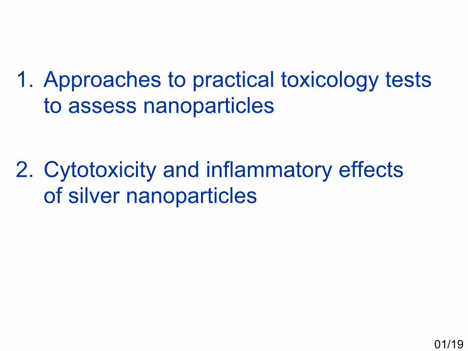
1. Approaches to practical toxicology tests to assess nanoparticles
2. Cytotoxicity and inflammatory effects of silver nanoparticles
01/19

• The rapidly developing field of nanotechnology will result in exposure of nanoparticles to humans via several routes (e.g., inhalation, ingestion, skin, etc.). Nanoparticles can translocate from the route of exposure to other vital organs and penetrate cells.
• Toxicity studies to determine the deleterious effects of nanoparticles on living cells are required.
• Due to the nanosize and the nature of agglomeration, simple standard methods to characterize the biological effects of nanoparticles are currently unavailable.
• In this study, practical information regarding the optimal in vitro tests for nanotoxicity were evaluated.
Nanoparticles and toxicity assay
02/19

• Antimicrobial reagents, detergents, water purificants, wall paints, textiles
Silver nanoparticles
03/19
Antimicrobial applications
Ink
Cosmetics
200nm 200nm 500nm
20 nm(synthetic
)
180 nm (commercial,
Aldrich)
80 nm(synthetic
)
04/19
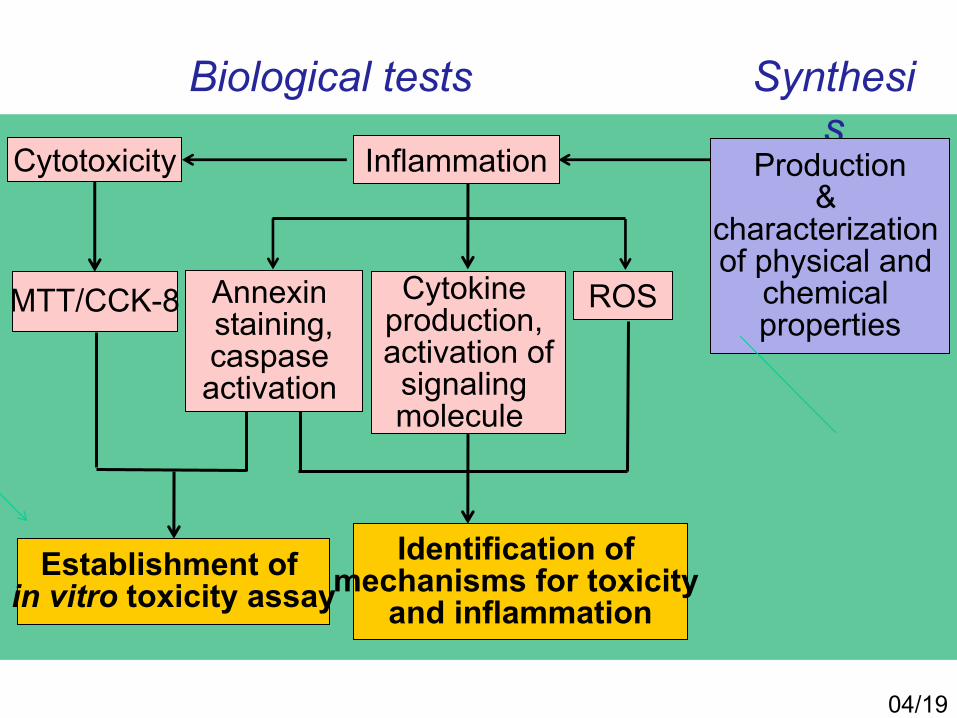
MTT/CCK-8
Establishment of in vitro toxicity assay
Identification of mechanisms for toxicity
and inflammation
Cytotoxicity
ROS
Synthesis
Biological tests
Annexin staining,caspase activation
Production&
characterization of physical and
chemical properties
Cytokine production, activation ofsignaling molecule
Inflammation

• Production of diverse particles (size, surface)
• Assess biological activities
Assesstoxicity
tests
•ISO/TC229
• OECD• U.S NCL
In vitro tests for nanoparticles
Review in vitro
methods
• Understanding of proper methods for nanoparticles
Establish proper
methods
05/19

Exposure routes of nanomaterials
Skin
06/19
Respiratory tract
Immune system
Immune system
Skin immune systemRespiratoryimmune system
Direct invasion
Cytotoxicity & Inflammation
Cell line Origin Characteristics
Respiratory A549 Lung epithelial Proper for cytotoxicity
BEAS-2B Bronchial epithelial Proper for cytokineproduction
Immune U937 Macrophage Proper for cytotoxicity and cytokine production
Skin SK-Mel Skin epithelial Proper for cytotoxicity and cytokine production
A375 Skin epithelial Too fast growing

Category Tests Mechanism Method SuggestionIn Vitro
Immunology Hemolysis Release of hemoglobin Standard Proper
(Blood contact
Properties)Complement
activationActivation of C3
complement Standard Inappropriate
In VitroImmunology Leukocyte
proliferation
Leukocyte proliferation with
mitogenstimulation
Standard CCK-8
(Cell-based assays) Phagocytosis Zymosan assay Standard Proper
Cytokine induction Cytokine production Standard Proper
Toxicity Oxidative stress Detection of ROS Standard Proper
Cytotoxicity (necrosis)Cell viability and
mitochondrial integrity
Standard CCK-8
Cytotoxicity(apoptosis)
Activation of caspase 3 Standard Annexin-V
Standard toxicology tests and silver nanoparticles
07/19
Targeting Cell binding/internalization N/S N/S
TEM, confocalmicroscope or other
methods

• Nanoparticles larger than 100 nm tend to aggregate relatively quickly in vitro when compared to nanoparticles smaller than 100 nm. Fresh samples within two weeks after synthesis is recommended for tests.
• Each standard toxicology method must be verified before use. (ex. interference with a specific wavelength, electrophoresis)
Characteristics specific to metal nanomaterials
09/19

Larger
Flow chart for nanotoxicity tests
- Aggregation- Particle sizeAnalysis of biological
properties- Cytotoxicity- Apoptosis- Cytokine production- Hemolysis- Leukocyte proliferation- ROS production
Smaller
Analysis of chemical/physical properties
10/19
Particle size
100 nm

Biological reactivity of silver nanoparticles

Cytotoxicity of silver nanoparticles
0 3.125 6.25 12.5 25 50
Conc. (µg/mL)
0
20 nm120
100
80
60
40
20
80 nm
0 50 100 200 400 800
Conc. (µg/mL)
120
100
80
60
40
20
0C
ell v
iabi
lity
(%)
SK-Mel28(skin)
A375(skin)
A549(lung)
Cel
l via
bilit
y (%
)
11/19
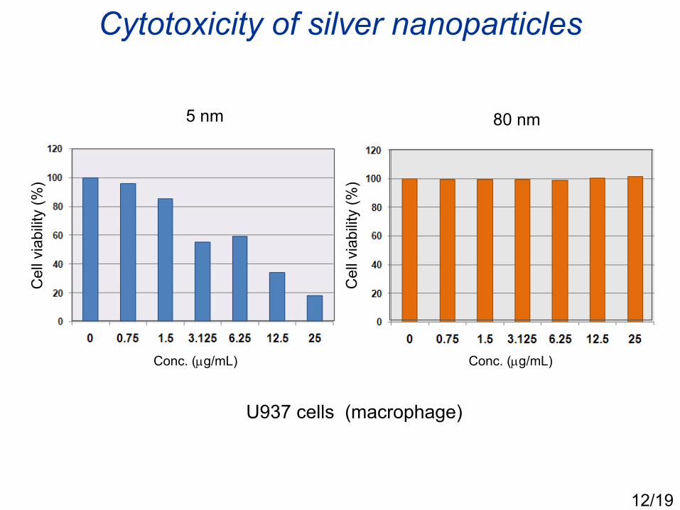
Cytotoxicity of silver nanoparticles
Conc. (µg/mL) Conc. (µg/mL)
U937 cells (macrophage)
12/19
5 nm 80 nm
Cel
l via
bilit
y (%
)
Cel
l via
bilit
y (%
)
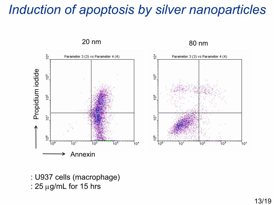
Induction of apoptosis by silver nanoparticles
Annexin
Pro
pidi
um io
dide
13/19
: U937 cells (macrophage): 25 µg/mL for 15 hrs
20 nm 80 nm

Lysosomal aggregation by silver nanoparticles
14/19
: U937 cells (macrophage): 20 nm, 25 µg/mL for 24 hrs

ROS production by silver nanoparticles
H2O2 Silver nanoparticle (20 nm)Unstained control Stained control
15/19
: BEAS-2B (lung): 20 nm, 30 µg/mL, for 3 hrs : stained with CM-H2DCFDA
H2O2
Silver nanoparticle

Cytokine production by silver nanoparticles
• Cytokine array
Positive control Positive control
IL-8IL-16
MIF
RANTES (CCL5)
Serpin E1Positive control
Positive: chemokines (IL-8, MIF, RANTES), Serpin E1, IL-16Negative: TNF-α, IL-6, IL-1
IL-1αIL-1β IL-6
Negative controlTNF-α
15/19

Cytokine production by silver nanoparticles
0 0.75 1.5 3.1 6.2 12.5
Conc. (µg/mL)
• ELISA (IL-8)
16/19
2,000
1,500
1,000
500
0
: U937 cells (macrophages): 20 nm for 24 hrs
IL-8
(pg/
mL)

Activation of signaling molecule by silver nanoparticles
• MAP kinase (ex. ERK) activation
0 15 30 60 0 15 30 60 (min)
: Protein 30 µg loading: LPS (E. coli lipopolysaccharide) 50 ng/mL: 5 nm silver nanoparticles, 1.5 µg/mL
LPS Silver nanoparticles
Phospho-ERKTotal ERK
17/19

• In human cells, epithelial cells from skin or lung, and macrophages, 5 nm and 20 nm silver particles induced stronger cytotoxicity and ROS synthesis than 80 nm particles did.
• 5 nm and 20 nm silver particles induced chemokine production, mainly IL-8, MIF and RANTES, while proinflammatory cytokines, IL-1, IL-6 and TNF-α were not induced significantly in the same conditions.
• Some MAP kinase signaling pathways were activated during exposure to silver nanoparticles at lower concentrations which do not induce cytotoxicity.
Summary
18/19

• The toxicity and inflammatory effects of nanoparticles are dependent on their size. In silver nanoparticles smaller than 20 nm induce cytotoxicity significantly in vitro.
• Nanoparticles induce inflammatory immune responses at lower concentrations and chemokines are the major cytokines induced at early stages of exposure to silver nanoparticles.
Conclusion
19/19


0 50 100 200 800400
0
0.1
0.2405 nm
450 nm
490 nm