d the outcome of intracranial subdural empyema at …
TRANSCRIPT

THE OUTCOME OF INTRACRANIAL SUBDURAL EMPYEMA AT STEVE BIKO ACADEMIC
HOSPITAL: RETROSPECTIVE STUDY
By
EMMANUEL KGORO THOBEJANE
Submitted in fulfilment of the requirements for the degree
MASTER OF MEDICINE IN NEUROSURGERY
In the
FACULTY OF HEALTH SCIENCES
At the
UNIVERSITY OF PRETORIA
Supervisor: Professor MS Mokgokong
Co-supervisor: Doctor TP Moja
15 March 2012
©© UUnniivveerrssiittyy ooff PPrreettoorriiaa

ii
DECLARATION
I declare that the master’s script, which I hereby submit for the degree Master of Medicine
in Neurosurgery at the University of Pretoria, is my own work and has not previously been
submitted by me for a degree at another university.
The study will also be submitted for journal publication.

iii
ACKNOWLEDGEMENTS
I wish to extend my sincere gratitude to the following people and institutions for their
contribution to this research script:
• My supervisor, Professor SM Mokgokong and co-supervisor, Doctor TP Moja for their
assistance and encouragement throughout the study
• Professor MM Sathekge and Doctor DP Maseko-Magongo for the guidance and
support they offered during writing of this research
• Miss Lieketseng Masenyetse from Medical Research Council for assisting with
statistical analysis of this work
• Chief Executive Officer of Steve Biko Academic Hospital, Doctor ME Kenoshi and his
deputy Doctor M Mathebula for affording me the opportunity to study at their
hospital, reviewing of patients records and their unwavering support throughout my
studies
• Professor D Lombard for proof reading of this text
• Mister BR Khoza from Mpumalanga Department of Health for his support and
prayers
• My colleagues for their support and motivation throughout my studies

iv
DEDICATION
I dedicate this work to my beautiful and loving wife, Doctor Sebotse Thobejane for having
stood by me throughout my hurdles in life till today, your support, prayers and
encouragements were second to none my love
To my handsome boys, Ofentje and Seetja Thobejane thank you for always managing to
bring a smile on my face when I came home
To my parents, Setino and Kanyane Thobejane, my parents-in-law Masolo and Mpetje Ratau
and my godparents Doctors Zukiswa and Gaolatlhoe Motlhale, thank you for your prayers
and believing in my abilities
Lastly Doctor Tembile Songabe, the man with whom I published my first scientific paper

v
ABSTRACT
Objectives: Intracranial subdural empyema (ICSDE) can be a devastating condition, with a
sequelae ranging from epilepsy, focal deficits to death. Factors affecting the outcome in
subdural empyema range from level of consciousness, the extend of subdural pus at the
time of diagnosis and the type of surgical procedure performed. Previous studies have
conflicting results of unfavourable prognostic factors associated with ICSDE. The outcome of
this condition at Steve Biko Academic Hospital (SBAH) is reported, as well as factors
influencing the outcome.
Methods: A retrospective analysis of all the patients admitted at neurosurgery unit of SBAH
during 2006 – 2010 period with confirmed subdural empyema on brain CT scan and at
surgery. Data sheet was used to collect all clinical information from patients’ records.
Glasgow Outcome Scale and Henk W. Mauser grading were used to report on the outcome.
Results: A total of 34 patients (20 males and 14 females) with mean age of 16.1 years were
admitted with a diagnosis of ICSDE. The common presenting features were headache
(58.8%), fever and seizures (47.0% each). Over 61% of patients had hemiplegia at
presentation. CT scan confirmed subdural collections with 70.6% over the convexity, 23.5%
at the convexity and parafalx and only 5.9% had bilateral collections. Complicated paranasal
sinusitis was the origin of infection in 82.3%, followed by meningitis with 8.8%. Burr hole
washout was done in 52.9% of patients, while 38.2% had burr holes with drains in situ and
8.8% had craniotomy to evacuate the subdural pus. All the patients were given empiric
triple antibiotic therapy. Streptococci species were the most cultured organisms in the 19
(56.0%) patients who had positive cultures, however 15 (44.0%) patients had negative
cultures. Resistance to penicillin was noted in 5.0% of cases only. Sixty-five percent of
patients had good outcome with no seizures nor neurological deficits. The overall mortality
was 15.0% in this study, with none from patients who had craniotomy.
Conclusion: Clinical presenting features and organisms cultured seems to be the same
internationally, particularly those due to complicated sinusitis. Empiric triple antibiotic
therapy of 3rd
generation cephalosporin plus vancomycin plus metronidazole is still relevant
at SBAH. Factors associated with favourable outcome were ages between 11 and 20 years,
and craniotomy as the surgical procedure of choice.

vi
TABLE OF CONTENTS
ITEM DESCRIPTION PAGE
Declaration i.
Acknowledgements ii.
Dedication iii.
Abstract iv.
Table of Contents v.
List of Tables vi.
List of Figures vii.
1 CHAPTER 1: ORIENTATION AND GENERAL BACKGROUND 1
1.1 Introduction 1
1.2 Background 1
1.3 Research Problem 2
1.4 Aim and Objectives 3
1.4.1 Primary objectives 3
1.4.2 Secondary objectives 3
2 CHAPTER 2: LITERATURE OVERVIEW 4
2.1 Introduction 4
2.2 Literature Review 4
3 CHAPTER 3: RESEARCH METHODOLOGY 8
3.1 Study Designs 8
3.2 Settings 8
3.3 Patient/Research Object Selection 8
3.3.1 Inclusion criteria 8
3.3.2 Exclusion criteria 8
3.4 Measurements 9
3.5 Data Analysis 9
3.6 Ethical Considerations 10
4 CHAPTER 4: RESEARCH FINDINGS 11
5 CHAPTER 5: DISCUSSION AND CONCLUSION 17
5.1 Discussion 17
5.2 Limitations 18
5.3 Conclusion 18
6 REFERENCES 19
7 APPENDICES 21
7.1 Appendix A – Glasgow Coma Scale 21
7.2 Appendix B – Data Collection Form 22
7.3 Appendix C – Henk W. Mauser grading system 23
7.4 Appendix D – Glasgow Outcome Scale 24

vii
LIST OF TABLES
Table no. Description Page
1 Prognostic factors associated with SDE 5
2 Empiric Treatment Recommendations for SDE and EDA 6
3 Demographics 11
4 Infection origin 11
5 Pathogens 13
6 Clinical features 14
7 Surgical procedures 14
8 Henk W. Mauser grading for morbidity of survivors of ICSDE 15
9 Contingency Table of GOS and Age groups 16

viii
LIST OF FIGURES
Figure Description Page
1 Treatment plan of cranial subdural empyema 5
2 CT scan demonstrating left subdural and parafalx empyema 12
3 Culture results 12
4 Pus location on contrasted brain CT scan 13
5 Glasgow Outcome Scale 15
6 Admission GCS and Mortality 16

1
CHAPTER 1: ORIENTATION AND GENERAL BACKGROUND
1.1 INTRODUCTION
This chapter gives the overview of the research project. It includes background of the
study, the problem statement and the importance of conducting a study of this kind. The
aim and objectives are defined.
1.2 BACKGROUND
Intracranial subdural empyema (ICSDE) refers to a collection of pus in the space
between the dura and the arachnoid layers covering the brain1,2,3
. It has no anatomic
barriers to spread except the falx cerebri and tentorial cerebelli2. It is distinguished from
abscesses within the brain substance with capsule around it. Incidence is higher in
developing countries, with male:female ratio 2:1 to 3:12,3,4
. It can occur at any age group
with mean of 10 – 40 years1,2,3
. Predisposing conditions for the development of ICSDE
are direct extension of local infection (contiguous spread) e.g. complicated pansinusitis
in 40 – 80%, penetrating cranial trauma, post neurosurgical procedure, haematogenous
spread or as a complication of meningitis3.
Clinical features may be rapidly progressive with symptoms and signs related to either
raised intracranial pressure, meningeal irritation, focal cortical inflammation or
thrombophlebitis of cerebral vein and/or venous sinuses. Most patients have headache,
fever and focal neurological deficits3. Seizures occur in up to 50% of patients
3.
Diagnosis of ICSDE should be suspected in any patient with meningeal signs and a focal
neurological deficit. Imaging tools are of valuable importance and include brain CT scan
and brain-MRI. Lumbar puncture carries high risk of herniation and may be sterile,
particularly in patients with focal signs or raised intracranial pressure5. Organisms
cultured depends on the source of infection, with aerobic and anaerobic streptococci
common in patients with complicated sinusitis, and staphylococci and gram negative
species observed in trauma/postsurgical patients. However up to 40% of cases have
sterile cultures3.
ICSDE is a surgical emergency because anti-microbial therapy alone does not reliably
sterilise the empyema. Antibiotic penetration into this space is poor. Optimal surgical
approach is controversial. The following has been reported as preferred surgical
methods for drainage with different outcomes – craniotomy6,7,8,9
, burr hole washout10,11
or burr holes with drain in situ12
. The emphasis should be put on source control when
dealing with ICSDE, as it carries mortality of between 6 – 35%2.

2
1.3 RESEARCH PROBLEM
Subdural empyema despite being reported as rare in developed countries (reporting
only 48 cases in 34 years in Europe)11
, it remains a common intracranial infection seen in
South Africa (with some studies reporting 699 cases in 15 years)8 with devastating
outcome if not well managed. There has not been a baseline study evaluating the
outcome of this condition at Steve Biko Academic Hospital (SBAH).
Mortality in South Africa is variable with Durban reporting as high figures as 22.3%8
compared to 7.7% at Cape Town7. Previous studies have conflicting results of
unfavourable prognostic factors associated with ICSDE. Hence the need to document the
outcome and factors affecting the outcome of ICSDE at SBAH, as this can have positive
clinical implications for the treating physicians.

3
1.4 AIM AND OBJECTIVES
1.4.1 Primary Objectives
To document the outcome of intracranial subdural empyema at Steve Biko Academic
Hospital
To determine factors influencing the outcome of ICSDE at SBAH
1.4.2 Secondary Objective
To compare the above findings with existing world literature on subdural empyema

4
CHAPTER 2: LITERATURE OVERVIEW
2.1 INTRODUCTION
In this chapter, the literature is reviewed and the collected knowledge divided into
manageable, sequential sections.
2.2 LITERATURE REVIEW
Several studies are available on this condition but very few reported on the factors affecting
the outcome in patients with cranial subdural empyema. Overall mortality of patients with
subdural empyema is reported to be between 10 and 20% but can be as high as 75% in
patients who are comatose13
.
Henk W Mauser et al reviewed factors affecting the outcome in subdural empyema. They
concluded that the level of consciousness and the extent of the subdural pus at the time of
diagnosis have a significant bearing on the outcome14
. Earlier on they have also reported
that the delay in diagnosis and surgical treatment had a negative bearing on the
outcome11
.Agrawal Amit et al reviewed subdural empyema and its management, they
identified the following as unfavourable prognostic factors : encephalopathy, elderly or
younger than 10 years, delay in starting antibiotics, and sterile cultures3- Table 1.
In an attempt to resolve the issue on the ideal surgical method in treating subdural
empyema, S.A. Mat Nayan et al analysed the efficacy of these two (burr holes and
craniotomy) surgical methods in terms of the outcome. They concluded that wide cranial
opening (craniotomy) is the better surgical method for the treatment of intracranial
subdural empyema compared to a limited procedure such as burr holes9. This was of course
in contrast to earlier studies that reported better outcome with burholes than craniotomy
by Miller et al10
.
Pasquale de Bonis et al developed treatment plan for both cranial and spinal subdural
empyema (figure 1), especially after recognizing that the two surgical options both have
their places in the management of patients with subdural empyema15
. Steve Ryu et al
compiled a comprehensive guide on the appropriate choice of antibiotics based on the likely
source of infection, taking into consideration the possibility of multiple organisms13
- Table
2.

5
TABLE 1: Prognostic Factors Associated With SDE3
Unfavourable prognostic factors
• Encephalopathy or coma at the time of presentation
• Elderly or younger than 10 years
• Delay in starting antibiotics
• Sterile cultures
Favourable prognostic factors
• Craniotomy as surgical modality (rather than burr holes)
• Early treatment (surgery and antibiotics)
• Young age (10 -20 years is optimal)
• Patient is alert, awake, and orientated at the time of presentation
• Paranasal sinus as source of initial infection
• Isolation of aerobic streptococci in the culture
FIGURE 1: Treatment Plan of SDE15
6
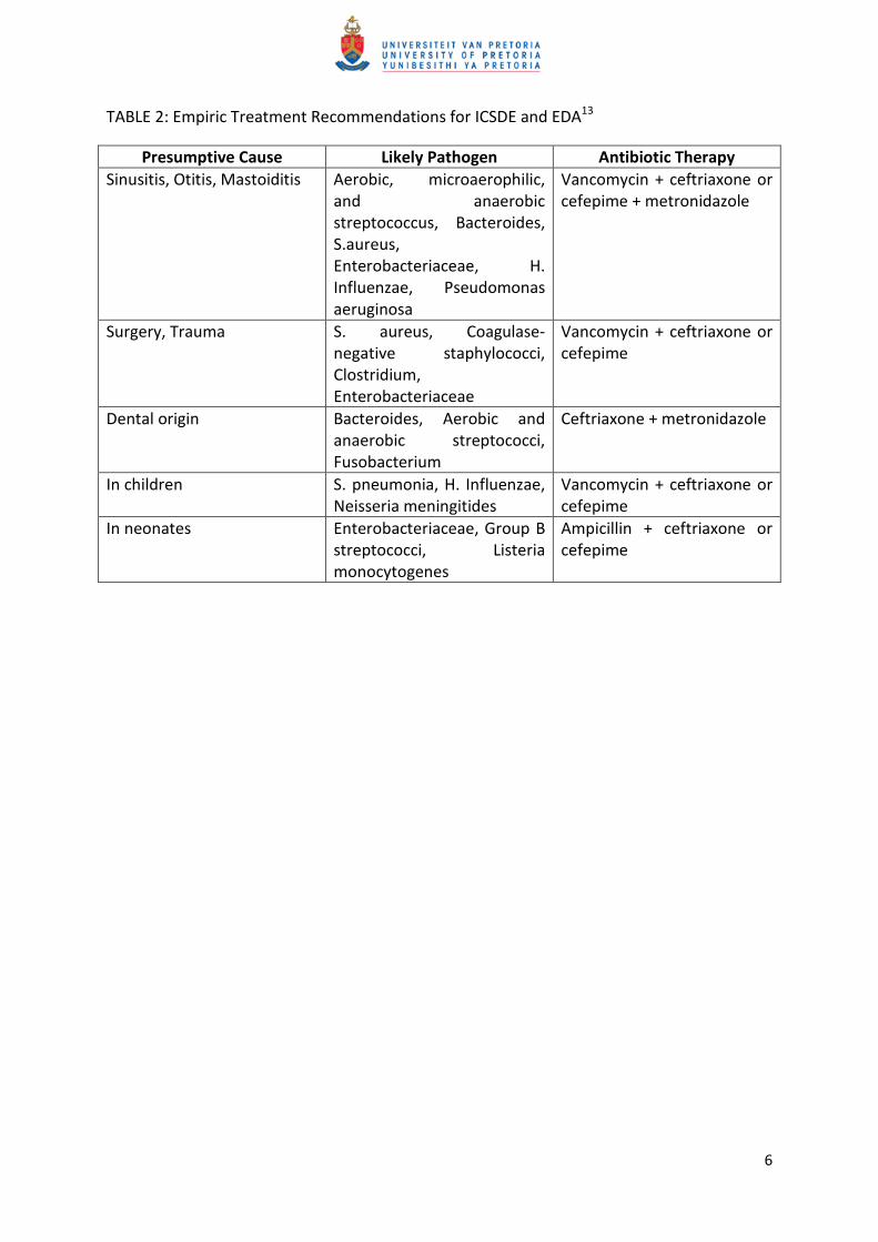
TABLE 2: Empiric Treatment Recommendations for ICSDE and EDA13
Presumptive Cause Likely Pathogen Antibiotic Therapy
Sinusitis, Otitis, Mastoiditis Aerobic, microaerophilic,
and anaerobic
streptococcus, Bacteroides,
S.aureus,
Enterobacteriaceae, H.
Influenzae, Pseudomonas
aeruginosa
Vancomycin + ceftriaxone or
cefepime + metronidazole
Surgery, Trauma S. aureus, Coagulase-
negative staphylococci,
Clostridium,
Enterobacteriaceae
Vancomycin + ceftriaxone or
cefepime
Dental origin Bacteroides, Aerobic and
anaerobic streptococci,
Fusobacterium
Ceftriaxone + metronidazole
In children S. pneumonia, H. Influenzae,
Neisseria meningitides
Vancomycin + ceftriaxone or
cefepime
In neonates Enterobacteriaceae, Group B
streptococci, Listeria
monocytogenes
Ampicillin + ceftriaxone or
cefepime

7
Local Studies
Several studies were done locally (South Africa) at tertiary and regional hospitals to evaluate
the incidence, the outcome of both surgical modes of treatments, pathogens cultured and
the overall outcome of this condition at their institutions.
Arnold P.L. et al analysed 90 cases at Groote Schuur Hospital to determine the outcome
looking at both surgical treatment (burr holes versus craniotomy). They concluded that burr
holes were easier and safer surgical intervention for the treatment of subdural empyema
compared to craniotomies. They reported 86% good recovery and a mortality of 7.7%
only7.Nathoo N. et al looked at 699 patients at Nelson R. Mandela Hospital in a retrospective
study, again assessing the outcome when using burr holes or craniotomy. They concluded
that craniotomy improves the outcome for cranial subdural empyema and hence their
recommended surgical procedure of choice. Mortality for burr holes was 23.3% and that in
craniotomy was 8.4%.8
Tshifularo M et al reported mortality as low as 5% on his work at Doctor George Mukhari
Hospital16
.Olwoch I.P. looked at local microbiology and spectrum of sensitivity and
resistance of organisms to antibiotics. This of course will assist in adapting the
recommended international guidelines on empiric antibiotics for an effective and
appropriate choice of antibiotic coverage based on local prevalence at Chris Hani and
Charlotte Maxeke Hospitals17
.
At SBAH there is no study done to evaluate this condition or report on the outcome of
intervention.

8
CHAPTER 3: RESEARCH METHODOLOGY
3.1 STUDY DESIGNS
This was a retrospective descriptive study over a 5 year period.
3.2 SETTINGS
This study was conducted at the Neurosurgery Unit at SBAH. Steve Biko Academic Hospital is
the referring hospital for local district hospitals in Gauteng province and the Mpumalanga
province.
3.3 PATIENT/RESEACH OBJECT SELECTION
3.3.1 Inclusion Criteria
All patients who were admitted at the neurosurgery unit (wards and the intensive care unit)
from 2006 – 2010 with confirmed subdural empyema on CT scan and at surgery. Patient’s
records were reviewed from time of admission till discharge and outpatient follow-up of up
to 6 months (average). Included were all the patients who presented with symptoms
suggestive of intracranial pathology and were confirmed to have subdural collection(s) with
contrast enhancement on Brain CT scan – figure 2, CT of the air sinuses was also included to
assist in demonstrating the origin of pus. Patients were then taken to theatre to evacuate
the subdural collection and it was confirmed to be pus intraoperatively. The patients
underwent different surgical procedures depending on the attending neurosurgery registrar
and consultant on duty’s discretion. They either had multiple burr hole washout repeatedly
until pus was evacuated, or burr holes leaving drains in situ to encourage on-going drainage
in the ward, or just had large craniotomy washout only. All the patients received triple
antibiotic therapy of ceftriaxone or meropenem plus vancomycin plus metronidazole. The
specimen was send to the laboratory for microscopy, culture and sensitivity. On average
those with positive cultures, the results were available within 7 days and antibiotics were
changed according to the sensitivity results. The antibiotics were given intravenously for
two weeks or until the pus was cleared as demonstrated by the follow-up scan (done every
4-7 days until no pus was seen on scan) then discharged on oral antibiotics (Cotrimoxazole
and/or Co-amoxiclav) for another four weeks. Patients were then followed up at out-patient
department for a minimum period of 6 months. All patients with deficits (physical or
cognitive) received evaluation and management by appropriate specialities – physiotherapy,
occupational therapy, speech and audiology.
3.3.2 Exclusion Criteria
Patients who were not operated to confirm presence of subdural pus.
Patients with brain abscesses.
9
3.4 MEASUREMENTS
Data was collected from patients’ files. Factors such as age, sex, level of consciousness
using Glasgow Coma Scale (appendix A) at the time of presentation and interventions were
recorded on Data Collection Form (appendix B). Criteria for diagnosing subdural empyema :
patient with fever, headache, new onset seizures/neurological deficits, this followed by CT
scan imaging confirming contrast enhancing subdural collection. The treatment guidelines
applied (burr holes versus craniotomy) on the management of these patients was recorded,
including the culture results from microbiology and choice of antibiotics used. Neurological
deficits before and after treatment, the source/cause of infection was documented. Henk
W. Mauser grading system for morbidity of survivors of intracranial subdural empyema and
Glasgow Outcome Scale were used to evaluate the outcome of this condition (appendices C
and D respectively).
3.5 DATA ANALYSIS
The sample size consisted of all patients who were diagnosed with intracranial subdural
empyema during the period 2006 to 2010. There were a total of 34 patients who met the
criteria. The analytical tools that were used in order to achieve the objectives of the study
firstly included descriptive statistics which mainly consist of frequency tables and graphical
representation of the data. Secondly, contingency tables were used to determine
proportions between the surgical procedure and mortality, Glasgow Outcome Scale and
other variables such as gender, age and clinical features. Data was analysed using STATA
version 11.

10
3.6 ETHICAL CONSIDERATIONS
This study has been approved by the Research Ethics Committee, faculty of Health Sciences,
University of Pretoria – 237/2011.
All the patients who met the inclusion criteria for the study were assigned numbers to
protect their identities in the research manuscripts, publication and presentations. No
conflict of interest existed with all people involved in the study. The raw data will be kept
for 15 years in the Department of Neurosurgery (Steve Biko Academic Hospital).

11
CHAPTER 4: RESEARCH FINDINGS
Data
TABLE 3: Demographics
Sex Frequency Percentage (%)
Male 20 58.82
Female 14 41.18
Age groups (Years)
0 - 10 9 26.47
11 - 20 16 47.06
21 - 30 6 17.65
31 above 3 8.82
A total of 34 patients were enrolled in the study having satisfied the criteria, 20 males and
14 females. Their ages were ranging from 5 months to 43 years, with mean age of 16.1 and
standard deviation of 9.4 years
TABLE 4: Infection origin
Infection Origin Frequency Percentage (%)
Complicated sinusitis 28 82.35
Head trauma 1 2.94
Surgical procedure 1 2.94
Meningitis 3 8.82
Pneumonia 1 2.94
TOTAL 34 100.00
Twenty eight patients (82.3%) had complicated paranasal sinusitis as the origin of the
infection, followed by 3 (8.8%) with meningitis.

FIGURE 2: CT scan demonstrating left subdural and
FIGURE 3: Culture Results
Pus drained was send for microscopy and sensitivity. 56% had positive cultures while 44%
had negative growths.
Negative
cultures
44% (n=15)
Culture Results
FIGURE 2: CT scan demonstrating left subdural and parafalx empyema
Pus drained was send for microscopy and sensitivity. 56% had positive cultures while 44%
Positive
cultures
56% (n=19)
Negative
44% (n=15)
Culture Results
12
Pus drained was send for microscopy and sensitivity. 56% had positive cultures while 44%

13
FIGURE 4: Pus Location on Contrasted Brain CT scan
Twenty four patients had pus located on the convexity of the hemisphere involved and only
2 had pus bilaterally.
TABLE 5: Pathogens
Pathogens Number Sensitive (S)/Resistance (R)
1. ß - haemolytic streptococci 8 S
2. ą - haemolytic streptococci 5 S
3. Staphylococci 1 S
4. Gram negative – Klebsiella 1 S
H.Influenza 1 S
Serratia 1 S
Salmonella sp 1 S
5. Mixed 1 S
Streptococci were identified in 13 (68%) patients and gram-negative organisms identified in
4 (21%) cases.
70.59% (n=24)
23.53% (n=8)
5.88% (n=2)
0
10
20
30
40
50
60
70
80
Convexity Convexity and parafalx Bilateral
Pe
rce
nta
ge
s
Pus Location
Pus Location on CT Scan
14
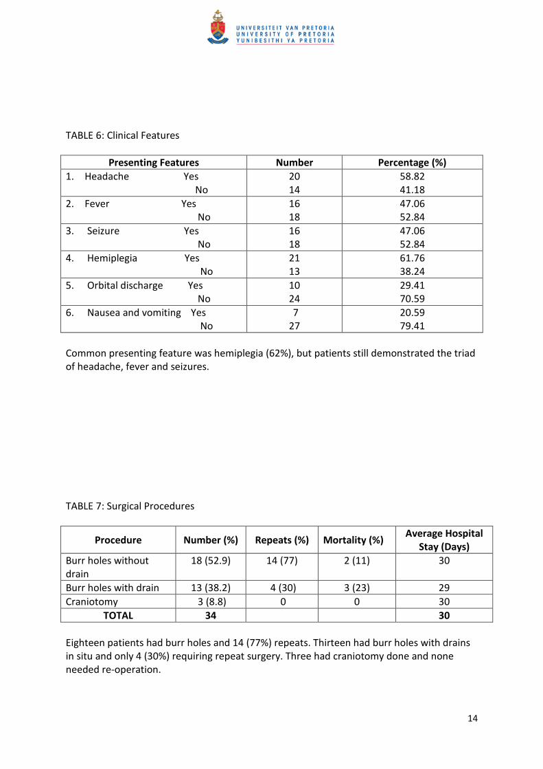
TABLE 6: Clinical Features
Presenting Features Number Percentage (%)
1. Headache Yes
No
20
14
58.82
41.18
2. Fever Yes
No
16
18
47.06
52.84
3. Seizure Yes
No
16
18
47.06
52.84
4. Hemiplegia Yes
No
21
13
61.76
38.24
5. Orbital discharge Yes
No
10
24
29.41
70.59
6. Nausea and vomiting Yes
No
7
27
20.59
79.41
Common presenting feature was hemiplegia (62%), but patients still demonstrated the triad
of headache, fever and seizures.
TABLE 7: Surgical Procedures
Procedure Number (%) Repeats (%) Mortality (%) Average Hospital
Stay (Days)
Burr holes without
drain
18 (52.9) 14 (77) 2 (11) 30
Burr holes with drain 13 (38.2) 4 (30) 3 (23) 29
Craniotomy 3 (8.8) 0 0 30
TOTAL 34 30
Eighteen patients had burr holes and 14 (77%) repeats. Thirteen had burr holes with drains
in situ and only 4 (30%) requiring repeat surgery. Three had craniotomy done and none
needed re-operation.
15
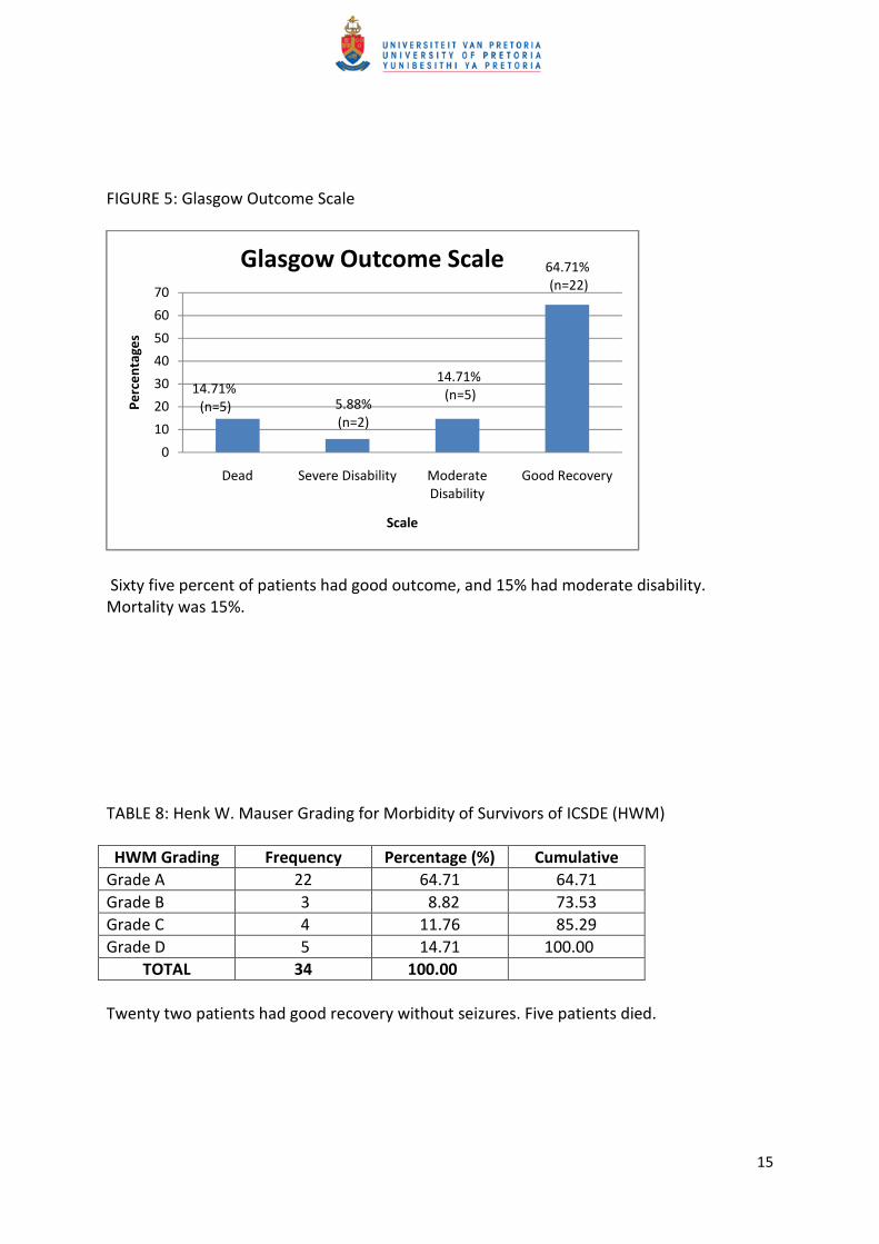
FIGURE 5: Glasgow Outcome Scale
Sixty five percent of patients had good outcome, and 15% had moderate disability.
Mortality was 15%.
TABLE 8: Henk W. Mauser Grading for Morbidity of Survivors of ICSDE (HWM)
HWM Grading Frequency Percentage (%) Cumulative
Grade A 22 64.71 64.71
Grade B 3 8.82 73.53
Grade C 4 11.76 85.29
Grade D 5 14.71 100.00
TOTAL 34 100.00
Twenty two patients had good recovery without seizures. Five patients died.
14.71%
(n=5) 5.88%
(n=2)
14.71%
(n=5)
64.71%
(n=22)
0
10
20
30
40
50
60
70
Dead Severe Disability Moderate
Disability
Good Recovery
Pe
rce
nta
ge
s
Scale
Glasgow Outcome Scale

16
FIGURE 6: Admission GCS and Mortality
Graph showing mortality against admission Glasgow Coma Scale, of the 34 patients
admitted (blue colour) and 5 died (brown colour) – independent of admission GCS.
TABLE 9: Contingency table of GOS and Age groups
GOS Age Groups (years)
0 - 10 11 - 20 21 - 30 31 above
Dead 0 (0.00) 2 (12.50) 2 (33.33) 1 (33.33)
Severe Disability 0 (0.00) 0 (0.00) 1 (16.67) 1 (33.33)
Moderate
Disability
3 (33.33) 1 (6.25) 0 (0.00) 1 (33.33)
Good Recovery 6 (66.67) 13 (81.25) 3 (50.00) 0 (0.00)
Total 9 (100.00) 16 (100.00) 6 (100.00) 3 (100.00)
1110
13
21
2
0
2
4
6
8
10
12
14
3 - 8 9 -12 13 - 15
Fre
qu
en
cy
Admission GCS
Admission GCS and Mortality
Series1
Series2

17
CHAPTER 5: DISCUSSION AND CONCLUSION
5.1 DISCUSSION
There were 20 (60%) males in the study with 14 (40%) females – Table 3. This is in keeping
with other international studies2,3,7
. The reason for this male dominance remains unclear.
The study also confirms what Dawodu noticed that the subdural empyema can affect any
age group2, patients’ age varied from five months to 45 years. Complicated sinusitis was the
main cause in 82% of patients, followed by meningitis at 8% with head trauma, surgical
operations and pneumonia contributing 3% each – Table 3. Of the patients with
complicated sinusitis all had paranasal sinuses as the origin of infection. None had otitis
media as the origin of infection as observed by Reeves Dill.18
Sterile cultures were reported in 44% of cases with 56% having positive cultures – Figure 3.
From the positive cultures 70% of the identified organisms were alpha and beta haemolytic
streptococci – Table 5. All patients were on empiric triple antibiotic therapy consisting of 3rd
generation cephalosporin/meropenem plus vancomycin plus metronidazole. The majority
were de-escalated to penicillinG/ampicillin according to sensitivity results. Resistance to
penicillin was not a major feature in this study, accounting for only 5% of cases as opposed
64,3% reported by Olwoch17
.Presenting features were similar to other studies3, namely the
triad of headache, fever and seizures. The most common presenting feature was
hemiplegia (accounting for 60% - Table 6) as also seen by Gradidge et al19
.
It is worth noting that from this study the type of surgical procedure had no influence on the
average hospital stay (Table 7).Leaving drains after burr holes reduced the likelihood for
repeat surgery from 77% to 30% (Table7) as was also reported by HE et al12
. There was no
mortality associated with craniotomy in this study which then suggests craniotomy as a
preferred surgical procedure of choice, as suggested by Feuerman et al6, Bok et al
7 and Mat
Nayan et al9. This is in contrast with what Mauser et al, who preferred burr holes to
craniotomy11
.
Glasgow Outcome Scale and Henk W. Mauser grading system were used to determine
outcome (Figure 5 and Table 8 respectively). Sixty-five percent of patients had good
outcome with no seizures and neurological deficits at discharge. Mortality was 15% in this
study, comparable to international figures. Locally this figure is higher compared to a study
at Cape Town7 and George Mukhari Hospital
16 but less when compared with the Durban
study by Nathoo et al8.None of the poor prognostic features as suggested by Agrawal et al
3,
Miller et al10
and Mauser et al14
were found to influence mortality or morbidity in this study,
e.g. admission GCS – Figure 5. Favourable outcome was associated with patients who
underwent craniotomy and patients between the ages of 11 – 20 years (Table 9), this is in
agreement with what Agrawal et al reported3.

18
5.2 LIMITATIONS
The limitation for this study is the enrolled number of patients. A larger group might show
some factors associated with poor outcome and those with good outcome.
5.3 CONCLUSION
ICSDE is still associated with high mortality if not treated early. Clinical features and
organisms cultured seem to be the same internationally, particularly those from
complicated sinusitis. Empiric triple antibiotic therapy of 3rd generation cephalosporin plus
vancomycin plus metronidazole is still relevant at SBAH.
The type of surgical operation done has no influence on length of hospital stay. Craniotomy
emerges as a procedure associated with no mortality in stable patients, multiple burr holes
with drains in situ is recommended in unstable patients to avoid repeated operations. Age
was another factor associated with better outcomes, particularly those between 11 and 20
years of age.
19
REFERENCES
1. D.A. Nica, R. Moroti-Constantinescu, R Copacium M. Nica. Multidisciplinary
management and outcome in subdural empyema – a case report. Chirurgia (2011)
106:673-676
2. Segun T Dawodu. Subdural Empyema. Emedicine.medscape.com/article/1168415 –
overview Jul 14, 2011
3. Amit Agrawal, Jake Timothy, Lekha Pandit, Lathika Shetty and J.P. Shetty. A Review
of Subdural Empyema and Its Management. Infectious Disease in Clinical Practice.
Volume 15, Number 3, May 2007
4. Manoj K Tewari, Rewati R Sharma, Vinod K Shiv, Santosh D Lad. Spectrum of
intracranial subdural empyema in a series of 45 patients: Current surgical options
and outcome. Neurology India, 2004. Volume: 52, Issue : 3, Page: 346-349
5. Syed S Nadvi, Narendra Nathoo, James R van Dellen. Lumbar puncture in brain
abscess or subdural empyema: Not an innocuous procedure. African Journal of
Neurological Sciences, Vol20, Num 1, 2001, p 14-16
6. Feuerman T, Wackym PA, Gade GF, Dubrow T. Craniotomy improves outcome in
subdural empyema. Surg Neurol. 1989 Aug;32(2): 105-10
7. Arnold P.L. Bok, Jonathan C. Peter. Subdural empyema: burr holes or craniotomy? J
Neurosurg 78: 574-578, 1993
8. Nathoo N, Nadvi SS, Gouws E, van Ellen JR. Craniotomy improves outcomes for
cranial subdural empyemas: computed tomography-era experience with 699
patients. Neurosurgery 2001 Oct;49(4): 872-7. Discussion 877-8
9. Mat Nayan SA, Mohd Haspani MS, Abd Latiff AZ, Abdullah JM, Abdullah S. Two
surgical methods used in 90 patients with intracranial subdural empyema. J Clin
Neurosci. 2009 Dec: 16(12): 1567-71. Epub 2009 Sep 29
10. Elizabeth S Miller, P S Dias, David Uttley. Management of subdural empyema: a
series of 24 cases. Journal of Neurology, Neurosurgery, and Psychiatry 1987;50:
1415-1418
11. Mauser HW, Telleken CA. Subdural empyema. A review of 48 patients. Clin Neurol
Neurosurg. 1984;86(4):255-63
12. AK HE, Ozkan U, Devecioglu C, et al. Treatment of subdural empyema by burr hole.
Isr J Med Sci 1996 Jul; 32(7): 542-4
13. Stephen Ryu, Michael Lim and Griffin R. Harsh IV. Management of Epidural Abscess
and Subdural Empyemas. Oper Tech Neurosurg 7: 182-187. 2005. Elsevier Inc.
14. Henk W Mauser, Hans C van Houwelingen, Cees A F Tulleken. Factors affecting the
outcome in subdural empyema. Journal of Neurology, Neurosurgery, and Psychiatry
1987; 50:1136-1141
15. Pasquale De Bonis, Carmmelo Anile, Angelo Pompucci, Maria Labonia, Carrado
Lucantoni & Annunziato Mangiola. Cranial and spinal empyema. British Journal of
Neurosurgery, June 2009; 23(3): 335-340
16. Tshifularo M, Monama GM. Complications of inflammatory sinusitis in children:
institutional review. SA Fam Pract 2006; 48(10): 16
17. Ian Paul Olwoch. The microbiology of acute complicated bacterial sinusitis at the
University of the Witwatersrand. S Afr Med J 2010; 100: 529-533
18. S Reeves Dill, C. Glenn Cobbs, and Cheryl K. McDonald. Subdural Empyema: Analysis
of 32 cases and Review. Clinical Infectious Diseases 1995;20:372-86
20
19. Katherine Gradidge, Denise Franzen. Record review of patients with Brain Abscess
and Empyema at Chris Hani Baragwanath Hospital. The Internet Journal of Neurology
ISSN:1531-295X

21
APPENDICES
Appendix A – Glasgow Coma Scale
Eye Opening Verbal Response Motor
1 = no response 1 = no response 1 = no response
2 = to painful stimuli 2 = incomprehensible sounds 2 = decerebrate
3 = to verbal stimuli 3 = inappropriate words 3 = decorticate
4 = spontaneous 4 = disorientated 4 = flexion withdrawal
5 = orientated 5 = localizes pain
6 = obey commands

22
Appendix B – Data Collection Form
PATIENT DATA SHEET: THE OUTCOME OF INTRACRANIAL SUBDURAL EMPYEMA AT STEVE
BIKO ACADEMIC HOSPITAL – RETROSPETIVE STUDY
PATIENT DATA
Name: ______________________________ Age: _____________________
Hospital Number: _____________________ Sex: ______________________
Date of Admission: ____________________ Admission Diagnosis: ________
PRESENTING FEATURES
Headache: _____yes/no Fever: ______yes/no
Seizures: _______yes/no Hemiplegia: __yes/no
Others (specify): ________________________
Admission Glasgow Coma Scale: ___________
INVESTIGATIONS
Brain CT scan – Pus Location: ____________ Pus swab results: _________
Source of Infection: Complicated sinusitis ______
Penetrating head trauma ____
Other (specify) _____________
TREATMENT
Date started Days given
1. Antibiotics
2. Anti-epileptic
3. Surgical procedure
Burr holes
Burr holes with
drain
Craniotomy
(document repeat surgery)
CLINICAL COURSE
GOS at discharge: __________________ HWM grade: ____________________
Length of hospital stay: _____________ Death: ____________________yes/no

23
Appendix C – Henk W. Mauser grading system
Grade Description
A Survival without or with a minor, not disabling focal deficit
B Survival with not disabling seizures and with or without a minor focal deficit
C Survival with severe disability
D Death

24
Appendix D – Glasgow Outcome Scale
Grade Description
1 Dead
2 Persistent vegetative state
3 Severe disability
4 Moderate disability
5. Good recovery