d1-asn-298inphotosystemiiisinvolvedinahydrogen-bond ... · how the four protons are transferred...
TRANSCRIPT

D1-Asn-298 in photosystem II is involved in a hydrogen-bondnetwork near the redox-active tyrosine YZ for proton exitduring water oxidationReceived for publication, August 29, 2017, and in revised form, October 4, 2017 Published, Papers in Press, October 18, 2017, DOI 10.1074/jbc.M117.815183
Ryo Nagao1, Hanayo Ueoka-Nakanishi, and Takumi Noguchi2
From the Division of Material Science, Graduate School of Science, Nagoya University, Furo-cho, Chikusa-ku,Nagoya 464-8602, Japan
Edited by Joseph Jez
In photosynthetic water oxidation, two water molecules areconverted into one oxygen molecule and four protons at theMn4CaO5 cluster in photosystem II (PSII) via the S-state cycle.Efficient proton exit from the catalytic site to the lumen is essen-tial for this process. However, the exit pathways of individualprotons through the PSII proteins remain to be identified. Inthis study, we examined the involvement of a hydrogen-bondnetwork near the redox-active tyrosine YZ in proton transferduring the S-state cycle. We focused on spectroscopic analysesof a site-directed variant of D1-Asn-298, a residue involved in ahydrogen-bond network near YZ. We found that the D1-N298Amutant of Synechocystis sp. PCC 6803 exhibits an O2 evolutionactivity of �10% of the wild-type. D1-N298A and the wild-typeD1 had very similar features of thermoluminescence glowcurves and of an FTIR difference spectrum upon YZ oxidation,suggesting that the hydrogen-bonded structure of YZ and elec-tron transfer from the Mn4CaO5 cluster to YZ were little affectedby substitution. In the D1-N298A mutant, however, the flash-number dependence of delayed luminescence showed a mono-tonic increase without oscillation, and FTIR difference spectraof the S-state cycle indicated partial and significant inhibition ofthe S23 S3 and S33 S0 transitions, respectively. These resultssuggest that the D1-N298A substitution inhibits the protontransfer processes in the S23 S3 and S33 S0 transitions. This inturn indicates that the hydrogen-bond network near YZ can befunctional as a proton transfer pathway during photosyntheticwater oxidation.
Plants and cyanobacteria utilize water as an ultimate electrondonor to reduce carbon dioxide in the synthesis of sugars (1).This reaction of water oxidation is performed in photosystem II(PSII)3 protein complexes, which are embedded in thylakoid
membranes. Water oxidation produces protons and molecularoxygen (2–9); protons are released into the thylakoid lumen,making a proton gradient across the membrane, and are used tosynthesize ATP, whereas molecular oxygen is liberated to theair to be the major oxygen source of the atmosphere.
In PSII, photochemical reactions start with light-inducedcharge separation between the primary donor chlorophyll(Chl), P680, and the pheophytin electron acceptor, Pheo (10,11). An electron is transferred from Pheo to the primary qui-none electron acceptor, QA, and then the secondary quinoneelectron acceptor, QB (12). On the electron donor side, the P680cation abstracts an electron from the redox-active tyrosine, YZ(D1-Tyr-161), which immediately becomes a neutral radical(YZ
�) by shifting a proton to the neighboring D1-His-190through a strong hydrogen bond, forming a protonated Hiscation (13–16). YZ
� then oxidizes the water-oxidizing center(WOC), the catalytic site of water oxidation, which consists of aMn4CaO5 cluster as an inorganic core, its amino acid (D1-Asp-170, D1-Glu-189, D1-His-332, D1-Glu-333, D1-Asp-342,D1-Ala-344, and CP43-Glu-354) and water (W1–W4) ligands,two Cl ions (Cl-1 and Cl-2), a hydrogen-bonded networkformed by nearby water molecules and amino acid residues(Fig. 1A) (17–21). The water-oxidation reaction proceeds by alight-driven cycle of five intermediates, called Si-state (i � 0 – 4;with a larger i value representing a higher oxidation state), inwhich the one-electron-oxidized S1-state is the most stable inthe dark (Fig. 1B) (22–23). Each S-state advances to the nextstate upon one-electron oxidation, except that the S4-state,which is the most oxidized but unstable intermediate, immedi-ately relaxes to the S0-state, by releasing an O2 molecule (2–9).During this S-state cycle, four protons are released to the lumenwith a stoichiometry of 1:0:1:2 for the S0 3 S1:S1 3 S2:S2 3S
3:S33 S0 transitions (24 –26). Because no proton is released in
the S13 S2 transition, an excessive positive charge is accumu-lated on the Mn4CaO5 cluster in the S2- and S3-states.
Efficient proton exit from the catalytic site is essential in themechanism of water oxidation. However, it remains unclearhow the four protons are transferred from WOC to the lumenthrough the PSII proteins during the S-state cycle. The X-ray
This work was supported by the Grants-in-Aid for Scientific Research fromJapan Society for the Promotion of Science 26840091 and 17K07442 (toR. N.) and 17H03662 and 17H06435 (to T. N.). The authors declare that theyhave no conflicts of interest with the contents of this article.
1 To whom correspondence may be addressed: Research Institute for Inter-disciplinary Science, Okayama University, 3-1-1 Tsushima-naka, Okayama700-8530, Japan. Tel.: 81-86-251-8630; Fax: 81-86-251-8630; E-mail:[email protected].
2 To whom correspondence may be addressed. Tel: 81-52-789-2881; Fax:81-52-789-2883; E-mail: [email protected].
3 The abbreviations used are: PSII, photosystem II; Chl, chlorophyll; DCBQ,2,6-dichloro-p-benzoquinone; DL, delayed luminescence; DM, n-dodecyl
�-D-maltoside; MD, molecular dynamics; QM/MM, quantum mechanics/molecular mechanics; TL, thermoluminescence; DCMU, 3-(3,4)-dichloro-phenyl-1,1-dimethylurea; Km, kanamycin; Cm, chloramphenicol; Em,erythromycin; Sm, spectinomycin.
croARTICLE
20046 J. Biol. Chem. (2017) 292(49) 20046 –20057
© 2017 by The American Society for Biochemistry and Molecular Biology, Inc. Published in the U.S.A.
by guest on June 27, 2019http://w
ww
.jbc.org/D
ownloaded from

structures of PSII complexes (17, 18, 27, 28) and theoreticalcalculations based on them have shown that there are severalchannels suitable for proton transfer around the Mn4CaO5cluster leading to the lumen (Fig. 1A) (29 –39). These channelsinvolve a number of water molecules forming hydrogen-bondnetworks, as revealed by the recent high-resolution X-raystructures that resolved water oxygen atoms (17–21). Near theentrance of each channel, either D1-Asp-61 or YZ is located,and a water cluster is formed between these two residuesincluding water ligands to Mn4 (W1 and W2) and Ca2� (W3and W4).
The channel from D1-Asp-61 to PsbO through the Cl-1 siteand the D1-Glu-65/D2-Glu-312/D1-Arg-334 triad has beenmost extensively studied and proposed to function as a pathwayfor protons or substrate water (designated “Cl path” in the pres-ent report; Fig. 1A) (29 –37, 39). Theoretical calculations sug-gested that D1-Asp-61 plays a crucial role in proton transfer(40, 41), and that the Cl� ion has a function to regulate protontransfer through this pathway (34). D1-Asp-61 is also locatednear the entrance of a water chain starting at O4 and leading toPsbU (designated “O4 path”; Fig. 1A), which has been proposedto function as a proton exit pathway especially in the S03 S1transition (38, 39). The recent X-ray free electron laser study forthe S3-state showed the displacement of a water molecule nearO4, suggesting the involvement of water molecules near O4 inproton transfer (21). The hydrogen-bond network near YZ hasalso been suggested to be a proton pathway (16, 17, 42, 43). YZform a hydrogen-bonded triad with D1-His-190 and D1-Asn-298, which is connected with a hydrogen-bond network leadingto PsbV through D1-Asn-322 (designated “YZ-Asn-298 path”;Fig. 1A). Molecular dynamics (MD) simulations (36, 39) sug-
gested a tight hydrogen-bond network of this pathway, which isadvantageous to proton transfer. A large water cluster interact-ing the YZ/D1-His-190/D1-Asn-298 triad also forms anotherhydrogen-bond network through O1 and D1-Glu-329 leadingto PsbV (designated “YZ-O1 path”). MD simulation showedthat this channel contains a number of mobile water molecules(33, 39). It is noted that the above key amino acid residues in thehydrogen-bond networks in cyanobacterial PSII (Fig. 1A) are allconserved in higher plants whose PSII-LHCII structures wererecently resolved by cryo-electron microscopy (44, 45).
The finding of several channels around the Mn4CaO5 clusterraises a question, which proton pathways are used for individ-ual protons released in the S-state transitions? The most directexperimental method to answer this question is examining theeffects of site-directed mutations, by which amino acid residueson putative proton channels are altered to different aminoacids, on the reactions of individual S-state transitions. Manymutants of amino acid residues in putative channels have beeninvestigated so far (46 – 60). Most of them are located on the Clpath; mutations of D1-Asp-61 to Ala and Asn significantlyretarded the kinetic rate of O2 release in the S33 S0 transition(47, 53, 58) and changed the property of a nearby water network(57). Mutations of D2-Lys-317 and D1-Asn-181, which areligands to Cl-1, also retarded the O2 kinetics or decreased theefficiency of the S3 3 S0 transition (54, 55, 59). In addition,mutants of any of the D1-Glu-65/D2-Glu-312/D1-Arg-334triad substantially inhibited the S3 3 S0 transition (49, 52).These data suggest that the Cl path is involved in proton orwater transfer at least in the S3 3 S0 transition, although acaution is necessary in the interpretation of results for chargedresidues, whose mutations could affect the redox potential of
Figure 1. A, hydrogen-bond network around the Mn4CaO5 cluster and putative proton transfer pathways. The X-ray free electron laser structure of Thermos-ynechococcus vulcanus at 1.95-Å resolution (PDB code 4UB6 (18)) was used to draw the picture. Mn, purple; Ca, orange; Cl, green; O, red; N, blue. Oxygen atomsof water molecules are colored cyan. The numbering of the Mn and oxygen atoms in the Mn4CaO5 cluster and its water ligands follows the numbering in Umenaet al. (17). D1-Asn-298, which was replaced with Ala in this work, is circled with a red dotted line. Magenta arrows represent putative proton pathways, which weredesignated Cl path, O4 path, YZ-O1 path, and YZ-Asn-298 path in this work. Hydrogen bonds are shown with green dotted lines. Amino acid residues with labelsare on the D1 subunit except for Lys-317 and Glu-312 on the D2 subunit, and Glu-354 and Ala-411 on the CP43 subunit. B, the S-state cycle of water oxidation.
Proton transfer pathway in photosynthetic water oxidation
J. Biol. Chem. (2017) 292(49) 20046 –20057 20047
by guest on June 27, 2019http://w
ww
.jbc.org/D
ownloaded from

the Mn4CaO5 cluster and hence the electron transfer reaction.In contrast, fewer mutagenesis studies have been performed forthe channels near the YZ site. Mutation of D1-Gln-165, which ishydrogen-bonded with W4 near YZ, to Glu decreased the effi-ciency of the S3 3 S0 transition (56), whereas mutation ofD1-Glu-329 on the YZ-O1 path to Gln little affected the S-statetransitions (52). Fourier transform infrared (FTIR) analysis ofthe S-state cycle using various mutants by Debus and co-work-ers (52, 54, 56, 57) further showed the presence of an extensivehydrogen-bond network around the Mn4CaO5 cluster.
D1-Asn-298 is located at a crucial position to keep a hydro-gen-bond network involving water molecules in the YZ-Asn-298 and YZ-O1 pathways (Fig. 1A). This has also been proposedto be a key residue to determine the hydrogen-bonded struc-ture of YZ and the rate of its proton-coupled electron trans-fer through the interaction with D1-His-190 (15, 16). Thus, muta-tion of D1-Asn-298 could affect the proton transfer from thecatalytic site as well as electron transfer from the Mn4CaO5cluster to YZ
� during S-state transitions. Yamasato et al. (50)previously found that the D1-N298I mutant obtained by ran-dom mutagenesis inhibited electron flow from water to theelectron acceptor side. More recently, Kuroda et al. (60) gener-ated 19 site-directed mutants of D1-Asn-298 using Chlamy-domonas reinhardtii, and showed that only seven mutants (D1-N298G, -A, -C, -M, -S, -Q, and -H) have O2 evolution activitybut with significantly low rates. However, detailed properties ofthe mutants of D1-Asn-298, such as specific inhibition of eitherelectron or proton transfer in individual S-state transitions,have not yet been investigated, and hence no clear answer wasobtained about the involvement of pathways near the YZ site inthe water oxidation mechanism.
In this work, we investigated whether the hydrogen-bondnetwork near YZ is functional or not in proton transfer duringwater oxidation, by constructing a D1-N298A mutant using acyanobacterium Synechocystis sp. PCC 6803, and analyzing thismutant by means of light-induced FTIR difference spectros-copy and detection of thermoluminescence (TL) and delayedluminescence (DL). Because Asn and Ala are both non-chargedamino acids and the replacement with a small Ala side chaincan avoid the undesirable alterations of main chains, only theperturbation of the hydrogen-bond network is expected tooccur without changes in the electrostatic interaction and pro-tein conformations. FTIR difference spectroscopy is a powerfulmethod for detecting the structural perturbations of amino acid
residues, water molecules, and the hydrogen-bond networkaround the Mn4CaO5 cluster, as well as monitoring the inhibi-tion of the S-state transitions (61–70). The results providedsolid evidence for the involvement of D1-Asn-298, and hencethe hydrogen-bond network near YZ, in proton transfer in theS
23 S3 and S33 S0 transitions of water oxidation.
Results
In vivo analyses of the D1-N298A mutant
The strains of Synechocystis sp. PCC 6803 used in the presentstudy lacked the psbA1 and psbA3 genes, and hence PsbA2 wasexpressed as a D1 protein (71). In addition, a histidine tag wasattached to the C terminus of the CP47 protein. Thus, the strainhaving a PsbA2 protein with a native sequence is designated“WT*” hereafter. In contrast, the Asn-298 of PsbA2 wasreplaced by Ala in the D1-N298A mutant. The D1-N298Amutant grew photoautotrophically with a rate slightly slowerthan the WT* (Fig. 2A). The O2 evolution activities of WT* andD1-N298A were 600 – 670 and 60 –70 �mol of O2 (mg ofChl)�1 h�1, respectively, in cells, and 2800 –3300 and 190 –300�mol of O2 (mg of Chl)�1 h�1, respectively, in isolated PSIIcomplexes. Thus, the O2 evolution activity of the D1-N298Amutant is about 10% of WT* in both cells and PSII core com-plexes. This reduced activity of the D1-N298A mutant is ingood agreement with that of a corresponding mutant of a greenalga C. reinhardtii, which showed a 11.5% activity of WT in cells(60).
TL glow curves of PSII in the presence and absence of DCMUoriginate from charge recombination of S2QA
� (Q band) andS2QB
� (B band), respectively (72). WT* cells showed Q and Bbands at about 14 and 34 °C, respectively, and they were virtu-ally unaffected by the D1-N298A mutation (Fig. 2B). In con-trast, the flash-number dependence of DL, which was moni-tored at 0.8 s after each flash, showed significantly differentpatterns between WT* and D1-N298A cells (Fig. 2C). WhereasWT* cells showed a typical period-four oscillation of DL sig-nals, D1-N298A cells did not show any oscillation and the DLamplitude monotonically increased with a flash number.
FTIR difference spectrum upon YZ oxidation
Light-induced FTIR difference spectra upon YZ oxidation(YZ
�/YZ difference spectra) were obtained using manganese-depleted PSII core complexes of WT* and D1-N298A (Fig. 3).Characteristic bands of the YZ
�/YZ spectra were virtually iden-
Figure 2. In vivo characterization of WT* (black) and D1-N298A (red) cells. A, growth curves of cells. B, TL glow curves in the (a) presence and (b) absenceof DCMU representing charge recombination of the S2QA
� and S2QB� charged pairs, respectively. C, flush-number dependence of DL induced by 10 successive
flashes (1 Hz). DL was measured at 25 °C and the amplitude at 0.8 s after each flash was plotted.
Proton transfer pathway in photosynthetic water oxidation
20048 J. Biol. Chem. (2017) 292(49) 20046 –20057
by guest on June 27, 2019http://w
ww
.jbc.org/D
ownloaded from
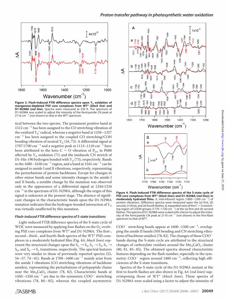
tical between the two species. The prominent positive band at1512 cm�1 has been assigned to the CO stretching vibration ofthe oxidized YZ
� radical, whereas a negative band at 1258 –1257cm�1 has been assigned to the coupled CO stretching/COHbending vibration of neutral YZ (16, 73). A differential signal at1707/1700 cm�1 and a negative peak at 1113–1110 cm�1 havebeen attributed to the keto C � O vibration of PD1 in P680affected by YZ oxidation (71) and the imidazole CN stretch ofD1-His-190 hydrogen bonded with YZ (73), respectively. Bandsin the 1680 –1630 cm�1 region, and a band at 1541 cm�1 can beassigned to amide I and II vibrations, respectively, representingthe perturbations of protein backbones. Except for changes inother minor bands and some intensity changes in the amide Iand II bands, a notable change by the mutation was observedonly in the appearance of a differential signal at 1244/1234cm�1 in the spectrum of D1-N298A, although the origin of thissignal is unknown at the present stage. The absence of signifi-cant changes in the characteristic bands upon the D1-N298Amutation indicates that the hydrogen-bonded interaction of YZwas virtually unaffected by this mutation.
Flash-induced FTIR difference spectra of S-state transitions
Light-induced FTIR difference spectra of the S-state cycle ofWOC were measured by applying four flashes on the O2-evolv-ing PSII core complexes from WT* and D1-N298A. The first-,second-, third-, and fourth-flash spectra of the WT* PSII com-plexes in a moderately hydrated film (Fig. 4A, black lines) rep-resent the structural changes upon the S13 S2, S23 S3, S33S0, and S03 S1 transitions, respectively. The spectral featureswere very similar to those of previously reported spectra (52,54 –57, 74 – 81). Bands at 1700 –1600 cm�1 mainly arise fromthe amide I vibrations (CO stretching vibrations of backboneamides), representing the perturbations of polypeptide chainsnear the Mn4CaO5 cluster (78, 82). Characteristic bands at1450 –1350 cm�1 are due to the symmetric COO� stretchingvibrations (78, 80 – 85), whereas the coupled asymmetric
COO� stretching bands appear at 1600 –1500 cm�1, overlap-ping the amide II bands (NH bending and CN stretching vibra-tions of backbone amides) (78, 82). The changes of these COO�
bands during the S-state cycle are attributed to the structuralchanges of carboxylate residues around the Mn4CaO5 cluster(80, 81, 83– 85). The obtained spectra showed characteristicfeatures depending on the flash number, especially in the sym-metric COO� region around 1400 cm�1, reflecting high effi-ciencies of the S-state transitions.
Spectra of the S-state cycle of the D1-N298A mutant at thefirst to fourth flashes are also shown in Fig. 4A (red lines) sup-erimposing those of WT* (black lines). These spectra ofD1-N298A were scaled using a factor to adjust the intensity of
Figure 3. Flash-induced FTIR difference spectra upon YZ oxidation ofmanganese-depleted PSII core complexes from WT* (black line) andD1-N298A (red line). Spectra were measured at 250 K. The spectrum ofD1-N298A was scaled to adjust the intensity of the ferricyanide CN peak at2116 cm�1 (not shown) to that in the WT* spectrum.
Figure 4. Flash-induced FTIR difference spectra of the S-state cycle ofPSII core complexes from WT* (black lines) and D1-N298A (red lines) inmoderately hydrated films. A, mid-infrared region (1800 –1200 cm�1) ofprotein vibrations. Difference spectra were measured upon the (a) first, (b)second, (c) third, and (d) fourth flashes. B, expanded view of the C � O stretch-ing region of COOH groups (1770 –1720 cm�1) at the (a) first and (b) secondflashes. The spectra of D1-N298A were scaled with a factor to adjust the inten-sity of the ferricyanide CN peak at 2116 cm�1 (not shown) in the first-flashspectrum to that of WT*.
Proton transfer pathway in photosynthetic water oxidation
J. Biol. Chem. (2017) 292(49) 20046 –20057 20049
by guest on June 27, 2019http://w
ww
.jbc.org/D
ownloaded from
the CN band of ferricyanide at 2116 cm�1 at the first-flash (notshown) to that of WT*, representing the same extent of an elec-tron flow in PSII in the S13 S2 transition. Although the first-flash spectrum due to the S1 3 S2 transition showed overallfeatures similar to those of WT*, some differences wereobserved in several bands (Fig. 4A, a). In the symmetric COO�
region, band intensities at 1417(�) and 1364(�) cm�1 weresignificantly reduced. In addition, bands at 1652(�), 1587(�),1543(�), 1523(�), and 1508(�) cm�1 in the amide I and theasymmetric COO�/amide II regions decreased their intensi-ties. Furthermore, a negative band at 1747 cm�1, which hasbeen assigned to the C � O stretching vibration of an uniden-tified COOH group involved in a hydrogen-bond networkaround the Mn4CaO5 cluster (52, 56), diminished and isreplaced by a positive band at 1745 cm�1 (Fig. 4B, a). In con-trast, spectral features of the second-flash spectra of D1-N298Awere very similar to those of WT* (Fig. 4Ab). However, an over-all intensity in the COO� region (1450 –1300 cm�1) was �60%of WT*. A small positive peak at 1745 cm�1 due to a COOHgroup, which has a different origin from the negative band at1747 cm�1 at the first flash (56), also diminished (Fig. 4B, b).
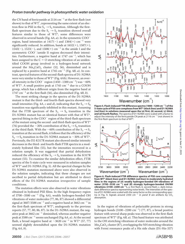
The most striking change in the spectra of the D1-N298Amutant is that the third- and fourth-flash spectra showed verysmall intensities (Fig. 4A, c and d), indicating that the S33 S0transition was significantly inhibited in this mutant. Assumingthat the FTIR spectrum of the S3 3 S0 transition in theD1-N298A mutant has an identical feature with that of WT*,spectral fitting in the COO� region of the third-flash spectrumof the mutant using the second- and third-flash spectra of WT*(76) provided the �20% contribution of the S33 S0 transitionat the third flash. With the �60% contribution of the S23 S3transition at the second flash, it follows that the efficiency of theS33 S0 transition in the D1-N298A mutant is �30% of WT*.Previously, the D2-K317R mutant also showed similar intensitydecreases in the third- and fourth-flash FTIR spectra in a mod-erately hydrated film (55), but the intensities recovered in asolution sample. It was suggested that partial dehydrationreduced the efficiency of the S33 S0 transition in the K317Rmutant (55). To examine the similar dehydration effect, FTIRspectra of the S-state cycle were measured in solution samplesof WT* and D1-N298A (Fig. 5). All the spectral changes by theD1-N298A mutation mentioned above were also observed inthe solution samples, indicating that these changes are notascribed to partial dehydration but are attributed to directeffects of the D1-N298A mutation irrespective of sampleconditions.
The mutation effects were also observed in water vibrationsobtained in hydrated PSII films. In the high frequency regionof 3700 –3500 cm�1 (Fig. 6A), weakly hydrogen-bonded OHvibrations of water molecules (77, 86, 87) showed a differentialsignal at 3607/3588 cm�1 and a negative band at 3662 cm�1 inthe first-flash spectrum of WT*, analogously to the previousreports (57, 77, 80, 86, 87). In the D1-N298A mutant, the neg-ative peak at 3662 cm�1 diminished, whereas another negativepeak at 3588 cm�1 seems unchanged (Fig. 6A, a). At the secondflash, a broad negative band at �3620 cm�1 in WT* (57, 77)seemed slightly downshifted upon the D1-N298A mutation(Fig. 6A, b).
In the region of vibrations of polarizable protons in stronghydrogen bonds (3100 –2200 cm�1) (77, 87), a broad positivefeature with several sharp peaks was observed in the first-flashspectrum of WT* (Fig. 6B, a). This band feature was attributedto the OH stretching vibrations of water molecules around theMn4CaO5 cluster (87), overlapping the NH stretching vibrationwith Fermi resonance peaks of a His side chain (D1-His-337)
Figure 5. Flash-induced FTIR difference spectra (1800 –1200 cm�1) of theS-state cycle of PSII core complexes from WT* (black lines) and D1-N298A(red lines) in solutions measured upon the (a) first, (b) second, (c) third,and (d) fourth flashes. The spectra of D1-N298A were scaled with a factor toadjust the intensity of the ferricyanide CN peak at 2116 cm�1 (not shown) inthe first-flash spectrum to that of WT*.
Figure 6. Flash-induced FTIR difference spectra of PSII core complexesfrom WT* (black lines) and D1-N298A (red lines) in moderately hydratedfilms in the regions of (A) weakly hydrogen-bonded OH vibrations ofwater (3700 –3530 cm�1) and (B) strongly hydrogen-bonded OH/NHvibrations (3100 –2200 cm�1). a, first flash; b, second flash; c, dark-minus-dark difference spectra representing noise levels. The intensities of the spec-tra of D1-N298A were scaled with the same factor as the corresponding spec-tra in Fig. 4. Signals around 2350 cm�1 in panel B are artifacts by CO2absorption.
Proton transfer pathway in photosynthetic water oxidation
20050 J. Biol. Chem. (2017) 292(49) 20046 –20057
by guest on June 27, 2019http://w
ww
.jbc.org/D
ownloaded from

hydrogen-bonded with the O3 oxo bridge of the Mn4CaO5cluster (88). The intensities of these bands were weakened uponthe D1-N298A mutation. In the second-flash spectra (Fig. 6B,b), WT* exhibited broad positive features at 2681 and 2456cm�1 (57, 77), whereas a new positive band appeared at 2907cm�1 upon the D1-N298A mutation leaving the broad features.
Discussion
D1-Asn-298 is a crucial amino acid residue in retaining ahydrogen-bond network near YZ. It supports a water chain thatleads from W4 to PsbV through D1-Asn-191 and D1-Asn-322(YZ-Asn-298 path), and a water cluster that interacts with O1 ofthe Mn4CaO5 cluster and D1-Glu-329 leading to PsbV (YZ-O1path) (Fig. 1A), both of which have been candidates of proton/water pathways (31–33, 35–37, 39). Thus, mutation of D1-Asn-298 to non-hydrogen bonding Ala is expected to significantlyperturb the structures of these hydrogen-bond networks byreorganizing nearby water molecules. In addition, D1-Asn-298is a key amino acid residue in determining the hydrogen-bonded structure of YZ. The amide C � O of the Asn-298 sidechain accepts an hydrogen bond from the N�-H of D1-His-190,whereas the N� of His-190 functions as a hydrogen-bondacceptor of the OH of YZ (15–18). This hydrogen-bondedstructure of the YZ/D1-His-190/D1-Asn-298 triad realizesrapid proton-coupled electron transfer of YZ, in which uponoxidation by P680�, the proton of YZ is immediately shifted toD1-His-190 forming a HisH� cation (15, 16). The D1-N298Amutation hence could also perturb the hydrogen-bonded struc-ture of YZ and its reaction. In contrast, mutation from Asn toAla, both of which are non-charged amino acids, should havelittle electrostatic effect on YZ and the Mn4CaO5 cluster tochange their redox potentials. Furthermore, a relatively remotelocation (�10 Å) of D1-Asn-298 from the Mn4CaO5 clusterwithout any direct interactions even with the first shell ligandsmakes less likely the possibility that its mutation has a directeffect on the catalytic reaction of water oxidation.
We examined the effect of the D1-N298A mutation on thehydrogen-bonded structure of YZ by comparing the YZ
�/YZFTIR difference spectrum of the manganese-depleted PSII corecomplex from the D1-N298A mutant with that from WT*. Theresults showed that they had very similar features (Fig. 3). Inparticular, the vibrational frequencies of the CO stretchingband of YZ
� (1512 cm�1) and the CO stretching/COH bendingband of neutral YZ (1257 cm�1), which are sensitive to changesin hydrogen-bonding interactions, were identical within 1cm�1, indicating that the hydrogen-bonded interaction of YZwas little affected by this mutation. It is presumed that thereplacement of Asn with a smaller Ala produced enough spacefor a new water molecule to bind to the N� of D1-His-190instead of the Asn C � O. Thus, the hydrogen-bonding patternof the YZ/D1-His-190/D1-Asn-298 triad was probablyunchanged in the YZ/D1-His-190/H2O triad in the mutant,although the possibility remains that in intact PSII with theMn4CaO5 cluster, the mutation has some effect on the YZ in-teraction through the hydrogen-bond network around theMn4CaO5 cluster.
The reaction process of the S13 S2 transition is thought tobe simple electron transfer from the Mn4CaO5 cluster to YZ
�.
Hence, if electron transfer from the Mn4CaO5 cluster to YZ� is
impaired by the D1-N298A mutation, the efficiency of the S13S2 transition should also be affected. The S2/S1 FTIR differencespectra of WT* and the D1-N298A mutant at the first flash(Figs. 4A, a, and 5a) showed similar overall features but withintensity changes and slight frequency shifts in some bands. Atthe second flash, the spectrum of D1-N298A showed very sim-ilar features to the spectrum of WT* representing the S23 S3transition (Figs. 4A, b, and 5b), although the mutant spectrumhad a smaller overall intensity (�60% of WT*). The absence ofthe features of the S2/S1 spectrum in the second-flash spectrumof the mutant (e.g. 1432 cm�1 peak characteristic of the first-flash spectrum was not involved in the second-flash spectrum)indicates that the S13 S2 efficiency in D1-N298A is as high asthat in WT*. In addition, the peak temperatures of the TL glowcurves at 14 and 34 °C arising from charge recombination ofS2QA
� (Q band) and S2QB� (B band), respectively, little
changed upon the D1-N298A mutation (Fig. 2B). This indicatesthat the redox potential of the S1 3 S2 transition of theMn4CaO5 cluster, which should regulate the electron transferreaction, is virtually unchanged by this mutation. These obser-vations suggest that the D1-N298A mutation little affected theelectron transfer from the Mn4CaO5 cluster to YZ
� at least dur-ing the S13 S2 transition.
In contrast to unaffected electron transfer, the hydrogen-bond network around the Mn4CaO5 cluster was clearly affectedby the D1-N298A mutation, as revealed by the following obser-vations. (i) The symmetric COO� region (1450 –1300 cm�1)in the S2/S1 FTIR difference spectra showed characteristicchanges, such as intensity decreases of the 1417 and 1364 cm�1
bands by the mutation (Figs. 4A, a, and 5a). These bands wererecently assigned to the symmetric COO� vibrations ofD1-Asp-342/D1-Asp-61 and D1-Asp-170/D1-Glu-333/D1-Asp-342/D1-Glu-189, respectively, by quantum mechanics/molecular mechanics (QM/MM) calculations (85), suggestingslight perturbations of these carboxylate groups by the muta-tion. (ii) A negative band at 1747 cm�1 in the S2/S1 spectrumand a positive band at 1745 cm�1 in the S3/S2 spectrum (Fig.4B), which have been assigned to the CO stretching vibrationsof unidentified COOH groups (52, 56), diminished in the spec-tra of the D1-N298A mutant, indicating the pKa changes ofthese COOH groups were located on a common hydrogen-bond network involving D1-Asn-298. (iii) The intensities of theamide II bands in the 1590 –1500 cm�1 region together with anamide I band at 1652 cm�1 in the S2/S1 spectrum significantlydecreased (Figs. 4A, a, and 5a), suggesting the perturbation ofthe backbone interactions near D1-Asn-298. (iv) A negativeband at 3662 cm�1 in the weakly hydrogen-bonded OH regionof water (57, 77, 87) in the S2/S1 spectrum diminished and abroader negative band at 3620 cm�1 in the S3/S2 spectrum (57,77) seemed slightly downshifted (Fig. 6A). (v) The intensity ofthe feature in the 3000 –2600 cm�1 region, which have beenassigned to strongly hydrogen-bonded OH vibrations of watermolecules (77, 87) overlapping the NH vibration of protonatedD1-His-337 (88), in the S2/S1 difference spectrum decreased bythe D1-N298A mutation (Fig. 6B, a). In addition, in the similarregion in the S3/S2 spectrum, a positive band newly appeared at2907 cm�1 (Fig. 6B, b). These spectral changes in (iv) and (v)
Proton transfer pathway in photosynthetic water oxidation
J. Biol. Chem. (2017) 292(49) 20046 –20057 20051
by guest on June 27, 2019http://w
ww
.jbc.org/D
ownloaded from

represent some perturbations in the hydrogen-bonded struc-ture of a water network near the Mn4CaO5 cluster by theD1-N298A mutation.
Similar changes in the symmetric COO�, COOH, and amideII regions in FTIR difference spectra have been previouslyobserved in site-directed mutants of D1-Asp-61, D1-Glu-65,D2-Lys-317, D2-Glu-312, and D1-Arg-334 on the Cl path, andthose of D1-Gln-165 near YZ and D1-Glu-329 on the YZ-O1path (Fig. 1A) (52, 54 –57). In addition, the similar decrease inthe weakly hydrogen-bonded OH band at �3662 cm�1 and thebroad feature in the 3000 –2400 cm�1 region in the S2/S1 dif-ference spectrum was observed in the D1-D61A mutant (57).Indeed, the contribution of W1, which directly interacts withD1-Asp-61, to the bands at �3662 and 3000 –2400 cm�1 wassuggested by the QM/MM calculation (87). Recently, diminish-ing of the �3662 cm�1 band in the S2/S1 spectrum was alsoobserved in PSII in which Ca2� is substituted with Sr2� (89). Allof the above observations, therefore, indicate that a hydrogen-bond network structurally coupled with the Mn4CaO5 cluster iswidely extended across the channels near Cl and YZ involvingD1-Asn-298.
The S3/S2 FTIR difference spectrum at the second flashdecreased its overall intensity to �60% of WT* by theD1-N298A mutation (Figs. 4A, b, and 5b). Furthermore, theFTIR spectra at the third and fourth flashes showed very smallintensities (Figs. 4A and 5). These observations were identicalbetween samples in hydrated films (Fig. 4) and solutions (Fig. 5).These FTIR data of isolated PSII core complexes are consistentwith the in vivo data of DL measurements using D1-N298Acells, in which the DL intensity monotonically increased withthe flash number and reached near maximum by 4 or 5 flashes,in contrast to WT* cells that showed a clear period-four oscil-lation (Fig. 2C). In addition, the 10-fold decrease in the O2 evo-lution activity by the D1-N298A mutation was identicalbetween cells and PSII core complexes. These results indicatethat the intensity changes in the FTIR spectra by mutation areascribed to neither the core preparations nor the measurementconditions, but represent the real changes in efficiencies in theS-state transitions. It is thus concluded that the efficiency of theS23 S3 transition is lowered to �60% and the S33 S0 transi-tion is significantly inhibited upon the D1-N298A mutation.
It is generally accepted that one and two protons are releasedin the S2 3 S3 and S3 3 S0 transitions, respectively (24 –26).With the absence of the prominent effect on the electron trans-fer from the Mn4CaO5 cluster to YZ
� in the S13 S2 transition,together with the unlikeliness of direct involvement of D1-Asn-298 in the catalytic reaction at the Mn4CaO5 cluster because ofits remote distance, the observed efficiency decreases in theS23 S3 and S33 S0 transitions suggest that the proton transferfrom the catalytic site are partially and significantly inhibited inthe S2 3 S3 and S3 3 S0 transitions, respectively, in theD1-N298A mutant. Alternative possibilities, however, cannotbe excluded at the present stage that electron transfer reactionsare affected in the S-state transitions other than S13 S2 and themutation provides some indirect effect on the catalytic reactionat the Mn4CaO5 cluster through a hydrogen-bond network.So far, various mutants at residues near the putative proton/water channels, such as D1-D61A/N/E (46 – 48, 53, 57, 58),
D2-K317R/A/E/Q (54, 55), D1-N181A/S (59), D1-D59N (48),D1-E65A (52), D2-E312A (52), D1-R334A/V/E (49, 56),D1-V185N (58), CP43-R357K (51), D1-Q165E (56), and D1-E329Q (52), have been examined to investigate the effects ofmutations on the S-state transitions. However, none of thesemutants apparently blocked the S23 S3 transition. Althoughsome changes in the symmetric COO� stretching region of thesecond-flash FTIR spectra in mutants at the D1-Glu-65/D2-Glu-312/D1-Arg-334 triad were interpreted to be due to adecreased S2 3 S3 efficiency (52, 56), electrostatic effects bymutations of charged amino acid residues have not been iden-tified. In addition, although the D1-D61N mutation slowed theS23 S3 transition by a factor of 2– 4, the S13 S2 transition wasalso slowed by a similar extent, and hence this retardation wassuggested to be caused by the increase in the redox potential ofthe Mn4CaO5 cluster (47, 53). The partial inhibition of the S23S3 efficiency in the D1-N298A mutant observed in our studysuggests that when the main proton pathway was blocked bythis mutation, another pathway was used for the proton exit inthe S23 S3 transition or this transition originally uses multipleproton pathways. Such flexible proton transfer through differ-ent pathways may be realized by a widely extended hydrogen-bond network involving several different pathways around theMn4CaO5 cluster mentioned above.
Because significant inhibition by the D1-N298A mutationwas observed in the S33 S0 transition, at least one of two pro-tons released in this transition most likely uses the pathwaynear YZ perturbed by this mutation. The other proton may usethe Cl path because mutations at D1-Asp-61, D2-Lys-317,D1-Asn-181, and the D1-Glu-65/D2-Glu-312/D1-Arg-334triad substantially lowered the efficiency of this transition orsignificantly retarded the O2 evolution kinetics.
A tight hydrogen-bond network is advantageous to rapidproton transfer using the Grotthuss mechanism rather than anetwork of mobile water molecules. Previous MD simulation byOgata et al. (36) showed that water molecules in the YZ-Asn-298 path (“path 1” in Ref. 36) showed only small fluctuations. Inaddition, Sakashita et al. (39) recently showed by MD simula-tion of water accessibility that water molecules near D1-Asn-298 are less exchangeable in contrast to exchangeable watermolecules in the YZ-O1 path and the Cl path (“O1-water chain”and “Glu-65/Glu-312 channel,” respectively, in Ref. 39), andsuggested the involvement of the hydrogen-bond network nearD1-Asn-298 in proton transfer. In the D1-N298A mutant, thetight hydrogen-bond network near D1-Asn-298 should be dis-rupted and may be replaced by a loose water network, whichwould retard the proton transfer from the catalytic site.Although this mutation may also loosen the structure of a watercluster near O1 in the YZ-O1 path, the effect would be minorbecause water molecules in this channel are originally mobile asshown in previous MD simulations (33, 35, 36, 39). In contrastto proton transfer, channels involving mobile water moleculesare advantageous to water transfer. Although insertion of sub-strate water to the catalytic site has been suggested to occur inthe S23 S3 and S33 S0 transitions (40, 42, 43, 66, 76, 90 –93),the loosened water network possibly produced by the D1-N298A mutation would not inhibit water insertion. Thus,the N298A-induced inhibition of the S23 S3 and S33 S0 tran-
Proton transfer pathway in photosynthetic water oxidation
20052 J. Biol. Chem. (2017) 292(49) 20046 –20057
by guest on June 27, 2019http://w
ww
.jbc.org/D
ownloaded from

sitions observed in the present study is more likely caused bythe impairment of proton transfer rather than water transfer.One possible mechanism of proton transfer through the YZ sitehas been proposed from FTIR and QM/MM analyses (16, 70).QM/MM calculations showed that upon YZ oxidation, the net-work of nearby water molecules is rearranged and one watermolecule moves toward D1-His-190. The proton that wasshifted from YZ to the N� of D1-His-190 has a large polarizabil-ity as revealed by a broad feature at �2800 cm�1 in a YZ
�/YZFTIR spectrum (16). It was thus proposed that this protonbetween YZ
� and HisH� may have a chance to hop to the nearbywater, probably using NH bending vibration, followed byimmediate proton transfer through a water chain supported byD1-Asn-298. An alternative proposal was proton transfer fromW4, a ligand to Ca2�, or a nearby water to the water chain nearD1-Asn-298. The transferred proton from D1-His-190 or W4will be immediately replenished by a proton from a substratewater through a water network between YZ and Mn4 (Fig. 1A)using coupled OH stretching vibrations extended to severalwater molecules (87). Note that this proton transfer mechanismis different from the previous so-called hydrogen abstractionmodel (94), in which the proton of YZ is first released to the bulkupon its oxidization. The driving force of the above protontransfer process is charge repulsion between the protonatedcation of D1-His-190 and the excess positive charge on theMn4CaO5 cluster in the S2 and S3 states. Also, proton releaseneeds to take place before electron transfer in the S23 S3 andS3 3 S0 transitions to decrease the redox potential of theMn4CaO5 cluster (42). Thus, this proton transfer mechanismthrough the YZ site can be functional in the S23 S3 transitionand for the first proton in the S33 S0 transition. Indeed, protontransfer as a lag phase (�30 �s) before electron transfer (42) oras a rate-limiting step (�350 �s) coupled to electron transfer(43) have been proposed in the S23 S3 transition, whereas a lagphase (�200 �s) before the electron transfer/O2 evolutionphase in the S33 S0 transfer has been attributed to a protontransfer process (58, 95–97). These putative proton transferprocesses during the S23 S3 and S33 S0 transitions take placein the similar time regime of tens or hundreds of microseconds(42, 43, 58, 95–97), suggesting that they are performed by asimilar proton transfer mechanism. However, the extent ofinhibition by the D1-Asn-298 mutation was different betweenthese transitions: the S23 S3 transition is only partially inhib-ited, whereas the S3 3 S0 transition is more significantlyblocked. This difference could be related to different hydrogen-bonded structures around the Mn4CaO5 cluster between theS2- and S3-states and their changes upon mutation, which arerevealed by a broad positive feature in the strongly hydrogen-bonded OH region at 2800 –2400 cm�1 in the S3/S2 FTIR dif-ference spectrum (57, 77) and a change in the mutant such asthe appearance of a positive feature at 2907 cm�1 (Fig. 6B, b).
In conclusion, spectroscopic analyses of the D1-N298Amutant suggest that the hydrogen-bond network near YZ isfunctional in proton transfer during water oxidation, especiallyin the S2 3 S3 and S3 3 S0 transitions. D1-Asn-298 plays apivotal role in forming a tight hydrogen-bond network near YZ,which is crucial for efficient proton transfer. At the presentstage, however, possibilities other than proton transfer as an
inhibited process by the mutation cannot be fully excluded.Further studies using this mutant as well as other site-directedmutants of residues on putative proton pathways, in combina-tion with spectroscopic analyses such as time-resolved infraredspectroscopy, are necessary for identifying proton release path-ways for four individual protons in the S-state cycle. Such stud-ies of proton transfer are crucial for full understanding of themechanism of photosynthetic water oxidation.
Experimental procedures
Construction of site-directed mutant
The wild-type control strain of the D1 subunit (WT*) and theD1-N298A strain were constructed in Synechocystis sp. PCC6803 with an analogous method of Nagao et al. (71). PlasmidpRN123, which involved the coding region of psbA2, was usedas a parental vector for site-directed mutagenesis. A hostSynechocystis strain, which was transformed with pRN123 toobtain the WT* strain, lacked all of the three psbA genes(�psbA1/�psbA2/�psbA3) and contains a His6 tag attached tothe C terminus of CP47 (71). Mutation of D1-Asn-298 to Alawas introduced into pRN123 by replacing an AAC codon at atarget site with a GCC codon, and the resultant plasmid wasintroduced into the host �psbA1/�psbA2/�psbA3 strain.
The WT* and D1-N298A strains were maintained on BG-11(98) agar plates containing 5 �g ml�1 of kanamycin (Km), 5 �gml�1 of chloramphenicol (Cm), 5 �g ml�1 of erythromycin(Em), and 5 �g ml�1 of spectinomycin (Sm) in the presence of 5mM glucose and 10 �M 3-(3,4)-dichlorophenyl-1,1-dimethyl-urea (DCMU) under a continuous low-light condition. Thegenotype of the D1-N298A mutant was confirmed by PCR anal-ysis and DNA sequencing in both cases of in vivo analyses andpreparation of PSII core complexes for FTIR measurements.No trace of the wild-type psbA2 gene was detected in any cul-tures of the D1-N298A strain.
Cell growth
WT* and D1-N298A cells were grown photoautotrophicallyin 40 ml of a BG-11 medium (98) supplemented with 4 mM
Hepes-NaOH (pH 7.5) and 5 �g ml�1 Km/Cm/Em/Sm by bub-bling with air containing 1% (v/v) CO2 at 30 °C under continu-ous illumination (20 �mol of photons m�2 s�1) by white fluo-rescence lamps. The cell density was monitored as an opticaldensity at 730 nm (A730) using a spectrophotometer (ShimadzuUV-3100PC). For in vivo analyses, cells grown to the concen-tration of A730 � 0.5–1.0 were inoculated into 40 ml of a freshBG-11 medium without antibiotics (A730 � �0.1), and werecultured under the same condition. Log-phase cells were col-lected by centrifugation (1000 � g for 5 min at 25 °C) and sus-pended in a BG-11 medium. For PSII preparation, cells weregrown in an 8-liter culture bottle without antibiotics under thephotoautotrophic growth condition. Cells cultured in six bot-tles (total volume of 48 liters) were used for preparation of PSIIcore complexes from each strain.
Preparation of PSII core complexes
O2-evolving PSII core complexes were purified using themethod by Nagao et al. (71) with a minor modification. Thyla-
Proton transfer pathway in photosynthetic water oxidation
J. Biol. Chem. (2017) 292(49) 20046 –20057 20053
by guest on June 27, 2019http://w
ww
.jbc.org/D
ownloaded from

koid membranes suspended in a buffer (pH 6.0) containing 50mM Mes-NaOH, 5 mM CaCl2, 10 mM MgCl2, and 25% (w/v)glycerol (buffer A) were solubilized with 1% (w/v) n-dodecyl�-D-maltoside (DM) at a Chl concentration of 1.0 mg ml�1 bystirring for 10 min on ice. After centrifugation at 27,000 � g for15 min, the resultant supernatant was applied to a Ni2� affinitycolumn equilibrated with buffer A containing 0.04% DM(buffer B). The column was washed with 1 volume of buffer Bcontaining 5 mM L-histidine followed by further washing withbuffer B. PSII complexes were eluted with buffer B containing50 mM L-histidine and then concentrated by ultrafiltration(Vivaspin 20, 100 kDa MWCO, Sartorius Stedim). The isolatedPSII complexes were stored in liquid nitrogen.
Measurement of O2 evolution activity
The O2 evolution activity was measured using a Clark-typeoxygen electrode at 30 °C under a saturating light condition.The activities of cell samples (10 �g of Chl) were measured inBG-11 medium in the presence of 1 mM DCBQ and 1 mM potas-sium ferricyanide as electron acceptors, whereas those of PSIIcomplexes (3.5–5 �g of Chl) were measured in a buffer (pH 6.0)containing 1 M sucrose, 5 mM CaCl2, 10 mM NaCl, and 50 mM
Mes-NaOH in the presence of 0.1 mM DCBQ and 4 mM potas-sium ferricyanide as electron acceptors.
TL and DL measurements
TL and DL were measured using a laboratory-built appara-tus, as described previously (99). Before measurements, cellssuspended in BG-11 medium (250 �g of Chl ml�1) wereexposed to white continuous light (200 �mol of photons m�2
s�1; �16 milliwatt cm�2 at the sample point) for 30 s at 30 °C,followed by incubation at this temperature for 5 min in thedark. A cell suspension (70 �l) was then loaded onto a piece offilter paper. For detection of a TL B band (S2QB
� recombina-tion), the sample was illuminated by a single saturating flashfrom a Xe lamp (SL-230S, Sugawara) at 5 °C, whereas for a TL Qband (S2QA
� recombination), the sample in the presence of 50�M DCMU was illuminated with continuous white light (�55milliwatt cm�2 at the sample point) from a halogen lamp(MEJIRO PRECISION PHL-150) for 10 s at �20 °C. The illu-minated sample was quickly cooled down and then warmed at arate of 40 °C min�1 to record a TL glow curve.
For DL measurement, the sample was illuminated by a seriesof saturating Xe flashes (1 Hz) at 25 °C, and DL emission uponeach flash was recorded. The DL amplitude at 0.8 s after eachflash was plotted against a flush number. The measurement wasperformed once for each sample, and the data obtained usingthree different samples were averaged.
FTIR measurements
Light-induced FTIR difference spectra were recorded using aBruker IFS-66/S spectrophotometer equipped with an MCTdetector (InfraRed D313-L) at 4 cm�1 resolution. Flash illumi-nation was performed by a Q-switched Nd:YAG laser (Quanta-Ray GCR-130; 532 nm, �7 ns full width at half-maximum) witha power of �7 mJ pulse�1 cm�2 at a sample point.
FTIR measurements of the S-state transitions in PSII corecomplexes were performed following the previous methods
(26, 74, 76, 77). PSII complexes were washed with a buffer (pH6.0) containing 10 mM Mes-NaOH, 5 mM NaCl, 5 mM CaCl2, 40mM sucrose, and 0.06% DM (buffer C) and concentrated to�2.5 mg of Chl ml�1 by ultrafiltration (Apollo 7-ml High-Per-formance Centrifugal Concentrators, 150 kDa MWCO, OrbitalBiosciences). Two sample types, solutions and moderatelyhydrated films, were used for measurements. For solution sam-ples, 10 �l of the PSII solution (�2.5 mg of Chl ml�1) in bufferC mixed with 1 �l of 100 mM potassium ferricyanide (totalvolume: 11 �l) was lightly dried on a CaF2 plate (25 mm indiameter) under N2 gas flow. The resultant sample was thenmixed with 1 �l of Milli-Q water, and sandwiched with anotherCaF2 plate with a circular groove (14-mm inner diameter;1-mm width). The sample cell was sealed with silicone grease inthe outer part of the groove, where a tiny piece of aluminum foilwas placed as a spacer. For preparation of a hydrated film, 4.5 �lof PSII solution (�2.5 mg of Chl ml�1) in buffer C mixed withadditional 5.5 �l of buffer C and 1 �l of 100 mM potassiumferricyanide (total volume: 11 �l) was dried on a CaF2 plate(25 � 25 mm) in a circle shape (8 mm in diameter) under N2 gasflow. The resultant sample was sealed using another CaF2 plateand a silicone spacer (0.5 mm in thickness), enclosing 2 �l of40% (v/v) glycerol solution (95% relative humidity) withouttouching the sample to moderately hydrate the film (76). Forboth types of samples, the sample temperature was kept at 10 °Cby circulating cold water through a copper holder.
In the measurement of a moderately hydrated film, two pre-flashes with a 1-s interval were first applied to the sample fol-lowed by dark adaptation for 15 min to synchronize all centersto the S1 state. Four flashes were then applied with intervals of10 s; a single-beam spectrum with 20 scans (10-s scan) wasmeasured twice before the first flash and once after individualflashes. The whole measurement scheme was repeated 12times, and the spectra were averaged using five different sam-ples (total 1200 scans). The measurement of a solution samplewas performed using the same scheme, but with the dark inter-val of 10 min. The measurement was repeated 24 times usingone sample, and the spectra were averaged (total 480 scans). Toobtain better signal-to-noise ratios in the 3700 –2200 cm�1
region of the spectra in the S1 3 S2 and S2 3 S3 transitions,measurements by two flashes were performed using thehydrated films. In this case, preflashes except for the first oneswere omitted because the S2- and S3-states returned back to theS1-state during dark adaptation for 15 min. Other conditionswere the same as the four-flash measurements of the hydratedfilms. The measurement was repeated 120 times, and the spec-tra using four samples were averaged (total 8400 scans). Theseaverage spectra were used to calculate flash-induced spectra ofthe S-state transitions together with a dark-minus-dark spec-trum representing a noise level.
YZ�/YZ FTIR difference spectra were measured following the
method described previously (16) with a slight modification.Mn depletion was performed by 10 mM NH2OH treatment for1 h on ice, followed by washing four times with a buffer (pH 6.5)containing 20 mM Mes-NaOH, 5 mM NaCl, and 0.06% DMusing ultrafiltration (Vivaspin 500, 100 kDa MWCO, SartoriusStedim), which finally concentrated the sample to 2.5 mg of Chlml�1. An aliquot (8 �l) of the sample solution, which was mixed
Proton transfer pathway in photosynthetic water oxidation
20054 J. Biol. Chem. (2017) 292(49) 20046 –20057
by guest on June 27, 2019http://w
ww
.jbc.org/D
ownloaded from

with 1 �l of 100 mM potassium ferricyanide, was dried on a BaF2
plate (13 mm in diameter) under a N2 gas flow, and then sand-wiched with another BaF2 plate together with 0.85 �l of Milli-Qwater. The sample temperature was adjusted to 250 K in a cry-ostat (Oxford DN1704). Single-beam spectra with 50 scans(25-s scan) were recorded twice before and once after a singleflash. This measurement was repeated 90 and 160 times forWT* and D1-N298A, respectively, with a dark interval of 225 s,and the average spectra were used to calculate a YZ
�/YZ spec-trum together with a dark-minus-dark spectrum representing anoise level.
Author contributions—R. N. and T. N. designed the study and wrotethe manuscript. R. N. and H. U.-N. performed the experiments. Allauthors reviewed the results and approved the final version of themanuscript.
References1. Blankenship, R. E. (2002) Molecular Mechanisms of Photosynthesis, Black-
well Science, Oxford, UK2. Debus, R. J. (1992) The manganese and calcium ions of photosynthetic
oxygen evolution. Biochim. Biophys. Acta 1102, 269 –3523. McEvoy, J. P., and Brudvig, G. W. (2006) Water-splitting chemistry of
photosystem II. Chem. Rev. 106, 4455– 44834. Dau, H., and Haumann, M. (2008) The manganese complex of photosys-
tem II in its reaction cycle: basic framework and possible realization at theatomic level. Coord. Chem. Rev. 252, 273–295
5. Renger, G. (2012) Mechanism of light induced water splitting in Photo-system II of oxygen evolving photosynthetic organisms. Biochim. Biophys.Acta 1817, 1164 –1176
6. Messinger, J., Noguchi, T., and Yano, J. (2012) in Molecular Solar Fuels(Wydrzynski, T. J., and Hillier, W., eds) pp. 163–207, Royal Society ofChemistry, Cambridge, UK
7. Cox, N., and Messinger, J. (2013) Reflections on substrate water and di-oxygen formation. Biochim. Biophys. Acta 1827, 1020 –1030
8. Yano, J., and Yachandra, V. (2014) Mn4Ca cluster in photosynthesis:where and how water is oxidized to dioxygen. Chem. Rev. 114, 4175– 4205
9. Shen, J.-R. (2015) The structure of photosystem II and the mechanism ofwater oxidation in photosynthesis. Annu. Rev. Plant Biol. 66, 23– 48
10. Diner, B. A., and Rappaport, F. (2002) Structure, dynamics, and energeticsof the primary photochemistry of photosystem II of oxygenic photosyn-thesis. Annu. Rev. Plant Biol. 53, 551–580
11. Renger, G., and Holzwarth, A. R. (2005) in Photosystem II: The Light-Driven Water:Plastoquinone Oxidoreductase (Wydrzynski, T. J., and Sa-toh, K., eds) pp. 139 –175, Springer, Dordrecht, The Netherlands
12. Petrouleas, V., and Crofts, A. R. (2005) in Photosystem II: The Light-DrivenWater:Plastoquinone Oxidoreductase (Wydrzynski, T. J., and Satoh, K.,eds) pp. 177–206, Springer, Dordrecht, The Netherlands
13. Diner, B. A., and Britt, R. D. (2005) in Photosystem II: The Light-DrivenWater:Plastoquinone Oxidoreductase (Wydrzynski, T. J., and Satoh, K.,eds) pp. 207–233, Springer, Dordrecht, The Netherlands
14. Styring, S., Sjöholm, J., and Mamedov, F. (2012) Two tyrosines thatchanged the world: Interfacing the oxidizing power of photochemistry towater splitting in photosystem II. Biochim. Biophys. Acta 1817, 76 – 87
15. Saito, K., Shen, J.-R., Ishida, T., and Ishikita, H. (2011) Short hydrogenbond between redox-active tyrosine YZ and D1-His190 in the photosys-tem II crystal structure. Biochemistry 50, 9836 –9844
16. Nakamura, S., Nagao, R., Takahashi, R., and Noguchi, T. (2014) Fouriertransform infrared detection of a polarizable proton trapped betweenphotooxidized tyrosine YZ and a coupled histidine in photosystem II: rel-evance to the proton transfer mechanism of water oxidation. Biochemistry53, 3131–3144
17. Umena, Y., Kawakami, K., Shen, J.-R., and Kamiya, N. (2011) Crystal struc-ture of oxygen-evolving photosystem II at a resolution of 1.9 Å. Nature473, 55– 60
18. Suga, M., Akita, F., Hirata, K., Ueno, G., Murakami, H., Nakajima, Y.,Shimizu, T., Yamashita, K., Yamamoto, M., Ago, H., and Shen, J.-R. (2015)Native structure of photosystem II at 1.95-Å resolution viewed by femto-second X-ray pulses. Nature 517, 99 –103
19. Young, I. D., Ibrahim, M., Chatterjee, R., Gul, S., Fuller, F. D., Koroidov, S.,Brewster, A. S., Tran, R., Alonso-Mori, R., Kroll, T., Michels-Clark, T.,Laksmono, H., Sierra, R. G., Stan, C. A., Hussein, R., et al. (2016) Structureof photosystem II and substrate binding at room temperature. Nature540, 453– 457
20. Tanaka, A., Fukushima, Y., and Kamiya, N. (2017) Two different struc-tures of the oxygen-evolving complex in the same polypeptide frame-works of photosystem II. J. Am. Chem. Soc. 139, 1718 –1721
21. Suga, M., Akita, F., Sugahara, M., Kubo, M., Nakajima, Y., Nakane, T.,Yamashita, K., Umena, Y., Nakabayashi, M., Yamane, T., Nakano, T., Su-zuki, M., Masuda, T., Inoue, S., Kimura, T., et al. (2017) Light-inducedstructural changes and the site of O�O bond formation in PSII caught byXFEL. Nature 543, 131–135
22. Joliot, P., Barbieri, G., and Chabaud, R. (1969) A new model of the photo-chemical centers in system II. Photochem. Photobiol. 10, 309 –329
23. Kok, B., Forbush, B., and McGloin, M. (1970) Cooperation of charges inphotosynthetic O2 evolution: I. a linear four step mechanism. Photochem.Photobiol. 11, 457– 475
24. Förster, V., and Junge, W. (1985) Stoichiometry and kinetics of protonrelease upon photosynthetic water oxidation. Photochem. Photobiol. 41,183–190
25. Schlodder, E., and Witt, H. T. (1999) Stoichiometry of proton release fromthe catalytic center in photosynthetic water oxidation: reexamination by aglass electrode study at pH 5.5–7.2. J. Biol. Chem. 274, 30387–30392
26. Suzuki, H., Sugiura, M., and Noguchi, T. (2009) Monitoring proton releaseduring photosynthetic water oxidation in photosystem II by means ofisotope-edited infrared spectroscopy. J. Am. Chem. Soc. 131, 7849 –7857
27. Ferreira, K. N., Iverson, T. M., Maghlaoui, K., Barber, J., and Iwata, S.(2004) Architecture of the photosynthetic oxygen-evolving center. Science303, 1831–1838
28. Guskov, A., Kern, J., Gabdulkhakov, A., Broser, M., Zouni, A., and Saenger,W. (2009) Cyanobacterial photosystem II at 2.9-Å resolution and the roleof quinones, lipids, channels and chloride. Nat. Struct. Mol. Biol. 16,334 –342
29. Ishikita, H., Saenger, W., Loll, B., Biesiadka, J., and Knapp, E.-W. (2006)Energetics of a possible proton exit pathway for water oxidation in pho-tosystem II. Biochemistry 45, 2063–2071
30. Murray, J. W., and Barber, J. (2007) Structural characteristics of channelsand pathways in photosystem II including the identification of an oxygenchannel. J. Struct. Biol. 159, 228 –237
31. Ho, F. M., and Styring, S. (2008) Access channels and methanol bindingsite to the CaMn4 cluster in Photosystem II based on solvent accessibilitysimulations, with implications for substrate water access. Biochim. Bio-phys. Acta 1777, 140 –153
32. Gabdulkhakov, A., Guskov, A., Broser, M., Kern, J., Müh, F., Saenger, W.,and Zouni, A. (2009) Probing the accessibility of the Mn4Ca cluster inphotosystem II: channels calculation, noble gas derivatization, and cocrys-tallization with DMSO. Structure 17, 1223–1234
33. Vassiliev, S., Comte, P., Mahboob, A., and Bruce, D. (2010) Tracking theflow of water through photosystem II using molecular dynamics andstreamline tracing. Biochemistry 49, 1873–1881
34. Rivalta, I., Amin, M., Luber, S., Vassiliev, S., Pokhrel, R., Umena, Y.,Kawakami, K., Shen, J.-R., Kamiya, N., Bruce, D., Brudvig, G. W., Gunner,M. R., and Batista, V. S. (2011) Structural-functional role of chloride inphotosystem II. Biochemistry 50, 6312– 6315
35. Vassiliev, S., Zaraiskaya, T., and Bruce, D. (2012) Exploring the energeticsof water permeation in photosystem II by multiple steered molecular dy-namics simulations. Biochim. Biophys. Acta 1817, 1671–1678
36. Ogata, K., Yuki, T., Hatakeyama, M., Uchida, W., and Nakamura, S. (2013)All-atom molecular dynamics simulation of photosystem II embedded inthylakoid membrane. J. Am. Chem. Soc. 135, 15670 –15673
Proton transfer pathway in photosynthetic water oxidation
J. Biol. Chem. (2017) 292(49) 20046 –20057 20055
by guest on June 27, 2019http://w
ww
.jbc.org/D
ownloaded from

37. Linke, K., and Ho, F. M. (2014) Water in Photosystem II: Structural, func-tional and mechanistic considerations. Biochim. Biophys. Acta 1837,14 –32
38. Saito, K., Rutherford, A. W., and Ishikita, H. (2015) Energetics of protonrelease on the first oxidation step in the water-oxidizing enzyme. Nat.Commun. 6, 8488
39. Sakashita, N., Watanabe, H. C., Ikeda, T., Saito, K., and Ishikita, H. (2017)Origins of water molecules in the photosystem II crystal structure. Bio-chemistry 56, 3049 –3057
40. Siegbahn, P. E. (2012) Mechanisms for proton release during water oxida-tion in the S2 to S3 and S3 to S4 transitions in photosystem II. Phys. Chem.Chem. Phys. 14, 4849 – 4856
41. Amin, M., Vogt, L., Szejgis, W., Vassiliev, S., Brudvig, G. W., Bruce, D., andGunner, M. R. (2015) Proton-coupled electron transfer during the S-statetransitions of the oxygen-evolving complex of photosystem II. J. Phys.Chem. B 119, 7366 –7377
42. Klauss, A., Haumann, M., and Dau, H. (2012) Alternating electron andproton transfer steps in photosynthetic water oxidation. Proc. Natl. Acad.Sci. U.S.A. 109, 16035–16040
43. Sakamoto, H., Shimizu, T., Nagao, R., and Noguchi, T. (2017) Monitoringthe reaction process during the S23 S3 transition in photosynthetic wateroxidation using time-resolve infrared spectroscopy. J. Am. Chem. Soc. 139,2022–2029
44. Wei, X., Su, X., Cao, P., Liu, X., Chang, W., Li, M., Zhang, X., and Liu, Z.(2016) Structure of spinach photosystem II-LHCII supercomplex at 3.2-Åresolution. Nature 534, 69 –74
45. Su, X., Ma, J., Wei, X., Cao, P., Zhu, D., Chang, W., Liu, Z., Zhang, X., andLi, M. (2017) Structure and assembly mechanism of plant C2S2M2-typePSII-LHCII supercomplex. Science 357, 815– 820
46. Chu, H.-A., Nguyen, A. P., and Debus, R. J. (1995) Amino acid residuesthat influence the binding of manganese or calcium to photosystem II: 1.the lumenal interhelical domains of the D1 polypeptide. Biochemistry 34,5839 –5858
47. Hundelt, M., Hays, A.-M., Debus, R. J., and Junge, W. (1998) Oxygenicphotosystem II: the mutation D1-D61N in Synechocystis sp. PCC 6803retards S-state transitions without affecting electron transfer from YZ toP680�. Biochemistry 37, 14450 –14456
48. Qian, M., Dao, L., Debus, R. J., and Burnap, R. L. (1999) Impact of muta-tions within the putative Ca2�-binding lumenal interhelical a-b loop of thephotosystem II D1 protein on the kinetics of photoactivation and H2O-oxidation in Synechocystis sp PCC6803. Biochemistry 38, 6070 – 6081
49. Li, Z., and Burnap, R. L. (2002) Mutations of basic arginine residue 334 inthe D1 protein of Photosystem II lead to unusual S2 state properties inSynechocystis sp. PCC 6803. Photosynth. Res. 72, 191–201
50. Yamasato, A., Kamada, T., and Satoh, K. (2002) Random mutagenesistargeted to the psbAII gene of Synechocystis sp. PCC 6803 to identifyfunctionally important residues in the D1 protein of the photosystem IIreaction center. Plant Cell Physiol. 43, 540 –548
51. Hwang, H. J., Dilbeck, P., Debus, R. J., and Burnap, R. L. (2007) Mutation ofarginine 357 of the CP43 protein of photosystem II severely impairs thecatalytic S-state cycle of the H2O oxidation complex. Biochemistry 46,11987–11997
52. Service, R. J., Hillier, W., and Debus, R. J. (2010) Evidence from FTIRdifference spectroscopy of an extensive network of hydrogen bonds nearthe oxygen-evolving Mn4Ca cluster of photosystem II involving D1-Glu65, D2-Glu312, and D1-Glu329. Biochemistry 49, 6655– 6669
53. Dilbeck, P. L., Hwang, H. J., Zaharieva, I., Gerencser, L., Dau, H., andBurnap, R. L. (2012) The D1-D61N mutation in Synechocystis sp. PCC6803 allows the observation of pH-sensitive intermediates in the forma-tion and release of O2 from photosystem II. Biochemistry 51, 1079 –1091
54. Pokhrel, R., Service, R. J., Debus, R. J., and Brudvig, G. W. (2013) Mutationof lysine 317 in the D2 subunit of photosystem II alters chloride bindingand proton transport. Biochemistry 52, 4758 – 4773
55. Suzuki, H., Yu, J., Kobayashi, T., Nakanishi, H., Nixon, P. J., and Noguchi,T. (2013) Functional roles of D2-Lys317 and the interacting chloride ion inthe water oxidation reaction of photosystem II as revealed by Fouriertransform infrared analysis. Biochemistry 52, 4748 – 4757
56. Service, R. J., Hillier, W., and Debus, R. J. (2014) Network of hydrogenbonds near the oxygen-evolving Mn4CaO5 cluster of photosystem IIprobed with FTIR difference spectroscopy. Biochemistry 53, 1001–1017
57. Debus, R. J. (2014) Evidence from FTIR difference spectroscopy that D1-Asp61 influences the water reactions of the oxygen-evolving Mn4CaO5
cluster of photosystem II. Biochemistry 53, 2941–295558. Bao, H., and Burnap, R. L. (2015) Structural rearrangements preceding
dioxygen formation by the water oxidation complex of photosystem II.Proc. Natl. Acad. Sci. U.S.A. 112, E6139 –E6147
59. Pokhrel, R., Debus, R. J., and Brudvig, G. W. (2015) Probing the effect ofmutations of asparagine 181 in the D1 subunit of photosystem II. Bio-chemistry 54, 1663–1672
60. Kuroda, H., Kodama, N., Sun, X.-Y., Ozawa, S., and Takahashi, Y. (2014)Requirement for Asn298 on D1 protein for oxygen evolution: Analyses byexhaustive amino acid substitution in the green alga Chlamydomonasreinhardtii. Plant Cell Physiol. 55, 1266 –1275
61. Chu, H.-A., Hillier, W., Law, N. A., and Babcock, G. T. (2001) Vibrationalspectroscopy of the oxygen-evolving complex and of manganese modelcompounds. Biochim. Biophys. Acta 1503, 69 – 82
62. Noguchi, T., and Berthomieu, C. (2005) in Photosystem II: The Light-Driven Water:Plastoquinone Oxidoreductase (Wydrzynski, T. J., and Sa-toh, K., eds) pp. 367–387, Springer, Dordrecht, The Netherlands
63. Noguchi, T. (2007) Light-induced FTIR difference spectroscopy as a pow-erful tool toward understanding the molecular mechanism of photosyn-thetic oxygen evolution. Photosynth. Res. 91, 59 – 69
64. Debus, R. J. (2008) Protein ligation of the photosynthetic oxygen-evolvingcenter. Coord. Chem. Rev. 252, 244 –258
65. Noguchi, T. (2008) Fourier transform infrared analysis of the photosyn-thetic oxygen-evolving center. Coord. Chem. Rev. 252, 336 –346
66. Noguchi, T. (2008) FTIR detection of water reactions in the oxygen-evolv-ing center of photosystem II. Phil. Trans. R. Soc. B 363, 1189 –1195
67. Chu, H.-A. (2013) Fourier transform infrared difference spectroscopy forstudying the molecular mechanism of photosynthetic water oxidation.Front. Plant Sci. 4, 146
68. Noguchi, T. (2013) Monitoring the reactions of photosynthetic water oxida-tion using infrared spectroscopy. Biomed. Spectrosc. Imaging 2, 115–128
69. Debus, R. J. (2015) FTIR studies of metal ligands, networks of hydrogenbonds, and water molecules near the active site Mn4CaO5 cluster in Pho-tosystem II. Biochim. Biophys. Acta 1847, 19 –34
70. Noguchi, T. (2015) Fourier transform infrared difference and time-re-solved infrared detection of the electron and proton transfer dynamics inphotosynthetic water oxidation. Biochim. Biophys. Acta 1847, 35– 45
71. Nagao, R., Yamaguchi, M., Nakamura, S., Ueoka-Nakanishi, H., and No-guchi, T. (2017) Genetically introduced hydrogen bond interactions revealan asymmetric charge distribution on the radical cation of the special-pairchlorophyll P680. J. Biol. Chem. 292, 7474 –7486
72. Vass, I., and Govindjee. (1996) Thermoluminescence from the photosyn-thetic apparatus. Photosynth. Res. 48, 117–126
73. Berthomieu, C., Hienerwadel, R., Boussac, A., Breton, J., and Diner, B. A.(1998) Hydrogen bonding of redox-active tyrosine Z of photosystem IIprobed by FTIR difference spectroscopy. Biochemistry 37, 10547–10554
74. Noguchi, T., and Sugiura, M. (2001) Flash-induced Fourier transform in-frared detection of the structural changes during the S-state cycle of theoxygen-evolving complex in photosystem II. Biochemistry 40, 1497–1502
75. Hillier, W., and Babcock, G. T. (2001) S-state dependent Fourier trans-form infrared difference spectra for the photosystem II oxygen evolvingcomplex. Biochemistry 40, 1503–1509
76. Noguchi, T., and Sugiura, M. (2002) Flash-induced FTIR difference spec-tra of the water oxidizing complex in moderately hydrated photosystem IIcore films: effect of hydration extent on S-state transitions. Biochemistry41, 2322–2330
77. Noguchi, T., and Sugiura, M. (2002) FTIR detection of water reactionsduring the flash-induced S-state cycle of the photosynthetic water-oxidiz-ing complex. Biochemistry 41, 15706 –15712
78. Noguchi, T., and Sugiura, M. (2003) Analysis of flash-induced FTIR differencespectra of the S-state cycle in the photosynthetic water-oxidizing complex byuniform 15N and 13C isotope labeling. Biochemistry 42, 6035–6042
Proton transfer pathway in photosynthetic water oxidation
20056 J. Biol. Chem. (2017) 292(49) 20046 –20057
by guest on June 27, 2019http://w
ww
.jbc.org/D
ownloaded from

79. Kimura, Y., Mizusawa, N., Ishii, A., and Ono, T. (2005) FTIR detection ofstructural changes in a histidine ligand during S-state cycling of photosyn-thetic oxygen-evolving complex. Biochemistry 44, 16072–16078
80. Shimada, Y., Suzuki, H., Tsuchiya, T., Tomo, T., Noguchi, T., andMimuro, M. (2009) Effect of a single-amino acid substitution of the 43 kDachlorophyll protein on the oxygen-evolving reaction of the cyanobacte-rium Synechocystis sp. PCC 6803: analysis of the Glu354Gln mutation.Biochemistry 48, 6095– 6103
81. Service, R. J., Yano, J., McConnell, I., Hwang, H. J., Niks, D., Hille, R.,Wydrzynski, T., Burnap, R. L., Hillier, W., and Debus, R. J. (2011) Partici-pation of glutamate-354 of the CP43 polypeptide in the ligation of man-ganese and the binding of substrate water in photosystem II. Biochemistry50, 63– 81
82. Kimura, Y., Mizusawa, N., Ishii, A., Yamanari, T., and Ono, T. (2003)Changes of low-frequency vibrational modes induced by universal 15N-and 13C-isotope labeling in S2/S1 FTIR difference spectrum of oxygen-evolving complex. Biochemistry 42, 13170 –13177
83. Noguchi, T., Ono, T., and Inoue, Y. (1995) Direct detection of a carboxy-late bridge between Mn and Ca2� in the photosynthetic oxygen-evolvingcenter by means of Fourier transform infrared spectroscopy. Biochim.Biophys. Acta 1228, 189 –200
84. Chu, H.-A., Hillier, W., and Debus, R. J. (2004) Evidence that the C-termi-nus of the D1 polypeptide of photosystem II is ligated to the manganeseion that undergoes oxidation during the S1 to S2 transition: an isotope-edited FTIR study. Biochemistry 43, 3152–3166
85. Nakamura, S., and Noguchi, T. (2016) Quantum mechanics/molecularmechanics simulation of the ligand vibrations of the water-oxidizingMn4CaO5 cluster in photosystem II. Proc. Natl. Acad. Sci. U.S.A. 113,12727–12732
86. Noguchi, T., and Sugiura, M. (2000) Structure of an active water moleculein the water-oxidizing complex of photosystem II as studied by FTIRspectroscopy. Biochemistry 39, 10943–10949
87. Nakamura, S., Ota, K., Shibuya, Y., and Noguchi, T. (2016) Role of a waternetwork around the Mn4CaO5 cluster in photosynthetic water oxidation:a Fourier transform infrared spectroscopy and quantum mechanics/mo-lecular mechanics calculation study. Biochemistry 55, 597– 607
88. Nakamura, S., and Noguchi, T. (2017) Infrared determination of the pro-tonation state of a key histidine residue in the photosynthetic water oxi-dizing center. J. Am. Chem. Soc. 139, 9364 –9375
89. Kim, C. J., and Debus, R. J. (2017) Evidence from FTIR difference spectros-copy that a substrate H2O molecule for O2 formation in photosystem II isprovided by the Ca ion of the catalytic Mn4CaO5 cluster. Biochemistry 56,2558 –2570
90. Suzuki, H., Sugiura, M., and Noguchi, T. (2008) Monitoring water reac-tions during the S-state cycle of the photosynthetic water-oxidizing cen-ter: Detection of the DOD bending vibrations by means of Fourier trans-form infrared spectroscopy. Biochemistry 47, 11024 –11030
91. Isobe, H., Shoji, M., Shen, J.-R., and Yamaguchi, K. (2015) Strong couplingbetween the hydrogen bonding environment and redox chemistry duringthe S2 to S3 transition in the oxygen-evolving complex of photosystem II.J. Phys. Chem. B 119, 13922–13933
92. Cox, N., Retegan, M., Neese, F., Pantazis, D. A., Boussac, A., and Lubitz, W.(2014) Electronic structure of the oxygen-evolving complex in photosys-tem II prior to O-O bond formation. Science 345, 804 – 808
93. Capone, M., Narzi, D., Bovi, D., and Guidoni, L. (2016) Mechanism ofwater delivery to the active site of photosystem II along the S2 to S3 tran-sition. J. Phys. Chem. Lett. 7, 592–596
94. Hoganson, C. W., and Babcock, G. T. (1997) A metalloradical mechanismfor the generation of oxygen from water in photosynthesis. Science 277,1953–1956
95. Rappaport, F., Blanchard-Desce, M., and Lavergne, J. (1994) Kinetics ofelectron transfer and electrochromic change during the redox transitionsof the photosynthetic oxygen-evolving complex. Biochim. Biophys. Acta1184, 178 –192
96. Haumann, M., Liebisch, P., Müller, C., Barra, M., Grabolle, M., and Dau,H. (2005) Photosynthetic O2 formation tracked by time-resolved X-rayexperiments. Science 310, 1019 –1021
97. Noguchi, T., Suzuki, H., Tsuno, M., Sugiura, M., and Kato, C. (2012) Time-resolved infrared detection of the proton and protein dynamics duringphotosynthetic oxygen evolution. Biochemistry 51, 3205–3214
98. Stanier, R. Y., Kunisawa, R., Mandel, M., and Cohen-Bazire, G. (1971)Purification and properties of unicellular blue-green algae (orderChroococcales). Bacteriol. Rev. 35, 171–205
99. Noguchi, T., Katoh, M., and Inoue, Y. (2002) A new system for detec-tion of thermoluminescence and delayed luminescence from photo-synthetic apparatus with precise temperature control. Spectroscopy 16,89 –94
Proton transfer pathway in photosynthetic water oxidation
J. Biol. Chem. (2017) 292(49) 20046 –20057 20057
by guest on June 27, 2019http://w
ww
.jbc.org/D
ownloaded from

Ryo Nagao, Hanayo Ueoka-Nakanishi and Takumi Noguchi for proton exit during water oxidationZredox-active tyrosine Y
D1-Asn-298 in photosystem II is involved in a hydrogen-bond network near the
doi: 10.1074/jbc.M117.815183 originally published online October 18, 20172017, 292:20046-20057.J. Biol. Chem.
10.1074/jbc.M117.815183Access the most updated version of this article at doi:
Alerts:
When a correction for this article is posted•
When this article is cited•
to choose from all of JBC's e-mail alertsClick here
http://www.jbc.org/content/292/49/20046.full.html#ref-list-1
This article cites 93 references, 11 of which can be accessed free at
by guest on June 27, 2019http://w
ww
.jbc.org/D
ownloaded from