danil hammoudi - sinoe medical...
TRANSCRIPT

Danil Hammoudi.MD


Virchow’s Triad

Dr. Rudolph Virchow1821-1902
AbnormalBlood Flow
AbnormalVessel Wall
AbnormalBlood
The Hypercoagulable State


Cause – Bleeding DiathesisAcquired Anticoagulation with warfarin / heparin Liver failure / Vitamin K deficiency / DIC Snake venom e.g Rattle snake, viper Viral hemorrhagic fever LeukemiaAutoimmune Acquired antibodies to coagulation factors Inhibitor directed
Against Factor VIII Antiphospholipid
GeneticLack of coagulation factor protienproducing genes
•Hemoplilia (VIIIA, IXBdeficiency)•Von willebrand (proteinregured for platlet adhesion)•Bernard souller (GpIb), thereceptor for vWF)•Wiskott Aldrich (autoimmunehaemolytic anaemia-defects inhomeostasis)•Glenzmann thromasthenia(platelets lack GP IIb/IIIa.Hence, no fibrinogen bridging
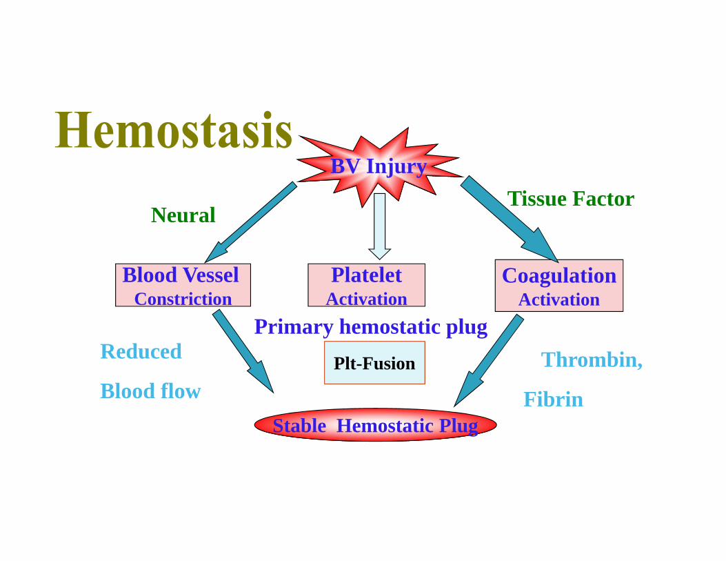
BV Injury
PlateletActivation
Plt-Fusion
Blood VesselConstriction
CoagulationActivation
Stable Hemostatic Plug
Thrombin,
Fibrin
Reduced
Blood flow
Tissue Factor
Primary hemostatic plug
Neural

Overview of Hemostasis:Clot Formation & VesselRepair

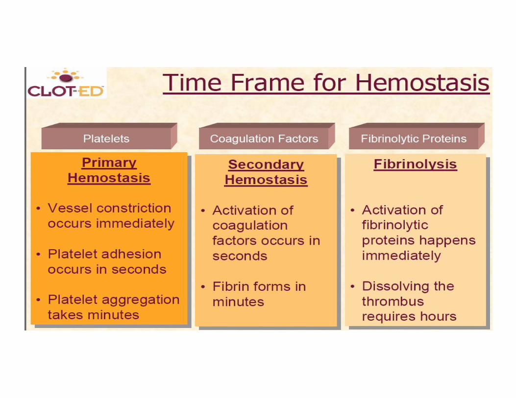
Hemostasis A series of reactions for stoppage of
bleeding During hemostasis, three phases occur
in rapid sequence Vascular spasms – immediate
vasoconstriction in response to injury Platelet plug formation Coagulation (blood clotting)
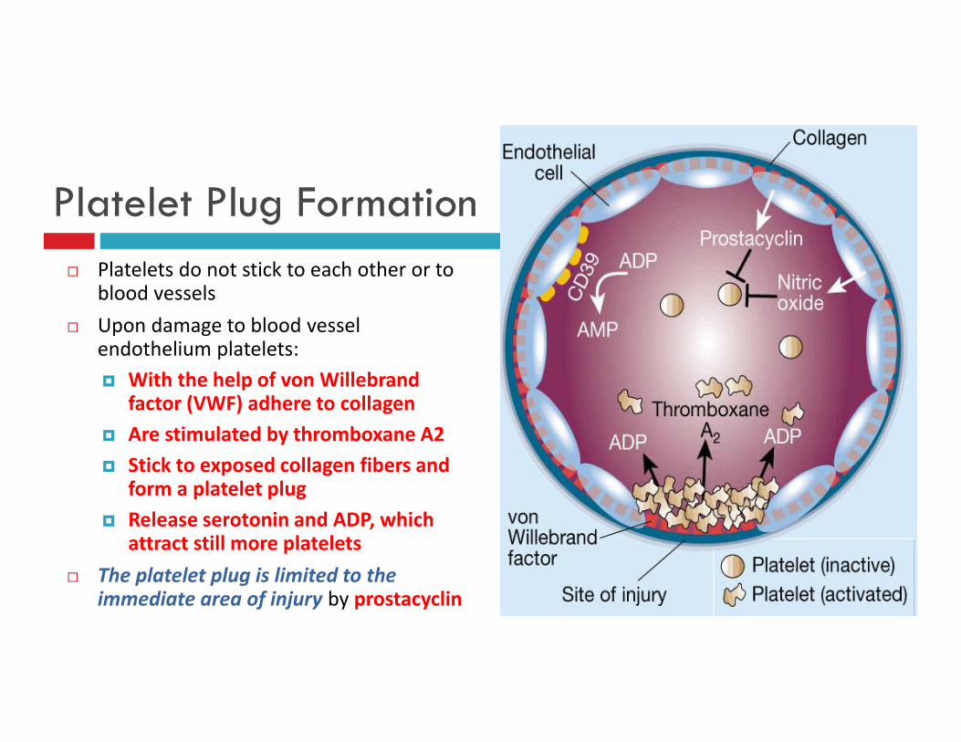
Platelet Plug Formation Platelets do not stick to each other or to
blood vessels Upon damage to blood vessel
endothelium platelets: With the help of von Willebrand
factor (VWF) adhere to collagen Are stimulated by thromboxane A2 Stick to exposed collagen fibers and
form a platelet plug Release serotonin and ADP, which
attract still more platelets The platelet plug is limited to the
immediate area of injury by prostacyclin

https://www.youtube.com/watch?v=HFNWGCx_Eu4

Platelets release
PF3
tissue factor other clotting factors
Prothrombin activator is formed,
Activator transforms prothrombin
Prothrombin becomes thrombin
catalyzes fibrinogen activates factor XIII
fibrinogen becomes fibrin
fibrin stabilizing factor
Fibrin Mesh Forms
Clot Forms

Antithrombotic Properties of the Endothelium
• Anti-platelet properties
– Covers highly thrombogenic basement membrane– Uninjured endothelium does not bind platelets– PGI2 (prostacyclin) and NO from uninjured endothelium inhibit platelet
binding– ADPase counters the platelet aggregating effects of ADP

Antithrombotic Properties of the EndotheliumAnticoagulant properties
*HEPARIN-LIKE MOLECULES: activate anti-thrombin III (inactivates activeproteases)
*THROMBOMODULIN: changes specificity of thrombin (activates protein C, which inactivates factors Va and VIIIa
*Endothelial cells produce tPA which activates fibrinolysis via plasminogento plasmin

Prothrombotic Properties of the Endothelium
•Synthesis of von Willebrand factor
•Release of tissue factor
•Production of plasminogen activator inhibitors (PAI)
•Membrane phospholipids bind and facilitate activation of clotting factors via Ca bridges

Hemostasis
Copyright © The McGraw-Hill Companies, Inc. Permission required for reproduction or display.
1 2 3 Coagulation phaseCoagulation cascade converts inactiveproteins to active forms and forms ablood clot.
Platelet plug formationPlatelets arrive at site of injury and stickto exposed collagen fibers.
Vascular spasmBlood vessel constricts to limit blood escape.
Erythrocytein clot
Fibrinstrandsin clotBlood
clot
VasoconstrictionPlatelets
Collagenfibers
Vesselinjury
Erythrocyte
Endothelialcells
Plateletplug


A set of reactions in which blood is transformed from a liquid to a gel Coagulation follows intrinsic and extrinsic pathways
The final three steps of this series of reactions are: Prothrombin activator is formed Prothrombin is converted into thrombin Thrombin catalyzes the joining of fibrinogen into a fibrin mesh
Coagulation

Coagulation
Figure 17.13a

Fibrinogen Fibrin

Fibrinogen FibrinThrombin

Fibrinogen FibrinThrombin
Prothrombin
XaVa

Fibrinogen FibrinThrombin
Prothrombin
XaVa
VIIa
TF
Extrinsic Pathway

Fibrinogen FibrinThrombin
Prothrombin
XaVa
VIIa
TF
Extrinsic Pathway
IXaVIIIa
XIa
XIIa
Intrinsic pathway

Fibrinogen FibrinThrombin
Prothrombin
XaVa
VIIa
TF
Extrinsic Pathway
IXaVIIIa
XIa
XIIa
Intrinsic pathway
XIIIa
Soft clot
FibrinHard clot

Fibrinogen FibrinThrombin
Prothrombin
XaVa
VIIa
TF
Extrinsic Pathway
IXaVIIIa
XIa
XIIa
Intrinsic pathway
XIIIa
Soft clot
FibrinHard clot
VVIII


The prothrombin time (PT) and its derived measures of prothrombin ratio (PR)and international normalized ratio (INR) are measures of the extrinsic pathwayof coagulation.
They are used to determine the clotting tendency of blood, in the measure of warfarin dosage, liver damage, vitamin K status.
The reference range for prothrombin time is usually around 12–15 seconds; the normal range for the INR is 0.8–1.2. PT measures factors I, II, V, VII, and X. It is used in conjunction with the activated partial thromboplastin time (aPTT)
which measures the intrinsic pathway.
Coagulation


The partial thromboplastin time (PTT) or activated partial thromboplastin time(aPTT or APTT) is a performance indicator measuring the efficacy of both the "intrinsic" (now referred to as
the contact activation pathway) and the common coagulation pathways.
monitor the treatment effects with heparin, a majoranticoagulant

Kaolin cephalin clotting time (KccT) is a historic name for the activated partialthromboplastin time


•Prolonged APTT may indicate:1. use of heparin (or contamination of the sample)2. antiphospholipid antibody (especially lupus
anticoagulant, which paradoxically increasespropensity to thrombosis)
3. coagulation factor deficiency (e.g. hemophilia)


Hemostasis: Vasoconstriction & PlugFormation
Figure 16-12: Platelet plug formation

Coagulation Phase 1:Two Pathways to Prothrombin Activator
May be initiated by either the intrinsic or extrinsic pathway
Triggered by tissue-damaging events
Involves a series of procoagulants
Each pathway cascades toward factor X
Once factor X has been activated, it complexes with calcium ions, PF3,and factor V to form prothrombin activator
Coagulation Phase 2:Pathway to Thrombin
Prothrombin activator catalyzes the transformationof prothrombin to the active enzyme thrombin

Coagulation Phase 3: Common Pathwaysto the Fibrin Mesh
Thrombin catalyzes the polymerization of fibrinogen into fibrin Insoluble fibrin strands form the structural basis of a clot Fibrin causes plasma to become a gel-like trap Fibrin in the presence of calcium ions activates factor XIII that:
Cross-links fibrin Strengthens and stabilizes the clot

Clot Retraction and Repair
Clot retraction – stabilization of the clot by squeezing serum from the fibrin strands Repair
Platelet-derived growth factor (PDGF) stimulates rebuilding of blood vessel wall Fibroblasts form a connective tissue patch Stimulated by vascular endothelial growth factor (VEGF), endothelial cells
multiply and restore the endothelial lining

Factors Limiting Clot Growth or Formation
Two homeostatic mechanisms prevent clots frombecoming large Swift removal of clotting factors Inhibition of activated clotting factors

Inhibition of Clotting Factors
Fibrin acts as an anticoagulant by binding thrombinand preventing its: Positive feedback effects of coagulation Ability to speed up the production of prothrombin
activator via factor V Acceleration of the intrinsic pathway by activating
platelets

Inhibition of Clotting Factors
Thrombin not absorbed to fibrin is inactivated byantithrombin III
Heparin, another anticoagulant, also inhibitsthrombin activity

Unnecessary clotting is prevented by endotheliallining the blood vessels
Platelet adhesion is prevented by: The smooth endothelial lining of blood vessels Heparin and PGI2 secreted by endothelial cells Vitamin E quinone, a potent anticoagulant
Factors Preventing Undesirable Clotting

Hemostasis: Coagulation & Clot Stabilization
Figure 16-13: The coagulation cascade
Prothrombin Ca++ Fibrinogen Fibrin Polymerization

Dissolving the Clot and Anticoagulants
Figure 16-14: Coagulation and fibrinolysis

Tests for Primary HemostasisBleeding Time
– Assesses all components of Virchow’s triad– in vivo test – performed directly on patient– Has fallen into disrepute and replaced by instruments that perform “invitro” bleeding times
Platelet Aggregation studies– Measure ability of platelets to aggregate, in vitro, when subjected to various stimulators (agonists)– Predominantly assesses function of platelet glycoprotein IIb/IIIa receptor
Von Willebrand Factor (VWF) assays– Measure amount and function of VWF, a protein that works with platelets so that they adhere to
site of injury– Assesses function of VWF ligand in its interaction with platelet glycoprotein Ib receptor

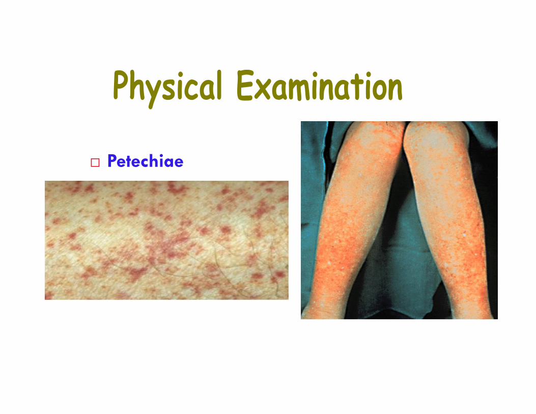
• ExaminationPlatelet DiseaseMucosal/cutaneous bleedingLack vessel protection by submucosal tissueBleed immediately after vascular trauma
Petechiae From small capillary In areas of increased venous pressure (dependent parts
of the body) Asymptomatic and not palpable D/D small telangiectasias
(Angiomas, Vasculitic purpura, Wiskott-AldrichSyndrome, Leukaemia, Vit K deficiency
Purpura Characteristically purple in colour Small, multiple, and superficial in location Develop without noticeable trauma / not spread into
deeper tissues Seen in – (Acute / Chronic leukaemia, Vitamin K
deficiency)

ExaminationCoagulation Disorders
Ecchymoses Large palpable ecchymoses
Spreading into deep tissue -haematomas – Hemarthrosis-severe coagulation disorder-haemophilia
Coagulation disorder bleedingonset may be delayed aftersurgery

Role of PT, PTT:Warfarin, Heparin Monitoring
Anticoagulant PT PTT
Low dose Heparin Normal Prolonged
High dose heparin Prolonged Prolonged
Low dose warfarin Prolonged Normal
High dose warfarin Prolonged Prolonged
Petechiae

Ecchymoses
Hemarthrosis

52 year old male
Severe Hemophilia
Now bleeds 3x month
Severe muscle wasting
Joint immobility
Atrophic skin changes
HIV and HCV +ve

Play key roles in the regulation of three physiological processes:
•Blood coagulation: (prothrombin (factor II), factors VII, IX, X, protein C, protein S, andprotein Z).
•Bone metabolism: osteocalcin, also called bone Gla-protein (BGP), and matrix gla protein(MGP).
•Vascular biology.
Like other liposoluble vitamins (A, D, E), vitamin K is stored in the fat tissue of the humanbody.
Role of vitamin K

Blood group (refer to lab)

Population Distribution of Major Blood Groups
O Rh pos 38%O Rh neg 7%A Rh pos 34%A Rh neg 6%B Rh pos 9%B Rh neg 2%AB Rh pos 3%AB Rh neg 1%

RBC membranes haveglycoprotein antigens on theirexternal surfaces
These antigens are: Unique to the individual Recognized as foreign if
transfused into anotherindividual
Promoters of agglutinationand are referred to asagglutinogens
Presence or absence of theseantigens is used to classifyblood groups
Human Blood Groups Humans have 30 varieties of naturally occurring RBC
antigens The antigens of the ABO and Rh blood groups cause
vigorous transfusion reactions when they areimproperly transfused
Other blood groups (M, N, Dufy, Kell, and Lewis) aremainly used for legalities
The ABO blood groups consists of: Two antigens (A and B) on the surface of the RBCs Two antibodies in the plasma (anti-A and anti-B)
ABO blood groups may have various types of antigens andpreformed antibodies
Agglutinogens and their corresponding antibodies cannot bemixed without serious hemolytic reactions

62
AGGLUTINOGENS Also called antigens. These agglutinogens are present on the
outer surface of the Erythrocytemembranes.
They are antigenic and have epitopes orantigenic determinants, which areglycoproteins.
In ABO groups, three types ofagglutinogens can be present.
• Some individuals will have Erythrocyteswith an agglutinogen called as “A”.
• Others have one called “B”
• The third type of agglutinogen is nonantigenic and it is called “H”
• H doesn’t cause production of antibodies.
• So those having H antigen are called Ogroup individuals.

A medicalproblem -some bloodtransfusionsproduce lethalclumping ofcells.
Don’t worryabout detailsyet...
Multiple alleles
ABO blood group s
Type A bloodtransfused intoType B person
Type B bloodtransfused intoType B person -OK

ABO BLOOD GROUPSBlood Group Antigens on
RBCs Antibodies in Serum Genotypes
A A Anti-B AA or AO
B B Anti-A BB or BO
AB A and B Neither AB
O Neither Anti-A and Anti-B OO
65
A AND B, INDIVIDUALS Those having the A agglutinogen on
their erythrocytes are called A bloodgroup people.
Those having the B agglutinogen arecalled the B blood group people.
•Some have both the A and B agglutinogenson their erythrocytes and they are called ABtype.
•Others have neither A nor B agglutinogens.They have the non antigenic H on their RBCsand are called O group people.

66
AGGLUTININS The antibodies to the agglutinogens are
called Agglutinins. These are present naturally in ABO
groups. They are always present in the plasma of
the individual. There are two types of agglutinins in the
ABO blood system: Anti A or α: Alpha Anti B or β: Beta
•The A group people have the Beta or anti Bagglutinin in their plasma.
•Similarly the B group people have the Alphaor Anti-A agglutinin in their plasma.
•The AB group of people have no agglutininsin their plasma.
•The O group people have both Alpha andBeta types of agglutinins in their plasma


Table 17.4
NOMAD:2006: BP:BldgpsI
69
HEMAGGLUTINATIONAgglutination or clumping is seen whenever the respective agglutinogens and
agglutinins are mixed.
Agglutinogen A + Agglutinin Alpha = Agglutination.
Agglutinogen B + Agglutinin Beta = Agglutination.
Both agglutinogens + Both antisera = Agglutination.
No agglutinogens = No agglutination.

HEMOGGLUTINATION WHICH CANLEAD TO HEMOLYSIS


The observation of red blood cell agglutination(also referred to as autoagglutination) must bedistinguished from rouleaux formation which isa physiological phenomenon. The presence ofantibodies (usually IgM) on the surface of redblood cells is responsible for the phenomenonof autoagglutination.
Agglutination can be observed during immune-mediated hemolytic anemia, but also during'cryoglobulinemia' ( a far more rare condition).
Agglutinating red blood cells resemble grapelikeclusters whereas red blood cells in rouleauxformation resemble a stack of coins.
In order to clearly distinguish erythrocyteagglutination from rouleaux formation, asimple saline test can be performed.


75Rh TYPING: INTRODUCTION It is the second most
important typing of blood. These blood groups were
originally discovered inRhesus monkeys
Rh is another type ofagglutinogen.
It is also present on theouter surface of theerythrocytes.
There are eight different Rh agglutinogens, three ofwhich (C, D, and E) are common
Presence of the Rh agglutinogens on RBCs is indicated asRh+
Anti-Rh antibodies are not spontaneously formed in Rh–
individuals However, if an Rh– individual receives Rh+ blood, anti-Rh
antibodies form A second exposure to Rh+ blood will result in a typical
transfusion reaction

76
ABO System & Pregnancy Majorities of hemolytic diseases are due to ABO incompatibility Foetus inherits one gene from each parent.
O + O = O, O + A= O or A, O + B= O or B, O + AB= A or B.
There is a 20% chance of ABO incompatibility of mother & foetus Only 5% chance of developing hemolytic disease only in type A & B infants of type
O mothers, that too only of milder forms

Rhesus47 Antigens make up the
Rhesus Blood GroupThe most significant is the
D antigen

78
Rh or D Agglutinins Anti-D agglutinins or
antibodies do not occurnaturally.
They are produced by theImmune systems as andwhen it is exposed to theD antigens.
So these Anti Dagglutinins are found onlyin some of the RhNegative people.
Those who have beenexposed to the Rh or Dantigen

Copyright © The McGraw-Hill Companies, Inc. Permission required for reproduction or display.
Type B recipient erythrocyte
Anti-A antibody in recipientplasma
Type A donor erythrocyte
Agglutinated erythrocytesfrom type A donor blocksmall vessels.
(a) Erythrocyte agglutination
Type Brecipient
Blood fromtype A donor

80
Exposure to Antigens: How?
The Rh+ve people will never manufacture Anti D antibodies. Only Rh – ve individuals can develop these Agglutinins.
When these Rh-ve people receive Rh+ve blood by mistake, they get exposed to theantigen.
Then they will develop the antibody.

The disease, called erythroblastosis fetalis or hemolytic disease of the newborn, may be sosevere as to kill the fetus or even the newborn infant. It is an example of an antibody-mediatedcytotoxicity disorder.

Pathogenesis Of Rh Iso-immunisation
Rh Negative Women Man Rh positive (Homo/Hetero)
Fetus Rh Neg FetusNo problem
Rh positive Fetus
Rh+ve R.B.C.s enterMaternal circulation
Mother previously sensitizedSecondary immune response
? Iso-antibody (IgG)
Non sensitized Mother Primaryimmune response
Fetus unaffected, 1st Babyusually escapes. Mother getssensitised?
Fetus
Hemolysis
?

83
ERYTHROBLASTOSIS FETALIS The second child in such a woman, if also Rh+ve, can
develop a disease called as Erythroblastosis fetalis. This is due to the Anti D antibodies developed in the
mother. These antibodies traverse through the placenta,
enter the fetal circulation and cause agglutination ofthe erythrocytes of the fetus.


BIRTH OF AN AFFECTED INFANT - Wide spectrum of presentations. Rapid deterioration ofthe infant after birth.May contiune for few days to few months. Chance of delayed anaemia at6-8 weeks probably due to persistance of anti Rh antibodies.
Pathology Of Iso-immunisation
HEMOLYSIS IN UTEROAFTER BIRTH
BILLIRUBIN
ANEMIA
MAT. LIV NO
EFFECT
HEPATICERYTHROPOESIS &
DYSFUNCTION
PORTAL & UMBILICAL VEIN HYPERTNSION,HEART FAILURE
Jaundice
KernicterusHepatic Failure
DEATH
ERYTHROBLASTOSISFETALIS
IUD

Hemolytic Disease of the Newborn
The drug RhoGAM can prevent the Rh– mother frombecoming sensitized
Treatment of hemolytic disease of the newborn involvespre-birth transfusions and exchange transfusions afterbirth

Transfusion reactions occur when mismatched blood is infused
Donor’s cells are attacked by the recipient’s plasma agglutinins causing:
Diminished oxygen-carrying capacity
Clumped cells that impede blood flow
Ruptured RBCs that release free hemoglobin into the bloodstream
Transfusion Reactions
Circulating hemoglobin precipitates in the kidneysand causes renal failure

Blood type beingtested RBC agglutinogens Serum Reaction
Anti-A Anti-B
AB A and B + +
B B – +
A A + –
O None – –
Blood Typing

Plasma Volume Expanders
When shock is imminent from low blood volume,volume must be replaced
Plasma or plasma expanders can be administered

Aging changes in the blood
The properties of blood change as we grow older. It is thought that these changes might contribute to the increased incident of clot
formation and atherosclerosis in older people. Some of the most prominent findings on these changes include:
Rise in fibrinogen Rise in blood viscosity Rise in plasma viscosity Increased red blood cell rigidity Increased formation of fibrin degradation products Earlier activation of the coagulation system