dann lab presentation
TRANSCRIPT

Dann Lab Presentation -Kayla
Thursday, April 15th, 2010

• How does manipulation of this arrangement
effect “stemness” or differentiation, OCT4, GFP, myc levels?

• Going from vector to cell line (SOHLH1)
• GFP analysis of cell lines (including TEX17)
• Immunostaining showing sub-cellular localization in COS cells
• Immunostaining analysis of cell lines: myc, and OCT4

Creating a cell line from a vector
• Plasmid backbone + insert (desired DNA sequence)
-Digest, CIP, ligate
-Gel electrophoresis for expected size purification, extract
• Bacterial transformation-Tina’s Homemade Fusion-Blue Competent Cells
*competent cells can take up the plasmid DNA
*bacterial cells in Ca2+ (permeable) mixed with plasmid DNA on
ice, heat shocked, plated (Amp)
• Mini preps for multiple colonies
• Restriction digest, gel electrophoresis
• Transfection into COS or 293
• Analyze by immunostaining or FACS

Creating pLSOGFS (SOHLH1-GFP)
• SOHLH1 - spermatogenesis and oogenesis specific basic helix-loop-helix 1 – Found in both male and female germ cells
– Essential for differentiation
• Replace Ubc promoter in Ubc-GFP– Increase differentiation activity
• SOHLH1 insert Ligation pLLU2G vector– Both cut w/ Xba1 and Age1
• Transformation

Nhe1 Age1/Xba1
Control vector – pLLU2G #3
Control vector – pLLU2G #3
8 kb
4 kb
.5 kb
1 12 23 34 45 5
Expected Age1/Xba1 band @ 4.2 kb
SOHLH1 digests following mini preps of colonies 1-5
• #1, 2, and 5 were sequenced• BLAST-ed sequencing results

• Test transfection for FACS- Increased SOHLH1 = decreased GFP
• According to FACS analysis and results of gel pLSOGFS #1 chosen for subsequent transformation and maxi prep
• Ex. of later use: test if Oct4 overexpression inhibits SOHLH1 expression (and therefore differentiation) by binding to the SOHLH1 promoter and repressing its’ transcription
Analyzing GFP reporter - FACS

GFP Analysis of Cell Lines
• 2 methods I use:– FACS (flow cytrometry)
– Immunostaining

GFP Analysis of Cell Lines
• SOGFS (SOHLH1)– Described earlier
FACS

GFP Analysis of Cell Lines• TEX17 – from ES cell mmlv,
ligated to pCDNA Not1/Xho1 (added myc), cut at Age1, ligated to pLUTG Age/CIP
Immunostaining : anti-myc + mouse IgG
pLLU2G – positive control
pLUTG-Tex17 pLUTG-WTOct #3
pCDNA-Tex17 negative control displayed no visible
GFP
Transient transfection in COS7
FACSLentivirus transduction in
DGC1
95% positive
GFP Analysis of Cell Lines
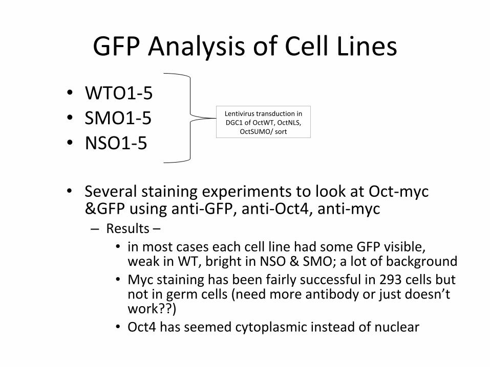
• WTO1-5• SMO1-5• NSO1-5
• Several staining experiments to look at Oct-myc &GFP using anti-GFP, anti-Oct4, anti-myc– Results –
• in most cases each cell line had some GFP visible, weak in WT, bright in NSO & SMO; a lot of background
• Myc staining has been fairly successful in 293 cells but not in germ cells (need more antibody or just doesn’t work??)
• Oct4 has seemed cytoplasmic instead of nuclear
Lentivirus transduction in DGC1 of OctWT, OctNLS,
OctSUMO/ sort
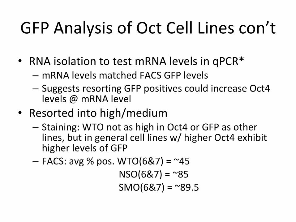
GFP Analysis of Oct Cell Lines con’t
• RNA isolation to test mRNA levels in qPCR*– mRNA levels matched FACS GFP levels – Suggests resorting GFP positives could increase Oct4
levels @ mRNA level• Resorted into high/medium– Staining: WTO not as high in Oct4 or GFP as other
lines, but in general cell lines w/ higher Oct4 exhibit higher levels of GFP
– FACS: avg % pos. WTO(6&7) = ~45 NSO(6&7) = ~85
SMO(6&7) = ~89.5
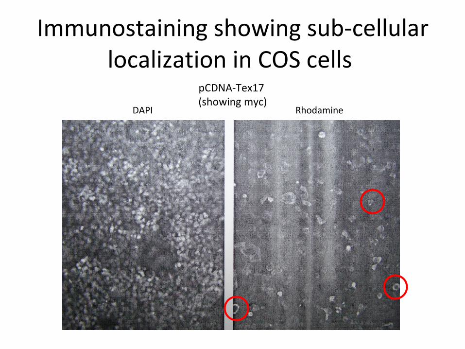
Immunostaining showing sub-cellular localization in COS cells
pCDNA-Tex17 (showing myc)
DAPI Rhodamine