data on morphometric analysis of anterior teeth from
TRANSCRIPT
ISSN 0973-2063 (online) 0973-8894 (print)
Bioinformation 17(1): 60-66 (2021)
©Biomedical Informatics (2021)
60
www.bioinformation.net
Volume 17(1) Research Article
Data on morphometric analysis of anterior teeth from Hazaribag College of Dental Sciences and Hospital, Jharkhand, India
Ankur Bhargava1, Sonal Saigal2,*, Pragya Thakur3, Uddipan Kumar4, Shreedevi Bhoi5 & Shandar Siddiqui6
1Department of Oral Pathology & Microbiology, Hazaribag College of Dental Sciences & Hospital, Hazaribag; 2Department of Oral Pathology, Microbiology and Forensic Odontology, Dental Institute, Rajendra Institute of Medical Sciences, Ranchi; 3Department of Conservative Dentistry & Endodontics, Awadh Dental College & Hospital, Jamshedpur; 4Smile World Dental Hospital, Hazaribag; 5Department of Oral & Maxillofacial Surgery, Hazaribag College of Dental Sciences & Hospital, Hazaribag; 6Department of Pedodontics and Preventive Dentistry, Clinic - Patna Health Care, Patna. *Corresponding author, Dr. Sonal Saigal - Email: [email protected] Received December 5, 2020; Revised December 31, 2020; Accepted January 2, 2021, Published January 31, 2021
DOI: 10.6026/97320630017060 Declaration on Publication Ethics: The author’s state that they adhere with COPE guidelines on publishing ethics as described elsewhere at https://publicationethics.org/. The authors also undertake that they are not associated with any other third party (governmental or non-governmental agencies) linking with any form of unethical issues connecting to this publication. The authors also declare that they are not withholding any information that is misleading to the publisher in regard to this article. Author responsibility: The authors are responsible for the content of this article. The editorial and the publisher have taken reasonable steps to check the content of the article in accordance to publishing ethics with adequate peer reviews deposited at PUBLONS. Declaration on official E-mail: The corresponding author declares that official e-mail from their institution is not available for all authors Abstract: It is of interest to document data on morphometric (measurement of external form) analysis of maxillary and mandibular anterior teeth collected from a dental set up using mesio-distal (MD) dimension. The mesiodistal dimensions of all permanent anterior teeth (central incisor, lateral incisor and canine) of 25 males and 25 females patients were recorded using digital vernier calipers. Data were charted and statistical analysis was done using Mann Whitney U test. Data shows sexual dimorphism for every tooth between males and females. However, dimorphism was exhibited only in maxillary and mandibular canine, mandibular central incisors, and lateral incisor. Hence, odontometric parameters offer simple, reliable and cost-effective in forensic investigation for recording gender discrimination. Keywords: Dimorphism; odontometric; forensic

ISSN 0973-2063 (online) 0973-8894 (print)
Bioinformation 17(1): 60-66 (2021)
©Biomedical Informatics (2021)
61
Background: Seventy percent of the identifications in the event of mass disasters have been confirmed by forensic dentistry [1]. The characteristics of the teeth can remain unchanged even after exposure to extreme environmental conditions, making the tooth an excellent forensic investigative tool [2]. Gender dimorphism, refers to those differences in size, stature and appearance between male and female that can be applied to dental identification because no two mouths are alike [3]. Teeth are readily accessible for examination and since no two teeth have similar morphology, they form an excellent forensic tool for gender determination [4]. The variations in tooth form are a common occurrence and these variations have an ethnic, forensic and anthropological significance [5]. The anterior teeth are esthetically important as they are readily seen during eating, speech, mastication and facial gesticulation. Its size, shape, color and position add to determine and create a definite coherence and order in the arrangement of natural anterior teeth [6]. Gender determination is completed using osteometry, DNA analysis and odontometric parameters. Accurate result is obtained using the DNA analysis. However, it is expensive and not readily available at all locations. It is difficult for DNA requiring qualified trained staff [7]. On the other hand, osteometry is a favoured procedure because it is more effective in determining gender in forensic investigations. However, bodies that are badly mutilated consisting of fragmentary remains of a skeleton are often not trivial for investigation [8]. Odontometric parameters such as mesio-distal and vestibulo-lingual diameters of some permanent teeth show statistically significant differences between men and women [9]. Therefore, It is of interest to document data on morphometric (measurement of external form) analysis of maxillary and mandibular anterior teeth collected from a dental set up using mesio-distal (MD) dimension. Material and Method: Dataset nature: This is a retrospective, cross‑sectional, descriptive study conducted using 50 dental stone models to measure the greatest mesio-distal width of upper and lower anteriors of undergraduate students at a dental college in Hazaribag, India. Consent: Informed consents were obtained from the subjects who included twenty-five male and twenty-five female dental. Parameter estimation: A digital Vernier caliper was used to measure the greatest mesio-distal dimension of the crown of each of the [twelve] teeth investigated, namely the left and right maxillary and mandibular
central incisors, lateral incisors and canine. The mesio-distal [MD] dimension has been defined as the greatest distance between the contact points on the proximal surfaces of the dental crown. Table 1: Statistical analysis of mesiodistal dimensions in the central incisor series
Position Gender N Mean SD Result Male 25 8.96 0.55 Right Maxillary Central Incisor Female 25 8.77 0.55 NS Male 25 8.96 0.54 Left Maxillary Central Incisor Female 25 8.8 0.54 NS Male 25 5.54 0.19 Right Mandibular Central Incisor Female 25 5.18 0.4 *** Male 25 5.54 0.19 Left Mandibular Central Incisor Female 25 5.13 0.22 ***
* = p<0.05; ** = p <0.01; *** = p <0.001; NS = p > 0.05
Inclusion criteria: The study included subjects in age range of 19-23 years with fully erupted teeth, periodontally healthy, non-carious teeth. Exclusion criteria: Subjects with physiological or pathological wearing of teeth (attrition, abrasion, erosion), misaligned teeth (crowding, rotation or malocclusion, spacing), partially erupted teeth, any history of restoration, orthodontic treatment or trauma were excluded from the study sample. Table 2: Statistical analysis of mesiodistal dimensions in the lateral incisor series.
Position Gender N Mean SD Result Male 25 6.77 1.45 Right Maxillary Lateral Incisor Female 25 6.87 0.51 NS Male 25 7.04 0.37 Left Maxillary Lateral Incisor Female 25 6.84 0.46 NS Male 25 6.11 0.22 Right Mandibular Lateral Incisor Female 25 5.62 0.17 *** Male 25 6.15 0.33 Left Mandibular Lateral Incisor Female 25 5.62 0.17 ***
* = p<0.05; ** = p <0.01; *** = p <0.001; NS = p > 0.05
Table 3: Statistical analysis of mesiodistal dimensions in the canine series.
Position Gender N Mean SD Result Male 25 7.98 1.71 Right Maxillary Canine Female 25 7.93 0.33
**
Male 25 8.29 0.43 Left Maxillary Canine Female 25 7.94 0.33 **
Male 25 7.46 0.41 Right Mandibular Canine Female 25 7.13 0.43 **
Male 25 7.46 0.38 Left Mandibular Canine Female 25 6.84 1.50 **
* = p<0.05; ** = p <0.01; *** = p <0.001; NS = p > 0.05
ISSN 0973-2063 (online) 0973-8894 (print)
Bioinformation 17(1): 60-66 (2021)
©Biomedical Informatics (2021)
62
Data collection: The width of 25 samples of each type of tooth per gender was measured. In each case, the teeth 11, 12, 13, 21, 22, 23, 31, 32, 33, 41, 42 and 43 were measured (FDI tooth notation). A single examiner to eliminate inter observer error was maintained. All measured dimensions of maxillary and mandibular anteriors are presented as mean and standard deviation (SD). Mann Whitney U test was used for satatistcal analysis.
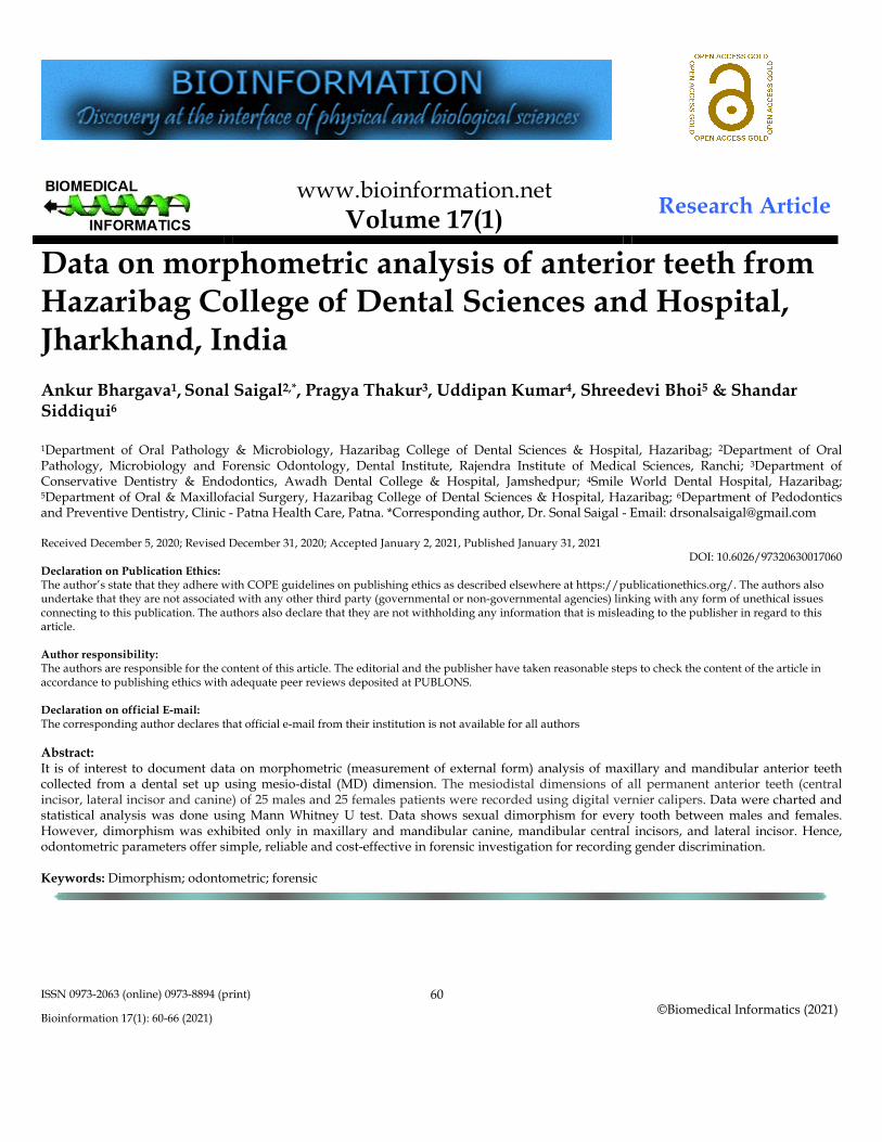
Figure 1: Mean value of mesiodistal dimensions of maxillary and mandibular anterior teeth in male and females. Results: The morphometric measurements taken from the representative teeth in the maxillary and mandibular series were analyzed statistically for their viability in the expression of values between genders. The mean value of MD dimension of the right maxillary central incisor at the level of contact area was 8.96mm and 8.77mm for male and female, respectively, which was statistically not significant (p>0.05). The left maxillary central incisor was also statistically not significant with the mean value of 8.96 mm and 8.80mm for male and female, respectively (Table 1). But the right and left mandibular central incisor for male and female were highly statistically significant (p<0.001). The mean value of right side was 5.54mm and 5.18mm and left side 5.54mm and 5.13mm for male and female respectively (Table 1, Figure 1). The mean value of the right and left maxillary lateral incisor at the level of contact area was statistically not significant (p>0.05). The mean value of right side was 6.77mm and 6.87mm and left side 7.04mm and 6.84mm for male and female respectively. But result were highly statistically
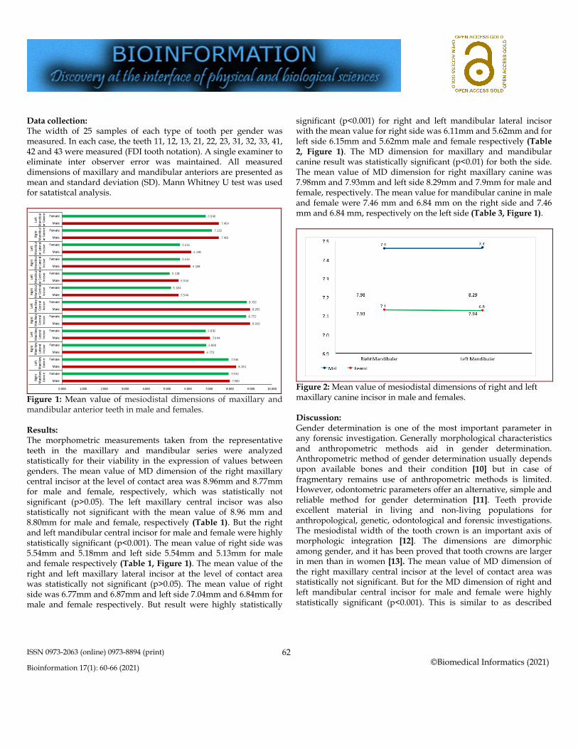
significant (p<0.001) for right and left mandibular lateral incisor with the mean value for right side was 6.11mm and 5.62mm and for left side 6.15mm and 5.62mm male and female respectively (Table 2, Figure 1). The MD dimension for maxillary and mandibular canine result was statistically significant (p<0.01) for both the side. The mean value of MD dimension for right maxillary canine was 7.98mm and 7.93mm and left side 8.29mm and 7.9mm for male and female, respectively. The mean value for mandibular canine in male and female were 7.46 mm and 6.84 mm on the right side and 7.46 mm and 6.84 mm, respectively on the left side (Table 3, Figure 1).
Figure 2: Mean value of mesiodistal dimensions of right and left maxillary canine incisor in male and females. Discussion: Gender determination is one of the most important parameter in any forensic investigation. Generally morphological characteristics and anthropometric methods aid in gender determination. Anthropometric method of gender determination usually depends upon available bones and their condition [10] but in case of fragmentary remains use of anthropometric methods is limited. However, odontometric parameters offer an alternative, simple and reliable method for gender determination [11]. Teeth provide excellent material in living and non-living populations for anthropological, genetic, odontological and forensic investigations. The mesiodistal width of the tooth crown is an important axis of morphologic integration [12]. The dimensions are dimorphic among gender, and it has been proved that tooth crowns are larger in men than in women [13]. The mean value of MD dimension of the right maxillary central incisor at the level of contact area was statistically not significant. But for the MD dimension of right and left mandibular central incisor for male and female were highly statistically significant (p<0.001). This is similar to as described

ISSN 0973-2063 (online) 0973-8894 (print)
Bioinformation 17(1): 60-66 (2021)
©Biomedical Informatics (2021)
63
elsewhere [14] where the mean width of the right maxillary central incisor for males was found to be 8.944mms and for females 8.613mms. The mean value of the left maxillary central incisor was found out to be 9.056mms for males and 8.664mm for females. Some other studies also show that their statistically significant result in the MD dimension of right and left mandibular central incisor for male and female [15,16]. Very few studies were conducted for MD dimension for lateral incisor for sex determination. In the study of Dash KC [17] right and left maxillary lateral incisor the result were statistically significant with the p value 0.0108 and 0.0009 respectively but for the right and left mandibular lateral incisor result were no significant with the p value 0.2400 and 0.2478. But in our study observation obtained for the maxillary right and left lateral incisor were statistically not significant but for mandibular right and left lateral incisor result statistically significant. This is similar to as described elsewhere [18] where mandibular lateral incisor shows statistically significant in comparison to maxillary lateral incisor in male and female for MD dimension. Recent studies present the canine as the most dimorphic tooth in human dentistry. Mandibular canines are considered reliable elements of human identification, as they are the last teeth to be extracted and are rarely affected by oral diseases and are more likely to survive severe trauma such as an air crash, hurricane or fire [19]. Lebanese subjects a statistically significant difference between men and women p≤ 0.001 in the mesio-distal diameter of the mandibular canine [20]. Similar results of canine dimorphism were also found in other studies. [21,22,23,24]. The MD dimension for maxillary and mandibular canine result was statistically significant (p<0.01) for both the side in male and female. The mean value was higher in male [right side 7.98 and left side 8.29mm] in comparison to female in maxillary teeth (right side 7.93 and left side 7.94mm). Similar results were found for mandibular canine where the mean value was higher in male (right side and left side) in comparison to female canine teeth (right side and left side). The mean value of MD dimension for the both arches of right and left canine of male and female was also not significant (Figures 2 & 3). Moreover, very few studies report a significant difference between the right and left side. Saudi population aged 13-20 years showed that the canines were the only teeth to show real dimorphism [25]. They also determined that there was no statistically significant difference between the left and right canines, suggesting that the measurement of teeth on one side could be truly representative when the corresponding measurement on the other side was not possible.
Figure 3: Mean value of mesiodistal dimensions of right and left mandibular canine incisor in male and females. Various theories have been given in the literature for this sexual dimorphism. According to Moss, it is because of the greater thickness of enamel in males due to the long period of amelogenesis as compared to females. However, in females the completion of calcification of the crown occurs earlier in both deciduous and permanent dentition as quoted by de Vito. [26] Gender chromosomes are also known to cause different effects on tooth size. The ‘Y’ chromosome influences the timing and rate of body development, thus producing slower male maturation, and acts additively and to a greater extent than the ‘X’ chromosome [26] ‘Y’ chromosome has a direct effect on tooth size which may be related to a more non-specific effect of hetrero chromatism or cellular activity [27]. The difference in size has been attributed to differently balanced hormonal production between the sexes consequent to the differentiation of either male or female gonads during the sixth or seventh week of embryogenesis rather than any direct effect of gender chromosome themselves [28]. Reason for this dimorphism could be a biologic variation, which is a characteristic of life and is attributed to family, genetics and environmental factors [29]. Variation in food resources exploited by different populations has also been explained as one such environmental cause [27] Previous studies indicate that MD dimensions are more accurate in determining sexual dimorphism [30,31]. These can be useful in archeological, odontologic, genetic, and forensic and crime investigations, as ethnicity/race, culture and environment is known to affect odontometrics. Data shows that gender dimorphism was observed for every tooth included in the study between males and

ISSN 0973-2063 (online) 0973-8894 (print)
Bioinformation 17(1): 60-66 (2021)
©Biomedical Informatics (2021)
64
females. Besides this, statistically significant dimorphism was exhibited only in maxillary canine, mandibular anteriors i.e. central incisors, lateral incisor and canines. Linear dimensions of the tooth act as an excellent parameter, which is a simple, affordable, and reliable method for gender determination from the dental remains. Using MD dimensions, the gender dimorphism becomes far better and accurate. Further studies can be done to procure extended data, which can be used by forensic experts as adjuncts to establish gender dimorphism in mass disasters. Conclusion: We document data on morphometric analysis of anterior teeth from a dental college in Hazaribag, India to help in forensic investigation. References:
[1] Lund H and Mornstad H J Forensic Odontotomatol 1999 17:30. [PMID: 10709560]
[2] Soumboundou S et al. J Crim Forensic Studies 2018 1:1. [Discontinued Journal: ISSN: 2640-6578; IC: 52203]
[3] Banerjee A et al. JFDS 2016 8:22. [PMID: 27051219] [4] Boaz K & Gupta C. J Forensic Dent Sci 2009 1:42. [5] Williams PL, et al. Gray’s Anatomy; The Teeth. 38th ed.
Churchill Livingstone: New York; 2000. [6] Renner RP. Anterior dental esthetics: An introduction to dental
anatomy and esthetics. 1st Ed. Quintessence Publishing Co; 1985.
[7] Iwamura E et al. Rev Hosp Clin Fac Med S Paulo 2004 59:383. [8] Bilge Y et al. Forensic Sci Int 2003 137:141. [PMID: 14609649] [9] Acharya AB & Mainali S. J Forensic Legal Med 2009 16:67.
[PMID: 19135000]
[10] Hasegawa I et al. Leg Med [Tokyo] 2009 11:260. [PMID: 19736033]
[11] Rao NG et al. Forensic Sci Int 1989 42:249. [PMID: 2792982] [12] Lombardi AV Am J Phys Anthropol 1975 42:99. [PMID:
1115230] [13] Santoro M et al. Angle Orthod 2000 70:303. [PMID:
10961780] [14] Kaushal S et al. JPAFMAT 2005 5:13. [15] Hattab FN et al. Arch Oral Biol 1996 41:641. [PMID:
9015564] [16] Nair P et al. Forensic Med Toxicol 1999 16:10. [17] Dash KC et al. J Int Soc Prevent Communit Dent 2018 8:174.
[PMID: 29780744] [18] Srinivasprasad M et al. Indian J Foren Med Toxicol 2016
10:172. [19] Costa YTF et al. Braz J Oral Sci 2011 11:406. [20] Ayoub F et al. Int J dent 2014 1:1. [PMID: 24672548] [21] Narang RS et al. Indian J Oral Sci 2014 5:16. [22] Iscana MY & Kedici PS. Forensic Sci Int 2003 137:160.
[PMID: 14609652] [23] Staka G & Bimbashi V. Int J Pharm Bio Sci 2013 4:927. [24] Lakhanpal M et al. JSM Dent 2013 1:10. [25] Hashim HA & Murshid ZA. Mesiodistal Tooth Width. Egypt
Dent J 1993 39:343. [PMID: 8299533] [26] Bunger E et al. Int J Dent Health Sci 2014 1:13. [27] Prathibha Rani RM et al. J Forensic Dent Sci 2009 1:88. [28] Metgud R et al. J Forensic Investigation. 2015 3:1. [29] Garn SM et al. J Dent Res 1967 46:963. [PMID: 5234039] [30] Acharya AB & Mainali S. J Forensic Odontostomatol 2008
26:53. [PMID: 22717790] [31] Narang RS et al. J Forensic Dent Sci 2015 7:54.
Edited by P Kangueane
Citation: Bhargava et al. Bioinformation 17(1): 60-66 (2021) License statement: This is an Open Access article which permits unrestricted use, distribution, and reproduction in any medium, provided
the original work is properly credited. This is distributed under the terms of the Creative Commons Attribution License

ISSN 0973-2063 (online) 0973-8894 (print)
Bioinformation 17(1): 60-66 (2021)
©Biomedical Informatics (2021)
65
Articles published in BIOINFORMATION are open for relevant post publication comments and criticisms, which will be published immediately linking to the original article for FREE of cost without open access charges. Comments should be concise, coherent and critical in less than 1000 words.

ISSN 0973-2063 (online) 0973-8894 (print)
Bioinformation 17(1): 60-66 (2021)
©Biomedical Informatics (2021)
66