dataset for histopathological reporting of tumours of the
TRANSCRIPT

CEff 090821 1 V3 Final
Dataset for histopathological reporting of tumours of the urinary collecting system (renal pelvis, ureter, urinary bladder and urethra)
August 2021
Authors: Dr Murali Varma, University Hospital of Wales, Cardiff
Dr Jonathan H Shanks, The Christie NHS Foundation Trust, Manchester Dr Ashish Chandra, Guys and St Thomas’ NHS Foundation Trust, London Dr Lorna McWilliam, Central Manchester University Hospitals NHS Foundation Trust
Unique document number G044 Document name Dataset for tumours of the urinary collecting system (renal pelvis, ureter, urinary
bladder and urethra) Version number 3 Produced by Each of the authors is a consultant histopathologist with a special interest in
urological pathology. Dr Murali Varma is a founder member and Chairman of the British Association of Urological Pathologists (BAUP), organises biannual uropathology courses on prostate and bladder pathology, and has published several original and review papers related to urological pathology. Dr Jonathan H Shanks regularly speaks at national and international meetings and has co-authored over 95 publications, mostly in the field of urological pathology. Dr Ashish Chandra is co-author of ACP Best Practice: Cut up and reporting of bladder cancer specimens and is on the NICE Clinical Guidelines Group for diagnosis and clinical management of bladder cancer. Dr Lorna McWilliam is a founder member of BAUP and speaks on bladder and prostate pathology at their biannual courses. She helped to develop national and regional guidelines for best practice in urological pathology.
Date active August 2021 (to be implemented within three months) Date for review August 2024 Comments This document will replace the 2nd edition published in 2013.
In accordance with the College’s pre-publications policy, this document was on the Royal College of Pathologists’ website for consultation from 27 May to 24 June 2021. Responses and authors’ comments are available to view, following final publication of this dataset. Dr Brian Rous Clinical Lead for Guideline Review
The Royal College of Pathologists 6 Alie Street, London E1 8QT Tel: 020 7451 6700 Fax: 020 7451 6701 Web: www.rcpath.org
Registered charity in England and Wales, no. 261035 © 2021, The Royal College of Pathologists
This work is copyright. You may download, display, print and reproduce this document for your personal, non-commercial use. Requests and inquiries concerning reproduction and rights should be addressed to the Royal College of Pathologists at the above address. First published: 2021.
CEff 090821 2 V3 Final
Contents Foreword……………. ......................................................................................................................... 3 1 Introduction ............................................................................................................................... 4 2 Clinical information required on the specimen request form .................................................... 5 3 Preparation of specimens before dissection ............................................................................. 6 4 Specimen handling and block selection ................................................................................... 9 5 Reporting recommendations .................................................................................................. 12 6 Reporting of small biopsy specimens ..................................................................................... 23 7 Reporting of frozen sections ................................................................................................... 24 8 Criteria for audit ...................................................................................................................... 24 9 References ............................................................................................................................. 25 Appendix A TNM classification of malignant tumours (8th edition 2016) ..................................... 32 Appendix B SNOMED codes ....................................................................................................... 37 Appendix C Histopathology reporting proforma: radical resections of renal pelvis and/or ureter ............................................................................................................ 39 Appendix D Histopathology reporting proforma: transurethral specimens (biopsy or TUR)……. 41 Appendix E Histopathology reporting proforma: urinary bladder
(cystectomy or diverticulectomy) .............................................................................. 43 Appendix F Histopathology reporting proforma: urethrectomy or urethral diverticulectomy ........ 46 Appendix G Histopathology reporting proforma: radical resections of renal pelvis
and/or ureter in list format ......................................................................................... 49 Appendix H Histopathology reporting proforma: transurethral specimens
(biopsy or TUR) in list format .................................................................................... 53 Appendix I Histopathology reporting proforma: urinary bladder (cystectomy or diverticulectomy) in list format .................................................................................. 57 Appendix J Histopathology reporting proforma: urethrectomy or urethral
diverticulectomy in list format ................................................................................... 62 Appendix K Summary table – Explanation of grades of evidence ............................................... 67 Appendix L AGREE II guideline monitoring sheet ....................................................................... 68
NICE has accredited the process used by the Royal College of Pathologists to produce its cancer datasets. Accreditation is valid for five years from 25 July 2017. More information on accreditation can be viewed at www.nice.org.uk/accreditation. For full details on our accreditation visit: www.nice.org.uk/accreditation
CEff 090821 3 V3 Final
Foreword The cancer datasets published by the Royal College of Pathologists (RCPath) are a combination of textual guidance, educational information and reporting proformas. The datasets enable pathologists to grade and stage cancers in an accurate, consistent manner in compliance with international standards and provide prognostic information, thereby allowing clinicians to provide a high standard of care for patients and appropriate management for specific clinical circumstances. This guideline has been developed to cover most common circumstances. However, we recognise that guidelines cannot anticipate every pathological specimen type and clinical scenario. Occasional variation from the practice recommended in this guideline may therefore be required to report a specimen in a way that maximises benefit to the patient. Each dataset contains core data items (see Appendices C–J) that are mandated for inclusion in the Cancer Outcomes and Services Dataset (COSD – previously the National Cancer Data Set) in England. Core data items are items that are supported by robust published evidence and are required for cancer staging, optimal patient management and prognosis. Core data items meet the requirements of professional standards (as defined by the Information Standards Board for Health and Social Care [ISB]) and it is recommended that at least 95% of reports on cancer resections should record a full set of core data items. Other non-core data items are described. These may be included to provide a comprehensive report or to meet local clinical or research requirements. All data items should be clearly defined to allow the unambiguous recording of data. The following stakeholders were contacted to consult on this document:
• British Association of Urological Surgeons (BAUS) Section of Oncology
• British Uro-oncology Group
• National Cancer Research Institute (NCRI) Bladder Cancer Clinical Studies Group
• British Association of Urological Pathologists (BAUP)
• UK and Ireland Association of Cancer Registries (UKIACR)
• National Cancer Intelligence Network (NCIN) Urology Clinical Reference Group.
Supporting evidence and recommendations in this dataset are based on:
• WHO classifications, 1973 and 2016
• National Institute for Health and Care Excellence (NICE) Improving Outcomes Guidance, 2002
• NICE guidance NG2
• TNM 8th edition staging classifications – the Union for International Cancer Control (UICC) and the American Joint Committee on Cancer (AJCC)
• International Collaboration on Cancer Reporting (ICCR) datasets for cancers of the urinary tract.1–7 The evidence has been evaluated according to the modified Scottish Intercollegiate Guidelines Network (SIGN) guidance and the level of evidence for the recommendations has been summarised according to College guidance (see Appendix K). Most of the supporting evidence is level C or D at least or meets the Good Practice Point (GPP) criteria. No major conflicts in the evidence have been identified and any minor discrepancies between evidence have been resolved by expert consensus. No major organisational changes have been identified that would hinder the implementation of the dataset and there are no new major financial or work implications arising from the implementation, compared with the 2013 dataset. A formal revision cycle for all cancer datasets takes place on a three-yearly basis. However, each year, the College will ask the author(s) of the dataset, in conjunction with the relevant subspecialty adviser to the College, to consider whether or not the dataset needs to be updated or revised. A full consultation process

CEff 090821 4 V3 Final
will be undertaken if major revisions are required, i.e. revisions to core data items (the only exception being changes to international tumour grading and staging schemes that have been approved by the Specialty Advisory Committee on Cellular Pathology and affiliated professional bodies; these changes will be implemented without further consultation). If minor revisions or changes to non-core data items are required, an abridged consultation process will be undertaken, whereby a short note of the proposed changes will be placed on the College website for two weeks for members’ attention. If members do not object to the changes, the changes will be incorporated into the dataset and the full revised version (incorporating the changes) will replace the existing version on the College website. The dataset has been reviewed by the Clinical Effectiveness team, Working Group on Cancer Services and Lay Advisory Group, and was placed on the College website for consultation with the membership from 27 May to 24 June 2021. All comments received from the Working Group and membership were addressed by the authors to the satisfaction of the Chair of the Working Group and the Clinical Lead for Guideline Review. This dataset was developed without external funding to the writing group. The College requires the authors of datasets to provide a list of potential conflicts of interest; these are monitored by the Clinical Effectiveness team and are available on request. Three authors of this document have declared that they have participated in a Roche Bladder Cancer Advisory Board Meeting and one participated in a Bladder Cancer Advisory Board Meeting organised by AstraZeneca. There are no other conflicts of interest. 1 Introduction
This document is the third edition of the dataset for tumours of the urinary collecting system and follows publication of the second edition in April 2013.8 Tumours of the urinary collecting system (renal pelvis, ureter, urinary bladder and urethra) are common. Most are reported by local teams that should include a uropathology lead who has a special interest in the field. Cystectomies are performed in larger centres where more than 50 radical procedures (to include cystectomies, cystoprostatectomies and radical prostatectomies) are performed per year according to recommendations in NICE’s Improving Outcomes Guidance published over 17 years ago.3 The recommended minimum is five such radical resections per surgeon. The most frequent tumour encountered in the urinary collecting system is urothelial carcinoma. The term ‘transitional cell carcinoma’ is not recommended, as this is less specific and could also apply to unrelated tumours arising from other sites such as the nasal sinuses. A peculiar feature of the classification of this tumour type is that, by longstanding convention, the term ‘carcinoma’ had also been applied to most non-invasive papillary lesions. At least half of urothelial carcinomas are non-invasive at presentation. This dataset applies only to malignant epithelial tumours (invasive or non-invasive) of the urinary collecting system. It is not intended to cover other tumour types, such as sarcoma or melanoma. However, note that for sarcomatoid tumours of the lower urinary tract the possibility of a sarcomatoid urothelial carcinoma (which is covered by this dataset) should be considered. In 1998, the International Society of Urological Pathology (ISUP) proposed a new classification, which was subsequently adopted in both the 2004 and 2016 WHO publications, although it has been controversial (see section 5 for further information on core data items).2,9,10 In the UK, the 1973 WHO classification remained in widespread use after 2004 and is currently recommended to be used in conjunction with the 2004/2016 WHO classification, as specified later in this document.
Urothelial carcinoma often displays divergent differentiation.3 The subtypes of bladder cancer that are now recognised, including variant forms of urothelial carcinoma, are listed in the 2016 WHO book (see Box 1 in section 5.3.1). Thus, urothelial carcinoma can show single or multiple divergent histological patterns. Pure squamous cell carcinoma, small cell carcinoma and primary adenocarcinoma also occur, but are uncommon.3 Spread/metastasis from elsewhere should be considered and excluded, especially for pure squamous cell carcinoma and adenocarcinoma.

CEff 090821 5 V3 Final
Two 8th editions of the TNM staging system were published separately by the AJCC and the UICC towards the end of 2016.5,6 Although there are some significant differences between the two versions, these were relatively minor in the chapters relating to tumours of the urinary tract and many of the differences were eliminated following publication of errata that are now incorporated in UICC TNM 8.11 In addition to incorporation of TNM 8th edition criteria, the 3rd edition of this dataset retains the separate reporting proformas for biopsy/transurethral resection (TUR) specimens and radical resections that were introduced in the last dataset. The 3rd edition of this dataset has also been updated in line with the recommendations in the ICCR datasets for cancers of the urinary tract.7 Referral pathways should be established for difficult cases and, as a minimum, the uropathology lead at each site reporting tumours of the urinary collecting system should participate in the national urological external quality assessment (EQA) scheme. Discussion of cases will be at the local or specialist multidisciplinary team (MDT) meetings, according to the type of case. The uropathology lead should be a member of such a team.
1.1 Target users and health benefits of this guideline
The target primary users of the dataset are trainee and consultant cellular pathologists and, on their behalf, the suppliers of IT products to laboratories. The secondary users are surgeons and oncologists, cancer registries and the National Cancer Intelligence Network. Standardised cancer reporting and MDT working reduce the risk of histological misdiagnosis or misinterpretation of histopathology reports and help to ensure that clinicians have all of the relevant pathological information required for tumour staging, management and prognosis. Collection of standardised cancer-specific data also provides information for healthcare providers and epidemiologists and facilitates international benchmarking and research.
1.2 Changes from previous version The significant changes from the previous version of the dataset are as follows:
• the dataset has been updated based on UICC TNM 8, WHO 2016 classification of tumours of the urinary tract and ICCR recommendations
• necrosis has been added as a non-core data item
• extent of invasion in TUR of bladder tumour (TURBT) specimens has been categorised as part of clinical rather than pathological stage (rare exceptions described in section 5.3.7)
• PDL-1 testing issue is addressed. Routine PDL-1 testing is not recommended. 2 Clinical information required on the specimen request form
In addition to demographic information about the patient and details of destination of the report, several items of clinical information can help the pathologist in the handling and reporting of specimens of the urinary collecting system. These should be available to the pathologist either on the specimen request form or by access to the electronic notes of the patient. For bladder biopsy/TUR specimens, the anatomical location(s) within the bladder should be given to help distinguish between the muscularis mucosae and muscularis propria (detrusor muscle), as there are regional variations in the morphology of the bladder wall.12
Awareness of cystoscopic appearances is critical in the assessment of biopsies and TURBT specimens. If the papillary lesion seen on cystoscopy is not identified in the initial levels of the biopsy/TURBT, examination of further levels is mandatory. In some borderline cases the morphology,

CEff 090821 6 V3 Final
while not diagnostic, would be consistent with origin from a small, low-grade papillary urothelial neoplasm if the biopsy was from a papillary lesion. It is essential to know the clinical/radiological appearances in cases with small endoscopic biopsies of the ureter or renal pelvis, as there is particular potential for misinterpretation of tiny, folded, fragmented pieces of mucosa, polypoid ureteritis or pyelitis, or other reactive changes, as tumour at these sites.13
Awareness of a urine cytology finding of high-grade urothelial neoplasia may indicate the need to examine further levels of a biopsy that shows only a low-grade urothelial neoplasm to exclude adjacent urothelial carcinoma in situ. Alternatively, this may prompt a search for a high-grade tumour elsewhere in the bladder. Results of staging investigations can be important, as there is little point in exhaustive examination of a TURBT specimen if the patient has distant metastasis or unequivocal locally advanced bladder cancer on radiological investigation. Patients with a history of urothelial neoplasia are at risk of developing urothelial tumours elsewhere in the urinary tract, so this information must be provided to the reporting pathologist. Knowledge of history of cancer arising from other sites such as the cervix, prostate and large bowel can also inform pathological interpretation, particularly in biopsy and TUR specimens. Details of current and previous therapy can aid morphological interpretation and inform the pathologist of the potential clinical implications of the report.14 For example, recurrent carcinoma in situ following intravesical Bacillus Calmette–Guérin (BCG) therapy may be an indication for radical cystectomy. Various epithelial alterations have been described following intravesical chemotherapy or occasionally as a result of non-therapeutic agents such as ketamine that can mimic neoplastic changes. Pseudocarcinomatous epithelial proliferation can occur following treatments such as radiotherapy or, occasionally, in the absence of therapy.14–18 Any history of recent procedures, stones, infections or obstruction should be given. In cystectomy specimens, it is useful for the pathologist to be aware of the rationale for the surgery in that patient. If cystectomy was performed to palliate pain, or for bleeding or urinary frequency, there is no need to exhaustively sample the specimen for residual cancer. On the other hand, if cystectomy was performed following a radiological impression of extravesical extension of the tumour, it is important to sample appropriate areas of the specimen to confirm or refute the radiological impression. It is important to be aware of the findings in a previous TURBT specimen when making an overall assessment of a cystectomy. This point is illustrated by cases where no invasive carcinoma is found at cystectomy, despite thorough sampling, even though muscularis propria (detrusor muscle) invasion was present in the prior TURBT specimen. If there is no history of neoadjuvant chemotherapy, UICC recommends that such a patient should be staged as pT2 following cystectomy (advice from UICC TNM help desk). An appropriate comment can be made in the report, ideally accompanied by review of the previous pathology. In cystoprostatectomy specimens, raised serum prostate-specific antigen (PSA) or radiological evidence of prostate cancer may indicate the need to sample the prostate gland more extensively, although elevations of serum PSA levels may accompany TURs and BCG therapy.
3 Preparation of specimens before dissection Specimen types received from the urinary collecting system include the following.
3.1 Renal pelvis and ureter
• Ureteroscopic biopsies.
• Transurethral resection.
CEff 090821 7 V3 Final
• Nephroureterectomy.
• Ureterectomy (including bladder cuff if distal ureterectomy).
• Accompanying lymphadenectomy.
• Adherent adjacent organs in advanced cases.
• Cytological specimens – urine from renal pelvis or nephrostomy, brushings and washings.
3.2 Bladder and urethra
• Cystoscopic biopsies.
• Transurethral resection.
• Cystectomy (partial or radical).
• Diverticulectomy.
• Urethrectomy.
• Anterior exenteration.
• Accompanying lymphadenectomy.
• Cytological specimens – brushings and washings from the bladder, urethra or ileal conduit. 3.3 Request forms/tracking
Appropriate labelling of request form and containers must be observed by the requesting clinical team to avoid delays in the booking in of specimens. If available, specimen tracking with bar coding should enable the progress of specimens to be followed during transport and processing in the laboratory, which would help auditing of turnaround times for reporting.
3.4 Tissue banking/fixation
Most histological specimens are received in 10% buffered formalin. Adequate fixation requires five to ten times the volume of formalin compared to the size of the specimen and a suitable size of container must be selected by the requestor. Adequate fixation is essential for good morphology, which is required for grading of urothelial carcinoma and for the recognition of in situ neoplasia. However, if fresh tissue is required for research or bio-banking, this should be collected according to agreed protocols and under the guidance of the pathologist or a trained biomedical scientist (BMS). Specimens may be transported on dry ice for collecting fresh tissue in the laboratory or snap frozen in theatres by biobank personnel. Detailed protocols for tissue banking, including ethical and consent issues, are beyond the scope of this document but, as a general principle, fresh tissue banking protocols should be designed so that diagnosis, staging and resection margin assessment are not compromised. If this is likely in a given case, then tissue banking should not occur, and the reasons should be recorded. Endoscopic biopsies from the renal pelvis and ureter may be collected in Bouin’s fluid, which provides good nuclear detail in these tiny specimens.19
Whether or not tissue banking is undertaken, once received in the laboratory, large specimens should be incised promptly for formalin penetration (if not already inflated with formalin), while small specimens that only require tissue transfer may be submitted by a BMS. With appropriate training and under the guidance of a histopathologist, advanced practitioners may prepare, as well as cut up, urological specimens.

CEff 090821 8 V3 Final
3.5 Nephrectomy specimens for pelvic tumour Nephrectomy specimens should be incised into anterior and posterior coronal halves for fixation, exposing the renal pelvic tumour but leaving the hilum intact. Further transverse slices are usually required if the tumour is large or to fix the kidney adequately. Vascular and ureteric margins at the hilum may be sampled at this time, placed in cassettes and returned to the container (within a small separate formalin-filled pot to avoid carry-over/contamination) until the remaining specimen is cut up. The perinephric fat and renal capsule should not be stripped for examination of the external surface.
3.6 Ureterectomy specimens
Ureterectomy specimens are generally received with one luminal end open and do not require incision prior to dissection.
3.7 Cystectomy/cystoprostatectomy with or without urethrectomy/anterior exenteration specimens
Partial cystectomy specimens are in the shape of a disc and may need serial slicing for fixation if large. Diverticulectomy specimens are open at the site of communication with the bladder lumen and generally require no incision prior to dissection. Radical cystectomy specimens may be received fresh or inflated with 150–250 ml buffered formalin for fixation of the mucosal surface, and the specimen immersed in a large container of formalin.20 After overnight fixation, the formalin within the bladder lumen is drained and the specimen incised in the following way. The specimen may be inked to indicate resection margins, anterior and posterior or left and right halves or simply to identify areas of interest to guide sampling. If the prostate is present, it may be severed below the level of the bladder neck. The bladder may then be bisected in the sagittal or coronal plane, depending on the location of the tumour, and may be left attached at the fundus or bladder neck. If the urethra is attached, it should be severed at the level of the prostatic apex. The proximal end is usually wider and more muscular than the distal end, but it is helpful to mark the specimen at this time to ensure reliable orientation later. Alternatively, the distal end may be sampled at this time in a cassette and returned to the container (within a small separate formalin-filled pot to avoid carry-over or contamination). In anterior exenteration specimens from female patients, the urethral margin is usually small and irregular, and best sampled before bisecting the bladder. The urethral margin can be sampled as a shave placed face down. The bladder should be bisected in the coronal plane into anterior and posterior halves and may be left attached at the fundus. The uterus and cervix should be opened.
3.8 Lymphadenectomy specimens
Lymph nodes from different node groups should be sent in different containers to allow pN subcategorisation. Lymphadenectomy specimens usually do not require incising, unless there is a large mass that requires slicing to facilitate fixation.
3.9 Cytology specimens Cytological specimens are generally processed as cytospins and stained with the Papanicolaou (Pap) stain. Pap-stained liquid-based cytology (LBC) preparations may also be used, and unstained LBC slides may be prepared for FISH analysis, if required.21

CEff 090821 9 V3 Final
4 Specimen handling and block selection 4.1 Biopsies
The number of biopsies and the largest dimension of each piece should be recorded. These should be examined at three levels.
4.2 Bladder TUR specimens The weight of the sample must be recorded and ideally all of the tissue should be submitted for microscopic examination for optimal assessment of tumour type, grade and stage. In resections of large tumours, it would be reasonable to sample the specimen and review the radiological findings. Further tissue should be submitted if initial sections do not show muscularis propria invasion, and there is no clear radiological evidence of locally advanced or metastatic disease. Transurethral en bloc resection of bladder tumours is rarely encountered in clinical practice. Unless the specimen is too small, it can be orientated and the margins inked to assess completeness of excision.
4.3 Nephroureterectomy
The specimen components, including presence of bladder cuff, should be recorded. Specimen dimensions are of little clinical utility and do not need to be recorded unless there are some unusual features. The location, size and number of the tumours (if multiple) should be recorded. The depth of invasion is easier to assess by transverse slicing through each half of the bisected kidney (performed when the specimen was received). Macroscopic invasion of the pelvic and perinephric fat, and renal parenchyma must be reported, as these determine the TNM classification of renal pelvic tumours. Invasion of the perinephric fat by urothelial carcinoma of the renal pelvis is reported as pT4, in contrast to renal parenchymal tumours (renal cell carcinoma), which would be staged as pT3. Areas of interest such as a close margin should be inked on the surface of the specimen. To minimise the risk of carry-over, blocks from the ureteric margin and vascular margins, the adrenal gland (if included) and normal renal parenchyma should be sampled before cutting into the friable, papillary tumour. The ureter should be sliced in cross-sections at regular (10 mm) intervals and a few cross-sections submitted from each third of the ureter including any abnormal areas. A block identification key should be recorded. Blocks should include:
• ureteric margin
• vascular margins
• selected cross-sections of ureter at 10 mm intervals
• adrenal gland
• normal renal parenchyma
• normal renal pelvis
• tumour, including the deepest point of invasion
• tumour invading fat or renal parenchyma
• ureteric tumour invading periureteric tissues
• hilar lymph nodes or tumour deposits in fat
• para-aortocaval lymph nodes (if included).

CEff 090821 10 V3 Final
4.4 Partial cystectomy Any margins or other areas indicated by orientating sutures should be inked and recorded in a schematic diagram. Generally, sampling of the specimen in serial slices perpendicular to the luminal cavity is adequate. If partial cystectomy is performed for a urachal tumour at the fundus of the bladder, serial slices of the tumour bulging into the perivesical connective tissue and the remaining urachus should be examined. Slices of the urachus up to the umbilicus should be inspected and a few pieces selected for histological examination. The soft tissue margins of the urachal tract and the umbilical skin margins should be evaluated if tumour is present at these locations.
4.5 Diverticulectomy
These specimens should be sampled to include representative blocks of tumour with deepest point of invasion and the excision margins. Flat mucosa should also be sampled to look for carcinoma in situ. Muscularis propria (detrusor muscle) is typically absent in the attenuated wall.
4.6 Radical cystectomy (with prostatectomy or anterior exenteration)
The included organs should be recorded. Specimen dimensions are of little clinical utility and do not need to be recorded unless there are some unusual features. The prostate gland and seminal vesicles are inked and may be separated below the level of the bladder neck at the time of receipt (see above). The bisected bladder is inspected for tumour and other significant features in each half. A photograph of the specimen may be appropriate. Thorough macroscopic examination by thin slicing of properly fixed specimens is more important than random histological sampling as only about 0.2% of a specimen is examined under the microscope even if the specimen is all embedded. Specimen blocking should be aimed at answering specific questions; the number of routine background blocks should be limited.22
The ureteric margins are usually sent as separate specimens, and each may bear orientating sutures to indicate the proximal and distal ends. There is no need to sample the ureteric margins of the bladder specimen in this instance. However, if a length of ureter is received attached to the bladder, this should be sampled to detect carcinoma in situ. If no separate ureteric resection margins are received, a section from the ureteric margins of the cystectomy specimen should be examined histologically. If a polypoid or ulcerated tumour is identified, this should be described and sampled together with flat mucosa to identify co-existing carcinoma in situ. Careful gross examination of the specimen for extravesical extension and recording of its presence or absence is mandatory, as any direct tumour spread into the perivesical fat that is found on macroscopic examination is regarded as pT3b in the TNM classification. The perivesical fat should also be carefully examined for any lymph nodes or tumour deposits, which should then be sampled. When no obvious tumour is evident, a scenario most common after neoadjuvant therapy, the key is careful macroscopic examination of the bladder by thin slicing after proper fixation and sampling of previous TURBT site and any area that appears abnormal. Extensive sampling of the bladder for identification of residual microscopic disease is of little clinical utility. The studies that have found the maximum tumour diameter in cystectomy specimens to be an independent predictor of outcome have used cut-offs around 3 cm diameter.23,24 The background flat urothelium should be carefully examined and any abnormal areas sampled. If flat epithelium appears normal, then a single representative section is sufficient. According to UICC TNM (8th edition), discrete tumour deposits (satellites) that are present separately from the main tumour mass in the perivesical fat, without histological evidence of residual lymph node in the nodule/deposit, may represent discontinuous spread, venous invasion or a completely replaced

CEff 090821 11 V3 Final
lymph node.5 A nodule (generally having a smooth contour) considered by the pathologist to be a totally replaced lymph node should be recorded as a positive lymph node, and each such nodule should be counted separately as a lymph node in the final pN determination. In cystoprostatectomy specimens, the urethral specimen margin should be sampled. Unlike in a radical prostatectomy specimen for prostate cancer (in which the cone method is recommended for the apex), the prostatic apical margin in a cystoprostatectomy for bladder cancer is best sampled as a transverse slice (shave), with a section from the cut flat surface examined. This slice could be slightly thicker than the shave section from the apex of radical prostatectomy specimens to ensure that the distal prostatic urethra (which tends to retract into the specimen) is sampled. The prostate gland should be sampled with a view to identifying involvement by urothelial carcinoma rather than incidental prostatic adenocarcinoma. Hence, it is not mandatory to submit the entire gland for histological examination. Sampling should also be focussed on identification of urothelial carcinoma in situ within the prostatic urethra. A protocol for greater urethral sampling would be a couple of sagittal or coronal plane sections of the prostate gland to include the entire length of the prostatic urethra. If a tumour is present at the bladder neck, then sections that include both the bladder neck and prostate base in continuity should be submitted. Bladder carcinoma infiltrating through the full thickness of the bladder wall to directly invade into the prostate gland (but not prostatic stromal invasion alone by a urothelial carcinoma arising in the urethra or prostatic ducts) is classified as pT4. For anterior exenteration specimens in females, if the bladder tumour is on the posterior wall and invasion into the uterus/cervix is suspected, transverse incisions should be made through the posterior wall of the bladder in continuity with the anterior half of the uterus and cervix to demonstrate the macroscopic depth of invasion of the tumour. Block selection of the uterus, cervix and vagina should include examination of these transverse slices. The vaginal resection margin may rarely have to be sampled when the tumour appears in close proximity to it. Lymph nodes from different node groups should be submitted separately to allow pN subcategorisation in accordance with the recommendations of TNM. The weight of a lymphadenectomy specimen can be used as a surrogate of specimen volume. Lymph nodes should be identified by careful examination and palpation of the fat and all nodal tissue should be submitted. The maximum dimension of a grossly involved lymph node should be recorded if it cannot be ascertained by microscopic examination. The number of lymph nodes in each tissue cassette should be recorded. A block key for other blocks taken should also be recorded. Blocks should include:
• ureteric and urethral margins
• tumour including the deepest point of invasion
• other mucosal abnormalities
• suspicious areas identified on imaging
• prostate and seminal vesicles to exclude involvement by urothelial carcinoma
• anterior wall of uterus, cervix and vagina to assess direct spread of tumour in continuity with the posterior bladder wall
• vaginal margin, if tumour appears in close proximity
• other representative blocks from included organs
• all lymph nodes sent, including a block key to facilitate determination of the number of lymph nodes present.

CEff 090821 12 V3 Final
4.7 Urethrectomy Urethrectomy specimens should be sampled in cross sections at 10 mm intervals and include sampling of the resection margins.25 Tumour location is important, as proximal urethral carcinoma correlates with significantly lower relapse-free survival compared with distal urethral carcinoma.26 Squamous carcinoma of the distal penile urethra is covered in the RCPath’s Dataset for Penile and Distal Urethra Cancer Histopathology Reports.27
5 Reporting recommendations Core and non-core data items are discussed together in the following subsections. The rationale for categorising a data item as core is also indicated. Tables 1 and 2 enumerate the core and non-core data items. Table 1. Biopsy/TURBT specimens: core and non-core data items.
Core data items Non-core items General Clinical and demographic information
Nature (biopsy/TURBT) and sites of specimen(s)
Macroscopy Specimen size (biopsies) or weight (TURBT)
Microscopy Histological tumour type Necrosis
Histological subtype/variant Substaging T1 disease
Tumour grade (WHO 1973 and WHO 2004) Associated epithelial lesions
Extent of invasion Other co-existent pathology
Status (presence/absence) of muscularis propria Ancillary studies (including PD-L1 status)
Lymphovascular invasion Best block identification
Carcinoma in situ Record if fresh tissue banked

CEff 090821 13 V3 Final
Table 2. Resection specimens: core and non-core data items.
Core Non-core General Clinical and demographic information
Nature of specimen
Macroscopy
Tumour size
Tumour focality (or number)
Tumour location
Block identification key
Microscopy
Histological tumour type Substaging T1 disease
Histological subtype/variant Associated epithelial lesions
Tumour grade (WHO 1973 and WHO 2004) Other co-existent pathology
Lymphovascular invasion Ancillary studies
Carcinoma in situ Extranodal extension
Extent of invasion Best block identification
Regional lymph node status Reference to previous specimens, especially if final stage is pT0
Tumour stage (TNM UICC 8th edition)
Margin status
5.1 Clinical information Clinical information is a core data item, as it is important to document the clinical context within which the specimen was interpreted. If no information is available, then this should be specified. Pathologists should try to obtain relevant clinical information, but it is ultimately the responsibility of the requesting clinician to provide information that could impact histopathological interpretation. It is good practice to include any information obtained verbally or from the electronic notes in this section. See section 2 for more details. [Level of evidence GPP – It is important to document the clinical information available to the reporting pathologist.]
5.2 Macroscopic data items
The nature of the specimen and components should be recorded. [Level of evidence GPP – It is important to document what was submitted for histopathological examination.]
5.2.1 Biopsies/TURBT An estimation of specimen size should be recorded. Number of pieces and size range should be
recorded for biopsies. Weight of the TURBT specimen should be recorded as a surrogate for tumour volume. [Level of evidence GPP – It is important to document how much tissue was submitted for histopathological examination.]

CEff 090821 14 V3 Final
5.2.2 Resection specimens
Tumour size Size of the tumour in resection specimens is prognostically relevant to progression and outcome.24,28–
30 The maximum tumour dimension must be reported; other dimensions are of limited clinical utility but may be recorded to allow correlation with radiological findings.
Tumour focality Tumour focality has been found to be a significant prognostic indicator in nephroureterectomy specimens. Upper tract urothelial carcinomas (UTUC) that are either multifocal or located in the ureter have been associated with worse prognosis in many but not all studies.31 Multifocal urothelial carcinoma is more commonly observed in the urinary bladder, where multifocality has been found to be associated with recurrences in the upper tract and urethra.32,33
Tumour location Tumour location has been reported to be a significant prognostic factor in urothelial carcinomas arising in upper urinary tract and in the male urethra.26,31 In nephrouretectomy specimens, the risk of developing subsequent intravesical disease is also higher in ureteral tumours; highest for tumours located in the lower ureter. Tumour location also influences pT categorisation of primary urethral carcinoma.6 Tumour location at the dome of the bladder is a defining feature of urachal carcinoma that is generally but not always an adenocarcinoma.2
Macroscopic extent of invasion Documentation of macroscopic perivesical invasion in cystectomy or diverticulectomy specimens is critical as this feature separates pT3a from pT3b. It is also important to document direct invasion of the prostate by a bladder neck tumour as this would amount to pT4 (in contrast to prostatic stromal invasion by a urothelial carcinoma arising in the urethra or prostatic ducts which would be pT2).
5.3 Microscopic data items 5.3.1 Tumour types/variants
This is a core data item because it is often of prognostic and therapeutic significance. Assignment of tumour type and subtype/variant should be based on the 2016 WHO classification (Box 1). A tumour is categorised as a urothelial carcinoma if it shows any evidence of urothelial differentiation (including urothelial carcinoma in situ) with any other types (such as squamous or glandular) reported with an estimated percentage. For example, a tumour that shows 20% urothelial and 80% squamous differentiation would be reported as urothelial carcinoma (20% urothelial, 80% squamous). An exception to this rule is small cell neuroendocrine carcinoma. The presence of this component would guide patient management, so a tumour with any small cell carcinoma component should be reported as small cell neuroendocrine carcinoma with estimated percentage of other components, if any. For example, a tumour that shows 20% small cell, 60% urothelial and 20% squamous differentiation would be reported as small cell carcinoma (20% small cell, 60% urothelial, 20% squamous). The percentage of various components is recorded to indicate whether the variant morphology is a predominant or minor component of the tumour. There is uncertainty regarding the reproducibility of variant percentage estimation as well as the amounts of each variant that would be clinically significant. Hence, this needs to be reported only as an approximate percentage (nearest 10%). Recording the presence of squamous or glandular differentiation within urothelial carcinoma can also assist in interpretation of any subsequent biopsies of recurrences or metastases. Although squamous or glandular differentiation are more likely to be seen in urothelial tumours of advanced grade and stage, there is no proven independent effect on survival after radical cystectomy compared with pure urothelial tumours.34 WHO 2016 designates urachal carcinoma as a separate type of bladder cancer, though part of the definition remains its location/distribution (including absence of lesion elsewhere) rather than
CEff 090821 15 V3 Final
histological type.2 Most urachal carcinomas are adenocarcinoma, though occasional non-glandular neoplasms such as urothelial carcinoma or squamous carcinoma occur. Justification for recognising urachal tumours as a distinct group recognises that their treatment strategy, unlike for other primary bladder cancers, includes partial cystectomy with urachectomy and umbilicectomy. In addition to the more common non-cystic adenocarcinomas of urachus of various subtypes, a proportion of primary mucinous tumours of the urachus are cystic and of low malignant potential, very similar in appearance to primary cystic mucinous tumours of the ovary (or appendix).35 Although numerous histological variants of urothelial carcinoma have been described, only some of these have important prognostic or therapeutic significance. Recognition of some patterns would also prevent aggressive tumours with deceptively benign morphology (e.g. nested variant) being misdiagnosed as benign or low-grade tumours. Small cell, plasmacytoid and sarcomatoid carcinomas are more aggressive than pure urothelial carcinoma without one or more of those elements. Plasmacytoid urothelial carcinoma has a high propensity for peritoneal spread and spread along fascial planes, the latter being a particular feature when it involves the ureter(s).36,37 Although nested variant urothelial carcinoma has the same prognosis as usual urothelial carcinoma when corrected for stage, the overall prognosis is worse because it more often presents as a higher stage tumour.38,39 Micropapillary urothelial carcinoma also more often presents at higher stage and with a worse prognosis, even when corrected for stage, and has been found in one cystectomy series but not in another.40,41
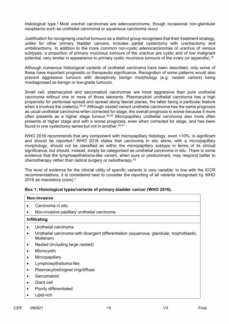
WHO 2016 recommends that any component with micropapillary histology, even <10%, is significant and should be reported.2 WHO 2016 states that carcinoma in situ alone, with a micropapillary morphology, should not be classified as within the micropapillary subtype in terms of its clinical significance, but should, instead, simply be categorised as urothelial carcinoma in situ. There is some evidence that the lymphoepithelioma-like variant, when pure or predominant, may respond better to chemotherapy rather than radical surgery or radiotherapy.42 The level of evidence for the clinical utility of specific variants is very variable. In line with the ICCR recommendations, it is considered best to consider the reporting of all variants recognised by WHO 2016 as mandatory (core).7
Box 1: Histological types/variants of primary bladder cancer (WHO 2016).
Non-invasive
• Carcinoma in situ • Non-invasive papillary urothelial carcinoma
Infiltrating
• Urothelial carcinoma • Urothelial carcinoma with divergent differentiation (squamous, glandular, trophoblastic,
Mullerian) • Nested (including large nested) • Microcystic • Micropapillary • Lymphoepithelioma-like • Plasmacytoid/signet ring/diffuse • Sarcomatoid • Giant cell • Poorly differentiated • Lipid-rich

CEff 090821 16 V3 Final
• Clear cell (glycogen-rich) • Squamous carcinoma • Verrucous carcinoma • Adenocarcinoma • Enteric • Mucinous • Mixed • Urachal carcinoma (this is listed in WHO 2016 as a separate type, though it can have
various histological features, most commonly adenocarcinoma) • Tumours of Mullerian type • Clear cell carcinoma • Endometrioid carcinoma • Neuroendocrine tumours • Small cell carcinoma • Large cell neuroendocrine carcinoma • Well-differentiated endocrine tumour
[Level of evidence D – Histological variants are important for cancer registration and prognosis.]
5.3.2 Squamous tumours of the distal penile urethra Most tumours of the distal penile urethra are squamous in type. These are dealt with in more detail in
Dataset for penile and distal urethral cancer histopathology reports (RCPath) and that document should be referred to when reporting such tumours.27
5.3.3 Tumour grade
Grading is critical for prognostication and management of non-invasive urothelial carcinomas, though less important in those with lamina propria invasion and of limited clinical utility in muscularis propria invasive tumours. The overwhelming majority of T1 tumours are high grade. Low-grade invasive papillary urothelial carcinoma exists but is rare.2,43 The well-established WHO 1973 grading system was modified by ISUP in 1998, adopted in WHO 2004
and retained in WHO 2016.1,2,9,10 These changes have been controversial and caused significant confusion among epidemiologists, pathologists and urologists. The systems cannot be easily mapped to each other, which poses difficulties for cancer registration and comparison of results of recent studies with historic data. There has been considerable debate on the merits and issues of both grading systems.44–47 The WHO 1973 system, although repeatedly validated, has some significant drawbacks, particularly the vague definitions of the grades. WHO 1973 Grade 2 is heterogeneous with the reported proportion of bladder tumours categorised as Grade 2 varying from 13% to 69%, suggesting significant interobserver variation.48,49 Moreover, non-invasive Grade 2 urothelial carcinoma is associated with a stage progression risk of about 10%, suggesting that a significant number of these patients have been under-treated.49 The WHO 2004 system provides detailed architectural and cytological criteria for the various grades of tumour. Adoption of a two-tier classification of carcinomas eliminated the issue of most tumours being categorised in the middle grade, while expansion of the high-grade category ensured that more patients who are likely to benefit would be treated with BCG. Moreover, the categorisation of tumours at the ‘good end’ of WHO 1973 Grade 1 tumours, as papillary urothelial neoplasm of low malignant potential (PUNLMP), has avoided labelling these biologically indolent tumours as carcinomas. However, there are some significant problems with the WHO 2004 system. It has issues with reproducibility, particularly in the distinction of PUNLMP from low-grade urothelial carcinoma. Since
CEff 090821 17 V3 Final
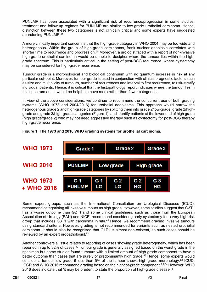
PUNLMP has been associated with a significant risk of recurrence/progression in some studies, treatment and follow-up regimes for PUNLMP are similar to low-grade urothelial carcinoma. Hence, distinction between these two categories is not clinically critical and some experts have suggested abandoning PUNLMP.44 A more clinically important concern is that the high-grade category in WHO 2004 may be too wide and heterogenous. Within the group of high-grade carcinomas, frank nuclear anaplasia correlates with shorter time to recurrence and progression.50 Moreover, a urologist faced with a report of non-invasive high-grade urothelial carcinoma would be unable to decipher where the tumour lies within the high-grade spectrum. This is particularly critical in the setting of post-BCG recurrence, where cystectomy may be considered for high-grade recurrence. Tumour grade is a morphological and biological continuum with no quantum increase in risk at any particular cut-point. Moreover, tumour grade is used in conjunction with clinical prognostic factors such as size and multiplicity of tumours, number of recurrences and interval to first recurrence, to risk-stratify individual patients. Hence, it is critical that the histopathology report indicates where the tumour lies in this spectrum and it would be helpful to have more rather than fewer categories. In view of the above considerations, we continue to recommend the concurrent use of both grading systems (WHO 1973 and 2004/2016) for urothelial neoplasms. This approach would narrow the heterogenous grade 2 and high-grade categories by splitting them into grade 2/low-grade, grade 2/high-grade and grade 3/high-grade categories (Figure 1), and identify patients at the lower end of high grade (high grade/grade 2) who may not need aggressive therapy such as cystectomy for post-BCG therapy high-grade recurrence.
Figure 1: The 1973 and 2016 WHO grading systems for urothelial carcinoma.
Some expert groups, such as the International Consultation on Urological Diseases (ICUD), recommend categorising all invasive tumours as high grade. However, some studies suggest that G3T1 has a worse outcome than G2T1 and some clinical guidelines, such as those from the European Association of Urology (EAU) and NICE, recommend considering early cystectomy for a very high-risk group that includes G3T1 with carcinoma in situ.49 Hence, we recommend grading invasive tumours using standard criteria. However, grading is not recommended for variants such as nested urothelial carcinoma. It should also be recognised that G1T1 is almost non-existent, so such cases should be reviewed by an expert uropathologist.51 Another controversial issue relates to reporting of cases showing grade heterogeneity, which has been reported in up to 32% of cases.52 Tumour grade is generally assigned based on the worst grade in the specimen but some studies found tumours with a limited amount of high-grade component to have a better outcome than cases that are purely or predominantly high grade.53 Hence, some experts would consider a tumour low grade if less than 5% of the tumour shows high-grade morphology.52 ICUD, ICCR and WHO 2016 recommend grading based on the highest-grade component.2,7,54 However, WHO 2016 does indicate that ‘it may be prudent to state the proportion of high-grade disease’.2

CEff 090821 18 V3 Final
The issue of grade heterogeneity may be related to tumour multifocality. Multiple papillary tumours could coalesce and appear as a single tumour upon cystoscopic examination. Thus, if the highest grade is assigned, then a 5 cm low-grade tumour coalescing with a 1 cm high-grade tumour may be interpreted as a 6 cm high-grade tumour, placing the patient inappropriately in a higher-risk category. We recommend grading based on the highest-grade component but suggest including a comment in cases where the high-grade component is estimated to be less than 10%, reflecting the uncertainty regarding the best approach to such cases. Squamous carcinoma or adenocarcinoma should be graded as well, moderately or poorly differentiated. Tumour variants such as small cell carcinoma, plasmacytoid carcinoma or sarcomatoid carcinoma often occur mixed with areas of urothelial carcinoma rather than in pure form. WHO grading of the variant elements is not recommended but the associated conventional urothelial element present can be graded (usually high grade) with a comment regarding the prognostic significance of the variant component(s). [Level of evidence B – Histological grade is important for prognostication.]
5.3.4 Associated carcinoma in situ Carcinoma in situ associated with papillary or invasive urothelial carcinoma is generally of urothelial type but may show other differentiations such as squamous and glandular. It is good practice to indicate the type of carcinoma in situ. The presence and extent of associated urothelial carcinoma in situ is a criterion for selection for intravesical BCG therapy, and failure to respond to initial treatment is a risk factor for subsequent progression.55 Urothelial carcinoma in situ may occur in the immediate vicinity of a tumour and/or further away from a tumour, for example in separate biopsies sent with a TUR specimen and the proformas allow for recording of this information in summarised form. The site(s) of separate positive biopsies should be specified in the ‘further comments’ section if applicable, as this may have a bearing on subsequent management decisions. It is important to indicate the type of carcinoma in situ, as BCG therapy would not be appropriate for pure squamous carcinoma in situ. The type of carcinoma in situ may also determine tumour type. If an invasive carcinoma with pure squamous differentiation is associated with urothelial carcinoma in situ, then as explained earlier it should be classified as a urothelial carcinoma with extensive squamous differentiation. However, if the in-situ component is of squamous type, then the tumour would be classed as a squamous cell carcinoma. [Level of evidence C.]
5.3.5 Lymphovascular invasion
Several studies have found the presence of lymphovascular invasion (LVI) in cystectomy and nephroureterectomy specimens to be an independent predictor of outcome.56−58 LVI has been demonstrated to be significantly associated with cancer-specific survival after radical cystectomy in both lymph node negative and lymph node positive patients. Data on LVI in biopsy/TUR of bladder specimens is more limited; some but not all studies found LVI to be a significant predictor of adverse outcome in T1 urothelial cancer.59,60 No data is available regarding the significance of LVI in urothelial carcinoma of the urethra. Another limitation of available data on the prognostic significance of LVI is that most studies are retrospective analysis of pathology data without central review.61 Most studies support LVI as an adverse prognostic indicator and LVI is part of some nomograms to guide patient management, so LVI is considered a core data item in all sites and specimens with primary urothelial carcinoma. LVI should be reported as being present only when it is unequivocal. 62,63 There is potential to mistake retraction artefact around tumour cells for LVI, and immunohistochemistry for endothelial markers may be helpful in selected cases.
CEff 090821 19 V3 Final
[Level of evidence B – Lymphovascular invasion predicts disease progression and adverse survival.] 5.3.6 Necrosis
Tumour necrosis has been identified as an adverse prognostic factor in T2/pT2 urothelial carcinoma.23,64,65 It has also been found to predict benefit from hypoxia modification in patients enrolled in the bladder carbogen and nicotinamide (BCON) trial.65 Since this parameter is used to modify radiotherapy only in some centres, it has been categorised as a non-core data item.
5.3.7 Extent of invasion (biopsy and TURBT specimens)
Extent of invasion in biopsy and TURBT specimens is a core data item, as it is an important prognostic indicator that guides patient management. However, one of the general rules of the TNM classification
is that ‘the pathological assessment of the primary tumour (pT) entails a resection of the primary tumour or a biopsy adequate to evaluate the highest pT category’.5 Hence, a pT category can be assigned only to definitive resection specimens, such as total or partial cystectomy specimens and not routinely to biopsy or TURBT specimens. Stage is therefore not a data item in the dataset for latter specimens. If stage is reported for ease of communication, then it would be part of the clinical stage and should be designated as T category rather than pT. This is consistent with ICCR recommendations.7 A comment such as ‘at least’ may be added in selected circumstances to emphasise that the T classification has a higher likelihood of not being representative (e.g. for T1 tumour where no muscularis propria present or only smooth muscle of indeterminate type present). The 5th edition of the TNM supplement (related to the TNM 8th edition) clarified that the precondition for pT categorisation after only TURBT would be met in the case of a histologically confirmed complete tumour resection with additional separately submitted tissues from adjacent (deep and lateral) grossly tumour-free areas that are histologically negative.66 However, ‘lateral’ biopsies are not usually submitted, so most tumour extent in TURBT specimens would generally contribute to the cT category. If the smooth muscle that is present in bladder biopsy/TURBT cases is indeterminate in type, this should be indicated – as alluded to above. It is important to state whether muscularis propria (detrusor muscle) is present or absent in bladder biopsies and TURBTs, especially in T1 tumours. Absence of muscularis propria (detrusor muscle) should prompt early re-resection in most instances (following MDT discussion). Ideally, tumour base biopsies should be performed at initial resection to sample muscularis propria (detrusor muscle) and submitted in a separate container. An alternative TUR method, the en bloc resection of bladder tumours, allows better specimen orientation for staging purposes and completeness of excision can be more readily assessed, but this is not standard practice.67,68 Routine early re-resection following standard TUR is performed in some centres to ensure complete excision, as complete eradication of all visible tumours at first resection is not always achieved.69 Tumour extent in resection specimens is discussed in staging section 5.3.8 (pT category).
Substaging of T1 urothelial carcinoma in bladder Urothelial carcinoma invading lamina propria is heterogenous and, particularly when high grade, is associated with significant risk of recurrence and cancer related mortality up to 33%.70 Several studies have found that risk of tumour progression and cancer-related deaths is higher with increasing depth of invasion.71 Hence, there have been several efforts to ‘substage’ T1 urothelial carcinoma based on either its relationship to the muscularis mucosae (superficial to, into or deep to this muscle layer) or the absolute extent in millimetres (maximum dimension or depth of invasion). However, it is often difficult to substage tumours accurately in TURBTs. Unlike in the colon, the muscularis mucosae layer is interrupted in the bladder, so may not be seen in relation to the invasive tumour. Tumour quantitation is hindered by tangential sectioning and difficulty in orientation of the chips and identification of the mucosal surface or basement membrane. There is also lack of consensus regarding the tumour depth or dimension cut-off that should be used to determine treatment.

CEff 090821 20 V3 Final
Owing to these issues, T1 substaging is categorised as a non-core data item. However, in view of its potential to impact clinical decision-making, it is recommended that some assessment of the extent of lamina propria invasion (by one of the above methods or descriptive terminology such as ‘superficial’ or ‘deep’) should be provided where possible.
5.3.8 Tumour stage and nodal status
Tumour stage is an important predictor of outcome and hence a core data item for excision specimens. The TNM classification is produced by the UICC in joint collaboration with the AJCC. There are some differences between the two systems, but these are relatively minor in the staging of tumours of the urinary tract.72
The Royal College of Pathologists recommends the use of UICC TNM 8th edition for staging tumours of the urinary collecting system (Appendix A). Readers must be aware of errata published by UICC subsequent to the publication of the initial print version, which has resulted in better synchronisation of the two TNM versions.11
(p)T subcategorisation For tumours invading only the lamina propria, the depth and extent of invasion correlates with outcome. Previously there was no international agreement that this information should be included in the report or which method should be used for its assessment. WHO 2016 now recommends providing an assessment of the depth and/or extent of subepithelial invasion in T1 cases.2 The AJCC 8th edition TNM also states that, although not formally endorsed by the AJCC staging system, an attempt to categorise pT1 disease is strongly recommended, using one of the methods mentioned. For tumours invading muscularis propria (detrusor muscle), subdivision in cystectomy specimens into pT2a and pT2b according to inner and outer half of the muscularis propria (detrusor muscle), or macroscopic (pT3b) versus microscopic (pT3a) extension into perivesical fat, had prognostic significance in several studies, although one group detected no difference in outcome between these stage categories.73‒77
Note that fat can be present normally in all layers of the bladder wall and tumour involvement of fat per se in biopsy/TUR material is not necessarily indicative of perivesical fat involvement. Microscopic assessment of perivesical fat invasion can be problematic due to poor definition of the boundary between muscularis propria of the bladder and perivesical fat, compounded by tumour-related factors such as stromal desmoplasia.78 Staging of cystectomies with limited residual tumour There is some uncertainty regarding the assignment of pT category in cystectomy specimens that show limited residual tumour. For example, no residual tumour may be identified in a cystectomy performed without neoadjuvant chemotherapy following a TURBT diagnosis of muscularis propria invasive urothelial carcinoma. AJCC recommends that pathological stage of cystectomy specimens should be assigned independently of previous biopsy information, so such a case would be categorised as pT0. However, UICC TNM rules state that cystectomy pT determination should include TURBT information so such a case would be assigned a pT2 category (confirmed by the UICC TNM help desk).5 Since the outcome of such patients would be of detrusor invasive bladder cancer, the authors of this dataset recommend following the UICC recommendation and categorising such specimens as pT2 with an appropriate reference to the transurethral resection and the subsequent negative cystectomy. However, if the patient has received neoadjuvant therapy prior to surgery, then the negative cystectomy specimen should be categorised as ypT0. Staging of urachal tumours There are particular issues with the staging of urachal tumours that generally arise within the bladder wall. The most widely used method for staging such tumours is the Sheldon staging system.79 Several other approaches for staging these rare tumours have been proposed but these remain to be validated.80 Despite its limitations, we recommend using the Sheldon system to stage urachal tumours.

CEff 090821 21 V3 Final
[Level of evidence A – Tumour stage predicts outcome.] Staging of tumours in diverticula In diverticulectomy specimens, staging may be difficult, as the muscularis propria (detrusor muscle) may be absent in the attenuated wall and the muscularis mucosae may be hyperplastic. The pT2 category will not be applicable for tumour in a diverticulum lacking muscularis propria. pT1 should be used for infiltrative tumours involving up to and including the muscularis mucosae, but not beyond. pT3 will be applicable for invasion into perivesical tissue. Staging of tumours involving prostate Urothelial tumours involving/arising from the prostatic urethra and/or prostatic ducts with a concurrent bladder tumour are staged separately, as though they were primary bladder and urethral tumours. Such tumours should not be classified as T4 bladder cancer unless they directly invade the prostatic stroma by invasion through the full thickness of the bladder wall. Subepithelial invasion of the prostatic urethra or stroma from the urethra does not constitute T4 bladder cancer.5,6
pN categorisation There is some ambiguity regarding the definition of regional lymph nodes for carcinomas of the urinary bladder, but we recommend adopting the AJCC definition, which includes the perivesical lymph nodes.6
It is worth noting that common iliac lymph nodes are considered regional nodes for bladder carcinoma (pN3 if positive) but non-regional for prostate cancer (pM1a if positive).5,6 The number of positive lymph nodes and lymph node density, defined as the ratio of positive nodes to the total number of nodes sampled, are predictors of cancer survival.81 The presence of nodal extracapsular spread was found in some studies to confer a worse outcome and decreased recurrence free survival.82–84 However, this was not incorporated into TNM 8th edition by the AJCC as there was some conflicting literature evidence.6 The size of metastatic nodal deposits needs to be taken into account for pN staging in nephroureterectomy specimens (see Appendix A). Unlike in the previous (7th edition) of TNM, this size is no longer relevant to the pN classification for urethral tumours, which has been simplified in the 8th
edition. AJCC recommendation is to assign pN status, regardless of the number of lymph nodes assessed, though they comment that optimised staging should result in an average of >12 lymph nodes from primary nodal regions.6 According to UICC TNM (8th edition), discrete tumour deposits (satellites) that are present separately from the main tumour mass in the perivesical fat, without histological evidence of residual lymph node in the nodule/deposit, may represent discontinuous spread, venous invasion or a completely replaced lymph node.5 A nodule (generally having a smooth contour) considered by the pathologist to be a totally replaced lymph node should be recorded as a positive lymph node, and each such nodule should be counted separately as a lymph node in the final pN determination.5 [Level of evidence B – Nodal status predicts survival.]
5.3.9 Specimen margin status
Positive margin status confers a worse outcome. Positive soft tissue margins, defined as tumour present at specimen margin, are associated with an increased risk of local recurrence and cancer-specific mortality after cystectomy.85,86 Distance of tumour to the nearest resection margin is not a core requirement (although it can be mentioned in a comment). Although it might be clinically important, it is not yet validated or in regular use for clinical management.
[Level of evidence A – Positive margins predict recurrence and cancer specific mortality.]
5.3.10 Best block It is helpful to include a record of the best tumour block in the pathology report (to which can be added the percentage of tumour in that block) to enable further study (with appropriate ethical approval and
CEff 090821 22 V3 Final
consent) and also to enable material to be sent, as appropriate, for clinical trials or for further future investigation. If this is done routinely, it avoids having to look through all the sections at a later date when a later request for tissue is made. Recording a block that has normal/uninvolved tissue here may also be helpful. This data item is categorised as non-core because although useful, it is not required for cancer staging, optimal patient management and prognosis.
5.3.11 Other neoplastic urothelial abnormalities
Associated carcinoma in situ is a core data item, as discussed in a previous section. In the context of a bladder tumour, urothelial dysplasia not amounting to carcinoma in situ does not influence patient management and is not recorded by UK cancer registries, so it has been classified as a non-core data item. De novo urothelial dysplasia has been described but should be diagnosed with caution.87 Urothelial dysplasia in background, flat epithelium adjacent to a papillary urothelial neoplasm suggests an unstable urothelium with a higher risk of recurrence. Exophytic urothelial papilloma, inverted urothelial papilloma and urothelial proliferation of uncertain malignant potential are also of little clinical significance when associated with a urothelial carcinoma.
5.3.12 Other co-existing pathology
Several benign abnormalities such as nephrogenic adenoma, florid von Brunn’s nests and metaplastic changes may be associated with bladder cancer. Although useful to recognise to avoid misdiagnosis or overstaging, they do not influence patient management and are hence categorised as non-core. Associated keratinising squamous metaplasia or intestinal metaplasia (particularly when dysplastic) would favour a diagnosis of squamous cell carcinoma or adenocarcinoma in the appropriate clinicopathological setting. Benign findings in organs removed with urinary tract resections are generally of little clinical significance but are sometimes important, e.g. when glomerular pathology is identified in a nephroureterectomy specimen. If significant pathology, such as carcinomas of prostate, uterus, ovaries or renal parenchyma, is identified, then it should be reported in accordance with the requirements of the relevant dataset. As indicated earlier, urethral urothelial carcinoma in situ involving prostatic ducts/stroma in a cystoprostatectomy specimen should be staged as a separate lesion.
5.3.13 Ancillary tests (immunohistochemistry, molecular)
No ancillary studies are recommended for routine use in urothelial carcinoma of the urinary tract and hence this data item is categorised as non-core. However, it is recommended that results of immunohistochemical studies performed to aid diagnosis, staging or detection of LVI, or to predict response to treatment should be included in the histopathology report. If other ancillary studies are performed, these should also be listed. Immunohistochemical assessment for PD-L1 expression can predict response to anti-PD-L1 immunotherapy.88–90 However, a number of different anti-PD-L1 clones are available from different manufacturers and the published trials have examined specific clones linked to the activity of specific anti-PD-L1 immunotherapy agents. Moreover, these tests use different algorithms and cut-offs to identify patients more likely to benefit from each immunotherapeutic agent.91,92 Since PD-L1 testing is required only for some patients with advanced bladder cancer, and each immunotherapeutic agent needs a different PD-L1 test, reflex testing of all TURBT and/or cystectomy specimens with muscle invasive bladder cancer is not recommended at present. However, departments should set up a process to enable prompt PD-L1 testing by a trained pathologist in an accredited laboratory for any patient requiring this test.

CEff 090821 23 V3 Final
Participation in relevant immunohistochemistry EQA is mandatory for laboratories involved in PD-L1 assessment. The results of such testing should be incorporated into the pathology record, when it is available; such testing should not delay the primary report. Biomarkers (immunohistochemical and molecular) have been recently proposed to predict risk of recurrence and progression in patients with Ta/T1 bladder cancer and identify patients that would benefit from early aggressive management.93 However, their clinical utility remains to be validated and recent NICE guidance recommends their use be assessed within the context of clinical trials.4
Molecular classification of bladder cancer using immunohistochemistry and gene expression analysis is another promising approach that requires standardisation and validation before it can be incorporated into routine clinical practice. UTUC is reported to be the third most common malignancy (after colorectal and endometrial carcinoma) and is associated with hereditary nonpolyposis colorectal carcinoma (Lynch syndrome).94 It has been estimated that 1–3% of all UTUC may represent Lynch syndrome-associated carcinoma.95 EAU guidelines recommend germline DNA testing for Lynch syndrome mutations in patients who are clinically identified to be at higher risk of Lynch syndrome.96 Some papers recommend reflex mismatch repair (MMR) immunohistochemical screening followed by microsatellite instability (MSI) testing for all UTUC cases.95,97 In our opinion, there is insufficient evidence to indicate that routine MMR or MSI testing of UTUC specimens is cost-effective.
5.3.14 Diagnostic coding
Coding is recommended for data retrieval, workload measurement and audit. SNOMED coding should be applied (see Appendix B).
Morphologic coding of non-invasive urothelial neoplasia poses particular problems. Traditionally, non-invasive papillary urothelial carcinoma (Ta) has been coded as M81303, which is inappropriate, as the behaviour code represented by the last digit (3) would indicate an invasive tumour. Current recommendations are that PUNLMP should be coded as M81301, all non-invasive papillary urothelial carcinomas as M81302 and papillary tumours with invasion as M81303.
6 Reporting of small biopsy specimens
Tumours encountered in small biopsies should be reported using the tumour protocol described in previous sections. Flat abnormalities such as dysplasia and carcinoma in situ should be reported. Carcinoma in situ encountered in the bladder is generally of urothelial type but may show other differentiations such as squamous and glandular. It is good practice to indicate the type of carcinoma in situ. Urothelial carcinoma in situ has a number of morphological variants that may cause diagnostic issues but recording these is generally of limited clinical significance.98,99 Urothelial carcinoma in situ is often associated with loss of epithelial cell cohesion and the surface may be almost totally denuded, hence the need for levels, particularly in cases with positive urine cytology. If surface epithelium is denuded, cytological follow-up would be advisable with clinical correlation. Biopsies showing flat abnormalities should be assessed primarily by morphology, although immunohistochemistry may be of value in a proportion of cases. The markers most commonly used in conjunction with the morphology to assist in the separation of dysplasia/carcinoma in situ from normal/reactive changes are CK20, CD44s and p53.98–100 Immunohistochemistry must be interpreted with caution as there is significant overlap in the immunostaining patterns. For example, urothelial carcinoma in situ may be immunonegative for CK20 while morphologically benign urothelium may rarely show full thickness CK20 immunoreactivity.101 MIB-1/Ki-67 has also been used in this context and although increased staining can overlap in different disease states, carcinoma in situ is less likely to be MIB-1/Ki-67 negative.

CEff 090821 24 V3 Final
Immunohistochemistry does not help to distinguish urothelial dysplasia from carcinoma in situ; a distinction that is based on morphology. These markers can be used individually but are available commercially as a combined cocktail using different chromogens. [Level of evidence D.]
7 Reporting of frozen sections Intraoperative frozen sections may be used to assess ureteric and urethral specimen margins at cystectomy and occasionally the ureteric margin at nephroureterectomy. Generally, only infiltrating carcinoma and carcinoma in situ can be diagnosed on frozen sections, as freezing artefact precludes reliable identification of urothelial dysplasia in these specimens. The clinical utility of frozen section examination of these margins is controversial, as skip lesions of multifocal carcinoma in situ may result in false-negative frozen sections. Frozen sections of the urethral margin are usually performed only in cases of bladder reconstruction. In general, urethral frozen sections are more critical, as a false-positive diagnosis would preclude bladder reconstruction. In contrast, a false-positive ureteric frozen section would only result in unnecessary excision of an additional segment of ureter. Segments of ureter submitted for frozen section should be orientated, preferably with a stitch at the end, opposite the resection margin to avoid disrupting the margin from which the frozen section is taken. There should always be a clear understanding between the pathologist and surgeon as to the meaning of the terms ‘proximal’ and ‘distal’ (regarding excision margins, e.g. ureter with respect to kidney versus ureter with respect to bladder) to avoid any ambiguity of interpretation. Urethral frozen sections are difficult, especially in cystoprostatectomy specimens in which the urothelium at the specimen margin tends to retract into the specimen. In contrast to frozen sections from radical prostatectomy specimens for prostate cancer, the urethral margin section from cystoprostatectomy specimens should, therefore, be taken a little deeper to include the urethral urothelium. In view of this technical difficulty in examining urethral margins, as well as false negativity due to multifocality of carcinoma in situ, urethral biopsies prior to radical surgery are preferred and routine intraoperative frozen sections of urethral margins are to be discouraged. Routine frozen section examination of pelvic lymph nodes at cystectomy is not recommended due to problems with histological sampling. Frozen sections should be limited to lymph nodes that appear to be grossly involved by tumour.
8 Criteria for audit The availability of pathology reports and data at MDT meetings:
• standard: 90% of cases discussed at MDT meetings where biopsies or resections have been taken should have pathology reports/core data available for discussion at the time of the meeting
• standard: 90% of cases where pathology has been reviewed for the MDT meeting should have the process of review recorded.

CEff 090821 25 V3 Final
9 References 1. Mostofi FK, Sobin LH, Torloni H. Histological Typing of Urinary Bladder Tumours. Geneva,
Switzerland: WHO, 1973.
2. Moch H, Humphrey PA, Ulbright TM, Reuter VE. WHO Classification of Tumours of the Urinary System and Male Genital Organs. Lyon, France: IARC Press, 2016.
3. NICE. Guidance on Cancer Services: Improving Outcomes in Urological Cancers. The Manual. London, UK: NICE, 2002. Accessed December 2020. Available at: www.nice.org.uk/guidance/csg2/resources/improving-outcomes-in-urological-cancers-pdf-773372413
4. NICE. Bladder Cancer: Diagnosis and Management. London, UK: NICE, 2015. Accessed September 2020. Available at: www.nice.org.uk/guidance/ng2
5. Union for International Cancer Control. Brierley JD, Gospodarowicz MK, Wittekind Ch (eds). TNM Classification of Malignant Tumours (8th edition). Oxford, UK: Wiley-Blackwell, 2016.
6. American Joint Committee on Cancer. Amin MB, Edge S, Greene F, Byrd DR, Brookland RK, Washington MK et al (eds). AJCC Cancer Staging Manual (8th edition). Chicago, US: Springer, 2016.
7. International Collaboration on Cancer Reporting. Urinary/Male Genital Cancer Datasets. Accessed September 2020. Available from: www.iccr-cancer.org/datasets/published-datasets/urinary-male-genital.
8. Shanks JH, Chandra A, McWilliam L, Varma M. Dataset for Tumours of the Urinary Collecting System (Renal Pelvis, Ureter, Urinary Bladder and Urethra) (2nd edition). London, UK: Royal College of Pathologists, 2013. Accessed Sept 2020. Available at: www.rcpath.org/uploads/assets/e2c11ff6-780a-471e-a21a4dc48788d35b/Dataset-for-tumours-of-the-urinary-collecting-system-renal-pelvis-ureter-urinary-bladder-and-urethra.pdf
9. Epstein JI, Amin MB, Reuter VE, Mostoki FK. The World Health Organization/International Society of
Urological Pathology consensus classification of urothelial (transitional cell) neoplasms of the urinary bladder. Bladder Consensus Conference Committee. Am J Surg Pathol 1998;22:1435–1448.
10. Eble J, Sauter G, Epstein J, Sesterhenn I. Pathology and Genetics of Tumours of the Urinary System and Male Genital Organs. Lyon, France: IARC Press, 2004.
11. Union for International Cancer Control. TNM Classification (8th edition). Accessed September 2020. Available at: www.uicc.org/resources/tnm/publications-resources
12. Paner GP, Ro JY, Wojcik EM, Venkataraman G, Datta MW, Amin MB. Further characterization of the muscle layers and lamina propria of the urinary bladder by systematic histologic mapping: implications for pathologic staging of invasive urothelial carcinoma. Am J Surg Pathol 2007;31:1420–1429.
13. Tavora F, Fajardo DA, Lee TK, Lotan T, Miller JS, Miyamoto H et al. Small endoscopic biopsies of the ureter and renal pelvis: pathologic pitfalls. Am J Surg Pathol 2009;33:1540–1546.
14. Lopez-Beltran A, Luque RJ, Mazzucchelli R, Scarpelli M, Montironi R. Changes produced in the urothelium by traditional and newer therapeutic procedures for bladder cancer. J Clin Pathol 2002;55:641–647.
15. Oxley JD, Cottrell AM, Adams S, Gillatt D. Ketamine cystitis as a mimic of carcinoma in situ.
Histopathology 2009;55:705–708.

CEff 090821 26 V3 Final
16. Baker PM, Young RH. Radiation-induced pseudocarcinomatous proliferations of the urinary bladder: a report of 4 cases. Hum Pathol 2000;31:678-683.
17. Chan TY, Epstein JI. Radiation or chemotherapy cystitis with “pseudocarcinomatous” features. Am J Surg Pathol 2004;28:909–913.
18. Lane Z, Epstein JI. Pseudocarcinomatous epithelial hyperplasia in the bladder unassociated with prior irradiation or chemotherapy. Am J Surg Pathol 2008;32:92–97.
19. Bultitude MF, Ghani KR, Horsfield C, Glass J, Chandra A, Thomas K. Improving the interpretation of ureteroscopic biopsies: use of Bouin’s fixative. BJU Int 2011;108:1373–1375.
20. Chandra A, Griffiths D, McWilliam LJ. Best Practice: gross examination and sampling of surgical specimens from the urinary bladder. J Clin Pathol 2010;63:475–479.
21. Cross P, Chandra A, Maddox A, Narine N, Giles Tl. Tissue Pathways for Diagnostic Cytopathology (2nd edition). London, UK: Royal College of Pathologists, 2019. Accessed September 2020. Available at: www.rcpath.org/uploads/assets/b328ab3d-f574-40f1-8717c32ccfc4f7d8/G086-Tissue-pathways-for-diagnostic-cytopathology.pdf
22. Liebmann R, Varma M. Best Practice Recommendations Histopathology and Cytopathology of Limited or No Clinical Value (3rd edition). London, UK: Royal College of Pathologists, 2019. Accessed September 2020. Available at: www.rcpath.org/uploads/assets/6516e9e7-4a23-4839-a8537c40c882a3b9/cbc1d100-3372-482d-a34268bafa829100/G177-BPR-histopathology-and-cytopathology-limited-or-no-clinical-value.pdf
23. Soave A, John LM, Dahlem R, Minner S, Engel O, Schmidt S et al. The impact of tumor diameter and tumor necrosis on oncologic outcomes in patients with urothelial carcinoma of the bladder treated with radical cystectomy. Urology 2015;86:92–98.
24. Cheng L, Neumann RM, Scherer BG, Weaver AL, Leibovich BC, Nehra A et al. Tumor size predicts the survival of patients with pathologic stage T2 bladder carcinoma: a critical evaluation of the depth of muscle invasion. Cancer 1999;85:2638–2647.
25. McKenney JK, Amin MB, Epstein JI, Grignon DJ, Esther O, Reuter VE et al. Protocol for the examination of specimens from patients with carcinoma of the urethra. Arch Pathol Lab Med 2010;134:345–350.
26. Gakis G, Morgan TM, Daneshmand S, Keegan KA, Todenhofer T, Mischinger J et al. Impact of perioperative chemotherapy on survival in patients with advanced primary urethral cancer: results of the international collaboration on primary urethral carcinoma. Ann Oncol 2015;26:1754–1759.
27. Corbishley CM, Oxley J, Shanks JH, Tinwell B. Dataset for Penile and Distal Urethral Cancer Histopathology Reports (3rd edition). London, UK: Royal College of Pathologists, 2015. Accessed September 2020. Available at: www.rcpath.org/uploads/assets/d0e70985-300e-4835-af4583df56025869/Dataset-for-penile-and-distal-urethral-cancer-histopathology-reports.pdf
28. Cheng L, Weaver AL, Leibovich BC, Ramnani DM, Neumann RM, Scherer BG et al. Predicting the survival of bladder carcinoma patients treated with radical cystectomy. Cancer 2000;88:2326–2332.
29. Heney NM, Ahmed S, Flanagan MJ, Frable W, Corder MP, Hafermann MD et al. Superficial bladder
cancer: progression and recurrence. J Urol 1983;130:1083–1086.
30. Chaux A, Karram S, Miller JS, Fajardo DA, Lee TK, Miyamoto H et al. High-grade papillary urothelial carcinoma of the urinary tract: a clinicopathologic analysis of a post-World Health Organization/International Society of Urological Pathology classification cohort from a single academic center. Hum Pathol 2012;43:115–120.

CEff 090821 27 V3 Final
31. Lughezzani G, Burger M, Margulis V, Matin SF, Novara G, Roupret M et al. Prognostic factors in upper urinary tract urothelial carcinomas: a comprehensive review of the current literature. Eur Urol 2012;62:100–114.
32. Picozzi S, Ricci C, Gaeta M. Upper urinary tract recurrence following radical cystectomy for bladder cancer: a meta-analysis on 13,185 patients. J Urol 2012;188:2046–2054
33. Boorjian SA, Kim SP, Weight CJ, Cheville JC, Thapa P, Frank I. Risk factors and outcomes of urethral recurrence following radical cystectomy. Eur Urol 2011;60:1266–1272.
34. Kim SP, Frank I, Cheville JC, Thompson RH, Weight CJ, Thapa P et al. The impact of squamous and glandular differentiation on survival after radical cystectomy for urothelial carcinoma. J Urol 2012;188:405–409.
35. Amin MB, Smith SC, Eble JN, Rao P, Choi WWL, Tamboli P et al. Glandular neoplasms of the urachus: a report of 55 cases emphasizing mucinous cystic tumors with proposed classification. Am J Surg Pathol 2014;38:1033–1045.
36. Dayyani F, Czerniak BA, Sircar K, Munsell MF, Millikan RE, Dinney CP et al. Plasmacytoid urothelial carcinoma, a chemosensitive cancer with poor prognosis, and peritoneal carcinomatosis. J Urol 2013;189:1656–1661.
37. Kaimakliotis HZ, Monn MF, Cheng L, Masterson TA, Cary KC, Pedrosa JA et al. Plasmacytoid bladder cancer: variant histology with aggressive behaviour and a new mode of invasion along fascial planes. Urology 2014;83:1112–1116.
38. Linder BJ, Frank I, Cheville JC, Thompson RH, Thapa P, Tarrell RF et al. Outcomes following radical cystectomy for nested variant of urothelial carcinoma: a matched cohort analysis. J Urol 2013;189:1670–1675.
39. Lopez-Beltran A, Cheng L, Montironi R, Blanca A, Leva M, Rouprêt M et al. Clinicopathological characteristics and outcome of nested carcinoma of the urinary bladder. Virchows Arch 2014;465:199–205.
40. Monn MF, Kaimaklotis HZ, Pedrosa JA, Cary KA, Bihrle R, Cheng L et al. Contemporary bladder cancer: variant histology may be a significant driver of disease. Urol Oncol 2015;33:15–20.
41. Fairey AS, Daneshmand S, Wang L, Schuckman A, Liesovsky G, Djaladat H et al. Impact of micropapillary urothelial carcinoma variant histology on survival after radical cystectomy. Urol Oncol 2014;32:110–116.
42. Amin MB, Ro JY, Lee KM, Ordonez NG, Dinney CP, Ayala AG et al. Lymphoepithelioma-like carcinoma of the urinary bladder. Am J Surg Pathol 1994;18:466–473.
43. Toll AD, Epstein JI. Invasive low-grade papillary urothelial carcinoma: a clinicopathologic analysis of 41 cases. Am J Surg Pathol 2012;36:1081–1086.
44. Cheng L, MacLennan GT, Lopez-Beltran A. Histologic grading of urothelial carcinoma: a reappraisal. Hum Pathol 2012;43:2097–2108.
45. Varma M, Delahunt B, van der Kwast T. Grading noninvasive bladder cancer: World Health Organisation 1973 or 2004 may be the wrong question. Eur Urol 2019;76:413–415.
46. Compérat E, Amin M, Reuter V. Two decades of World Health Organisation/International Society of Urological Pathology bladder cancer grading: Time to reflect on accomplishments and plan refinement in the molecular era, not regress to readoption of a 45-year-old classification. Eur Urol 2019;76:416–417.

CEff 090821 28 V3 Final
47. Varma M, Delahunt B, van der Kwast T. Precision medicine requires more not fewer grade categories. Eur Urol 2020;77:28–29.
48. Sylvester RJ, van der Meijden AP, Oosterlinck W, Witjes JA, Bouffioux C, Denis L et al. Predicting recurrence and progression in individual patients with stage Ta T1 bladder cancer using EORTC risk tables: a combined analysis of 2596 patients from seven EORTC trials. Eur Urol 2006;49:466–477.
49. Soukup V, Capoun O, Cohen D, Hernandez V, Babjuk M, Burger M et al. Prognostic performance and reproducibility of the 1973 and 2004/2016 World Health Organization grading classification systems in non-muscle-invasive bladder cancer: a European Association of Urology Non-muscle Invasive Bladder Cancer Guidelines Panel systematic review. Eur Urol 2017;72:801–813.
50. Bircan S, Candir O, Serel TA. Comparison of WHO 1973, WHO/ISUP 1998, WHO 1999 grade and combined scoring systems in evaluation of bladder carcinoma. Urol Int 2004;73:201–208.
51. Mikulowski P, Hellsten S. T1 G1 urinary bladder carcinoma: fact or fiction? Scand J Urol Nephrol 2005;39:135–137.
52. Cheng L, Neumann RM, Nehra A, Spotts BE, Weaver AL, Bostwick DG. Cancer heterogeneity and its biologic implications in the grading of urothelial carcinoma. Cancer 2000;88:1663–1670.
53. Duvdevani M, Yutkin V, Landau EH, Zorn KC, Hidas G, Pode D. Mixed high and low grade bladder tumors—are they clinically high or low grade? J Urol 2014;191:1693–1696.
54. Amin MB, Smith SC, Reuter VE, Epstein JI, Grignon DJ, Hansel DE et al. Update for the practicing pathologist: The International Consultation on Urologic Disease – European Association of Urology Consultation on bladder cancer. Mod Pathol 2015;28:612–630.
55. Lerner SP, Tangen CM, Sucharew H, Wood D, Crawford ED. Failure to achieve a complete response to induction BCG therapy is associated with increased risk of disease worsening and death in patients with high risk non-muscle invasive bladder cancer. Urol Oncol 2009;27:155–159.
56. Mari A, Kimura S, Foerster B, Abufaraj M, D’Andrea D, Gust KM et al. A systematic review and meta-analysis of lymphovascular invasion in patients treated with radical cystectomy for bladder cancer. Urol Oncol 2018;36:293–305.
57. Muppa P, Gupta S, Frank I, Boorjian SA, Karnes RJ, Thompson RH et al. Prognostic significance of lymphatic, vascular and perineural invasion for bladder cancer patients treated by radical cystectomy. Pathology 2017;49:259–266.
58. Liu W, Sun L, Guan F, Wang F, Zhang G. Prognostic value of lymphovascular invasion in upper urinary tract urothelial carcinoma after radical nephroureterectomy: A systematic review and meta-analysis. Dis Markers 2019;7386140.
59. Mari A, Kimura S, Foerster B, Abufaraj M, D’Andrea D, Hassler M et al. A systematic review and meta-analysis of the impact of lymphovascular invasion in bladder cancer transurethral resection specimens. BJU Int 2019;123:11–21.
60. Orsola A, Trias I, Raventos CX, Espanol I, Cecchini L, Bucar S et al. Initial high-grade T1 urothelial cell carcinoma: feasibility and prognostic significance of lamina propria invasion microstaging (T1a/b/c) in BCG-treated and BCG-non-treated patients. Eur Urol 2005;48:231–238.
61. Reuter V. Lymphovascular invasion as an independent predictor of recurrence and survival in node-negative bladder cancer remains to be proven. J Clin Oncol 2005; 23:6450–641.

CEff 090821 29 V3 Final
62. Eisenberg MS, Boorjian SA, Cheville JC, Thompson RH, Thapa P, Kaushik D et al. The SPARC score: a multifactorial outcome prediction model for patients undergoing radical cystectomy for bladder cancer. J Urol 2013;190:2005–2010.
63. Seisen T, Colin P, Hupertan V, Yates DR, Xylinas E, Nison L et al. Postoperative nomogram to predict cancer-specific survival after radical nephroureterectomy in patients with localised and/or locally advanced upper tract urothelial carcinoma without metastasis. BJU Int 2014;114:733–740.
64. Ord JJ, Agrawal S, Thamboo TP, Roberts I, Campo L, Turley H et al. An investigation into the prognostic significance of necrosis and hypoxia in high grade and invasive bladder cancer. J Urol 2007;178:677–682.
65. Eustace A, Irlam JJ, Taylor J, Denley H, Agrawal S, Choudhury A et al. Necrosis predicts benefit from hypoxia-modifying therapy in patients with high risk bladder cancer enrolled in a phase III randomised trial. Radiother Oncol 2013;108:40–47.
66. Wittekind Ch, Brierley JD, Lee A, van Eycken E. TNM Supplement: A Commentary for Uniform Use
(5th edition). Hoboken, NJ, US: Wiley-Blackwell, 2019.
67. Thomas K, O’Brien T. Improving transurethral resection of bladder tumour: the gold standard for diagnosis and treatment of bladder tumours. Eur Urol 2008;7:524–528.
68. Ukai R, Hashimoto K, Iwasa T, Nakayama H. Transurethral resection in one piece (TURBO) is an accurate tool for pathological staging of bladder tumor. Int J Urol 2010;17:708–714.
69. Adiyat KT, Katkoori D, Soloway CT, De los Santos R, Manoharan M, Soloway MS. “Complete transurethral resection of bladder tumor”: are the guidelines being followed? Urology 2010;75:365–367.
70. Cookson MS, Herr HW, Zhang ZF, Soloway S, Sogani PC, Fair WR. The treated natural history of high risk superficial bladder cancer: 15-year outcome. J Urol 1997;158:62–67.
71. Martin-Doyle W, Leow JJ, Orsola A, Chang SL, Bellmunt J. Improving selection criteria for early cystectomy in high-grade t1 bladder cancer: a meta-analysis of 15,215 patients. J Clin Oncol 2015;33:643–650.
72. Delahunt B, Egevad L, Samaratunga H, Varma M, Verrill C, Cheville J et al. UICC drops the ball in the 8th edition TNM staging of urological cancers. Histopathology 2017;71:5–11.
73. Tilki D, Reich O, Karakiewicz PI, Novara G, Kassouf W, Ergün S et al. Validation of the AJCC TNM substaging of pT2 bladder cancer: deep muscle invasion is associated with significantly worse outcome. Eur Urol 2010;58:112–117.
74. Tilki D, Svatek RS, Karakiewicz PI, Novara G, Seitz M, Sonpavde G et al. pT3 substaging is a prognostic indicator for lymph node negative urothelial carcinoma of the bladder. J Urol 2010;184:470–474.
75. Sonpavde G, Khan MM, Svatek RS, Lee R, Novara G, Tilki D et al. Prognostic risk stratification of pathological stage T3N0 bladder cancer after radical cystectomy. J Urol 2011;185:1216–1221.
76. Boudreaux KJ Jr, Chang SS, Lowrance WT, Rumohr JA, Barocas DA, Cookson MS et al. Comparison of American Joint Committee on Cancer pathologic stage T3a versus T3b urothelial carcinoma: analysis of patient outcomes. Cancer 2009;115:770–775.
CEff 090821 30 V3 Final
77. Boudreaux KJ Jr, Clark PE, Lowrance WT, Rumohr JA, Barocas DA, Cookson MS et al. Comparison of American Joint Committee on Cancer pathological stage T2a versus T2b urothelial carcinoma: analysis of patient outcomes in organ confined bladder cancer. J Urol 2009;181:540–545.
78. Ananthanarayanan V, Pan Y, Tretiakova M, Amin MB, Cheng L, Epstein JI et al. Influence of histologic criteria and confounding factors in staging equivocal cases for microscopic perivesical tissue invasion (pT3a): an interobserver study among genitourinary pathologists. Am J Surg Pathol 2014;38:167–175.
79. Sheldon CA, Clayman RV, Gonzalez R, Williams RD, Fraley EE. Malignant urachal lesions. J Urol 1984;131:1–8.
80. Paner GP, Lopez-Beltran A, Sirohi D, Amin MB. Updates in the pathologic diagnosis and classification of epithelial neoplasms of urachal origin. Adv Anat Pathol 2016;23:71-83.
81. Stein JP, Cai J, Groshen S, Skinner DG. Risk factors for patients with pelvic lymph node metastases
following radical cystectomy with en bloc pelvic lymphadenectomy: concept of lymph node density. J Urol 2003;170:35–41.
82. Fleischmann A, Thalmann GN, Markwalder R, Studer UE. Prognostic implications of extracapsular extension of pelvic lymph node metastases in urothelial carcinoma of the bladder. Am J Surg Pathol 2005;29:89–95.
83. Fajkovic H, Cha EK, Jeldres C, Donner G, Chromecki TF, Margulis V et al. Prognostic value of extranodal extension and other lymph node parameters in patients with upper tract urothelial carcinoma. J Urol 2012;187:845–851.
84. Seiler R, von Gunten M, Thalmann GN, Fleischmann A. Extracapsular extension but not the tumour burden of lymph node metastases is an independent adverse risk factor in lymph node-positive bladder cancer. Histopathology 2011;58:571–578.
85. Novara G, Svatek RS, Karakiewicz PI, Skinner E, Ficarra V, Fradet Y et al. Soft tissue surgical margin status is a powerful predictor of outcomes after radical cystectomy: a multicentre study of more than 4,400 patients. J Urol 2010;183:2165–2170.
86. Dotan ZA, Kavanagh K, Yossepowitch O, Kaag M, Olgac S, Donat M et al. Positive surgical margins in soft tissue following radical cystectomy for bladder cancer and cancer specific survival. J Urol 2007;178:2308–2312.
87. Cheng L, Cheville JC, Neumann RM, Bostwick DG. Natural history of urothelial dysplasia of the urinary bladder. Am J Surg Pathol 1999;23:443–447.
88. Rosenberg JE, Hoffman-Censits J, Powles T, van der Heijden MS, Balar AV, Necchi A et al. Atezolizumab in patients with locally advanced and metastatic urothelial carcinoma who have progressed following treatment with platinum-based chemotherapy: a single arm, multicentre, phase 2 trial. Lancet 2016:387:1909–1920.
89. Bellmunt J, de Wit R, Vaughn YF, Lee J-L, Fong NJ, Vogelzang MA et al. Pembrolizumab as second-line therapy for advanced urothelial carcinoma. N Eng J Med 2017;376:1015–1026.
90. Stenehjem DD, Tran D, Nkrumah MA, Gupta S. PD1/PDL1 inhibitors for the treatment of advanced urothelial bladder cancer. Onco Targets Ther 2018;11:5973–5989.
91. NICE. Pembrolizumab for Untreated PD-L1-positive Locally Advanced or Metastatic Urothelial Cancer When Cisplatin Is Unsuitable. London, UK: NICE, 2018. Accessed September 2020. Available from: www.nice.org.uk/guidance/ta522
CEff 090821 31 V3 Final
92. NICE. Atezolizumab For Untreated PD-L1-positive Locally Advanced or Metastatic Urothelial Cancer When Cisplatin Is Unsuitable. London, UK: NICE, 2017. Accessed September 2020. Available from: www.nice.org.uk/guidance/ta492
93. Netto GJ, Cheng L. Emerging critical role of molecular testing in diagnostic genitourinary pathology. Arch Pathol Lab Med 2012;136:372–390.
94. Watson P, Vasen HFA, Mecklin J, Bernstein I, Aarnio M, Järvinen HJ et al. The risk of extra-colonic, extra-endometrial cancer in the Lynch syndrome. Int J Cancer 2008;123:444–449.
95. Ju JY, Mills AM, Mahadevan MS, Fan J, Culp SH, Thomas MH et al. Universal Lynch syndrome screening should be performed in all upper tract urothelial carcinomas. Am J Surg Pathol 2018;42:1549–1555.
96. Rouprêt M, Babjuk M, Burger M, Capoun O, Cohen D, Compérat EM et al. European Association of Urology guidelines on upper urinary tract urothelial carcinoma: 2020 update. Eur Urol 2021;79:62–79.
97. Lindner AK, Schachtner G, Tulchiner G, Thurnher M, Untergasser G, Obrist P, et al. Lynch syndrome: Its impact on urothelial carcinoma. Int J Mol Sci 2021;22:531.
98. McKenney JK, Gomez JA, Desai S, Lee MW, Amin MB. Morphologic expressions of urothelial carcinoma in situ: a detailed evaluation of its histologic patterns with emphasis on carcinoma in situ with microinvasion. Am J Surg Pathol 2001;25:356–362.
99. Lopez-Beltran A, Jimenez RE, Montironi R, Patriarca C, Blanca A, Menendez CL et al. Flat urothelial carcinoma in situ of the bladder with glandular differentiation. Hum Pathol 2011;42:1653–1659.
100. Harnden P, Eardley I, Joyce AD, Southgate J. Cytokeratin 20 as an objective marker of urothelial dysplasia. Br J Urol 1996;78:870–875.
101. Amin MB, Epstein JI, Ulbright TM, Humphrey PA, Egevad L, Montironi R et al. Best practices
recommendations in the application of immunohistochemistry in urologic pathology: report from the International Society of Urological Pathology consensus conference. Am J Surg Pathol 2014;38:1017–1022.

CEff 090821 32 V3 Final
Appendix A TNM classification of malignant tumours (8th edition 2016) [UICC Brierley et al. 2016, including errata]11
Italicised text in square brackets denotes an explanatory note added by the authors of this dataset. Renal, pelvis and ureter
pT Primary tumour
pTX Primary tumour cannot be assessed
pT0 No evidence of primary tumour
pTa Non-invasive papillary carcinoma
pTis Carcinoma in situ
pT1 Tumour invades subepithelial connective tissue
pT2 Tumour invades muscularis
pT3 (Renal pelvis): Tumour invades beyond muscularis into peripelvic fat or renal parenchyma
(Ureter): Tumour invades beyond muscularis into periureteric fat
pT4 Tumour invades adjacent organs or through the kidney into perinephric fat pN Regional lymph nodes [defined as ‘hilar, abdominal para-aortic and paracaval nodes and, for
the ureter, intrapelvic nodes’.]
pNX Regional lymph nodes cannot be assessed
pN0 No regional lymph node metastasis
pN0 (i+) Isolated tumour cells [defined as ‘single tumour cells or small clusters of tumour cells (in a regional lymph node) not more than 0.2 mm in greatest extent that can be detected by routine H&E stains or immunohistochemistry’.]
pN1 Metastasis to a single lymph node 2 cm or less in greatest dimension [refers to the size of the largest metastasis, not the size of the largest lymph node.]
pN1(mi) Micrometastasis [defined as a metastasis in a regional lymph node >0.2 mm but ≤2.0 mm].
pN2 Metastasis in a single lymph node more than 2 cm, or multiple lymph nodes [refers to the size of the largest metastasis; pN3 has been removed in TNM 8th edition.]
pM Distant metastasis
pM1 Distant metastasis
[Categories pMX and pM0 remain invalid in 8th editions of TNM. M0 can only be assigned clinically, not pathologically.]

CEff 090821 33 V3 Final
Stage grouping Stage 0a Ta N0 M0
Stage 0is Tis N0 M0
Stage I T1 N0 M0
Stage II T2 N0 M0
Stage III T3 N0 M0
Stage IV T4 N0 M0
Any T N1, N2 M0
Any T Any N M1 Urinary bladder
pT Primary tumour
pTX Primary tumour cannot be assessed
pT0 No evidence of primary tumour
pTa Non-invasive papillary carcinoma
pTis Carcinoma in situ: ‘flat tumour’
pT1 Tumour invades subepithelial connective tissue
pT2 Tumour invades muscularis propria
pT2a Tumour invades muscularis propria (inner half)
pT2b Tumour invades muscularis propria (outer half)
pT3 Tumour invades perivesical tissue
pT3a Microscopically
pT3b Macroscopically (extravesical mass)
pT4 Invades any of the following: prostate stroma, seminal vesicles, uterus, vagina, pelvic wall, abdominal wall
pT4a Tumour invades prostate stroma, seminal vesicles, uterus or vagina
pT4b Tumour invades pelvic wall or abdominal wall

CEff 090821 34 V3 Final
pN Regional lymph nodes [defined as ‘the nodes of the true pelvis which essentially are the pelvic nodes below the bifurcation of the common iliac arteries but include the lymph nodes along the common iliac artery too’ – see also node groups specified below.]
pNX Regional lymph nodes cannot be assessed
pN0 No regional lymph node metastasis
pN0 (i+) Isolated tumour cells [defined as ‘single tumour cells or small clusters of tumour cells (in a regional lymph node) not more than 0.2 mm in greatest extent that can be detected by routine H&E stains or immunohistochemistry’]
pN1 Metastasis in a single lymph node in the true pelvis (hypogastric, obturator, external iliac or presacral)
pN1(mi) Micrometastasis [defined as a metastasis in a regional lymph node >0.2 mm but ≤2.0 mm]
pN2 Metastasis in multiple lymph nodes in the true pelvis (hypogastric, obturator, external iliac or presacral)
pN3 Metastasis in a common iliac lymph node(s) pM Distant metastasis
pM1 Distant metastasis pM1a Non-regional lymph nodes
pM1b Other distant metastasis
[pMX and pM0 are not valid categories in the 8th edition. M0 can only be assigned clinically, not pathologically.]
Stage grouping
Stage 0a Ta N0 M0
Stage 0is Tis N0 M0
Stage I T1 N0 M0
Stage II T2a, T2b N0 M0
Stage IIIA T3a, T3b, T4a N0 M0
T1, T2, T3, T4a N1 M0
Stage IIIB T1, T2, T3, T4a N2, N3 M0
Stage IVA T4b Any N M0
Any T Any N M1a
Stage IVB Any T Any N M1b

CEff 090821 35 V3 Final
Urethra pT Primary tumour pTX Primary tumour cannot be assessed
pT0 No evidence of primary tumour Urethra (male and female) pTa Non-invasive papillary, polypoid or verrucous carcinoma [most verrucous carcinomas arise from
the penile skin rather than urethra; readers are referred to the penile dataset for clarification. Authors of the RCPath penile dataset27 have argued that a non-invasive variant of verrucous carcinoma does not exist.]
pTis Carcinoma in situ
pT1 Tumour invades subepithelial connective tissue
pT2 Tumour invades any of the following: corpus spongiosum, prostate, periurethral muscle
pT3 Tumour invades any of the following: corpus cavernosum, beyond prostatic capsule anterior vagina, bladder neck (extraprostatic extension)
pT4 Tumour invades other adjacent organs (invasion of the bladder) Prostatic urothelial (transitional cell) carcinoma pTis Carcinoma in situ, involving the prostatic urethra, periurethral or prostatic ducts without stromal
invasion
pT1 Tumour invades subepithelial connective tissue (for tumours involving prostatic urethra only)
pT2 Tumour invades any of the following: prostatic stroma, corpus spongiosum, periurethral muscle
pT3 Tumour invades any of the following: corpus cavernosum, beyond prostatic capsule, bladder neck (extraprostatic extension)
pT4 Tumour invades other adjacent organs (invasion of bladder or rectum) pN Regional lymph nodes [defined by TNM as the inguinal and the pelvic nodes. Laterality does
not affect the N classification.] pNX Regional lymph nodes cannot be assessed
pN0 No regional lymph node metastasis
pN0 (i+) Isolated tumour cells [defined as ‘single tumour cells or small clusters of tumour cells (in a regional lymph node) not more than 0.2 mm in greatest extent that can be detected by routine H&E stains or immunohistochemistry’.]
pN1 Metastasis in a single lymph node (UICC)
pN1(mi) Micrometastasis (defined as a metastasis in a regional lymph node >0.2 mm but ≤2.0 mm)
pN2 Metastasis in multiple lymph nodes (UICC)

CEff 090821 36 V3 Final
pM Distant metastasis pM1 Distant metastasis
[Categories pMX and pM0 remain not applicable in TNM 8th edition. M0 can only be assigned clinically, not pathologically.]
Stage grouping Stage 0a Ta N0 M0
Stage 0is Tis N0 M0
Stage I T1 N0 M0
Stage II T2 N0 M0
Stage III T1, T2 N1 M0
T3 N0, N1 M0
Stage IV T4 N0, N1 M0
Any T N2 M0
Any T Any N M1

CEff 090821 37 V3 Final
Appendix B SNOMED codes Topographical codes (T) and morphological codes (M) Topographical codes are used in SNOMED 2 and SNOMED 3 to indicate the site of lesions and morphological codes (M) are used to indicate the morphological diagnosis. Common topography and morphology codes are given in the table below, although the list is not exhaustive. SNOMED versions Different versions of SNOMED are in use and are compared in the table below. For the sites and disease entities applicable to the current dataset, the older coding systems known as SNOMED 2 and SNOMED 3 (including version 3.5, its most recent update released in 1998) use the same codes (shown in the two left-hand columns of the table). SNOMED-CT, also known as SNOMED International, is the newer SNOMED system, first introduced in 2002 with multiple updates (it is shown in the two right-hand columns) and uses different codes from SNOMED 2 and SNOMED 3 (numerical code only is used for SNOMED-CT, rather than T and M codes followed by a number). A comparison of SNOMED 2 or 3 with SNOMED-CT codes.
Topographical codes SNOMED 2 or 3 SNOMED-CT terminology SNOMED-CT code Kidney T-71000 Kidney structure
(body structure) 64033007
Kidney, right T-71010 Right kidney structure (body structure)
9846003
Kidney, left T-71020 Left kidney structure (body structure)
18639004
Renal pelvis T-72000 Renal pelvis structure (body structure)
25990002
Renal pelvis, right T-72010 Structure of right renal pelvis (body structure)
54444007
Renal pelvis, left T-72020 Structure of left renal pelvis (body structure)
38594006
Ureter T-73000 Ureteric structure (body structure)
87953007
Ureter, right T-73010 Structure of right ureter (body structure)
25308007
Ureter, left T-73020 Structure of left ureter (body structure)
26559004
Urinary bladder T-74000 Urinary bladder structure (body structure)
89837001
Urethra T-75000 Urethral structure (body structure)
13648007
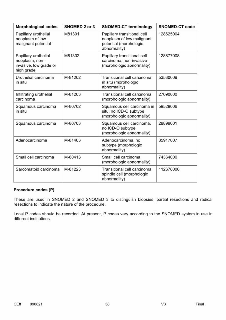
CEff 090821 38 V3 Final
Morphological codes SNOMED 2 or 3 SNOMED-CT terminology SNOMED-CT code
Papillary urothelial neoplasm of low malignant potential
M81301 Papillary transitional cell neoplasm of low malignant potential (morphologic abnormality)
128625004
Papillary urothelial neoplasm, non-invasive, low grade or high grade
M81302 Papillary transitional cell carcinoma, non-invasive (morphologic abnormality)
128877008
Urothelial carcinoma in situ
M-81202 Transitional cell carcinoma in situ (morphologic abnormality)
53530009
Infiltrating urothelial carcinoma
M-81203 Transitional cell carcinoma (morphologic abnormality)
27090000
Squamous carcinoma in situ
M-80702 Squamous cell carcinoma in situ, no ICD-O subtype (morphologic abnormality)
59529006
Squamous carcinoma M-80703 Squamous cell carcinoma, no ICD-O subtype (morphologic abnormality)
28899001
Adenocarcinoma M-81403 Adenocarcinoma, no subtype (morphologic abnormality)
35917007
Small cell carcinoma M-80413 Small cell carcinoma (morphologic abnormality)
74364000
Sarcomatoid carcinoma M-81223 Transitional cell carcinoma, spindle cell (morphologic abnormality)
112676006
Procedure codes (P) These are used in SNOMED 2 and SNOMED 3 to distinguish biopsies, partial resections and radical resections to indicate the nature of the procedure. Local P codes should be recorded. At present, P codes vary according to the SNOMED system in use in different institutions.

CEff 090821 39 V3 Final
Appendix C Histopathology reporting proforma: radical resections of renal pelvis and/or ureter
Surname……………………… Forenames………………….… Date of birth…………….. Sex…....
Hospital………….……….…… Hospital no……………….……. NHS/CHI no……………..
Date of receipt………….……. Date of reporting………..…….. Report no………………...
Pathologist……….…………… Surgeon………………….…….
RELEVANT CLINICAL INFORMATION
…………………………………………………………………………………………………………………..
NATURE OF SPECIMEN/PROCEDURE
Right ureterectomy □ Left ureterectomy □
Right nephroureterectomy □ Left nephroureterectomy □
MACROSCOPY
Tumour location…………………………………………………………………...…………………………
Number of tumours…….
Maximum tumour diameter (mm)…… or No obvious tumour visible macroscopically □
Resection margins: Not assessable □ Not involved □
Involved □ Site(s)…………………………………………………
Lymph nodes: Present □ Absent □
Site of lymph nodes………………………………………..………………..
Size of largest visible regional lymph node metastasis……. or Not applicable □
MICROSCOPY
Tumour type
Urothelial carcinoma □ Squamous cell carcinoma □ Adenocarcinoma □
Mullerian type tumour □ Small cell neuroendocrine carcinoma □
Large cell neuroendocrine carcinoma □ Other (specify) ………………………….…………..
Urothelial carcinoma subtype/variant (specify percentage if present)
Not identified □
Squamous □ ……% Glandular □ ……% Micropapillary □ ……%
Nested □ ……% Plasmacytoid □ ……% Sarcomatoid □ ……%
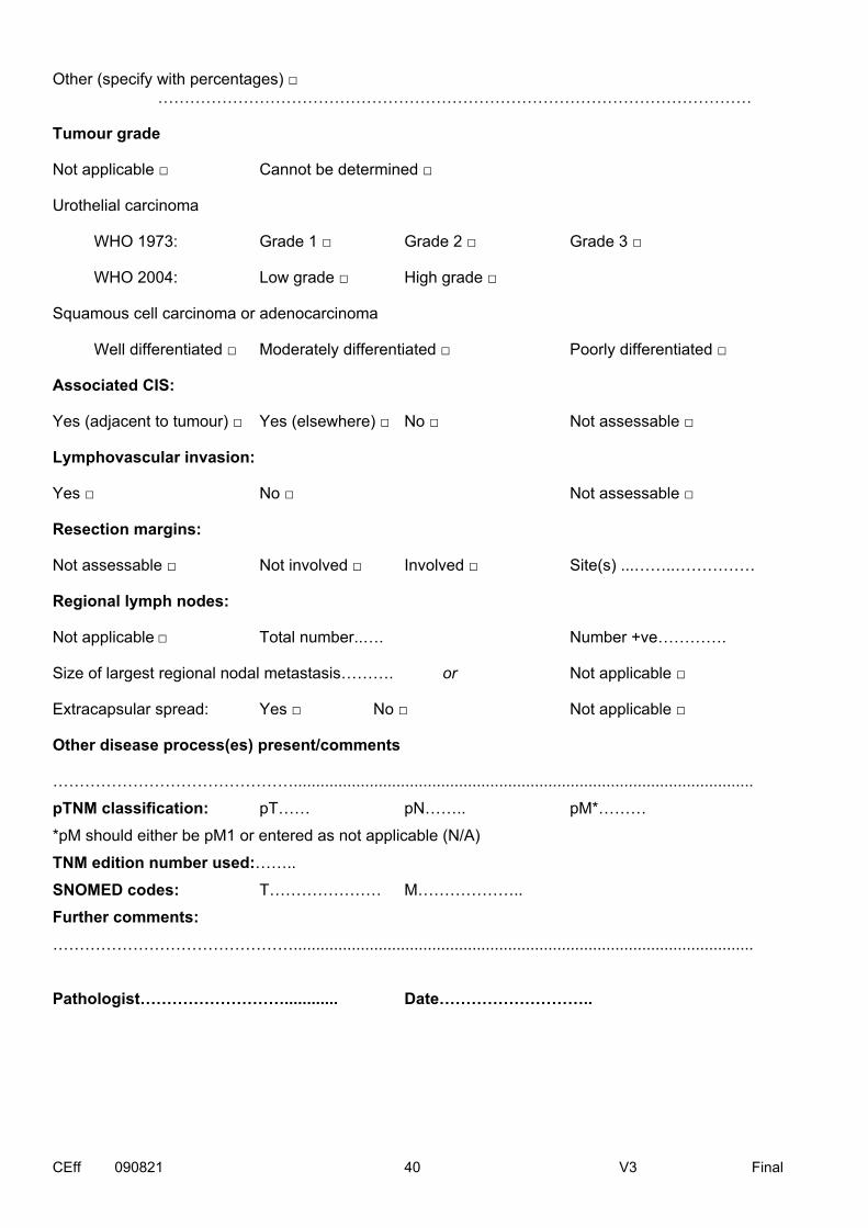
CEff 090821 40 V3 Final
Other (specify with percentages) □ …………………………………………………………………………………………………
Tumour grade
Not applicable □ Cannot be determined □
Urothelial carcinoma
WHO 1973: Grade 1 □ Grade 2 □ Grade 3 □
WHO 2004: Low grade □ High grade □
Squamous cell carcinoma or adenocarcinoma
Well differentiated □ Moderately differentiated □ Poorly differentiated □
Associated CIS:
Yes (adjacent to tumour) □ Yes (elsewhere) □ No □ Not assessable □
Lymphovascular invasion:
Yes □ No □ Not assessable □
Resection margins:
Not assessable □ Not involved □ Involved □ Site(s) ...……..……………
Regional lymph nodes:
Not applicable □ Total number..…. Number +ve………….
Size of largest regional nodal metastasis………. or Not applicable □
Extracapsular spread: Yes □ No □ Not applicable □
Other disease process(es) present/comments
………………………………………....................................................................................................... pTNM classification: pT…… pN…….. pM*……… *pM should either be pM1 or entered as not applicable (N/A) TNM edition number used:…….. SNOMED codes: T………………… M……………….. Further comments: ………………………………………....................................................................................................... Pathologist………………………............ Date………………………..

CEff 090821 41 V3 Final
Appendix D Histopathology reporting proforma: transurethral specimens (biopsy or TUR)
Surname……………………… Forenames………………….… Date of birth…………….. Sex…....
Hospital………….……….…… Hospital no……………….……. NHS/CHI no……………..
Date of receipt………….……. Date of reporting………..…….. Report no………………...
Pathologist……….…………… Surgeon………………….…….
RELEVANT CLINICAL INFORMATION
…………………………………………………………………………………………………………………..
Site of the specimen
Renal pelvis □ Ureter □ Urethra □
Bladder □ Site(s) in bladder (if known)……………………………………………….
Nature of specimen/procedure
Biopsy □ TUR □
MACROSCOPY
Weight of TUR……….(g)
MICROSCOPY
Tumour type
Urothelial carcinoma □ Squamous cell carcinoma □ Adenocarcinoma □
Mullerian type tumour □ Small cell neuroendocrine carcinoma □
Large cell neuroendocrine carcinoma □ Other (specify)………………………………….…..
Urothelial carcinoma subtype/variant (specify percentage if present)
Not identified □
Squamous □……% Glandular □……% Micropapillary □……%
Nested □……% Plasmacytoid □……% Sarcomatoid □……%
Other (specify with percentages) □ ………………………………………………………………..……

CEff 090821 42 V3 Final
Tumour grade
Not applicable □ Cannot be determined □
Urothelial carcinoma
WHO 1973: Grade 1 □ Grade 2 □ Grade 3 □
WHO 2004: Low grade □ High grade □
Squamous cell carcinoma or adenocarcinoma
Well differentiated □ Moderately differentiated □ Poorly differentiated □
Maximum extent of tumour invasion
Not assessable □ Non-invasive papillary carcinoma □
Tumour invades lamina propria (submucosa) □ Tumour invades muscularis propria □
Tumour involves prostatic ducts/acini □ Tumour invades prostatic stroma □
Other (specify) ………………………
Associated CIS:
Yes (adjacent to tumour) □ Yes (elsewhere) □ No □ Not assessable □
Lymphovascular invasion:
Yes □ No □ Not assessable □
Status of muscularis propria:
Present □ Not present □ Indeterminate □
Not applicable (e.g. for prostatic urethra biopsy) □
Other disease process(es) present/comments
……………………………………….......................................................................................................
SNOMED codes: T………………… M………………..
Further comments:
……………………………………….......................................................................................................
Pathologist………………………............ Date………………………..

CEff 090821 43 V3 Final
Appendix E Histopathology reporting proforma: urinary bladder (cystectomy or diverticulectomy)
Surname……………………… Forenames………………….… Date of birth…………….. Sex…....
Hospital………….……….…… Hospital no……………….……. NHS/CHI no……………..
Date of receipt………….……. Date of reporting………..…….. Report no………………...
Pathologist……….…………… Surgeon………………….…….
RELEVANT CLINICAL INFORMATION
…………………………………………………………………………………………………………………..
NATURE OF SPECIMEN/PROCEDURE
Radical cystectomy □ Partial cystectomy □ Diverticulectomy □
MACROSCOPY
Specimen components
Bladder □ Prostate □ Seminal vesicles □ Penile urethra □ Uterus □ Vaginal cuff □
Fallopian tubes: right □ left □ Laterality not specified □
Ovaries: right □ left □ Laterality not specified □
Ureters: right □ left □ Laterality not specified □
Regional lymph nodes: right □ left □ Laterality not specified □
Sites of regional lymph nodes (specify):…………………………………………………………….……
Non-regional lymph nodes (specify)…………………………………………………...………………….
Macroscopic tumour assessment
No macroscopically visible tumour □
or
Tumour location(s)……………………………..
Maximum tumour diameter.................. (mm) Number of tumours……….
Macroscopic extent of invasion:
Cannot be assessed □ Non-invasive tumour □
Invasion into bladder wall □ Invasion into perivesical tissue □
Invasion into peritoneal surface □
Involvement of other adjacent tissues (specify) □
Resection margins: Not assessable □ Not involved □

CEff 090821 44 V3 Final
Involved □ Site(s)…………………………………………………
Comments…………………………………………………………………………………………………….
MICROSCOPY
Tumour type
Urothelial carcinoma □ Squamous cell carcinoma □ Adenocarcinoma □
Mullerian type tumour □ Small cell neuroendocrine carcinoma □
Large cell neuroendocrine carcinoma □ Other (specify)……………………….……………..
Urothelial carcinoma subtype/variant (specify percentage if present)
Not identified □
Squamous □……% Glandular □……% Micropapillary □……%
Nested □……% Plasmacytoid □……% Sarcomatoid □……%
Other (specify with percentages) □…………………………………………………………………………
Tumour grade
Not applicable □ Cannot be determined □
Urothelial carcinoma
WHO 1973: Grade 1 □ Grade 2 □ Grade 3 □
WHO 2004: Low grade □ High grade □
Squamous cell carcinoma or adenocarcinoma
Well differentiated □ Moderately differentiated □ Poorly differentiated □
Associated CIS:
Yes (adjacent to tumour) □ Yes (elsewhere) □ No □ Not assessable □
Lymphovascular invasion:
Yes □ No □ Not assessable □
Resection margins:
Not assessable □ Not involved □ Involved □ Site(s)……………...……
Regional lymph nodes:
Not applicable □ Total number..……..... Number +ve..…….....
Extracapsular spread: Yes □ No □ Not applicable □
Common iliac nodal metastasis: Yes □ No □ Not assessable □

CEff 090821 45 V3 Final
pTNM classification: pT…… pN…….. pM*………
*pM should either be pM1 or not applicable (N/A)
TNM edition number used:……..
SNOMED codes: T………………… M………………..
Further comments:
……………………………………….......................................................................................................
Pathologist………………………............ Date………………………..

CEff 090821 46 V3 Final
Appendix F Histopathology reporting proforma: urethrectomy or urethral diverticulectomy
(For squamous tumours of distal penile urethra refer to RCPath penile dataset, 2nd edition.27)
Surname……………………… Forenames………………….… Date of birth…………….. Sex…....
Hospital………….……….…… Hospital no……………….……. NHS/CHI no……………..
Date of receipt………….……. Date of reporting………..…….. Report no………………...
Pathologist……….…………… Surgeon………………….…….
RELEVANT CLINICAL INFORMATION
…………………………………………………………………………………………………………………..
NATURE OF SPECIMEN/PROCEDURE
Urethrectomy □ Urethral diverticulectomy □ Other (specify)……………………….......................
MACROSCOPY
Other tissues/organs included (specify)……………………………………............................................
Macroscopic tumour assessment
No macroscopically visible tumour □
or
Tumour location(s) ………………………………………………………………………………..
Maximum tumour diameter........ (mm) Number of tumours……….
Macroscopic extent of invasion:
No invasion identified □
Tumour invades: Muscular wall □ Corpus spongiosum □ Corpus cavernosum □
Vagina □ Prostate □ Periprostatic tissue □
Other adjacent structure (specify)…………………………………………...……….
Resection margins:
Not assessable □ Not involved □ Involved □ Site(s)……………………
Comments…………………………………………………………………………………………………….

CEff 090821 47 V3 Final
MICROSCOPY
Tumour type
Urothelial carcinoma □ Squamous cell carcinoma □ Adenocarcinoma □
Mullerian type tumour □ Small cell neuroendocrine carcinoma □
Large cell neuroendocrine carcinoma □ Other (specify)……………………………………...
Urothelial carcinoma subtype/variant (specify percentage if present)
Not identified □
Squamous □……% Glandular □ ……% Micropapillary □……%
Nested □……% Plasmacytoid □……% Sarcomatoid □……%
Other (specify with percentages) □……………………………………
Tumour grade
Not applicable □ Cannot be determined □
Urothelial carcinoma
WHO 1973: Grade 1 □ Grade 2 □ Grade 3 □
WHO 2004: Low grade □ High grade □
Squamous cell carcinoma or adenocarcinoma
Well differentiated □ Moderately differentiated □ Poorly differentiated □
Associated CIS:
Yes (adjacent to tumour) □ Yes (elsewhere) □ No □ Not assessable □
Lymphovascular invasion:
Yes □ No □ Not assessable □
Resection margins:
Not assessable □ Not involved □ Involved □ Site(s)…………...………
Regional lymph nodes:
Not applicable □ Total number..……..... Number +ve…….
Extracapsular spread: Yes □ No □ Not applicable □
Non-regional nodal metastasis: Yes □ No □ Not assessable □
pTNM classification: pT…… pN…….. pM*………
*pM should either be pM1 or not applicable (N/A)

CEff 090821 48 V3 Final
TNM edition number used:……..
SNOMED codes: T………………… M………………..
Further comments:
……………………………………….......................................................................................................
Pathologist………………………............ Date………………………..

CEff 090821 49 V3 Final
Appendix G Histopathology reporting proforma: radical resections of renal pelvis and/or ureter in list format
Element name Values Implemen-
tation comments
COSD v8 COSD v9
Nature of Specimen/ Procedure
Single selection value list: • Right ureterectomy • Left ureterectomy • Right
nephroureterectomy • Left
nephroureterectomy
CR0760 – All values = EX (Excision) CR0970 – All values = 1 (Primary tumour) CR0820 • Right ureterectomy
= R (Right) • Left ureterectomy =
L (Left) • Right
nephroureterectomy = R (Right)
• Left nephroureterectomy = L (Left)
pCR0760 – All values = EX (Excision) pCR0970 – All values = 1 (Primary tumour) pCR0820 • Right ureterectomy
= R (Right) • Left ureterectomy =
L (Left) • Right
nephroureterectomy = R (Right)
• Left nephroureterectomy = L (Left)
Tumour location
Free text
Number of tumours
Number pCR0840 Blank = 9 (Not known) 1 = N (No, no synchronous tumours present) >1 = Y (Yes, synchronous tumours present)
pCR0840 Blank = 9 (Not known) 1 = N (No, no synchronous tumours present) >1 = Y (Yes, synchronous tumours present)
Maximum tumour diameter (mm)
Size (in mm) or ‘No obvious tumour visible macroscopically’
CR0830 pCR0830
Macroscopic resection margins
Single selection value list: • Not assessable • Not involved • Involved Site(s)…
Lymph nodes
Single selection value list: • Present • Absent Site of lymph nodes…

CEff 090821 50 V3 Final
Size of largest visible regional lymph node metastasis
Size in mm Or not applicable
Tumour type
• Urothelial carcinoma
• Squamous cell carcinoma
• Adenocarcinoma • Mullerian type
tumour • Small cell
neuroendocrine carcinoma
• Large cell neuroendocrine carcinom
• Other (specify) …
Urothelial carcinoma subtype/ variant (specify percentage if present)
• Not identified • Squamous… % • Glandular… % • Micropapillary…% • Nested… % • Plasmacytoid… % • Sarcomatoid… % • Other (specify with
percentages)
Tumour grade
• Not applicable • Cannot be
determined • Urothelial
carcinoma o WHO 1973: Grade 1 Grade 2 Grade 3
o WHO 2004: Low grade High grade
• Squamous cell carcinoma or adenocarcinoma o Well
differentiated o Moderately
differentiated
CR0860 • Not applicable = GX
(Grade of differentiation is not appropriate or cannot be assessed)
• Cannot be determined = = GX (Grade of differentiation is not appropriate or cannot be assessed)
• Urothelial carcinoma o WHO
1973/2004: Grade 1/Low
grade = G1 Grade 2/Low
grade = G2
pCR0860 • Not applicable = GX
(Grade of differentiation is not appropriate or cannot be assessed)
• Cannot be determined = = GX (Grade of differentiation is not appropriate or cannot be assessed)
• Urothelial carcinoma o WHO
1973/2004: Grade 1/Low
grade = G1 Grade 2/Low
grade = G2

CEff 090821 51 V3 Final
o Poorly differentiated
Grade 2/High grade = G3
Grade 3/High grade = G4
• Squamous cell carcinoma or adenocarcinoma o Well
differentiated = G1
o Moderately differentiated = G2
o Poorly differentiated = G3
Grade 2/High grade = G3
Grade 3/High grade = G4
• Squamous cell carcinoma or adenocarcinoma o Well
differentiated = G1
o Moderately differentiated = G2
o Poorly differentiated = G3
Associated CIS
Single selection value list: • Yes (adjacent to
tumour • Yes (elsewhere) • No • Not assessable
Lympho vascular invasion
Single selection value list: • Yes • No • Not assessable
CR0870 Yes = YU (Yes - vascular/lymphatic invasion present) No = NU (No - vascular/lymphatic invasion not present) Not assessable = XX (Cannot be assessed)
pCR0870 Yes = YU (Yes - vascular/lymphatic invasion present) No = NU (No - vascular/lymphatic invasion not present) Not assessable = XX (Cannot be assessed)
Microscopic resection margins
Single selection value list: • Not assessable • Not involved • Involved Site(s)…
CR0880 • Not assessable =
98 (Not applicable) • Not involved = 01
(Excision margins are clear (distance from margin not stated))
• Involved = 05 (Tumour reaches excision margin)
pCR0880 • Not assessable =
98 (Not applicable) • Not involved = 01
(Excision margins are clear (distance from margin not stated))
• Involved = 05 (Tumour reaches excision margin)
Regional lymph nodes
Total number… Number + ve… Or not applicable…
Total number = CR0890 Number +ve = CR0900
Total number = pCR0890 Number +ve = pCR0900

CEff 090821 52 V3 Final
Regional lymph nodes: Size of largest regional nodal metastasis
Size in mm Or not applicable
Regional lymph nodes: Extracapsular spread
Single selection value list: • Yes • No • Not applicable
Other disease process(es) present/ comments
Free text
pTNM classification
pT… pN… pM*
*pM should either be pM1 or entered as not applicable (N/A)
pT = CR0910 pN = CR0920 pM = CR0930
pT = pCR0910 pN = pCR0920 pM = pCR0930
TNM edition number used
Free text CR6820 pCR6820
SNOMED codes
T… M…
T = CR6410 M = CR6420
T = pCR6410 M = pCR6420
Further comments
Free text
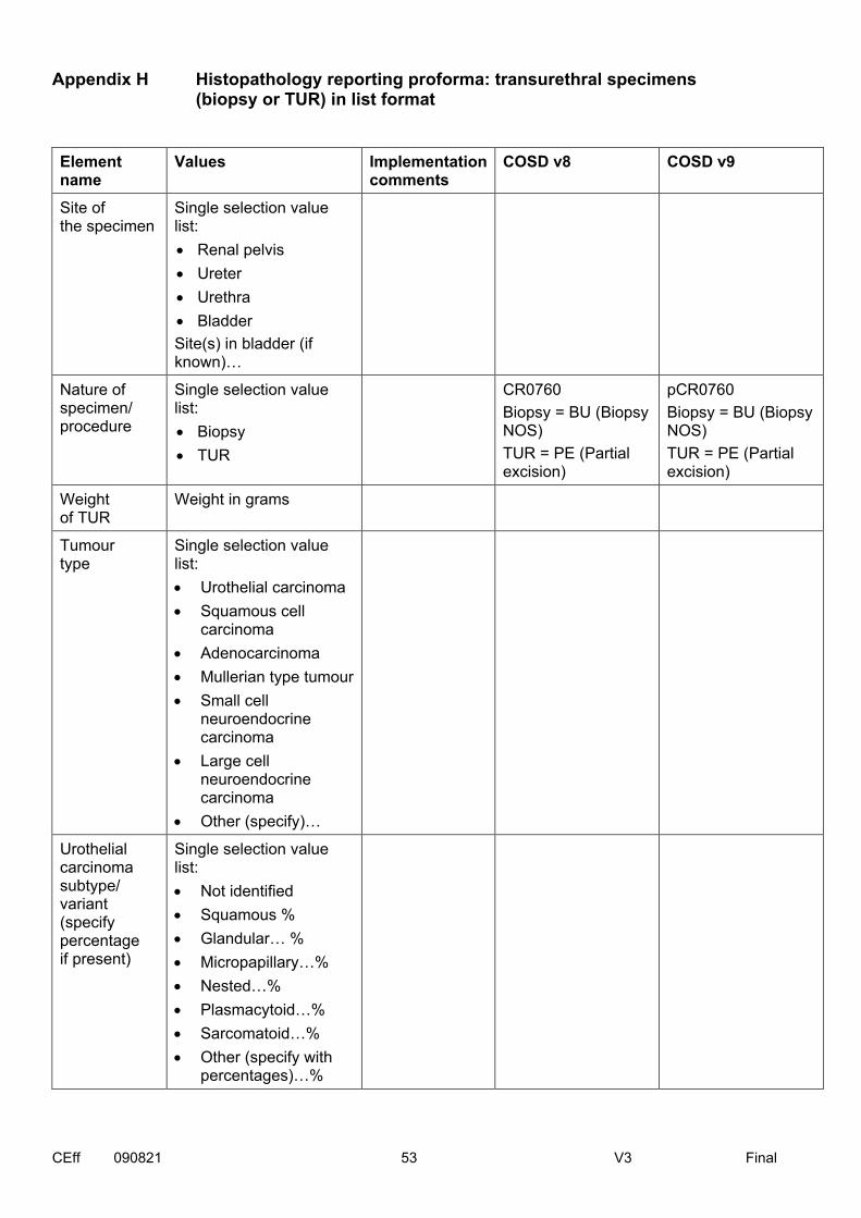
CEff 090821 53 V3 Final
Appendix H Histopathology reporting proforma: transurethral specimens (biopsy or TUR) in list format
Element name
Values Implementation comments
COSD v8 COSD v9
Site of the specimen
Single selection value list: • Renal pelvis • Ureter • Urethra • Bladder Site(s) in bladder (if known)…
Nature of specimen/ procedure
Single selection value list: • Biopsy • TUR
CR0760 Biopsy = BU (Biopsy NOS) TUR = PE (Partial excision)
pCR0760 Biopsy = BU (Biopsy NOS) TUR = PE (Partial excision)
Weight of TUR
Weight in grams
Tumour type
Single selection value list: • Urothelial carcinoma • Squamous cell
carcinoma • Adenocarcinoma • Mullerian type tumour • Small cell
neuroendocrine carcinoma
• Large cell neuroendocrine carcinoma
• Other (specify)…
Urothelial carcinoma subtype/ variant (specify percentage if present)
Single selection value list: • Not identified • Squamous % • Glandular… % • Micropapillary…% • Nested…% • Plasmacytoid…% • Sarcomatoid…% • Other (specify with
percentages)…%

CEff 090821 54 V3 Final
Tumour grade
Single selection value list: • Not applicable • Cannot be determined • Urothelial carcinoma • WHO 1973:
o Grade 1 o Grade 2 o Grade 3
• Who 2004: o Low grade o High grade
CR0860 • Not applicable =
GX (Grade of differentiation is not appropriate or cannot be assessed)
• Cannot be determined = = GX (Grade of differentiation is not appropriate or cannot be assessed)
UR15290 (BLADDER UROTHELIAL TUMOURS ONLY) • Not applicable =
X (Not applicable)
• Urothelial carcinoma o WHO
1973/2004: Grade
1/Low-grade = L (Low)
Grade 2/Low-grade = L (Low)
Grade 2/High-grade = H (High)
Grade 3/High-grade = H (High)
pCR0860 • Not applicable =
GX (Grade of differentiation is not appropriate or cannot be assessed)
• Cannot be determined = = GX (Grade of differentiation is not appropriate or cannot be assessed)-
pUR15290 (BLADDER UROTHELIAL TUMOURS ONLY) • Not applicable =
X (Not applicable)
• Urothelial carcinoma o WHO
1973/2004: Grade
1/Low-grade = L (Low)
Grade 2/Low-grade = L (Low)
Grade 2/High-grade = H (High)
Grade 3/High-grade = H (High)

CEff 090821 55 V3 Final
Squamous cell carcinoma or adeno carcinoma
Single selection value list: • Well differentiated • Moderately
differentiated • Poorly differentiated
CR0860 • Squamous cell
carcinoma or adenocarcinoma o Well
differentiated = G1
o Moderately differentiated = G2
o Poorly differentiated = G3
pCR0860 • Squamous cell
carcinoma or adenocarcinoma o Well
differentiated = G1
o Moderately differentiated = G2
o Poorly differentiated = G3
Maximum extent of tumour invasion
Single selection value list: • Not assessable • Non-invasive
papillary carcinoma • Tumour invades
lamina propria (submucosa)
• Tumour invades muscularis propria
• Tumour involves prostatic ducts/acini
• Tumour invades prostatic stroma
• Other(specify)
Associated CIS
Single selection value list: • Yes (adjacent to
tumour) • Yes (elsewhere) • No • Not assessable
Lymphovascular invasion
Single selection value list: • Yes • No • Not assessable
CR0870 Yes = YU (Yes - vascular/ lymphatic invasion present) No = NU (No - vascular/ lymphatic invasion not present)
pCR0870 Yes = YU (Yes - vascular/lymphatic invasion present) No = NU (No - vascular/lymphatic invasion not present) Not assessable = XX (Cannot be assessed)

CEff 090821 56 V3 Final
Not assessable = XX (Cannot be assessed)
Status of muscularis propria
Single selection value list: • Present • Not present • Indeterminate • Not applicable (e.g.
for prostatic urethra biopsy)
UR15120 (BLADDER ONLY) • Present = 1
(Present) • Not present = 2
(Absent) • Indeterminate =
9 (Not known)
• Not applicable (e.g. for prostatic urethra biopsy) = Leave blank
pUR15120 (BLADDER ONLY) • Present = 1
(Present) • Not present = 2
(Absent) • Indeterminate =
9 (Not known) • Not applicable
(e.g. for prostatic urethra biopsy) = Leave blank
Other disease process(es) present/ comments
Free text
SNOMED codes
T… M…
T = CR6410 M = CR6420
T = pCR6410 M = pCR6420
Further comments
Free text

CEff 090821 57 V3 Final
Appendix I Histopathology reporting proforma: urinary bladder (cystectomy or diverticulectomy) in list format
Element name Values Implementation
comments COSD v8 COSD v9
Nature of specimen/ procedure
Single selection value list: • Radical
cystectomy • Partial
cystectomy • Diverticulectomy
CR0760 • Radical
cystectomy = RE (Radical excision)
• Partial cystectomy = PE (Partial excision)
• Diverticulectomy = EX (Excision)
CR0970 – All values = 1 (Primary tumour)
pCR0760 • Radical
cystectomy = RE (Radical excision)
• Partial cystectomy = PE (Partial excision)
• Diverticulectomy = EX (Excision)
pCR0970 – All values = 1 (Primary tumour)
Specimen components
Single selection value list: • Bladder • Prostate • Seminal vesicles • Penile urethra • Uterus • Vaginal cuff
Fallopian tubes
Single selection value list: • Right • Left • Laterality not
specified
Ovaries Single selection value list: • Right • Left • Laterality not
specified
Ureters Single selection value list: • Right • Left • Laterality not
specified

CEff 090821 58 V3 Final
Regional lymph nodes
Single selection value list: • Right • Left • Laterality not
specified
Site of regional lymph nodes (specify)
Free text
Non-regional lymph nodes (specify)
Free text
Macroscopic tumour assessment (or indicate if no macroscopically visible tumour)
Tumour location… Maximum tumour diameter… Number of tumours…
Macroscopic extent of invasion
Single selection value list: • Cannot be
assessed • Non-invasive
tumour • Invasion into
bladder wall • Invasion into
perivesical tissue • Invasion into
peritoneal surface
• Involvement of other adjacent tissues (specify)
Resection margins
Single selection value list:
• Not assessable • Not involved • Involved Site(s)…

CEff 090821 59 V3 Final
Tumour type
Single selection value list: • Urothelial
carcinoma • Squamous cell
carcinoma • Adenocarcinoma • Mullerian type
tumour • Small cell
neuroendocrine carcinoma
• Large cell neuroendocrine carcinoma
• Other (specify)…
Urothelial carcinoma subtype/ variant (specify percentage if present)
• Not identified • Squamous…% • Glandular…% • Micropapillary…
% • Nested…% • Plasmacytoid…
% • Sarcomatoid…% • Other (specify
with percentages)…%
Tumour grade
Single selection value list: • Not applicable • Cannot be
determined • Urothelial
carcinoma o WHO 1973: Grade 1 Grade 2 Grade 3
o WHO 2004: Low grade High grade
CR0860 • Not applicable =
GX (Grade of differentiation is not appropriate or cannot be assessed)
• Cannot be determined = = GX (Grade of differentiation is not appropriate or cannot be assessed)-
UR15290 (BLADDER UROTHELIAL TUMOURS ONLY) • Not applicable = X
(Not applicable)
pCR0860 • Not applicable =
GX (Grade of differentiation is not appropriate or cannot be assessed)
• Cannot be determined = = GX (Grade of differentiation is not appropriate or cannot be assessed)-
pUR15290 (BLADDER UROTHELIAL TUMOURS ONLY) • Not applicable =
X (Not applicable)

CEff 090821 60 V3 Final
• Urothelial carcinoma o WHO
1973/2004: Grade 1/Low
grade = L (Low)
Grade 2/Low grade = L (Low)
Grade 2/High grade = H (High)
Grade 3/High grade = H (High)
• Urothelial carcinoma o WHO
1973/2004: Grade
1/Low-grade = L (Low)
Grade 2/Low-grade = L (Low)
Grade 2/High-grade = H (High)
Grade 3/High-grade = H (High)
Squamous cell carcinoma or adenocarcinoma
Single selection value list: • Well
differentiated • Moderately
differentiated • Poorly
differentiated
CR0860 • Squamous cell
carcinoma or adenocarcinoma o Well
differentiated = G1
o Moderately differentiated = G2
o Poorly differentiated = G3
pCR0860 • Squamous cell
carcinoma or adenocarcinoma o Well
differentiated = G1
o Moderately differentiated = G2
o Poorly differentiated = G3
Associated CIS
Single selection value list: • Yes (adjacent to
tumour) • Yes (elsewhere) • No • Not assessable
Lymphovascular invasion
Single selection value list: • Yes • No • Not assessable
CR0870 Yes = YU (Yes – vascular/ lymphatic invasion present) No = NU (No – vascular/ lymphatic invasion not present) Not assessable = XX (Cannot be assessed)
pCR0870 Yes = YU (Yes - vascular/lymphatic invasion present) No = NU (No - vascular/lymphatic invasion not present) Not assessable = XX (Cannot be assessed)

CEff 090821 61 V3 Final
Resection margins
Single selection value list: • Not assessable • Not Involved • Involved Site(s)…
CR0880 • Not assessable =
98 (Not applicable)
• Not involved = 01 (Excision margins are clear (distance from margin not stated))
• Involved = 05 (Tumour reaches excision margin)
pCR0880 • Not assessable =
98 (Not applicable)
• Not involved = 01 (Excision margins are clear (distance from margin not stated))
• Involved = 05 (Tumour reaches excision margin)
Regional lymph nodes
Not applicable Total number… Number + ve…
Total number = CR0890 Number +ve = CR0900
Total number = pCR0890 Number +ve = pCR0900
Extracapsular spread
Single selection value list: • Yes • No • Not applicable
Common iliac nodal metastasis
Single selection value list: • Yes • No • Not assessable
pTNM classification
pT… pN… pM*…
*pM should either be pM1 or not applicable (N/A)
pT = CR0910 pN = CR0920 pM = CR0930
pT = pCR0910 pN = pCR0920 pM = pCR0930
TNM edition number used
Free text CR6820 pCR6820
SNOMED codes
T… M…
T = CR6410 M = CR6420
T = pCR6410 M = pCR6420
Further comments
Free text
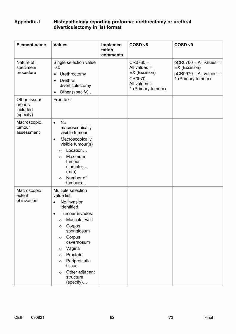
CEff 090821 62 V3 Final
Appendix J Histopathology reporting proforma: urethrectomy or urethral diverticulectomy in list format
Element name Values Implemen
tation comments
COSD v8 COSD v9
Nature of specimen/ procedure
Single selection value list: • Urethrectomy • Urethral
diverticulectomy • Other (specify)…
CR0760 – All values = EX (Excision) CR0970 – All values = 1 (Primary tumour)
pCR0760 – All values = EX (Excision) pCR0970 – All values = 1 (Primary tumour)
Other tissue/ organs included (specify)
Free text
Macroscopic tumour assessment
• No macroscopically visible tumour
• Macroscopically visible tumour(s) o Location… o Maximum
tumour diameter… (mm)
o Number of tumours…
Macroscopic extent of invasion
Multiple selection value list: • No invasion
identified • Tumour invades:
o Muscular wall o Corpus
spongiosum o Corpus
cavernosum o Vagina o Prostate o Periprostatic
tissue o Other adjacent
structure (specify)…
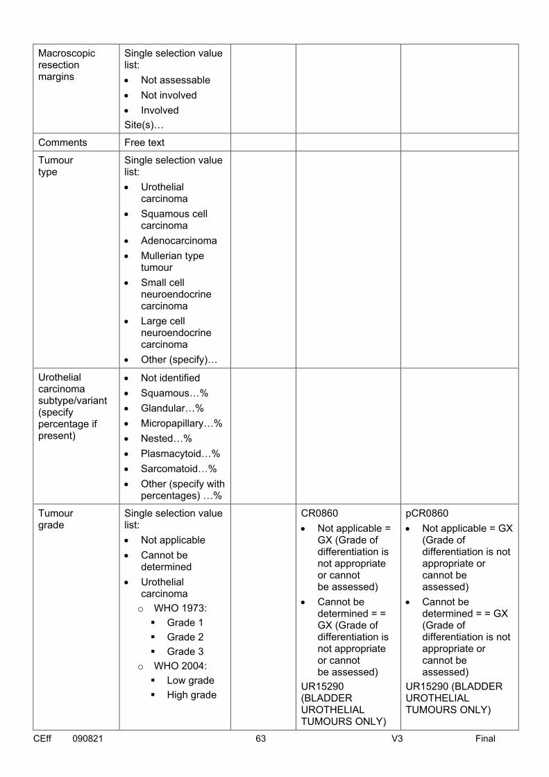
CEff 090821 63 V3 Final
Macroscopic resection margins
Single selection value list: • Not assessable • Not involved • Involved Site(s)…
Comments Free text
Tumour type
Single selection value list: • Urothelial
carcinoma • Squamous cell
carcinoma • Adenocarcinoma • Mullerian type
tumour • Small cell
neuroendocrine carcinoma
• Large cell neuroendocrine carcinoma
• Other (specify)…
Urothelial carcinoma subtype/variant (specify percentage if present)
• Not identified • Squamous…% • Glandular…% • Micropapillary…% • Nested…% • Plasmacytoid…% • Sarcomatoid…% • Other (specify with
percentages) …%
Tumour grade
Single selection value list: • Not applicable • Cannot be
determined • Urothelial
carcinoma o WHO 1973: Grade 1 Grade 2 Grade 3
o WHO 2004: Low grade High grade
CR0860 • Not applicable =
GX (Grade of differentiation is not appropriate or cannot be assessed)
• Cannot be determined = = GX (Grade of differentiation is not appropriate or cannot be assessed)
UR15290 (BLADDER UROTHELIAL TUMOURS ONLY)
pCR0860 • Not applicable = GX
(Grade of differentiation is not appropriate or cannot be assessed)
• Cannot be determined = = GX (Grade of differentiation is not appropriate or cannot be assessed)
UR15290 (BLADDER UROTHELIAL TUMOURS ONLY)

CEff 090821 64 V3 Final
• Not applicable = X (Not applicable)
• Urothelial carcinoma o WHO
1973/2004: Grade 1/
Low grade = L (Low)
Grade 2/ Low grade = L (Low)
Grade 2/ High grade = H (High)
Grade 3/ High grade = H (High)
• Not applicable = X (Not applicable)
• Urothelial carcinoma o WHO
1973/2004: Grade 1/Low
grade = L (Low)
Grade 2/Low grade = L (Low)
Grade 2/High grade = H (High)
Grade 3/High grade = H (High)
Squamous cell carcinoma or adenocarcinoma
Single selection value list: • Well differentiated • Moderately
differentiated • Poorly
differentiated
CR0860 • Squamous cell
carcinoma or adenocarcinoma o Well
differentiated = G1
o Moderately differentiated = G2
o Poorly differentiated = G3
pCR0860 • Squamous cell
carcinoma or adenocarcinoma o Well
differentiated = G1
o Moderately differentiated = G2
o Poorly differentiated = G3
Associated CIS
Single selection value list: • Yes (adjacent to
tumour) • Yes (elsewhere) • No • Not assessable

CEff 090821 65 V3 Final
Lymphovascular invasion
Single selection value list: • Yes • No • Not assessable
CR0870 Yes = YU (Yes - vascular/ lymphatic invasion present) No = NU (No - vascular/ lymphatic invasion not present) Not assessable = XX (Cannot be assessed)
pCR0870 Yes = YU (Yes - vascular/lymphatic invasion present) No = NU (No - vascular/lymphatic invasion not present) Not assessable = XX (Cannot be assessed)
Microscopic resection margins
Single selection value list: • Not Involved • Involved • Not assessable Site(s)…
CR0880 • Not assessable
= 98 (Not applicable)
• Not involved = 01 (Excision margins are clear (distance from margin not stated))
• Involved = 05 (Tumour reaches excision margin)
pCR0880 • Not assessable =
98 (Not applicable) • Not involved = 01
(Excision margins are clear (distance from margin not stated))
• Involved = 05 (Tumour reaches excision margin)
Regional lymph nodes
Not applicable Total number… Number + ve…
Total number = CR0890 Number +ve = CR0900
Total number = pCR0890 Number +ve = pCR0900
Extracapsular spread
Single selection value list: • Yes • No • Not applicable
Non-regional nodal metastasis
Single selection value list: • Yes • No • Not assessable
pTNM classification
pT… pN… pM*…
*pM should either be pM1 or not applicable (N/A)
pT = CR0910 pN = CR0920 pM = CR0930
pT = pCR0910 pN = pCR0920 pM = pCR0930
TNM edition number used
Free text CR6820 pCR6820

CEff 090821 66 V3 Final
SNOMED codes
T… M…
T = CR6410 M = CR6420
T = pCR6410 M = pCR6420
Further comments
Free text

CEff 090821 67 V3 Final
Appendix K Summary table – explanation of grades of evidence (modified from Palmer K et al. BMJ 2008;337:1832) Grade (level) of evidence
Nature of evidence
Grade A At least one high-quality meta-analysis, systematic review of randomised controlled trials or a randomised controlled trial with a very low risk of bias and directly attributable to the target cancer type or A body of evidence demonstrating consistency of results and comprising mainly well-conducted meta-analyses, systematic reviews of randomised controlled trials or randomised controlled trials with a low risk of bias, directly applicable to the target cancer type.
Grade B A body of evidence demonstrating consistency of results and comprising mainly high-quality systematic reviews of case-control or cohort studies and high-quality case-control or cohort studies with a very low risk of confounding or bias and a high probability that the relation is causal and which are directly applicable to the target cancer type or Extrapolation evidence from studies described in A.
Grade C A body of evidence demonstrating consistency of results and including well-conducted case-control or cohort studies and high-quality case-control or cohort studies with a low risk of confounding or bias and a moderate probability that the relation is causal and which are directly applicable to the target cancer type or Extrapolation evidence from studies described in B.
Grade D Non-analytic studies such as case reports, case series or expert opinion or Extrapolation evidence from studies described in C.
Good practice point (GPP)
Recommended best practice based on the clinical experience of the authors of the writing group.
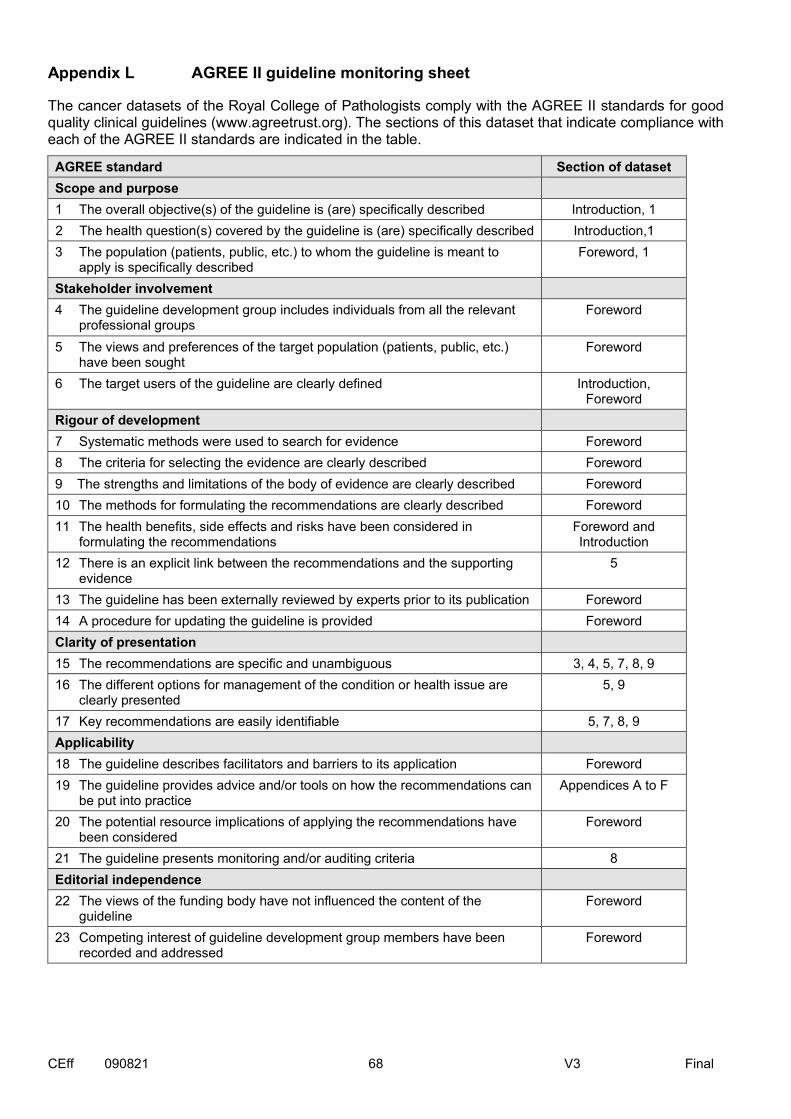
CEff 090821 68 V3 Final
Appendix L AGREE II guideline monitoring sheet The cancer datasets of the Royal College of Pathologists comply with the AGREE II standards for good quality clinical guidelines (www.agreetrust.org). The sections of this dataset that indicate compliance with each of the AGREE II standards are indicated in the table.
AGREE standard Section of dataset Scope and purpose 1 The overall objective(s) of the guideline is (are) specifically described Introduction, 1 2 The health question(s) covered by the guideline is (are) specifically described Introduction,1 3 The population (patients, public, etc.) to whom the guideline is meant to
apply is specifically described Foreword, 1
Stakeholder involvement 4 The guideline development group includes individuals from all the relevant
professional groups Foreword
5 The views and preferences of the target population (patients, public, etc.) have been sought
Foreword
6 The target users of the guideline are clearly defined Introduction, Foreword
Rigour of development 7 Systematic methods were used to search for evidence Foreword 8 The criteria for selecting the evidence are clearly described Foreword 9 The strengths and limitations of the body of evidence are clearly described Foreword 10 The methods for formulating the recommendations are clearly described Foreword 11 The health benefits, side effects and risks have been considered in
formulating the recommendations Foreword and Introduction
12 There is an explicit link between the recommendations and the supporting evidence
5
13 The guideline has been externally reviewed by experts prior to its publication Foreword 14 A procedure for updating the guideline is provided Foreword Clarity of presentation 15 The recommendations are specific and unambiguous 3, 4, 5, 7, 8, 9 16 The different options for management of the condition or health issue are
clearly presented 5, 9
17 Key recommendations are easily identifiable 5, 7, 8, 9 Applicability 18 The guideline describes facilitators and barriers to its application Foreword 19 The guideline provides advice and/or tools on how the recommendations can
be put into practice Appendices A to F
20 The potential resource implications of applying the recommendations have been considered
Foreword
21 The guideline presents monitoring and/or auditing criteria 8 Editorial independence 22 The views of the funding body have not influenced the content of the
guideline Foreword
23 Competing interest of guideline development group members have been recorded and addressed
Foreword