david miller, dpm podiatry - affinity health · david miller, dpm podiatry . most common types of...
TRANSCRIPT

Foot and Ankle Tendinopathies David Miller, DPM
Podiatry

Most Common Types of
Tendinopathy Affecting the
Foot and Ankle
Achilles
Peroneal

Achilles Tendinopathy
• Terminology: confusing?
– Tendinitis, Tendonitis, Paratenonitis,
Tendovaginitis, Tenosynovitis, Achillodynia
– Definition: Umbrella term for disease of a
tendon (tendonitis/tendinosis)
– Suggested that pain, swelling, and impaired
performance be labeled “tendinopathy”
– Tendonitis Tendinosis

Achilles Tendinopathy
• “Tendonitis” is often used to depict tendon
pain and swelling, inflammatory cells are
infrequently seen except with tendon
rupture
• Many clinicians use term tendonitis to
describe what is actually a tendinosis
• Tendinosis is a degenerative process
without histological or clinical signs of
inflammation within the tendon

Achilles Tendinopathy
• Anatomy: Achilles forms at the junction of
the medial and lateral gastroc and soleus
muscles and inserts into the posterior
calcaneus
– Surrounded by the paratenon
– The mesotenon (middle layer)- the main blood
supply
– Blood flow lowered during contraction and can
cease completely


Achilles Tendinopathy
• Anatomy cont:
– Tendons transmit force generated by muscle
to bone
• Tensile strength is related to thickness and
collagen content
• Cross sectional area of 1sq cm can support 500-
1000 kg
• Loading of the Achilles reaches 9 kN during
running ~ 12.5 x’s the body weight and 2.6 kN
during slow walking

Achilles Tendinopathy
• Histology of biopsied tendon:
– Reveals cellular activation and increases in
cell numbers and ground substance, collagen
disarray, and neovascularization
– Prostaglandin inflammatory elements are not
present but substance P has been isolated

Achilles Tendinopathy
• Etiology
– Tendon injury: acute versus chronic
– Acute: extrinsic factors predominate
– Chronic: intrinsic and extrinsic factors interact
– Intrinsic factor: tendon vascularity, gastroc-
soleus dysfunction, age, gender, body weight
and height, pes cavus, lateral ankle instability,
excessive pronation (whipping action on the
Achilles), forefoot varus

Achilles Tendinopathy
• Etiology cont:
– Extrinsic factors: changes in training pattern,
poor technique, previous injury, footwear and
training on hard, slippery or slanted surfaces
– Excessive loading of tendons during training
is the #1 stimulus for degeneration
– Age: molecular properties of collagen,
decreased water content and decrease in
vascularity, Achilles becomes weak and stiff,
the older athlete needs a stretching program

Achilles Tendinopathy
• Etiology cont:
– Fluoroquinolones: affects tendon at a cellular
level

Achilles Tendinopathy
• Presentation
– Pain: initially at the beginning and end of a
training session and lessened pain in between
– Later: pain during exercise and then
interference with ADLs
– Acute phase the tendon is swollen and
edematous
– Chronic phase a tender nodular swelling
present and is believed to be tendinosis

Achilles Tendinopathy
• Presentation cont:
– Post static dyskinesia
– Inability to wear a closed shoe

Achilles Tendinopathy
Achilles Tendinopathy
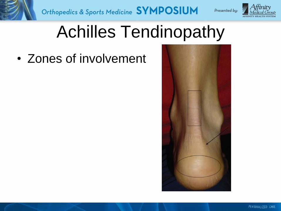
• Zones of involvement

Achilles Tendinopathy
• Zone 1
– Tendinitis
– Tendinosis
• Zone 2
– Retrocalcaneal bursitis
• Zone 3: spur causes pain?
– Tendinitis
– Tendinosis

• Imaging – X-ray
– Ultrasound
• Quick, safe, inexpensive
• Operator dependent, limited soft tissue contrast
• Less expensive than MRI
– MRI
• Provides extensive information on the internal morphology
and surrounding structures
• Peritendinitis vs tendinosis
• Good correlation between MRI findings and surgical
findings
Achilles Tendinopathy

Achilles Tendinopathy
• Xray
Asymptomatic left Symptomatic right
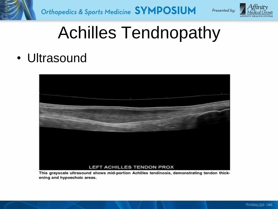
• Ultrasound
Achilles Tendnopathy
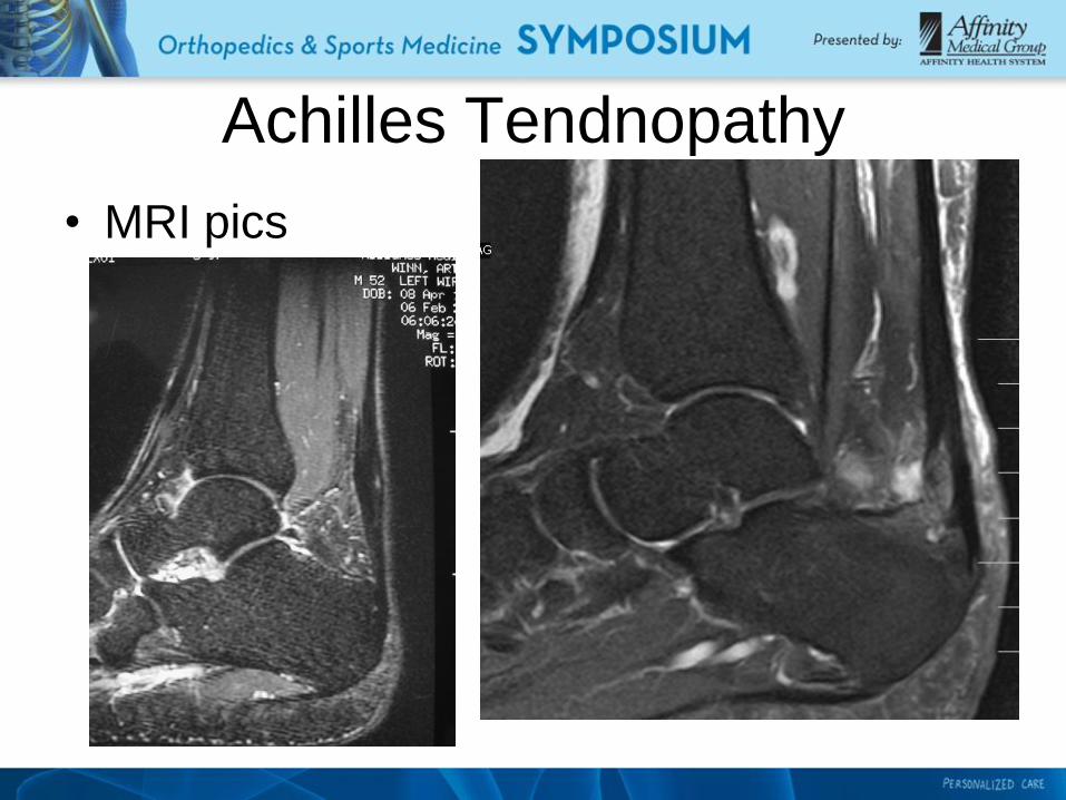
• MRI pics
Achilles Tendnopathy

• Management – Conservative treatment is customary
– Earlier treatment better outcome
– Initial treatment
• Activity modification- decrease activity at injured site
but normal activity elsewhere
• Correct training errors
• Addressing muscle weakness
• Correcting biomechanics
• Complete rest could be detrimental since collagen
repair and remodelling is stimulated by tendon loading
Achilles Tendnopathy

• Management cont:
– Cryotherapy in acute phase
– Therapeutic ultrasound
– Deep friction massage: advocated for
tendinopathy along with stretching
– Stretching and strengthening of the posterior
muscle group
– Eccentric muscle training
Achilles Tendnopathy
• Eccentric muscle training
– Superior to concentric muscle training
– More effective for mid substance tendinopathy
vs. insertional tendinopathy
– Some believe eccentric loading may lengthen
the muscle tendon unit over time and increase
its ability to bear load
– ? Repetitive eccentric training may damage
abnormal vessels and nerves in the tendon
Achilles Tendinopathy
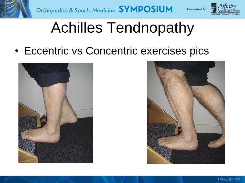
• Eccentric vs Concentric exercises pics
Achilles Tendnopathy

Achilles Tendinopathy
• Concentric stretching
– Concentric exercise is done by toe raises with
progressive weight applied
– Eccentric (“negatives” in weight-lifting)
• Management cont:
– NSAIDS questioned due to absence of
prostaglandin inflammatory mediators within
diseased tendon, especially chronic
– Corticosteroid injections: controversial due to
concern of Achilles rupture, generally should
be avoided
– Others: ESWT (low and high energy), PRP
• Investigational?
Achilles Tendinopathy

• Management continued
– Surgical management for those who fail an
exhaustive non-operative program
– Various surgical techniques have been used:
most involve removal of inflamed or diseased
tissue and decompression of mechanical
pressure from the adjacent calcaneus
(Haglund’s deformity / Exostosis)
Achilles Tendinopathy

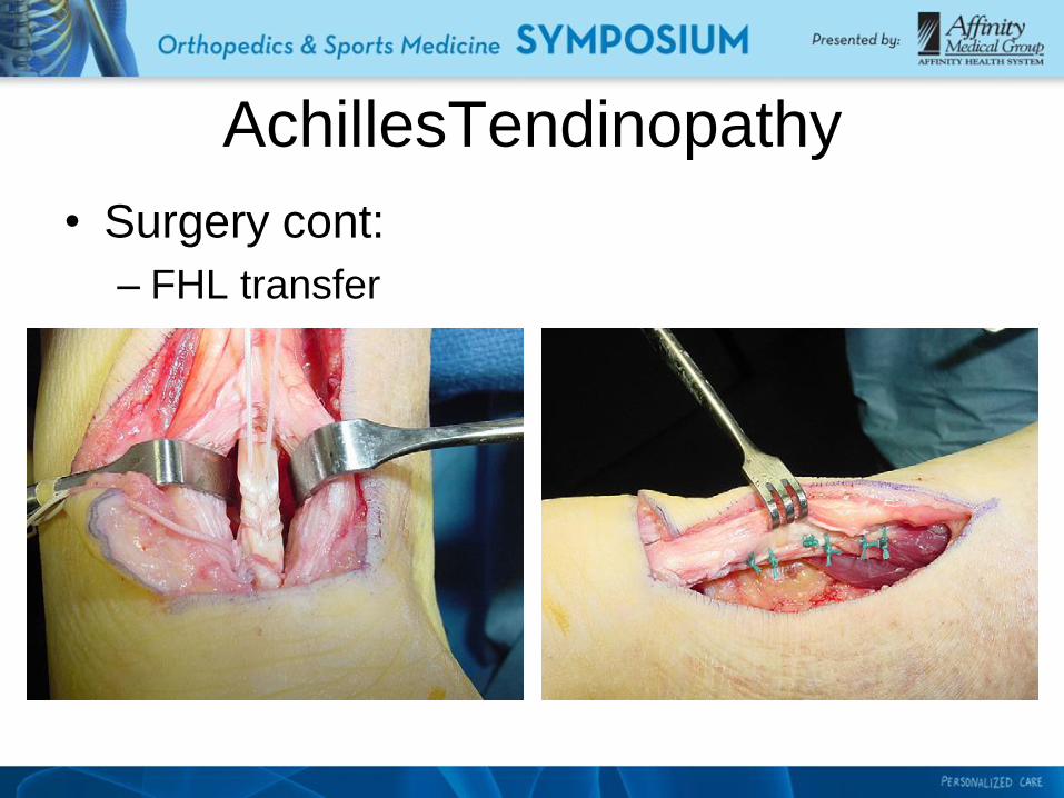
• Management cont:
– Surgery
• FHL transfer utilized if significantly diseased
tendon present
• Generally good results but can involve a lengthy
recovery.
Achilles Tendinopathy

• Surgery
– Non-insertional
AchillesTendinopathy

AchillesTendinopathy
• Surgery
– Insertional
AchillesTendinopathy
• Surgery cont:
– FHL transfer

Peroneal Tendinopathy
• Anatomy:
– Located in the lateral compartment of the leg
– Everters of the of the foot and ankle
– Share the same sheath until the peroneal
tubercle
– Os peroneum present 10% to 20% of the time
– Vascularity: watershed areas
• P. brevis: fibular groove
• P. longus: near the lateral malleolus and cuboid

• Anatomy cont:
– Accessory muscle: peroneus quartus
• Can be pathologic
• Pic
Peroneal Tendinopathy

• Etiology
– Overuse
– Trauma
• Severe ankle sprains, ankle fractures, ankle
instability, s/p calcaneal fracture
– Underlying biomechanics or structural
abnormality
• Pes cavus, anterior cavus, forefoot valgus,
plantarflexed first ray, met adductus, rearfoot varus
– Mechanical disadvantage
Peroneal Tendinopathy

• Presentation
– Pain along the course of the peroneal tendons
– Possible swelling
– Ankle instability, decreased resistance to
inversion forces
– Pain lateral lower leg
Peroneal Tendinopathy

• Examination
– Isolated muscle testing
• Edema, warmth, thickening
– Assess ankle stability
– Snapping peroneals: may indicate subluxation
– Biomechanical exam: heel position, forefoot
valgus
Peroneal Tendinopathy
• Mechanism of injury
– Usually occur from inversion or recurrent
inversion injuries to the ankle
– When the ankle sustains a sudden
dorsiflexion with reflexive contraction of the
peroneal mucles.
– Inversion injury may injure the superior
peroneal retinaculum causing laxity
– Usually have lateral ankle instablity
Peroneal Tendinopathy

Peroneal Tendinopathy
• Peroneal brevis tears
– Longitudinal tear most common
– “bucket handle tear”
– Low lying muscle belly: volume effect
– Distal injury associated with 5th met fractures
• Peroneal Longus tears
– Less common

Peroneal Tendinopathy
• Associated Pathology
– Ankle Instability: if pain, look for peroneal
tendon injury
– Hindfoot varus
– Hypertrophic peroneal tubercle: mechanical
irritation of the tendons
Peroneal Tendinopathy
• Imaging
– WB xrays foot/ankle
– Diagnostic US: operator dependant and
learning curve
– MRI: Standard imaging modality
• Can be unreliable with false positive and false
negative results reported
• Magic angle phenomenon with tendon at 55
degree angle to the magnetic field
• Rely on patient hx and clinical exam
Peroneal Tendinopathy
• Conservative treatment
– NSAIDS: ? With tendinosis
– Lateral heel wedge
– Bracing
– PT
– Helpful more with tendonitis, not as
successful for the treatment of tears, high
failure rate

Peroneal Tendinopathy
• Surgical treatment
– Tx of the acute/chronic tear of the peroneal
tendon is largely surgical in the symptomatic
patient
• Once torn the likelihood of the pathology
worsening is present and treatment options
become more complicated
– Extent of injury not known before surgical
exploration

Peroneal Tendinopathy
• Surgical treatment
– Repair based on surgical findings
• Retinaculum inspected
• Tendons inspected
• Low lying muscle belly
• Tears repaired, tendon tubularized
• >50% diameter intact – degenerated portion
excised
• <50% - tenodesis performed
• Os peroneum excision: P.L. under cuboid

Peroneal Tendinopathy
• Surgical treatment
– Correction of ankle instability
– Calcaneal osteotomy (calcaneal varus
deformity)
– Peroneal tendon dislocation
– Lateral wall ostectomy
– Dorsiflexory first met osteotomy

Peroneal Tendinopathy
• Outcomes
– Reports on outcomes are largely retrospective
reviews or case reviews
– Difficult to recommend one treatment or
another
– Can be associated with a protracted recovery
in terms of returning to athletics