dce–mri team activity - rsna · and siemens mr scanners based on imaging a specific phantom with...
TRANSCRIPT

DCE–MRI Team
Activity Gudrun Zahlmann, Edward Jackson, Sandeep Gupta

QIBA Groundwork – DCE-MRI
Technical Characteristics and Standards
Diagnostic Accuracy and Reproducibility
Clinical Efficacy and Real-world
Effectiveness
Groundwork
(“precursor
questions”)…
Focus: Cancer and imaging biomarkers
Three pre-selected Imaging biomarkers
6/28/2010 Buckler Biomedical 2
questions”)…
…Profiling
(“profile details”)…
Three pre-selected Imaging biomarkers
DCE – MRI was least mature
What is the specific DCE-MRI group
Activity?
Where do we stand today?

DCE–MRI: What are its uses?
� DCE–MRI: Quantitative analysis of T1-weighted dynamic contrast enhanced images
� Use cases: � Clinical trial related:� Clinical trial related:
� UC1: pharmacodynamic investigations (e.g. Ktrans) in early phase clinical trials
� UC2: biological effect assessment as predictive biomarker
� UC3: heterogeneity of disease/response
� Clinical routine use (future):� UC4: diagnostic decision making
� UC5: therapeutic progress assessment in a clinical environment

QIBA Groundwork – DCE-MRI
Technical Characteristics and Standards
Diagnostic Accuracy and Reproducibility
Clinical Efficacy and Real-world
Effectiveness
Groundwork
(“precursor
questions”)…Focus DCE-MRI group
Phantom definition and improvement
6/28/2010 Buckler Biomedical 4
questions”)…
…Profiling
(“profile details”)…
Phantom definition and improvement
Phantom study ongoing
Profiling activity regarding
technical characteristics and standards
for clinical application

Phantom study aimThe aim of this study is to compare DCE-MRI images from GE, Philips
and Siemens MR scanners based on imaging a specific phantom with
a specific imaging protocol.
The following questions will be addressed:
• What are the differences in the slope of relationship between the
change in signal intensity and the change in R1 across different
vendor’s MRI systems?vendor’s MRI systems?
• How reliable and practical is the phantom study as a tool for image
quality assessment prior to and during clinical trials?
• Are surface/body coil ratio images useful for correcting RF receiver
sensitivity variations?
• What is the reproducibility of R1, M0, SNR and CNR on each MRI
system?
•Phantom study protocol available at QIBA Wiki

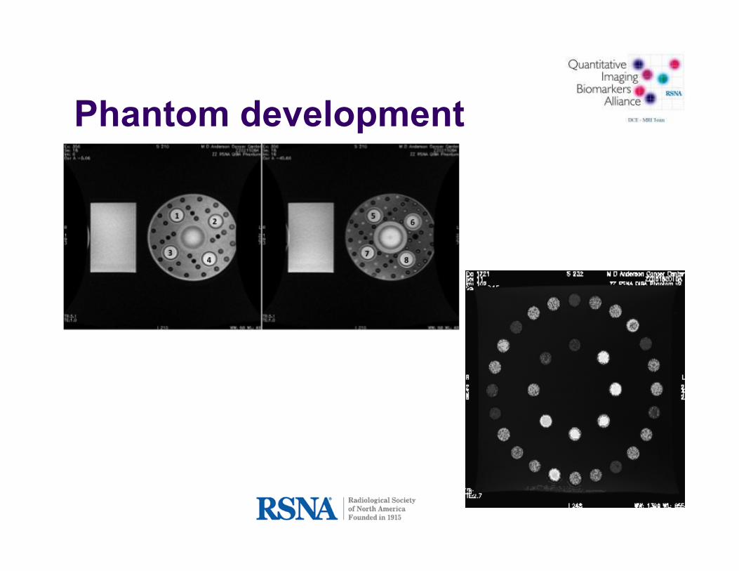
Phantom study design

Phantom Study - phantom
Phantom:
•Phantom design based on
ADNI and IRAT
experiences
•2 phantoms purchased by
MDACC based on NCI
grantgrant
•Phantom provider: The
Phantom Laboratory.
•Phantom delivery with 4
months delay
•Phantoms were not
identical, needed
adjustments that were
provided by MDACC
•Special thanks to Edward
Jackson for his efforts and
dedication
Image analysis / transport
� VirtualScopics provides analysis
� ftp based image transport
� Using NBIA infrastructure supported by NCI
(John Freyman)(John Freyman)
Phantom development

Test data and SW validation
� The synthetic test data initiative has one primary and two
secondary goals.
� Primary: to verify analysis software to be used in the DCE-MRI
clinical study
� Secondary 1: to evaluate software already available in the field� Secondary 1: to evaluate software already available in the field
� Secondary 2: to aid the development of new software tools
� Technique: synthetic DICOM compliant DCE-MRI
images are created using standard vascular input
functions and standard pharmacokinetic models.
� Special results presented by Sandeep Gupta
� Daniel P. Barboriak, Duke Univ. leads this initiative

QIBA DCE-MRI Timeline
2008 2009
Interim results
RSNA2009
AwarenessAwareness
RSNA2008RSNA2008
Q3 Q4 Q2Q1H1 H2
QIBA workshop
DCE-MRI team call
QIBA workshop
2010
Q3 Q4
1111
May – July
Start phase
Definition
phantom
and
protocol
August – December
Exploratory phase
DCE-MRI phantom
study definition
DCE-MRI team call
Evaluation start phase
Milestone 1 December 08 – June 09
DCE – MRI Phantom study
DCE-MRI clinical test-retest study
definition
Profiling activities

Lessons learned
� DCE-MRI core team drives activities.
� Volunteer activities can focus only on activities with limited scope.
� Clinical test – retest study is not doable as a � Clinical test – retest study is not doable as a volunteer activity.
� Phantom study results are very important for next steps.
� Phantom study experiences led to further phantom development.

Lessons learned
� DCE-MRI has today more clinical applications as two years ago.
� DCE-MRI is used in clinical trials in early phases.
� We need to decide on which phantom to use based on QIBA experiences and promote the immediate on QIBA experiences and promote the immediate use in several clinical trials to assess practical use and quality parameters over time – will this become a standard?
� An agreed basic imaging protocol is needed for phantom imaging and for DCE-MRI procedure understanding high spatial and temporal resolution.
� Speed of development is an issue.

May 2010 Workshop
� Objective:
� Phantom imaging study update and discussion on next
steps
� QIBA 2 phantom – first results and next steps
� DCE-MRI profile outline; imaging protocol discussion� DCE-MRI profile outline; imaging protocol discussion
� Roadmap DCE-MRI

Thank you
� Ed Jackson
� Sandeep Gupta
� Ed Ashton
� Greg Karczmar
� Michael Buonocore
� John Freyman
� Larry Clarke
� RSNA staff� Greg Karczmar
� Jeff Evelhoch
� Dan Barboriak
� Mitch Schnall
� Mark Rosen
� David Purdy
� RSNA staff
� Susan Anderson
� Joe Koudelik
� Sharon Skeen
� Fiona Miller

Results 2010 workshop
� Phantom study� Work further on Philips scanner sites to align scan
protocol and procedure
� Work further on study as scheduled (2nd GE site, back
to MDACC)
� Phantom� QIBA phantom 2 has been further discussed and
improved – next version shall be produced soon and
tested. Goal is to provide this as QIBA reommended
DCE-MRI phantom for future clinical trials.

Results 2010 workshop
� Profile� We agreed on profile claim for ktrans and iAUC profile.
� A first outline is available.
� Further writing has been agreed by appointed � Further writing has been agreed by appointed
persons.

Results 2010 workshop� Roadmap of the quantitative MR group
� To finish phantom study and publish results.
� To recommend a QIBA phantom as result of phantom study.
� To clarify next steps or handover of existing activity on clinical test – retest protocol to UPICT.clinical test – retest protocol to UPICT.
� DCE-MRI profiles:
� DCE-MRI imaging profile (1.5T, ktrans + iAUC)
� DCE-MRI imaging on 3T
� DCE-MRI based parameters will not become a surrogate for clinical outcome at today‘s knowledge level.
� Quantitative MR team will start working on initial states on additional MRI based biomarkers (e.g.DWI) profile.

Backup

Current Status:� Differences across MRI systems, field strengths, software platforms, site practices (e.g.
infusion protocols).
� Wide variety of pulse sequences. No standardization of T1 map acquisition.
� Different vendors use different internal system settings that complicate image comparisons.
Challenge: MEDIUM
DCE-MRI Protocol
Challenge: MEDIUM� Agree on all aspects of a single protocol for DCE-MRI image acquisition.
� Incorporate a method for motion correction that is common to all vendors.
Recommendations:� Select a single acquisition protocol including T1 mapping based on dynamic contrast-
enhanced imaging acquisition. Each vendor defines similar internal system settings to support the protocol (step #1 of QIBA DCE-MRI team).
� Incorporation of motion correction method is longer term issue.

DCE-MRI Phantom
Current Status:� An accepted systematic phantom calibration method does not exist for DCE-
MRI imaging protocols, especially for calibration across different MRI systems.
Challenge: HIGH � Phantoms will serve as a useful benchmark to test the accuracy and � Phantoms will serve as a useful benchmark to test the accuracy and
consistency of the acquisition protocols. Several phantoms to be developed, validated and distributed. Constructing phantom with actual inflow and perfusion is challenging.
Recommendations:� Priority 1: phantoms will focus on image intensity as a function of relaxation
rate (R1, determined by contrast agent concentration).
� Priority 2: Phantoms will allow R1 and proton density estimation.
� Determination of geometric distortion due to magnetic field changes and true inflow and perfusion will not be focus for first activities.

Biomarker SW validation
Current Status:� A plethora of software and quantification metrics exist. Many different semi-quantitative
and quantitative metrics are commonly calculated and reported. Even for a widely accepted parameter, Ktrans for example, many different models exist and they each have limitations and assumptions.
Challenge: HIGH � Picking the right algorithms for analysis is very important and challenging. It is � Picking the right algorithms for analysis is very important and challenging. It is
complicated due to the non-standardization of acquisition protocols, as well as the fact that different body applications have very different acquisition scenarios. Robustness of the algorithm, speed of computation, ease of use are vital. Accurate image registration prior to analysis and some form of automated segmentation and analysis of disease after analysis are key challenges. No real ground truth without simulation no gold standard.
Recommendation:� Simulated and real data are needed for reference purposes. It need to be used to
assess algorithm implementation and development .
� Demonstration of comparability of algorithm implementations and development. Mainly task for research/academic community.

Reference Clinical Data
Current Status:� No organized collection of reference data currently available for specific application
areas.
Challenge: MEDIUM � Large volume of clinical data can be obtained from multi-center trials that can serve as
use-cases for evaluation of software tools and acquisition robustness. It must be ensured that the data is of high quality and adheres to the requirements of the analysis ensured that the data is of high quality and adheres to the requirements of the analysis software. Various groups of datasets for each body application area and targeted types of cancer will have to formed. See validation for challenges - no easy way to build the data base; work with DICOM WG 18 regarding necessary meta data as part of the DICOM header plus SR data
Recommendation:� Ensure that appropriate DICOM SR is used to make a complete record of input and
results obtained (this DICOM SR is likely to support other biomarkers e.g. lesion segmentation, ROI definition).
� Start awareness campaign to ensure acquisition standardization becomes widely used in order to enable future population data based build up.