de motor in overdrive: de myeloproliferatieve syndromen g verhoef... · de motor in overdrive: de...
TRANSCRIPT

De motor in overdrive: de myeloproliferatieve
syndromen
G. Verhoef, Leuven Voordracht Iridium kankernetwerk
16 november 2011


Essentiele thrombocytose
Chronische myeloide leukemie
Primaire myelofibrose
Polycytemia rubra
vera
Myeloproliferatieve overlap

Usual features of myeloid disorders at diagnosis
Disease BM cellularity
% marrow blasts
Maturation morphology hematopoiesis Blood counts
Organo-megaly
Mypro ++ <10% ++ normal effective increased common
MDS ++/- <20% + dysplasia ineffective cytopenia uncommon
Mypro/MDS
++ <20% + dysplasia Effective/ ineffective
variable common
AML ++/- ≥20% +/-- Normal/ dysplasia
ineffective variable uncommon

Myeloproliferatieve neoplasieën
Polycythemia rubra vera
Essentiële trombocytose
Primaire Myelofibrose (myelosclerose)
Chronische myeloide leukemie

PV: epidemiology
• age at diagnosis (average): 60 yr
• incidence: 20 / 10 6 (prevalence 250-300 / 106)
• M / F ratio — 1.2 : 1

WHO criteria Polycythemia Vera 2001 A1. rode bloedcel massa >25% boven gemiddelde, of Hb >18.5 g/dL (mannen) of >16.5 g/dl (vrouwen)
A2. Geen oorzaak voor secundaire polycythemie 1. niet familiair 2. geen EPO verhoging door a. hypoxie (arterieel pO2 ≤92%) b. hoge affiniteit Hb voor O2
c. getruceerde EPO receptor d. EPO productie door tumor
A3. Splenomegalie
A4. clonale cytogenetische afwijkingen (geen t(9;22) of BCR-ABL fusie),
A5. endogene in vitro CFU-E vorming
B1. trombocytose >400 x 109/L
B2. leukocyten >12 x 109/L
B3. beenmergbiopt met prominente toename erytro- en megakaryocytaire reeks
B4. laag serum EPO
Nodig zijn: [A1 & A2 & A3/4/5] of [A1 & A2 & 2B criteria]


2° polycytemia
congenital acquired
hypoxia driven o chronic lung disease o R L cardio-pulmonary shunts o high altitude o smoking / carbon monoxide poisoning o sleep apnea / hypoventilation Σd o renal artery stenosis
oxygen-independent
o EPO injection o use of androgen preparations o RCC / renal cysts / postrenal Tx o meningioma / cerebellar hemangioblastoma o pheochromocytoma o uterine leiomyoma o hyperparathyroidism / parathyroid adenoma o HCC o other tumors Adapted from Silver & Tefferi, Informa, 2007
o high-oxygen-affinity hemoglobinopathy (autosomal dominant) o 2,3 BPG deficiency (autosomal recessive)
o VHL mutation (increased EPO)
o EPO-R mutation (EPO low/nl)

JAK 2 V617F mutatie
• William Vainchenker:
– Epo-independent growth characteristics of PV progenitors
• Kralovics – Precise mapping of minimal 9p
loss of heterozygosity region
• Gary Gilliland and Green – Analysis of tyrosine
kinome

.
Signal transduction to nucleus
Nucleus
Binding site
Tyrosine kinase activity
Cytoplasm
Plasma membrane
Growth factor
Gene activation CELL DIVISION
.Normal cell

polycythemia Vera cell
Signal transduction to nucleus
Nucleus
Binding site
Tyrosine kinase activity
Cytoplasm
Plasma membrane
Growth factor
Gene activation CELL DIVISION
X

JAK 2 V617F mutatie
• William Vainchenker:
– Epo-independent growth characteristics of PV progenitors
• Kralovics – Precise mapping of minimal 9p
loss of heterozygosity region
• Gary Gilliland and Green – Analysis of tyrosine
kinome

WHO criteria Polycythemia Vera 2008
A1. rode bloedcel massa >25% boven gemiddelde, of Hb >18.5 g/dL (11.6 mmol/l) (mannen) of >16.5 g/dl (10.3 mmol/l)(vrouwen) of Hb >17 g/dL (mannen) of >15 g/dl (vrouwen) in combinatie met toename ≥2 g/dL niet toe te schrijven aan correctie Fe tekort A2. Aanwezigheid van JAK2V617F or vergelijkbare mutatie (JAK2 exon 12)
B1. endogene in vitro CFU-E vorming
B2. laag serum EPO
B3. beenmergbiopt met prominente toename erytro- en megakaryocytaire reeks
Nodig zijn: [A1 & A2 & één B] of [A1 & twéé B criteria]

Symptomen van Polycytemie • Plethora • Splenomegalie • Hyperviscositeit
• Wazig zicht, diplopie • Hoofdpijn • Orgaanfalen : decompensatie, dyspnee ,
nierblokkage, cerebraal… • Epistaxis
• Trombotische fenomenen (extreem : hemorragische diathese)
• Jeuk, urticaria (vooral na warm bad) - basofielen en mastcellen
• Polyneuropathie • Jicht


risk factors:
– advanced age ( > 60-70 yr)
– history of thrombosis
– smoking and other cardiovascular risk factors
– leukocytosis (Gangat et al. BJH 2007) (Landolfi et al. Blood 2006)
– associated thrombocytosis ? conflicting data
– JAK2V617F allele burden prospective validation in clinical trials is needed !
PV: complications
A. thrombosis sometimes life-threatening
B. other life-threatening complications:
bleeding: < thrombosis, linked to acquired von Willebrand Σd
in the presence of thrombocytosis +++
leukemic transformation (incidence 5-10% in the first 10 yr)
development of post-PV myelofibrosis
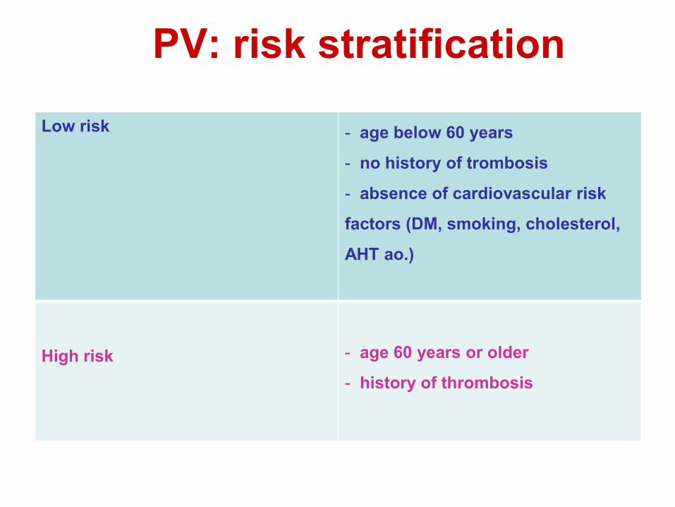
PV: risk stratification
Low risk
- age below 60 years
- no history of trombosis
- absence of cardiovascular risk
factors (DM, smoking, cholesterol,
AHT ao.)
High risk
- age 60 years or older
- history of thrombosis

PV: management
target Hct < 0.45
Low risk
- flebotomy
- low dose ASA (75-100 mg/d if no contra indication)
- manage C-V RF aggressively
High risk
- flebotomy
- low dose ASA (75-100 mg if no C-I)
- cytoreductive therapy (HU of
IFNα)
- manage C-V RF aggressively

Myeloproliferatieve neoplasieën
Polycythemia rubra vera
Essentiële trombocytose
Primaire Myelofibrose (myelosclerose)
Chronische myeloide leukemie

ET: epidemiology and symptoms • average age at diagnosis: 50 - 60 yr
• incidence: 15 - 25 / 106 estimated prevalence: 400 / 106
• F > M (peak F 30 yr)
• symptoms:
- mostly asymptomatic
- 10 – 15 %: thrombosis (art > ven)
- microcirculatory disturbances: headache, dizziness, distal
paresthesias, acrocyanosis, erythromelalgia
- bleedings (cave: BP > 1500000/µl)
- itching (15 – 40 %)

Oorzaken van Trombocytose
Primair of essentiëel Als teken van een andere myeloproliferatieve
aandoening (PRV, CML, myelofibrose) Secundair :
• Post hemorragisch • Posttraumatisch, postoperatief • Chronische inflammatie • Postsplenectomie • Ijzerdeficiëntie • Paraneoplastisch• Systeemziekte

WHO criteria Essentiële Trombocytemie 2001
Inclusie criteria: 1. trombocyten over 6 mnd stabiel >600 x 109/L 2. beenmergbiopt met voornamelijk megakaryocytaire toename met atypische grote megakaryocyten
Exclusie criteria: 1. geen aanwijzingen voor PV a. normale rode bloedcelmassa of Hb b. kleurbaar ijzer in beenmerg, normaal ferritine of normaal MCV c. indien niet voldaan aan bovenstaande criteria, dan proefbehandeling met ijzer 2. geen aanwijzingen voor CML, geen t(9;22) of BCR-ABL fusie 3. geen aanwijzingen voor CIMF a. geen collagene fibrose b. geen of minimale reticuline vervezeling 4. geen aanwijzingen voor MDS a. geen del(5q), t(3;3), inv(3) b. geen of minimale dysgranulopoiese of micromegakaryocyten 5. geen aanwijzingen voor reactieve trombocytose a. infectie of ontsteking b. maligniteit c. splenectomie

Frequencies of the JAK2V617F mutation in patients with myeloproliferative disorders
Authors PV (%) IMF (%) ET (%) James et al. Baxter et al. Levine et al. Kralovics et al.
89 73 74 65
43 44 35 57
43 12 32 23
Zhao et al. Jones et al. Steensma et al. Goerttler et al. Jelinek et al. Levine et al.
83 81 ND 100 86 ND
ND 43 ND 57 95 ND
ND 41 ND 33 30 ND
Total 422/557 76%) 80/160 (50%) 114/391 (29%)
Data obtained by allele-specific PCR*
71/93 (97%) 8/16 (50%) 29/51 (57%)
* Baxter et al.

WHO criteria Essentiële Trombocytemie 2008
Proposed criteria
1. Trombocyten ≥450 x 109/L
2. Beenmergbiopt met voornamelijk megakaryocytaire toename met atypische grote megakaryocyten. Geen belangrijke afwijkingen aan rood en wit
3. Voldoet niet aan WHO criteria PV, IMF, CML, MDS of andere entiteit
4. Aanwezigheid van JAK2V167F of andere clonale merker, of, in afwezigheid van clonale merker geen aanwijzingen voor secundaire trombocytose
Voor diagnose ET zijn alle vier criteria noodzakelijk

ET: beenmerg

ET and skin lesions
erythromelagia acrocyanosis
Raynaud‟s phenomenon
livedo reticularis

ET: risk stratification Low risk
- younger < 60 yr
- no history of thrombosis
- BP < 1500000/µl
Intermediate risk
- no „clear cut‟ definition (not low, not
high)
- „operational definition‟: low risk
patients with one or more of the
following risk factors: CV disease,
diabetes, smoking, hypertension,
familial thrombophilia, ea.
High risk
- older > 60 yr
- history of thrombo-embolic event(s)
- BP > 1500000/µl

ET: management
Low risk
• low dose ASA (if no contra-
indication)
Intermediate risk
• low dose ASA
• treat C-V risk factors „aggressively‟
• in some patients: BP reduction
(decision on individual base)
High risk
• low dose ASA
• reduce BP to < 400000/µl
• treat C-V risk factors

Note : smoking as independent risk factor
100
80
60
40
20 0
0 24 48 96 192
p=0.005
Thro
mbo
sis-
free
sur
viva
l (%
)
Months after randomization
Hydroxyurea - Anagrelide
Control
Outcome
Essential thrombocytosis Primary thrombocythemia

Myeloproliferatieve neoplasieën
Polycythemia rubra vera
Essentiële trombocytose
Primaire Myelofibrose (myelosclerose)
Chronische myeloide leukemie

Oorzaken van mergfibrose
Primair Secundair :
• Paraneoplastisch • Chronische infecties (TBC, osteomyelitis) • Paget • Botmetastasen (oa prostaat) • Bestraling • Benzeen, fluorvergiftiging • Andere myeloproliferatieve ziekten • Osteopetrose

Phi – MPN
PV
ET
MF
PMF
post PV – MF post ET - MF

Epidemiology PMF
• rare: incidence ~ 1/100.000
• M = F
• average age at diagnosis: 61 yr

Chronische idiopathische myelofibrose: fibrotisch stadium WHO 2001
Clinical findings Morphological findings Spleen and liver: Moderate to marked splenomegaly and hepatomegaly
Blood: Leukoerythroblastosis Prominent red blood cell poikilocytosis with dacrocytes
Hematology: Anemia: ↓, ↓↓ WBC: ↓, =, ↑ Platelet count: ↓, =, ↑
Bone marrow: Reticulin and/or collagen fibrosis Decreased cellularity Dilated marrow sinuses with intraluminal hematopoiesis Prominent megakaryocytic proliferation and atypia (clustering of megakaryocytes, abnormally lobulated megakaryocytic nuclei, naked megakaryocytic nuclei) New bone formation

Frequencies of the JAK2V617F mutation in patients with myeloproliferative disorders
Authors PV (%) IMF (%) ET (%) James et al. Baxter et al. Levine et al. Kralovics et al.
89 73 74 65
43 44 35 57
43 12 32 23
Zhao et al. Jones et al. Steensma et al. Goerttler et al. Jelinek et al. Levine et al.
83 81 ND 100 86 ND
ND 43 ND 57 95 ND
ND 41 ND 33 30 ND
Total 422/557 76%) 80/160 (50%) 114/391 (29%)
Data obtained by allele-specific PCR*
71/93 (97%) 8/16 (50%) 29/51 (57%)
* Baxter et al.

Proposed criteria for PMF WHO 2008 Proposed criteria for PMF
Major criteria 1. Presence of megakaryocyte proliferation and atypia, usually accompanied by either reticulin and/or collagen fibrosis, or, in the absence of significant reticulin fibrosis, the megakaryocyte changes must be accompanied by an increased bone marrow cellularity characterized by granulocytic proliferation and often decreased erythropoiesis 2. Not meeting WHO criteria for PV, CML, MDS or other myeloid neoplasm
3. Demonstation of JAK2V167F or other clonal marker (MPL515W>L/K), or in the absence of a clonal marker, no evidence of bone marrow fibrosis due to underlying inflammatory or other neoplastic diseases
Minor criteria
1. Leukoerythroblastosis
2. Increase in LDH
3. Anemia
4. Palpable splenomegaly
Diagnosis of PMF requires meeting all three major criteria and two minor criteria

Symptomen van primaire myelofibrose
• Indien ernstige Pancytopenie: dan gereleerde symptomen (anemie, granulocytopenie, trombopenie)
• Leuco-erythroblastische formule ! • Massieve Hepatosplenomegalie • Hyperuricemie • Sterk verhoogd LDH • Hoog risiko op leukemische transformatie
(1/3!)


PMF bloed: teardrop cells, erytroblast


Tefferi. Blood 2011
MF: risico stratificatie

Behandeling primaire myelofibrose Symptomatisch en supportief ( transfusies PC en
bloedplaatjes, antibiotica, fungistatica) Lage dosis chemotherapie in proliferatieve fase
(Hydrea, Lanvis) Splenectomie bij hinderlijke splenomegalie,
miltinfarcten, refractaire hemolyse, pooling, portale hypertensie Per-Operatoire mortaliteit ongeveer 20 % !
Myelo-ablatieve stamceltransplantatie Niet-myeloablatieve stamceltransplantatie Thalidomide (al dan niet in combinatie met
dexamethasone) – Zolendronaat (?)

• JAKs (Janus kinasen): JAK1, JAK2, JAK3, TYK2
• V617F mutatie van JAK2 activatie ++
cytokine receptor – kinase complexen
constitutieve activatie van EPOR-JAK2 en
TPOR-JAK2 complexen MPN fenotype
• JAK2-inhibitie: expansie blokkeren van de
gedifferentieerde (myeloïde) HPC en symptomen
van MPN ↓. Zwak effect op stamcel !
JAK2 V617F mutatie
Benekli M et al. Blood 2003
Knoops L et al. Belg J Hematol 2011

JAK inhibitors
Tefferi. Blood 2011

JAK remmers: fase 1/2
Verstovsek et al. NEJM 2010
INCB018424 (= ruxilotinib)
• starting dose 15 mg BID
25 mg BID
Pardanani JCO 2011
TG 101348
starting dose 400 mg 500 mg/d
miltvolume
constutionele
symptomen

• fase 2 studie (Tefferi et al. JCO 2009)
best respons met dosis 0.5 mg/d
anemie verbetert in 40 %
bloedplaatjes stijgen in 58%
geen effect op splenomegalie
NW: zelden neuropathie en myelosuppressie
• momenteel fase 3 studie in VS, Europa en Azië
Pomalidomide (« 3 de generatie IMID »)

Myeloproliferatieve neoplasieën
Polycythemia rubra vera
Essentiële trombocytose
Primaire Myelofibrose (myelosclerose)
Chronische myeloide leukemie

Kliniek CML
• Vaak toevallige bevinding, soms vermoeidheid, buikbezwaren, leukostase, vaak splenomegalie
• verhoogd aantal witte bloedcellen in alle rijpingsstadia, dikwijls trombocytose
• incidentie: 1-1.5 per 100.000 inwoners • Gemiddelde overleving 5-6 jaar

CML: perifeer bloed

Three phases of the Disease (before imatinib)
chronic phase accelerated blast phase
36-48 months 6 months 3-6 months
Surv
ivin
g pe
rcen
t
Years after diagnosis

The Philadelphia chromosome

Goldman et al. N Engl J Med 2003; 349:1451-64

Goldman et al. N Engl J Med 2003; 349:1451-64
↑Proliferation ↓Adherence ↓Apoptosis


NEJM, 346, 683, 2002
Imatinib targets CML at its source [t(9;22)]


• Comparison with historic data (Kantarjian et al., Cancer 98: 2336-2342 (2003):

Nowell and Hungerford
1845
CML, pionier voor ‘targeted therapy’
J.Rowley
2010
1997 IFN + ARA-C

1864 1903 1953 1964 1975 1983 1999 2005 2007 2008 2009
Palliative therapy Currative therapy
arsenic
Spleen irradication
Busulfan
Hydroxyurea
Stem cell transplantation
Combination chemo
Interferon
Imatinib
Dasatinib Nilotinib
Bosutinib
“first generation TKI” “second generation TKI”
MK0457,Pha739358 XL228, AP24534
“third generation TKI”
Developments of treatment for CML
68
