deciduous molar hypomineralisation, its nature and nurture · deciduous molar hypomineralisation,...
TRANSCRIPT

Deciduous Molar Hypomineralisation,
its nature and nurture
Marlies Elfrink

Colofon
This thesis was prepared at the department of Cariology, Endodontology and Pedodontology of the Academic Centre for Dentistry Amsterdam (ACTA), the combined faculty of the University of Amsterdam and VU University Amsterdam, the Netherlands.
Financial support for this dissertation was kindly provided by: GABA, Therwil, Switzerland (unrestricted grant).
The Generation R Study is made possible by financial support from the Erasmus MC, Rotterdam, Erasmus University Rotterdam and the Netherlands Organization for Health Research and Development (ZonMw).
The printing of this thesis has been financially supported by: IOT (Interuniversitair Onderzoekoverleg Tandheelkunde)Stichting Conserverende TandheelkundeTandtechnisch laboratorium Kesseler&Reuvekamp BV (http://www.kesseler-reuvekamp.nl/)
Printed and lay-out by: Gildeprint Drukkerijen – The NetherlandsISBN/EAN: 978-94-6108-292-3
Copyright © Marlies Elfrink, Almelo, the Netherlands
No part of this thesis may be reproduced in any form or by any means without written permission from the author.

Deciduous Molar Hypomineralisation,
its nature and nurture
ACADEMISCH PROEFSCHRIFT
ter verkrijging van de graad van doctor aan de Universiteit van Amsterdamop gezag van de Rector Magnificus
prof. dr. D.C. van den Boomten overstaan van een door het college voor promoties
ingestelde commissie, in het openbaar te verdedigen in de Agnietenkapel
op vrijdag 1 juni 2012, te 14:00 uur
door
Maria Elisabeth Christina Elfrink
geboren te Almelo

ProMotieCoMMissie
Promotoren: Prof. dr. J.M. ten Cate Prof. dr. H.A. Moll
Co-promotor: Dr. J.S.J. Veerkamp
Overige leden: Prof. dr. S. Alaluusua Prof. dr. C. van Loveren Dr. K.L. Weerheijm
Faculteit der Tandheelkunde

Contents
Chapter 1: Introduction and research questions 7 Chapter 2: Prevalence of Deciduous Molar Hypomineralisation in 5-year-old 23 Dutch children
Chapter 3: Validity of scoring Deciduous Molar Hypomineralisation on intra-oral 31 photographs
Chapter 4: Relationship between Deciduous Molar Hypomineralisation and caries 4.1: Caries pattern in primary molars in 5-year-old Dutch children 45 4.2: Factors increasing the caries risk of second primary molars in 57 5-year-old Dutch children
Chapter 5: MicroCT study on Deciduous Molar Hypomineralisation 69
Chapter 6: Determinants and associated factors for Deciduous Molar Hypomineralisation 6.1: Pre- and postnatal determinants for Deciduous Molar 81 Hypomineralisation 6.2: Is maternal medication use during pregnancy associated with 97 Deciduous Molar Hypomineralisation in the offspring?
Chapter 7: Relationship between Deciduous Molar Hypomineralisation and 109 Molar Incisor Hypomineralisation
Chapter 8: General discussion and clinical implications 123
Chapter 9: Summary and general conclusion / Samenvatting en conclusie (in Dutch) 135
Epilogue Dankwoord (Acknowledgements in Dutch) 145 List of publications 151 Curriculum Vitae 155


1
introduction and research questions

R1R2R3R4R5R6R7R8R9
R10R11R12R13R14R15R16R17R18R19R20R21R22R23R24R25R26R27R28R29R30R31R32R33R34R35R36R37R38R39
Chapter 1
8

R1R2R3R4R5R6R7R8R9R10R11R12R13R14R15R16R17R18R19R20R21R22R23R24R25R26R27R28R29R30R31R32R33R34R35R36R37R38R39
Introduction and research questions
9
1introDuCtion
Diseases of the dentition, such as dental caries and enamel malformations, are among the most common chronic illnesses worldwide (1, 2). Caries generally cause oral discomfort and pain and influence a child’s ability to eat, do schoolwork and sleep (2). The prevalence of caries in children in the Netherlands, as in other developed countries, has declined since 1975 (4). Although it had stabilised, the prevalence of caries is now slightly increasing (5). Paediatric dentists warned about the increasing number of children who need extensive dental treatment, which received renewed attention, even in the national newspapers, in 2011. Because the incidence of caries has been declining, the emphasis of research has been more on predicting caries (6) and other dental problems, such as developmental enamel defects. Developmental enamel defects are not uncommon, both in the primary and permanent dentitions, and can be divided into hypomineralisation and hypoplasia (7, 8). Enamel hypoplasia is a quantitative defect of the enamel, and enamel hypomineralisation is a qualitative defect of the enamel identified visually as an alteration in the translucency of the enamel, with a clear border, variable in degree, and a white, yellow or brown colour. It has also been termed a demarcated opacity (7, 9). The first permanent molars with hypomineralisations are often associated with affected permanent upper incisors and, more rarely, lower incisors (10). Therefore, the name Molar Incisor Hypomineralisation (MIH) is currently used (10-12). In the primary dentition, hypomineralisations are also found in the second primary molars, a process known as Deciduous Molar Hypomineralisation (DMH).
HistoryAmong the earliest authors publishing on hypomineralisations in the permanent dentition were Koch et al. (13) who reported its prevalence in Swedish children in various birth cohorts. This observation led to work by many researchers, who between them collectively defined the name, definition and scoring criteria of hypomineralisations (9, 10).
Many different names have been used for Molar Incisor Hypomineralisation (MIH): hypomineralised first permanent molars, non-fluoride hypomineralisation, idiopathic enamel hypomineralisation, non-endemic mottling of enamel and cheese molars (10). Experts of the European Academy of Paediatric Dentistry (EAPD) developed diagnostic criteria for MIH in 2003, and these criteria were updated in 2009 (9, 14). These criteria (see Table 1.1) should be interpreted in the same way in all future research on MIH and DMH to improve the comparability of results. In this thesis, we used the MIH criteria and recommendations for DMH but made some modifications: the definition of DMH only involves the second primary molar, and atypical caries were added because many cavities are not restored in the primary dentition.

R1R2R3R4R5R6R7R8R9
R10R11R12R13R14R15R16R17R18R19R20R21R22R23R24R25R26R27R28R29R30R31R32R33R34R35R36R37R38R39
Chapter 1
10
table 1.1: Criteria for the diagnosis of MIH and DMHMild:
• Opacity: A defect that changes the translucency of the enamel, variable in degree. The defective enamel is of normal thickness with a smooth surface and can be white, yellow or brown in colour. The demarcated opacity is not caused by caries, ingestion of excess fluoride during tooth development or amelogenesis imperfecta, etc.
Severe:• Posteruptive enamel loss: A defect that indicates surface enamel loss after the eruption of the
tooth, e.g., hypomineralisation-related attrition. Enamel loss due to erosion was excluded, and/or• Atypical caries: The size and form of the caries lesion do not match the present caries
distribution in the child’s mouth, and/or• Atypical restoration: The size and form of the restoration do not match the present caries
distribution in the child’s mouth, and/or• Atypical extraction: The absence of a molar that does not fit with the dental development and
caries pattern of the child.
The association between DMH and MIH is only speculated on in the literature (9). We performed a large prospective cohort study to evaluate this association.
PrevalenceIn many countries, researchers have established the prevalence of MIH in healthy children. The reported prevalence varies between 2.4% and 40.2% (14). A comparison of the various studies proved difficult due to differences in patient selection (at random or not, age of the children), different scoring criteria ((modified) Developmental Defects of Enamel ((m)DDE) index, EAPD criteria or other criteria) and differences in the examination circumstances (clinically or by photographs, in a dental chair or in a classroom, etc.) (3, 15, 16).In the Netherlands, the most recently reported prevalence of MIH is 14.3% (17). The prevalence differs from country to country and changes per birth-year. In the study of Koch et al. (13), the prevalence varied between the different birth-years from 6.3% to 15.4%, with a high prevalence peak in children born in 1970. The prevalence of MIH in the Netherlands also differed between the various cohorts in the TJZ (Tandheelkundige verzorging Jeugdige Ziekenfondsverzekerden) study: 9.7% in the study from 1999 and 14.3% in the study from 2003 (17, 18). Data on the prevalence of DMH were lacking. In the recent TJZ studies, second primary molars were also investigated for DMH, and we established the prevalence of DMH in the Netherlands.
enamel hypomineralisationEnamel is the hardest tissue in the human body, but its formation can be disturbed rather easily (1). Disturbances in enamel formation leave a permanent mark in the tooth. These disturbances can be inherited (e.g., amelogenesis imperfecta), acquired (e.g., induced by chemicals such as in fluorosis) or idiopathic (e.g., DMH and MIH). DMH and MIH are probably caused by a disturbance in the initial calcification and/or during the maturation phase of the enamel, causing demarcated opacities (10, 19, 20). In MIH molars, these opacities contain more carbon and less calcium and

R1R2R3R4R5R6R7R8R9R10R11R12R13R14R15R16R17R18R19R20R21R22R23R24R25R26R27R28R29R30R31R32R33R34R35R36R37R38R39
Introduction and research questions
11
1phosphate (21, 22). Although the mineral composition of the enamel has not yet been investigated in DMH, the same results can be expected as for MIH. The vulnerability of teeth with DMH or MIH can be explained by the lower mineral content or other mineral composition of the enamel. The colour of the demarcated opacity in MIH molars (white, yellow or brown) was reported to be associated with the mineral density of the enamel (23). Opacities in MIH molars contained 3- to 21-fold more protein than normal enamel (20, 23), and brown opacities in particular contained more protein (20). The mineral content of the enamel is reflected in the mechanical properties of the enamel (20, 24). In MIH molars, the enamel density in the hypomineralised areas is lower than in sound areas (19). Little is known about the mineral content and density in DMH molars. Studies used the micro-computer tomography (microCT) technique to determine mineral content in MIH molars. MicroCT, a miniaturised version of the whole body CT scan, is a non-destructive x-ray absorption microscopic technique for the 3D visualisation of teeth. It can also perform quantitative measurements of the mineral content (19). In the permanent dentition, MIH molars showed a 19-20% reduction of mineral concentration in the affected enamel, the hypomineralised enamel had a mineral concentration gradient opposite that of normal enamel and the hypomineralised areas were distributed randomly throughout the MIH molars, with only the cervical region being less affected. No studies on the mineral concentration in hypomineralised areas of DMH molars have been performed yet. Therefore, we performed a microCT study to compare DMH molars with sound second primary molars.
relationship with cariesCaries can affect each tooth and surface, with a preference for pits, fissures and proximal surfaces (6, 25). Caries at other, less vulnerable, sites could be a sign of severe caries (6). Caries patterns can also be associated with aetiology (26). In early childhood caries (ECC), when the causative factor is a sweet(ened) liquid diet, especially at night, the primary teeth are affected following the eruption sequence, and the mandibular incisors are affected last (27). Not all caries lesions, however, follow the eruption sequence. Many investigators have tried to find a pattern for predicting caries (6, 28), which becomes more important when caries prevalence in the population is declining (6). The second primary molars were reported to be more often affected by caries than the first primary molars (6, 29-31). The second primary molars erupt 10-12 months after the first primary molars at the age of 24-30 months (32, 33), leading to the assumption that the first primary molars have a greater prevalence of caries due to a longer presence in the oral cavity. Both MIH and hypoplasia in the primary dentition influence caries prevalence in children (10, 34, 35). DMH could be an explanation for the differences in caries prevalence between the first and second primary molars (34, 35). Important in interpreting this hypothesis is that DMH had not been investigated as a putative caries-influencing factor previously, like we did now.

R1R2R3R4R5R6R7R8R9
R10R11R12R13R14R15R16R17R18R19R20R21R22R23R24R25R26R27R28R29R30R31R32R33R34R35R36R37R38R39
Chapter 1
12
Determinants and associated factorsTooth development, although genetically controlled, is reported to be sensitive to disturbances from the environment (3). Because enamel is not remodelled like bone, disturbances acquired during its development leave a permanent record in the tooth (36).Dental development starts with the formation of the dental lamina from the ectodermal epithelium. Tooth development follows the bud, cap and bell stages, generating the shape of the tooth. The cells from the dental lamina differentiate into, among others, ameloblasts and dentinoblasts (37). Dentin and enamel formation occur simultaneously along a line that will develop into the dentino-enamel junction (1) (see Figure 1.1 and 1.2). Amelogenesis is a slow developmental process that can be divided into the following steps: secretory stage, transitional stage and maturation stage (3). At the secretory stage, the enamel matrix is formed in large amounts. The ameloblasts secrete enamel proteins, and enamel crystals grow in length, resulting in a thickening of the enamel layer (1, 3).
At a certain point, the secretory ameloblasts undergo a transition (transitional stage), and the maturation of the enamel will start. During the maturation stage, the enamel layer hardens. The crystals stop their growth in length and start to grow in width and thickness, which results in a mineralised tissue with more than 95% mineral content (1, 3). After the maturation stage, the ameloblasts degenerate with the other layers of the enamel-epithelium during tooth eruption (37).
The development of the second primary molars occurs somewhat earlier than the development of the first permanent molars and permanent incisors, but the periods of their development overlap (32, 33) and the maturation of the permanent molar is slower (38). If a risk factor occurs during this overlapping period, a hypomineralisation might occur in the primary and permanent dentition (39). Because the second primary molars erupt 4 years earlier in life than the first permanent molars, DMH might be a clinically useful predictor for MIH.
A number of recent studies on MIH focused on the possible determinants (3, 15, 16). Numerous determinants have been identified in the literature, but the conclusions of these different studies have been contradictory (3, 15, 16, 40).Commonly mentioned determinants for MIH are summarised in Table 1.2.

R1R2R3R4R5R6R7R8R9R10R11R12R13R14R15R16R17R18R19R20R21R22R23R24R25R26R27R28R29R30R31R32R33R34R35R36R37R38R39
Introduction and research questions
13
1
figure 1.1: schematic picture of a developing molar. The cells of the cervical loop (cl) proliferate and develop into presecretory ameloblasts (pa) and further into secretory ameloblasts (sa). After odontoblasts (od) have deposited a small predentine layer, secretion of the enamel matrix can start. Secretory ameloblasts have deposited the protein rich enamel matrix, which contains only small quantities of minerals, in the cusp tips. e: enamel; d: dentine (and predentine) (3).
figure 1.2: schematic picture of a developing molar. In the occlusal half of the tooth, the ameloblasts are at the maturation stage. The enamel matrix is resorbed by the ameloblasts (ma) and the massive mineralisation of the enamel is carried out. More cervically, ameloblasts are at a short, so-called transitional stage before entering the maturation stage (transitional-stage ameloblasts). In the most cervical part of the crown, the secretion of the enamel matrix is still on going by secretory ameloblasts. Apoptotic cell death of the ameloblasts begins at the transitional stage and peaks at the maturation stage. Most ameloblasts die before the tooth erupts into the oral cavity. pa: presecretory ameloblasts; cl: cervical loop; od: odontoblasts (3).(Courtesy: S. Alaluusua, Helsinki, Finland).

R1R2R3R4R5R6R7R8R9
R10R11R12R13R14R15R16R17R18R19R20R21R22R23R24R25R26R27R28R29R30R31R32R33R34R35R36R37R38R39
Chapter 1
14
table 1.2: Determinants for Molar Incisor Hypomineralisation (MIH), overview from the literature.
Determinants of Molar Incisor Hypomineralisation (MIH)
Nut
ritio
n
Med
ical
pro
blem
s
Med
ical
pro
blem
s
Prem
atur
e bi
rth
Oxy
gen
shor
tage
Nut
ritrio
n
Brea
stfe
edin
g
Chi
ldho
od d
iseas
es
Med
icat
ion
Envi
ronm
enta
l pol
lutio
n (d
ioxi
ns)
Reference prenatal perinatal postnatal
Aine et al., 2000 (39) + +
Alaluusua et al., 1996a (41) +
Alaluusua et al., 1996b (42) +
Alaluusua et al., 2004 (43) +
Van Amerongen& Kreulen, 1995 (44) + + +
Beentjes et al., 2002 (8) - - +
fagrell et al., 2011 (15) - - + + - -
Holtta et al., 2001 (45) +
Jalevik&noren, 2000 (7) - - -
Jalevik et al., 2001 (46) - - - + +
Jontell&linde, 1986 (47) + +
Kuscu et al., 2008 (48) +
Kuscu et al., 2009 (49) -
laisi et al., 2008 (50) +
laisi et al., 2009 (51) +
lygidakis et al., 2008 (52) ± + +
salmela et al., 2011 (53) +
Whatling&fearne, 2008 (54) + - - - + +
Wogelius et al., 2010 (55) +
Crombie et al., 2009 (16) (review) ± ± ± + ± + + +
Alaluusua, 2010 (3) (review) + + + - + + + + +
- no influence± possible influence+ influence

R1R2R3R4R5R6R7R8R9R10R11R12R13R14R15R16R17R18R19R20R21R22R23R24R25R26R27R28R29R30R31R32R33R34R35R36R37R38R39
Introduction and research questions
15
1Identifying the cause of MIH is still difficult. Several possible reasons for this difficulty have been reported:- The cause of MIH is multifactorial and/or a threshold level needs to have been reached before enamel defects are caused or become apparent (3, 8, 12, 16).- Most studies on the determinants are retrospective, giving biased data. Parents are unable to remember health and nutritional details after approximately 8 years (3, 15, 16).- The study populations were small and selected (3, 15, 16).
Compared with hypomineralisation defects in the permanent dentition, very little has been written on hypomineralisation defects in the primary dentition. The few articles on this topic have stated that in the primary dentition, the second primary molar is the tooth most often affected by hypomineralisation (56-59). Possible determinants have only been hypothesised about. The same determinants are expected as for MIH molars, although occurring somewhat earlier in life (perinatal instead of postnatal) (39, 57, 60, 61). The developmental period of the first permanent molars and second primary molars have some overlap, but the second primary molars start to develop earlier and quicker. Pre- and perinatal factors do not seem to have much influence on MIH, but they may be determinants for DMH. To study these factors, information during pregnancy and early life needs to be collected prospectively in a large cohort of children. In the Generation R study, a population-based prospective cohort study following pregnant women and their children from foetal life until young adulthood in Rotterdam, the Netherlands, determinants for DMH were studied.
AimsThe overall aim of this thesis was to describe and provide more insight into Deciduous Molar Hypomineralisation (DMH), including its prevalence, enamel mineral content, pre-, peri- and postnatal determinants and associations with Molar Incisor Hypomineralisation (MIH) and caries.
PrevalenceThe aim of this study was to report on the prevalence of Deciduous Molar Hypomineralisation (DMH) in 5-year-old Dutch children.
Validity and reliability of intra-oral photographsThe aims of this study were (i) to assess whether intra-oral photographs could be used to score caries and hypomineralisation on primary molars (using the adapted Molar Incisor Hypomineralisation (MIH) criteria) and (ii) to assess the reliability and validity of these scores in 3- to 7-year-old Dutch children by comparing them with direct clinical scorings.

R1R2R3R4R5R6R7R8R9
R10R11R12R13R14R15R16R17R18R19R20R21R22R23R24R25R26R27R28R29R30R31R32R33R34R35R36R37R38R39
Chapter 1
16
Relationship between Deciduous Molar Hypomineralisation (DMH) and cariesThe aims of this study were (i) to look for a difference in caries prevalence between the surfaces of the first and second primary molars and (ii) to investigate determinants both directly and indirectly associated with caries in second primary molars.
Mineral density in Deciduous Molar Hypomineralisation (DMH)The aim of this study was to determine the mineral (hydroxyapatite) density of sound and opaque areas in DMH molars and healthy teeth.
Determinants and associated factors of Deciduous Molar Hypomineralisation (DMH)The aim of this study was to examine the possible determinants of DMH in a prospective cohort study in the prenatal period and the first year of life of the children. The association between antibiotics and asthma medication used during pregnancy with DMH was also studied.
Relationship between Deciduous Molar Hypomineralisation (DMH) and Molar Incisor Hypomineralisation (MIH)The aim of this study was to determine the association between DMH in the second primary molars and MIH in the first permanent molars.
Some overlap between chapters can be seen because the chapters are based on separate publications on the same topic. The chapters are not arranged chronologically for editorial reasons.
study populations
Dental practicesFor the study on the validity of the intra-oral camera, a convenience sample of 62 children (aged 2.92-7.17 years, mean 4.96 years [SD±1.27]; 38.7% girls) visiting the dental practice of one of the investigators between November 2007 and February 2008 was asked to participate. All invited children participated in the study. The accompanying parent gave consent for taking the intra-oral photographs.Children from the same dental practices were asked to donate their extracted second primary molars for the study on the mineral content of DMH molars.
TJZ studyAs part of a Dutch standardised epidemiological survey (Tandheelkundige verzorging Jeugdige Ziekenfondsverzekerden (TJZ); dental care for children insured by Health Insurance Funds), the dentition of 5-year-old children were examined every six years. The children were living in Gouda, Alphen aan de Rijn, ´s Hertogenbosch or Breda, and their parents received a letter about

R1R2R3R4R5R6R7R8R9R10R11R12R13R14R15R16R17R18R19R20R21R22R23R24R25R26R27R28R29R30R31R32R33R34R35R36R37R38R39
Introduction and research questions
17
1the investigation and were asked to give permission for the participation of their child in the investigation. The parents of these children were insured by Health Insurance Funds, under which approximately 60% of the Dutch population was insured. Professional oral care for children was included in this insurance plan (4). The dental examination was performed by calibrated dentists in a dental van. Ethical approval was given for this study. All teeth were examined using the dmfs score.The second primary molars of 5-year-olds were evaluated for DMH by visual examination, using criteria adapted from the EAPD criteria for diagnosing MIH in the permanent dentition (9). During the calibration sessions, the examiners were trained in diagnosing DMH molars.
table 1.3: Participants in the TJZ study.Year 1999 2005Children invited 692 974Permission 540 (78%) 495 (51%)Clinically examined 435 (63%) 386 (38%)2nd primary molars examined - 1517DMH children - 19 (4,9%)DMH molars - 55 (3,6%)
Generation R studyThe Generation R study is a population-based prospective cohort study from foetal life until young adulthood. It has previously been described in detail (62, 63). The cohort included 9778 mothers and their children living in Rotterdam, the Netherlands. Enrolment of mothers was aimed at early pregnancy (gestational age <18 weeks) but was possible until the birth of the child. All children were born between April 2002 and January 2006 and formed a prenatally enrolled birth-cohort. Sixty-one percent of all the eligible children in the study area, participated in this study (63). The study was approved by the Medical Ethics Committee of the Erasmus Medical Centre, Rotterdam. Written informed consent was obtained from all participants.For the postnatal phase of the study, 7893 children were available (63). Most mothers (51.0%) and children were of Dutch origin (54.8%). Measurements during pregnancy included questionnaires, foetal ultrasounds and physical examinations. From pharmacy reports, data on medication use of the mother during pregnancy is got. Birth parameters, like birth weight and length, were measured at time of birth. Many other data on both mother and child were collected by means of regular questionnaires.At age 5 to 6, the children were invited for a check-up visit at the Sophia’s Children’s Hospital, Erasmus Medical Centre. From March 2008 until January 2012, 6690 children visited the Erasmus Medical Centre. As a part of this visit, intra-oral photographs of their teeth were taken.In Figure 1.3 a flow diagram of the participants of the Generation R study is shown.

R1R2R3R4R5R6R7R8R9
R10R11R12R13R14R15R16R17R18R19R20R21R22R23R24R25R26R27R28R29R30R31R32R33R34R35R36R37R38R39
Chapter 1
18
Assessed for eligibility
Pregnancies (n=9778)
Children (n=9897)
Excluded (n=152)
intra-uterine death (n=78)
abortion (n=29)
loss to follow-up (n=45)
Live births (n=9745)
Excluded (n=1852)
no postnatal participation (n=38)
other reasons (n=1163)
loss to follow-up (n=651)
Postnatal participants
(n=7893)
Excluded (n=1203)
not visited research center (n=1203)
Eligible for analysis (n=6690)
Excluded (n=365)
no or only one photograph was taken
(n=365)
Analysed
(n=6325)
Enrolment
Analysis
Availability
figure 1.3: Flow diagram participants Generation R study

R1R2R3R4R5R6R7R8R9R10R11R12R13R14R15R16R17R18R19R20R21R22R23R24R25R26R27R28R29R30R31R32R33R34R35R36R37R38R39
Introduction and research questions
19
1literAture
1. Simmer JP, Hu JC. Dental enamel formation and its impact on clinical dentistry. J Dent Educ 2001;65(9):896-905.
2. Oral health: prevention is key. Lancet 2009;373(9657):1.
3. Alaluusua S. Aetiology of Molar-Incisor Hypomineralisation: A systematic review. Eur Arch Paediatr Dent 2010;11(2):53-8.
4. Kalsbeek H, Verrips GH, Eijkman MA, Kieft JA. Changes in caries prevalence in children and young adults of Dutch and Turkish or Moroccan origin in The Netherlands between 1987 and 1993. Caries Res 1996;30(5):334-41.
5. Poorterman JH, Schuller AA. Tandheelkundige verzorging Jeugdige Ziekenfondsverzekerden (TJZ). Een onderzoek naar veranderingen in mondgezondheid en preventief tandheelkundig gedrag. Amsterdam: ACTA; 2006.
6. Holt RD. The pattern of caries in a group of 5-year-old children and in the same cohort at 9 years of age. Community Dent Health 1995;12(2):93-9.
7. Jalevik B, Noren JG. Enamel hypomineralization of permanent first molars: a morphological study and survey of possible aetiological factors. Int J Paediatr Dent 2000;10(4):278-89.
8. Beentjes VE, Weerheijm KL, Groen HJ. Factors involved in the aetiology of molar-incisor hypomineralisation (MIH). Eur J Paediatr Dent 2002;3(1):9-13.
9. Weerheijm KL, Duggal M, Mejare I, Papagiannoulis L, Koch G, Martens LC, Hallonsten AL. Judgement criteria for molar incisor hypomineralisation (MIH) in epidemiologic studies: a summary of the European meeting on MIH held in Athens, 2003. Eur J Paediatr Dent 2003;4(3):110-3.
10. Weerheijm KL. Molar incisor hypomineralisation (MIH). Eur J Paediatr Dent 2003;4(3):114-20.
11. Weerheijm KL, Jalevik B, Alaluusua S. Molar-incisor hypomineralisation. Caries Res 2001;35(5):390-1.
12. William V, Messer LB, Burrow MF. Molar incisor hypomineralization: review and recommendations for clinical management. Pediatr Dent 2006;28(3):224-32.
13. Koch G, Hallonsten AL, Ludvigsson N, Hansson BO, Holst A, Ullbro C. Epidemiologic study of idiopathic enamel hypomineralization in permanent teeth of Swedish children. Community Dent Oral Epidemiol 1987;15(5):279-85.
14. Jalevik B. Prevalence and Diagnosis of Molar-Incisor- Hypomineralisation (MIH): A systematic review. Eur Arch Paediatr Dent 2010;11(2):59-64.
15. Fagrell TG, Ludvigsson J, Ullbro C, Lundin SA, Koch G. Aetiology of severe demarcated enamel opacities - an evaluation based on prospective medical and social data from 17,000 children. Swed Dent J 2011;35(2):57-67.
16. Crombie F, Manton D, Kilpatrick N. Aetiology of molar-incisor hypomineralization: a critical review. Int J Paediatr Dent 2009;19(2):73-83.
17. Jasulaityte L, Weerheijm KL, Veerkamp JS. Prevalence of molar-incisor-hypomineralisation among children participating in the Dutch National Epidemiological Survey (2003). Eur Arch Paediatr Dent 2008;9(4):218-23.
18. Weerheijm KL, Groen HJ, Beentjes VE, Poorterman JH. Prevalence of cheese molars in eleven-year-old Dutch children. ASDC J Dent Child 2001;68(4):259-62, 29.

R1R2R3R4R5R6R7R8R9
R10R11R12R13R14R15R16R17R18R19R20R21R22R23R24R25R26R27R28R29R30R31R32R33R34R35R36R37R38R39
Chapter 1
20
19. Fearne J, Anderson P, Davis GR. 3D X-ray microscopic study of the extent of variations in enamel density in first permanent molars with idiopathic enamel hypomineralisation. Br Dent J 2004;196(10):634-8; discussion 25.
20. Farah RA, Swain MV, Drummond BK, Cook R, Atieh M. Mineral density of hypomineralised enamel. J Dent 2010;38(1):50-8.
21. Jalevik B. Enamel hypomineralization in permanent first molars. A clinical, histo-morphological and biochemical study. Swed Dent J Suppl 2001(149):1-86.
22. Fagrell TG, Dietz W, Jalevik B, Noren JG. Chemical, mechanical and morphological properties of hypomineralized enamel of permanent first molars. Acta Odontol Scand 2010;68(4):215-22.
23. Farah RA, Monk BC, Swain MV, Drummond BK. Protein content of molar-incisor hypomineralisation enamel. J Dent 2010;38(7):591-6.
24. Jalevik B, Dietz W, Noren JG. Scanning electron micrograph analysis of hypomineralized enamel in permanent first molars. Int J Paediatr Dent 2005;15(4):233-40.
25. Bimstein E, Eidelman E, Klein H, Chosack A. Distribution of caries in different tooth surfaces in 7-year-old children. Caries Res 1981;15(4):324-30.
26. Al-Malik MI, Holt RD, Bedi R. Prevalence and patterns of caries, rampant caries, and oral health in two- to five-year-old children in Saudi Arabia. J Dent Child (Chic) 2003;70(3):235-42.
27. Veerkamp JS, Weerheijm KL. Nursing-bottle caries: the importance of a development perspective. ASDC J Dent Child 1995;62(6):381-6.
28. Roeters F. Prediction of future caries prevalence in preschool children. Nijmegen1992.
29. Li SH, Kingman A, Forthofer R, Swango P. Comparison of tooth surface-specific dental caries attack patterns in US schoolchildren from two national surveys. J Dent Res 1993;72(10):1398-405.
30. Douglass JM, Wei Y, Zhang BX, Tinanoff N. Caries prevalence and patterns in 3-6-year-old Beijing children. Community Dent Oral Epidemiol 1995;23(6):340-3.
31. Gizani S, Vinckier F, Declerck D. Caries pattern and oral health habits in 2- to 6-year-old children exhibiting differing levels of caries. Clin Oral Investig 1999;3(1):35-40.
32. Proffit W, Fields H. Contemporary Orthodontics. 3rd ed. St. Louis: Mosby Inc; 2000.
33. Linden vd, FP. Numerieke en grafische informatie over de gebitsontwikkeling. In: Linden vd, FP, editor. Gebitsontwikkeling. Houten: Bohn Stafleu Van Loghum; 1994. p. 163-200.
34. Pascoe L, Seow WK. Enamel hypoplasia and dental caries in Australian aboriginal children: prevalence and correlation between the two diseases. Pediatr Dent 1994;16(3):193-9.
35. Montero MJ, Douglass JM, Mathieu GM. Prevalence of dental caries and enamel defects in Connecticut Head Start children. Pediatr Dent 2003;25(3):235-9.
36. Fearne JM, Elliott JC, Wong FS, Davis GR, Boyde A, Jones SJ. Deciduous enamel defects in low-birth-weight children: correlated X-ray microtomographic and backscattered electron imaging study of hypoplasia and hypomineralization. Anat Embryol (Berl) 1994;189(5):375-81.
37. ten Cate A. Oral histology, development, structure and function. 5th ed. St Louis: Mosby Inc.; 1998.
38. Butler PM. The prenatal development of the human first upper permanent molar. Arch Oral Biol 1967;12(4):551-63.
39. Aine L, Backstrom MC, Maki R, Kuusela AL, Koivisto AM, Ikonen RS, Maki M. Enamel defects in primary and permanent teeth of children born prematurely. J Oral Pathol Med 2000;29(8):403-9.
40. Elfrink ME, Weerheijm KL. Molar Incisor Hypomineralisation. ACTA QP 2009;4(4):8-15.

R1R2R3R4R5R6R7R8R9R10R11R12R13R14R15R16R17R18R19R20R21R22R23R24R25R26R27R28R29R30R31R32R33R34R35R36R37R38R39
Introduction and research questions
21
141. Alaluusua S, Lukinmaa PL, Koskimies M, Pirinen S, Holtta P, Kallio M, Holttinen T, Salmenpera L.
Developmental dental defects associated with long breast feeding. Eur J Oral Sci 1996;104(5-6): 493-7.
42. Alaluusua S, Lukinmaa PL, Vartiainen T, Partanen AM, Torppa J, Tuomisto J. Polychlorinated dibenzo-p-dioxins and dibenzofurans via mother’ s milk may cause developmental defects in the child’ s teeth. Environ Toxicol Pharmacol 1996;1:193-7.
43. Alaluusua S, Calderara P, Gerthoux PM, Lukinmaa PL, Kovero O, Needham L, Patterson DG, Jr., Tuomisto J, Mocarelli P. Developmental dental aberrations after the dioxin accident in Seveso. Environ Health Perspect 2004;112(13):1313-8.
44. van Amerongen WE, Kreulen CM. Cheese molars: a pilot study of the etiology of hypocalcifications in first permanent molars. ASDC J Dent Child 1995;62(4):266-9.
45. Holtta P, Kiviranta H, Leppaniemi A, Vartiainen T, Lukinmaa PL, Alaluusua S. Developmental dental defects in children who reside by a river polluted by dioxins and furans. Arch Environ Health 2001;56(6):522-8.
46. Jalevik B, Noren JG, Klingberg G, Barregard L. Etiologic factors influencing the prevalence of demarcated opacities in permanent first molars in a group of Swedish children. Eur J Oral Sci 2001;109(4):230-4.
47. Jontell M, Linde A. Nutritional aspects on tooth formation. World Rev Nutr Diet 1986;48:114-36.
48. Kuscu OO, Caglar E, Sandalli N. The prevalence and aetiology of molar-incisor hypomineralisation in a group of children in Istanbul. Eur J Paediatr Dent 2008;9(3):139-44.
49. Kuscu OO, Caglar E, Aslan S, Durmusoglu E, Karademir A, Sandalli N. The prevalence of molar incisor hypomineralization (MIH) in a group of children in a highly polluted urban region and a windfarm-green energy island. Int J Paediatr Dent 2009;19(3):176-85.
50. Laisi S, Kiviranta H, Lukinmaa PL, Vartiainen T, Alaluusua S. Molar-incisor-hypomineralisation and dioxins: new findings. Eur Arch Paediatr Dent 2008;9(4):224-7.
51. Laisi S, Ess A, Sahlberg C, Arvio P, Lukinmaa PL, Alaluusua S. Amoxicillin may cause molar incisor hypomineralization. J Dent Res 2009;88(2):132-6.
52. Lygidakis NA, Dimou G, Marinou D. Molar-incisor-hypomineralisation (MIH). A retrospective clinical study in Greek children. II. Possible medical aetiological factors. Eur Arch Paediatr Dent 2008;9(4):207-17.
53. Salmela E, Lukinmaa PL, Partanen AM, Sahlberg C, Alaluusua S. Combined effect of fluoride and 2,3,7,8-tetrachlorodibenzo-p-dioxin on mouse dental hard tissue formation in vitro. Arch Toxicol 2011;85(8):953-63.
54. Whatling R, Fearne JM. Molar incisor hypomineralization: a study of aetiological factors in a group of UK children. Int J Paediatr Dent 2008;18(3):155-62.
55. Wogelius P, Haubek D, Nechifor A, Norgaard M, Tvedebrink T, Poulsen S. Association between use of asthma drugs and prevalence of demarcated opacities in permanent first molars in 6-to-8-year-old Danish children. Community Dent Oral Epidemiol 2010;38(2):145-51.
56. Seow WK. Effects of preterm birth on oral growth and development. Aust Dent J 1997;42(2):85-91.
57. Slayton RL, Warren JJ, Kanellis MJ, Levy SM, Islam M. Prevalence of enamel hypoplasia and isolated opacities in the primary dentition. Pediatr Dent 2001;23(1):32-6.
58. Lunardelli SE, Peres MA. Prevalence and distribution of developmental enamel defects in the primary dentition of pre-school children. Braz Oral Res 2005;19(2):144-9.

R1R2R3R4R5R6R7R8R9
R10R11R12R13R14R15R16R17R18R19R20R21R22R23R24R25R26R27R28R29R30R31R32R33R34R35R36R37R38R39
Chapter 1
22
59. Lunardelli SE, Peres MA. Breast-feeding and other mother-child factors associated with developmental enamel defects in the primary teeth of Brazilian children. J Dent Child (Chic) 2006;73(2):70-8.
60. Li Y, Navia JM, Bian JY. Prevalence and distribution of developmental enamel defects in primary dentition of Chinese children 3-5 years old. Community Dent Oral Epidemiol 1995;23(2):72-9.
61. Elfrink ME, Schuller AA, Weerheijm KL, Veerkamp JS. Hypomineralized second primary molars: prevalence data in Dutch 5-year-olds. Caries Res 2008;42(4):282-5.
62. Jaddoe VW, Mackenbach JP, Moll HA, Steegers EA, Tiemeier H, Verhulst FC, Witteman JC, Hofman A. The Generation R Study: Design and cohort profile. Eur J Epidemiol 2006;21(6):475-84.
63. Jaddoe VW, van Duijn CM, van der Heijden AJ, Mackenbach JP, Moll HA, Steegers EA, Tiemeier H, Uitterlinden AG, Verhulst FC, Hofman A. The Generation R Study: design and cohort update 2010. Eur J Epidemiol 2010;25(11):823-41.

2
Prevalence of Deciduous Molar Hypomineralisation
in 5-year-old Dutch children
Based on:
Hypomineralised second Primary Molars: Prevalence Data in Dutch 5-Year-olds
MEC ElfrinkAA SchullerKL WeerheijmJSJ Veerkamp
Caries Res 2008;42:282–285

R1R2R3R4R5R6R7R8R9
R10R11R12R13R14R15R16R17R18R19R20R21R22R23R24R25R26R27R28R29R30R31R32R33R34R35R36R37R38R39
Chapter 2
24
ABstrACt
Aim: The aim of this cross-sectional observational study was to report on the prevalence of hypomineralisations in second primary molars in 5-year-old Dutch children.
Materials and methods: In the study 386 (45% girls) 5-year-old Dutch children, all insured by a Health Insurance Fund, participated. Scoring criteria for Molar Incisor Hypomineralisation (MIH)were adapted to score the second primary molars on Deciduous Molar Hypomineralisation (DMH).
Results: In 19 (4.9%) children a second primary molar was seen with a demarcated opacity, an atypical restoration or posteruptive enamel loss, with a mean of 2.5 DMH molars per child. At tooth level, 55 of the 1517 scored primary second molars were diagnosed as DMH (3.6%) of which most had more than one of the required characteristics. No differences were seen in the presence of MIH characteristics between lower and upper jaws, or between left and right sides. Opacities (87%) were most frequently scored in the DMH molars followed by posteruptive enamel loss (40%). In the population studied, atypical restorations were hardly found (15%).
Conclusion: The prevalence of Deciduous Molar Hypomineralisation (DMH) was 4.9% at child level and 3.6% at tooth level. Most DMH molars (87%) showed demarcated opacities, followed by posteruptive enamel loss (40%).

R1R2R3R4R5R6R7R8R9R10R11R12R13R14R15R16R17R18R19R20R21R22R23R24R25R26R27R28R29R30R31R32R33R34R35R36R37R38R39
Prevalence of Deciduous Molar Hypomineralisation in 5-year-old Dutch children
25
2
introDuCtion
Developmental defects of tooth enamel are not uncommon, both in the primary and permanent dentitions, and can be divided into hypomineralisation and hypoplasia (1, 2). Enamel hypoplasia is a quantitative defect of the enamel, while enamel hypomineralisation is a qualitative defect of the enamel identified visually as an alteration in the translucency of the enamel with a clear border, variable in degree and can be white, yellow or brown in colour. It is also denominated as a demarcated opacity (1, 3). First permanent molars with hypomineralisations are often associated with affected permanent upper incisors and, more rarely, lower incisors (4). Therefore the name Molar Incisor Hypomineralisation (MIH) is used nowadays (3, 5, 6). The definition of MIH is: hypomineralisation of systemic origin of 1-4 permanent first molars, frequently associated with affected incisors (5). In the literature a number of possible causes for MIH are mentioned. Many factors, such as diseases early in life and environmental pollution with dioxin, may be responsible for MIH (1, 2, 7). The cause of MIH is possibly a combination of factors (2, 6). Probably a threshold level has to be reached before enamel defects are caused (6).In the primary dentition enamel hypomineralisations similar to those observed in MIH in the permanent dentition are present as well. Weerheijm et al. (3) stated that MIH can also be noticed on second primary molars. For these developmental defects, the same possible causes are mentioned as for MIH molars, though somewhat earlier in life (perinatal instead of postnatal) (8-10).Investigations on second primary molars with hypomineralisations comparable to those observed in MIH are scarce. The quality of the investigations is often poor, because important variables are not given. The prevalence of hypomineralisations varies. In only a few articles it is stated that in the primary dentition second molars are most often affected by hypomineralisation (10-12). Hypomineralisations can be an important explanation for the differences in caries prevalence between first and second primary molars (13). The aim of this study is to report on the prevalence of Deciduous Molar Hypomineralisation (DMH) in 5-year-old Dutch children. In this investigation, we refer to DMH, defined as idiopathic hypomineralisation of 1-4 second primary molars.
MAteriAls AnD MetHoDs
Participants. As part of a Dutch standardized epidemiological survey in 2005, the second primary molars of 386 children were examined for hypomineralisations. The parents of 974 5-year-old children living in Gouda, Alphen aan de Rijn, ’s Hertogenbosch or Breda received a letter about the investigation and were asked to give permission for participation of their child in the investigation. The parents of 495 children (51%) gave permission and in the clinical part of the study 386 children (37.8%) participated. The parents of these children were insured by Health Insurance Funds, under which approximately 60% of the Dutch population is insured. Professional

R1R2R3R4R5R6R7R8R9
R10R11R12R13R14R15R16R17R18R19R20R21R22R23R24R25R26R27R28R29R30R31R32R33R34R35R36R37R38R39
Chapter 2
26
oral care for children is included in this insurance (14). The dental examination was performed by 5 calibrated dentists in a dental van. Ethical approval was given for this study. All teeth were examined registering the dmfs score.Measures. Second primary molars of 5-year-olds were evaluated by visual examination for MIH-characteristic hypomineralisation such as demarcated opacities, posteruptive enamel loss and atypical restorations, using criteria adapted from the EAPD criteria for diagnosing MIH in the permanent dentition (3), so teeth with fluorosis were excluded.Calibration. During calibration sessions the examiners were trained in diagnosing hypomineralised molars, using the photographs shown in Figure 2.1. In 12% of the children a repeat investigation was done to determine interexaminer agreement. There is no water fluoridation in the Netherlands. The most common source of fluoride is toothpaste. Toothbrushing is done with fluoridated toothpaste with an age-related concentration between 250 and 1500 ppm.
figure 2.1 Photographs used for calibrating examiners. a Deciduous Molar Hypomineralisation (DMH) with white to yellow-brown demarcated opacity on the occlusal and buccal surface. b DMH with a yellow-brown demarcated opacity on the occlusal surface. Also some enamel loss is seen on the buccal cusps. c DMH with white-yellow demarcated opacity on the buccal and occlusal surface, next to a compomere restoration. d DMH with an atypical restoration: a stainless steel crown in a caries-free dentition.
a b
c d

R1R2R3R4R5R6R7R8R9R10R11R12R13R14R15R16R17R18R19R20R21R22R23R24R25R26R27R28R29R30R31R32R33R34R35R36R37R38R39
Prevalence of Deciduous Molar Hypomineralisation in 5-year-old Dutch children
27
2
results
In this study 386 (45% girls) of the 974 selected children participated (37.8%). Causes for non-participation were: not interested (41%), lack of time (5%), fearful child (15%), language problems (16%), not present (16%), other reasons (18%). In 19 (4.9%) children a second primary molar was seen with a demarcated opacity, an atypical restoration or posteruptive enamel loss, with a mean of 2.5 DMH molars per child. Among the 19 affected children, 4 had 1 molar affected with DMH, 4 had 2 DMH molars, 1 had 3 DMH molars and 10 had 4 DMH molars. More boys than girls had DMH (13 vs. 6), however, without a statistically significant difference (χ2 test; p=0.222). At tooth level, 55 of the 1517 scored primary second molars were diagnosed as DMH (3.6%) of which most had more than one of the required characteristics. No differences were seen in the presence of DMH characteristics between lower and upper jaws, or between left and right sides. Opacities (87%) were most frequently scored in the DMH molars followed by posteruptive enamel loss (40%). In the population studied atypical restorations were hardly found (15%) (Table 2.1). Inter-examiner agreement, expressed as the test-retest correlation, was r=0.96.At the time of publication only the total numbers of restorations and carious lesions were available, resulting in a restorative care index of 17%.
table 2.1 Distribution of demarcated opacities, posteruptive enamel loss, atypical restorations and number of teeth diagnosed with DMH in the total population
toothDemarcated opacity Posteruptive enamel loss Atypical restoration DMHn % n % n % n %
55 13 3.4 6 1.6 1 0.3 15 3.965 12 3.1 3 0.8 2 0.5 14 3.775 11 2.9 7 1.9 2 0.5 13 3.585 12 3.2 6 1.6 3 0.8 13 3.5
DisCussion
The study population consisted of children insured by the Health Insurance Funds, so in this sample the lower social classes were overrepresented. Nation et al. (15) did not find differences in developmental enamel defects in the primary dentition between different social classes. It is assumed that non-participation is associated with less favourable dental health, especially in terms of caries experience. This means that caries experience could be underestimated in the group participants (14). However, it is uncertain whether there is an association between non-participation and the prevalence of DMH. In this study we only scored second primary molars on MIH criteria. Other investigations in which all primary teeth are scored also found that second primary molars are most affected by demarcated opacities (10-12). The second primary molars develop just before the permanent first molars and incisors start to develop (16). For DMH, the same possible causes are mentioned as for MIH molars, though somewhat earlier in life (perinatal

R1R2R3R4R5R6R7R8R9
R10R11R12R13R14R15R16R17R18R19R20R21R22R23R24R25R26R27R28R29R30R31R32R33R34R35R36R37R38R39
Chapter 2
28
instead of postnatal) (8-10). If a molar was diagnosed with DMH in this investigation, most of the time it had a demarcated opacity. Atypical restorations were only seen a few times. Of the caries lesions in the primary dentition, 17% were restored. This fact could possibly also explain the low prevalence of atypical restorations. The prevalence of DMH in the primary dentition was 4.9% at child level and 3.6% at tooth level. Thus, in a child with DMH, not all second primary molars were affected. This is in line with studies on permanent MIH molars (2, 6).Our prevalence falls within the lower range compared to other studies looking at hypomineralisations. For example, Slayton et al. (10) reported a prevalence of 27% in the primary dentition, Seow et al. (17) found 20%, and Nation et al. (15) reported 12.3%. Lower prevalence rates have also been reported: Lunardelli and Peres (12) found a prevalence of 6.1% at child level and 4.6% at surface level, while Li et al. (8) even found 1.6% at child level. The first reason for the differences found between the investigations might be that the criteria used to score enamel hypomineralisation were different. Unfortunately there is no unambiguous definition for hypomineralisations in the primary dentition. In this study for the first time the strict MIH criteria were adapted for use in second primary molars. No (modified) Developmental Defects of Enamel ((m)DDE) index was used because this index does not differentiate well between hypomineralisation and other enamel defects such as opacities due to fluorosis (3). In many other studies fluorosis was not excluded and drinking water fluoridation or the use of fluoride toothpaste were not described. Second, we only looked at the second primary molars in our investigation, whereas the others included all teeth, sometimes without distinguishing between different teeth, so their prevalence at child level would have been higher. Third, the conditions in which the teeth were scored were very different. Sometimes the teeth were dried or cleaned (15). Also the illumination of the teeth varied. In some investigations an external light source was used (15), while in others no dental lamp or other light source was used (8, 12). It is thus very difficult to compare the scarce studies on hypomineralisations in the primary dentition.In the primary dentition, molars are the teeth most often affected by caries (13, 18, 19) and second molars are more often affected than first molars (13, 18, 19). A positive correlation between enamel hypoplasia and caries in the primary dentition was found in some investigations (10). In teeth with hypomineralisations we can also expect more caries, so DMH can be an explanation for the differences in caries seen between first and second primary molars. Further investigations, including especially the other teeth, have to be done to confirm this.
ConClusion
From this study we can conclude that in the Netherlands, the prevalence of DMH molars in the primary dentition is 4.9% at child level and 3.6% at tooth level and most DMH molars (around 87%) show demarcated opacities.

R1R2R3R4R5R6R7R8R9R10R11R12R13R14R15R16R17R18R19R20R21R22R23R24R25R26R27R28R29R30R31R32R33R34R35R36R37R38R39
Prevalence of Deciduous Molar Hypomineralisation in 5-year-old Dutch children
29
2
literAture
1. Jalevik B, Noren JG. Enamel hypomineralization of permanent first molars: a morphological study and survey of possible aetiological factors. Int J Paediatr Dent 2000;10(4):278-89.
2. Beentjes VE, Weerheijm KL, Groen HJ. Factors involved in the aetiology of molar-incisor hypomineralisation (MIH). Eur J Paediatr Dent 2002;3(1):9-13.
3. Weerheijm KL, Duggal M, Mejare I, Papagiannoulis L, Koch G, Martens LC, Hallonsten AL. Judgement criteria for molar incisor hypomineralisation (MIH) in epidemiologic studies: a summary of the European meeting on MIH held in Athens, 2003. Eur J Paediatr Dent 2003;4(3):110-3.
4. Weerheijm KL. Molar incisor hypomineralisation (MIH). Eur J Paediatr Dent 2003;4(3):114-20.
5. Weerheijm KL, Jalevik B, Alaluusua S. Molar-incisor hypomineralisation. Caries Res 2001;35(5):390-1.
6. William V, Messer LB, Burrow MF. Molar incisor hypomineralization: review and recommendations for clinical management. Pediatr Dent 2006;28(3):224-32.
7. Alaluusua S, Lukinmaa PL, Vartiainen T, Partanen AM, Torppa J, Tuomisto J. Polychlorinated dibenzo-p-dioxins and dibenzofurans via mother’ s milk may cause developmental defects in the child’ s teeth. Environ Toxicol Pharmacol 1996;1:193-7.
8. Li Y, Navia JM, Bian JY. Prevalence and distribution of developmental enamel defects in primary dentition of Chinese children 3-5 years old. Community Dent Oral Epidemiol 1995;23(2):72-9.
9. Aine L, Backstrom MC, Maki R, Kuusela AL, Koivisto AM, Ikonen RS, Maki M. Enamel defects in primary and permanent teeth of children born prematurely. J Oral Pathol Med 2000;29(8):403-9.
10. Slayton RL, Warren JJ, Kanellis MJ, Levy SM, Islam M. Prevalence of enamel hypoplasia and isolated opacities in the primary dentition. Pediatr Dent 2001;23(1):32-6.
11. Seow WK. Effects of preterm birth on oral growth and development. Aust Dent J 1997;42(2):85-91.
12. Lunardelli SE, Peres MA. Prevalence and distribution of developmental enamel defects in the primary dentition of pre-school children. Braz Oral Res 2005;19(2):144-9.
13. Elfrink ME, Veerkamp JS, Kalsbeek H. Caries pattern in primary molars in Dutch 5-year-old children. Eur Arch Paediatr Dent 2006;7(4):236-40.
14. Kalsbeek H, Verrips GH, Eijkman MA, Kieft JA. Changes in caries prevalence in children and young adults of Dutch and Turkish or Moroccan origin in The Netherlands between 1987 and 1993. Caries Res 1996;30(5):334-41.
15. Nation WA, Matsson L, Peterson JE. Developmental enamel defects of the primary dentition in a group of Californian children. ASDC J Dent Child 1987;54(5):330-4.
16. Linden vd, FP. Numerieke en grafische informatie over de gebitsontwikkeling. In: Linden vd, FP, editor. Gebitsontwikkeling. Houten: Bohn Stafleu Van Loghum; 1994. p. 163-200.
17. Seow WK, Brown JP, Tudehope DA, O’Callaghan M. Dental defects in the deciduous dentition of premature infants with low birth weight and neonatal rickets. Pediatr Dent 1984;6(2):88-92.
18. Holt RD. The pattern of caries in a group of 5-year-old children and in the same cohort at 9 years of age. Community Dent Health 1995;12(2):93-9.
19. Gizani S, Vinckier F, Declerck D. Caries pattern and oral health habits in 2- to 6-year-old children exhibiting differing levels of caries. Clin Oral Investig 1999;3(1):35-40.


3
Validity of scoring Deciduous Molar
Hypomineralisation on intra-oral photographs
Based on:
Validity of scoring caries and deciduous molar hypomineralization (DMH) on intraoral photographs
MEC ElfrinkJSJ VeerkampIHA AartmanHA MollJM ten Cate
Eur Arch Paediatr Dent 2009;10(S1):5-10

R1R2R3R4R5R6R7R8R9
R10R11R12R13R14R15R16R17R18R19R20R21R22R23R24R25R26R27R28R29R30R31R32R33R34R35R36R37R38R39
Chapter 3
32
ABstrACt
Aim: The aims of this study were to assess whether intra-oral photographs could be used to score caries and hypomineralisation on primary molars (Using adapted Molar Incisor Hypomineralisation (MIH) criteria), and also to assess the reliability and validity in 3-7 year-old Dutch children of these scores by comparing them to direct clinical scorings.
Materials and methods: In this cross-sectional study 62 children (38.7% girls) with a mean age of 4.96 years (SD±1.27) participated. The children were rated clinically by their own dentist (JV or ME) for caries reaching the dentine in their primary molars and also for Deciduous Molar Hypomineralisation using the adapted MIH-criteria.For the intra-oral photographs, a digital intra-oral camera was used. The two paediatric dentists rated all the intra-oral photographs on caries and hypomineralisations on the second primary molars, using the same criteria for the clinical scoring as for the scoring of the photographs. They scored independently, at least 2 weeks after the initial clinical scoring to avoid observational bias with the clinical scoring. This clinical observation was used as the gold standard from which sensitivity, specificity, Positive Predictive Value (PPV), Negative Predictive Value (NPV) and the Positive and Negative Likelihood Ratio (LR+, LR-) were computed. To test the intra-observer agreement 25% of the photographs was scored again, at least 2 weeks after the initial scoring of the images. Inter- and intra-observer agreement were tested using Cohen’s Kappa.
Results: The mean prevalence of clinically detected caries at tooth level was 46.7% and the mean prevalence of clinically detected hypomineralisations in second primary molars at tooth level was 21.8%. The sensitivity of assessing caries using intra-oral photographs was 85.5%, the specificity 83.6%, the positive likelihood ratio 5.2 and the negative likelihood ratio was 0.17. For Deciduous Molar Hypomineralisation (DMH) the sensitivity was 72.3%, the specificity 92.8%, the positive likelihood ratio 10.1 and the negative likelihood ratio was 0.30. The inter-observer agreement yielded the following Cohen’s Kappa scores: for caries 0.76 and for DMH 0.62. The intra-observer agreement was for caries 0.80 (ME) and 0.72 (JV) and for DMH 0.95 (both ME and JV).
Conclusion: From this investigation it was concluded that the sensitivity, specificity and the likelihood ratio of scoring caries and DMH on photographs made with an intra-oral camera were good. The inter- and intra-observer reliability for caries and DMH were good to excellent. These findings suggest that intra-oral photographs may be used in clinical practice and large epidemiological studies.

R1R2R3R4R5R6R7R8R9R10R11R12R13R14R15R16R17R18R19R20R21R22R23R24R25R26R27R28R29R30R31R32R33R34R35R36R37R38R39
Validity of scoring Deciduous Molar Hypomineralisation on intra-oral photographs
33
3
introDuCtion
Enamel hypomineralisation is defined as a qualitative defect of the enamel visually identified as an abnormality in the translucency of the enamel and also denominated as a demarcated opacity of enamel (1). Developmental defects of dental enamel are common, both in primary and permanent dentition (1-4). Assessment of this defect is usually done in a clinical setting under direct observation. An alternative way is offered by the use of intra-oral photographs, either conventional (non-digital) or digital and with an intra-oral camera or an extra-oral camera in combination with a mouth mirror. Digital photography has been available since 1981 and in 1999 the first mega-pixel cameras became available (5). The image quality of digital photographs can be related to the pixels (photograph elements) the photograph is made up of three colours of light used (green, red and blue) and each colour can be set at a level between 0 and 255. If all colours are set at 0, black is the result, and if all colours are set at 255, white is the result. By varying the level of each of the colours, 16.7 million different colours are possible. The numerical values for the colours are stored on a charged couple device (CCD), which is made up of pixels. The number of pixels and the degree of compression determine the quality of the photograph (5). In general, the extra-oral cameras make photographs with more pixels than the intra-oral cameras.Nowadays, digital photographs are seen as an important part of the clinical documentation of paediatric dentists and orthodontists and they may serve as a tool in scoring dental defects (5). Advantages of intra-oral photographs compared with a clinical examination are that they are more objective, less invasive for the patient (6), more convenient for the investigator (6, 7). Records may be used for other investigations in the future (6) and photographs may be magnified (7). In addition, Tsuzuki et al. (8) concluded that intra-oral cameras may be useful if mouth opening is restricted. However, problems have also been reported in using the intra-oral camera. For example, there may be difficulties in focussing when the camera is positioned close to the teeth (8, 9), difficulty in capturing an image of the teeth in the molar regions due to the magnification factor (8). Problems may also occur involving colour tone due to excessive light (8, 9), the photographs are more difficult to reproduce (9) and show a poorer image quality (9).In some studies the reliability of scoring intra-oral photographs has been assessed. Wong et al. (6) took 5 standardized photographs with a conventional Single Lens Reflex (SLR) camera, with built-in ring flash, of the incisors of a child and investigated the agreement between clinical diagnoses and the photographic examinations of developmental defects by using the Developmental Defects of Enamel (DDE) index. They found that the agreement between the methods was good to excellent (Cohen’s Kappa = 0.73-0.86). For erosion, Al-Malik et al. (7) found good agreement (Cohen’s Kappa = 0.64) between clinical and photographic evaluation.They also used a conventional SLR camera with ring flash. Their conclusion was that photographs can be used as an alternative for measuring erosion, but the method may benefit from refinement. In addition, Tavener et al. (10) reported a moderate to good inter-examiner agreement for scoring

R1R2R3R4R5R6R7R8R9
R10R11R12R13R14R15R16R17R18R19R20R21R22R23R24R25R26R27R28R29R30R31R32R33R34R35R36R37R38R39
Chapter 3
34
dental fluorosis from photographs made with a digital SLR camera using the Thylstrup-Fejerskov fluorosis index (TFI) (weighted Cohen’s Kappa varied from 0.40 and 0.71). They concluded that different investigators might interpret the criteria of the TFI differently. This may explain some of the variation found between earlier studies on the prevalence and severity of fluorosis (10). Smith et al. (9) compared a digital SLR camera and an intra-oral camera for scoring disclosed dental plaque. The photographs taken with the intra-oral camera had less quality (659x494 pixels), but scoring disclosed dental plaque was adequate in these photographs. They concluded that the digital SLR camera (1051x1524 pixels) was superior, because of the high reproducibility of the photographs, resulting in higher reliability (9). They used the Fleiss coefficient of reliability for the inter- and intra-observer agreement, which showed an excellent agreement (inter-observer: R= 0.830, intra-observer 1: R=0.899, intra-observer 2: R=0.924).To conclude, two types of digital cameras (SLR or intra-oral) are available and were used to make intra-oral photographs of a variety of dental variables, both with their own advantages and disadvantages. The aims of this study were (i) to assess whether intra-oral photographs could be used to score caries and hypomineralisation on primary molars (Using adapted Molar Incisor Hypomineralisation (MIH) criteria), and (ii) to assess the reliability and validity in 3- to 7-year-old Dutch children of these scores by comparing them to direct clinical scorings.
MAteriAls AnD MetHoDs
Participants. For this study a convenience sample of 62 children (mean age 4.96 years (SD±1.27), range 2.92-7.17 years; 38.7% girls) visiting two dental practices (JV and ME) between November 2007 and February 2008 were asked to participate. All invited children participated in the study. Consent for an intra-oral photograph was given by the accompanying parent. The Medical Ethics committee of the VU University Medical Centre (VUMC) gave permission for the study. Measures. The clinical observations were carried out by two dentists (JV and ME). The teeth were examined wet; only debris and saliva were removed with a cotton pellet just before clinical scoring and taking of the photographs. The dentist who did the clinical observation also took the photographs. It took 1-2 minutes to take photographs of all primary molars of the child. For this purpose, an intra-oral camera was used [Poscam USB intra-oral camera (Digital Leader PointNix), 640 x 480 pixels]. An example of a photograph made with this camera can be seen in Figure 3.1. The minimal scene illumination is f 1.4 and 30 lx. The camera used had autofocus.

R1R2R3R4R5R6R7R8R9R10R11R12R13R14R15R16R17R18R19R20R21R22R23R24R25R26R27R28R29R30R31R32R33R34R35R36R37R38R39
Validity of scoring Deciduous Molar Hypomineralisation on intra-oral photographs
35
3
figure 3.1: Intra-oral photograph made with an intra-oral camera [Poscam USB intra-oral camera (Digital Leader PointNix)], showing a lower right second primary molar with Deciduous Molar Hypomineralisation (DMH).
Calibration. The two dentists, JV and ME, were calibrated using intra-oral photographs taken earlier while trying to get used to handling the camera. The photographs were shown on a computer in full-screen mode and scored by both paediatric dentists independently at least two weeks after the photographs were taken to reduce recall bias. Dental caries and Deciduous Molar Hypomineralisation (DMH) were scored clinically and on the intra-oral photographs using the same criteria. With respect to caries (World Health Organisation (WHO) criteria), only the lesions on both first and second primary molars most probably reaching into the dentine were scored as carious. With respect to hypomineralisations, the second primary molars were scored by using the adapted MIH criteria (Table 3.1). A second primary molar was diagnosed as having DMH when one of the aspects in Table 3.1 or a combination of these characteristics was found.Statistics. Using clinical investigation as the gold standard, the sensitivity, specificity, Positive Predictive Value (PPV), Negative Predictive Value (NPV) and the Positive and Negative Likelihood Ratio (LR+ and LR-) were computed (Table 3.2). Some 25 percent of the photographs were scored again, at least two weeks after the first scoring of the photographs. To test the inter- and intra-observer agreement, Cohen’s Kappa was calculated.

R1R2R3R4R5R6R7R8R9
R10R11R12R13R14R15R16R17R18R19R20R21R22R23R24R25R26R27R28R29R30R31R32R33R34R35R36R37R38R39
Chapter 3
36
table 3.1: Scoring criteria for Deciduous Molar Hypomineralisation (DMH), adapted from the EAPD criteria on MIH (15).Atypical caries The size and form of the caries lesion do not fit in the caries distribution in the
child’s mouth.Atypical restoration The size and form of the restoration do not fit in the present caries distribution.Opacity The defect involves an alteration in the translucency of the enamel, variable in
degree. The defective enamel is of normal thickness with a smooth surface and can be white, yellow or brown in colour. The demarcated opacity is not caused by caries, fluorosis or amelogenesis imperfecta etc.
Posteruptive enamel loss A defect indicating a deficiency of the surface after eruption of the tooth, e.g. hypomineralisation-related attrition. Enamel loss due to erosion was excluded.
table 3.2: Definitions of statistical terms (16)statistical term DefinitionSensitivity The ability of a diagnostic test to correctly identify the presence of
disease. Calculation: True Positives / (True Positives + False Negatives) Specificity The ability of a diagnostic test to correctly identify the absence of
disease. Calculation: True Negatives / (True Negatives + False Positives) Positive Predictive Value (PPV) Indication of the proportion of patients correctly identified by the test
as having disease. Calculation: True Positives / (True Positives + False Positives)
Negative Predictive Value (NPV) Indication of the proportion of patients correctly identified by the test as not having disease. Calculation: True Negatives / (True Negatives + False Negatives)
Positive Likelihood Ratio (LR+) The likelihood that a given positive test result would be expected in a patient with the target disorder compared to the likelihood that the same result would be expected in a patient without that disorder. Calculation: sensitivity / (1-specificity)
Negative Likelihood Ratio (LR-) The likelihood that a given negative test result would be expected in a patient with the target disorder compared to the likelihood that the same result would be expected in a patient without that disorder. Calculation: (1-sensitivity) / specificity
results
In this investigation, 62 children participated (mean age 4.96 years (SD±1.27), range 2.92-7.17 years; 38.7 % girls). At a tooth level, the prevalence of clinically scored caries was 46.7% and of clinically scored DMH 21.8%. In Table 3.3 the sensitivity, specificity, PPV, NPV, LR+ and LR- for the different scorings of caries and DMH are presented. For caries, the sensitivity and specificity were 85.5% and 83.6%, respectively; the PPV was 82.0% and NPV 86.8%. The Positive Likelihood Ratio was 5.2 and the Negative Likelihood Ratio was 0.17. For DMH the sensitivity and specificity were 72.3% and 92.8%, the PPV was 73.7% and the NPV was 92.3%, the LR+ 10.1 and LR- 0.30. The inter- and intra-observer agreements yielded the following Cohen’s Kappa scores: for caries 0.76 (inter),

R1R2R3R4R5R6R7R8R9R10R11R12R13R14R15R16R17R18R19R20R21R22R23R24R25R26R27R28R29R30R31R32R33R34R35R36R37R38R39
Validity of scoring Deciduous Molar Hypomineralisation on intra-oral photographs
37
3
0.72 (intra JV) and 0.80 (intra ME), and for DMH 0.62 (inter), 0.95 (intra JV) and 0.95 (intra ME).Scoring atypical caries and atypical restorations gave rather high validity scores; on tooth level the sensitivity, specificity, LR+ and LR- for atypical caries were 53.7%, 92.5%, 7.1 and 0.5, respectively. For atypical restorations the corresponding parameters were 81.3%, 98.8%, 69.5 and 0.19 (see Table 3.3). The inter-observer agreement showed good agreement (Cohen’s Kappa=0.68 for atypical caries and Cohen’s Kappa= 0.77 for atypical restorations).Scoring of post eruptive enamel loss and opacities gave low validity scores, especially for sensitivity and PPV, 26.1% and 38.7% at surface level and 38.5% and 58.8% at tooth level (Table 3.3). The Cohen’s Kappa scores of the inter-observer agreement for post eruptive enamel loss also showed a poor agreement (Cohen’s Kappa=0.11 at the surface level and Cohen’s Kappa=0.21 at the tooth level). The Negative Predictive Values were high for all scorings. The intra-observer agreement showed a good to excellent agreement for most sites. All the above mentioned data are summarized in Table 3.4.
table 3.3: Validity of scoring caries and DMH on primary molars sensitivity
[95%Ci]
specificity
[95%Ci]
Positive Predictive Value (PPV)[95% Ci]
negative Predictive Value (nPV)[95% Ci]
Positive likelihood ratio (lr+)[95% Ci]
negative likelihood ratio (lr-)[95% Ci]
Caries per surface 79.9%[69.9-89.9%]
94.8%[89.3-100%]
72.1%[61.0-83.2%]
96.6%[92.1-100%]
15.3[15.2-15.4]
0.21[0.20-0.22]
Caries per tooth 85.5%[76.7-94.3%]
83.6%[74.4-92.8%]
82.0%[72.4-91.6%]
86.8%[78.4-95.2%]
5.2[5.1-5.3]
0.17[0.05-0.29]
Atypical caries per surface
56.4%[44.1-68.7%]
97.1%[92.9-100%]
46.9%[34.5-59.3%]
98.0%[94.5-100%]
19.7[19.6-19.9]
0.45[0.33-0.57]
Atypical caries per tooth
53.7%[41.3-66.1%]
92.5%[85.9-99.1%]
41.5%[29.2-53.8%]
95.3%[90.0-100%]
7.1[6.9-7.4]
0.50[0.33-0.67]
Atypical restoration per surface
67.3%[55.6-79.0%]
99.5%[97.7-100%]
78.7%[68.5-88.9%]
99.1%[96.7-100%]
131.5[131.2-131.8]
0.33[0.14-0.52]
Atypical restoration per tooth
81.3%[71.6-91.0%]
98.8%[96.1-100%]
72.2%[61.0-83.4%]
99.3%[97.2-100%]
69.5[69.1-70.0]
0.19[0-0.71]
Posteruptive enamel loss per surface
26.1%[15.2-37.0%]
99.1%[96.8-100%]
38.7%[26.6-50.8%]
98.5%[95.5-100%]
30.5[30.2-30.9]
0.75[0.66-0.83]
Posteruptive enamel loss per tooth
38.5%[26.4-40.6%]
98.4%[95.2-100%]
58.8%[46.5-71.1%]
96.3%[91.6-100%]
23.5[23.1-24.0]
0.63[0.48-0.78]
Opacity per surface 36.2%[24.2-48.2%]
97.8%[94.1-100%]
33.8%[22.0-45.6%]
98.0%[94.5-100%]
16.3[16.0-16.5]
0.65[0.56-0.74]
Opacity per tooth 48.2%[35.8-60.6%]
92.7%[86.2-99.2%]
48.2%[25.8-60.6%]
92.7%[86.2-99.2%]
6.6[6.4-6.8]
0.56[0.43-0.69]
DMH 72.3%[61.2-83.4%]
92.8%[86.4-99.2%]
73.7%[62.7-84.7%]
92.3%[85.7-98.9%]
10.1[9.9-10.3]
0.30[0.13-0.46]

R1R2R3R4R5R6R7R8R9
R10R11R12R13R14R15R16R17R18R19R20R21R22R23R24R25R26R27R28R29R30R31R32R33R34R35R36R37R38R39
Chapter 3
38
table 3.4: The inter- and intra-observer agreement for scoring caries and DMH on primary molars.inter-observer agreement
intra-observer agreement Me
intra-observer agreement JV
Caries per surface 0.76 0.86 0.75Caries per tooth 0.76 0.80 0.72Atypical caries per surface 0.64 0.96 0.58Atypical caries per tooth 0.68 0.90 0.68Atypical restoration per surface 0.53 * 0.56Atypical restoration per tooth 0.77 * 0.65Posteruptive enamel loss per surface 0.11 0.91 0Posteruptive enamel loss per tooth 0.21 0.85 0Opacity per surface 0.34 0.59 0.36Opacity per tooth 0.33 0.80 0.34DMH 0.62 0.95 0.95
* not seen in duplo-investigation
DisCussion
For developmental defects of dental enamel in primary teeth several names (e.g., Hypomineralised Second Primary Molars (HSPM) (4), enamel hypoplasia (11), enamel defects (12)) are used in the literature. This article follows the name and definition used for first permanent molars by Weerheijm et al. (13). As no detailed studies on the issue are available yet, the global name Deciduous Molar Hypomineralisation (DMH) is proposed. Thus, DMH is defined as idiopathic hypomineralisation of 1-4 second primary molars (4). The results of the present study showed that the validity of scoring DMH and caries on primary molars using intra-oral photographs with 640 x 480 pixels was good. It can be concluded that intra-oral photographs can be used as an alternative to score caries and DMH on primary molars. Furthermore, the reliability was high, as shown by the Cohen’s Kappa scores for inter- and intra-observer agreement. A few of the disadvantages of the intra-oral camera, as described by Tsuzuki et al. (8) and Smith et al. (9), were encountered occasionally, but could be dealt with. For example, asking the child to open the mouth as wide as possible can prevent problems with focussing and magnification so that several teeth in the molar region could be captured on one photograph. Furthermore, the colour tone of the photographs was a bit different from the natural colour tone. Changes in colour tone due to excessive light can be influenced by a larger distance between the teeth and the camera and by adjusting the light in the room. Although changes in colour tone did not give problems in diagnosing DMH, it might explain some of the results of the present study. Firstly, the difficulty in seeing the difference between the criteria of scoring post eruptive enamel loss and opacities on the photographs might have resulted in the low validity and low Cohen’s Kappa scores. Likewise, opacities due to caries and opacities due to DMH appeared more difficult to differentiate. And finally, the lower Cohen’s Kappa scores for scoring atypical restorations could be explained in

R1R2R3R4R5R6R7R8R9R10R11R12R13R14R15R16R17R18R19R20R21R22R23R24R25R26R27R28R29R30R31R32R33R34R35R36R37R38R39
Validity of scoring Deciduous Molar Hypomineralisation on intra-oral photographs
39
3
this way, because mostly tooth-coloured restorations were used. The Cohen’s Kappa score for inter- and intra-observer reliability of caries and DMH showed good to excellent agreement. Our intra- and inter-observer reliability scores were better than those published by Tavener et al. (10) and almost the same as published by Al-Malik et al. (7) in their studies on fluorosis and erosion, respectively. Wong et al. (6) found a comparable high intra-examiner reliability for developmental defects (Cohen’s Kappa = 0.88) as we did for DMH (Cohen’s Kappa = 0.95). No other studies on the scoring of developmental dental defects from photographs have calculated sensitivity and specificity, so these outcomes could not be compared. We used the clinical examination as a gold standard. Other investigations on scoring dental defects also used the clinical examination as the gold standard to which the outcome of the photographs was compared (6, 7). A test is considered accurate when the sum of the specificity and the sensitivity is 160 or more (14). For caries and DMH in this investigation this was the case, so the intra-oral photographs seem to be a valid way to score caries and DMH.Accurate scoring of enamel defects in primary teeth requires a reproducible and valid index. In the literature the Developmental Defects of Enamel (DDE) index and the Enamel Defects Index (EDI) are commonly used. The DDE index, however, is time consuming and post eruptive enamel breakdown can not be scored with it (15). The EDI does not show differences between diffuse and demarcated opacities. Diffuse opacities, caused by fluorosis, should not be incorporated in the scoring index for hypomineralised teeth, but demarcated opacities should (15). Therefore, Elfrink et al. (4) used the same criteria for scoring the hypomineralised second primary molars as was used for scoring Molar Incisor Hypomineralisation in the permanent dentition by Weerheijm et al. (15). The photographs taken with this digital intra-oral camera consisted of 640 x 480 pixels. This was less than a photograph made with a digital SLR camera (around 2000 x 2600 pixels). The amount of 640 x 480 pixels seems enough to score the photographs adequately. The advantage of this smaller size was that a lot of photographs could be stored on the computer and that they could easily be sent by e-mail to the other investigator.The prevalence data for caries and DMH were relatively high in this convenience sample of 62 children, higher for example than in a previous study representative for the Netherlands in Dutch 5-year-old children (DMH: 3.6% at tooth level) (4). This was in fact predictable as both paediatric dentists were working in a secondary dental care setting, so their patients were referred for behaviour management problems, excessive caries or developmental defects, such as DMH. The children should therefore not be considered a random selection representative of the population.
ConClusion
This investigation has shown that the sensitivity and specificity of scoring caries and Deciduous Molar Hypomineralisation on intra-oral photographs made with a digital intra-oral camera are satisfactory. The inter- and intra-observer reliability are good to excellent. Also the likelihood ratios

R1R2R3R4R5R6R7R8R9
R10R11R12R13R14R15R16R17R18R19R20R21R22R23R24R25R26R27R28R29R30R31R32R33R34R35R36R37R38R39
Chapter 3
40
give moderate to large probabilities that caries or Deciduous Molar Hypomineralisation are either present or absent. Intra-oral photographs may be used in clinical practice and epidemiological studies. The technique clearly creates opportunities for epidemiological research and storing data in clinical settings.

R1R2R3R4R5R6R7R8R9R10R11R12R13R14R15R16R17R18R19R20R21R22R23R24R25R26R27R28R29R30R31R32R33R34R35R36R37R38R39
Validity of scoring Deciduous Molar Hypomineralisation on intra-oral photographs
41
3
literAture
1. Jalevik B, Noren JG. Enamel hypomineralization of permanent first molars: a morphological study and survey of possible aetiological factors. Int J Paediatr Dent 2000;10(4):278-89.
2. Koch G, Hallonsten AL, Ludvigsson N, Hansson BO, Holst A, Ullbro C. Epidemiologic study of idiopathic enamel hypomineralization in permanent teeth of Swedish children. Community Dent Oral Epidemiol 1987;15(5):279-85.
3. Beentjes VE, Weerheijm KL, Groen HJ. Factors involved in the aetiology of molar-incisor hypomineralisation (MIH). Eur J Paediatr Dent 2002;3(1):9-13.
4. Elfrink ME, Schuller AA, Weerheijm KL, Veerkamp JS. Hypomineralized second primary molars: prevalence data in Dutch 5-year-olds. Caries Res 2008;42(4):282-5.
5. Sandler J, Murray A. Digital photography in orthodontics. J Orthod 2001;28(3):197-201.
6. Wong HM, McGrath C, Lo EC, King NM. Photographs as a means of assessing developmental defects of enamel. Community Dent Oral Epidemiol 2005;33(6):438-46.
7. Al-Malik MI, Holt RD, Bedi R. Clinical and photographic assessment of erosion in 2-5-year-old children in Saudi Arabia. Community Dent Health 2001;18(4):232-5.
8. Tsuzuki T, Ueno A, Kajiwara M, Hanaoka Y, Uchiyama H, Agawa Y, Takagi T, Sato Y. Evaluation of intraoral CCD camera for dental examination in forensic inspection. Leg Med (Tokyo) 2002;4(1): 40-6.
9. Smith RN, Rawlinson A, Lath DL, Brook AH. A digital SLR or intra-oral camera: preference for acquisition within an image analysis system for measurement of disclosed dental plaque area within clinical trials. J Periodontal Res 2006;41(1):55-61.
10. Tavener J, Davies RM, Ellwood RP. Agreement amongst examiners assessing dental fluorosis from digital photographs using the TF index. Community Dent Health 2007;24(1):21-5.
11. Pascoe L, Seow WK. Enamel hypoplasia and dental caries in Australian aboriginal children: prevalence and correlation between the two diseases. Pediatr Dent 1994;16(3):193-9.
12. Aine L, Backstrom MC, Maki R, Kuusela AL, Koivisto AM, Ikonen RS, Maki M. Enamel defects in primary and permanent teeth of children born prematurely. J Oral Pathol Med 2000;29(8):403-9.
13. Weerheijm KL, Jalevik B, Alaluusua S. Molar-incisor hypomineralisation. Caries Res 2001;35(5):390-1.
14. Powell LV. Caries risk assessment: relevance to the practitioner. J Am Dent Assoc 1998;129(3):349-53.
15. Weerheijm KL, Duggal M, Mejare I, Papagiannoulis L, Koch G, Martens LC, Hallonsten AL. Judgement criteria for molar incisor hypomineralisation (MIH) in epidemiologic studies: a summary of the European meeting on MIH held in Athens, 2003. Eur J Paediatr Dent 2003;4(3):110-3.
16. Brunette DM. Critical Thinking, Understanding and Evaluating Dental Research. Carol Stream, Illinois: Quintessence Publishing Company, Inc; 1996.


4
relationship between Deciduous Molar
Hypomineralisation and caries


4.1
Caries pattern in primary molars in
5-year-old Dutch children
Based on:
Caries pattern in primary molars in Dutch 5-year-old children
MEC Elfrink JSJ VeerkampH Kalsbeek
Eur Arch Paediatr Dent 2006;7(4):236-240

R1R2R3R4R5R6R7R8R9
R10R11R12R13R14R15R16R17R18R19R20R21R22R23R24R25R26R27R28R29R30R31R32R33R34R35R36R37R38R39
Chapter 4.1
46
ABstrACt
Aim: The aim of this study was to investigate the difference in caries prevalence based on quadrant decayed missing filled surfaces (dmfs) data between first and second primary molars in 5-year-old Dutch children.
Materials and methods: For this cross-sectional observational study 692 children, all insured by a Health Insurance Fund, living in one of four selected cities in the Netherlands were asked to participate in the study. From the original cohort 435 children (49% girls) participated. Clinical examinations were performed and only carious lesions with involvement of the dentine were reported. Lesions on the occlusal, buccal, palatal/lingual, mesial and distal surfaces as well as lesions in buccal and palatal pits and fissures were reported separately. No radiographs were taken. Systematic differences in dmfs between first and second molars in the same quadrant of each primary dentition were tested with the Wilcoxon signed rank test.
Results: Second primary molars, even after correction for caries in pits and buccal/palatal fissures, had a statistically significant higher total dmfs than the first primary molars. The differences were mainly found on the occlusal surfaces. On proximal surfaces, the first primary molars had significant more caries than the second primary molars. The d-component constituted the major part of the caries index.
Conclusions: Second primary molars, corrected for decay in the pits and buccal/palatal fissures of this molar, are more affected by caries than first primary molars and the differences in caries prevalence are the largest on the occlusal surface. The specific site of the caries found suggests that developmental disturbances in second primary molars may attribute to their prevalence.

R1R2R3R4R5R6R7R8R9R10R11R12R13R14R15R16R17R18R19R20R21R22R23R24R25R26R27R28R29R30R31R32R33R34R35R36R37R38R39
Caries pattern in primary molars in 5-year-old Dutch children
47
4
introDuCtion
The caries prevalence in 5-year-old children in the Netherlands, as in other developed countries, has declined since 1975 (1). Caries can affect each tooth and surface, with a predilection for pits, fissures and proximal surfaces (2, 3). Caries at other, less vulnerable, sites could be a sign of severe caries (3). However, caries patterns can also be associated with aetiology (4). Many investigators have tried to find a pattern for predicting caries (3, 5) as this becomes more important when caries prevalence in the population is declining (3). In the primary dentition, molars are the teeth most often affected (3, 6-9). The occlusal surface seems to be most vulnerable (3, 6, 8, 10). The second primary molars are more often affected by caries than the first primary molars (3, 6, 8, 10, 11). In European countries investigations have described the caries pattern. In the United Kingdom, Holt found that at the age of 5 caries mainly affects the primary molars, especially the second primary molar (3). In 4-year-olds in Ireland it was also noted by Holland and Crowley (6) that the second primary molars are most commonly affected by caries. Not only in Europe has this been seen but also in the USA 5-year-olds show more caries lesions on the second molars, especially in the mandible (12). Elsewhere in the world, for example in 4-year-olds in Beijing (China), occurrence of caries is also higher in the second primary molar than the first primary molar. The most striking differences were seen on the occlusal surfaces (11). This is also not a new phenomenon: Watt et al. (13) investigated the caries prevalence in the primary dentition of a mediaeval population in Scotland. They noticed that first primary molars generally showed a lower caries prevalence than second primary molars, significantly lower for the older age band (6-12.9 years). Up to now however all the studies mentioned above were based on dmft data and the dmfs has rarely been studied. Accordingly, the aim of this study is to look for a comparable difference in caries prevalence between the surfaces of first and second primary molars in 5-year-old Dutch children.
Materials and methodsParticipants. In 1999 an epidemiological study was performed to evaluate the oral health in young people insured by Health Insurance Funds. In the Netherlands, insurance by such funds was compulsory for individuals earning less than some income criterion and their family members, covering altogether approximately 60% of the Dutch population. Professional oral care for children is included in this insurance (1).The study was located in four Dutch cities; Gouda, Alphen aan de Rijn, ’s Hertogenbosch and Breda. In each city, three districts were chosen. The trends seen in these cities are accepted to be representative for the trends in the Netherlands (14). The parents of 692 5-year-old children received a letter about the investigation and were asked to give permission for participation of their child in the investigation. In the clinical part of the study 435 children (63%) participated.

R1R2R3R4R5R6R7R8R9
R10R11R12R13R14R15R16R17R18R19R20R21R22R23R24R25R26R27R28R29R30R31R32R33R34R35R36R37R38R39
Chapter 4.1
48
Measures. A non-response investigation was completed in 164 5-year-olds to look for differences between participants and non-participants. The parents of non-participating children completed a questionnaire about feeding, fluoride, oral hygiene, dental visits and dental treatments. Also parents of participating children filled out such a questionnaire. The dental examination was performed in a dental van equipped with dental chair, lamp etc. Dental examinations were performed by seven previously calibrated dentists. Tooth surfaces were evaluated by visual examination. If in doubt, a dental probe was used for plaque removal, detection of fissure sealants and careful examination of surfaces. Only carious lesions with involvement of the dentine were reported. Lesions on the occlusal, buccal, palatal/lingual, mesial and distal surfaces as well as lesions in buccal and palatal pits and fissures were reported separately. Due to medical ethical reasons, no radiographs were taken. The inter-examiner agreement was tested by a duplo-investigation in fifty children. The test-retest-correlation was calculated and was very high for dmfs (r=0.99). Statistics. The data were entered in a computer file, decayed missing filled teeth/surfaces (dmft/dmfs) indices were computed, using SPSS 11.0. To test the statistical differences between first and second primary molars, the Wilcoxon’s signed rank test was used. For the test to compare dmfs of both primary molars, a p-value <0.05 was used as an indication of significance, for different surfaces we use a p-value <0.01 as a correction for multiple testing.
results
In this study 435 (49% girls) of the 692 selected children participated. Causes for non-participation were: not at home when visited (9%), no consent (22%), not at school at time of examination (4%), fearful child (1%), failing appointment (1%). The non-response study for variables with respect to gender, social economic status and oral health, resulted in comparable outcomes in both studied groups. The mean dmft was 2.5 for all primary teeth, 51% of the children had no carious lesions in their primary dentition. The distribution of the children according to the number of dmft is shown in Table 4.1.
table 4.1: Distribution of 5-year-old children according to the total number of decayed, missing and filled teeth (dmft) based on oral examinations without the support of radiographs.
dmft0 1-5 6-10 11-15 16-20
Percentage of children 51 32 11 5 1
The d-component constituted the major part of the dmft-index, see Figure 4.1. In the 55, for example, the mean dmft was 0.28 and the mean decayed teeth (dt) 0.22. Therefore, almost 80% of the dmft number was due to untreated decay. Furthermore, the dmft number of second primary

R1R2R3R4R5R6R7R8R9R10R11R12R13R14R15R16R17R18R19R20R21R22R23R24R25R26R27R28R29R30R31R32R33R34R35R36R37R38R39
Caries pattern in primary molars in 5-year-old Dutch children
49
4
molars was higher than that of first primary molars in the same quadrant. The mean dmft in second primary molars varied between 0.26 and 0.31, the mean dmft in first primary molars varied between 0.14 and 0.21 (Figure 4.1). There were no fissure sealants found in primary molars in this investigation. After this, the second primary molars had still significantly more dmfs (Figure 4.2). Because first primary molars have no buccal pit or palatal fissure, we excluded these sites in the comparison between first and second primary molars with respect to dmfs.
figure 4.1: Mean caries as dmft per tooth in a Dutch population of 5-year-old children.ft = filled teethdt = decayed teethmt = missing teeth
In Table 4.2 and Figure 4.2, the mean dmfs per surface is given for each primary molar separately. There were not only significant differences in total dmfs between first and second primary molars, but also at the surface level where significant differences were seen (Table 4.2 and Figure 4.2). The differences on the occlusal surface were most prominent; in each quadrant the second primary molar had significantly more caries than the first primary molar in the same quadrant (p<0.001). The second primary molars had a mean dmfs score on the occlusal surface between 0.23 and 0.28.The first primary molars had a mean dmfs score between 0.07 and 0.15. The differences between first and second primary molars on the smooth surfaces (pits and fissures excluded) were only significant for the mandible. The mean dmfs of the 75 and 85 was 0.05 and 0.06, for teeth 74 and 84 the mean dmfs was 0.01 (p<0.001). On the proximal surface it was the other way around: in all quadrants the first primary molars had more caries (mean dmfs of 0.16-0.19) than the second primary molars (mean dmfs of 0.08-0.12) (p<0.05 and p<0.001).

R1R2R3R4R5R6R7R8R9
R10R11R12R13R14R15R16R17R18R19R20R21R22R23R24R25R26R27R28R29R30R31R32R33R34R35R36R37R38R39
Chapter 4.1
50
table 4.2: Average dmfs score in 5-year-old children based on oral examinations (without x-rays) of individual primary teeth.tooth Dmfs occlusal
surfacedmfs approximal surface
dmfs smooth surface
dmfs excl pit and fissure
55 0.23*** 0.10** 0.03 0.36***54 0.07 0.16 0.02 0.2465 0.23*** 0.08*** 0.03 0.33*64 0.09 0.16 0.01 0.2575 0.26*** 0.11*** 0.05*** 0.42**74 0.14 0.19 0.01 0.2585 0.28*** 0.12* 0.06*** 0.46***84 0.15 0.17 0.01 0.33
* significant difference with the same surface of the adjacent first primary molar (* p<0.05, ** p<0.01, ***p<0.001)
figure 4.2: Mean caries as dmfs per tooth in a Dutch population of 5-year-old children.
DisCussion
The study population consisted of children insured by the Heath Insurance Funds, so in this sample, the lower social classes were over-represented. Children from higher social classes are reported to have on average lower dmfs scores than those from lower classes (1). Also the dmfs score in this report may be higher than the dmfs of Dutch children in general. However as the participation rate in this investigation was 63%, the opposite may also be true. Moreover, as no radiographs were taken, caries lesions are under recorded, which can result in a difference in caries estimation up to a level of 60% (5).

R1R2R3R4R5R6R7R8R9R10R11R12R13R14R15R16R17R18R19R20R21R22R23R24R25R26R27R28R29R30R31R32R33R34R35R36R37R38R39
Caries pattern in primary molars in 5-year-old Dutch children
51
4
As can be seen, the d-component constituted the major part of the caries index. This finding has been confirmed by earlier studies (8, 11, 12). This is seen as an important finding and there is still a need for further investigations on this subject (5). Our study confirms that second primary molars have more caries than first primary molars (3, 6, 8, 10, 11).Furthermore we also looked at the different surfaces to see if second primary molars have more caries on all these surfaces. On the proximal surface the opposite was noted: first primary molars had significantly more caries lesions than second primary molars. This can be explained by the fact that at the age of 5 the first permanent molars are not erupted yet, so the second primary molar has only one proximal surface (mesial) which is in contact with another tooth. On the contrary, the first primary molar has contact points with both the canine and the second primary molar, creating an additional predilection site to develop proximal caries. If radiographs had been used, the difference between the number of proximal dmfs of both molars might even have been larger. The total difference in dmfs between first and second primary molars was mainly found to be related to the caries incidence on the occlusal and buccal surfaces.Possible causes for the difference in caries prevalence are:- plaque retention: brushing the second primary molar is more difficult than brushing the first
primary molar and natural cleaning is probably better on the first primary molar;- eruption-time: the first primary molars erupt earlier than the second primary molars;- anatomy of the tooth;- prevalence of developmental disturbances in the primary dentition.Plaque retention could be an explanation, but plaque is not the only cause of caries. Feeding pattern, tooth brushing and fluoride intake are also very important and most likely comparable for first and second primary molars in the same oral cavity (4).The second primary molars erupt 10-12 months later than the first primary molars, at an age of 24-30 months (15). One could assume that the first primary molar has more caries due to a longer presence in the oral cavity. But this is not supported by the literature. Only in special cases (e.g., early childhood caries) are the teeth attacked by caries in sequence of eruption (16, 17). The anatomy of the tooth could also be an explanation. In 1981, Bimstein et al. investigated which tooth surface is most likely to develop caries. They found that the difference in caries prevalence between the first and second primary molar could be explained by the buccal pit in the second molar in the mandible and the palatal fissure in the second molar in the maxilla (2). Since we excluded these surfaces, the tooth-anatomy was not a major explanation for the differences seen. This is supported by other authors (17). Fissure sealants in the primary molars also influence the anatomy of the fissures. But in this investigation, the children did not have sealants in their primary molars. So that was also not an explanation for the differences found.Developmental defects can also be an explanation. In the permanent dentition Molar Incisor Hypomineralisation (MIH), hypomineralisation of systemic origin of 1-4 permanent first molars, frequently associated with affected incisors, occurs. Clinically, MIH molars have an abnormality in

R1R2R3R4R5R6R7R8R9
R10R11R12R13R14R15R16R17R18R19R20R21R22R23R24R25R26R27R28R29R30R31R32R33R34R35R36R37R38R39
Chapter 4.1
52
the translucency of the enamel due to hypomineralisation (18). Although it does not always occur, the hypomineralised enamel can chip off easily leading to unprotected dentine and unexpected rapid caries development (18). Due to a higher sensitivity to caries, these molars are sometimes restored extensively (19). The unusual form of the restoration often indicates, however, that a caries lesion may not have been the only reason for restoration (19). Fissure sealants in MIH molars seem to protect against breakdown and encourage further post-eruptive maturation (20). In addition, teeth with hypoplasia in the primary dentition are more vulnerable for caries (21, 22). Sometimes in second primary molars MIH-like opacities are seen (Figure 4.3). This is completely different from a caries lesion, as seen in Figure 4.4. In this investigation we can only hypothesize on the size and form of the restorations, so more investigations are needed. Further studies, looking at the amount of dental decay and possible developmental disturbances, are needed, such as investigations done in the permanent dentition for MIH molars. Furthermore, other possible causes have to be taken into account. Discussion of the possible causes suggests that developmental disturbances are among the best explanation for the differences in caries prevalence found. If developmental disturbances in the second primary molars are an important cause for this difference, their prevalence has to be rather high, which needs confirmation.
figure 4.3: Intra-oral photograph of tooth 65 (upper left second primary molar) showing a compomer restoration and developmental disturbances of the enamel.

R1R2R3R4R5R6R7R8R9R10R11R12R13R14R15R16R17R18R19R20R21R22R23R24R25R26R27R28R29R30R31R32R33R34R35R36R37R38R39
Caries pattern in primary molars in 5-year-old Dutch children
53
4
figure 4.4: Intra-oral photograph of tooth 85 (lower right second primary molar) shows a caries lesion on occlusal surface, reaching into the dentine.
ConClusion
Second primary molars have more caries than first primary molars and the differences in caries prevalence are the largest on the occlusal surface. The causes are yet unknown, but developmental disturbances may be amongst them.

R1R2R3R4R5R6R7R8R9
R10R11R12R13R14R15R16R17R18R19R20R21R22R23R24R25R26R27R28R29R30R31R32R33R34R35R36R37R38R39
Chapter 4.1
54
literAture
1. Kalsbeek H, Verrips GH, Eijkman MA, Kieft JA. Changes in caries prevalence in children and young adults of Dutch and Turkish or Moroccan origin in The Netherlands between 1987 and 1993. Caries Res 1996;30(5):334-41.
2. Bimstein E, Eidelman E, Klein H, Chosack A. Distribution of caries in different tooth surfaces in 7-year-old children. Caries Res 1981;15(4):324-30.
3. Holt RD. The pattern of caries in a group of 5-year-old children and in the same cohort at 9 years of age. Community Dent Health 1995;12(2):93-9.
4. Al-Malik MI, Holt RD, Bedi R. Prevalence and patterns of caries, rampant caries, and oral health in two- to five-year-old children in Saudi Arabia. J Dent Child (Chic) 2003;70(3):235-42.
5. Roeters F. Prediction of future caries prevalence in preschool children. Nijmegen: Catholic University Nijmegen; 1992.
6. Holland TJ, Crowley MJ. Detailed examination of caries progression in 4-year-old children in a non-fluoridated area in Ireland. Community Dent Oral Epidemiol 1982;10(3):144-7.
7. Margolis MQ, Hunt RJ, Vann WF Jr., Stewart PW. Distribution of primary tooth caries in first-grade children from two nonfluoridated US communities. Pediatr Dent 1994;16(3):200-5.
8. Gizani S, Vinckier F, Declerck D. Caries pattern and oral health habits in 2- to 6-year-old children exhibiting differing levels of caries. Clin Oral Investig 1999;3(1):35-40.
9. Menghini G, Steiner M, Leisebach T, Weber R. Caries prevalence among 5-year-olds in the city of Winterthur in the year 2001. Schweiz Monatsschr Zahnmed 2003;113(5):519-23.
10. Li SH, Kingman A, Forthofer R, Swango P. Comparison of tooth surface-specific dental caries attack patterns in US schoolchildren from two national surveys. J Dent Res 1993;72(10):1398-405.
11. Douglass JM, Wei Y, Zhang BX, Tinanoff N. Caries prevalence and patterns in 3-6-year-old Beijing children. Community Dent Oral Epidemiol 1995;23(6):340-3.
12. Autio-Gold JT, Tomar SL. Prevalence of noncavitated and cavitated carious lesions in 5-year-old head start schoolchildren in Alachua County, Florida. Pediatr Dent 2005;27(1):54-60.
13. Watt ME, Lunt DA, Gilmour WH. Caries prevalence in the deciduous dentition of a mediaeval population from the south-west of Scotland. Arch Oral Biol 1997;42(12):811-20.
14. Kalsbeek H, Poorterman JH, Eijkman MA, Verrips GH. Dental care for young people insured by health insurance fund 1. Prevalence and treatment of dental caries between 1987 and 1999. Ned Tijdschr Tandheelkd 2002;109(7):250-4.
15. Linden vd, FP. Numerieke en grafische informatie over de gebitsontwikkeling. In: Linden vd, FP, editor. Gebitsontwikkeling. Houten: Bohn Stafleu Van Loghum; 1994. p. 163-200.
16. Veerkamp JS, Weerheijm KL. Nursing-bottle caries: the importance of a development perspective. ASDC J Dent Child 1995;62(6):381-6.
17. Douglass JM, Tinanoff N, Tang JM, Altman DS. Dental caries patterns and oral health behaviors in Arizona infants and toddlers. Community Dent Oral Epidemiol 2001;29(1):14-22.
18. Weerheijm KL. Molar incisor hypomineralisation (MIH). Eur J Paediatr Dent 2003;4(3):114-20.
19. van Amerongen WE, Kreulen CM. Cheese molars: a pilot study of the etiology of hypocalcifications in first permanent molars. ASDC J Dent Child 1995;62(4):266-9.

R1R2R3R4R5R6R7R8R9R10R11R12R13R14R15R16R17R18R19R20R21R22R23R24R25R26R27R28R29R30R31R32R33R34R35R36R37R38R39
Caries pattern in primary molars in 5-year-old Dutch children
55
4
20. Fearne J, Anderson P, Davis GR. 3D X-ray microscopic study of the extent of variations in enamel density in first permanent molars with idiopathic enamel hypomineralisation. Br Dent J 2004;196(10):634-8; discussion 25.
21. Pascoe L, Seow WK. Enamel hypoplasia and dental caries in Australian aboriginal children: prevalence and correlation between the two diseases. Pediatr Dent 1994;16(3):193-9.
22. Montero MJ, Douglass JM, Mathieu GM. Prevalence of dental caries and enamel defects in Connecticut Head Start children. Pediatr Dent 2003;25(3):235-9.


4.2
factors increasing the caries risk of second primary
molars in 5-year-old Dutch children
Based on:
factors increasing the caries risk of second primary molars in 5-year-old Dutch children
MEC Elfrink AA SchullerJSJ VeerkampJHG PoortermanHA MollJM ten Cate
Int J Pediatr Dent 2010;20:151-157

R1R2R3R4R5R6R7R8R9
R10R11R12R13R14R15R16R17R18R19R20R21R22R23R24R25R26R27R28R29R30R31R32R33R34R35R36R37R38R39
Chapter 4.2
58
ABstrACt
Aim: Caries is still a prevalent condition in 5-year-old children. At present, knowledge regarding some aetiological factors, like Deciduous Molar Hypomineralisation (DMH), is limited. The aim was to investigate aetiological factors both directly and indirectly associated with caries in second primary molars.
Materials and methods: Of 974 children invited to participate in the study, 386 children were examined clinically with visual detection of caries. Only carious lesions determined to have reached the dentine were recorded. Information about tooth brushing frequency, education level of the mother, and country of birth of mother and child, was collected by means of a multiple-choice questionnaire. Parents of 452 children filled in the questionnaire. Complete clinical and questionnaire data were available for 242 children. Statistical analysis of the effect of the independent variables was undertaken using the Pearson’s chi-square test.
Results: Deciduous Molar Hypomineralisation (p=0.02) and the country of birth of the mother (p<0.001) were positively associated with caries prevalence.
Conclusions: Deciduous Molar Hypomineralisation and the country of birth of the mother play a role in the prevalence of dental caries in the second primary molar. These aetiological factors associated with childhood dental caries need to be investigated further in longitudinal clinical trials.

R1R2R3R4R5R6R7R8R9R10R11R12R13R14R15R16R17R18R19R20R21R22R23R24R25R26R27R28R29R30R31R32R33R34R35R36R37R38R39
Factors increasing the caries risk of second primary molars in 5-year-old Dutch children
59
4
introDuCtion
Caries prevalence of 5-year-old children in the Netherlands, as in other developed countries, has declined since 1975 (1). Since 1993, the decayed missing filled surfaces (dmfs) score of 5-year-olds has slightly increased again, however, the differences between the data of 1999 and 2005 are not statistically significant (2). To improve the efficacy of preventive measures it has become more important to identify increased caries risk as caries prevalence in the population is declining (3). Many investigators have tried to develop a method for predicting caries (3, 4) or tried to identify aetiological factors (5). In the primary dentition, molar teeth were most often reported to be affected by dental caries (3, 6-8) and of these, the occlusal surface seemed to be most susceptible (3, 7, 9). The second primary molars were reported to be more often affected by caries than the first primary molars (3, 7, 9-11). The second primary molars erupt 10-12 months after the first primary molars at the age of 24-30 months (12), leading to the assumption that the first primary molars have a greater prevalence of caries due to a longer presence in the oral cavity. Hypomineralisation of the second molars could be an explanation for the differences in caries prevalence between first and second primary molars (11, 13, 14). Hypomineralisation in the second primary molars has not been investigated as a putative caries-influencing factor previously. Most of the putative aetiological factors for dental caries have been studied extensively. Feeding pattern, tooth brushing, and fluoride intake influence the prevalence of caries in general and are most likely comparable for first and second primary molars in the same oral cavity (5). Other factors, such as the education level of the mother, country of birth, and gender of the child are also seen as influencing factors for caries in general (15), but not influencing the caries in second primary molars alone.The aim of this study was to investigate aetiological factors both directly and indirectly associated with caries in second primary molars.
MAteriAls AnD MetHoDs
Participants. As part of a Dutch standardized epidemiological survey in 2005, the parents of 974 5-year old children received a letter describing the study and were asked to provide consent for the participation of their child. The parents of 495 children (51%) gave permission. The dentitions of 386 children were examined for caries and Deciduous Molar Hypomineralisation (DMH). DMH is defined as idiopathic hypomineralisation of 1-4 second primary molars (16).The parents of these children were insured by Health Insurance Funds, under which approximately 60% of the Dutch population was insured. Professional oral care for children was included in this insurance (1).The study was located in four Dutch cities; Gouda, Alphen aan de Rijn, ’s Hertogenbosch and Breda. In each city, three districts were chosen. The trends seen in these cities are considered representative for the trends in the Netherlands (17).

R1R2R3R4R5R6R7R8R9
R10R11R12R13R14R15R16R17R18R19R20R21R22R23R24R25R26R27R28R29R30R31R32R33R34R35R36R37R38R39
Chapter 4.2
60
Ethical approval was given for this study by the Medical Ethics committee from Amsterdam Medical Centre.Measures. To obtain information regarding toothbrushing frequency, education level of the mother, and country of birth of mother and child, a multiple-choice questionnaire was used. The parents of 452 5-year-old children completed the questionnaire. Toothbrushing frequency was scored as either less than one time a day, one time a day, and two or more times a day. The other factors were scored dichotomously: the education level of the mother scored ‘high’ if highschool was completed and/or a bachelors or masters degree obtained and ‘low’ for all other educational standards. The country of birth was divided in ‘the Netherlands’ and ‘other countries’.Parents who did not return the consent form for the clinical component were contacted personally. Of these parents, 146 were willing to fill out a short questionnaire (the non-response questionnaire) to complete investigating differences between participants and non-participants. The parents of non-participating children completed the same questionnaire about tooth brushing frequency, education level of the mother, and country of birth of mother and child.In the clinical component of the study, 386 of 974 children (39.6%) participated. The dental examination was performed by five calibrated dentists in a dental van, equipped with dental chair, lamp, syringe, etc. Tooth surfaces were evaluated by visual examination. If in doubt, a dental probe was used for plaque removal, detection of fissure sealants, and careful examination of the surfaces. Due to medical ethical reasons, no radiographs were taken. A dmfs score was recorded in all teeth. Only carious lesions determined to have reached into the dentine were scored. The second primary molars of 5-year-olds were evaluated by visual examination for DMH characteristic hypomineralisation, such as demarcated opacities, posteruptive enamel loss and atypical restorations, using the criteria shown in Table 4.3. Teeth with fluorosis were excluded from the DMH scorings. During calibration sessions the examiners were trained in detecting the dentinal caries and hypomineralised molars. Twelve per cent of the children were re-examined. The inter-examiner agreement was high (r=0.96).Statistics. The data were entered in a computer spreadsheet; dmft and dmfs indices were calculated using SPSS version 15.0 (SPSS Inc, Chicago, IL, USA). To determine the influence of the independent variables DMH, education level of the mother, gender of the child, brushing frequency, and country of birth of mother and child separately on the prevalence of caries in the second primary molars, the Pearson’s chi-square test was used. The critical level for alpha was set at 0.05. Subsequently, the statistically significantly related factors with caries as a dependent variable were also examined using binary logistic regression analysis.

R1R2R3R4R5R6R7R8R9R10R11R12R13R14R15R16R17R18R19R20R21R22R23R24R25R26R27R28R29R30R31R32R33R34R35R36R37R38R39
Factors increasing the caries risk of second primary molars in 5-year-old Dutch children
61
4
table 4.3: Scoring criteria for DMH (Deciduous Molar Hypomineralisation). Adapted from the EAPD criteria for scoring MIH (Molar Incisor Hypomineralisation) in the permanent dentition (31).Atypical restoration The size and form of the restoration do not fit in the present caries distribution.Opacity There is a defect involving an alteration in the translucency of the enamel,
variable in degree. The defective enamel is of normal thickness with a smooth surface and may be white, yellow or brown in colour. The demarcated opacity is not caused by caries, fluorosis or amelogenesis imperfecta etc.
Posteruptive enamel loss A defect indicating a deficiency of the surface after eruption of the tooth, possibly caused by factors such as trauma and attrition. Enamel loss due to erosion is excluded.
results
In this study, 386 (45% of whom were female) of the 974 selected children participated in the clinical part of the investigation (39.6%). Causes for non-participation were: not interested (n = 106), lack of time (n = 13), fearful child (n = 39), language problems (n = 15), no show (n = 40), other reasons (n = 46).The questionnaire was completed by 452 parents of the 5-year-olds (response rate 46%). There were no statistically significant differences between participating and non-participating children for tooth brushing frequency, education level of the mother, and country of birth of mother and child. In 242 children, both the results of the clinical examination and the questionnaire were available. The majority of participants (85%) brushed with fluoridated toothpaste. The parents of 5% of the participants did not know if the toothpaste contained fluoride or not.Of the 386 children examined, 171 (44%) were caries-free. The mean dmft score in the primary dentition was 2.9 (Fig. 4.5). The d-component constituted the major part of the dmft index. Sealants were also scored, but only present in a few cases. From these 386 children, the mean dmft per tooth of second primary molars varied between 0.26 and 0.35; the mean dmft per tooth of first primary molars was (significantly) lower than in second primary molars; 0.19 and 0.27 (Fig. 4.5). There were statistically significant differences between dmfs scores in first and second primary molars on the occlusal surface (paired t-test, p<0.001) (Fig. 4.6). Of the children with dmft≥1, 80% had caries on one or more occlusal surfaces of the second primary molars. For the occlusal surface of the first primary molars it was 53.5%. Gender, tooth brushing frequency, education level of the mother, and country of birth of the child were not related to the presence of caries in the second primary molar with any statistical significance. The data analysis indicated that DMH and the country of birth of the mother had statistically significant influence on caries prevalence (χ2 = 5.31, d.f. = 1, p = 0.02) (χ2 = 19.42, d.f. = 1, p<0.001) respectively (Table 4.4). The binary logistic regression indicated that children with DMH have 3.2 times (95% CI: 1.13-9.09) the risk of having caries in the second primary molars than children without DMH and that children

R1R2R3R4R5R6R7R8R9
R10R11R12R13R14R15R16R17R18R19R20R21R22R23R24R25R26R27R28R29R30R31R32R33R34R35R36R37R38R39
Chapter 4.2
62
with a mother not born in the Netherlands have 3.5 times (95% CI: 1.98-6.07) the risk of caries in the second primary molars than children with a mother born in the Netherlands.
table 4.4: Factors associated with caries prevalence in second primary molars.factor Children with
caries (%)Children without caries (%)
p-value
DMH (Deciduous Molar Hypomineralisation)
Yes 14 (7.6 %) 5 (2.5%) 0.02*No 171 (92.4%) 196 (97.5%)
Education level mother High 16 (17.0%) 34 (25.4%) 0.13Low 78 (83.0%) 100 (74.6%)
Brushing frequency 2≤ times 55 (56.1%) 79 (55.6%) 0.990 or 1 times 43 (43.9%) 63 (44.4%)
Country of birth mother The Netherlands 48 (48.5%) 108 (76.1%) <0.001*Other country 51 (51.5%) 34 (23.9%)
Country of birth child The Netherlands 96 (97.0%) 138 (97.9%) 0.66Other country 3 (3.0%) 3 (2.1%)
Gender Girl 65 (42.8%) 72 (47.4%) 0.42Boy 87 (57.2%) 80 (52.6%)
* statistically significant difference
.
.
.
.
.
.
.
.
.
figure 4.5: Mean dmft of the primary molars of Dutch 5-year-olds* statistically significant difference between first and second primary molar (p<0,05)mt = missing teethft = filled teethdt = decayed teeth

R1R2R3R4R5R6R7R8R9R10R11R12R13R14R15R16R17R18R19R20R21R22R23R24R25R26R27R28R29R30R31R32R33R34R35R36R37R38R39
Factors increasing the caries risk of second primary molars in 5-year-old Dutch children
63
4
figure 4.6: The proportion of primary molars scoring d (decayed), m (missing) and f (filled) per surface in Dutch 5-year-oldsdmf buc = decayed, missing or filled buccal surfacesdmf dist = decayed, missing or filled distal surfacesdmf ling = decayed, missing or filled lingual surfacesdmf mes = decayed, missing or filled mesial surfacesdmf occ = decayed, missing or filled occlusal surfacesdmf pit = decayed, missing or filled surfaces in the buccal pit or palatinal groove of the second primary molar
DisCussion
The study population consisted of children insured by the Health Insurance Funds, possibly overrepresenting those with a lower social economic status in this sample. Differences in developmental enamel defects in the primary dentition between different social classes were not reported (18). Children from higher social classes are reported to have on average lower dmfs scores than those from lower classes (1). Moreover, as no radiographs were taken, caries lesions would have been underrecorded, up to a level of 60% (4). A biased sample of participants for the dental visit was not likely present as no differences between participants and non-participants were determined with regard to tooth brushing frequency, education level of the mother, and country of birth of mother and child. Caries was present in 56% of the children. The d-component constituted the major part of the caries index. This finding has been confirmed by earlier studies (7, 10, 19). This study confirms the reports of other studies that second primary molars have more caries than first primary molars (3, 7, 9, 10, 20). The anatomy of the tooth could also be an explanation for differences in caries between the first and second primary molar. In 1981, Bimstein et al. (21) investigated which tooth surface is most likely to develop caries. They reported that the difference in caries prevalence between the first and second primary molar could be explained by the buccal pit in the second molar in the lower jaw and the palatinal fissure in the second molar in the upper jaw. As we recorded caries on these surfaces separately not including the

R1R2R3R4R5R6R7R8R9
R10R11R12R13R14R15R16R17R18R19R20R21R22R23R24R25R26R27R28R29R30R31R32R33R34R35R36R37R38R39
Chapter 4.2
64
pits and fissures in the analysis, the tooth-anatomy is not a major explanation for the differences we observed. This conclusion is supported by other authors (11, 22). In this study, gender did not influence the prevalence of caries in second primary molars significantly, which is in line with other research (15, 23-27). Previous studies have reported that low parental education is associated with a greater prevalence of caries (23, 25, 26, 28), in contrast to this study in which the education level of the mother did not influence the caries differences in second primary molars. In most studies, ‘brushing once a day’ is chosen as the lower frequency cut-off point (15). In this study, only a few children (seven out of 240) brushed less than once a day, which may explain why we did not find a relationship between caries in second primary molars and the tooth brushing frequency. Brushing the second primary molar could be more difficult than brushing the first primary molar (11); however, Al-Malik et al. (5) stated that feeding pattern, toothbrushing and fluoride intake are very important for caries development and most likely comparable for first and second primary molars in the same oral cavity. It should be mentioned that there is no water fluoridation in the Netherlands, with the most common source of fluoride being toothpaste. The majority of participants (85%) brushed with fluoridated toothpaste. The parents of 5% of the participants did not know if the toothpaste contains fluoride or not. The most common advise in the Netherlands is to brush with toothpaste with a reduced fluoride concentration (500-750 ppm) until the age of 5, and children of 5 and that older should brush with a toothpaste with a fluoride concentration between 1000 and 1500 ppm (29). Studies on the influence of ethnicity of the child on caries prevalence are difficult to compare due to the different definitions of ethnicity (15). The study of Vanobbergen et al. (15) shows no differences between Belgian and non-Belgian 7-year-old children. In this study, the group of children not born in the Netherlands was too small to draw conclusions. In most studies, only the country of birth or the ethnicity of the child is looked at (15). But the country of birth of the mother can also influence the caries experience of the child. In the country of birth of the mother there could be, for example, different feeding habits. From this study, it can be concluded that the country of birth of the mother has influence on the caries in the second primary molars. More investigations are needed into how the country of birth influences caries. In this investigation, DMH is shown to be related to a higher dmft score in second primary molars in 5-year-old Dutch children. This study did not investigate the aetiological factors associated with the occurrence of DMH, however, this warrants further research. In the primary dentition, molars are the teeth most often affected by caries (3, 7, 11) and second molars are more often affected than first molars (3, 7, 11). A positive correlation between enamel hypoplasia and caries in the primary dentition was found in some investigations (13, 14, 30). As DMH is defined as hypomineralisation of 1-4 second primary molars (16), we did not look at possible hypomineralisations on the first primary molars in this study. In this investigation, it is shown that DMH is related to a higher dmft score in second primary molars in 5-year-old Dutch children. This study asks for further research on the influence DMH on the caries prevalence.

R1R2R3R4R5R6R7R8R9R10R11R12R13R14R15R16R17R18R19R20R21R22R23R24R25R26R27R28R29R30R31R32R33R34R35R36R37R38R39
Factors increasing the caries risk of second primary molars in 5-year-old Dutch children
65
4
From this study, we conclude that DMH as well as the country of birth of the mother play a role in the prevalence of caries in the second primary molar. The aetiological factors associated with childhood dental caries need to be investigated further in longitudinal clinical trials.
What this paper adds* This paper identifies Deciduous Molar Hypomineralisation as a factor influencing the prevalence of caries in second primary molars.
Why this paper is important to paediatric dentists* Paediatric dentists should be aware of the factors influencing the prevalence of caries.* Deciduous Molar Hypomineralisation can influence the caries pattern in the primary dentition.
AcknowledgementsThe authors thank Dr. K.L. Weerheijm and Dr. D.J. Manton for their advice.

R1R2R3R4R5R6R7R8R9
R10R11R12R13R14R15R16R17R18R19R20R21R22R23R24R25R26R27R28R29R30R31R32R33R34R35R36R37R38R39
Chapter 4.2
66
literAture
1. Kalsbeek H, Verrips GH, Eijkman MA, Kieft JA. Changes in caries prevalence in children and young adults of Dutch and Turkish or Moroccan origin in The Netherlands between 1987 and 1993. Caries Res 1996;30(5):334-41.
2. Poorterman JH, Schuller AA. Tandheelkundige verzorging Jeugdige Ziekenfondsverzekerden (TJZ). Een onderzoek naar veranderingen in mondgezondheid en preventief tandheelkundig gedrag. Amsterdam: ACTA; 2006.
3. Holt RD. The pattern of caries in a group of 5-year-old children and in the same cohort at 9 years of age. Community Dent Health 1995;12(2):93-9.
4. Roeters F. Prediction of future caries prevalence in preschool children. Nijmegen: Catholic University Nijmegen; 1992.
5. Al-Malik MI, Holt RD, Bedi R. Prevalence and patterns of caries, rampant caries, and oral health in two- to five-year-old children in Saudi Arabia. J Dent Child (Chic) 2003;70(3):235-42.
6. Margolis MQ, Hunt RJ, Vann WF, Jr., Stewart PW. Distribution of primary tooth caries in first-grade children from two nonfluoridated US communities. Pediatr Dent 1994;16(3):200-5.
7. Gizani S, Vinckier F, Declerck D. Caries pattern and oral health habits in 2- to 6-year-old children exhibiting differing levels of caries. Clin Oral Investig 1999;3(1):35-40.
8. Menghini G, Steiner M, Leisebach T, Weber R. Caries prevalence among 5-year-olds in the city of Winterthur in the year 2001. Schweiz Monatsschr Zahnmed 2003;113(5):519-23.
9. Li SH, Kingman A, Forthofer R, Swango P. Comparison of tooth surface-specific dental caries attack patterns in US schoolchildren from two national surveys. J Dent Res 1993;72(10):1398-405.
10. Douglass JM, Wei Y, Zhang BX, Tinanoff N. Caries prevalence and patterns in 3-6-year-old Beijing children. Community Dent Oral Epidemiol 1995;23(6):340-3.
11. Elfrink ME, Veerkamp JS, Kalsbeek H. Caries pattern in primary molars in Dutch 5-year-old children. Eur Arch Paediatr Dent 2006;7(4):236-40.
12. Linden vd, FP. Numerieke en grafische informatie over de gebitsontwikkeling. In: Linden vd, FP, editor. Gebitsontwikkeling. Houten: Bohn Stafleu Van Loghum; 1994. p. 163-200.
13. Pascoe L, Seow WK. Enamel hypoplasia and dental caries in Australian aboriginal children: prevalence and correlation between the two diseases. Pediatr Dent 1994;16(3):193-9.
14. Montero MJ, Douglass JM, Mathieu GM. Prevalence of dental caries and enamel defects in Connecticut Head Start children. Pediatr Dent 2003;25(3):235-9.
15. Vanobbergen J, Martens L, Lesaffre E, Bogaerts K, Declerck D. Assessing risk indicators for dental caries in the primary dentition. Community Dent Oral Epidemiol 2001;29(6):424-34.
16. Elfrink ME, Schuller AA, Weerheijm KL, Veerkamp JS. Hypomineralized second primary molars: prevalence data in Dutch 5-year-olds. Caries Res 2008;42(4):282-5.
17. Kalsbeek H, Poorterman JH, Eijkman MA, Verrips GH. Dental care for young people insured by health insurance fund 1. Prevalence and treatment of dental caries between 1987 and 1999. Ned Tijdschr Tandheelkd 2002;109(7):250-4.
18. Nation WA, Matsson L, Peterson JE. Developmental enamel defects of the primary dentition in a group of Californian children. ASDC J Dent Child 1987;54(5):330-4.

R1R2R3R4R5R6R7R8R9R10R11R12R13R14R15R16R17R18R19R20R21R22R23R24R25R26R27R28R29R30R31R32R33R34R35R36R37R38R39
Factors increasing the caries risk of second primary molars in 5-year-old Dutch children
67
4
19. Autio-Gold JT, Tomar SL. Prevalence of noncavitated and cavitated carious lesions in 5-year-old head start schoolchildren in Alachua County, Florida. Pediatr Dent 2005;27(1):54-60.
20. Holland TJ, Crowley MJ. Detailed examination of caries progression in 4-year-old children in a non-fluoridated area in Ireland. Community Dent Oral Epidemiol 1982;10(3):144-7.
21. Bimstein E, Eidelman E, Klein H, Chosack A. Distribution of caries in different tooth surfaces in 7-year-old children. Caries Res 1981;15(4):324-30.
22. Douglass JM, Tinanoff N, Tang JM, Altman DS. Dental caries patterns and oral health behaviors in Arizona infants and toddlers. Community Dent Oral Epidemiol 2001;29(1):14-22.
23. Al-Hosani E, Rugg-Gunn A. Combination of low parental educational attainment and high parental income related to high caries experience in pre-school children in Abu Dhabi. Community Dent Oral Epidemiol 1998;26(1):31-6.
24. Mascarenhas AK. Oral hygiene as a risk indicator of enamel and dentin caries. Community Dent Oral Epidemiol 1998;26(5):331-9.
25. Hallett KB, O’Rourke PK. Social and behavioural determinants of early childhood caries. Aust Dent J 2003;48(1):27-33.
26. Ferreira SH, Beria JU, Kramer PF, Feldens EG, Feldens CA. Dental caries in 0- to 5-year-old Brazilian children: prevalence, severity, and associated factors. Int J Paediatr Dent 2007;17(4):289-96.
27. Wyne AH. Caries prevalence, severity, and pattern in preschool children. J Contemp Dent Pract 2008;9(3):24-31.
28. Declerck D, Leroy R, Martens L, Lesaffre E, Garcia-Zattera MJ, Vanden Broucke S, Debyser M, Hoppenbrouwers K. Factors associated with prevalence and severity of caries experience in preschool children. Community Dent Oral Epidemiol 2008;36(2):168-78.
29. Ivoren Kruis. Fluoride basis advies. 2001; Available from: http://www.ivorenkruis.nl/index.cfm?PAGE=De-totstandkoming-van-adviezen&S=32&T=14&ID=1&I=28&M=5&&showfulltext=1.
30. Slayton RL, Warren JJ, Kanellis MJ, Levy SM, Islam M. Prevalence of enamel hypoplasia and isolated opacities in the primary dentition. Pediatr Dent 2001;23(1):32-6.
31. Weerheijm KL, Duggal M, Mejare I, Papagiannoulis L, Koch G, Martens LC, Hallonsten AL. Judgement criteria for molar incisor hypomineralisation (MIH) in epidemiologic studies: a summary of the European meeting on MIH held in Athens, 2003. Eur J Paediatr Dent 2003;4(3):110-3.


5
MicroCt study on Deciduous Molar
Hypomineralisation
Based on:
MicroCt study on Deciduous Molar Hypomineralisation (DMH)
MEC Elfrink JM ten Cate LJ van Ruijven JSJ Veerkamp
Submitted

R1R2R3R4R5R6R7R8R9
R10R11R12R13R14R15R16R17R18R19R20R21R22R23R24R25R26R27R28R29R30R31R32R33R34R35R36R37R38R39
Chapter 5
70
ABstrACt
Aim: In this paper, we report the mineral (hydroxyapatite) density of sound and opaque areas in Deciduous Molar Hypomineralisation (DMH) molars with the healthy parts of carious teeth serving as controls.
Materials and methods: Sixteen extracted second primary molars obtained from six children were studied. Five of these molars were DMH molars with yellow opacities, three were DMH molars with white opacities, one was a DMH molar with both yellow and white opacities and seven were molars without DMH. Prior to microCT scanning, the teeth were mounted in impression material (Impregum®) and stored in water with a thymol crystal. Spot analysis and line scans were performed in areas with yellow or white opacities and in sound areas.
Results: The average density of the hydroxyapatite (HA) in the yellow opacities (1245 mg HA/cm2) was significantly lower than in clinically unaffected enamel (1569 mg HA/cm2) of the DMH teeth or of sound molars (1768 mg HA/cm2). The mineral density in the white opacities (1731 mg HA/cm2) was the same as that in the enamel of sound molars (1768 mg HA/cm2). The mineral density values in the yellow enamel opacities (1245 mg HA/cm2) were in between those of dentin (986 mg HA/cm2) and sound enamel (1768 mg HA/cm2).
Conclusion: DMH molars with yellow opacities had a 30% lower mineral density in the hypomineralised enamel compared with unaffected molars. The white opacities do not show a lower mineral content. The total reduction in enamel mineral content in the DMH molars is approximately the same as in white spot lesions, stressing the need for a preventive approach in DMH.

R1R2R3R4R5R6R7R8R9R10R11R12R13R14R15R16R17R18R19R20R21R22R23R24R25R26R27R28R29R30R31R32R33R34R35R36R37R38R39
MicroCT study on Deciduous Molar Hypomineralisation
71
5
introDuCtion
Enamel, the hardest non-vital human tissue, is formed by cells that degrade after the formation of the enamel. Enamel is not remodelled like bone; therefore, disturbances acquired during its development leave a permanent record in the tooth (1). Hypomineralisations of the tooth enamel are observed both in the primary and permanent dentition. Enamel hypomineralisations are defects that occur due to a disturbance during initial calcification and/or during maturation. They are identified visually as alterations in the translucency of the enamel and have a clear border with unaffected enamel. They are variable in severity and can have either a white, yellow or brown colour (2, 3). In the permanent dentition these hypomineralised teeth are known as Molar Incisor Hypomineralisation (MIH) and in the primary dentition they were named Deciduous Molar Hypomineralisation (DMH) (3, 4). In MIH the mineral density of the enamel is reported to determine its mechanical properties (5). Therefore, all hypomineralised parts of the tooth are weaker, and the enamel can chip off easily during regular chewing motions. This weakness may result in posteruptive enamel loss (2, 3), and caries is also more likely to occur (4). Children with MIH and DMH require more dental treatment and are, as a result, more fearful of dental treatment (6). No reports are available on the mineral density in DMH molars. In MIH molars, the mineral content has been assessed by microCT. MicroCT, a miniaturised version of the whole body CT scan, is a non-destructive x-ray analysis technique for 3D visualisation at the microscopic level. Analyses can be performed both qualitatively and quantitatively (7, 8). This technique holds promise for measuring mineral densities, which should allow the comparison of the mineral densities of sound enamel, dentine and affected tissues. MIH molars were reported to have a 19-20% reduced mineral density in the affected enamel. In a cross section hypomineralised enamel had a mineral density gradient opposite that of normal enamel, with the lowest mineral density found at the outer surface. Hypomineralised areas were seemed to be distributed randomly throughout the surface of MIH molars, with only the cervical region being less affected. The hypomineralisation defects, however, follow the natural incremental lines of enamel formation. Because no reduction in enamel thickness is found, the defects are not hypoplastic, which suggests a disturbance during the maturation process (5, 7).As no studies have yet reported on the mineral density patterns of DMH molars, this study aimed to determine mineral density distributions in teeth affected by DMH and in sound control deciduous teeth.

R1R2R3R4R5R6R7R8R9
R10R11R12R13R14R15R16R17R18R19R20R21R22R23R24R25R26R27R28R29R30R31R32R33R34R35R36R37R38R39
Chapter 5
72
MAteriAls AnD MetHoDs
Participants. Under full anaesthesia, 24 second primary molars were extracted from six children (mean age 5 years, 6 months; 2 girls) referred for dental treatment. All the children had at least one tooth diagnosed with DMH. The teeth were extracted because of pain or for orthodontic reasons. Parents of the children gave permission to further analyse the teeth. Teeth with deep carious lesions or demineralisations of uncertain origin were excluded. One to four teeth per child could be used, in total 16 teeth were included. The selected teeth were mounted together in a block of impression material (Impregum®, 3M ESPE). Each block contained one to four second primary molars from the same child. The teeth were stored in a box containing tap water and thymol crystal to prevent fungal or bacterial growth. Measures. The teeth were scanned with the µCT 40 (Scanco Medical AG, Brüttisellen, Switzerland). During the scanning procedure, the teeth were kept wet in a cylindrical specimen holder (36.9mm diameter) containing tap water. The integration time was set at 600 ms, the beam intensity at 70 kV, the current at 114 mA, and the resolution at 0.036 mm. Three-dimensional reconstructions were made with the cone-beam reconstruction algorithm.After the scanning procedure, which took approximately 1.5 hours per block, the hypomineralised areas were assessed by comparing the scan and the clinical picture. Only areas of the tooth without signs of caries were studied. Measurement points in the enamel and dentin were chosen based on the locations of the yellow or white opacities in the tooth. In areas of sound enamel and dentin, the parts without any sign of caries were chosen (see Figure 5.1). Horizontal and vertical microCT cross sections were studied (Figure 5.1 and 5.2). The measurements were done in one horizontal cross section, which was selected based on the location of the opaque areas. In the case of those teeth with sound enamel, an image of approximately half the crown length was selected. Additional measurements in teeth with yellow opacities were taken coronally and cervically from the opacity. The mineral density found in these locations was within the confidence intervals of the registrations of mineral density left and right of the opacity. Therefore, only the measurements in the horizontal microCT cross sections were used for the results.Statistics. In all sixteen teeth mineral density of the enamel and dentine were measured at different sites in the teeth (mesial, distal, buccal, lingual). From these measurements means per tooth could be calculated. The mean densities, the median value and the range in the different areas of the tooth were also calculated. The measurements are checked for normal distribution. Statistical analyses were performed with SPSS version 18.0 (SPSS Inc, Chicago, IL, USA) to test differences in mean mineral density with the paired t-test.

R1R2R3R4R5R6R7R8R9R10R11R12R13R14R15R16R17R18R19R20R21R22R23R24R25R26R27R28R29R30R31R32R33R34R35R36R37R38R39
MicroCT study on Deciduous Molar Hypomineralisation
73
5
figure 5.1: MicroCT cross section with measurement points (squares and circles) of the horizontal section in the upper third part of the crown in tooth 55.
figure 5.2: MicroCT cross section of a vertical section in tooth 65. Note the posteruptive enamel loss on the left side (arrow).

R1R2R3R4R5R6R7R8R9
R10R11R12R13R14R15R16R17R18R19R20R21R22R23R24R25R26R27R28R29R30R31R32R33R34R35R36R37R38R39
Chapter 5
74
results
The measured hydroxyapatite densities per tooth are shown in Table 5.1. Differences between the sound areas and opacities were clearly seen. The mean hydroxyapatite density in the yellow opacities (1245 mg HA/cm2) was 21% less than that in the clinically unaffected enamel (1569 mg HA/cm2) in the six DMH molars (p=0.002). The density of the enamel in the yellow opacities (1245 mg HA/cm2) was reduced by 30% (p=0.001) and the density of the clinically unaffected DMH enamel (1569 mg HA/cm2) was reduced by 11% (p=0.015) compared with the mean enamel density of the seven sound molars (1768 mg HA/cm2). In contrast, the mean density of the enamel in the white opacities (1731 mg HA/cm2) was not significantly different from the density of sound enamel (1768 mg HA/cm2) (p>0.05).The density of hydroxyapatite in the yellow opacities was between that of sound enamel and of dentin (sound enamel: 1768 mg HA/cm2; DMH enamel: 1245 mg HA/cm2; sound dentin: 986 mg HA/cm2). In a cross-section of a yellow opacity the outer layer showed a peak in the density (Figure 5.3).
figure 5.3: Mineral-density measurement of the enamel of tooth 65. Note the peak at the outer enamel surface.

R1R2R3R4R5R6R7R8R9R10R11R12R13R14R15R16R17R18R19R20R21R22R23R24R25R26R27R28R29R30R31R32R33R34R35R36R37R38R39
MicroCT study on Deciduous Molar Hypomineralisation
75
5
table 5.1: Overview of the teeth and hydroxyapatite densities within sound and opaque areas. Child (age) tooth
sound
(min-max)
Clinically unaffected
(min-max)
White opacity
(min-max)
Yellow opacity
(min-max)
Dentin
(min-max)
1 (4.1 yrs)55
1431(1239-1712)
945(717-1038)
801(733-851)
1 (4.1 yrs) 65
1453(1387-1526)
1078(1000-1148)
859(749-1042)
2 (4.3 yrs)55
1642(1371-1925)
1199 (1098-1364)
963 (930-1004)
2 (4.3 yrs)65
1539(1464-1585)
1353 (1329-1376)
919 (767-995)
2 (4.3 yrs)75
1788(1763-2003)
1743(1659-1912)
1145 (1141-1150)
3 (8.1 yrs)85
1771 (1526-1989)
1617 (1561-1674)
977 (947-1015)
4 (5.4 yrs)55
1735 (1610-1847)
1025 (987-1051)
4 (5.4 yrs)75
1772 (1694-1911)
985 (940-1033)
4 (5.4 yrs)85
1831 (1560-2071)
902 (815-990)
5 (5.5 yrs)55
1695 (1540-1889)
1028(1005-1052)
5 (5.5 yrs)65
1717 (1511-2006)
996 (995-996)
5 (5.5 yrs)75
1752 (1625-2013)
1892 (1877-1908)
990 (942-1044)
6 (5.8 yrs)55
1673 (1522-1857)
1670 (1661-1687)
1434 (1377-1475)
1040 (938-1141)
6 (5.8 yrs)65
1675 (1585-1775)
1463 (1426-1505)
1182 (1115-1258)
6 (5.8 yrs)75
1807 (1680-1970)
972 (890-1011)
6 (5.8 yrs)85
1816 (1658-1986)
997 (966-1028)
Mean (sd) 1768 (53) 1569 (110) 1731 (120) 1245 (207) 986 (94)Median (range)
1771(1695-1831)
1590 (1430-1675)
1643(1617-1892)
1276(946-1463)
988(801-1182)

R1R2R3R4R5R6R7R8R9
R10R11R12R13R14R15R16R17R18R19R20R21R22R23R24R25R26R27R28R29R30R31R32R33R34R35R36R37R38R39
Chapter 5
76
DisCussion
The current microCT analysis revealed that the density of hydroxyapatite in hypomineralised primary molars (DMH) was much lower than in sound enamel. In earlier studies on permanent molars, a reduction of approximately 20% between sound and hypomineralised enamel was found (5, 7). This is quantitatively in line with our findings. Interestingly, the white opacities in DMH molars did not show a reduction in mineral density. This observation agrees with MIH reports, in which the consensus is that darker opacities are weaker (9). In the permanent dentition, white spot lesions due to caries also have a lower mineral content than sound enamel (74-100% of the mineral content of sound enamel) (10). The mineral content in the hypomineralised molars is therefore comparable with smooth carious white spot areas.The areas with yellow opacities showed the highest reduction in hydroxyapatite density. In this case, the reduction covered the full thickness of the enamel, suggesting that the disturbance causing the hypomineralisation might have extended over a longer period of time (chronic nature) and/or occurred during an early phase of maturation (7, 11). The higher density in the outer layer of the enamel with yellow opacities has been described previously (5, 7) and could be explained by posteruptive remineralisation of the outer layer or fluoride uptake from the oral cavity surroundings.Because the number of teeth used in this study is relatively low, the differences in density were considered more important than the absolute density values. Previous reports on MIH molars also included a limited number of teeth, similar to this study, but showed clear changes in the density of the enamel (5, 7, 9). We note that it is relatively difficult to get access to affected primary teeth, also given their prevalence.The age of the children at which the teeth were lost likely also influenced the enamel density as fluoride could have been incorporated in the outer layer of enamel for several years (12). This report is the first to describe the density of the hypomineralisations in DMH molars. The enamel in the second primary molars is now confirmed to be hypomineralised, justifying the clinically assessed name of DMH. Studies on MIH molars, such as that performed by Fearne et al. (7), showed a very comparable picture of hypomineralisation. DMH was also clinically resembled in the mineral content of the enamel of MIH molars. The clinical consequences of the lower mineral content can obviously lead to a more rapid onset of caries. However, the lower mineral content can also cause a higher sensitivity to temperature changes. The DMH molar can cause pain, even in the absence of caries. Because the DMH molars have approximately the same reduced mineral content as white spot lesions, the clinical treatment of DMH can reasonably be assumed to be in line with the treatment of white spot lesions.

R1R2R3R4R5R6R7R8R9R10R11R12R13R14R15R16R17R18R19R20R21R22R23R24R25R26R27R28R29R30R31R32R33R34R35R36R37R38R39
MicroCT study on Deciduous Molar Hypomineralisation
77
5
ConClusion
DMH molars with yellow opacities had a 30% lower mineral density in the hypomineralised enamel compared with unaffected molars. The white opacities did not show differences between clinically unaffected and hypomineralised areas in the DMH molar. The total reduction in enamel mineral content in DMH molars is approximately the same as in white spot lesions, stressing the need for a preventive approach in DMH. Our observations confirmed that enamel in the second primary DMH molars did indeed have a lower mineral content, justifying the clinically assessed name of DMH.

R1R2R3R4R5R6R7R8R9
R10R11R12R13R14R15R16R17R18R19R20R21R22R23R24R25R26R27R28R29R30R31R32R33R34R35R36R37R38R39
Chapter 5
78
literAture
1. Fearne JM, Elliott JC, Wong FS, Davis GR, Boyde A, Jones SJ. Deciduous enamel defects in low-birth-weight children: correlated X-ray microtomographic and backscattered electron imaging study of hypoplasia and hypomineralization. Anat Embryol (Berl). 1994 May;189(5):375-81.
2. Jalevik B, Noren JG. Enamel hypomineralization of permanent first molars: a morphological study and survey of possible aetiological factors. International journal of paediatric dentistry / the British Paedodontic Society [and] the International Association of Dentistry for Children. 2000 Dec;10(4):278-89.
3. Weerheijm KL, Mejare I. Molar incisor hypomineralization: a questionnaire inventory of its occurrence in member countries of the European Academy of Paediatric Dentistry (EAPD). International journal of paediatric dentistry / the British Paedodontic Society [and] the International Association of Dentistry for Children. 2003 Nov;13(6):411-6.
4. Elfrink ME, Veerkamp JS, Aartman IH, Moll HA, ten Cate JM. Validity of scoring caries and primary molar hypomineralisation (DMH) on intraoral photographs. Eur Arch Paediatr Dent. 2009;10(S1): 5-10.
5. Farah RA, Swain MV, Drummond BK, Cook R, Atieh M. Mineral density of hypomineralised enamel. J Dent. 2010 Jan;38(1):50-8.
6. Jalevik B, Klingberg GA. Dental treatment, dental fear and behaviour management problems in children with severe enamel hypomineralization of their permanent first molars. International journal of paediatric dentistry / the British Paedodontic Society [and] the International Association of Dentistry for Children. 2002 Jan;12(1):24-32.
7. Fearne J, Anderson P, Davis GR. 3D X-ray microscopic study of the extent of variations in enamel density in first permanent molars with idiopathic enamel hypomineralisation. British dental journal. 2004 May 22;196(10):634-8; discussion 25.
8. Suckling GW, Nelson DG, Patel MJ. Macroscopic and scanning electron microscopic appearance and hardness values of developmental defects in human permanent tooth enamel. Adv Dent Res. 1989 Sep;3(2):219-33.
9. Farah R, Drummond B, Swain M, Williams S. Linking the clinical presentation of molar-incisor hypomineralisation to its mineral density. International journal of paediatric dentistry / the British Paedodontic Society [and] the International Association of Dentistry for Children. 2010 Sep 1;20(5):353-60.
10. Cochrane NJ, Anderson P, Davis GR, Adams GG, Stacey MA, Reynolds EC. An X-ray Microtomographic Study of Natural White-spot Enamel Lesions. Journal of dental research. 2012 Feb;91(2):185-91.
11. Suga S. Enamel hypomineralization viewed from the pattern of progressive mineralization of human and monkey developing enamel. Adv Dent Res. 1989 Sep;3(2):188-98.
12. Cardoso CA, Magalhaes AC, Rios D, Lima JE. Cross-sectional hardness of enamel from human teeth at different posteruptive ages. Caries research. 2009;43(6):491-4.

6
Determinants and associated factors for Deciduous
Molar Hypomineralisation


6.1
Pre- and postnatal determinants of Deciduous
Molar Hypomineralisation
Based on:
Pre- and postnatal determinants of Deciduous Molar Hypomineralisation in children
MEC ElfrinkHA MollJC Kiefte-de JongVWV JaddoeA HofmanJM ten CateJSJ Veerkamp

R1R2R3R4R5R6R7R8R9
R10R11R12R13R14R15R16R17R18R19R20R21R22R23R24R25R26R27R28R29R30R31R32R33R34R35R36R37R38R39
Chapter 6.1
82
ABstrACt
Aim: The occurrence of Deciduous Molar Hypomineralisation (DMH) and Molar Incisor Hypomineralisation (MIH) is related. Determinants of DMH are only hypothesised on yet. The same determinants are expected as for MIH-molars, though somewhat earlier in life. However, this is still not fully elucidated. The aim of this study was to identify possible determinants of DMH in a prospective cohort study.
Materials and methods: This study was embedded in the Generation R study, a population-based prospective cohort study from foetal life until young adulthood and focused on the determinants of DMH. Clinical photographs of clean, moist teeth were taken with an intra-oral camera in 6690 children (mean age 6.2 years, SD±0.53; 49.9% girls). Possible determinants occurring during pregnancy and/or the child’s first year of life were based on measurements (Apgar scores, low birth weight, small for gestational age) and questionnaires (ethnicity, education level, household income, additional use of folic acid, hospitalisation of the child within the 1st week of life, breastfeeding at 6 months, fever and antibiotic use by the child). To test the possible determinants, logistic regression analysis was used. Using univariate logistic regression analyses, a list of possible determinants was selected. These factors were tested in a multivariate model, using backward and forward selection procedures.
Results: Ethnicity, alcohol consumption by the mother during pregnancy, low birth weight and fever in the first year of the child’s life remained in the final model as determinants for DMH (p-value<0.05).
Conclusion: This study suggest that ethnic background, low birth weight, and alcohol consumption by the mother during pregnancy, and any history of fever in the first year of the child’s life may play a role in the development of DMH.

R1R2R3R4R5R6R7R8R9R10R11R12R13R14R15R16R17R18R19R20R21R22R23R24R25R26R27R28R29R30R31R32R33R34R35R36R37R38R39
Pre- and postnatal determinants of Deciduous Molar Hypomineralisation
83
6
introDuCtion
Enamel formation is a slow developmental process that can be divided in the following steps: secretory stage, transitional stage and maturation stage (1). Tooth development is genetically controlled but is also sensitive to disturbances from the environment (1). Because enamel is not remodelled like bone, disturbances acquired during its development leave a permanent record in a tooth (2). In both the primary and permanent dentition, hypomineralisation of tooth enamel is observed. Enamel hypomineralisation is a qualitative defect that occurs as a consequence of a disturbance during the transitional or maturation stage. It is identified visually as an alteration in the translucency of the enamel, with a clear border, variable in degree, and with a white, yellow or brown colour (3, 4). In the permanent dentition, these hypomineralised teeth are known as Molar Incisor Hypomineralisation (MIH) and in the primary dentition they are known as Deciduous Molar Hypomineralisation (DMH) (4, 5). The development of the second primary molars occurs somewhat earlier than the development of the first permanent molars and permanent incisors, but the periods of their development overlap (6). The development of the second primary molar and the first permanent molar start at the same time, but the maturation of the permanent molar is slower (7). If a risk factor occurred during this overlapping period, a hypomineralisation might develop in the primary and permanent dentition (8). Because the second primary molars erupt four years earlier in life than the first permanent molars, DMH might be a clinically useful predictor for MIH (9).Some recent reviews on MIH focus on possible determinants (1, 10). Numerous factors have been identified in the literature, but the conclusions of these different studies have sometimes been contradictory (1, 10, 11). Commonly reported determinants for MIH are summarised in Table 6.1.Possible determinants of DMH have only been hypothesised about. The same determinants are expected as for MIH molars, although occurring somewhat earlier in life (pre- and perinatal instead of postnatal) (8, 12-14). Moreover, additional determinants might be found. Although pre- and perinatal factors do not seem to have much influence on MIH, they might play an interesting role in DMH. The aim of this study was to identify determinants of DMH using a prospective cohort study.

R1R2R3R4R5R6R7R8R9
R10R11R12R13R14R15R16R17R18R19R20R21R22R23R24R25R26R27R28R29R30R31R32R33R34R35R36R37R38R39
Chapter 6.1
84
table 6.1: Determinants of Molar Incisor Hypomineralisation (MIH), overview from the literature.
Determinants of Molar Incisor Hypomineralisation (MIH)
Nut
ritio
n
Med
ical
pro
blem
s
Med
ical
pro
blem
s
Prem
atur
e bi
rth
Oxy
gen
shor
tage
Nut
ritrio
n
Brea
stfe
edin
g
Chi
ldho
od d
iseas
es
Med
icat
ion
Envi
ronm
enta
l pol
lutio
n (d
ioxi
ns)
Reference prenatal perinatal postnatal
Aine et al., 2000 (8) + +
Alaluusua et al., 1996a (26) +
Alaluusua et al., 1996b (27) +
Alaluusua et al., 2004 (28) +
Van Amerongen& Kreulen, 1995 (29) + + +
Beentjes et al., 2002 (20) - - +
fagrell et al., 2011 (11) - - + + - -
Holtta et al., 2001 (30) +
Jalevik&noren, 2000 (3) - - -
Jalevik et al., 2001 (31) - - - + +
Jontell&linde, 1986 (32) + +
Kuscu et al., 2008 (33) +
Kuscu et al., 2009 (34) -
laisi et al., 2008 (35) +
laisi et al., 2009 (36) +
lygidakis et al., 2008 (37) ± + +
salmela et al., 2011 (38) +
Whatling&fearne, 2008 (39) + - - - + +
Wogelius et al., 2010 (40) +
Crombie et al., 2009 (10) (review) ± ± ± + ± + + +
Alaluusua, 2010 (1) (review) + + + - + + + + +
- no influence± possible influence+ influence

R1R2R3R4R5R6R7R8R9R10R11R12R13R14R15R16R17R18R19R20R21R22R23R24R25R26R27R28R29R30R31R32R33R34R35R36R37R38R39
Pre- and postnatal determinants of Deciduous Molar Hypomineralisation
85
6
MAteriAls AnD MetHoDs
Participants. This study was embedded in the Generation R study. This study, which was previously described in detail (15, 16), was a population-based prospective cohort study from foetal life until young adulthood and was designed to identify early environmental and genetic determinants of growth, development and health. At enrolment, the cohort included 9778 mothers and their children living in Rotterdam, the Netherlands. All children were born between April 2002 and January 2006 and formed a prenatally enrolled birth-cohort. Of all the eligible children in the study area, 61% participated at birth in the study (16). The Medical Ethics Committee of the Erasmus Medical Centre, Rotterdam approved the study and all participants gave written informed consent. For the postnatal phase of the study, 7893 children were available. From March 2008 until January 2012, 6690 5 to 6-year-old children, including 88 twins, visited the Erasmus Medical Centre. A flowchart of the participants is shown in Figure 6.1.
Assessed for eligibility
Pregnancies (n=9778)
Children (n=9897)
Excluded (n=152)
intra-uterine death (n=78)
abortion (n=29)
loss to follow-up (n=45)
Live births (n=9745)
Excluded (n=1852)
no postnatal participation (n=38)
excluded from follow-up (n=1163)
loss to follow-up (n=651)
Postnatal participants
(n=7893)
Excluded (n=1203)
not visited research center (n=1203)
Eligible for analysis (n=6690)
Enrolment
Analysis
Availability
figure 6.1: Flow Diagram Participants

R1R2R3R4R5R6R7R8R9
R10R11R12R13R14R15R16R17R18R19R20R21R22R23R24R25R26R27R28R29R30R31R32R33R34R35R36R37R38R39
Chapter 6.1
86
Measures. Assessments were planned in early pregnancy (gestational age <18 weeks), mid pregnancy (gestational age 18-25 weeks) and late pregnancy (gestational age >25 weeks) and included questionnaires, physical examinations and foetal ultrasound examinations. Postnatal information on the growth, development and health of the participating children at the ages of 2, 6 and 12 months was obtained from hands-on measurements at the routine child health centres and by questionnaires. Apgar scores and other birth parameters, such as weight and length, were measured at the time of birth. Other data, such as ethnicity (17), education level (18), household income, additional use of folic acid, and the health of the mother and child, were collected via questionnaires.At ages of 5-6, children visited the research centre for hands-on measurements and to have photographs of their teeth taken. After brushing their teeth, photographs of clean, moist teeth were taken, which was successfully done in 6325 children (94.5%).Trained nurses and dental students took approximately ten photographs of all the teeth within 1-2 minutes per child. An intra-oral camera (Poscam USB intra-oral autofocus camera, Digital Leader PointNix, 640 x 480 pixels) was used for the photographs of the teeth, with a minimal scene illumination of f 1.4 and 30 lx. In an earlier study, the validity of this camera for visualising DMH was shown to be high (5).DMH was scored from the intra-oral photographs using the EAPD criteria (see Table 6.2) (4, 5). When at least one of these criteria was fulfilled, a second primary molar was diagnosed as having DMH. In cases in which a few teeth could not be scored, only the teeth visible in the photographs were used in the analysis. If the tooth or the place where the tooth should be was not shown on the photographs, the tooth was scored as ‘not able to be judged’.
table 6.2: Criteria for the diagnosis of DMH, based on the EAPD criteria for MIH.Mild:
• Opacity: A defect that changes the translucency of the enamel, variable in degree. The defective enamel is of normal thickness with a smooth surface and can be white, yellow or brown in colour. The demarcated opacity is not caused by caries, ingestion of excess fluoride during tooth development or amelogenesis imperfecta, etc.
Severe:• Posteruptive enamel loss: A defect that indicates surface enamel loss after the eruption of the
tooth, e.g., hypomineralisation-related attrition. Enamel loss due to erosion was excluded, and/or• Atypical caries: The size and form of the caries lesion do not match the present caries
distribution in the child’s mouth, and/or• Atypical restoration: The size and form of the restoration do not match the present caries
distribution in the child’s mouth, and/or• Atypical extraction: The absence of a molar that does not fit with the dental development and
caries pattern of the child.
The photographs were displayed on a computer in full-screen mode and scored by a single calibrated dentist (ME). To test the inter-observer agreement in this study, the data of 648 children

R1R2R3R4R5R6R7R8R9R10R11R12R13R14R15R16R17R18R19R20R21R22R23R24R25R26R27R28R29R30R31R32R33R34R35R36R37R38R39
Pre- and postnatal determinants of Deciduous Molar Hypomineralisation
87
6
were scored independently by another calibrated dentist (JV). The Cohen’s kappa score in this study was 0.73 for DMH and 0.64 for MIH. In the event of a disagreement, the photographs were studied again, and a joint consensus decision was made. A separate group of 649 children were scored again by the first dentist (ME), at least six weeks after the first scoring. The intra-observer agreement reached the following Cohen’s kappa scores: 0.82 (DMH) and 0.85 (MIH). Statistics. Statistical analyses were performed with SPSS version 18.0 (SPSS Inc, Chicago, IL, USA). To test the possible determinants of DMH, logistic regression analysis was used. With univariate logistic regression analyses a list of possible determinants were selected (p<0.20). These factors were then tested in a multivariate model, using backward and forward selection procedures retaining only the strongest determinants of DMH with p=0.05 as endpoint. A multiple imputation procedure was used (n=10 imputations) to complete the data from the 6690 children (19). The imputations were repeated for 10 times and the data were imputed according to the Markov Chain Monte Carlo (MCMC) method (assuming no monotone missing pattern). In each data set the data were separately analysed and the results of the 10 imputed analyses were pooled. In this paper only the original data were reported because the results on the original data were not significantly different from the imputed data. A p-value<0.05 was considered as statistically significant.
table 6.3: Possible determinants for Deciduous Molar Hypomineralisation (DMH)PrenatalLifestyle factorsEthnicity child*, education level mother*, household income*, smoking during pregnancy mother, alcohol consumption during pregnancy mother*, additional use folium acid*Environmental factorsEnvironmental pollution, twin pregnancyHealth related factorsIllnesses during pregnancy, vomiting and/or diarrhea during pregnancy*, diabetes gravidarum, pregnancy induced high bloodpressure, low birth weight*, pre-eclampsia, intrauterine growth retardation, small for gestational age*, preterm birth, gestational age at birth
PerinatalApgar score 1 min*, Apgar score 5 min*, hospitalisation first week of life*Postnatal (1st year)Lifestyle factorsBreastfeeding at six months*, additional vitamin D, additional fluoride, age for introduction other feedingEnvironmental factorsAntibiotic use mother during breastfeeding, asthma medication during breastfeeding, allergy medication during breastfeedingHealth related factorsAntibiotic use child first year of life*, fever*, illnesses, shortness of breath or wheezing, diarrhea*
Factors marked with* have a p-value below 0.20 in the univariate logistic regression analysis and were used in the multivariable regression analysis.
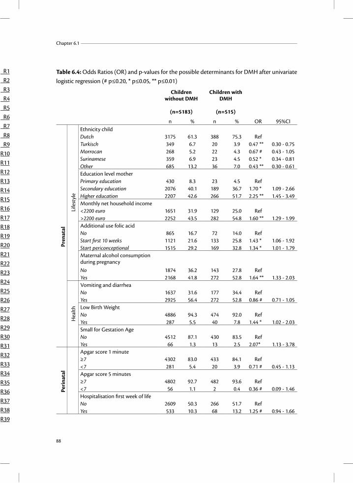
R1R2R3R4R5R6R7R8R9
R10R11R12R13R14R15R16R17R18R19R20R21R22R23R24R25R26R27R28R29R30R31R32R33R34R35R36R37R38R39
Chapter 6.1
88
Table 6.4: Odds Ratios (OR) and p-values for the possible determinants for DMH after univariate logistic regression (# p≤0.20, * p≤0.05, ** p≤0.01)
Children without DMH
(n=5183)
Children with DMH
(n=515)
n % n % OR 95%CI
Pren
atal
Ethnicity childDutch 3175 61.3 388 75.3 RefTurkisch 349 6.7 20 3.9 0.47 ** 0.30 - 0.75Morrocan 268 5.2 22 4.3 0.67 # 0.43 - 1.05Surinamese 359 6.9 23 4.5 0.52 * 0.34 - 0.81Other 685 13.2 36 7.0 0.43 ** 0.30 - 0.61Education level motherPrimary education 430 8.3 23 4.5 RefSecondary education 2076 40.1 189 36.7 1.70 * 1.09 - 2.66Higher education 2207 42.6 266 51.7 2.25 ** 1.45 - 3.49Monthly net household income<2200 euro 1651 31.9 129 25.0 Ref>2200 euro 2252 43.5 282 54.8 1.60 ** 1.29 - 1.99Additional use folic acidNo 865 16.7 72 14.0 RefStart first 10 weeks 1121 21.6 133 25.8 1.43 * 1.06 - 1.92Start periconceptional 1515 29.2 169 32.8 1.34 * 1.01 - 1.79Maternal alcohol consumption during pregnancyNo 1874 36.2 143 27.8 RefYes 2168 41.8 272 52.8 1.64 ** 1.33 - 2.03Vomiting and diarrheaNo 1637 31.6 177 34.4 RefYes 2925 56.4 272 52.8 0.86 # 0.71 - 1.05Low Birth WeightNo 4886 94.3 474 92.0 RefYes 287 5.5 40 7.8 1.44 * 1.02 - 2.03Small for Gestation AgeNo 4512 87.1 430 83.5 RefYes 66 1.3 13 2.5 2.07* 1.13 - 3.78
Peri
nata
l
Apgar score 1 minute≥7 4302 83.0 433 84.1 Ref<7 281 5.4 20 3.9 0.71 # 0.45 - 1.13Apgar score 5 minutes≥7 4802 92.7 482 93.6 Ref<7 56 1.1 2 0.4 0.36 # 0.09 - 1.46Hospitalisation first week of lifeNo 2609 50.3 266 51.7 RefYes 533 10.3 68 13.2 1.25 # 0.94 - 1.66
Life
styl
eH
ealth

R1R2R3R4R5R6R7R8R9R10R11R12R13R14R15R16R17R18R19R20R21R22R23R24R25R26R27R28R29R30R31R32R33R34R35R36R37R38R39
Pre- and postnatal determinants of Deciduous Molar Hypomineralisation
89
6
Post
nata
l
Life
styl
e Breastfeeding at 6 months
No 2438 47.0 290 56.3 RefYes 1191 23.0 112 21.7 0.79* 0.63 - 0.99
Hea
lth
Antibiotic use child first year No 2237 43.2 222 43.1 RefYes 1495 28.8 178 34.6 1.20 # 0.98 - 1.48Fever child first yearNo 652 12.6 47 9.1 RefYes 3100 59.8 356 69.1 1.59 ** 1.16 - 2.18Vomiting and diarrheaNo 1802 34.8 169 32.8 RefYes 1936 37.4 234 45.4 1.29 * 1.05 - 1.59
ResulTs
From the 6690 participating children, a good series of photographs was made in 94.5%, only one photograph was made in 3.2% and no photographs were made in 2.3%. In this study, on a child level, the data from 6325 children were used (mean age 6.2 years, SD±0.53; 49.9% girls). On a tooth level, the data from 5697 children could be used for DMH diagnosis, mostly due to the limitations in judging individual teeth. The prevalence of DMH was 9.0% (n=515) at the child level. Of all eligible second primary molars (n=24347), DMH was present in 4.1% (n=987). Often children only had one molar affected, and the mean number of DMH molars per child was 1.9.Several determinants were tested - based on the determinants for MIH, with some prenatal factors added - and these are all listed in Table 6.3. The eligible data vary per determinant due to missing data. Sensitivity analyses showed that effect sizes were not significantly different after the multiple imputation procedure.The determinants that reached a p-value less than 0.2 in the univariate logistic regression (Table 6.4) were used in the multivariate model. After backward and forward selection procedures, the final model was reached. Ethnicity, alcohol consumption by the mother during pregnancy, low birth weight and fever in the first year of life were identified as the determinants for DMH (Table 6.5). Results were not significantly different after the multiple imputation procedure (data not shown).

R1R2R3R4R5R6R7R8R9
R10R11R12R13R14R15R16R17R18R19R20R21R22R23R24R25R26R27R28R29R30R31R32R33R34R35R36R37R38R39
Chapter 6.1
90
table 6.5: Final model after multivariate analysis. Odds Ratios (OR) and p-values for the determinants are given. Determinants p-value or 95%CiEthnicity (Dutch vs Turkish) 0.035 0.49 0.25 - 0.95Ethnicity (Dutch vs Moroccan) 0.290 0.68 0.34 - 1.39Ethnicity (Dutch vs Surinamese) 0.046 0.56 0.32 - 0.99Ethnicity (Dutch vs “other”) <0.001 0.45 0.28 - 0.70Low Birth Weight 0.007 1.91 1.19 - 3.05Alcohol consumption during pregnancy 0.013 1.39 1.07 - 1.80Fever child 1st year of life 0.035 1.48 1.03 - 2.12
DisCussion
Ethnicity, alcohol consumption by the mother during pregnancy, low birth weight and any fever in the first year of life were associated with DMH. These results are partly in line with the expectations based on MIH research. The second primary molar and first permanent molar have a shared period of development and mineralisation, and an observed relationship between DMH and MIH had already been hypothesised (4, 9). The development of the second primary molar and first permanent molar start at the same time, but the maturation phase of the permanent molar is considerably longer (7). If a risk factor occurred during this overlapping period, hypomineralisation might occur in both the primary and permanent dentition (8). The determinants for DMH are expected to be more pre- and perinatal than postnatal. The cause of MIH and most possibly also for DMH, is a combination of factors and/or a threshold level needs to have been reached before enamel defects are caused (1, 10, 20, 21). Most studies are retrospective, giving biased data. Parents are not able to remember details what happened about eight years before (1, 10, 11), in our study the questionnaires were filled in every 3 to 6 months. In other studies the populations were also small and selected (1, 10). The first point is still the most difficult. Ethnicity and alcohol consumption were not mentioned previously in MIH research. Most studies on MIH and DMH are performed in Europe, probably because they are seen most often here and the Caucasian background may cause a lower threshold for DMH and MIH.Animal research has shown that ethanol (alcohol) can lead to changes in, among others, cellular differentiation and enamel mineralisation (22). This association with hypomineralisation was not found before.Low birth weight children seemed to be more at risk for enamel defects in the primary dentition than children with normal birth weight (23, 24). For MIH, low birth weight does not seem to act as a determinant. Low birth weight can be associated with DMH, but caution should be taken because the research from Vello et al. (23) and Rugg-Gunn et al. (24) used another index (modified Developmental Defects of Enamel (mDDE)) for scoring the enamel defects. Enamel defects on all primary teeth were taken into account, and low birth weight is likely to be biased with other possible interacting factors (e.g., intake of medication, fever, dehydration).

R1R2R3R4R5R6R7R8R9R10R11R12R13R14R15R16R17R18R19R20R21R22R23R24R25R26R27R28R29R30R31R32R33R34R35R36R37R38R39
Pre- and postnatal determinants of Deciduous Molar Hypomineralisation
91
6
Fever is often mentioned in MIH research (1) as a possible determinant. In an animal study, hypomineralisation of incisors could be induced by fever in rats (25). Our study shows that fever is one of the determinants of DMH.The cause of DMH seems to be multifactorial, but some of the same determinants were found here as proposed in the MIH research. This observation supports the earlier finding of a direct relationship between DMH and MIH (9).To appreciate the results some limitations need to be discussed. The percentage of mothers from different ethnicities and lower socio-economic statuses were lower among the participants than expected from the population figures in Rotterdam (16). The selection towards a more affluent and healthier population might influence the generalisability of the results. The identified determinants are not, however, expected to be different in the participating population compared with the non-participating population.Taking the photographs was difficult in some of the young children. Unsuccessful pictures were generally seen in cases in which the child was not able to breathe nasally, e.g., due to the common cold, thus creating moisture on the lens of the camera. Due to the small number of missing photographs, the results are considered representative.There can be several reasons that it is challenging to identify the cause of MIH and DMH. Therefore, more research is needed to elucidate the relationship between ethnicity, low birth weight, alcohol consumption during pregnancy and fever in the first year of life and DMH. Also, possible confounding factors and mediators need to be studied in more detail.
ConClusion
This study shows that ethnicity, low birth weight, alcohol consumption by the mother during pregnancy and any fever in the first year of the child’s life are determinants for DMH. This result shows that not only childhood factors have to be taken into account but also mother-related factors when studying determinants for DMH. More in-depth studies on the association with DMH need to focus on the prenatal factors.
AcknowledgementsThe Generation R study is conducted by the Erasmus MC in close collaboration with the Erasmus University Rotterdam, School of Law and Faculty of Social Sciences, the Municipal Health Service Rotterdam area, Rotterdam, the Rotterdam Homecare Foundation, Rotterdam, and the Stichting Trombosedienst & Artsenlaboratorium Rijnmond (STAR), Rotterdam. We gratefully acknowledge the contribution of general practitioners, hospitals, midwives, and pharmacies in Rotterdam. The first phase of the Generation R study is made possible by financial support from the Erasmus MC, Rotterdam, Erasmus University Rotterdam and the Netherlands Organization for Health Research and Development (ZonMw). The present study was supported by an additional and unrestricted grant of GABA, Therwil, Switzerland.

R1R2R3R4R5R6R7R8R9
R10R11R12R13R14R15R16R17R18R19R20R21R22R23R24R25R26R27R28R29R30R31R32R33R34R35R36R37R38R39
Chapter 6.1
92
The authors have no conflict of interest.
These results are currently being considered for publication.
R1R2R3R4R5R6R7R8R9R10R11R12R13R14R15R16R17R18R19R20R21R22R23R24R25R26R27R28R29R30R31R32R33R34R35R36R37R38R39
Pre- and postnatal determinants of Deciduous Molar Hypomineralisation
93
6
literAture
1. Alaluusua S. Aetiology of Molar-Incisor Hypomineralisation: A systematic review. Eur Arch Paediatr Dent 2010;11(2):53-8.
2. Fearne JM, Elliott JC, Wong FS, Davis GR, Boyde A, Jones SJ. Deciduous enamel defects in low-birth-weight children: correlated X-ray microtomographic and backscattered electron imaging study of hypoplasia and hypomineralization. Anat Embryol (Berl) 1994;189(5):375-81.
3. Jalevik B, Noren JG. Enamel hypomineralization of permanent first molars: a morphological study and survey of possible aetiological factors. Int J Paediatr Dent 2000;10(4):278-89.
4. Weerheijm KL, Duggal M, Mejare I, Papagiannoulis L, Koch G, Martens LC, Hallonsten AL. Judgement criteria for molar incisor hypomineralisation (MIH) in epidemiologic studies: a summary of the European meeting on MIH held in Athens, 2003. Eur J Paediatr Dent 2003;4(3):110-3.
5. Elfrink ME, Veerkamp JS, Aartman IH, Moll HA, ten Cate JM. Validity of scoring caries and primary molar hypomineralisation (DMH) on intraoral photographs. Eur Arch Paediatr Dent 2009;10(S1): 5-10.
6. Profitt W, Fields H. Contemporary Orthodontics. 3rd ed. St Louis: Mosby; 2000.
7. Butler PM. Comparison of the development of the second deciduous molar and first permanent molar in man. Arch Oral Biol 1967;12(11):1245-60.
8. Aine L, Backstrom MC, Maki R, Kuusela AL, Koivisto AM, Ikonen RS, Maki M. Enamel defects in primary and permanent teeth of children born prematurely. J Oral Pathol Med 2000;29(8):403-9.
9. Elfrink ME, ten Cate JM, Jaddoe VW, Hofman A, Moll HA, Veerkamp JS. Deciduous Molar Hypomineralization and Molar Incisor Hypomineralization. J Dent Res 2012;accepted.
10. Crombie F, Manton D, Kilpatrick N. Aetiology of molar-incisor hypomineralization: a critical review. Int J Paediatr Dent 2009;19(2):73-83.
11. Fagrell TG, Ludvigsson J, Ullbro C, Lundin SA, Koch G. Aetiology of severe demarcated enamel opacities-an evaluation based on prospective medical and social data from 17,000 children. Swed Dent J 2011;35(2):57-67.
12. Li Y, Navia JM, Bian JY. Prevalence and distribution of developmental enamel defects in primary dentition of Chinese children 3-5 years old. Community Dent Oral Epidemiol 1995;23(2):72-9.
13. Slayton RL, Warren JJ, Kanellis MJ, Levy SM, Islam M. Prevalence of enamel hypoplasia and isolated opacities in the primary dentition. Pediatr Dent 2001;23(1):32-6.
14. Elfrink ME, Schuller AA, Weerheijm KL, Veerkamp JS. Hypomineralized second primary molars: prevalence data in Dutch 5-year-olds. Caries Res 2008;42(4):282-5.
15. Jaddoe VW, Mackenbach JP, Moll HA, Steegers EA, Tiemeier H, Verhulst FC, Witteman JC, Hofman A. The Generation R Study: Design and cohort profile. Eur J Epidemiol 2006;21(6):475-84.
16. Jaddoe VW, van Duijn CM, van der Heijden AJ, Mackenbach JP, Moll HA, Steegers EA, Tiemeier H, Uitterlinden AG, Verhulst FC, Hofman A. The Generation R Study: design and cohort update 2010. Eur J Epidemiol 2010;25(11):823-41.
17. Statistics Netherlands. Allochtonen in Nederland 2004. Voorburg/Heerlen2004.
18. Statistics Netherlands. Standaard onderwijs indeling 2003. Voorburg/Heerlen2004.
19. Sterne JA, White IR, Carlin JB, Spratt M, Royston P, Kenward MG, Wood AM, Carpenter JR. Multiple imputation for missing data in epidemiological and clinical research: potential and pitfalls. BMJ 2009;338:b2393.

R1R2R3R4R5R6R7R8R9
R10R11R12R13R14R15R16R17R18R19R20R21R22R23R24R25R26R27R28R29R30R31R32R33R34R35R36R37R38R39
Chapter 6.1
94
20. Beentjes VE, Weerheijm KL, Groen HJ. Factors involved in the aetiology of molar-incisor hypomineralisation (MIH). Eur J Paediatr Dent 2002;3(1):9-13.
21. William V, Messer LB, Burrow MF. Molar incisor hypomineralization: review and recommendations for clinical management. Pediatr Dent 2006;28(3):224-32.
22. Jimenez-Farfan D, Guevara J, Zenteno E, Hernandez-Guerrero JC. Alteration of the sialylation pattern of the murine tooth germ after ethanol exposure. Birth Defects Res A Clin Mol Teratol 2005;73(12):980-8.
23. Vello MA, Martinez-Costa C, Catala M, Fons J, Brines J, Guijarro-Martinez R. Prenatal and neonatal risk factors for the development of enamel defects in low birth weight children. Oral Dis 2010;16(3): 257-62.
24. Rugg-Gunn AJ, Al-Mohammadi SM, Butler TJ. Malnutrition and developmental defects of enamel in 2- to 6-year-old Saudi boys. Caries Res 1998;32(3):181-92.
25. Tung K, Fujita H, Yamashita Y, Takagi Y. Effect of turpentine-induced fever during the enamel formation of rat incisor. Arch Oral Biol 2006;51(6):464-70.
26. Alaluusua S, Lukinmaa PL, Koskimies M, Pirinen S, Holtta P, Kallio M, Holttinen T, Salmenpera L. Developmental dental defects associated with long breast feeding. Eur J Oral Sci 1996;104(5-6): 493-7.
27. Alaluusua S, Lukinmaa PL, Vartiainen T, Partanen AM, Torppa J, Tuomisto J. Polychlorinated dibenzo-p-dioxins and dibenzofurans via mother’ s milk may cause developmental defects in the child’ s teeth. Environ Toxicol Pharmacol 1996;1:193-7.
28. Alaluusua S, Calderara P, Gerthoux PM, Lukinmaa PL, Kovero O, Needham L, Patterson DG, Jr., Tuomisto J, Mocarelli P. Developmental dental aberrations after the dioxin accident in Seveso. Environ Health Perspect 2004;112(13):1313-8.
29. van Amerongen WE, Kreulen CM. Cheese molars: a pilot study of the etiology of hypocalcifications in first permanent molars. ASDC J Dent Child 1995;62(4):266-9.
30. Holtta P, Kiviranta H, Leppaniemi A, Vartiainen T, Lukinmaa PL, Alaluusua S. Developmental dental defects in children who reside by a river polluted by dioxins and furans. Arch Environ Health 2001;56(6):522-8.
31. Jalevik B, Noren JG, Klingberg G, Barregard L. Etiologic factors influencing the prevalence of demarcated opacities in permanent first molars in a group of Swedish children. Eur J Oral Sci 2001;109(4):230-4.
32. Jontell M, Linde A. Nutritional aspects on tooth formation. World Rev Nutr Diet 1986;48:114-36.
33. Kuscu OO, Caglar E, Sandalli N. The prevalence and aetiology of molar-incisor hypomineralisation in a group of children in Istanbul. Eur J Paediatr Dent 2008;9(3):139-44.
34. Kuscu OO, Caglar E, Aslan S, Durmusoglu E, Karademir A, Sandalli N. The prevalence of molar incisor hypomineralization (MIH) in a group of children in a highly polluted urban region and a windfarm-green energy island. Int J Paediatr Dent 2009;19(3):176-85.
35. Laisi S, Kiviranta H, Lukinmaa PL, Vartiainen T, Alaluusua S. Molar-incisor-hypomineralisation and dioxins: new findings. Eur Arch Paediatr Dent 2008;9(4):224-7.
36. Laisi S, Ess A, Sahlberg C, Arvio P, Lukinmaa PL, Alaluusua S. Amoxicillin may cause molar incisor hypomineralization. J Dent Res 2009;88(2):132-6.
37. Lygidakis NA, Dimou G, Marinou D. Molar-incisor-hypomineralisation (MIH). A retrospective clinical study in Greek children. II. Possible medical aetiological factors. Eur Arch Paediatr Dent 2008;9(4):207-17.

R1R2R3R4R5R6R7R8R9R10R11R12R13R14R15R16R17R18R19R20R21R22R23R24R25R26R27R28R29R30R31R32R33R34R35R36R37R38R39
Pre- and postnatal determinants of Deciduous Molar Hypomineralisation
95
6
38. Salmela E, Lukinmaa PL, Partanen AM, Sahlberg C, Alaluusua S. Combined effect of fluoride and 2,3,7,8-tetrachlorodibenzo-p-dioxin on mouse dental hard tissue formation in vitro. Arch Toxicol 2011;85(8):953-63.
39. Whatling R, Fearne JM. Molar incisor hypomineralization: a study of aetiological factors in a group of UK children. Int J Paediatr Dent 2008;18(3):155-62.
40. Wogelius P, Haubek D, Nechifor A, Norgaard M, Tvedebrink T, Poulsen S. Association between use of asthma drugs and prevalence of demarcated opacities in permanent first molars in 6-to-8-year-old Danish children. Community Dent Oral Epidemiol 2010;38(2):145-51.


6.2
is maternal medication use during
pregnancy associated with Deciduous Molar
Hypomineralisation in the offspring?
Based on:
is maternal medication use during pregnancy associated with Deciduous Molar Hypomineralisation in the offspring?
MEC ElfrinkHA MollJC Kiefte-de JongVWV Jaddoe A HofmanH El MarrounBHC StrickerJM ten CateJSJ Veerkamp

R1R2R3R4R5R6R7R8R9
R10R11R12R13R14R15R16R17R18R19R20R21R22R23R24R25R26R27R28R29R30R31R32R33R34R35R36R37R38R39
Chapter 6.2
98
ABstrACt
Aim: The influence of maternal medication use during pregnancy on tooth-development has scarcely been studied, only for tetracycline the negative effects on the teeth are known. Therefore, we focused on the influence of medication use on the prevalence of Deciduous Molar Hypomineralisation (DMH). The aim of this study is to investigate whether antibiotics and allergy and asthma medication used during pregnancy are associated with DMH and, if so, which ones.
Materials and methods: To study this association, an intra-oral camera was used to take clinical photographs of clean, moist teeth. In the photographs, the second primary molars of 6690 children (mean age 6.2 years, SD±0.53; 49.9% girls) were scored for DMH. Data on medication use during pregnancy were retrieved from pharmacies.
Results: There is no association between the use of either asthma and allergy medication or antibiotics during pregnancy and early life of the child and the DMH.
Conclusion: Maternal use of antibiotics and allergy or asthma medication during pregnancy does not seem to play a major role in the development of DMH in the offspring.

R1R2R3R4R5R6R7R8R9R10R11R12R13R14R15R16R17R18R19R20R21R22R23R24R25R26R27R28R29R30R31R32R33R34R35R36R37R38R39
Is maternal medication use during pregnancy associated with Deciduous Molar Hypomineralisation in the offspring?
99
6
introDuCtion
Some medications induce changes to the teeth, such as discoloration and physical damage to the tooth structure (1). Extrinsic tooth discoloration, which is removable, is often seen with the use of chlorhexidine, amoxicillin with clavulanic acid, essential oils and oral iron salts in liquid form (1). Intrinsic tooth discoloration, which is permanent, occurs if the medication had been used during tooth formation. The most common medications causing intrinsic tooth discoloration are fluorides, tetracyclines, minocycline, and ciprofloxacin (1). But more antibiotics have proven in animal research to influence the amelogenesis (2, 3). Physical damage to the tooth structure can be caused by medications that contain sugar, have a low pH, or reduce salivary secretion. Some anticonvulsants and chemotherapeutic drugs used during the tooth development period cause changes in tooth size, tooth agenesis, arrested tooth development and disturbances affecting enamel, dentin and cementum (1). Also other factors such as dioxins (4), fluoride (5) and temperature/fever (6, 7) were found to disturb amelogenesis in animal research.Deciduous Molar Hypomineralisation (DMH) was defined as hypomineralisation of 1-4 second primary molars. The hypomineralisations in DMH were similar to those observed in Molar Incisor Hypomineralisation (MIH) in the permanent dentition (8). There were also MIH-like defects on the second primary molars and permanent cuspids (9). These MIH-like defects in the deciduous molars have now been described as DMH (10). Most teeth with DMH had white, yellow or brown demarcated opacities (8). In the Netherlands, 4.9% of the children had DMH (8).For DMH and MIH, the same possible causes were suggested, although occurring somewhat earlier in life for DMH than for MIH (8, 11-13).MIH was associated with perinatal problems, common childhood diseases, medication usage (e.g., antibiotics and asthma medication) and pollution (e.g., dioxins) during the tooth development period (14-17).Antibiotics can cause early childhood caries and primary tooth fluorosis (18, 19). Amoxicillin, the most commonly prescribed antibiotic has been said to cause MIH (3, 17). Asthma medication and severe demarcated opacities also seemed to be associated (20). However, the influence of these medications taken during pregnancy on hypomineralisation has not been studied before. This study focused on the influence of medication use on the prevalence of DMH. The aim of this study is to investigate whether antibiotics and asthma medication used during pregnancy are associated with DMH. MAteriAls AnD MetHoDs
Participants. The participants took part in the Generation R study, a population-based prospective cohort study from foetal life until young adulthood. The Generation R study, which was designed to identify early environmental and genetic determinants of growth, development and health, has

R1R2R3R4R5R6R7R8R9
R10R11R12R13R14R15R16R17R18R19R20R21R22R23R24R25R26R27R28R29R30R31R32R33R34R35R36R37R38R39
Chapter 6.2
100
previously been described in detail (21, 22). The cohort included 9897 children and their mothers living in Rotterdam, the Netherlands. Enrolment of mothers was aimed at early pregnancy (gestational age <18 weeks). All children were born between April 2002 and January 2006. Of all the eligible children in the study area, 61% participated at birth in the study (22). The study had approved by the Medical Ethics Committee of the Erasmus Medical Centre, Rotterdam; from all participants written informed consent was obtained. For the postnatal phase of the study 7893 children were available. Approximately half of the mothers (51.0%) and children were of Dutch origin (54.8%) (22). At the age of five to six years, the children were invited for a check-up visit at the Erasmus Medical Centre. From March 2008 until January 2012, 6690 children, including 88 twins, visited the Erasmus Medical Centre. As part of this visit, intra-oral photographs of their teeth were taken, which was successfully done in 6325 children (94.5%). In cases in which a few teeth could not be scored, only the teeth visible in the photographs were used in the analysis. A flowchart of the participants is shown in Figure 6.1 (page 85).Measures. Assessments included questionnaires, physical examinations and foetal ultrasound examinations and were planned in early pregnancy (gestational age <18 weeks), mid pregnancy (gestational age 18-25 weeks) and late pregnancy (gestational age >25 weeks). Data on maternal medication use during pregnancy were retrieved from pharmacies. These data were available for 5613 pregnancies of 5654 children. At time of birth, Apgar scores and other birth parameters such as weight and length were measured. Other data, such as ethnicity (23), education level (24), household income, additional use of folic acid, and health of the mother and child, were collected via questionnaires at the ages of 2, 6 and 12 months.At ages of 5-6, children visited the research centre for hands-on measurements and to have photographs of their teeth taken. After brushing their teeth, photographs of clean, moist teeth were taken by trained nurses and dental students (excess saliva was removed with a cotton ball). Taking approximately ten photographs of all the teeth took 1-2 minutes for each child. An intra-oral camera [Poscam USB intra-oral autofocus camera (Digital Leader PointNix), 640 x 480 pixels] was used for the pictures of the teeth, with a minimal scene illumination of f 1.4 and 30 lx. In an earlier study, the validity of this camera for visualising DMH was shown to be high (10).From the intra-oral photographs DMH was scored using the EAPD criteria (see Table 6.6) (9, 10). A second primary molar was diagnosed as having DMH when at least one of these criteria was found. The tooth was scored as ‘not able to be judged’, if the tooth, or the place where the tooth should be was not shown on the photographs, The photographs were displayed on a computer in full-screen mode and scored by a single calibrated dentist (ME). To test the inter-observer agreement in this study, the data of 648 children were scored independently by another calibrated dentist (JV). The Cohen’s kappa score in this study was 0.73 for DMH and 0.64 for MIH. In the event of a disagreement, the photographs were studied again, and a consensus decision was made. At least six weeks after the first scoring, a separate group of 649 children were scored again by the first dentist (ME). The intra-observer agreement reached the following Cohen’s kappa scores: 0.82 (DMH) and 0.85 (MIH).

R1R2R3R4R5R6R7R8R9R10R11R12R13R14R15R16R17R18R19R20R21R22R23R24R25R26R27R28R29R30R31R32R33R34R35R36R37R38R39
Is maternal medication use during pregnancy associated with Deciduous Molar Hypomineralisation in the offspring?
101
6
Statistics. Statistical analyses were performed with SPSS version 18.0 (SPSS Inc, Chicago, IL, USA). To test for any association between medication use and DMH, logistic regression analysis was used with adjustment for potential confounders. Selection of potential confounders was based on the aetiological factors of MIH. To test the association between the medications and DMH, first a univariate logistic regression analysis was performed. This yielded no significant results. The potential confounder was kept in the final multivariate model when it gave 10% or more change of the beta-coefficient. Missing data were multiple imputed (n=10 imputations) based on the correlation between the variable with missing values with other patient characteristics. Data were imputed according to the Markov Chain Monte Carlo (MCMC) method (assuming no monotone missing pattern) and the imputations were repeated for 10 times. Data were analyzed in each data set separately and the results of the 10 imputed analyses were pooled and reported in this paper along with the original data. A p-value <0.05 was considered as statistically significant.
table 6.6: Criteria for the diagnosis of DMH Mild:
• Opacity: A defect that changes the translucency of the enamel, variable in degree. The defective enamel is of normal thickness with a smooth surface and can be white, yellow or brown in color. The demarcated opacity is not caused by caries, ingestion of excess fluoride during tooth development or amelogenesis imperfecta etc.
Severe:• Posteruptive enamel loss: A defect that indicates surface enamel loss after eruption of the tooth,
e.g., hypomineralisation related attrition. Enamel loss due to erosion was excluded, and/or• Atypical caries: The size and form of the caries lesion do not match the present caries
distribution in the child’s mouth, and/or• Atypical restoration: The size and form of the restoration do not match the present caries
distribution in the child’s mouth, and/or• Atypical extraction: Absence of a molar that does not fit in the dental development and caries
pattern of the child.
results
Among the 6690 participating children (mean age 6.2 years, SD±0.53; 49.9% girls), a good series of photographs was made in 94.5%, only one photograph was made in 3.2% and no photograph was made in 2.3%. The prevalence of DMH in the original dataset was 9.0% (n=515) at the child level. Of all eligible second primary molars (n=24347), DMH was present in 4.1% (n=987). In the original dataset, the mean age of the mothers was 30.6 years (SD±5.2), and 63.2% were of Dutch-Caucasian origin. More details about the study population can be found in Table 6.7.

R1R2R3R4R5R6R7R8R9
R10R11R12R13R14R15R16R17R18R19R20R21R22R23R24R25R26R27R28R29R30R31R32R33R34R35R36R37R38R39
Chapter 6.2
102
table 6.7: Subject characteristicsmean sD
Age mother at intake (years) 30.6 5.2Age child at visit research centre (years) 6.2 0.5
n %Deciduous Molar Hypomineralisation (DMH)No 5182 91.0Yes 515 9.0Maternal antibiotic use (prenatal)No 2230 79.7Yes 569 20.3Maternal amoxicillin use (prenatal)No 2433 86.9Yes 366 13.1Maternal tetracycline use (prenatal)No 2776 99.2Yes 23 0.8Maternal asthma medication use (prenatal)No 2455 87.7Yes 344 12.3Maternal allergy medication use (prenatal)No 2648 94.6Yes 151 5.4Maternal ethnicityDutch-Caucasian 3919 63.2Turkisch 509 8.2Morrocan 336 5.4Surinamese 476 7.7Other 959 15.5Additional use of folium acidNo 1138 25.0Start first 10 weeks 1445 31.8Start periconceptional 1963 43.2Addition use of fluoride child (1st year of life)No 4441 98.6Yes 62 1.4Maternal illnesses during pregnancyNo 1157 19.7Yes 4722 80.3Maternal fever during pregnancyNo 4895 83.5Yes 968 16.5Diabetes gravidarumNo 6301 98.9Yes 69 1.1

R1R2R3R4R5R6R7R8R9R10R11R12R13R14R15R16R17R18R19R20R21R22R23R24R25R26R27R28R29R30R31R32R33R34R35R36R37R38R39
Is maternal medication use during pregnancy associated with Deciduous Molar Hypomineralisation in the offspring?
103
6
Gestational hypertensionNo 5448 95.8Yes 240 4.2Apgar score 1 minute≥7 5582 94.4<7 333 5.6Infectious diseases child (1st year of life)No 201 4.1Yes 4661 95.9Fever child (1st year of life)No 846 17.4Yes 4017 82.6
During pregnancy, 569 mothers used antibiotics, with 366 using amoxicillin and 23 using tetracycline. Asthma medication (sympaticomimetics, corticosteroids etc.) was used by 344 mothers, and allergy medication (antihistaminics) by 151 mothers. Of those who used amoxicillin, 85% did so once during pregnancy, 11% twice and the remaining 4% 3-5 times. The most frequent daily dose was 1500 mg (84%), with the prescribed daily doses varying between 750 and 2500 mg.To test the association between the medications and DMH, a logistic regression analysis without correction was first performed. This analysis yielded no significant results. Four different correction models were then tested, also using logistic regression analysis (see Table 6.8).The first model corrects for the general factors including age and ethnicity of the mother. The second model for lifestyle factors includes the use of folic acid during pregnancy and the use of additional fluoride tablets during the first year of life by the child. The third model corrects for health problems of the mother during pregnancy, including illnesses, fever, diabetes gravidarum (gestational diabetes) and hypertension. The last model corrects for health problems of the child, including the Apgar score at 1 minute, infectious diseases and fever of the child in the first year of life.The association between DMH and antibiotics, asthma medication and allergy medication changed slightly in the multivariate analyses. No statistically significant association was found.
DisCussion
No associations between antibiotics, asthma medication or allergy medication and DMH were found in our research, which is only partly in line with the literature.Antibiotics are either supposed to be safe to use during pregnancy or data about the safety of individual antibiotics are lacking. Only tetracycline is advised against because of its effects on tooth development (25, 26).Asthma and allergy medication are also supposed to be safe to use during pregnancy, but little data are available on their safety (25).

R1R2R3R4R5R6R7R8R9
R10R11R12R13R14R15R16R17R18R19R20R21R22R23R24R25R26R27R28R29R30R31R32R33R34R35R36R37R38R39
Chapter 6.2
104
tabl
e 6.
8: O
dds R
atio
s (O
Rs) f
or th
e as
soci
atio
n be
twee
n m
edic
atio
n us
e du
ring
preg
nanc
y an
d D
ecid
uous
Mol
ar H
ypom
iner
alisa
tion
(DM
H).
(# p
≤0.2
0, *p
≤0.0
5, **
p≤0.
01)
uni
vari
ate
Corr
ecti
on g
ener
al
fact
ors
Corr
ecti
on li
fest
yle
fact
ors
Corr
ecti
on h
ealt
h m
othe
rCo
rrec
tion
hea
lth
child
or
95%
Ci
or
95%
Ci
or
95%
Ci
or
95%
Ci
or
95%
Ci
AB
Orig
inal
0.73
#0.
49 -
1.09
0.81
0.53
- 1.
220.
930.
57 -
1.53
0.71
#0.
47 -
1.09
0.74
0.46
- 1.
19Po
oled
0.98
0.56
- 1.
730.
990.
57 -
1.74
0.98
0.55
- 1.
730.
980.
56 -
1.73
0.99
0.56
- 1.
74A
MO
XO
rigin
al0.
64 #
0.38
- 1.
070.
690.
41 -
1.17
0.79
0.41
- 1.
510.
63 #
0.36
- 1.
080.
700.
39 -
1.26
Pool
ed1.
160.
39 -
3.49
1.16
0.38
- 3.
561.
160.
38 -
3.51
1.16
0.39
- 3.
491.
160.
39 -
3.50
TETR
AO
rigin
al1.
150.
27 -
4.95
2.00
0.45
- 8.
975.
04#
0.90
- 28
.33
1.39
0.32
- 6.
102.
180.
47 -
10.1
3Po
oled
0.86
0.25
- 2.
960.
860.
25 -
2.99
0.85
0.24
- 3.
000.
850.
23 -
3.10
0.86
0.24
- 3.
02A
STH
MA
Orig
inal
0.97
0.61
- 1.
541.
060.
67 -
1.70
0.95
0.53
- 1.
710.
950.
58 -
1.54
1.15
0.69
- 1.
91Po
oled
1.05
0.69
- 1.
581.
050.
70 -
1.60
1.04
0.68
- 1.
591.
040.
68 -
1.58
1.05
0.69
- 1.
60A
LLO
rigin
al1.
040.
54 -
2.03
1.11
0.57
- 2.
181.
260.
56 -
2.84
1.08
0.54
- 2.
181.
250.
61 -
2.55
Pool
ed1.
230.
59 -
2.57
1.25
0.58
- 2.
681.
230.
58 -
2.60
1.24
0.59
- 2.
601.
240.
58 -
2.64
AB=
ant
ibio
tics
AM
OX=
am
oxic
illin
TETR
A=
tetr
acyc
line
AST
HM
A=
asth
ma
med
icat
ion
ALL
= al
lerg
y m
edic
atio
nO
rigin
al: a
naly
sis b
ased
on
orig
inal
dat
aset
Pool
ed: b
ased
on
data
set a
fter
mul
tiple
impu
tatio
n fo
r rep
laci
ng m
issin
g va
lues

R1R2R3R4R5R6R7R8R9R10R11R12R13R14R15R16R17R18R19R20R21R22R23R24R25R26R27R28R29R30R31R32R33R34R35R36R37R38R39
Is maternal medication use during pregnancy associated with Deciduous Molar Hypomineralisation in the offspring?
105
6
Several factors, also prenatal factors, were found to be associated with hypomineralisation in the permanent dentition (MIH), such as medical problems, although sometimes controversial results are found (16, 17, 27-29). Possible determinants of DMH are only currently hypothesised about. The same determinants are expected as for MIH molars, though occurring somewhat earlier in life (pre- and perinatal instead of postnatal) (8, 11-13). Pre- and perinatal factors do not seem to have much influence on MIH, they may be interesting for DMH. In MIH, whether the disease or the medication to treat the disease cause the hypomineralisation, is not yet known. Most likely, the disease itself does not cause the hypomineralisation but rather the medications commonly prescribed to treat the disease. For antibiotics, especially the commonly prescribed amoxicillin, the relationship with hypomineralisation has been confirmed in animal research (3). In our study, however, we could not affirm this finding. Conclusions from our non-significant data are difficult. For tetracycline use, after correction for lifestyle factors, the Odds Ratios (ORs) of the original and pooled data were not within each other’s Confidence Intervals (CI). In the original dataset, the OR for tetracyclines especially after correction for lifestyle factors is high and needs to be investigated further.The relationship between hypomineralisation in the permanent dentition and the use of asthma medication by the child was found by Wogelius et al. (20). Our study could not find an association between maternal use of asthma medication and DMH.
limitations of the studyThe percentage of mothers from different ethnicities and lower socio-economic statuses were lower among the participants than is expected from the population figures in Rotterdam (22). The generalisability of the results might be influenced by a selection towards a more healthy and affluent population. The associations studied, however, are not expected to be different in the participating population compared with the non-participating population.Some groups, especially the group of mothers who used tetracycline during pregnancy, are very small. Due to the number of missing values, the missing data were imputed. There were no significant differences in the outcome between the analyses on the original data and on the imputed data (see Table 6.8). Photographs were difficult to take in some of the young children. Unsuccessful pictures were generally seen in cases in which the child was not able to breathe nasally, e.g., due to the common cold, thus creating moisture on the lens of the camera. But the overall number of missing photographs was low (5.5%).

R1R2R3R4R5R6R7R8R9
R10R11R12R13R14R15R16R17R18R19R20R21R22R23R24R25R26R27R28R29R30R31R32R33R34R35R36R37R38R39
Chapter 6.2
106
In the literature, many different factors influencing the tooth development have been studied. In animals, a specific relationship between one medication and its influence on amelogenesis were investigated. Because many medications, especially antibiotics, were found to be associated with disturbances in amelogenesis, more research is needed to determine if the disease or the treatment for the disease (medication used) influences the amelogenesis, thereby being one of the factors influencing DMH.
ConClusion
The use of asthma and allergy medication and antibiotics during pregnancy is not associated with DMH in the offspring suggesting that these factors do not play a major role in the etiology of DMH in children.
AcknowledgementsThe Generation R study was conducted by the Erasmus MC in close collaboration with the following: Erasmus University, Rotterdam; the School of Law and Faculty of Social Sciences, the Municipal Health Service Rotterdam area, Rotterdam; the Rotterdam Homecare Foundation, Rotterdam; and the Stichting Trombosedienst and Artsenlaboratorium Rijnmond (STAR), Rotterdam. We gratefully acknowledge the contribution of general practitioners, hospitals, midwives, and pharmacies in Rotterdam. The first phase of the Generation R study was made possible by financial support from the Erasmus MC, Rotterdam, Erasmus University, Rotterdam and the Netherlands Organisation for Health Research and Development (ZonMw). The present study was supported by an additional and unrestricted grant from GABA, Therwil, Switzerland.The authors have no conflict of interest.
These results are currently being considered for publication.

R1R2R3R4R5R6R7R8R9R10R11R12R13R14R15R16R17R18R19R20R21R22R23R24R25R26R27R28R29R30R31R32R33R34R35R36R37R38R39
Is maternal medication use during pregnancy associated with Deciduous Molar Hypomineralisation in the offspring?
107
6
literAture
1. Tredwin CJ, Scully C, Bagan-Sebastian JV. Drug-induced disorders of teeth. J Dent Res 2005;84(7):596-602.
2. Lyaruu DM, van Duin MA, Bervoets TJ, Woltgens JH, Bronckers AL. Effects of actinomycin D on developing hamster molar tooth germs in vitro. Eur J Oral Sci 1997;105(1):52-8.
3. Laisi S, Ess A, Sahlberg C, Arvio P, Lukinmaa PL, Alaluusua S. Amoxicillin may cause molar incisor hypomineralization. J Dent Res 2009;88(2):132-6.
4. Alaluusua S, Lukinmaa PL, Torppa J, Tuomisto J, Vartiainen T. Developing teeth as biomarker of dioxin exposure. Lancet 1999;353(9148):206.
5. Bronckers AL, Lyaruu DM, Bervoets TJ, Woltgens JH. Fluoride enhances intracellular degradation of amelogenins during secretory phase of amelogenesis of hamster teeth in organ culture. Connect Tissue Res 2002;43(2-3):456-65.
6. Tung K, Fujita H, Yamashita Y, Takagi Y. Effect of turpentine-induced fever during the enamel formation of rat incisor. Arch Oral Biol 2006;51(6):464-70.
7. Woltgens JH, Lyaruu DM, Bervoets TJ, Bronckers AL. Reversible and irreversible effects of temperature on amelogenesis of hamster tooth germs in vitro. Scanning Microsc 1993;7(3):1009-16.
8. Elfrink ME, Schuller AA, Weerheijm KL, Veerkamp JS. Hypomineralized second primary molars: prevalence data in Dutch 5-year-olds. Caries Res 2008;42(4):282-5.
9. Weerheijm KL, Duggal M, Mejare I, Papagiannoulis L, Koch G, Martens LC, Hallonsten AL. Judgement criteria for molar incisor hypomineralisation (MIH) in epidemiologic studies: a summary of the European meeting on MIH held in Athens, 2003. Eur J Paediatr Dent 2003;4(3):110-3.
10. Elfrink ME, Veerkamp JS, Aartman IH, Moll HA, ten Cate JM. Validity of scoring caries and primary molar hypomineralisation (DMH) on intraoral photographs. Eur Arch Paediatr Dent 2009;10(S1): 5-10.
11. Li Y, Navia JM, Bian JY. Prevalence and distribution of developmental enamel defects in primary dentition of Chinese children 3-5 years old. Community Dent Oral Epidemiol 1995;23(2):72-9.
12. Aine L, Backstrom MC, Maki R, Kuusela AL, Koivisto AM, Ikonen RS, Maki M. Enamel defects in primary and permanent teeth of children born prematurely. J Oral Pathol Med 2000;29(8):403-9.
13. Slayton RL, Warren JJ, Kanellis MJ, Levy SM, Islam M. Prevalence of enamel hypoplasia and isolated opacities in the primary dentition. Pediatr Dent 2001;23(1):32-6.
14. Alaluusua S, Lukinmaa PL, Vartiainen T, Partanen AM, Torppa J, Tuomisto J. Polychlorinated dibenzo-p-dioxins and dibenzofurans via mother’ s milk may cause developmental defects in the child’ s teeth. Environ Toxicol Pharmacol 1996;1:193-7.
15. Jalevik B, Noren JG. Enamel hypomineralization of permanent first molars: a morphological study and survey of possible aetiological factors. Int J Paediatr Dent 2000;10(4):278-89.
16. Beentjes VE, Weerheijm KL, Groen HJ. Factors involved in the aetiology of molar-incisor hypomineralisation (MIH). Eur J Paediatr Dent 2002;3(1):9-13.
17. Whatling R, Fearne JM. Molar incisor hypomineralization: a study of aetiological factors in a group of UK children. Int J Paediatr Dent 2008;18(3):155-62.
18. Alaki SM, Burt BA, Garetz SL. The association between antibiotics usage in early childhood and early childhood caries. Pediatr Dent 2009;31(1):31-7.

R1R2R3R4R5R6R7R8R9
R10R11R12R13R14R15R16R17R18R19R20R21R22R23R24R25R26R27R28R29R30R31R32R33R34R35R36R37R38R39
Chapter 6.2
108
19. Hong L, Levy SM, Warren JJ, Dawson DV, Bergus GR, Wefel JS. Association of amoxicillin use during early childhood with developmental tooth enamel defects. Arch Pediatr Adolesc Med 2005;159(10):943-8.
20. Wogelius P, Haubek D, Nechifor A, Norgaard M, Tvedebrink T, Poulsen S. Association between use of asthma drugs and prevalence of demarcated opacities in permanent first molars in 6-to-8-year-old Danish children. Community Dent Oral Epidemiol 2010;38(2):145-51.
21. Jaddoe VW, Mackenbach JP, Moll HA, Steegers EA, Tiemeier H, Verhulst FC, Witteman JC, Hofman A. The Generation R Study: Design and cohort profile. Eur J Epidemiol 2006;21(6):475-84.
22. Jaddoe VW, van Duijn CM, van der Heijden AJ, Mackenbach JP, Moll HA, Steegers EA, Tiemeier H, Uitterlinden AG, Verhulst FC, Hofman A. The Generation R Study: design and cohort update 2010. Eur J Epidemiol 2010;25(11):823-41.
23. Statistics Netherlands. Allochtonen in Nederland 2004. Voorburg/Heerlen2004.
24. Statistics Netherlands. Standaard onderwijs indeling 2003. Voorburg/Heerlen2004.
25. http://www.fk.cvz.nl.
26. Demers P, Fraser D, Goldbloom RB, Haworth JC, LaRochelle J, MacLean R, Murray TK. Effects of tetracyclines on skeletal growth and dentition. A report by the Nutrition Committee of the Canadian Paediatric Society. Can Med Assoc J 1968;99(17):849-54.
27. Alaluusua S. Aetiology of Molar-Incisor Hypomineralisation: A systematic review. Eur Arch Paediatr Dent 2010;11(2):53-8.
28. Jalevik B, Noren JG, Klingberg G, Barregard L. Etiologic factors influencing the prevalence of demarcated opacities in permanent first molars in a group of Swedish children. Eur J Oral Sci 2001;109(4):230-4.
29. Tapias-Ledesma MA, Jimenez R, Lamas F, Gonzalez A, Carrasco P, Gil de Miguel A. Factors associated with first molar dental enamel defects: a multivariate epidemiological approach. J Dent Child (Chic) 2003;70(3):215-20.

7
Deciduous Molar Hypomineralisation and Molar
incisor Hypomineralisation
Based on:
Deciduous Molar Hypomineralization and Molar incisor Hypomineralization
MEC ElfrinkJM ten CateVWV JaddoeA HofmanHA MollJSJ Veerkamp
J Dent Res 2012, Feb 27 [Epub ahead of print]

R1R2R3R4R5R6R7R8R9
R10R11R12R13R14R15R16R17R18R19R20R21R22R23R24R25R26R27R28R29R30R31R32R33R34R35R36R37R38R39
Chapter 7
110
ABstrACt
Aim: This study was embedded in the Generation R study, a population-based prospective cohort study from foetal life until young adulthood. This study focused on the relationship between Deciduous Molar Hypomineralisation (DMH) and Molar Incisor Hypomineralisation (MIH).
Materials and methods: First permanent molars develop during a period similar to that of second primary molars, with possible comparable risk factors for hypomineralisation. Children with DMH have a greater risk of developing MIH. Clinical photographs of clean, moist teeth were taken with an intra-oral camera in 6161 children (mean age 74.3 months, SD±6.1; 49.8% girls). First permanent molars and second primary molars were scored with respect to DMH or MIH.
Results: The prevalence of DMH and MIH was 9.0% and 8.7% at child level, and 4.0% and 5.4% at tooth level. The Odds Ratio (OR) for MIH based on DMH was 4.4 (95% CI: 3.1-6.4).
Conclusion: The relationship between the occurrence of DMH and MIH suggests a shared cause and indicates that, clinically, DMH can be used as a predictor for MIH.

R1R2R3R4R5R6R7R8R9R10R11R12R13R14R15R16R17R18R19R20R21R22R23R24R25R26R27R28R29R30R31R32R33R34R35R36R37R38R39
Deciduous Molar Hypomineralisation and Molar Incisor Hypomineralisation
111
7
introDuCtion
Developmental defects of dental enamel are common in both deciduous and permanent dentitions and are classified into hypomineralisation and hypoplasia (1, 2). Enamel hypoplasia is a quantitative defect of the enamel, resulting from a disturbance to the ameloblasts during matrix formation (1, 3, 4). Enamel hypomineralisation is a qualitative defect of the enamel because of a disturbance during initial calcification and/or during maturation (1, 3). Hypomineralised parts of teeth are weaker and the enamel may chip off easily, resulting in posteruptive loss of enamel. It can be difficult to distinguish between hypoplasia and posteruptive enamel loss (4). In the primary dentition, the second primary molar generally presents with more caries than the first primary molar (5-7). A recent study showed that hypomineralisation was an important risk factor for caries in the primary dentition (8). Also, in the permanent dentition, rapid caries progression was observed in hypomineralised molars (9, 10). Hypomineralised permanent molars are frequently combined with hypomineralised incisors. Molar Incisor Hypomineralisation (MIH) is defined as hypomineralisation of systemic origin of 1 to 4 permanent first molars combined with affected incisors (11). MIH-like defects are also seen on second primary molars and permanent cuspids (3). These MIH-like defects in the primary molars are now described as Deciduous Molar Hypomineralisation (DMH) (8, 12). In the Netherlands, the prevalence of DMH has been reported at 4.9% at child level (13) while the prevalence of MIH was higher (6-14%) (9, 14). The severity of MIH as well as DMH varies between patients, but also within a patient. Opacities are considered the mildest form of MIH and DMH, and atypical extractions as the most severe manifestation (9). MIH can cause serious pain due to posteruptive enamel loss, rapid caries progression, and pain during restorative treatment (9, 10). Children with MIH need more dental treatments and - probably as a consequence - are generally more fearful than their peers (10). Therefore, it is important to diagnose MIH as early as possible to reduce the vulnerability of the MIH-affected molars by focusing on their restorative and preventive needs. The development of the second primary molars starts at around the same time as the development of the first permanent molars and permanent incisors, but the maturation of the permanent teeth occurs more slowly (see Table 7.1) (15, 16). If a risk factor occurs during this overlapping period, hypomineralisation might occur in the primary as well as in the permanent dentition (17). Therefore, DMH might be used as a predictor for MIH. The parallel development of the second primary molar and the first permanent molar, both developmentally and with respect to their location in the jaw, might be indicative of a common cause for the hypomineralisation process. Our aim was therefore to study the association between DMH in the second primary molars and MIH in the first permanent molars.

R1R2R3R4R5R6R7R8R9
R10R11R12R13R14R15R16R17R18R19R20R21R22R23R24R25R26R27R28R29R30R31R32R33R34R35R36R37R38R39
Chapter 7
112
table 7.1. Starting age of development of the incisors and first molars of the permanent dentition and the second molars of the primary dentition (16).
Tooth 1st permanent incisor
2nd permanent incisor
1st permanent molar
2nd primary molar
Upper jawStart calcification 3 months 11 months 32 weeks in utero 19 weeks in uteroCrown completed 4.5 years 5.5 years 4.3 years 11 monthsEruption 7.3 years 8.3 years 6.25 years 29 months
Lower jawStart calcification 3 months 3 months 32 weeks in utero 18 weeks in uteroCrown completed 3.5 years 4 years 3.8 years 10 monthsEruption 6.3 years 7.5 years 6 years 27 months
MAteriAls AnD MetHoDs
Participants. This study was embedded in the Generation R study, a population-based prospective cohort study from foetal life until young adulthood. The Generation R study was designed to identify early environmental and genetic determinants of growth, development, and health and has previously been described in detail (18, 19). Briefly, the cohort included 9778 mothers and their children living in Rotterdam, the Netherlands. Enrolment of mothers occurred in their early pregnancy (gestational age <18 wks). All children were born between April 2002 and January 2006 and formed a prenatally enrolled birth-cohort. Of all eligible children in the study area, 61% were enrolled in the study at birth (18). The Medical Ethics Committee of the Erasmus Medical Centre, Rotterdam, approved the study. Written informed consent was obtained from all participants.For the postnatal phase of the study, 7893 children were available. About half of the mothers (51.0%) and the children were of Dutch origin (54.8%) (19). At the age of 5 to 6 yrs, the children were invited for a check-up visit at the Erasmus Medical Centre. From March 2008 until November 2011, 6487 children (77 twins) visited the Erasmus Medical Centre. As part of this visit, intra-oral photographs of the teeth were made, which was successfully done in 6161 children (95.0%). In cases where a few teeth could not be scored, only the teeth visible on the photographs were used in the analysis. A flowchart of the participants is shown in Figure 7.1.

R1R2R3R4R5R6R7R8R9R10R11R12R13R14R15R16R17R18R19R20R21R22R23R24R25R26R27R28R29R30R31R32R33R34R35R36R37R38R39
Deciduous Molar Hypomineralisation and Molar Incisor Hypomineralisation
113
7
Assessed for eligibility
Pregnancies (n=9778)
Children (n=9897)
Excluded (n=152)
intra-uterine death (n=78)
abortion (n=29)
various reasons (n=45)
Enrolment
Analysis
Live births (n=9745)
Availability
Excluded (n=1852)
no consent (n=38)
various reasons e.g. moved from study area
(n=1814)
Postnatal participants
(n=7893)
Excluded (n=1406)
failed participation (n=1406)
Eligible for analysis (n=6487)
Excluded (n=326)
no or only one photograph was made
(n=326)
Analysed
(n=6161)
figure 7.1. Flowchart of the participants.
Dental examination. After teeth were brushed, photographs of clean and moist teeth were taken by trained nurses and dental students (excess saliva was removed with a cotton roll). It took 1-2 min to take approximately 10 photographs of all teeth of the child. For this purpose, an intra-oral camera was used [Poscam USB intra-oral autofocus camera (Digital Leader PointNix), 640 x 480 pixels]. An example of such a photograph is shown in Figure 7.2.

R1R2R3R4R5R6R7R8R9
R10R11R12R13R14R15R16R17R18R19R20R21R22R23R24R25R26R27R28R29R30R31R32R33R34R35R36R37R38R39
Chapter 7
114
figure 7.2. Tooth 26 (1st permanent molar upper left) showing MIH (yellow opacity) and tooth 65 (2nd
primary molar upper left) showing DMH (yellow opacity and post-eruptive enamel loss).
The minimal scene illumination was f 1.4 and 30 lx. In an earlier study, the validity of this camera for visualizing DMH was shown to be high (sensitivity 72.3% and specificity 92.8%). The reliability was good for inter-observer agreement (kappa 0.62) and excellent for intra-observer agreement (kappa 0.95) (12).DMH and MIH were scored on the intra-oral photographs according to the EAPD criteria (3, 12):Mild:
• Opacity: A defect that changes the translucency of the enamel, variable in degree. The defective enamel is of normal thickness with a smooth surface and can be white, yellow or brown in colour. The demarcated opacity is not caused by caries, ingestion of excess fluoride during tooth development or amelogenesis imperfecta etc.
Severe:• Posteruptive enamel loss: A defect that indicates surface enamel loss after eruption of the
tooth, e.g., hypomineralisation related attrition. Enamel loss due to erosion was excluded, and/or

R1R2R3R4R5R6R7R8R9R10R11R12R13R14R15R16R17R18R19R20R21R22R23R24R25R26R27R28R29R30R31R32R33R34R35R36R37R38R39
Deciduous Molar Hypomineralisation and Molar Incisor Hypomineralisation
115
7
• Atypical caries: The size and form of the caries lesion do not match the present caries distribution in the child’s mouth, and/or
• Atypical restoration: The size and form of the restoration do not match the present caries distribution in the child’s mouth, and/or
• Atypical extraction: Absence of a molar that does not fit in the dental development and caries pattern of the child.
A first permanent molar or a second primary molar was diagnosed as having MIH or DMH when at least one of these criteria or a combination was found. If in a child one or more DMH molars were scored as severe, the child was scored as having a severe form of DMH.On the photographs, the number of shed teeth was also measured. The following criteria were used for the eruption of the permanent teeth:
• Not erupted / not shed: gingiva distally from the 2nd primary molar was shown on the photograph, but nothing from the 1st permanent molar can be seen. Permanent incisor not visible yet.
• Partly erupted / primary tooth missing due to shedding: 1st permanent molar was partly covered by the gingiva. Permanent incisor is not visible yet, but primary incisor was lost, most likely due to shedding.
• Erupted: permanent molar was not covered by the gingiva anymore, permanent incisor is (partly) visible.
• If the tooth, or the place were the tooth has to be, was not shown on the photographs, the tooth was scored as ‘not able to be judged’
The photographs were shown on a computer in full-screen mode, and were scored by one calibrated dentist (ME). To test the inter-observer agreement in this study, another calibrated dentist (JV) independently scored the data for 648 children. The Cohen’s kappa score in this study for DMH was 0.60 and for MIH 0.69. In the event of disagreement, the photographs were studied again and a joint consensus decision made. A separate group of 649 children was scored again by the first dentist (ME), at least 6 wks after the first scoring. The intra-observer agreement reached the following Cohen’s kappa scores: 0.82 (DMH) and 0.85 (MIH).Statistics. Statistical analyses were made with SPSS version 18.0 (SPSS Inc., Chicago, IL, USA). To test if children with DMH also had MIH more often, the Odds Ratio (OR) was computed (logistic regression analysis).
results
From the 6487 participating children, in 95.0% a good series of photographs was made, in 2.9% only one photograph was made, and in 2.1% no photographs were made. In this study, on a child level the data from 6161 children were used (mean age 74.3 months, SD±6.1; 49.8% girls). On tooth level, the data from 5561 children could be used for DMH diagnosis and from 2327 children

R1R2R3R4R5R6R7R8R9
R10R11R12R13R14R15R16R17R18R19R20R21R22R23R24R25R26R27R28R29R30R31R32R33R34R35R36R37R38R39
Chapter 7
116
for MIH diagnosis, mostly due to limitations in the judging of individual teeth. The prevalence of DMH was 9.0% (n=499) and of MIH was 8.7% (n=203) at child level. Of all eligible second primary molars (n=23722), DMH was present in 4.0% (n=955) of the teeth, and of all eligible first permanent molars (n=6545), MIH was present in 5.4% (n=355). Most children with DMH had a severe form of DMH (302 out of 499). Of the children with DMH, 76.6% (n=382) had opacities, 31.9% (n=159) post-eruptive enamel loss, 14.6% (n=73) atypical caries, 19.4% (n=97) atypical fillings, and 11.2% (n=56) atypical extractions. Often children had only one molar affected; the mean number of affected molars per child was 1.9 for DMH and 1.7 for MIH. These results are described in more detail in Table 7.2.
table 7.2: Distribution of DMH and MIH at child level and tooth level.Distribution DMH
n (%)MiHn (%)
Children 499/5561 (9.0%) 203/2327 (8.7%)Molars 955/23722 (4.0%) 355/6545 (5.4%)Number of affected molars per child per dentition DMH
n=499MiH
n=203One molar 216 (43.3%) 105 (51.7%)Two molars 157 (31.5%) 59 (29.1%)Three molars 79 (15.8%) 24 (11.8%)Four molars 47 (9.4%) 15 (7.4%)Mean number of affected molars (sd) 1.9 (1.0) 1.7 (0.9)Severity of hypomineralisation DMH
n=499MiH
Mild 197 (39.5%) Not scoredSevere 302 (60.5%)Affected molars per quadrant DMH
n=955MiH
n=355Maxillary right 245 (4.1%) 96 (5.0%)Maxillary left 221 (3.7%) 87 (4.6%)Mandibular left 265 (4.5%) 92 (6.5%)Mandibular right 224 (3.8%) 80 (6.0%)
Children with DMH in more than one molar had a higher Odds Ratio (OR) of developing MIH molars when compared with children with only one molar affected (Table 7.3). The OR seemed to increase when more molars were affected with DMH, and for children with three DMH molars, the OR was lower (table 7.3). Of the children with DMH, 49 (26.5%) children were also diagnosed as having MIH. Children with DMH had an OR of 4.4 (95% CI: 3.1-6.4) for MIH compared with children without DMH. Analysis on tooth level did not show statistically significant differences.Children with mild DMH (n=197) had an OR of 5.3 (95%CI: 2.9-9.4), and children with severe DMH had an OR of 4.0 (95% CI: 2.6-6.3).

R1R2R3R4R5R6R7R8R9R10R11R12R13R14R15R16R17R18R19R20R21R22R23R24R25R26R27R28R29R30R31R32R33R34R35R36R37R38R39
Deciduous Molar Hypomineralisation and Molar Incisor Hypomineralisation
117
7
tabl
e 7.
3: D
istrib
utio
n of
DM
H in
chi
ldre
n w
ith a
nd w
ithou
t MIH
.W
ithou
t sel
ectio
nO
ne o
r mor
e 1st
per
man
ent m
olar
s eru
pted
an
d al
l mol
ars a
ble
to b
e ju
dged
All
four
1st p
erm
anen
t mol
ars e
rupt
ed a
nd a
ll m
olar
s abl
e to
be
judg
edCh
ildre
n w
itho
ut M
iH
(n=
2124
)
Child
ren
wit
h M
iH
(n=
203)
or
(95%
Ci)
Child
ren
wit
hout
MiH
(n
=17
48)
Child
ren
wit
h M
iH
(n=
138)
or
(95%
Ci)
Child
ren
wit
hout
MiH
(n
=79
9)
Child
ren
wit
h M
iH
(n=
57)
or
(95%
Ci)
No
DM
H m
olar
s17
4215
4Re
fere
nce
1639
106
Refe
renc
e75
849
Refe
renc
eO
ne o
r mor
e m
olar
s w
ith D
MH
136
494.
4 (3
.1-6
.4)
109
324.
2 (2
.7-6
.6)
418
2.8
(1.2
-6.2
)
One
mol
ar w
ith D
MH
59
183.
9 (2
.3-6
.8)
4511
3.8
(1.9
-7.5
)13
44.
8 (1
.5-1
5.1)
Two
mol
ars w
ith D
MH
3816
5.4
(3.0
-10.
0)32
115.
3 (2
.6-1
0.8)
153
3.1
(0.9
-11.
0) n
sTh
ree
mol
ars w
ith D
MH
227
4.1
(1.7
-9.8
)18
43.
4 (1
.1-1
0.3)
71
2.2
(0.3
-18.
3) n
sFo
ur m
olar
s with
DM
H17
86.
1 (2
.6-1
4.3)
146
6.6
(2.5
-17.
6)6
00
(0-0
) ns
ns: n
ot si
gnifi
cant

R1R2R3R4R5R6R7R8R9
R10R11R12R13R14R15R16R17R18R19R20R21R22R23R24R25R26R27R28R29R30R31R32R33R34R35R36R37R38R39
Chapter 7
118
DisCussion
The prevalence of DMH found in this group was higher than the prevalence previously reported for 5-year-olds in the Netherlands (13). The prevalence of MIH was in line with earlier studies performed in the Netherlands and abroad (9, 14, 20). This paper is the first to present data on the relationship between DMH and MIH, showing that patients with DMH have an OR of 4.4 for developing MIH. There is a tendency that ORs increase when the number of DMH-affected molars goes up. Caution must be taken, since the number of children with three or four DMH-affected molars are rather small, resulting in considerably large confidence intervals.Most DMH-affected molars were scored as ‘severe’, meaning that they not only had opacities but also showed posteruptive enamel loss, atypical restoration, or atypical caries, or had been extracted. The age of the children probably influenced the severity.At the age of 5 or 6, the second primary molars have been in function for 3-4 yrs, thus increasing the likelihood of enamel breakdown or caries. The difference in OR found between the children with mild and those with severe DMH is interesting. This difference can possibly be explained by the onset and period of influence of possible aetiological factors. When the onset is early, the second primary molar is most actively formed (see Table 7.1). When the onset of the causative factor is later, the maturation in the second primary molar is already at a later stage, and the influence is less. In the first permanent molar, in contrast, the maturation process is more active and more influenced by the aetiological factor(s). For the results to be appreciated, some limitations need to be discussed. The percentages of mothers from different ethnicities and lower socio-economic status were lower among the participants than expected from the population figures in Rotterdam (19). The trend toward a more affluent and healthy population might influence the generalisability of the results. The inter- and intra-observer agreements were adequate for DMH, but good agreement was found for the inter-observer agreement for MIH. The Cohen’s kappa scores for DMH were similar to those found in the previous study for inter-observer agreement, but were somewhat lower for the intra-observer agreement (12). After the photographs were discussed with no initial disagreement, agreement was reached in all cases. Most discussions arose concerning partly erupted first permanent molars.In some of these young children, it was difficult to take the photographs. Unsuccessful pictures were generally seen in cases where the children were not able to breathe nasally, e.g., from common colds, thus creating moisture on the lens of the camera. Due to the limited numbers of missing photographs, the results are considered representative. In the present study, DMH was scored on intra-oral photographs, while in previous studies, DMH was scored clinically.The difference in prevalence of DMH between this study and a previous study in the Netherlands (12) should not be attributed to the scoring method, because the validity and reliability of scoring DMH on intra-oral photographs were good.

R1R2R3R4R5R6R7R8R9R10R11R12R13R14R15R16R17R18R19R20R21R22R23R24R25R26R27R28R29R30R31R32R33R34R35R36R37R38R39
Deciduous Molar Hypomineralisation and Molar Incisor Hypomineralisation
119
7
The second primary molar and first permanent molar have a shared period of development and mineralisation (see Table 7.1), and an observed relationship between DMH and MIH has already been hypothesized (3). The development of the second primary molar and the first permanent molar start at the same time, but the maturation phase of the permanent molar is considerably longer (15). If a risk factor occurred during this overlapping period, hypomineralisation might occur in the primary as well as in the permanent dentition (17).A genetic predisposition for hypomineralisation or a chronic or frequent recurrent disease within a particular time span, instead of only one risk factor, might affect, first, the second primary molars and, later, the first permanent molars.Because the second primary molars erupt four years earlier in life than first permanent molars, DMH might clinically be used as an indicator for MIH. Whether this will lead to clinical consequences such as more tooth destruction or pain in deciduous teeth is subject to further research. If MIH can be diagnosed as early as possible, a greater effect of preventive measures (e.g., fluoride applications and Casein PhosphoPeptides and Amorphous Calcium Phosphate (CPP-ACP) (21) can be expected.The relationship between DMH and MIH found in this study is an additional tool in the study of possible aetiological factors such as exposure to dioxins from breastfeeding, antibiotic use, perinatal problems, infectious diseases, etc. (9, 22-25), because they might lead to both DMH and MIH. MIH-affected molars are typically in need of restoration soon after eruption, and they may cause pain (9, 10). Therefore, in clinical practice, extra attention needs to be paid to children with DMH in the period when their permanent molars and incisors are erupting, given their increased risk of having MIH. The use of DMH as a predictor for MIH could help with this important early diagnosis.This study shows an association between the prevalence of DMH and MIH in 5- to 6-year-old children. This relationship suggests a shared cause and indicates that, especially, mild DMH can clinically be used as an indicator for MIH.
AcknowledgementsThe Generation R study was conducted by the Erasmus MC in close collaboration with the Erasmus University Rotterdam, School of Law and Faculty of Social Sciences, the Municipal Health Service Rotterdam area, Rotterdam, the Rotterdam Homecare Foundation, Rotterdam, and the Stichting Trombosedienst & Artsenlaboratorium Rijnmond (STAR), Rotterdam. We gratefully acknowledge the contribution of general practitioners, hospitals, midwives, and pharmacies in Rotterdam. The first phase of the Generation R study was made possible by financial support from the Erasmus MC, Rotterdam, Erasmus University Rotterdam and the Netherlands Organization for Health Research and Development (ZonMw). The present study was supported by an additional and unrestricted grant from GABA, Therwil, Switzerland. The authors declare no potential conflicts of interest with respect to the authorship and/or publication of this article.

R1R2R3R4R5R6R7R8R9
R10R11R12R13R14R15R16R17R18R19R20R21R22R23R24R25R26R27R28R29R30R31R32R33R34R35R36R37R38R39
Chapter 7
120
literAture
1. Jalevik B, Noren JG. Enamel hypomineralization of permanent first molars: a morphological study and survey of possible aetiological factors. Int J Paediatr Dent 2000;10(4):278-89.
2. William V, Messer LB, Burrow MF. Molar incisor hypomineralization: review and recommendations for clinical management. Pediatr Dent 2006;28(3):224-32.
3. Weerheijm KL, Duggal M, Mejare I, Papagiannoulis L, Koch G, Martens LC, Hallonsten AL. Judgement criteria for molar incisor hypomineralisation (MIH) in epidemiologic studies: a summary of the European meeting on MIH held in Athens, 2003. Eur J Paediatr Dent 2003;4(3):110-3.
4. Fearne J, Anderson P, Davis GR. 3D X-ray microscopic study of the extent of variations in enamel density in first permanent molars with idiopathic enamel hypomineralisation. Br Dent J 2004;196(10):634-8; discussion 25.
5. Holt RD. The pattern of caries in a group of 5-year-old children and in the same cohort at 9 years of age. Community Dent Health 1995;12(2):93-9.
6. Gizani S, Vinckier F, Declerck D. Caries pattern and oral health habits in 2- to 6-year-old children exhibiting differing levels of caries. Clin Oral Investig 1999;3(1):35-40.
7. Elfrink ME, Veerkamp JS, Kalsbeek H. Caries pattern in primary molars in Dutch 5-year-old children. Eur Arch Paediatr Dent 2006;7(4):236-40.
8. Elfrink ME, Schuller AA, Veerkamp JS, Moll HA, ten Cate JM. Factors increasing the caries risk of second primary molars in 5-year-old Dutch children. Int J Paediatr Dent 2010;20(1):151-7.
9. Weerheijm KL, Groen HJ, Beentjes VE, Poorterman JH. Prevalence of cheese molars in eleven-year-old Dutch children. ASDC J Dent Child 2001;68(4):259-62, 29.
10. Jalevik B, Klingberg GA. Dental treatment, dental fear and behaviour management problems in children with severe enamel hypomineralization of their permanent first molars. Int J Paediatr Dent 2002;12(1):24-32.
11. Weerheijm KL. Molar incisor hypomineralisation (MIH). Eur J Paediatr Dent 2003;4(3):114-20.
12. Elfrink ME, Veerkamp JS, Aartman IH, Moll HA, ten Cate JM. Validity of scoring caries and primary molar hypomineralisation (DMH) on intraoral photographs. Eur Arch Paediatr Dent 2009;10(S1): 5-10.
13. Elfrink ME, Schuller AA, Weerheijm KL, Veerkamp JS. Hypomineralized second primary molars: prevalence data in Dutch 5-year-olds. Caries Res 2008;42(4):282-5.
14. Jasulaityte L, Weerheijm KL, Veerkamp JS. Prevalence of molar-incisor-hypomineralisation among children participating in the Dutch National Epidemiological Survey (2003). Eur Arch Paediatr Dent 2008;9(4):218-23.
15. Butler PM. Comparison of the development of the second deciduous molar and first permanent molar in man. Arch Oral Biol 1967;12(11):1245-60.
16. Profitt W, Fields H. Contemporary Orthodontics. 3rd ed. St Louis: Mosby; 2000.
17. Aine L, Backstrom MC, Maki R, Kuusela AL, Koivisto AM, Ikonen RS, Maki M. Enamel defects in primary and permanent teeth of children born prematurely. J Oral Pathol Med 2000;29(8):403-9.
18. Jaddoe VW, Mackenbach JP, Moll HA, Steegers EA, Tiemeier H, Verhulst FC, Witteman JC, Hofman A. The Generation R Study: Design and cohort profile. Eur J Epidemiol 2006;21(6):475-84.
R1R2R3R4R5R6R7R8R9R10R11R12R13R14R15R16R17R18R19R20R21R22R23R24R25R26R27R28R29R30R31R32R33R34R35R36R37R38R39
Deciduous Molar Hypomineralisation and Molar Incisor Hypomineralisation
121
7
19. Jaddoe VW, van Duijn CM, van der Heijden AJ, Mackenbach JP, Moll HA, Steegers EA, Tiemeier H, Uitterlinden AG, Verhulst FC, Hofman A. The Generation R Study: design and cohort update 2010. Eur J Epidemiol 2010;25(11):823-41.
20. Jalevik B. Prevalence and Diagnosis of Molar-Incisor- Hypomineralisation (MIH): A systematic review. Eur Arch Paediatr Dent 2010;11(2):59-64.
21. Lygidakis NA, Wong F, Jalevik B, Vierrou AM, Alaluusua S, Espelid I. Best Clinical Practice Guidance for clinicians dealing with children presenting with Molar-Incisor-Hypomineralisation (MIH): An EAPD Policy Document. Eur Arch Paediatr Dent 2010;11(2):75-81.
22. Alaluusua S, Lukinmaa PL, Koskimies M, Pirinen S, Holtta P, Kallio M, Holttinen T, Salmenpera L. Developmental dental defects associated with long breast feeding. Eur J Oral Sci 1996;104(5-6): 493-7.
23. Alaluusua S, Lukinmaa PL, Torppa J, Tuomisto J, Vartiainen T. Developing teeth as biomarker of dioxin exposure. Lancet 1999;353(9148):206.
24. Whatling R, Fearne JM. Molar incisor hypomineralization: a study of aetiological factors in a group of UK children. Int J Paediatr Dent 2008;18(3):155-62.
25. Laisi S, Ess A, Sahlberg C, Arvio P, Lukinmaa PL, Alaluusua S. Amoxicillin may cause molar incisor hypomineralization. J Dent Res 2009;88(2):132-6.


8
General discussion and clinical implications

R1R2R3R4R5R6R7R8R9
R10R11R12R13R14R15R16R17R18R19R20R21R22R23R24R25R26R27R28R29R30R31R32R33R34R35R36R37R38R39
Chapter 8
124

R1R2R3R4R5R6R7R8R9R10R11R12R13R14R15R16R17R18R19R20R21R22R23R24R25R26R27R28R29R30R31R32R33R34R35R36R37R38R39
General discussion and clinical implications
125
8
Molar Incisor Hypomineralisation (MIH), the hypomineralisation defect in the permanent dentition, has been studied for many years. In 2003, Weerheijm et al. (1) were the first to state that the hypomineralisation defects described as MIH can also be seen in second primary molars, a process later known as Deciduous Molar Hypomineralisation (DMH). Recently, Jalevik (2) advised that the second primary molars and first permanent canines should also be examined when studying the prevalence of MIH. This thesis describes DMH in various aspects, such as its prevalence, relationships with caries and with Molar Incisor Hypomineralisation (MIH), mineral content in the DMH enamel and possible determinants and associated factors.
PrevalenceIn earlier years, DMH and MIH were of unknown influence on the caries prevalence of second primary molars. Weerheijm et al. (1) reported that not only first permanent molars and incisors are affected by hypomineralisation but also the second primary molars. Our study on the prevalence of DMH in 5-year-old Dutch children was the first study on the prevalence of DMH worldwide (3). Most studies on hypomineralisation in the primary dentition reported all primary teeth. Looking only at the second primary molars, as we did, will automatically give a lower prevalence at child level. In our studies DMH was diagnosed with the same criteria as used for MIH, which makes it possible to make a comparison: the prevalence of DMH is lower than the prevalence of MIH in Dutch children (9.7%-14.3%) (4, 5). The prevalence of DMH in the Generation R study was higher than in the TJZ study (9.0% vs 4.9%) (3, 6) and it was more in line with the prevalence of MIH in other studies in the Netherlands. Because the children in both studies were around the same age and the same scoring criteria were used, age differences and scoring differences can not be an explanation. Probably the fact that Rotterdam is a large multicultural city and the TJZ study comprised smaller cities could be one of the explanations. The exposure to possible determinants could also be an explanation, but data on the pre- and perinatal period of the children in the TJZ study are not available.From MIH research we also know that prevalence can differ between consecutive birth years without a clear cause (2). This unexplained variability could also be occurring in DMH, and additional research, for example in the TJZ study, is needed to examine this possibility.
relationship between DMH and cariesThe two studies described in chapter 4 showed that second primary molars are affected more by caries than first primary molars (7, 8). Many studies had previously been done on the description of caries patterns and the prediction of caries (9). The first primary molar could be assumed to have more caries due to its longer presence in the oral cavity, but this theory is not supported in the literature. Only in special cases (e.g., early childhood caries) are teeth attacked by caries in the sequence of eruption (10, 11). The difference in caries can be explained by the hypomineralisation of second primary molars. In our study, DMH was shown to be an important explanation, next

R1R2R3R4R5R6R7R8R9
R10R11R12R13R14R15R16R17R18R19R20R21R22R23R24R25R26R27R28R29R30R31R32R33R34R35R36R37R38R39
Chapter 8
126
to ethnicity, for the differences in caries found between the first and second primary molars (8). When DMH is the main reason for the caries differences, the hypomineralised areas would be found in the most vulnerable parts of the teeth. The differences in caries on the occlusal surface between first and second primary molars were the most pronounced in our research. Most opacities were also found in the occlusal third of the molar, which is in line with opacities being more prone to caries.
Mineral content of DMH molarsIn chapter 5, the mineral (hydroxyapatite) density of sound and opaque areas was determined in both DMH and unaffected molars. The reductions in mineral density of clinically unaffected enamel and in yellow opacities were similar to those reported for white spot lesions (12). This structural weakness could lead to an increased sensitivity to cold, warmth and sweets, and the teeth will be more vulnerable to the development of caries and posteruptive breakdown, similar to MIH molars. The mineral content of the yellow opaque areas in DMH molars is in between that of dentin and sound enamel. The hypomineralised enamel does not contain collagen fibres as does dentine, and therefore the structure of the hypomineralised areas could be even weaker. We suggest that the treatment of hypomineralisations should follow the same protocol as the treatment of demineralisation: regular applications of fluoride or Casein PhosphoPeptides and Amorphous Calcium Phosphate (CPP-ACP) to stimulate remineralisation of the enamel (13, 14). This treatment will protect the teeth against caries and decrease their sensitivity to warmth, cold and sweets.
Determinants and associated factors of DMHIn a prospective cohort study, the determinants of DMH were described. In this study, ethnicity, low birth weight, alcohol consumption by the mother during pregnancy and fever of the child in its first year of life were found to be determinants for DMH. The relationship between the use of either antibiotics or asthma medication during pregnancy and DMH was also studied, but no associations between medication and DMH were found. More research is needed to determine the principles behind the relationships that were identified. Ethnicity, as found in this study, can be an indication of the involvement of a genetic factor. Most studies on MIH and DMH are performed in Europe, probably because it is most often seen here. This suggests that the Caucasian background might be related to a lower threshold.For MIH, low birth weight did not appear to be a determinant. Low birth weight could be associated with DMH, but caution needs to be exercised because the research from Vello et al. (15) and Rugg-Gunn et al. (16) examined the enamel defects on all primary teeth and used a different index (mDDE) to score the enamel defects. Moreover, low birth weight is a variable that involves a significant number of co-morbidities.

R1R2R3R4R5R6R7R8R9R10R11R12R13R14R15R16R17R18R19R20R21R22R23R24R25R26R27R28R29R30R31R32R33R34R35R36R37R38R39
General discussion and clinical implications
127
8
Based on animal research, ethanol (alcohol) has been shown to lead to changes in, for example, cellular differentiation and enamel mineralisation (17). An association of alcohol use with hypomineralisation in humans has not been previously reported. In our study we found that alcohol consumption of the mother during pregnancy is one of the determinants of DMH.Fever is often mentioned in MIH research (18) as a possible aetiological factor. In MIH research at the moment, whether the disease, its co-morbidity, the fever that usually comes with the disease, or the medication used to treat the disease (antibiotics) causes the hypomineralisation is unknown. Several diseases were found to be associated with MIH, including otitis media, pneumonia, asthma, urinary tract infections and chickenpox, although controversial results were sometimes found (18-22). Most likely, the disease itself does not cause the hypomineralisation, but rather high temperature, as a common symptom, or antibiotics, as a common treatment of the abovementioned diseases, is responsible. In an animal study, hypomineralisation of incisors could be induced by fever in rats (23). The cause of DMH is multifactorial, and some of the same determinants were found as have been proposed for MIH. This overlap indicates a direct relationship between DMH and MIH, which was also shown in the study on the relationship between DMH and MIH.
relationship between DMH and MiH In this study (chapter 7), the association between DMH in the second primary molars and MIH in the first permanent molars was described. Children with DMH have a higher risk for MIH than children without DMH (OR: 4.4), but interestingly, children with mild DMH (opacities only) had a higher risk than children with severe DMH (6). The relationship between DMH and MIH is important for clinical practice: extra attention needs to be paid to those children with DMH during the period that their permanent molars and incisors are erupting, given their increased risk of having MIH. Using DMH as predictor for MIH could help with this clinically important early diagnosis. Predicting which teeth might be affected with MIH based on the molars affected with DMH will hopefully be possible in future.
MetHoDoloGiCAl ConsiDerAtions
In the previous chapters, specific strengths and limitations have been discussed for the specific studies and their populations. Now, a more general discussion will follow, also comparing the strengths and limitations of the different study populations.
study designIn this thesis, different study populations were used. The smallest group comprised the children who donated their teeth for the microCT research. These referred children were all treated under general anaesthesia in one of the paediatric dental practices in Amsterdam or Haarlem.

R1R2R3R4R5R6R7R8R9
R10R11R12R13R14R15R16R17R18R19R20R21R22R23R24R25R26R27R28R29R30R31R32R33R34R35R36R37R38R39
Chapter 8
128
The 62 children in the validation study were also referred children, visiting a dental practice in Amsterdam or Oldenzaal (small city in the eastern part of the Netherlands). Both populations are convenience samples of the population in the dental practice of paediatric dentists. Many children who are referred to a paediatric dentist have caries and/or hypomineralisation defects (DMH and MIH). The prevalence of caries and DMH is therefore higher than in the average Dutch population, and the children should therefore not be considered a random selection that is representative of the population. Outcomes on the prevalence of DMH and caries in these studies should not be compared with the other studies. The TJZ study was carried out in four medium-sized Dutch cities: Gouda, Alphen aan de Rijn, ´s Hertogenbosch and Breda. The trends seen in these cities are considered to be representative of the trends in the Netherlands (24). The comparison of the TJZ study with the Generation R study and the generalisability of the results for the whole Dutch population should be done with caution.The children participating in the TJZ study were all insured by Health Insurance Funds.Insurance by such funds was compulsory for individuals earning less than some income criterion and for their family members, covering altogether approximately 60% of the Dutch population (25). Therefore, the TJZ study included only the lower income groups and in the Generation R study all income-groups were aimed at. Because household income can be a confounder in the research on caries and hypomineralisation, comparison between the Generation R study and the TJZ study has to be done with caution.
Validity and reliability of intra-oral photographsIn chapter 3, the validity and reliability of intra-oral photographs are described. This study proved that the validity (based on the sensitivity and specificity) and reliability (based on Cohen´s kappa scores for inter- and intra-observer agreement) of the camera was high (26). Therefore, the camera could be used in the Generation R study. Of course there are also some drawbacks in using an intra-oral camera specifically for a large epidemiological study. Because the lifetime of the camera was less than the duration of the epidemiological study, cameras needed to be replaced. Differences between the photographs made with different types of cameras were checked and no differences in diagnosing DMH and caries were found. The major difference was the colour tone of the photographs. Due to these colour differences, the visualisation of tooth-coloured restorations was more difficult, and the colour of the opacities could not be judged reliably.One of the major advantages of taking photographs of the teeth is that the photographs can be stored and used in a later phase of study for comparisons or additional research, such as to assess inter-observer reliability.

R1R2R3R4R5R6R7R8R9R10R11R12R13R14R15R16R17R18R19R20R21R22R23R24R25R26R27R28R29R30R31R32R33R34R35R36R37R38R39
General discussion and clinical implications
129
8
selection biasIn the validation study and microCT study, all children who were asked to participate agreed. Because these samples were convenience samples, the outcome cannot be generalised. We decided to make a descriptive analysis of the differences in the mineral contents of the DMH molars with and without opacities using healthy molars as a control. Future studies will need to confirm the clinical consequences of hypomineralisation.The TJZ study, needed for the epidemiological data, had a participation rate of 63% in 1999 and 38% in 2005. In a group of non-participants, the reasons for non-participation were asked using a short questionnaire; the answers revealed that there were no statistically significant differences between participating and non-participating children with respect to tooth brushing frequency, education level of the mother, and country of birth of mother and child. The group participants were therefore considered to be representative.In Rotterdam, 61% of the eligible children participated in the Generation R study (27). In the Generation R study, mothers of lower socio-economic status and from ethnic minorities and mothers and children with medical problems seemed to be underrepresented. This underrepresentation would imply that the study population was healthier and had a higher social economic status (28).Most data in the Generation R study were collected via questionnaires. To also reach parents from ethnic minorities, questionnaires were also available in English and Turkish. For other languages, interpreters were available for support. Despite these efforts, participants who did not return their questionnaires were most often not of Dutch origin and were less educated, younger or had a less healthy lifestyle (28).At the age of 5 or 6, 6690 children visited the research centre, and most of them had photographs taken of their teeth. The ethnicity and education level of the mothers and children participating in this part of the study showed that Dutch mothers and mothers with higher education more often participated in this research.
information biasThe TJZ and Generation R studies both used questionnaires to collect data on factors including socio-economic status, ethnicity, health and behaviour. These questionnaires were based on earlier validated questionnaires.Data collected with questionnaires reported by the parents are generally believed as being less reliable because people tend to give socially desirable answers (29). Questionnaires also depend on the memory of the parents as questionnaires typically ask for situations and conditions in the past (30); details pertaining to last month are much more reliable than those of years ago. So, the frequent questionnaires in the Generation R study during pregnancy and the first year of life of the children should have provided a good picture of the possible medical and environmental factors and overcome some of the drawbacks of research with questionnaires.

R1R2R3R4R5R6R7R8R9
R10R11R12R13R14R15R16R17R18R19R20R21R22R23R24R25R26R27R28R29R30R31R32R33R34R35R36R37R38R39
Chapter 8
130
future PersPeCtiVes
PrevalenceBecause our study was the first to report on the prevalence of DMH, prevalence studies in other countries also need to be performed, by preference embedded in the MIH research. As ethnicity is found to be a determinant for DMH, the differences in prevalence between different countries and continents will be interesting.Most of the MIH research has been done in northern Europe (Sweden, Finland and the Netherlands), and the prevalence found in those countries seemed to be higher than in other countries. The same will likely be the case for DMH.
Mineral densityThe enamel in DMH molars has a lower mineral content, approximately the same as in white spot lesions (12). Hypomineralisation of the second primary molar is the main reason for the caries difference between first and second primary molars. In vitro and in vivo studies should be performed to see which preventive measures (e.g., application of fluoride or CPP-ACP, or micro-invasive infiltration therapy (ICON)) can be used for DMH molars to prevent them from getting carious or from posteruptive breakdown. The sensitivity of the DMH teeth could likely be treated in the same way.
Determinants and associated factorsThis study is only the start of elucidating the determinants of DMH. In the literature, many different factors influencing tooth development have been studied. These studies are typically animal studies, investigating a specific relationship between one factor and its influence on amelogenesis. Factors such as dioxins (31), medication (32, 33), fluoride (34) and temperature/fever (23, 35) were found to cause disturbances in amelogenesis. From these studies, the tissues involved in tooth formation could be concluded to be very sensitive during the period of amelogenesis. Since we found an association between DMH and MIH, comparable determinants must play a role. The children participating in the Generation R study will be invited to visit the research centre again at age 9. At that age, most children have all their first permanent molars and incisors erupted, so MIH can be diagnosed. Those data, together with the data collected on DMH in the same children at the ages of 5-6, will provide better insight into the determinants and their interactions involved in hypomineralisations. Genetic factors are likely to be also associated with DMH. Genetic variation could possibly explain the differences between individuals with the same exposure to risk factors. In the Generation R study, Single Nucleotide Polymorphism (SNP) data are available. To perform a Genome Wide Association (GWA) study, data from the children in Rotterdam need to be compared with data from a large group of children from another setting. Genes influencing tooth development have

R1R2R3R4R5R6R7R8R9R10R11R12R13R14R15R16R17R18R19R20R21R22R23R24R25R26R27R28R29R30R31R32R33R34R35R36R37R38R39
General discussion and clinical implications
131
8
been studied (36), but no studies were done for DMH or MIH. A GWA study in Caucasian children can bring more insight into the genetic factors of hypomineralisation.The genome of individuals from different races is different and the genome is continuously adapting to local environments. The genome is likely to be influenced also by infectious diseases and its pathogens (37). Probably also medical care influences the genome by opposing the natural selection. As a consequence, the factors influencing the genome can give a less healthy genome for hypomineralisations in the Caucasian children.In recent years, epigenetics, the change in gene expression without changes in the DNA, has received attention in the medical world, especially in cancer research (38). DNA methylation, which regulates genetic expression and integrity in various biological processes such as cell differentiation, is important in epigenetics. The areas where this DNA methylation can occur are not equally distributed along the DNA strand but tend to be clustered in so-called CpG-islands. The genome methylation pattern is inherited during mitosis and is tissue-specific. If a gene is methylated in its promoter region, the gene will not, or only at a low rate, be transcribed into messenger RNA (mRNA), thereby reducing the expression of that gene (38). DNA methylation can be influenced by excessive or deficient nutrient status. Amongst others, folate, vitamin B6, vitamin B12, vitamin A, alcohol, zinc and selenium are known to influence DNA methylation. Environmental toxins seem to affect epigenetic pathways mainly in the same way as nutritional factors and should be studied simultaneously (38). Because alcohol was identified as a determinant and nutritional factors and environmental toxins have also been mentioned as determinants for MIH (18, 39), their influence needs to be further studied taken epigenetics into account.Probably not the pollution of the environment but the human reaction on this is the cause of DMH and MIH. If this is true, epigenetics are more important than natural selection for the predisposition of hypomineralisation. As a consequence, DMH and MIH will be more prevalent in future.
CliniCAl iMPliCAtions
Because the relatively high prevalence of DMH and its relationship with MIH, clinical treatment is important. Children with MIH need more dental treatment and are - as a consequence - more fearful for dental treatment (40). The same could be expected for children with DMH. Early diagnosis of DMH is important, since regular application of fluoride or CPP-ACP can help in remineralising the enamel and prevent posteruptive enamel loss. Because the hypomineralised teeth develop caries more easily, regular dental check-ups are advised on. Around the age of 6, when the first permanent molars will erupt, the check-ups need to be more frequent for early diagnosing of MIH.

R1R2R3R4R5R6R7R8R9
R10R11R12R13R14R15R16R17R18R19R20R21R22R23R24R25R26R27R28R29R30R31R32R33R34R35R36R37R38R39
Chapter 8
132
referenCes
1. Weerheijm KL, Duggal M, Mejare I, Papagiannoulis L, Koch G, Martens LC, Hallonsten AL. Judgement criteria for molar incisor hypomineralisation (MIH) in epidemiologic studies: a summary of the European meeting on MIH held in Athens, 2003. Eur J Paediatr Dent 2003;4(3):110-3.
2. Jalevik B. Prevalence and Diagnosis of Molar-Incisor- Hypomineralisation (MIH): A systematic review. Eur Arch Paediatr Dent 2010;11(2):59-64.
3. Elfrink ME, Schuller AA, Weerheijm KL, Veerkamp JS. Hypomineralized second primary molars: prevalence data in Dutch 5-year-olds. Caries Res 2008;42(4):282-5.
4. Weerheijm KL, Groen HJ, Beentjes VE, Poorterman JH. Prevalence of cheese molars in eleven-year-old Dutch children. ASDC J Dent Child 2001;68(4):259-62, 29.
5. Jasulaityte L, Weerheijm KL, Veerkamp JS. Prevalence of molar-incisor-hypomineralisation among children participating in the Dutch National Epidemiological Survey (2003). Eur Arch Paediatr Dent 2008;9(4):218-23.
6. Elfrink ME, Ten Cate JM, Jaddoe VW, Hofman A, Moll HA, Veerkamp JS. Deciduous Molar Hypomineralization and Molar Incisor Hypomineralization. J Dent Res 2012, Feb 27 [Epub ahead of print].
7. Elfrink ME, Veerkamp JS, Kalsbeek H. Caries pattern in primary molars in Dutch 5-year-old children. Eur Arch Paediatr Dent 2006;7(4):236-40.
8. Elfrink ME, Schuller AA, Veerkamp JS, Moll HA, ten Cate JM. Factors increasing the caries risk of second primary molars in 5-year-old Dutch children. Int J Paediatr Dent 2010;20(1):151-7.
9. Roeters F. Prediction of future caries prevalence in preschool children. Nijmegen: Catholic University Nijmegen; 1992.
10. Veerkamp JS, Weerheijm KL. Nursing-bottle caries: the importance of a development perspective. ASDC J Dent Child 1995;62(6):381-6.
11. Douglass JM, Tinanoff N, Tang JM, Altman DS. Dental caries patterns and oral health behaviors in Arizona infants and toddlers. Community Dent Oral Epidemiol 2001;29(1):14-22.
12. Cochrane NJ, Anderson P, Davis GR, Adams GG, Stacey MA, Reynolds EC. An X-ray Microtomographic Study of Natural White-spot Enamel Lesions. J Dent Res 2012;91(2):185-91.
13. Ten Cate JM. Cariëslaesies. In: van Loveren C, van der Heijden GA, editors. Preventieve tandheelkunde, op weg naar een doelmatige aanpak. Houten: Bohn Stafleu Van Loghum; 1996.
14. Zhang Q, Zou J, Yang R, Zhou X. Remineralization effects of casein phosphopeptide-amorphous calcium phosphate creme on artificial early enamel lesions of primary teeth. Int J Paediatr Dent 2011;21(5):374-81.
15. Vello MA, Martinez-Costa C, Catala M, Fons J, Brines J, Guijarro-Martinez R. Prenatal and neonatal risk factors for the development of enamel defects in low birth weight children. Oral Dis 2010;16(3): 257-62.
16. Rugg-Gunn AJ, Al-Mohammadi SM, Butler TJ. Malnutrition and developmental defects of enamel in 2- to 6-year-old Saudi boys. Caries Res 1998;32(3):181-92.
17. Jimenez-Farfan D, Guevara J, Zenteno E, Hernandez-Guerrero JC. Alteration of the sialylation pattern of the murine tooth germ after ethanol exposure. Birth Defects Res A Clin Mol Teratol 2005;73(12):980-8.

R1R2R3R4R5R6R7R8R9R10R11R12R13R14R15R16R17R18R19R20R21R22R23R24R25R26R27R28R29R30R31R32R33R34R35R36R37R38R39
General discussion and clinical implications
133
8
18. Alaluusua S. Aetiology of Molar-Incisor Hypomineralisation: A systematic review. Eur Arch Paediatr Dent 2010;11(2):53-8.
19. Beentjes VE, Weerheijm KL, Groen HJ. Factors involved in the aetiology of molar-incisor hypomineralisation (MIH). Eur J Paediatr Dent 2002;3(1):9-13.
20. Jalevik B, Noren JG, Klingberg G, Barregard L. Etiologic factors influencing the prevalence of demarcated opacities in permanent first molars in a group of Swedish children. Eur J Oral Sci 2001;109(4):230-4.
21. Tapias-Ledesma MA, Jimenez R, Lamas F, Gonzalez A, Carrasco P, Gil de Miguel A. Factors associated with first molar dental enamel defects: a multivariate epidemiological approach. J Dent Child (Chic) 2003;70(3):215-20.
22. Whatling R, Fearne JM. Molar incisor hypomineralization: a study of aetiological factors in a group of UK children. Int J Paediatr Dent 2008;18(3):155-62.
23. Tung K, Fujita H, Yamashita Y, Takagi Y. Effect of turpentine-induced fever during the enamel formation of rat incisor. Arch Oral Biol 2006;51(6):464-70.
24. Kalsbeek H, Poorterman JH, Eijkman MA, Verrips GH. Dental care for young people insured by health insurance fund 1. Prevalence and treatment of dental caries between 1987 and 1999. Ned Tijdschr Tandheelkd 2002;109(7):250-4.
25. Kalsbeek H, Verrips GH, Eijkman MA, Kieft JA. Changes in caries prevalence in children and young adults of Dutch and Turkish or Moroccan origin in The Netherlands between 1987 and 1993. Caries Res 1996;30(5):334-41.
26. Elfrink ME, Veerkamp JS, Aartman IH, Moll H, ten Cate JM. Validity of scoring caries and primary molar hypomineralisation (DMH) on intraoral photographs. Eur Arch Paediatr Dent 2009;10(S1): 5-10.
27. Jaddoe VW, van Duijn CM, van der Heijden AJ, Mackenbach JP, Moll HA, Steegers EA, Tiemeier H, Uitterlinden AG, Verhulst FC, Hofman A. The Generation R Study: design and cohort update 2010. Eur J Epidemiol 2010;25(11):823-41.
28. Visser AM. Febrile Seizures and Other Paroxysmal Events in Early Childhood - The Generation R Study. Rotterdam: Erasmus Universiteit Rotterdam; 2012.
29. Boynton PM, Greenhalgh T. Selecting, designing, and developing your questionnaire. BMJ 2004;328(7451):1312-5.
30. Fagrell TG, Ludvigsson J, Ullbro C, Lundin SA, Koch G. Aetiology of severe demarcated enamel opacities-an evaluation based on prospective medical and social data from 17,000 children. Swed Dent J 2011;35(2):57-67.
31. Alaluusua S, Lukinmaa PL, Torppa J, Tuomisto J, Vartiainen T. Developing teeth as biomarker of dioxin exposure. Lancet 1999;353(9148):206.
32. Lyaruu DM, van Duin MA, Bervoets TJ, Woltgens JH, Bronckers AL. Effects of actinomycin D on developing hamster molar tooth germs in vitro. Eur J Oral Sci 1997;105(1):52-8.
33. Laisi S, Ess A, Sahlberg C, Arvio P, Lukinmaa PL, Alaluusua S. Amoxicillin may cause molar incisor hypomineralization. J Dent Res 2009;88(2):132-6.
34. Bronckers AL, Lyaruu DM, Bervoets TJ, Woltgens JH. Fluoride enhances intracellular degradation of amelogenins during secretory phase of amelogenesis of hamster teeth in organ culture. Connect Tissue Res 2002;43(2-3):456-65.
35. Woltgens JH, Lyaruu DM, Bervoets TJ, Bronckers AL. Reversible and irreversible effects of temperature on amelogenesis of hamster tooth germs in vitro. Scanning Microsc 1993;7(3):1009-16.
R1R2R3R4R5R6R7R8R9
R10R11R12R13R14R15R16R17R18R19R20R21R22R23R24R25R26R27R28R29R30R31R32R33R34R35R36R37R38R39
Chapter 8
134
36. Thesleff I. The genetic basis of tooth development and dental defects. Am J Med Genet A 2006;140(23):2530-5.
37. Fumagalli M, Sironi M, Pozzoli U, Ferrer-Admetlla A, Pattini L, Nielsen R. Signatures of environmental genetic adaptation pinpoint pathogens as the main selective pressure through human evolution. PLoS Genet 2011;7(11):e1002355.
38. Su LJ, Mahabir S, Ellison GL, McGuinn LA, Reid BC. Epigenetic Contributions to the Relationship between Cancer and Dietary Intake of Nutrients, Bioactive Food Components, and Environmental Toxicants. Front Genet 2011;2:91.
39. Crombie F, Manton D, Kilpatrick N. Aetiology of molar-incisor hypomineralization: a critical review. Int J Paediatr Dent 2009;19(2):73-83.
40. Jalevik B, Klingberg GA. Dental treatment, dental fear and behaviour management problems in children with severe enamel hypomineralization of their permanent first molars. Int J Paediatr Dent 2002;12(1):24-32.

9
summary and general conclusion

R1R2R3R4R5R6R7R8R9
R10R11R12R13R14R15R16R17R18R19R20R21R22R23R24R25R26R27R28R29R30R31R32R33R34R35R36R37R38R39
Chapter 9
136

R1R2R3R4R5R6R7R8R9R10R11R12R13R14R15R16R17R18R19R20R21R22R23R24R25R26R27R28R29R30R31R32R33R34R35R36R37R38R39
Summary and general conclusion
137
9
suMMArY AnD GenerAl ConClusion
This thesis, which focused on Deciduous Molar Hypomineralisation (DMH), gives an overview of the prevalence of DMH in the Netherlands, its relation to caries and Molar Incisor Hypomineralisation (MIH), the mineral content of the affected teeth and the possible determinants of DMH. More insight into DMH could bring better prevention and treatment options for children with hypomineralised primary molars. This overall aim was divided into separate aims that are described in the various chapters of this thesis.
PrevalenceIn chapter 2, a cross-sectional study (TJZ study) in collaboration with TNO on the prevalence of DMH in 5-year-old Dutch children is described. This study was the first to examine the prevalence of DMH worldwide. The results revealed that in the Netherlands, the prevalence of DMH is 4.9% at the child level and 3.6% at the tooth level. In addition, DMH molars most often (87%) show demarcated opacities. This prevalence falls within the lower range when compared with other studies dealing with hypomineralisations. Due to different scoring criteria, the studies on the prevalence of hypomineralisation in the primary dentition were not comparable. In our study, the same criteria were used for DMH as for MIH. The prevalence of DMH reported in the Generation R study (chapter 7) is higher than that in the TJZ study (9.0% vs. 4.9%) and more in line with the prevalence of MIH in the Netherlands (1, 2).
Validity of scoring DMH using intra-oral photographsIn chapter 3, the validity and reliability of intra-oral photographs is reported.The research questions in this study were to assess (i) whether intra-oral photographs could be used to score caries and DMH and (ii) the reliability and validity of these scores in 3- to 7-year-old Dutch children by comparing them with direct clinical scorings. This study demonstrated that the validity and reliability of the camera was high and, therefore, that intra-oral photographs could be used in large epidemiological studies, such as the Generation R study (described in chapter 6).
Caries in the second primary molarThe two studies described in chapter 4 involve caries in the primary molars and were aimed (i) to look for a difference in caries prevalence between the surfaces of the first and second primary molars and (ii) to investigate risk factors that are both directly and indirectly associated with caries in second primary molars. Because second primary molars seemed to be affected more by caries than first primary molars, determining if the hypomineralisation of the second primary molars could be one of the explanations, was interesting. With respect to the risk factors, we concluded ethnicity of the mother and DMH were significant factors for the caries in second primary molars.

R1R2R3R4R5R6R7R8R9
R10R11R12R13R14R15R16R17R18R19R20R21R22R23R24R25R26R27R28R29R30R31R32R33R34R35R36R37R38R39
Chapter 9
138
Mineral content of the DMH molarsIn chapter 5, a study to determine the mineral (hydroxyapatite) density of sound and opaque areas in DMH molars compared with non affected teeth is described. Yellow opacities had a significantly lower mineral content than clinically unaffected or sound enamel, whereas white opacities had approximately the same mineral content as sound enamel. The mineral density in the yellow opacities was reduced by 30% compared with sound enamel and by 21% compared with clinically unaffected enamel in the DMH molar.
DeterminantsThe aim of this study, described in chapter 6, was to explore the determinants and associated factors of DMH in a prospective cohort study. Possible determinants were selected from the Generation R dataset based on literature about MIH. After univariate testing of determinants, a multivariate model with the most important determinants was made. Ethnicity, alcohol consumption by the mother during pregnancy, low birth weight and fever in the child’s first year of life were determined to be statistically significant determinants for DMH.The oft-mentioned association of medication, especially antibiotics and asthma medication, and hypomineralisation was also investigated. No associations of DMH were found with either antibiotics and allergy or asthma medication.
relationship between DMH and MiHIn this study, the association between DMH in the second primary molars and MIH in the first permanent molars was investigated. The second primary molars erupt around the age of two, and the first permanent molars erupt around the age of six but these teeth develop during the same period. The second primary molar, which is present in the oral cavity four years before the eruption of the first permanent molar, could be an easy clinical tool to use in the prediction of whether a child might develop MIH or not. Chapter 7 shows that children with DMH have a higher risk of MIH than children without DMH (OR: 4.4) but that children with mild DMH (only opacities) have a higher risk than children with severe DMH. The relationship between MIH and DMH found in this study is an additional tool for studying possible determinants because they can cause both DMH and MIH. In clinical practice, extra attention needs to be paid to those children with DMH during the period that their permanent molars and incisors are erupting, given their increased risk of having MIH. Using DMH as a predictor for MIH could help with this important early diagnosis.

R1R2R3R4R5R6R7R8R9R10R11R12R13R14R15R16R17R18R19R20R21R22R23R24R25R26R27R28R29R30R31R32R33R34R35R36R37R38R39
Summary and general conclusion
139
9
General conclusion DMH is commonly seen in the primary dentition of Dutch children. Because the mineral content of the DMH molars is lower, they have more wear. Caries also occurs more rapidly and can affect large parts of the tooth quickly. Determinants for DMH included the following: ethnicity, low birth weight, alcohol consumption of the mother during pregnancy and fever of the child in its first year of life. These factors give some indications that the same determinants can be involved in both DMH and MIH. Because both pre- and postnatal determinants for DMH were identified, good medical care for the mother and child is important for the development of the teeth. Children with DMH have an increased risk to develop MIH, so these children need extra attention from the dentist when the first permanent molars begin erupting.

R1R2R3R4R5R6R7R8R9
R10R11R12R13R14R15R16R17R18R19R20R21R22R23R24R25R26R27R28R29R30R31R32R33R34R35R36R37R38R39
Chapter 9
140
literAture:
1. Weerheijm KL, Groen HJ, Beentjes VE, Poorterman JH. Prevalence of cheese molars in eleven-year-old Dutch children. ASDC J Dent Child 2001;68(4):259-62, 29.
2. Jasulaityte L, Weerheijm KL, Veerkamp JS. Prevalence of molar-incisor-hypomineralisation among children participating in the Dutch National Epidemiological Survey (2003). Eur Arch Paediatr Dent 2008;9(4):218-23.

R1R2R3R4R5R6R7R8R9R10R11R12R13R14R15R16R17R18R19R20R21R22R23R24R25R26R27R28R29R30R31R32R33R34R35R36R37R38R39
Samenvatting en conclusie
141
9
sAMenVAttinG en ConClusie
Dit proefschrift over Deciduous Molar Hypomineralisation (DMH) geeft een overzicht van de prevalentie van DMH in Nederland, de relatie met cariës en Molar Incisor Hypomineralisation (MIH), het mineraalgehalte van het aangedane glazuur en de mogelijke determinanten van DMH. Meer inzicht in DMH kan leiden tot betere preventie en behandelplannen voor kinderen met gehypomineraliseerde melkmolaren. Dit overkoepelende doel is onderverdeeld in verschillende doelen, die worden beschreven in de verschillende hoofdstukken van dit proefschrift.
PrevalentieIn hoofdstuk 2 wordt een onderzoek beschreven in samenwerking met TNO over de prevalentie van DMH bij 5-jarige Nederlandse kinderen in een cross-sectioneel onderzoek (TJZ studie). Dit was wereldwijd het eerste onderzoek naar de prevalentie van DMH. De resultaten laten zien dat in Nederland de prevalentie van DMH 4,9% is op kindniveau en 3,6% op elementniveau. Daarnaast blijkt dat DMH molaren het meest frequent (87%) duidelijk begrensde opaciteiten hebben.Deze prevalentie is aan de lage kant als deze vergeleken wordt met andere onderzoeken naar hypomineralisaties. Vanwege de verschillende score-criteria zijn de prevalentie-onderzoeken naar hypomineralisaties in de melkdentitie niet vergelijkbaar. In ons onderzoek worden voor DMH dezelfde criteria gebruikt als voor MIH. De prevalentie van DMH, zoals gerapporteerd in de Generation R studie (hoofdstuk 7), is hoger dan in de TJZ studie (9,0% vs 4,9%) en is meer vergelijkbaar met de prevalentie van MIH in Nederland (1, 2)
Validiteit van het scoren van DMH op intra-orale foto´sIn hoofdstuk 3 wordt gerapporteerd over de validiteit en betrouwbaarheid van intra-orale foto’s. De vraagstellingen van dit onderzoek waren (i) het bepalen of intra-orale foto´s gebruikt kunnen worden om cariës en DMH te scoren en (ii) het bepalen van de betrouwbaarheid en validiteit van deze scores bij 3- tot 7-jarige Nederlandse kinderen door ze te vergelijken met klinische beoordelingen. Deze studie toont aan dat de validiteit en betrouwbaarheid van de camera hoog is en daarom gebruikt kan worden in een grote epidemiologische studie, zoals Generation R (beschreven in hoofdstuk 6).
Cariës in de 2e melkmolaarDe twee studies in hoofdstuk 4 beschrijven cariës in de melkmolaren en hadden als doel (i) het verschil in cariësprevalentie tussen de vlakken van de eerste en tweede melkmolaren te vinden en (ii) de risicofactoren te onderzoeken die zowel direct als indirect geassocieerd zijn met cariës in de tweede melkmolaar. Omdat tweede melkmolaren vaker aangedaan lijken te zijn door cariës dan eerste melkmolaren, was het intrigerend om te bepalen of de hypomineralisatie van de tweede melkmolaar één van de verklaringen kan zijn. De risicofactoren bekijkend, concluderen we dat voor cariës in de tweede melkmolaar etniciteit van de moeder en DMH belangrijke factoren zijn.

R1R2R3R4R5R6R7R8R9
R10R11R12R13R14R15R16R17R18R19R20R21R22R23R24R25R26R27R28R29R30R31R32R33R34R35R36R37R38R39
Chapter 9
142
Mineraalgehalte van DMH molarenIn hoofdstuk 5 wordt het onderzoek naar de mineraaldichtheid (hydroxy-apatiet) in gezonde en opake gebieden van DMH molaren, vergeleken met niet aangedane elementen, beschreven. Gele opaciteiten hadden een significant lager mineraalgehalte dan klinisch niet aangedaan of gezond glazuur, terwijl witte opaciteiten ongeveer hetzelfde mineraal gehalte hadden als gezond glazuur. De mineraaldichtheid in de gele opaciteiten was 30% lager dan bij gezond glazuur en 21% lager vergeleken met klinisch niet aangedaan glazuur van de DMH molaar.
DeterminantenHet doel van deze studie, zoals beschreven in hoofdstuk 6, was het onderzoeken van de determinanten van en factoren die geassocieerd zijn met DMH in een prospectieve cohort studie. Mogelijke determinanten werden geselecteerd uit de Generation R dataset, gebaseerd op de literatuur over MIH. Na het univariaat testen van de determinanten, werd er een multivariaat model gemaakt met de belangrijkste determinanten. Etniciteit, alcoholconsumptie van de moeder tijdens de zwangerschap, laag geboortegewicht en koorts bij het kind in het eerste levensjaar waren geassocieerd met DMH.De vaak genoemde associatie van hypomineralisatie met medicijngebruik, in het bijzonder antibiotica en astma medicatie, is ook onderzocht. Er werd geen associatie gevonden tussen zowel antibiotica als allergie en astma medicatie enerzijds en DMH anderzijds.
relatie DMH en MiHIn dit onderzoek werd associatie tussen DMH in de 2e melkmolaar en MIH in de eerste blijvende molaar onderzocht. Tweede melkmolaren erupteren rond de leeftijd van 2 jaar, en eerste blijvende molaren rond het 6e jaar, maar deze elementen ontwikkelen zich in dezelfde periode. De tweede melkmolaar, die al in de mond aanwezig is 4 jaar voor de eruptie van de eerste blijvende molaar, zou een makkelijk klinisch hulpmiddel zijn om te voorspellen of een kind MIH zou kunnen ontwikkelen of niet. In hoofdstuk 7 wordt beschreven dat kinderen met DMH een hoger risico op MIH hebben dan kinderen zonder DMH (OR: 4.4), maar kinderen met milde DMH (alleen opaciteiten) hebben een hoger risico dan kinderen met ernstige DMH. De relatie tussen MIH en DMH zoals gevonden in dit onderzoek, is een aanvullend hulpmiddel bij het bestuderen van mogelijke determinanten omdat ze zowel DMH als MIH kunnen veroorzaken. In de praktijk moet er extra aandacht besteed worden aan kinderen met DMH in de periode dat hun blijvende molaren en incisieven doorbreken, vanwege hun hoger risico op MIH. Het gebruik van DMH als voorspeller voor MIH kan helpen bij de belangrijke vroege diagnose.

R1R2R3R4R5R6R7R8R9R10R11R12R13R14R15R16R17R18R19R20R21R22R23R24R25R26R27R28R29R30R31R32R33R34R35R36R37R38R39
Samenvatting en conclusie
143
9
Algemene conclusieDMH wordt regelmatig gezien in het melkgebit van Nederlandse kinderen. Omdat de DMH molaren minder mineraal bevatten, slijten ze sneller. Ook cariës kan sneller optreden en grote delen van het element binnen korte tijd aantasten. Als determinanten voor DMH zijn gevonden: etniciteit, laag geboortegewicht, alcoholconsumptie van de moeder tijdens de zwangerschap en koorts bij het kind in het eerste levensjaar. Deze factoren geven ons enige indicatie dat de determinanten vergelijkbaar zijn voor zowel DMH als MIH. Omdat we zowel pre- als postnatale determinanten hebben gevonden voor DMH, is goede medische zorg voor moeder en kind ook belangrijk voor de tandontwikkeling. Kinderen met DMH hebben een toegenomen kans om ook MIH te hebben, dus hebben deze kinderen extra aandacht van de tandarts nodig als de eerste blijvende molaren doorbreken.

R1R2R3R4R5R6R7R8R9
R10R11R12R13R14R15R16R17R18R19R20R21R22R23R24R25R26R27R28R29R30R31R32R33R34R35R36R37R38R39
Chapter 9
144
literAtuur:
1. Weerheijm KL, Groen HJ, Beentjes VE, Poorterman JH. Prevalence of cheese molars in eleven-year-old Dutch children. ASDC J Dent Child 2001;68(4):259-62, 29.
2. Jasulaityte L, Weerheijm KL, Veerkamp JS. Prevalence of molar-incisor-hypomineralisation among children participating in the Dutch National Epidemiological Survey (2003). Eur Arch Paediatr Dent 2008;9(4):218-23.

Dankwoord (Acknowledgements in Dutch)

R1R2R3R4R5R6R7R8R9
R10R11R12R13R14R15R16R17R18R19R20R21R22R23R24R25R26R27R28R29R30R31R32R33R34R35R36R37R38R39
Dankwoord (Acknowledgements in Dutch)
146

R1R2R3R4R5R6R7R8R9R10R11R12R13R14R15R16R17R18R19R20R21R22R23R24R25R26R27R28R29R30R31R32R33R34R35R36R37R38R39
Dankwoord (Acknowledgements in Dutch)
147
HORA EST: het is tijd. Tijd om iedereen te bedanken die direct of indirect heeft bijgedragen aan dit proefschrift. Zonder de steun, adviezen, inspiratie en vriendschap die ik hierbij in de afgelopen jaren heb ondervonden van heel veel mensen was dit niet gelukt. Tijdens mijn opleiding tot tandarts-pedodontoloog op ACTA werd ik door Jaap Veerkamp gevraagd om onderzoek te gaan doen naar kaasvijven. Na de eerste artikelen ontstond het idee om me “uit te lenen” aan Erasmus Universiteit Rotterdam voor het tandheelkunde-gedeelte van het Generation R onderzoek Focus@5. Nu, na 7GB tandenfoto’s van 6690 kinderen - in totaal zo’n 1115 uren werk (d.w.z. ±30 werkweken van 38 uur) voor uitsluitend de beoordeling van de foto’s en het maken van de bijbehorende ruim 6500 brieven - het verslijten van 2 laptops en een aantal publicaties, is het dan zover.
Een speciaal woord van dank wil ik wijden aan:
De promotorenProf. dr. dr.(h.c.) J.M. ten Cate / Academy Professor Royal Netherlands Academy of Arts and Sciences (KNAW): beste Bob, bij jou was ik letterlijk en figuurlijk aan het hoogste adres. Je kamer op de 13e verdieping heeft het mooiste uitzicht op Amsterdam. Ik vond het een eer dat je mijn promotor wilde zijn. Je was laagdrempelig, motiverend en inspirerend, maar ook relativerend. Je spoorde me aan om verder te kijken en over de grenzen van de kindertandheelkunde te gaan. Je inzet en begeleiding, de wijze van wetenschappelijke sturing aan de vele ideeën van Jaap en mij was onnavolgbaar, je correcties snel en waardevol. Je was altijd over de hele wereld bereikbaar, doorzag razendsnel waar het probleem zat en stuurde me vervolgens soepel naar de oplossing. Heel veel dank dat je mij zo deskundig begeleidde naast al je andere taken en internationale functies.Prof. dr. H.A.Moll: beste Henriëtte, een groot deel van mijn onderzoek betrof de kinderen van Generation R en zo kwam het dat je gevraagd werd om - als prof kindergeneeskunde - promotor te zijn voor een kindertandarts. Ik waardeer het dat je dat wilde doen en dat je vanuit de kindergeneeskundige hoek mijn onderzoek nog extra probeerde te verbreden, al moesten we elkaar af en toe wel “bijpraten” omdat we vanuit verschillende invalshoeken naar de gegevens keken. Dank je wel voor je aandeel kindergeneeskunde in het geheel!
De co-promotorDr. J.S.J. Veerkamp: beste Jaap, bij onze eerste ontmoeting op ACTA stond ik als een volledig verzopen kat voor je neus omdat de weergoden niet meewerkten. Gelukkig maakte dat niet zo’n slechte indruk op je, want ik mocht blijven als KIO (kindertandarts in opleiding). Het is moeilijk om in enkele zinnen weer te geven wat je voor mij hebt betekend tijdens dit traject. Ik heb je de afgelopen jaren goed leren kennen: als enthousiast docent, een “multi-tasker”, altijd overlopend van creatieve ideeën, stimulerend, maar ook steunend (indien nodig). Wat hebben we een

R1R2R3R4R5R6R7R8R9
R10R11R12R13R14R15R16R17R18R19R20R21R22R23R24R25R26R27R28R29R30R31R32R33R34R35R36R37R38R39
Dankwoord (Acknowledgements in Dutch)
148
gesprekken gevoerd (en niet alleen over onderzoek), gebrainstormd, gediscussieerd, maar ook gelachen! Je humor en positieve insteek hielden me op maandag wel wakker en op de been als ik vanuit Almelo om 06.15u weer in de trein was gestapt. Jammer toch dat het oosten voor jou zoveel verder weg lijkt dan het Amsterdamse westen voor mij….. Ook op de andere 6 dagen in de week was je bereikbaar: duizenden e-mails van ‘s morgens vroeg tot ’s avonds laat. Samen congressen in binnen- en buitenland bijwonen (met de bijbehorende diners) en presentaties houden; artikelen reviewen; bachelor- en master-studenten begeleiden, het hoorde er allemaal bij. En je hebt me daarin veel geleerd. Nu is “jouw kleine slimme heksje” klaar. Jaap, dank je wel, je was een supercoach!
De leescommissieProf. dr. S. Alaluusua: dear Satu, the first time we met, was in 2005 at ACTA. Your presentation about dioxins and MIH inspired me to continue my research on DMH. At the following congresses (EAPD, IAPD) we met and discussed our new findings. Last year in Istanbul we even spent a rainy afternoon in an internet-café, “playing” with my research data in SPSS. Thank you for your interest in my research and for coming to Amsterdam for my thesis defence. Prof. dr. C. van Loveren: beste Cor, je leerboek had ik natuurlijk al tijdens mijn opleiding tandheelkunde in Nijmegen bestudeerd, maar je colleges kende ik niet. Dat veranderde wel toen ik op ACTA kwam: ik zag een gedreven docent, die aanzette tot nadenken. Je input bij onze wekelijkse “research meetings” was vaak verrassend. Zo wist je de aanwezigen bij de les te houden, maar zette ook aan tot (zelf)reflectie. Eén ding weet ik zeker: jij hebt mijn proefschrift kritisch gelezen.Dr. K.L. Weerheijm: beste Karin, last but not least! Je was het liefst op de achtergrond aanwezig, maar als onze eerste Nederlandse expert op het gebied van kaasmolaren (MIH) was jij wel degene die wist wat er nog allemaal gedaan moest worden en in welke richting ik verder moest na mijn eerste voorzichtige stappen op onderzoeksgebied. Ik vond het een hele eer dat ik jouw kaasmolaren-onderzoek mocht vervolgen met onderzoek naar kaasvijven. Ik wil je bedanken voor al je hulp, adviezen en overleg, maar ook voor je opbeurende mailtjes als dat nodig was!
De co-auteursDank aan alle nog niet genoemde co-auteurs van de artikelen die bijdragen aan dit proefschrift voor het beschikbaar stellen van de onderzoeksgegevens en voor hun ondersteuning bij het schrijven van de artikelen en de statistiek.Vanuit ACTA: dr. I.H.A. (Irene) Aartman, dr. J.H.G. (Jan) Poorterman, dr. L.J. (Leo) van Ruijven; van TNO in Leiden: dr. H. (Huib) Kalsbeek, dr. A.A. (Annemarie) Schuller; en vanuit Rotterdam (Generation R/ErasmusMC): prof.dr. B.H.C. (Bruno) Stricker, prof.dr. A. (Albert) Hofman, dr. V.W.V. (Vincent) Jaddoe, dr. H. (Hanan) El Marroun, drs. J.C. (Jessica) Kiefte-de Jong.

R1R2R3R4R5R6R7R8R9R10R11R12R13R14R15R16R17R18R19R20R21R22R23R24R25R26R27R28R29R30R31R32R33R34R35R36R37R38R39
Dankwoord (Acknowledgements in Dutch)
149
De deelnemersNatuurlijk was mijn onderzoek niet mogelijk geweest zonder de TJZ gegevens van TNO en zonder medewerking van al die ouders en kinderen die deelnamen aan het Generation R onderzoek en met name Focus@5. Dit geldt ook voor de kinderen in verschillende kinderpraktijken die ons foto’s lieten maken en die zelfs bereid waren hun “kaaskiezen” af te staan voor mijn onderzoek in de microCT.
Collega’s en vriendenMijn dank gaat ook uit naar alle collega’s van ACTA, met name van de afdeling pedodontologie; de deelnemers aan de research meetings, de collega-kindertandartsen, de collega’s uit de praktijk in Nijverdal en Oldenzaal en alle collega’s die regelmatig belangstelling toonden voor de voortgang van mijn onderzoek.Natuurlijk wil ik niet de inzet vergeten te noemen van alle studenten die meewerkten aan de dataverzameling van het onderzoek en de onderzoeksgroep van Generation R (secretariaat, datamanagement, collega-promovendi en de medewerkers van Focus@5).De vriendinnenclub van tandheelkunde in Nijmegen, ontstaan tijdens de opleiding en in stand gehouden door minstens eens per jaar een “meidenweekend” te organiseren (waarbij de partners op de kinderen passen, indien nodig). Jolien, Laleh, Lette, Linda, Marjolein: we hebben samen jarenlang veel gedeeld als vriendinnen en collega’s. Af en toe vroegen jullie je af waarom ik toch echt wilde promoveren, maar jullie interesse en aandacht waren hartverwarmend.
De paranimfenSusanne: mijn vriendin vanaf de middelbare school; jij ging geneeskunde studeren in Groningen en bent nu hard op weg om neuroloog te worden. We bleven elkaar geregeld zien in Almelo (de frequentie nam alleen wat af). Je nuchterheid, je Twentse humor en je relativerende instelling maakten onze gesprekken altijd zo dat het leek alsof we gisteren nog contact hadden. Je hoefde gelukkig ook geen moment na te denken toen ik je vroeg of je mijn paranimf wilde zijn.Jolien: vriendin van het eerste uur in Nijmegen, we woonden het grootste deel van onze studietijd in hetzelfde gebouw en kookten geregeld samen, al plaagde ik je soms door te zeggen dat je de “microgolf-oven” alleen mocht lenen als je het Nederlandse woord wist. We hebben heel wat avonden samen doorgebracht met thee, koekjes en…natuurlijk chocola! Intussen is je Brabantse gastvrijheid beroemd. Gelukkig stond mijn promotiedatum jouw bevalling niet in de weg, en kun je er bij zijn als paranimf.
Mijn oudersLieve papa en mama, allereerst dank voor de fysieke en praktische steun, zoals al die ritjes van en naar het station en de op de meest onmogelijke tijden klaarstaande maaltijden, zodat ik me bij deadlines volledig op mijn onderzoek kon richten.

R1R2R3R4R5R6R7R8R9
R10R11R12R13R14R15R16R17R18R19R20R21R22R23R24R25R26R27R28R29R30R31R32R33R34R35R36R37R38R39
Dankwoord (Acknowledgements in Dutch)
150
Maar vooral jullie interesse, betrokkenheid en onvoorwaardelijke steun tijdens deze toch af en toe zware periode gaven mij de kracht om door te zetten en te bereiken wat ik zo graag wilde. Ik ben jullie dankbaar voor de mogelijkheden en de vrijheid die ik kreeg om dit te verwezenlijken.

Publications

R1R2R3R4R5R6R7R8R9
R10R11R12R13R14R15R16R17R18R19R20R21R22R23R24R25R26R27R28R29R30R31R32R33R34R35R36R37R38R39
Publications
152

R1R2R3R4R5R6R7R8R9R10R11R12R13R14R15R16R17R18R19R20R21R22R23R24R25R26R27R28R29R30R31R32R33R34R35R36R37R38R39
Publications
153
related to this thesis:
Elfrink MEC, Veerkamp JSJ, Kalsbeek H. Caries pattern in primary molars in Dutch 5-year-old children. Eur Arch Paediatr Dent 2006;7(4):236-240.
Elfrink MEC. Kaasmolaren in het melkgebit. In: Van Amerongen WE, Lenters M, Marks LAM, Veerkamp JSJ (red.). Casuïstiek in de kindertandheelkunde (deel 1). Houten, Prelum, 2007:7-11 (in Dutch).
Elfrink MEC, Schuller AA, Veerkamp JSJ. Hypomineralized second primary molars: prevalence data in Dutch 5-year-olds. Caries Res 2008;42(4):282-5.
Elfrink MEC, Veerkamp JSJ, Aartman IHA, Moll HA, ten Cate JM. Validity of scoring caries and deciduous molar hypomineralisation (DMH) on intraoral photographs. Eur Arch Paediatr Dent 2009;10(S1):5-10.
Elfrink MEC & Weerheijm KL. Molar Incisor Hypomineralisation. ACTA QP, 2009; 4(4):8-15 (in Dutch).
Elfrink MEC, Schuller AA, Veerkamp JSJ, Poorterman JHG, Moll HA, ten Cate JM. Factors increasing the caries risk of second primary molars in 5-year-old Dutch children. Int J Pediatr Dent 2010;20:151-157.
Elfrink MEC & Weerheijm KL. Molar Incisor Hypomineralisation. Quality Practice Mondhygiëne, 2011; 3(3):16-22 (in Dutch).
Elfrink MEC, ten Cate JM, Jaddoe VWV, Hofman A, Moll HA, Veerkamp JSJ. Deciduous Molar Hypomineralisation and Molar Incisor Hypomineralisation. J Dent Res, 2012, Feb 27 [Epub ahead of print].
Elfrink MEC, Veerkamp JSJ, van Ruijven LJ, ten Cate JM. MicroCT study on Deciduous Molar Hypomineralisation (DMH). Submitted.
Elfrink MEC, Moll HA, Kiefte-de Jong JC, Jaddoe VWV, Hofman A, ten Cate JM, Veerkamp JSJ. Pre- and postnatal determinants of Deciduous Molar Hypomineralisation. *
R1R2R3R4R5R6R7R8R9
R10R11R12R13R14R15R16R17R18R19R20R21R22R23R24R25R26R27R28R29R30R31R32R33R34R35R36R37R38R39
Publications
154
Elfrink MEC, Moll HA, Kiefte-de Jong JC, Jaddoe VWV, Hofman A, Stricker, BHC, ten Cate JM, Veerkamp JSJ. Is maternal medication use during pregnancy associated with Deciduous Molar Hypomineralisation in the offspring? *
* These results are currently being considered for publication.
other:
Elfrink MEC. Hoe behandel je kleine kinderen met een herpesinfectie in de mond? Welk advies geef je? Mag corsodyl spray/gel? Mondhygiëne vademecum september 2006:4(11) (in Dutch).
Elfrink MEC & Blom-Reukers HCCM. Zuigflescariës. In: Van Amerongen WE, Lenters M, Marks LAM, Veerkamp JSJ (red.). Casuïstiek in de kindertandheelkunde (deel 1). Houten, Prelum, 2007:97-101 (in Dutch).
Elfrink MEC. Obliteratie van het wortelkanaal na avulsie. ACTA QP 2008;3(4):40-44 (in Dutch).

Curriculum Vitae

R1R2R3R4R5R6R7R8R9
R10R11R12R13R14R15R16R17R18R19R20R21R22R23R24R25R26R27R28R29R30R31R32R33R34R35R36R37R38R39
Curriculum Vitae
156

R1R2R3R4R5R6R7R8R9R10R11R12R13R14R15R16R17R18R19R20R21R22R23R24R25R26R27R28R29R30R31R32R33R34R35R36R37R38R39
Curriculum Vitae
157
Maria Elisabeth Christina (Marlies) Elfrink was born on the 25th of June, 1981, in Almelo, the Netherlands.
After finishing the gymnasium (´t Noordik, Almelo, the Netherlands), she started her study in dentistry in Nijmegen (Radboud University) in 1999. Her interest in paediatric dentistry started there. After receiving her dental degree in 2004, she started at ACTA with the master-program in paediatric dentistry. In 2007, she received her master’s degree in paediatric dentistry.
During the master’s program in paediatric dentistry, she became interested in scientific research. From 2007, she worked part-time as a PhD student at ACTA in addition to her clinical work as a dentist. Part of her PhD research was performed in Rotterdam, at the Generation R study group.
She presented her research regularly at national and international conferences. During her PhD, she won the EAPD travel award in 2008 for her research on the prevalence of Deciduous Molar Hypomineralisation (DMH) in 5-year-old Dutch children. In 2010, she received an award for the best research proposal from the Netherlands Society of Paediatrics.
In the future, she would like to combine her interest in scientific research with the clinical work of a paediatric dentist.