decrease in epidermal growth factor receptor levels and production
TRANSCRIPT
Proc. Natl. Acad. Sci. USAVol. 76, No. 7, pp. 3377-3381, July 1979Cell Biology
Decrease in epidermal growth factor receptor levels and productionof material enhancing epidermal growth factor bindingaccompany the temperature-dependent changes fromnormal to transformed phenotype
(cell culture/temperature-sensitive mutant)
PHYLLIS GUINIVAN AND ROGER L. LADDA*Division of Genetics, Department of Pediatrics, College of Medicine, The Pennsylvania State University, Hershey, Pennsylvania 17033
Communicated by W. M. Krogman, April 12, 1979
ABSTRACT Normal rat kidney (NRK) cells infected witha temperature-sensitive mutant of Kirsten sarcoma virus (Tscells) exhibited normal monolayer morphology identical to thatobserved for uninfected cells (NRK cells) at the nonpermissivetemperature, 39°C, but grew as multilayered foci resemblingNRK cells transformed by the wild-type virus (KNRK cells) at32°C, the permissive temperature. NRK cell division wasstimulated by epidermal growth factor (EGF), and these cellsshowed high levels of EGF receptors, as determined by 25I-labeled EGF binding. KNRK cells were unresponsive to EGFand no EGF receptors were detectable. Ts cells also were un-responsive to EGF at both temperatures, but exhibited just de-tectable EGF binding at 32°C and 10-15% of NRK cell bindingat 39°C. Use ofEGF added to the culture medium by these cellsparalleled the receptor levels. Crossfeeding experiments amongNRK, KNRK, and Ts cultures indicated that Ts cells at the per-missive temperature and KNRK cells at both temperaturesproduced a heat-stable substance(s) which stimulated DNAsynthesis in NRK cells independent of the presence of serum orof EGF. Conditioned medium from the transformed culturesalso significantly enhanced EGF binding to NRK cells. Thesestudies demonstrated a correlation between the transformedphenotype and the receptor levels of a potent cell mitogen, EGF,which was readily reversible in the Ts cultures. In addition,cultures expressing the transformed phenotype produced ma-terial that did not compete for the EGF receptor but did enhanceEGF binding, in contrast to other reports involving sarcomavirus-transformed cells.
Normal cells in culture require serum for the initiation of celldivision and for the maintenance of viability during the sta-tionary phase of the cell cycle (1-3). Viral or chemical trans-formation produces a permanent cell line often with a signifi-cantly reduced serum requirement and enhanced proliferativeactivity (4). These changes suggest that serum normally exertssome regulatory function on cells that is lost upon transforma-tion. The growth-regulating components of serum remainpoorly characterized, but with the isolation of small polypep-tides with growth-promoting activity, experiments may nowbe designed to study the cellular events associated with theinitiation of cell division in normal and transformed cells (5-9).Polypeptide growth factors apparently bind to specific recep-tors on the cell membrane and thereby initiate the eventsleading to the regulation of cell division (10, 11). The differingserum requirements of normal and transformed cells may berelated to quantitative or qualitative alterations (or both) of theirspecific membrane receptors for growth factors (12), thus al-tering the response of the cells to serum factors.
In the following experiments, we investigated the growth-
promoting activity and binding characteristics of epidermalgrowth factor (EGF) on normal rat kidney (NRK) cells andderivatives of these cells transformed by Kirsten murine sar-coma virus (KNRK cells) and a temperature-sensitive mutantof the virus (Ts cells). Whereas NRK cells readily bound EGF,we found that KNRK cells did not bind EGF and that EGFbinding to the Ts cells was dependent on the normal phenotypeat the nonpermissive temperature. Absent or lowered EGFbinding by KNRK and Ts cells was accompanied by a lack ofcellular responsiveness to the mitogenic effect of EGF. Fur-thermore, Ts cells at the permissive temperature and KNRKcells at both temperatures produced a heat-stable substance(s)which stimulated the initiation of DNA synthesis and enhancedEGF binding to normal cells. Thus, in this Ts cell system thespecific modulation of EGF receptors reflects the changes ac-companying transformation.
MATERIALS AND METHODSCells. NRK, KNRK, and Ts mutant cultures were developed
by Scolnick and coworkers (13, 14); the Ts mutant originallywas designated Ts 371-clone 5. Each cell type was routinelycultured in plastic culture dishes or flasks in Dulbecco's modi-fied Eagle's enriched medium (GIBCO) with 10% fetal calfserum at 320C and 390C, the permissive and nonpermissivetemperatures, respectively, for Ts cells transformed by thetemperature-sensitive mutant. Initial Ts cultures showed het-erogeneous morphology and growth characteristics at thenonpermissive temperature. Ts cells were repeatedly clonedat both temperatures to select for specific characteristics:multilayered focal growth pattern at 320C and "normal" mo-nolayer growth pattern at 390C. Five Ts clones with stablecharacteristics were selected from over 100 surveyed; two cloneswere studied in detail and have demonstrated stable featureson transfer between 320C 390C over the past 4 years. Cellnumbers and cell size were determined with a Coulter Counterequipped with a Coulter Channelizer.
Epidermal Growth Factor. EGF was isolated from thesubmaxillary glands of 25- to 40-g male Swiss mice (CharlesRiver Breeding Laboratories) by the method of Savage andCohen (15). The purity and identity of the isolated EGF wascharacterized by biological assays (16), amino acid analysis, andthe presence of a single band after electrophoresis on sodiumdodecyl sulfate/polyacrylamide gels. 125I-Labeled EGF wasprepared by a modification of the chloramine-T method ofGreenwood et al. (17). Specific activities ranged from 28,000
Abbreviations: EGF, epidermal growth factor; NRK cells, normal ratkidney cells; KNRK, NRK cells transformed by Kirsten sarcoma virus;Ts, temperature sensitive.*To whom correspondence should be addressed.
3377
The publication costs of this article were defrayed in part by pagecharge payment. This article must therefore be hereby marked "ad-vertisement" in accordance with 18 U. S. C. §1734 solely to indicatethis fact.

378 Cell Biology: Guinivan and Ladda
to 36,000 cpm per ng of EGF. '25I-labeled EGF was diluted in50 mM phosphate buffer (pH 7.5), containing 0.5 mg of bovineserum albumin (Miles) per ml and stored in 1-ml aliquots at-20'C. In these experiments, 125I-labeled EGF was used within7-14 days after preparation.DNA Synthesis. Stimulation of DNA synthesis was evaluated
by growing cells in serum-free medium or in low serum me-dium (0.25%) for 4 days and then changing to test mediumcontaining EGF followed by 22 hr of further culture; [3H]-thymidine (1 ACi/ml) was added for 3 hr. Culture dishes wererinsed three times with Hanks' balanced salt solution andtrypsinized to remove cells. Cells were transferred to 0.45-AmGelman filters on a suction apparatus and rinsed twice with cold5% trichloroacetic acid. Filters were dried overnight and ra-dioactivity was measured in a Beckman scintillation counterby placing filters in 10 ml of nonaqueous scintillation cocktail(Amersham).
Binding Experiments. All bindings were performed in60-mm plastic petri dishes. Initially cells were plated at 5 X 10⁵cells per dish and assayed at confluence. For time course studies,a constant low dosage of 125I-labeled EGF (4 ng; 150,000cpm/dish) was added directly to the cells and incubated at 32'Cor 390C for 1, 3, 5, 15, 30, 90, and 180 min; the final volume ofreaction mixture was 1.5 ml. Cells were rinsed four times withcold Hanks' solution (pH 6.8), containing 1 mg of bovine serumalbumin per ml; 0.5 ml of 0.25% trypsin (GIBCO) was addedto remove cells and plates were rinsed twice with 0.5 ml oflysing buffer containing 50mM Tris-HCl (pH 7.2), 0.5% sodiumdodecyl sulfate, and 1 mM EDTA. The cell lysate was trans-ferred to plastic vials and radioactivity was measured in aBeckman Biogamma II counter.
Saturation binding was performed as above except that in-creasing amounts of 125I-labeled EGF were added to culturedishes and incubation was carried out for 55 min. Nonspecificbinding was determined in the presence of 7.5 pug of unlabeledEGF and was consistently less than 2% of total binding. Scat-chard plots (18) were used to estimate the number of EGF re-ceptors per cell and per pim2 of cell surface area. A radiore-ceptor assay for EGF was used to determine the concentrationof EGF in culture medium derived from NRK, KNRK, and Tscells at various times during a single passage. Details of the assayare described elsewhere (19). Fibroblastic growth factor,multiplication stimulating activity, dexamethasone, and KNRKand Ts cell conditioned media were evaluated for possiblecompetition for EGF receptors (see Fig. 4).
RESULTSCell Growth and Morphology. NRK cultures grew as a
monolayer, and cells were flattened and polygonal at both 320Cand 39°C. In the transformed wild-type derivative KNRK, cellswere rounded and spindle-shaped, with apparently much lesssurface adherent to the culture dish. These cells no longerformed a monolayer, but rather exhibited a focal multilayeredmorphology at both temperatures. The Ts cells developed themultilayered focal morphology at 320C, the permissive tem-perature, but resembled NRK cells at 390C, the nonpermissivetemperature. Morphological transition of Ts cells occurredwhen cultures were cycled between 32°C and 390C. For cellsadapted to 32°C and transferred to 390C, the transformedgrowth pattern started to revert to the monolayer form as earlyas 4 hr and was completed by 24 hr. Morphological transitionfrom 390C to 32°C required 3 days. The morphology of NRKand KNRK cells was not affected by temperature.
In 10% fetal calf serum growth medium, NRK, KNRK, andTs cells had similar rapid growth rates and achieved higher celldensities at 390C than at 320C (data not shown). Fig. 1 shows
9.0
6.0
3.0F
1.0
0.6
0.3
6.0
3.0[
1.0
0.6
0.3
2 4 6 8Days
6.01 I -
Ts 390C-
3.0F
1.0
0.6
0.3 =
2 4 6 8Days
10 12
FIG. 1. Effect of EGF on growth rate of NRK, KNRK, and Tscultures in low serum medium. Cells were plated in medium con-
taining 0.25% fetal calf serum, which sustained viability but limitedthe growth rate of the cultures as compared to 10% fetal calf serum.After 12 hr, culture medium was replaced with medium containing0.25% fetal calf serum with (- - -) or without (-) 25 ng of EGF perml. Cell counts were obtained every other day and the medium was
changed every 3 days. Similar growth patterns were observed in fourseparate experiments.
the growth patterns of NRK, KNRK, and Ts cells at 320C and390C grown in medium with 0.25% fetal calf serum, with or
without 25 ng of EGF per ml. NRK cells had a more rapid rateof growth at 390C and achieved maximum cell density.
In the presence of EGF and 0.25% fetal calf serum, NRK cellsshowed an enhanced growth rate, particularly at 320C, and thefinal cell density was comparable to NRK cells grown in me-dium containing 10% fetal calf serum at 320C and 390C. NRKcells exposed to EGF reached saturation densities of 3 X 10⁶ cellsper 60-mm culture dish within 5 days of plating, and the cellmonolayer would then slough off of the dish. In the absence ofEGF, cell proliferation reached a plateau at 4.5 days at a muchlower cell density, and the monolayer remained attached to theculture dish for up to 14 days at 320C.
Ts cells grew more slowly at 390C compared to NRK andKNRK cells, but at 320C, Ts cells showed growth comparable
NRK 320C
I-
* _
2 I6l1
2 4 6 8 10 12
2 4 6 8 10 12
KNRK 320C
6.I -I '
6.0 KNRK 390C
3.0-
1.0
0.6 /
0.3
Proc. Natl. Acad. Sci. USA 76 (1979)
Proc. Natl. Acad. Sci. USA 76 (1979) 3379
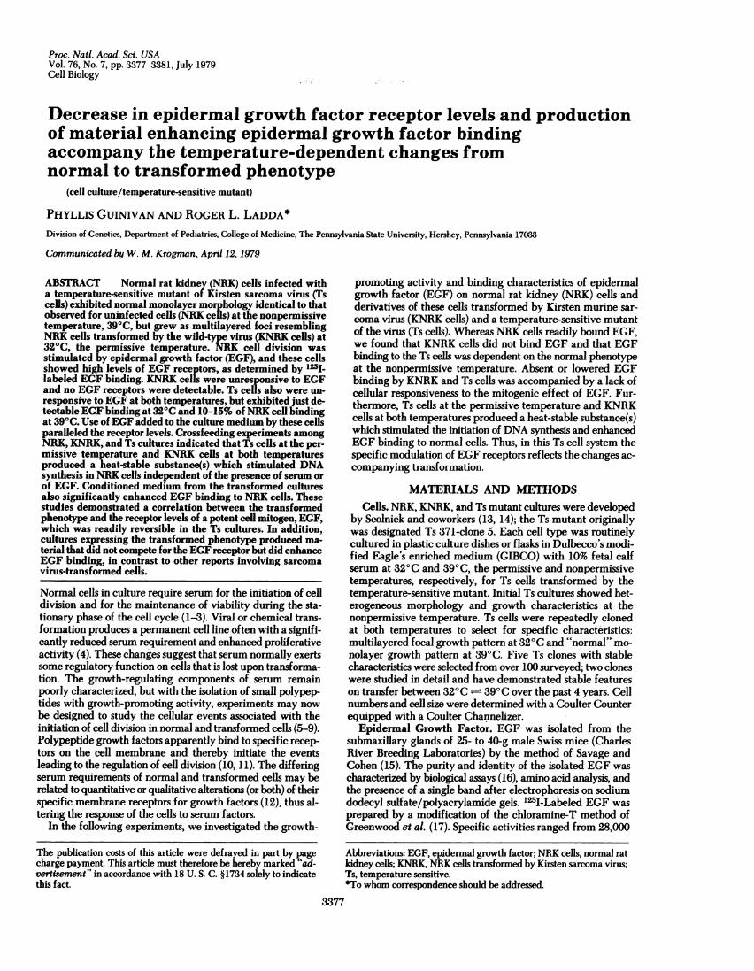
Table 1. EGF concentration in conditioned medium during asingle passage at 390C
Day NRK KNRK Ts
1 25.4 26.1 25.123 21.0 -4 - 24.7 23.46 10.5 23.1 18.08 8.1 - -
An EGF radioreceptor assay (19) was used to determine the con-
centration ofEGF in culture medium at various times during a singlepassage. The medium was originally prepared with 25 ng of EGF per
ml. Values are ng of EGF per ml of medium and are the average ofdeterminations on three separate dishes.
to KNRK cells. KNRK and Ts cells exhibited no apparent re-
sponse to EGF at either temperature.EGF Use by Cells in Culture. The disappearance of EGF
added to the culture medium by the three cultures was mea-
sured over an 8-day period (Table 1). NRK cell medium showeda striking reduction in EGF. In contrast, KNRK cell mediumshowed little evidence of EGF degradation even at high celldensities, but Ts cell medium showed about a 28% depletionof EGF at 390C and essentially none at 320 C.EGF Binding. Time course studies showed that maximum
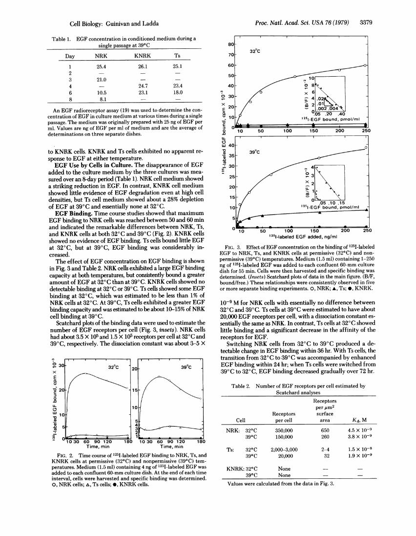
EGF binding to NRK cells was reached between 50 and 60 minand indicated the remarkable differences between NRK, Ts,and KNRK cells at both 320C and 390C (Fig. 2). KNRK cellsshowed no evidence of EGF binding. Ts cells bound little EGFat 320C, but at 390C, EGF binding was considerably in-creased.The effect of EGF concentration on EGF binding is shown
in Fig. 3 and Table 2. NRK cells exhibited a large EGF bindingcapacity at both temperatures, but consistently bound a greateramount of EGF at 320C than at 390C. KNRK cells showed no
detectable binding at 320C or 390C. Ts cells showed some EGFbinding at 320C, which was estimated to be less than 1% ofNRK cells at 320C. At 390C, Ts cells exhibited a greater EGFbinding capacity and was estimated to be about 10-15% of NRKcell binding at 390 C.
Scatchard plots of the binding data were used to estimate thenumber of EGF receptors per cell (Fig. 3, insets). NRK cellshad about 3.5 X 10⁵ and 1.5 X 10⁵ receptors per cell at 320C and390C, respectively. The dissociation constant was about 3-5 X
0330 6 320C 20 90 10Cx
E
U
-V20- 15-C
0
.0
0L 10
W10
453
~~~~~~~~2
10 30 60 90 120 180 10 30 60 90 120 180
Time, min Time, min
FIG. 2. Time course of 1251-labeled EGF binding to NRK, Ts, andKNRK cells at permissive (320C) and nonpermissive (390C) tem-peratures. Medium (1.5 ml) containing 4 ng of 1251-labeled EGF was
added to each confluent 60-mm culture dish. At the end of each timeinterval, cells were harvested and specific binding was determined.0, NRK cells; ,, Ts cells; 0, KNRK cells.
E30
C 20a1*
20 i- o
E OL002 .004
a i05 .20 .40
'251~~~~~-EGF bound, pn lm
s0
C
10 50 100 150 200 250
wu 40-
Q-
25-
20~~~~~~
15~~~~~~~~~~~~
10 1251-EGF bound, pmnol/mI
5
10 50 100 150 200 250
1251-labeled EGF added, ng/ml
FIG. 3. Effect of EGF concentration on the binding of '25I-labeledEGF to NRK, Ts, and KNRK cells at permissive (320C) and non-
permissive (390C) temperatures. Medium (1.5 ml) containing 1-250ng of 1251-labeled EGF was added to each confluent 60-mm culturedish for 55 min. Cells were then harvested and specific binding was
determined. (Insets) Scatchard plots of data in the main figure. (B/F,bound/free.) These relationships were consistently observed in fiveor more separate binding experiments. 0, NRK; *, Ts; *, KNRK.
10-9 M for NRK cells with essentially no difference between320C and 390C. Ts cells at 390C were estimated to have about20,000 EGF receptors per cell, with a dissociation constant es-
sentially the same as NRK. In contrast, Ts cells at 320C showedlittle binding and a significant decrease in the affinity of thereceptors for EGF.
Switching NRK cells from 320C to 390C produced a de-tectable change in EGF binding within 36 hr. With Ts cells, thetransition from 320C to 390C was accompanied by enhancedEGF binding within 24 hr; when Ts cells were switched from390C to 320 C, EGF binding decreased gradually over 72 hr.
Table 2. Number of EGF receptors per cell estimated byScatchard analyses
Receptorsper jim2
Receptors surfaceCell per cell area Kd, M
NRK: 320C 350,000 650 4.5 X 10-9390C 150,000 260 3.8 X 10⁻⁹
Ts: 320C 2,000-3,000 2-4 1.5 X 10-8390C 20,000 32 1.9 x 10⁻⁹
KNRK: 320C None390C None
Values were calculated from the data in Fig. 3.
Cell Biology: Guinivan and Ladda
3380 Cell Biology: Guinivan and Ladda
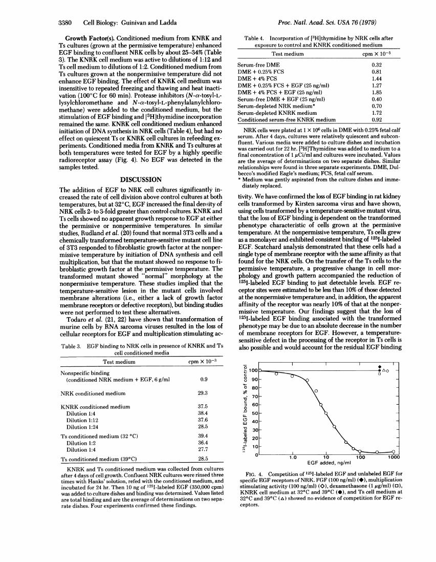
Growth Factor(s). Conditioned medium from KNRK andTs cultures (grown at the permissive temperature) enhancedEGF binding to confluent NRK cells by about 25-34% (Table3). The KNRK cell medium was active to dilutions of 1:12 andTs cell medium to dilutions of 1:2. Conditioned medium fromTs cultures grown at the nonpermissive temperature did notenhance EGF binding. The effect of KNRK cell medium wasinsensitive to repeated freezing and thawing and heat inacti-vation (1000C for 60 min). Protease inhibitors (N-a-tosyl-L-lysylchloromethane and N-ax-tosyl-L-phenylalanylchloro-methane) were added to the conditioned medium, but thestimulation of EGF binding and [3H]thymidine incorporationremained the same. KNRK cell conditioned medium enhancedinitiation of DNA synthesis in NRK cells (Table 4), but had noeffect on quiescent Ts or KNRK cell cultures in refeeding ex-periments. Conditioned media from KNRK and Ts cultures atboth temperatures were tested for EGF by a highly specificradioreceptor assay (Fig. 4). No EGF was detected in thesamples tested.
DISCUSSIONThe addition of EGF to NRK cell cultures significantly in-creased the rate of cell division above control cultures at bothtemperatures, but at 320C, EGF increased the final density ofNRK cells 2- to 3-fold greater than control cultures. KNRK andTs cells showed no apparent growth response to EGF at eitherthe permissive or nonpermissive temperatures. In similarstudies, Rudland et al. (20) found that normal 3T3 cells and achemically transformed temperature-sensitive mutant cell lineof 3T3 responded to fibroblastic growth factor at the nonper-missive temperature by initiation of DNA synthesis and cellmultiplication, but that the mutant showed no response to fi-broblastic growth factor at the permissive temperature. Thetransformed mutant showed "normal" morphology at thenonpermissive temperature. These studies implied that thetemperature-sensitive lesion in the mutant cells involvedmembrane alterations (i.e., either a lack of growth factormembrane receptors or defective receptors), but binding studieswere not performed to test these alternatives.
Todaro et al. (21, 22) have shown that transformation ofmurine cells by RNA sarcoma viruses resulted in the loss ofcellular receptors for EGF and multiplication stimulating ac-
Table 3. EGF binding to NRK cells in presence of KNRK and Tscell conditioned media
Test medium cpm X 10-3
Nonspecific binding(conditioned NRK medium + EGF, 6 g/ml 0.9
NRK conditioned medium 29.3
KNRK conditioned medium 37.5Dilution 1:4 38.4Dilution 1:12 37.6Dilution 1:24 28.5
Ts conditioned medium (32 °C) 39.4Dilution 1:2 36.4Dilution 1:4 27.7
Ts conditioned medium (39°C) 28.5
KNRK and Ts conditioned medium was collected from culturesafter 4 days of cell growth. Confluent NRK cultures were rinsed threetimes with Hanks' solution, refed with the conditioned medium, andincubated for 24 hr. Then 10 ng of 1251-labeled EGF (350,000 cpm)was added to culture dishes and binding was determined. Values listedare total binding and are the average of determinations on two sepa-rate dishes. Four experiments confirmed these findings.
Table 4. Incorporation of [3H]thymidine by NRK cells afterexposure to control and KNRK conditioned medium
Test medium cpm X 10-5
Serum-free DME 0.32DME + 0.25% FCS 0.81DME + 4% FCS 1.44DME + 0.25% FCS + EGF (25 ng/ml) 1.27DME + 4% FCS + EGF (25 ng/ml) 1.85Serum-free DME + EGF (25 ng/ml) 0.40Serum-depleted NRK medium* 0.70Serum-depleted KNRK medium 1.72Conditioned serum-free KNRK medium 0.92
NRK cells were plated at 1 X 10⁶ cells in DME with 0.25% fetal calfserum. After 4 days, cultures were relatively quiescent and subcon-fluent. Various media were added to culture dishes and incubationwas carried out for 22 hr. [3H]Thymidine was added to medium to afinal concentration of 1 ,uCi/ml and cultures were incubated. Valuesare the average of determinations on two separate dishes. Similarrelationships were found in three separate experiments. DME, Dul-becco's modified Eagle's medium; FCS, fetal calf serum.* Medium was gently aspirated from the culture dishes and imme-diately replaced.
tivity. We have confirmed the loss of EGF binding in rat kidneycells transformed by Kirsten sarcoma virus and have shown,using cells transformed by a temperature-sensitive mutant virus,that the loss of EGF binding is dependent on the transformedphenotype characteristic of cells grown at the permissivetemperature. At the nonpermissive temperature, Ts cells grewas a monolayer and exhibited consistent binding of '25I-labeledEGF. Scatchard analysis demonstrated that these cells had asingle type of membrane receptor with the same affinity as thatfound for the NRK cells. On the transfer of the Ts cells to thepermissive temperature, a progressive change in cell mor-phology and growth pattern accompanied the reduction of125I-labeled EGF binding to just detectable levels. EGF re-ceptor sites were estimated to be less than 10% of those detectedat the nonpermissive temperature and, in addition, the apparentaffinity of the receptor was nearly 10% of that at the nonper-missive temperature. Our findings suggest that the loss of125I-labeled EGF binding associated with the transformedphenotype may be due to an absolute decrease in the numberof membrane receptors for EGF. However, a temperature-sensitive defect in the processing of the receptor in Ts cells isalso possible and would account for the residual EGF binding
0
C 0
180 - \ 0
:3 60-X-0
50 R(D4030|>
320_-O G
0 1.0 10 100 1000EGF added, ng/ml
FIG. 4. Competition of 125I-labeled EGF and unlabeled EGF forspecific EGF receptors of NRK. FGF (100 ng/ml) (*), multiplicationstimulating activity (100 ng/ml) (O), dexamethasone (1 ,Ag/ml) (o),KNRK cell medium at 32°C and 39°C (-), and Ts cell medium at32°C and 39°C (A) showed no evidence of competition for EGF re-ceptors.
Proc. Natl. Acad. Sci. USA 76 (1979)

Proc. Natl. Acad. Sci. USA 76 (1979) 3381
and the affinity change of the receptors at the permissivetemperature. Since no biological response to EGF by Ts cellswas observed at the permissive or nonpermissive temperaturein spite of the significant difference in numbers of EGF re-ceptors, it is possible that either the level of EGF receptors ofTs cells is still too low to stimulate growth or the receptors maybe nonfunctional. The disappearance of exogenous EGF fromthe medium of NRK, KNRK, and Ts cells was found to be adirect reflection of the relative levels of receptors in the dif-ferent cell lines. NRK cell medium exhibited a striking reduc-tion in EGF concentration, whereas KNRK cell mediumshowed little change. Ts cells at 390C, however, showed a 28%depletion of EGF from the culture medium. The depletion ofEGF was similar to that observed in NRK cells, yet cell divisionwas not stimulated. Presumably the Ts cell EGF-receptorcomplexes at 390C are internalized, but perhaps these cellsurface events are no longer coupled to the process of cell di-vision (23, 24).
Modulation of EGF receptors was not peculiar to the tem-perature-sensitive mutant. NRK cells showed significantlygreater numbers of EGF receptors at 320C than at 390C. In-terestingly, this change was opposite to that demonstrated bythe temperature-sensitive mutant. The growth responses ofNRK cells to EGF in reduced serum were essentially the sameat both temperatures. Thus, the total number of EGF receptorsexhibited by NRK cells did not appear to affect the mitogenicresponse of cultures to EGF. This may be due to a differencein the relative turnover of the EGF-receptor complex as afunction of temperature.
In cross-feeding experiments among NRK, KNRK, and Tscells, we found that KNRK at both temperatures and Ts cellsat the permissive temperature secreted material capable ofstimulating the initiation of DNA synthesis and cell division innormal human diploid fibroblasts (unpublished observations)and in NRK cell cultures. DeLarco and Todaro (25) recentlydescribed the production of a growth factor by sarcoma virus-transformed 3T3 cells. The factor enhanced DNA synthesis andenabled untransformed cells to grow in agar, but competed with'25I-labeled EGF for cell receptors. However, in contrast to thecompetition for EGF receptors reported by DeLarco andTodaro (25) by the sarcoma growth factor, the material isolatedfrom KNRK and Ts cell conditioned media enhanced 125I-labeled EGF binding to NRK cells. Preliminary studies haveindicated that the active material(s) are polypeptide(s) with anestimated molecular weight range of 14,000-20,000.The loss of EGF receptors and the production of growth-
promoting material appear to be characteristic of cells trans-formed by murine sarcoma virus. In the cloned Ts cell systemwe have described, these features may be readily manipulated
to provide a model for the analysis of the expression of thetransformed phenotype.We thank Drs. Edward Scolnick and Charles Scher for providing
the normal and transformed cells for our study and Drs. Wayne Bardinand George Keller for helpful discussions and suggestions. This workwas supported by a grant from the National Institute on Aging (AG00421). R.L.L. is the recipient of a Research Career DevelopmentAward (K04 AG 00006) from the National Institute on Aging. P.G. isa predoctoral candidate in the Department of Anatomy.
1. Temin, H. (1972) in Growth Nutrition and Metabolism of Cells,eds. Rothblat, G. & Cristofalo, V. (Academic, New York), pp.50-81.
2. Temin, H. (1971) J. Cell Physiol 78, 161-170.3. Stoker, M. (1973) Nature (London) 246,200-203.4. Holley, R. & Kiernan, J. (1971) Proc. Natl. Acad. Sci. USA 60,
300-304.5. Dulbecco, R. (1970) Nature (London) 227,802-806.6. Paul, D., Lipton, A. & Klinger, I. (1971) Proc. Natl. Acad. Sci.
USA 68, 645-648.7. Gospodarowicz, D. (1974) Nature (London) 240, 123-127.8. Seifert, W. & Rudland, P. (1974) Nature (London) 248, 138-
140.9. Gospodarowicz, D. & Moran, S. (1975) J. Cell Biol. 66, 451-
457.10. Hollenberg, M. & Cuatrecasas, P. (1973) Proc. Natl. Acad. Sci.
USA 70,2964-2968.11. Cohen, S. & Carpenter, G. (1975) Proc. Natl. Acad. Sci. USA 72,
1317-1321.12. Pardee, A. (1975) Biochim. Biophys. Acta 417, 153-172.13. Carchman, R., Johnson, G., Pastan, I. & Scolnick, E. (1974) Cell
1,59-64.14. Scolnick, E., Stephenson, J. & Aaronson, S. (1972) J. Virol. 10,
653-657.15. Savage, C. & Cohen, S. (1972) J. Biol. Chem. 247, 7609-7611.16. Cohen, S. (1962) J. Biol. Chem. 237, 1555-1562.17. Greenwood, F., Hunter, W. & Glover, J. (1963) Biochem. J. 89,
114-123.18. Scatchard, G. (1949) Ann. N. Y. Acad. Sci. 51, 660-672.19. Ladda, R., Bullock, L., Gianopoulos, T. & McCormick, L. (1979)
Anal. Biochem. 93, 286-294.20. Rudland, P., Eckhart, W., Gospodarowicz, D. & Seifert, W. (1974)
Nature (London) 250,337-339.21. Todaro, G., DeLarco, J. & Cohen, S. (1976) Nature (London) 264,
26-31.22. Todaro, G., DeLarco, J., Nissley, S. & Rechler, M. (1977) Nature
(London) 267,526-528.23. Carpenter, G. & Cohen, S. (1976) J. Cell Biol. 71, 159-171.24. Das, M., Miyakawa, T., Fox, C., Pruss, R., Aharonov, A. &
Herschman, H. (1977) Proc. Natl. Acad. Sci. USA 74, 2790-2794.
25. DeLarco, J. & Todaro, G. (1978) Proc. Natl. Acad. Sci. USA 75,4001-4005.
Cell Biology: Guinivan and Ladda